Article

miR156-SPL and miR169-NF-YA Modules Regulate the Induction of Somatic Embryogenesis in Arabidopsis via LEC- and Auxin-Related Pathways

Institute of Biology, Biotechnology and Environmental Protection, Faculty of Natural Sciences, University of Silesia, 40-007 Katowice, Poland
*
Authors to whom correspondence should be addressed.
These authors contributed equally to this work.
Int. J. Mol. Sci. 2024, 25(17), 9217; https://doi.org/10.3390/ijms25179217 (registering DOI)
Submission received: 30 June 2024 / Revised: 21 August 2024 / Accepted: 23 August 2024 / Published: 25 August 2024
(This article belongs to the Special Issue Molecular Research on Embryo Developmental Potential)

Abstract

:
The embryogenic transition of plant somatic cells to produce somatic embryos requires extensive reprogramming of the cell transcriptome. The prominent role of transcription factors (TFs) and miRNAs in controlling somatic embryogenesis (SE) induction in plants was documented. The profiling of MIRNA expression in the embryogenic culture of Arabidopsis implied the contribution of the miR156 and miR169 to the embryogenic induction. In the present study, the function of miR156 and miR169 and the candidate targets, SPL and NF-YA genes, were investigated in Arabidopsis SE. The results showed that misexpression of MIRNA156 and candidate SPL target genes (SPL2, 3, 4, 5, 9, 10, 11, 13, 15) negatively affected the embryogenic potential of transgenic explants, suggesting that specific fine-tuning of the miR156 and target genes expression levels seems essential for efficient SE induction. The results revealed that SPL11 under the control of miR156 might contribute to SE induction by regulating the master regulators of SE, the LEC (LEAFY COTYLEDON) genes (LEC1, LEC2, FUS3). Moreover, the role of miR169 and its candidate NF-YA targets in SE induction was demonstrated. The results showed that several miR169 targets, including NF-YA1, 3, 5, 8, and 10, positively regulated SE. We found, that miR169 via NF-YA5 seems to modulate the expression of a master SE regulator LEC1/NF-YA and other auxin-related genes: YUCCA (YUC4, 10) and PIN1 in SE induction. The study provided new insights into miR156-SPL and miR169-NF-YA functions in the auxin-related and LEC-controlled regulatory network of SE.

1. Introduction

Somatic embryogenesis (SE) is a plant-specific process that results in the formation of embryos from in vitro cultured somatic cells. The studies on SE in different plants, especially Arabidopsis, provide a research model for understanding the regulatory processes controlling the embryogenic transition of somatic cells and the developmental plasticity of plants, which is called totipotency [1]. SE has been widely explored in plant biotechnology for the efficient regeneration and genetic modifications of plants (reviewed in [2]). Research on SE evidenced that embryogenic reprogramming of somatic cells is controlled by complex interactions between genetic and epigenetic factors, including transcription factors (TFs), miRNAs, DNA methylation, histone methylation, and acetylation (reviewed in [3,4]).
In particular, TFs play a central role in the genetic network, controlling the reprogramming of plant somatic cells [5]. In line with the prominent function of TFs in SE induction, the overexpression of several TFs improved the regeneration efficiency in the in vitro recalcitrant crops [6,7,8,9]. Within TFs involved in SE induction, those associated with auxin-related processes such as biosynthesis and the signaling of auxin are overrepresented [10]. Within these TFs, LEAFY COTYLEDON, including LEC1, LEC2, and FUS3, and PLETHORA proteins such as BBM (BABY BOOM) and members of the WUSCHEL/WOX TFs family were indicated to play a critical function [11,12,13]. Identifying both the up and downstream targets and mutual regulatory interactions between SE-decisive TFs provides a current challenge in understanding the molecular determinants of developmental plasticity in plant somatic cells.
In line, numerous efforts to identify miRNAs regulating TF genes during SE induction have been made (reviewed in [14]). Accordingly, differential expression of numerous MIRNA genes has been indicated in SE cultures of Arabidopsis [15] and other species [16,17,18]. However, only a few SE-modulated miRNAs have been functionally analyzed in SE, including miR160, miR166/165 [19], miR167 [20], miR393 [21], miR396 [22], and miR528 [23].
In concert with TF and miRNA, auxin plays a central role in the embryogenic transcriptome reprogramming associated with SE induction in plant somatic cells (reviewed in [24]). Accordingly, several miRNAs, such as miR160, miR166, miR167, miR390, and miR396 were shown to control auxin metabolism and signaling in the regulatory network of SE [19,21,22,23].
Besides auxin, stress-related responses also have a prevalent role in the genetic mechanism that governs SE induction (reviewed in [25]). Relevantly, the stress-related miR528 that targeted MATE (MULTIGRUD AND TOXIN EXTRUSION), bHLH152, and SOD1a (SUPEROXIDE DISMUTASE 1A) were indicated to have a regulatory function in SE [23].
The stress-related candidate miRNAs that might control SE induction also involve miR156 and miR169 [15]. The miR156 clade in Arabidopsis is encoded by eight genes producing three different miRNA isoforms: (1) miR156a-f, (2) miR156g, which has a single nucleotide substitution at the first position, and (3) miR156h with two nucleotide substitutions at positions 11 and 14 in relevance to miR156a-f [26]. In planta, miR156 isoforms modulating vegetative phase transition in Arabidopsis accumulate in the seedling stage and then gradually decline with plant development [27]. The genes targeted by miR156 play roles in multiple fundamental processes in plants, such as the development of embryos, siliques and the abiotic and biotic stress response (reviewed by [28]). The miR156 targets include SPLs from the TF family of highly conserved 76 amino acid residue and the SBP domain, with two zinc-binding sites essential for DNA binding [29,30]. Based on their functions in developmental phase transitions, miR156-regulated SPL genes have been divided into two groups: (1) SPL2, SPL9, SPL10, SPL11, SPL13, and SPL15 of the crucial roles in juvenile-to-adult vegetative and vegetative-to-reproductive transitions and (2) SPL3, SPL4, and SPL5, which promote the floral meristem identify transition [31]. In addition, SPL2, SPL10, and SPL11 redundantly control proper lateral organ development and shoot maturation in the reproductive phase [32].
The upregulation of MIR169 transcripts and mature miR169 accumulation was observed at a late stage of SE [15]. In Arabidopsis, the miR169 is encoded by fourteen genes producing three different miRNA isoforms: miR169a-c, miR169d-g differing in two nucleotide substitutions at the first and second position, and miR169h-n with single a substitution at the first position in comparison to miR169a-c [33].
The miR169 isoforms show diverse expression patterns during plant development [34] and in response to biotic/abiotic stresses [35,36,37]. The targets of miR169 in plant development involve the genes that encode the subunit A of Nuclear Factor Y (NF-Y) TF [38]. NF-Y TF is a heterotrimeric protein composed of NF-YA, NF-YB, and NF-YC subunits. NF-Y TF recognizes the CCAAT motif, which is frequent in eukaryotic gene promoters [39]. NF-YB and NF-YC subunits of a histone fold domain similar to H2A and H2B core histones [40,41] form a heterodimer interacting with NF-YA. The function of the miR169-NF-YA module in regulating various plant processes has been attributed to the abiotic and biotic stress responses [42,43,44]. miR169 negatively regulates rice immunity against Magnaporthe oryzae [44]. Arabidopsis seedlings under cold stress show an overaccumulation of miR169 molecules correlated with a reduction in some NF-YA target transcripts [42,43]. The accumulation of MIRNA169 transcripts was also observed in response to salt stress and ABA in poplar [45]. Moreover, the overexpression of miR169a resulted in reduced nitrogen accumulation and increased sensitivity to nitrogen starvation [37]. In root development, the miR169defg-targeted NF-YA2 and NF-YA10 control RAM (Root Apical Meristem) length and lateral root density [46].
In the present study, we analyze the function of miR156 and miR169 in SE regulation. In support of the contribution of these miRNAs to SE induction, we demonstrated the spatial–temporal expression patterns coinciding with the induction of SE and disturbed expression/function of MIR156 and MIR169 genes significantly impaired the embryogenic potential of Arabidopsis explants. We also indicated the candidate genes possibly targeted by miR156 and miR169 to regulate the embryogenic culture and their versatile relationships with stress and auxin responses. The results expand our knowledge of the SE regulatory network and recommend the miR156-SPL11 and miR169-NF-YA5 modules for further studies on LEC1- and auxin-mediated mechanisms of embryogenic transition in plant somatic cells.

2. Results

Here, two miRNA molecules, miR156 and miR169, of differential expression in SE culture [15] were subjected to functional analysis in the embryogenic culture of Arabidopsis. To answer the question of whether those miRNA molecules are regulators of SE, we analyzed the effect of mutations in the relevant MIRNA genes and candidate target genes on the embryogenic potential of explants. Moreover, the expression of MIRNA and target genes in the SE-induced explants was evaluated.

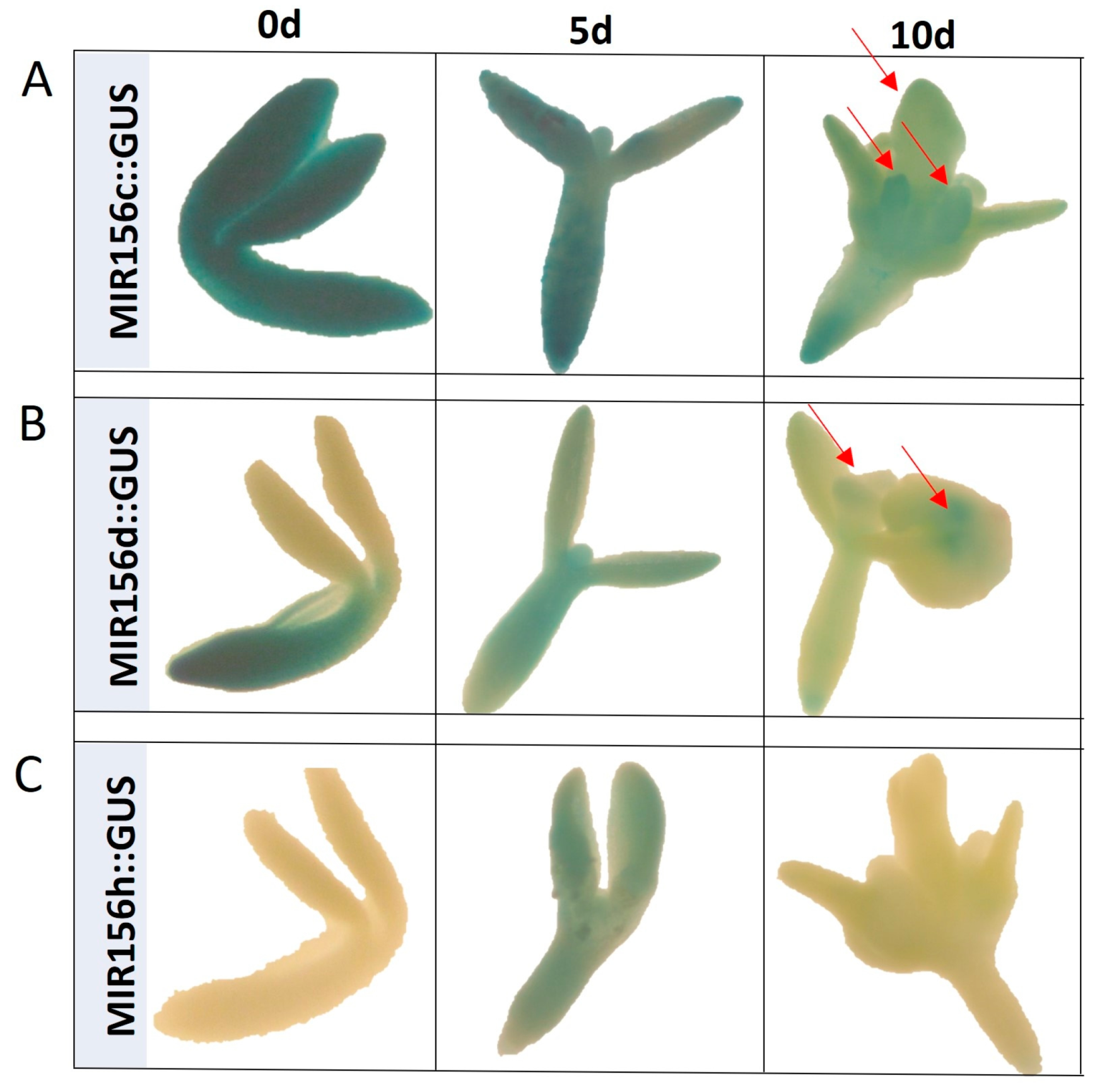
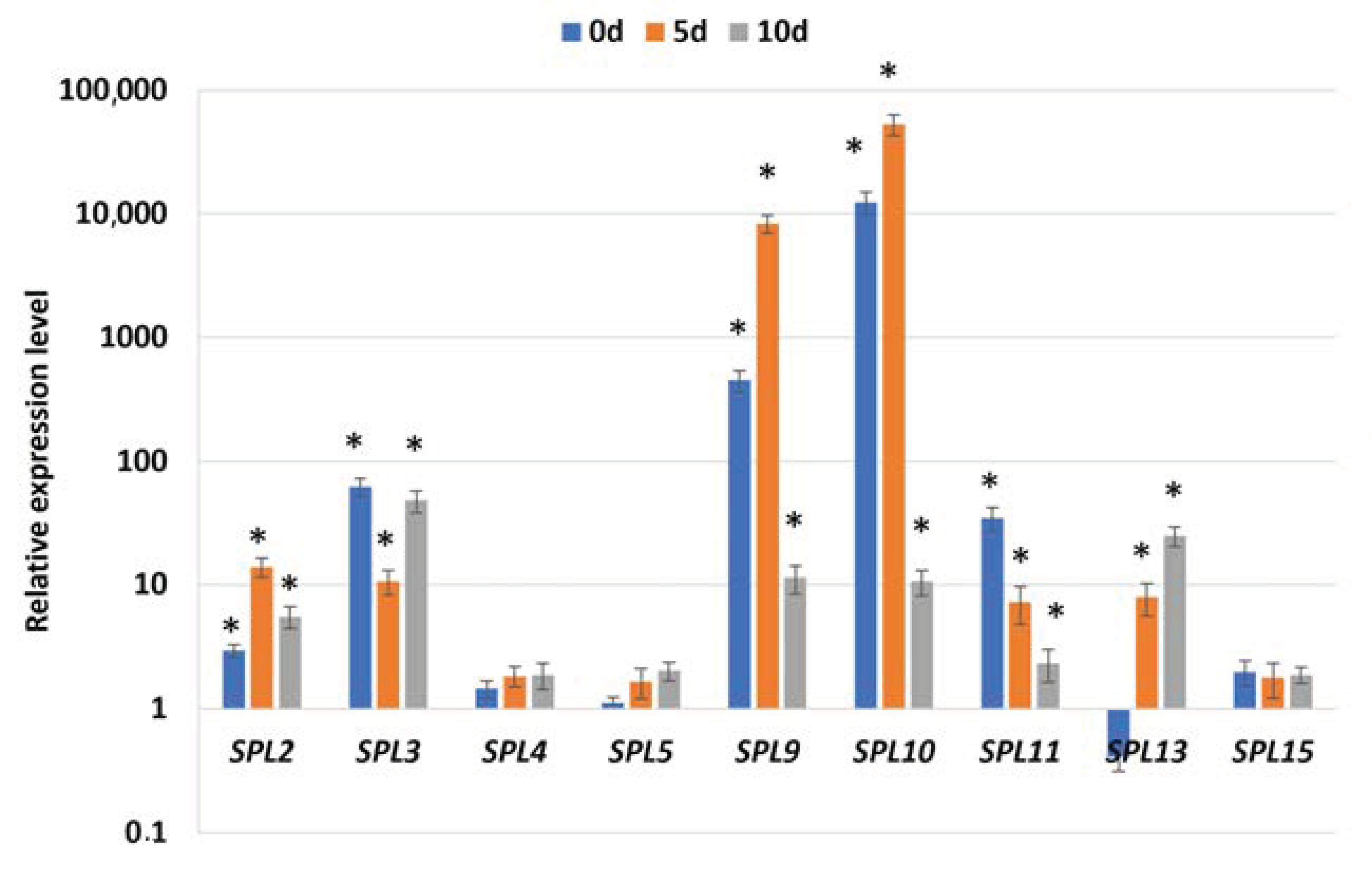
2.1. Different MIRNA156 Genes Regulate SE Induction

In support of the regulatory role of miR156 in SE induction, the inhibition of the miR156 function in the MIM line resulted in a strong reduction in the embryogenic response of the explants, both SE efficiency and SE productivity (Figure 1A,B). Similarly, the overexpression of miR156 in the 35S::MIR156A line negatively affected SE efficiency. Moreover, the impaired embryogenic response displayed was also observed in miR156b and miR156g insertion mutant cultures. These results implied that different MIR156 genes producing different miR156 isoforms, such as miR156a-f and miR156g, might contribute to regulating SE induction.
To study the spatiotemporal expression of MIRNA156 genes, we used the lines containing the sequence of GUS reporter gene under the promoter of selected MIRNA genes that allow the visualization of cells/tissues where MIRNA156 are transcriptionally active by monitoring the presence of blue color related to the activity of GUS. The analysis of the GUS reporter lines also provided evidence of the involvement of different MIR156 genes in controlling SE. Relevantly, the spatiotemporal expression patterns of MIR156 C, D, and H genes producing two different isoforms of miR156, miR156a-f, and miR156h, were analyzed in freshly isolated (0 d) and SE-induced explants (Figure 2). The results showed a diversity of GUS expression patterns between the MIR156 genes in IZEs both before (0 d) and during embryogenic (5 and 10 d) culture. We found that two genes, MIRNA156C and MIRNA156D, were expressed in freshly isolated (0 d) explants, and the MIRNA156C gene was particularly strongly expressed in the whole explant tissue, including SE-involved cotyledons. The auxin treatment used to induce SE distinctly affected the MIR156 gene expression pattern in the explants. The reporter line explants cultured for 5 days on an SE induction auxin medium (E5) showed the expression of all analyzed MIR156 genes in the explants. In the more advanced stage of SE induction (10 d), the GUS signal pattern was detected for two genes, MIR156 C and D. The expression of these genes is visible in the embryo-like protuberances that emerged on the adaxial side of the cotyledons.
Together, the mutant and reporter line results enhanced the assumption of the regulatory role of miR156 in SE induction and provided evidence that different MIR156 genes, including MIR156C and D, seem to contribute to miR156 production in SE induction.

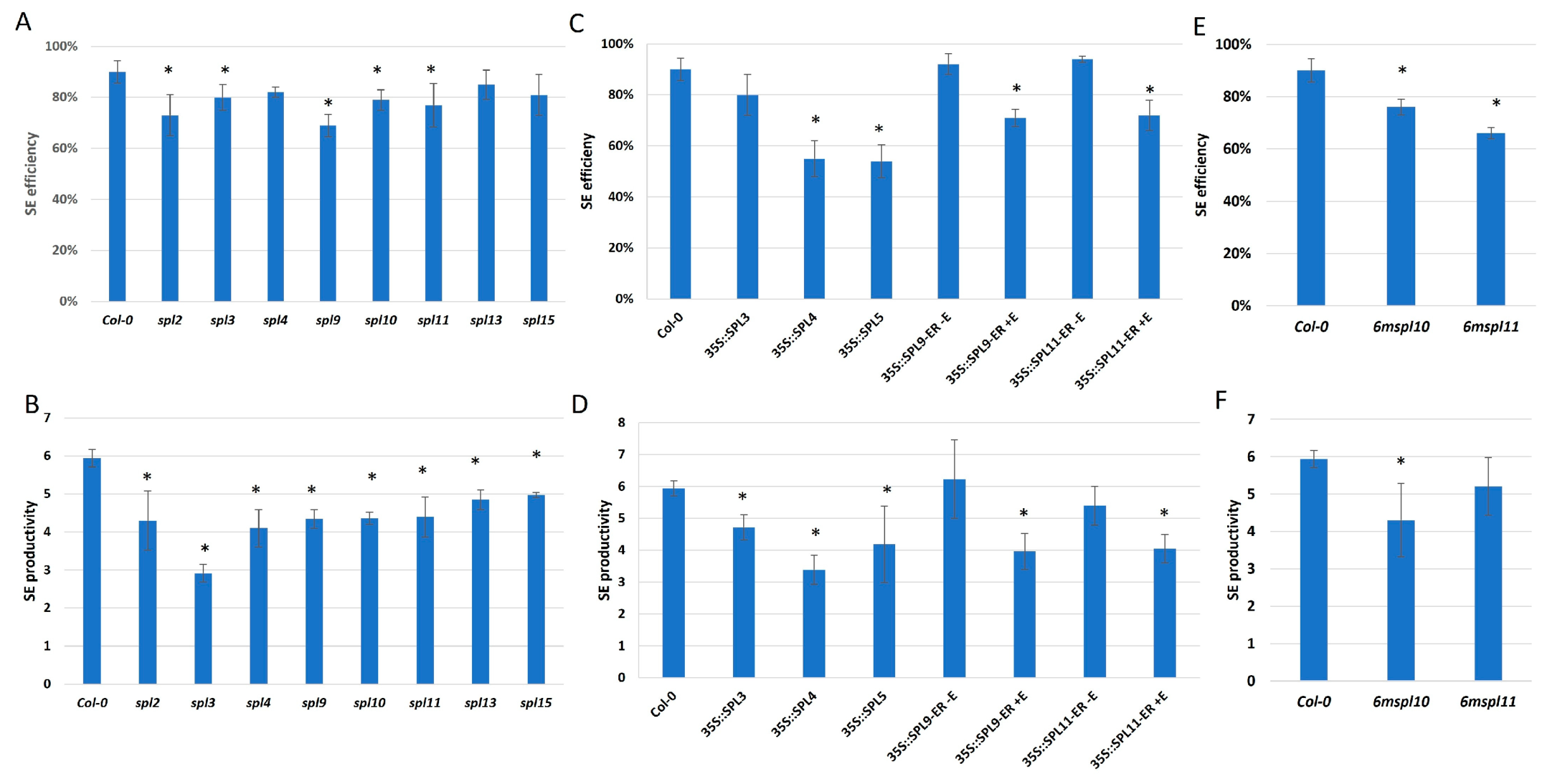
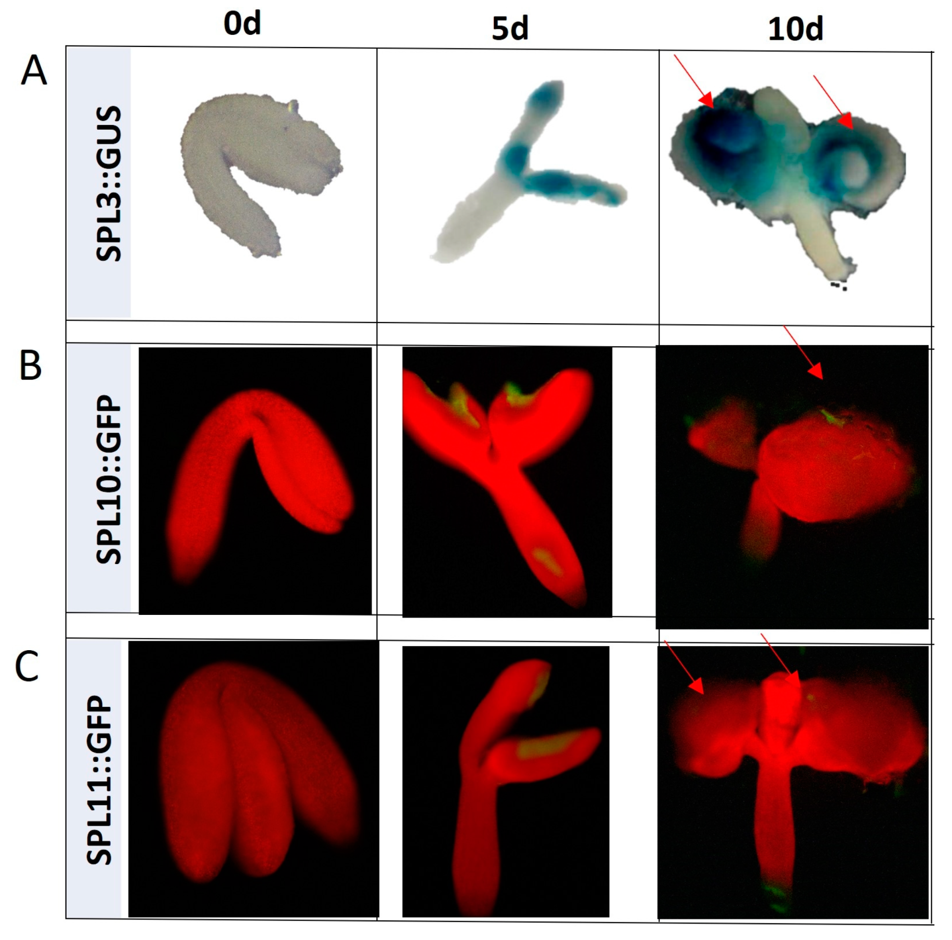
2.2. miR156 Regulates SE Induction via Controlling SPLs

The candidate miR156 targets include SPLs from the transcription factors family with a highly conserved SBP domain that binds to the targeted DNA. To validate the candidate miR156 target function in SE induction, we evaluated the embryogenic response of the genotypes with a disturbed expression/function of SPL genes, including spl2, 3, 4, 9, 10, 11, 13, 15 mutants and an overexpression of lines (35S::SPL3, 4, 5, 35S::SPL9-ER, 35S::SPL11-ER). The results showed that all of the mutants (Figure 3A,B) and overexpression lines (Figure 3C,D) in different SPL genes indicated a significantly reduced SE efficiency and/or productivity.
Further support for the SPL involvement in SE were provided by the spatiotemporal analysis of SPL3, 10, and 11 expression patterns in SE-induced explants with the use of reporter lines (Figure 4). We found that, in contrast to the freshly isolated explants (0 d), which showed no GUS/GFP signal, the explants cultured on SE-medium for 5 and 10 days indicated the signal of SPL3, 10, and 11 in the adaxial side of cotyledons where the somatic embryos were developed. Altogether, the mutant and reporter line results implied that the SPL2, 3, 4, 5, 9, 10, 11, 13, and 15 genes contribute to SE induction in Arabidopsis.
We found that the 6mSPL10 and 6mSPL11 lines of a disrupted miR156 binding site resulted in the overexpression of these genes and showed an impaired SE response (Figure 3E,F). This result provides evidence of the role of miR156 in regulating SPL10 and SPL11 in SE.
To further verify the regulatory relationship between miR156 and the SPL targets in SE induction, the transcription levels of SPL2, 3, 4, 5, 9, 10, 11, 13, and 15 were evaluated in the 35S::MIM156 culture, with a defective miR156 function (Figure 5). The analysis showed that all analyzed candidate targets of miR156 had increased expression levels in the 35S::MIM156 tissue, including freshly isolated (0 d) and SE-induced (5 and 10 d) explants. A particularly high level of transcript accumulation showed SPL9 and SPL10, suggesting a strong miR156-related regulation of these genes in SE induction.
Thus, we assumed that miR156 might control different SPLs, including SPL2, 3, 4, 5, 9, 10, 11, 13, and 15 during the SE process.

2.3. miR156 via SPL11 Might Regulate LEC Genes in SE

In plant development in vivo and in vitro, miR156 and SPL11 activity correlates with the expression of FUS3 from the LEC genes group, a key regulator of SE [11,47,48]. To verify the assumption of regulatory relation between LEC genes and miR156 in SE, we analyzed the expression level of LEC1, LEC2, and FUS3 in the 35S::MIM156 culture of the inhibited miR156 function (Figure 6A). We indicated that LEC1, LEC2, and FUS3 transcripts were significantly deregulated in early-stage SE (5 d) in MIM156 line culture compared to the Col-0 culture (Figure 6A), suggesting the role of miR156 in the regulation of LEC genes during SE induction. Moreover, these results showed that, in contrast to the negative regulation of LEC2 and FUS3, miR156 positively affected LEC1 expression implying indirect regulation of LEC1 by miR156 in SE. To assess whether the miR156-SPL11 module might control LEC expression in SE, we analyzed the LEC transcript levels in the 6mSPL11 mutant line overexpressing SPL11 due to disruption of the miR156-binding site in SPL11gene (Figure 6B). In support of an assumption on the role of miR156-SPL in SE, changes in LECs transcript levels in the early stage of SE (5 d) were indicated. Together, the results on the 35S::MIM156 and 6mSPL11 lines implied that miR156 via SPLs, including SPL11, might control LEC genes in SE induction (Figure 6).

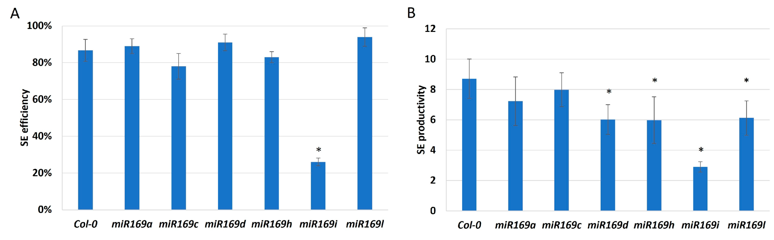
2.4. Different MIRNA169 Genes Contribute to miR169 Regulating SE Induction

To verify a hypothesis on miR169 contribution in SE, we analyzed the effect of mutations in MIRNA169 genes (MIRNA169a, c, d, h, i, l) on the embryogenic potential of the explants. The results indicated an impaired embryogenic response of four mutants, miR169d, miR169h, miR169i, and miR169l, implying that relevant MIR169 genes might contribute to SE induction (Figure 7). Since SE-involved MIR169 genes produce different miR169 isoforms [34], we assumed a role for miR169d-g and miR169h-n in the SE regulatory network.

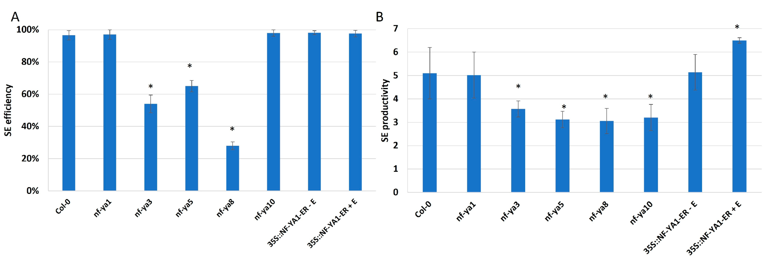
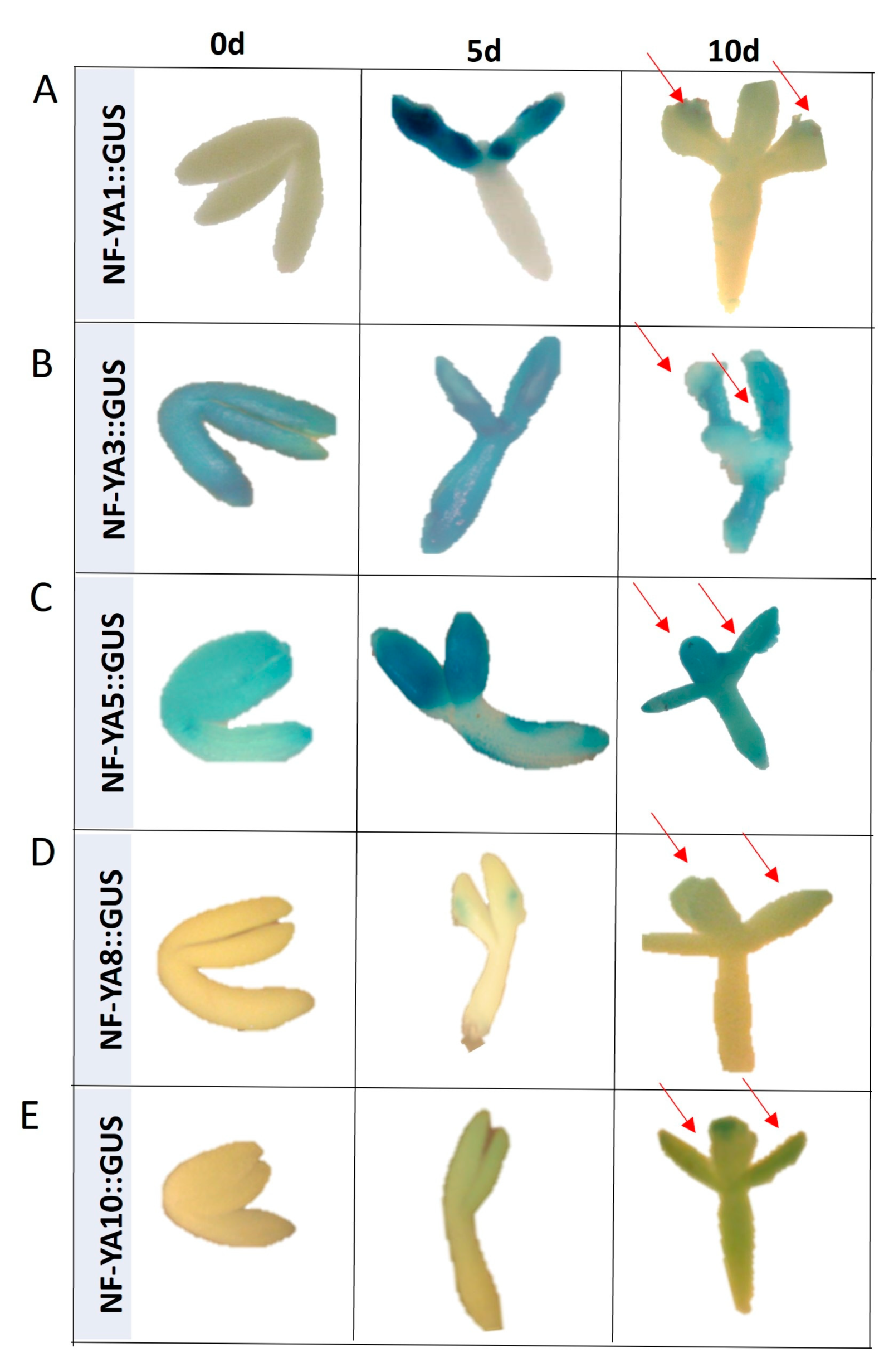
2.5. miR169 Regulates Embryogenic Induction via Controlling NF-YA

NF-YA Control SE Induction

The targets of miR169 in plant development belong to the transcription factors of the family that predominantly bind to the CCAAT-box present in the target gene promoters, such as NF-YA TFs [49,50]. To verify the engagement of NF-YA genes in the regulation of embryogenic induction, we analyzed the SE efficiency and productivity in line with a disturbed expression of NF-YA genes (Figure 8). The results showed an impaired embryogenic potential of explants with the mutations in NF-YA3, 5, 8, and 10 genes. In contrast, NF-YA1 overexpression stimulated somatic embryo production. The results indicated that different NF-YA, including NF-YA1, 3, 5, 8, and 10, seem to act as positive regulators of SE.
More lines of evidence on the role of NF-YA in SE provided the GUS reporter lines analysis that monitored the NF-YA1, 3, 5, 8, and 10 expression in freshly isolated (0 d) and SE-induced (5 and 10 d) explants (Figure 9). The analysis indicated differences in the spatiotemporal expression profile of the analyzed NF-YAs. The expression of two genes, NF-YA3 and 5, was found in freshly isolated and SE-induced explants, while the other NF-YA genes (NF-YA1, 8, and 10) were expressed exclusively in cotyledons, the explant part that effectively contributes to SE-induction (5 and 10 d). Altogether, the reporter line analysis provided some lines of evidence on the involvement of different NF-YAs (NF-YA1,3 5,8, and 10) in SE.

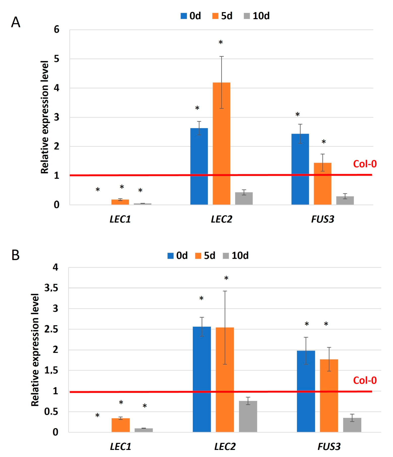
2.6. miR169 via NF-YA5 Regulates the Expression of Genes of Critical Role in SE

The impaired embryogenic potential of miR169i and nf-ya5 mutants (Figure 7 and Figure 8) and GUS-indicated expression of NF-YA5 in the SE-related region of explants (Figure 9C) indicated the involvement of miR169i and NF-YA5. To verify an assumption on the regulatory relationships between these molecules in SE induction, we analyze the expression of NF-YA5 in the culture of miR169i mutant (Figure 10A). The increased NF-YA5 transcript level in the freshly isolated (0 d) and SE-induced (5 d) mutant explants implied that the miR169i isoform might directly control the expression of NF-YA5 in the embryogenic transition of explants.
To identify the downstream elements of the miR169-NF-YA5 regulatory node, we focused the analysis on auxin and SE-related LEC1, YUC, and PIN1 genes [11,51], which are targeted by miR169 during in vivo plant development [37,52,53]. Accordingly, we analyzed the expression levels of the candidate SE-related targets in nf-ya5 mutant cultures. The results revealed decreased LEC1, YUC4, YUC10, and PIN1 levels in different stages (5 and 10 d) of the mutant cultures (Figure 10B). The gene expression results provided support for the assumption of the role of miR169-targeted NF-YA5 in regulatory networks controlling auxin-related processes in SE.

3. Discussion

3.1. miR156 through SPLs Controls SE Induction

Our global expression profiling of MIRNA genes and their mature miRNA transcripts suggested the SE-related functions of miR156 in the embryogenic culture of Arabidopsis [15]. Similar to Arabidopsis, the downregulation of miR156 was also characteristic of the embryogenic culture of coconut [54], Lilium [55], and Eucalyptus camaldulensis [56]. In contrast, the miR156 level was significantly higher in embryogenic than in non-embryogenic callus in citrus [57], implying the plant species-specific impact of miR156 on the SE regulatory network. Relevantly to this assumption, the overexpression of MIR156A in citrus enhanced the formation of the embryos [47], while it negatively affected SE response in Arabidopsis (present results).
Here, we observed that disturbing miR156 levels by both mutation and overexpression of different MIR156 genes negatively affected SE in Arabidopsis. Thus, a specific fine-tuned miR156 level seems essential for effective SE induction. The evidence for this assumption is the tightly controlled abundance and the level-dependent effect of miR156 in plant development in vivo. Accordingly, the increased accumulation of miR156 level was found to prolong the juvenile phase, whereas a reduction in miR156 activity led to an accelerated expression of adult traits [27].
In vivo, MIR156 genes encode three miR156 isoforms contributing to various developmental processes, including zygotic embryogenesis [48,58,59]. Increased expression of MIRNA (MIR156A, B, and C) genes producing a miR156a-f isoform was associated with the progress of ZE from the globular to the mature stage of zygotic embryos [58,59]. Our results indicated that miR156a-f, besides miR156g isoform, might also regulate embryogenic induction in vitro. In addition to obvious similarities to ZE, the miR156-related regulation of SE resembles a genetic network operating in plant flowering [60]. Accordingly, AGL15, the upstream TF regulator of MIR156A and C in Arabidopsis flowering [61], was evidenced to control miR156 in an embryogenic culture of Arabidopsis [60]. It was reported that in the SE regulatory pathway, AGL15 positively controls MIRNA156 transcription and limits the abundance of mature miR156 by repressing the miRNA biogenesis genes [60]. To identify targets of miR156 in SE, we analyzed SPL genes controlled by miR156 in plant development in vivo [62]. The role of miR156-controlled SPL genes in SE induction supports the contribution of the miR156-SPL to other SE-related processes, including the regulation of shoot regenerative competence in vitro [63] and stress responses in plant development [62]. We observed that, consistent with the declined SE potential of miR156 transgenic lines (present results), both mutations and overexpression of SPL genes also impaired the SE response of the explants. These results implied that similar to BBM TF [13], SPLs control SE induction in a gene-dose-dependent manner. The reports also suggested that LEC2 [11,64] and miRNA-TF regulatory modules, including miR393/TIR1/AFB, miR166/PHB/PHV, and miR396/GRFs [19,21,22] might control SE response in a dose-dependent mode. Thus, we assumed that a correctly fine-tuned and specific level of the SPLs expression conditions SE induction.
Similar to miR156, the role of SPLs in activating SE response might be species-specific. In contrast to impaired SE induction in Arabidopsis, a knockdown of the SPL3 or SPL14 genes enhanced the number of SE formed in citrus [47]. Also, the callus proliferation rate in cotton is significantly increased in SPL10 overexpression lines [65].
Our data provided by the miR156 and spl mutant and reporter lines analysis suggested that miR156 might control different SPLs, including SPL2, 3, 4, 5, 9, 10, 11, 13, and 15 during SE induction. The redundant function of multiple miR156-targeted SPL genes in regulating the same targets was also postulated in seed maturation [66]. Importantly, most of the SE-involved and miR156-targeted SPL candidates represent the SPL9 group of SPL genes (SPL2, 9, 10, 11, 13, and 15). Importantly, these SPL were identified as functional targets of miR156 in Arabidopsis shoot organogenesis [63], the alternative to the SE morphogenic process contributing to plant regeneration in vitro.
Consistent with the assumption of the miR156-SPL role in the SE regulation, we found that the genotypes of a disrupted miR156-binding site in transcripts of SPL10 and SPL11 showed distinctly impaired SE response. Moreover, a particularly high accumulation of SPL9 and 10 in the MIM156 culture, pointed to a distinctive function of the miR156-SPL9/SPL10 module in SE induction. In support of the SE-related function of this regulatory module, miR156-regulated SPL9, and SPL10 were postulated to contribute to the miR172-AP2-controlled repression of WUS TF in the SE induction of Arabidopsis [67].

3.2. Phytohormone and LEC Genes Related Mechanism of miR156-SPL Regulation in SE

The miR156-SPLs module functions as a hinge to integrate multiple phytohormones including auxin, gibberellin (GA), and ethylene (ET) in vegetative-phase transition, floral transition, lateral organ development, and callus production in different plants [65,68,69,70]. In shoot regeneration, the cytokinin-related function of miR156-SPL in controlling B-type ARR TFs, the key elements of the cytokinin signaling pathway, was also reported [63]. In support of the cytokinin-related function of the miR156-SPL in SE, the miR156-SPL was postulated to control cytokinin-responsive WUS TF [66,70].
We hypothesized that miR156-SPL function in SE might also be related to auxin, the phytohormone that a central role in the SE induction mechanism [71,72]. In support, miR156-SPL7-controlled IAA biosynthesis was reported in roots, and miR156-SPL3, 9, 10 were responsive to auxin signaling in lateral root development [73,74]. To verify the assumption that the miR156-SPL module contributes to auxin metabolism and signaling in SE, we focused the study on LEC TFs (LEC1, 2, FUS3) of a key regulatory function in the auxin-induced SE pathway [11,75]. The reports indicated the regulation of LEC genes by miR156 through SPL11 in Arabidopsis seed maturation [48,66] and FUS3 targeting by miR156-SPL3/14 in embryogenic citrus callus [47].
Our results on LEC transcript profiling in the embryogenic culture of 35::MIM156 and 6mSPL11 lines showed that miR156, possibly via controlling SPL11, might regulate the LEC gene expression in SE induction. The present results on LECs indicated the gene-specific effects of miR156-SPL, and depending on the targeted gene, the increased (LEC1) or decreased (LEC2, FUS3) level of LEC transcripts in SE of the MIM156 line was found. In conclusion, the complex regulatory interactions operated in the embryogenic induction between the miR156-SPL and TF genes, including that LECs might be expected. The mechanism and other elements involved in miR156-SPL regulation of LECs and other SE genes require identification, with particular consideration of phytohormones.

3.3. Two miR169 Isoforms via NF-YA TFs Control SE Induction

Changes in the levels of miR169 accumulation in embryogenic cultures of different plants, including Arabidopsis [15,56,75], point to its common role in SE induction. However, the diversity of miR169 impacts on SE regulation might be assumed since both increased and decreased miR169 accumulation in SE, depending on the SE stage and plant species, were indicated [15,54,56,76].
In the study, we obtained insights into the miR169-related SE regulatory pathways in Arabidopsis by identifying miR169 isoforms and their targets during embryogenic induction. The analysis of the embryogenic potential of mutants with impaired function on different MIRNA169 genes (MIRNA169A, C, D, H, I, L) producing three miR169 isoforms indicated that two of them, including miR169d-g, and miR169h-n, might operate in the SE regulatory network in Arabidopsis. The role of these miR169 isoforms in SE also suggested expression profiling of different miR169 isoforms in the embryogenic culture of Arabidopsis [15]. Similar to SE, in planta, different miR169 isoforms show distinct expression patterns [34], illustrating their functional specialization in the developmental processes of plants [46].
To identify the miR169-regulated genes in SE, we obtained insights into the NF-YA TFs, which are commonly targeted by this miRNA in plant development [77]. Our results on the embryogenic potential of different nf-ya mutant and overexpression lines indicated the role of NF-YA1, 3, 5, 8, and 10 in controlling the embryogenic response. Consistent with this postulate, the differential expression of NF-YA1, 8, and 10 in SE of Arabidopsis was reported [15]. Other miR169-regulated NF-YA TFs that control SE response in plants include NF-YA4 in Larix leptolepis [78] and NF-YA3 in Lilium pumilum [75]. Moreover, the embryogenic effect of the overexpression of NF-YA1, 5, 6, and 9, was observed in Arabidopsis seedlings [52].
At the transcriptomic level, the SE induction shows high similarity to the stress responses of plant cells, and numerous stress-responsive TFs of different families were identified within the SE regulators [72,79]. Consistent with the ubiquitous role of stress-related TFs in embryogenic induction, the NF-YA TFs were demonstrated to play pivotal roles in the responses of plants to different abiotic stresses (reviewed in [77]). Thus, the general function of SE-involved NF-YA, including those that are miR169-regulated, might be related to stress responses induced in cells undergoing embryogenic reprogramming under in vitro conditions. In support, the NF-YA5 presently indicated within the candidate regulators of SE was strongly induced by the downregulation of miR169, mostly miR169c, in drought stress [80].

3.4. miR169-NF-YA5 Module—A New Player in LEC1 and Auxin-Related Mechanisms Controlling SE Induction

To explore molecular interactions of the miR169-NF-YA module in SE, in particular its downstream elements, we focused on NF-YA5 due to its distinctly higher expression level in miR169i mutant culture and strongly impaired SE capacity of the relevant nf-ya5 and miR169i mutants, suggesting a prominent role of NF-YA5-miR169i in SE.
The results of the candidate target analysis implied the versatile links of NF-YA5 with auxin in the SE-regulated pathway. In support of this postulate, we found that NF-YA5 positively regulated the genes involved in auxin signaling (LEC1), biosynthesis (YUC4 and 10), and transport (PIN1). Interestingly, besides being a target of NF-YA5, LEC1 might mutually regulate NF-YA5, suggesting the complexity of the NF-YA5 factor regulation [52]. The assumption that LEC1 and NF-YA5 might act in the same regulatory pathway is strengthened by the similar SE-defective phenotype of the nf-ya5 to lec1 mutant (present results; [11]). Moreover, the overexpression of both NF-YA5 and LEC1 induced the embryogenic response in Arabidopsis seedlings [52,81].
The auxin-related mechanism of other NF-YA TFs was also reported, including NF-YA10, showing that they operated in leaf development and negatively regulated YUC2, ARF, and PIN1, controlling biosynthesis, signaling, and transport of auxin relevantly [76]. In response to temperature stress, the activation of NF-YA2 following the downregulation of miR169h resulted in an increased expression of the auxin biosynthesis YUC2 gene [82]. Also, in the cold stress response, the miR169/NF-YA module operated in the Aux/IAA14-mediated auxin signaling pathway in roots [83]
NF-Y TFs were documented to play rather ubiquitous and general functions in the development of plants and all eukaryotic organisms, from yeast to humans [77,84]. In this context, the postulate about the unique cell type-specific regulatory functions of the NF-Y complex seems more interesting. Accordingly, in mice, the NF-Y complex was found to promote chromatin accessibility for the cell type-specific master TFs required to maintain embryonic stem cell identity [85]. The cell-specificity of NF-Y-mediated gene regulation in SE induction seems especially intriguing. In plants, including Arabidopsis, SE capacity is limited to strictly defined tissue types, and even within the SE-responding explant, only a limited number of cells are pluripotent and can respond to the SE-inductive signal, mostly auxin treatment [5,86]. Similar to the induction of stem cells in mammals, SE induction in plants seems to require the activity of cell-specific pioneer TFs [87]. One of the candidate pioneer TFs is of NF-YB factors, which was postulated to act as a pioneer TF that epigenetically reprograms embryonic chromatin states in plants to activate the FLOWERING LOCUS (FLC) gene [88]. Thus, the identified candidate in the present study, NF-YA, especially NF-YA5, possibly interacting with LEC1 in the NF-Y complex, might be of special interest in deciphering cell-specific regulatory systems involved in SE induction.
Another issue to be solved in future studies on the NF-YA role in SE is related to the factors regulating these TFs. In addition to posttranscriptional regulation of NF-YA5 by miR169, transcriptional control of the gene by ABA was reported in response to drought stress [80]. Thus, identifying factors regulating NF-YA5 and other candidate NF-YA at the transcription level should be focused on TFs related to hormones, especially auxin. As a support, in the promoter of NF-YA, including NF-YA5, numerous ARF (Auxin Response Factor)-binding sequences were found (PlantPan3.0).
To fully understand the molecular function of distinct miR169-NF-YA modules in the regulation of SE, future studies need to aim at the identification of NF-YB and NF-YC elements interacting with NF-YA within the heterotrimeric NF-Y complex, other TFs interacting with this complex to regulate targeted genes, and the up-and downstream targets, with special consideration of LEC1 and other genes interacting with plant hormones, in particular with auxin.

4. Materials and Methods

4.1. Plant Material and Growth Conditions

Plants of Arabidopsis thaliana (L.) Heynh. Col-0 (WT) and insertional mutants in MIRNA156 (mir156b-N672188, mir156g-N521422), MIRNA169 (mir169a-N671905, mir169c-N557411, mir169d-N683188, mir169h-N645274, mir169i-N640264, mir169l-N684525) SPLs (spl2-N665518, spl3-N535917, spl4-N677087, spl9-N452192, spl10-N612209, spl11-GK-425E12-017811, spl13-N604630, spl15-N677130) and NF-YAs genes (nf-ya1-N573824, nf-ya3 -N568206, nf-ya5-N539175, nf-ya8-N872082, nf-ya10-N676859) were studied. Two transgenic lines with opposing miR156 levels were used, including the 35S::MIR156A (N67849) overexpression line and the 35S::MIM156 (N9953) line with disturbed miR156 function. In addition, the transgenic lines from NASC (The Nottingham Arabidopsis Stock Center, UK) with constitutive overexpression of SPL genes (35S::SPL3-N67850, 35S::SPL4-N67851, 35S::SPL5-N67852) and β-estradiol-induced overexpression of SPL genes (35S::SPL9-ER, 35S::SPL11-ER) and NF-YA1 (35S::NF-YA1-ER) genes from TRANSPLANTA collection, were used. In the 6mSPL10 and 6mSPL11 lines, the mutation disrupts the miR156-binding site in the target genes, SPL10 and SPL11 [48]. The reporter lines of MIR156C::GUS, MIR156D::GUS, and MIR156H::GUS were kindly provided by Peter Huijser (Max-Planck-Institut für Pflanzenzüchtungsforschung, Köln, Germany); seeds of SPL3::GUS line were kindly provided by Scot Poethig (University of Pennsylvania, Philadelphia, PA, USA), and Michael Nodine (Gregor Mendel Institute, Vienna, Austria) kindly shared with us the SPL10::GFP and SPL11::GFP line. The reporter lines, NF-YA1::GUS (N67009), NF-YA3::GUS (N67011), NF-YA5::GUS (N67013), NF-YA8::GUS (N67016), and NF-YA10::GUS (N67018) were purchased from NASC. The seeds were sown in 42 mm diameter Jiffy-7 peat pots (Jiffy), and plants were grown in a ‘walk-in’ type phytotron under controlled conditions: 22 °C, 16 h/8 h (light/dark), and a light intensity of 100 µE/m2s. Cultures grown in vitro were maintained in a controlled growth chamber at 22 °C, 16 h/8 h (light/dark) and a light intensity of 50 µE/m2s.

4.2. Somatic Embryogenesis Induction In Vitro

Immature zygotic embryos at the late cotyledonary stage of different Arabidopsis thaliana (L.) Heynh genotypes were used as the explants for SE induction under in vitro cultures according to standard protocol [86]. Explants were excised from siliques 10–12 days after pollination, sterilized with 20% commercial bleach (with sodium hypochlorite), and washed thoroughly with sterile water (3 × 5 min). Sterile explants were cultured on an E5 solid medium containing B5 basal medium [89] and supplemented with 5.0 µM 2,4-D (2,4-dichlorophenoxyacetic acid, Sigma), 20 g L−1 sucrose, and 8 g L−1 agar (Oxoid, Hampshire, United Kingdom). The explant capacity for SE was evaluated in a three-week-old culture, and two parameters were evaluated, including SE efficiency, i.e., the percentage of explants that formed somatic embryos, and SE productivity, i.e., the average number of somatic embryos produced by embryogenic explant.

4.3. Analysis of Target Gene Expression

Total RNA from the explants cultured on the SE-induction E5 medium for 0, 5, and 10 d. RNA was isolated with miRVana miRNA Isolation Kit and depending on the age of the culture, 250 (0 d) to 50 (10 d) explants were used for RNA isolation in one biological replicate. The concentration and quality of the isolated RNA were evaluated using an ND-1000 NanoDrop spectrophotometer. To synthesize cDNA for target gene analysis, the oligo-dT primers and RivertAid First Strand cDNA synthesis kit were applied. The product of the reverse transcription was diluted with water at a 1:4 ratio, and 2.0 µL of the solution was used for Real-Time RT qPCR. The oligonucleotides were designed and stem–loop reverse transcriptase reactions were performed according to Speth and Laubinger [90] to evaluate the accumulation level of mature miRNA. LightCycler Fast-Start DNA Master SYBR Green I (Roche) and the primers that were relevant to the genes and miRNAs molecules being studied were used in Real-Time RT qPCR reactions (Table 1). Relative expression levels were calculated and normalized to internal control—the At4g27090 gene encoded 60S ribosomal protein. All analyzed samples showed the control gene’s constant expression pattern (CT = 18 ± 1). Three biological repetitions of each sample and two technical replicates of each repetition were analyzed. The relative expression level was calculated using 2 –∆∆CT, where ∆∆CT represents ∆CTreference condition-∆CT compared condition.

4.4. GUS/GFP Signal Detection

Explants of the GUS/GFP reporter lines cultured on the E5 media for 0, 5, and 10 days were sampled. The samples were incubated in a GUS assay solution at 37 °C for 12 h [92] for GUS detection. Pigments from tissue were removed with 95% ethanol. The GFP signal was analyzed using a Nikon Eclipse Ni-E/Ni-U fluorescent microscope system. GFP fluorescence was excited using a wavelength of 488 nm (halogen lamphouses with a 100–240 VAC Prior Lumen200). The images were recorded with a Nikon Digital Sight DS-Fi2 with a DS-U3 camera.

4.5. Statistical Analysis

The T-student statistical test was applied to calculate any significant differences (at p = 0.05) between the combinations. The graphs show the averages with the standard deviation (SD); the statistical analysis was performed with the medians.

Author Contributions

Conceptualization, K.N., A.M.W., and M.D.G.; methodology, K.N. and A.M.W.; formal analysis, K.N., A.M.W., K.K., A.J., and K.D.; investigation, K.N., A.M.W., K.K., A.J., and K.D.; data curation, A.M.W., K.K., A.J., and K.D.; writing—original draft preparation, K.N. and A.M.W.; writing—review and editing, M.D.G.; visualization, K.N. and A.M.W.; supervision, M.D.G.; project administration, M.D.G.; funding acquisition, M.D.G. All authors have read and agreed to the published version of the manuscript.

Funding

This work was supported by a research grant from the National Science Centre in Poland (OPUS5 2013/09/B/NZ2/03233).

Institutional Review Board Statement

Not applicable.

Informed Consent Statement

Not applicable.

Data Availability Statement

The original contributions presented in the study are included in the article. Further inquiries can be directed to the corresponding author/s.

Acknowledgments

We thank Łukasz Gabrych, Daria Grzybkowska, and Daria Pająk for technical assistance in some experiments, including the selection and in vitro analysis of insertion mutants and transgenic lines.

Conflicts of Interest

The authors declare no conflicts of interest. The funders had no role in the design of the study, in the collection, analyses, or interpretation of data, in the writing of the manuscript, or in the decision to publish the results.

References

  1. Costa, S.; Shaw, P. “Open Minded” Cells: How Cells Can Change Fate. Trends Cell Biol. 2007, 17, 101–106. [Google Scholar] [CrossRef] [PubMed]
  2. Ochoa-Alejo, N. The Uses of Somatic Embryogenesis for Genetic Transformation. In Somatic Embryogenesis: Fundamental Aspects and Applications; Loyola-Vargas, V.M., Ochoa-Alejo, N., Eds.; Springer International Publishing: Cham, Switzerland, 2016; pp. 415–434. ISBN 978-3-319-33705-0. [Google Scholar]
  3. Wójcikowska, B.; Wójcik, A.M.; Gaj, M.D. Epigenetic Regulation of Auxin-Induced Somatic Embryogenesis in Plants. Int. J. Mol. Sci. 2020, 21, 2307. [Google Scholar] [CrossRef] [PubMed]
  4. Salaün, C.; Lepiniec, L.; Dubreucq, B. Genetic and Molecular Control of Somatic Embryogenesis. Plants 2021, 10, 1467. [Google Scholar] [CrossRef]
  5. Su, Y.H.; Tang, L.P.; Zhao, X.Y.; Zhang, X.S. Plant Cell Totipotency: Insights into Cellular Reprogramming. J. Integr. Plant Biol. 2021, 63, 228–243. [Google Scholar] [CrossRef] [PubMed]
  6. Deng, W.; Luo, K.; Li, Z.; Yang, Y. A Novel Method for Induction of Plant Regeneration via Somatic Embryogenesis. Plant Sci. 2009, 177, 43–48. [Google Scholar] [CrossRef]
  7. Heidmann, I.; de Lange, B.; Lambalk, J.; Angenent, G.C.; Boutilier, K. Efficient Sweet Pepper Transformation Mediated by the BABY BOOM Transcription Factor. Plant Cell Rep. 2011, 30, 1107–1115. [Google Scholar] [CrossRef]
  8. Belide, S.; Zhou, X.R.; Kennedy, Y.; Lester, G.; Shrestha, P.; Petrie, J.R.; Singh, S.P. Rapid Expression and Validation of Seed-Specific Constructs in Transgenic LEC2 Induced Somatic Embryos of Brassica Napus. Plant Cell Tissue Organ Cult. 2013, 113, 543–553. [Google Scholar] [CrossRef]
  9. Lowe, K.; Wu, E.; Wang, N.; Hoerster, G.; Hastings, C.; Cho, M.J.; Scelonge, C.; Lenderts, B.; Chamberlin, M.; Cushatt, J.; et al. Morphogenic Regulators Baby Boom and Wuschel Improve Monocot Transformation. Plant Cell 2016, 28, 1998–2015. [Google Scholar] [CrossRef]
  10. Gliwicka, M.; Nowak, K.; Balazadeh, S.; Mueller-Roeber, B.; Gaj, M.D. Extensive Modulation of the Transcription Factor Transcriptome during Somatic Embryogenesis in Arabidopsis Thaliana. PLoS ONE 2013, 8, e69261. [Google Scholar] [CrossRef]
  11. Gaj, M.D.; Zhang, S.; Harada, J.J.; Lemaux, P.G. Leafy Cotyledon Genes Are Essential for Induction of Somatic Embryogenesis of Arabidopsis. Planta 2005, 222, 977–988. [Google Scholar] [CrossRef]
  12. Su, Y.H.; Liu, Y.B.; Bai, B.; Zhang, X.S. Establishment of Embryonic Shoot-Root Axis Is Involved in Auxin and Cytokinin Response during Arabidopsis Somatic Embryogenesis. Front. Plant Sci. 2015, 5, 792. [Google Scholar] [CrossRef] [PubMed]
  13. Horstman, A.; Bemer, M.; Boutilier, K. A Transcriptional View on Somatic Embryogenesis. Regeneration 2017, 4, 201–216. [Google Scholar] [CrossRef]
  14. Siddiqui, Z.H.; Abbas, Z.K.; Ansari, M.W.; Khan, M.N. The Role of MiRNA in Somatic Embryogenesis. Genomics 2019, 111, 1026–1033. [Google Scholar] [CrossRef] [PubMed]
  15. Szyrajew, K.; Bielewicz, D.; Dolata, J.; Wójcik, A.M.; Nowak, K.; Szczygieł-Sommer, A.; Szweykowska-Kulinska, Z.; Jarmolowski, A.; Gaj, M.D. MicroRNAs Are Intensively Regulated during Induction of Somatic Embryogenesis in Arabidopsis. Front. Plant Sci. 2017, 8, 18. [Google Scholar] [CrossRef]
  16. Zhang, J.; Zhang, S.; Han, S.; Wu, T.; Li, X.; Li, W.; Qi, L. Genome-Wide Identification of MicroRNAs in Larch and Stage-Specific Modulation of 11 Conserved MicroRNAs and Their Targets during Somatic Embryogenesis. Planta 2012, 236, 647–657. [Google Scholar] [CrossRef]
  17. Yang, X.; Wang, L.; Yuan, D.; Lindsey, K.; Zhang, X. Small RNA and Degradome Sequencing Reveal Complex MiRNA Regulation during Cotton Somatic Embryogenesis. J. Exp. Bot. 2013, 64, 1521–1536. [Google Scholar] [CrossRef] [PubMed]
  18. Chávez-Hernández, E.C.; Alejandri-Ramírez, N.D.; Juárez-González, V.T.; Dinkova, T.D. Maize MiRNA and Target Regulation in Response to Hormone Depletion and Light Exposure during Somatic Embryogenesis. Front. Plant Sci. 2015, 6, 555. [Google Scholar] [CrossRef]
  19. Wójcik, A.M.; Nodine, M.D.; Gaj, M.D. MiR160 and MiR166/165 Contribute to the LEC2-Mediated Auxin Response Involved in the Somatic Embryogenesis Induction in Arabidopsis. Front. Plant Sci. 2017, 8, 2024. [Google Scholar] [CrossRef]
  20. Wójcik, A.M.; Mosiolek, M.; Karcz, J.; Nodine, M.D.; Gaj, M.D. Whole Mount in Situ Localization of Mirnas and Mrnas during Somatic Embryogenesis in Arabidopsis. Front. Plant Sci. 2018, 9, 1277. [Google Scholar] [CrossRef]
  21. Wójcik, A.M.; Gaj, M.D. MiR393 Contributes to the Embryogenic Transition Induced in Vitro in Arabidopsis via the Modification of the Tissue Sensitivity to Auxin Treatment. Planta 2016, 244, 231–243. [Google Scholar] [CrossRef]
  22. Szczygieł-Sommer, A.; Gaj, M.D. The MiR396–GRF Regulatory Module Controls the Embryogenic Response in Arabidopsis via an Auxin-Related Pathway. Int. J. Mol. Sci. 2019, 20, 5221. [Google Scholar] [CrossRef] [PubMed]
  23. Luján-Soto, E.; Juárez-González, V.T.; Reyes, J.L.; Dinkova, T.D. Microrna Zma-Mir528 Versatile Regulation on Target Mrnas during Maize Somatic Embryogenesis. Int. J. Mol. Sci. 2021, 22, 5310. [Google Scholar] [CrossRef] [PubMed]
  24. Tang, L.P.; Zhang, X.S.; Su, Y.H. Regulation of Cell Reprogramming by Auxin during Somatic Embryogenesis. aBIOTECH 2020, 1, 185–193. [Google Scholar] [CrossRef] [PubMed]
  25. Elhiti, M.; Stasolla, C. Transduction of Signals during Somatic Embryogenesis. Plants 2022, 11, 178. [Google Scholar] [CrossRef]
  26. Jones-Rhoades, M.W.; Bartel, D.P. Computational Identification of Plant MicroRNAs and Their Targets, Including a Stress-Induced MiRNA. Mol. Cell 2004, 14, 789–799. [Google Scholar] [CrossRef]
  27. Wu, G.; Park, M.Y.; Conway, S.R.; Wang, J.W.; Weigel, D.; Poethig, R.S. The Sequential Action of MiR156 and MiR172 Regulates Developmental Timing in Arabidopsis. Cell 2009, 138, 750–759. [Google Scholar] [CrossRef]
  28. Zhang, M.; An, P.; Li, H.; Wang, X.; Zhou, J.; Dong, P.; Zhao, Y.; Wang, Q.; Li, C. The MiRNA-Mediated Post-Transcriptional Regulation of Maize in Response to High Temperature. Int. J. Mol. Sci. 2019, 20, 1754. [Google Scholar] [CrossRef]
  29. Birkenbihl, R.P.; Jach, G.; Saedler, H.; Huijser, P. Functional Dissection of the Plant-Specific SBP-Domain: Overlap of the DNA-Binding and Nuclear Localization Domains. J. Mol. Biol. 2005, 352, 585–596. [Google Scholar] [CrossRef]
  30. Yamasaki, K.; Kigawa, T.; Inoue, M.; Tateno, M.; Yamasaki, T.; Yabuki, T.; Aoki, M.; Seki, E.; Matsuda, T.; Nunokawa, E.; et al. A Novel Zinc-Binding Motif Revealed by Solution Structures of DNA-Binding Domains of Arabidopsis SBP-Family Transcription Factors. J. Mol. Biol. 2004, 337, 49–63. [Google Scholar] [CrossRef]
  31. Xu, M.; Hu, T.; Zhao, J.; Park, M.Y.; Earley, K.W.; Wu, G.; Yang, L.; Poethig, R.S. Developmental Functions of MiR156-Regulated SQUAMOSA PROMOTER BINDING PROTEIN-LIKE (SPL) Genes in Arabidopsis Thaliana. PLoS Genet. 2016, 12, e1006263. [Google Scholar] [CrossRef]
  32. Shikata, M.; Koyama, T.; Mitsuda, N.; Ohme-Takagi, M. Arabidopsis SBP-Box Genes SPL10, SPL11 and SPL2 Control Morphological Change in Association with Shoot Maturation in the Reproductive Phase. Plant Cell Physiol. 2009, 50, 2133–2145. [Google Scholar] [CrossRef] [PubMed]
  33. Liang, G.; He, H.; Yu, D. Identification of Nitrogen Starvation-Responsive MicroRNAs in Arabidopsis Thaliana. PLoS ONE 2012, 7, e48951. [Google Scholar] [CrossRef]
  34. Gonzalez-Ibeas, D.; Blanca, J.; Donaire, L.; Saladié, M.; Mascarell-Creus, A.; Cano-Delgado, A.; Garcia-Mas, J.; Llave, C.; Aranda, M.A. Analysis of the Melon (Cucumis Melo) Small RNAome by High-Throughput Pyrosequencing. BMC Genom. 2011, 12, 393. [Google Scholar] [CrossRef] [PubMed]
  35. Singh, K.; Talla, A.; Qiu, W. Small RNA Profiling of Virus-Infected Grapevines: Evidences for Virus Infection-Associated and Variety-Specific MiRNAs. Funct. Integr. Genom. 2012, 12, 659–669. [Google Scholar] [CrossRef]
  36. Licausi, F.; Weits, D.A.; Pant, B.D.; Scheible, W.R.; Geigenberger, P.; van Dongen, J.T. Hypoxia Responsive Gene Expression Is Mediated by Various Subsets of Transcription Factors and MiRNAs That Are Determined by the Actual Oxygen Availability. New Phytol. 2011, 190, 442–456. [Google Scholar] [CrossRef]
  37. Zhao, M.; Ding, H.; Zhu, J.K.; Zhang, F.; Li, W.X. Involvement of MiR169 in the Nitrogen-Starvation Responses in Arabidopsis. New Phytol. 2011, 190, 906–915. [Google Scholar] [CrossRef]
  38. Rhoades, M.W.; Reinhart, B.J.; Lim, L.P.; Burge, C.B.; Bartel, B.; Bartel, D.P. Prediction of Plant MicroRNA Targets. Cells. 2002, 110, 513–520. [Google Scholar] [CrossRef]
  39. Mantovani, R. The Molecular Biology of the CCAAT-Binding Factor NF-Y. Gene 1999, 239, 15–27. [Google Scholar] [CrossRef] [PubMed]
  40. Baxevanis, A.D.; Arents, G.; Moudrianakis, E.N.; Landsman, D. A Variety of DNA-Binding and Multimeric Proteins Contain the Histone Fold Motif. Nucleic Acids Res. 1995, 23, 2685–2691. [Google Scholar] [CrossRef]
  41. Zemzoumi, K.; Frontini, M.; Bellorini, M.; Mantovani, R. NF-Y Histone Fold A1 Helices Help Impart CCAAT Specificity. J. Mol. Biol. 1999, 286, 327–337. [Google Scholar] [CrossRef]
  42. Zhou, X.; Wang, G.; Sutoh, K.; Zhu, J.K.; Zhang, W. Identification of Cold-Inducible MicroRNAs in Plants by Transcriptome Analysis. Biochim. Biophys. Acta Gene Regul. Mech. 2008, 1779, 780–788. [Google Scholar] [CrossRef] [PubMed]
  43. Lee, H.; Yoo, S.J.; Lee, J.H.; Kim, W.; Yoo, S.K.; Fitzgerald, H.; Carrington, J.C.; Ahn, J.H. Genetic Framework for Flowering-Time Regulation by Ambient Temperature-Responsive MiRNAs in Arabidopsis. Nucleic Acids Res. 2010, 38, 3081–3093. [Google Scholar] [CrossRef]
  44. Li, Y.; Zhao, S.L.; Li, J.L.; Hu, X.H.; Wang, H.; Cao, X.L.; Xu, Y.J.; Zhao, Z.X.; Xiao, Z.Y.; Yang, N.; et al. Osa-MiR169 Negatively Regulates Rice Immunity against the Blast Fungus Magnaporthe Oryzae. Front. Plant Sci. 2017, 8, 2. [Google Scholar] [CrossRef] [PubMed]
  45. Wang, R.; Wang, Y.; Gu, Y.; Yan, P.; Zhao, W.; Jiang, T. Genome-Wide Identification of MiR169 Family in Response to ABA and Salt Stress in Poplar. Forests 2023, 14, 961. [Google Scholar] [CrossRef]
  46. Sorin, C.; Declerck, M.; Christ, A.; Blein, T.; Ma, L.; Lelandais-Brière, C.; Njo, M.F.; Beeckman, T.; Crespi, M.; Hartmann, C. A MiR169 Isoform Regulates Specific NF-YA Targets and Root Architecture in Arabidopsis. New Phytol. 2014, 202, 1197–1211. [Google Scholar] [CrossRef]
  47. Long, J.M.; Liu, C.Y.; Feng, M.Q.; Liu, Y.; Wu, X.M.; Guo, W.W. MiR156-SPL Modules Regulate Induction of Somatic Embryogenesis in Citrus Callus. J. Exp. Bot. 2018, 69, 2979–2993. [Google Scholar] [CrossRef]
  48. Nodine, M.D.; Bartel, D.P. MicroRNAs Prevent Precocious Gene Expression and Enable Pattern Formation during Plant Embryogenesis. Genes Dev. 2010, 24, 2678–2692. [Google Scholar] [CrossRef] [PubMed]
  49. Petroni, K.; Kumimoto, R.W.; Gnesutta, N.; Calvenzani, V.; Fornari, M.; Tonelli, C.; Holt, B.F.; Mantovani, R. The Promiscuous Life of Plant NUCLEAR FACTOR Y Transcription Factors. Plant Cell 2013, 24, 4777–4792. [Google Scholar] [CrossRef]
  50. Calvenzani, V.; Testoni, B.; Gusmaroli, G.; Lorenzo, M.; Gnesutta, N.; Petroni, K.; Mantovani, R.; Tonelli, C. Interactions and CCAAT-Binding of Arabidopsis Thaliana NF-Y Subunits. PLoS ONE 2012, 7, e42902. [Google Scholar] [CrossRef]
  51. Su, Y.H.; Zhao, X.Y.; Liu, Y.B.; Zhang, C.L.; O’Neill, S.D.; Zhang, X.S. Auxin-Induced WUS Expression Is Essential for Embryonic Stem Cell Renewal during Somatic Embryogenesis in Arabidopsis. Plant J. 2009, 59, 448–460. [Google Scholar] [CrossRef]
  52. Mu, J.; Tan, H.; Hong, S.; Liang, Y.; Zuo, J. Arabidopsis Transcription Factor Genes NF-YA1, 5, 6, and 9 Play Redundant Roles in Male Gametogenesis, Embryogenesis, and Seed Development. Mol. Plant 2013, 6, 188–201. [Google Scholar] [CrossRef] [PubMed]
  53. Fornari, M.; Calvenzani, V.; Masiero, S.; Tonelli, C.; Petroni, K. The Arabidopsis NF-YA3 and NF-YA8 Genes Are Functionally Redundant and Are Required in Early Embryogenesis. PLoS ONE 2013, 8, e82043. [Google Scholar] [CrossRef] [PubMed]
  54. Sabana, A.A.; Antony, G.; Rahul, C.U.; Rajesh, M.K. In Silico Identification of MicroRNAs and Their Targets Associated with Coconut Embryogenic Calli. Agri Gene 2018, 7, 59–65. [Google Scholar] [CrossRef]
  55. Zhang, J.; Xue, B.; Gai, M.; Song, S.; Jia, N.; Sun, H. Small RNA and Transcriptome Sequencing Reveal a Potential MiRNA-Mediated Interaction Network That Functions during Somatic Embryogenesis in Lilium Pumilum DC. Fisch. Front. Plant Sci. 2017, 8, 566. [Google Scholar] [CrossRef] [PubMed]
  56. Qin, Z.; Li, J.; Zhang, Y.; Xiao, Y.; Zhang, X.; Zhong, L.; Liu, H.; Chen, B. Genome-Wide Identification of MicroRNAs Involved in the Somatic Embryogenesis of Eucalyptus. G3 Genes Genomes Genet. 2021, 11, jkab070. [Google Scholar] [CrossRef]
  57. Wu, X.M.; Kou, S.J.; Liu, Y.L.; Fang, Y.N.; Xu, Q.; Guo, W.W. Genomewide Analysis of Small RNAs in Nonembryogenic and Embryogenic Tissues of Citrus: MicroRNA- and SiRNA-Mediated Transcript Cleavage Involved in Somatic Embryogenesis. Plant Biotechnol. J. 2015, 13, 383–394. [Google Scholar] [CrossRef]
  58. Armenta-Medina, A.; Lepe-Soltero, D.; Xiang, D.; Datla, R.; Abreu-Goodger, C.; Gillmor, C.S. Arabidopsis Thaliana MiRNAs Promote Embryo Pattern Formation Beginning in the Zygote. Dev. Biol. 2017, 431, 145–151. [Google Scholar] [CrossRef]
  59. Plotnikova, A.; Kellner, M.J.; Schon, M.A.; Mosiolek, M.; Nodine, M.D. MicroRNA Dynamics and Functions During Arabidopsis Embryogenesis. Plant Cell 2019, 31, 2929–2946. [Google Scholar] [CrossRef]
  60. Nowak, K.; Morończyk, J.; Wójcik, A.; Gaj, M.D. AGL15 Controls the Embryogenic Reprogramming of Somatic Cells in Arabidopsis through the Histone Acetylation-Mediated Repression of the Mirna Biogenesis Genes. Int. J. Mol. Sci. 2020, 21, 6733. [Google Scholar] [CrossRef]
  61. Serivichyaswat, P.; Ryu, H.S.; Kim, W.; Kim, S.; Chung, K.S.; Kim, J.J.; Ahn, J.H. Expression of the Floral Repressor MiRNA156 Is Positively Regulated by the AGAMOUS-like Proteins AGL15 and AGL18. Mol. Cells 2015, 38, 259–266. [Google Scholar] [CrossRef]
  62. Jeyakumar, J.M.J.; Ali, A.; Wang, W.M.; Thiruvengadam, M. Characterizing the Role of the MiR156-SPL Network in Plant Development and Stress Response. Plants 2020, 9, 1206. [Google Scholar] [CrossRef] [PubMed]
  63. Zhang, T.Q.; Lian, H.; Tang, H.; Dolezal, K.; Zhou, C.M.; Yu, S.; Chen, J.H.; Chen, Q.; Liu, H.; Ljung, K.; et al. An IntrinsicmicroRNA Timer Regulates Progressive Decline in Shoot Regenerative Capacity in Plants. Plant Cell 2015, 27, 349–360. [Google Scholar] [CrossRef]
  64. Wang, L.; Liu, N.; Wang, T.; Li, J.; Wen, T.; Yang, X.; Lindsey, K.; Zhang, X. The GhmiR157a- GhSPL10 Regulatory Module Controls Initial Cellular Dedifferentiation and Callus Proliferation in Cotton by Modulating Ethylene-Mediated Flavonoid Biosynthesis. J. Exp. Bot. 2018, 69, 1081–1093. [Google Scholar] [CrossRef] [PubMed]
  65. Willmann, M.R.; Mehalick, A.J.; Packer, R.L.; Jenik, P.D. MicroRNAs Regulate the Timing of Embryo Maturation in Arabidopsis. Plant Physiol. 2011, 155, 1871–1884. [Google Scholar] [CrossRef] [PubMed]
  66. Nowak, K.; Morończyk, J.; Grzyb, M.; Szczygieł-Sommer, A.; Gaj, M.D. MiR172 Regulates WUS during Somatic Embryogenesis in Arabidopsis via AP2. Cells 2022, 11, 718. [Google Scholar] [CrossRef] [PubMed]
  67. Luo, P.; Di, D.W.; Wu, L.; Yang, J.; Lu, Y.; Shi, W. MicroRNAs Are Involved in Regulating Plant Development and Stress Response through Fine-Tuning of TIR1/AFB-Dependent Auxin Signaling. Int. J. Mol. Sci. 2022, 23, 510. [Google Scholar] [CrossRef]
  68. Curaba, J.; Singh, M.B.; Bhalla, P.L. MiRNAs in the Crosstalk between Phytohormone Signalling Pathways. J. Exp. Bot. 2014, 65, 1425–1438. [Google Scholar] [CrossRef]
  69. Yu, S.; Galvão, V.C.; Zhang, Y.C.; Horrer, D.; Zhang, T.Q.; Hao, Y.H.; Feng, Y.Q.; Wang, S.; Schmid, M.; Wang, J.W. Gibberellin Regulates the Arabidopsis Floral Transition through MiR156-Targeted SQUAMOSA PROMOTER BINDING-LIKE Transcription Factors. Plant Cell 2012, 24, 3320–3332. [Google Scholar] [CrossRef]
  70. Sablowski, R. Cytokinin and WUSCHEL Tie the Knot around Plant Stem Cells. Proc. Natl. Acad. Sci. USA 2009, 106, 16016–16017. [Google Scholar] [CrossRef]
  71. Wójcik, A.M.; Wójcikowska, B.; Gaj, M.D. Current Perspectives on the Auxin-Mediated Genetic Network That Controls the Induction of Somatic Embryogenesis in Plants. Int. J. Mol. Sci. 2020, 21, 1333. [Google Scholar] [CrossRef]
  72. Dai, Z.; Wang, J.; Yang, X.; Lu, H.; Miao, X.; Shi, Z. Modulation of Plant Architecture by the MiR156f-OsSPL7-OsGH3.8 Pathway in Rice. J. Exp. Bot. 2018, 69, 5117–5130. [Google Scholar] [CrossRef] [PubMed]
  73. Yu, N.; Niu, Q.W.; Ng, K.H.; Chua, N.H. The Role of MiR156/SPLs Modules in Arabidopsis Lateral Root Development. Plant J. 2015, 83, 673–685. [Google Scholar] [CrossRef]
  74. Braybrook, S.A.; Harada, J.J. LECs Go Crazy in Embryo Development. Trends Plant Sci. 2008, 13, 624–630. [Google Scholar] [CrossRef]
  75. Zhang, M.; Hu, X.; Zhu, M.; Xu, M.; Wang, L. Transcription Factors NF-YA2 and NF-YA10 Regulate Leaf Growth via Auxin Signaling in Arabidopsis. Sci. Rep. 2017, 7, 1395. [Google Scholar] [CrossRef] [PubMed]
  76. Zhang, H.; Liu, S.; Ren, T.; Niu, M.; Liu, X.; Liu, C.; Wang, H.; Yin, W.; Xia, X. Crucial Abiotic Stress Regulatory Network of NF-Y Transcription Factor in Plants. Int. J. Mol. Sci. 2023, 24, 4426. [Google Scholar] [CrossRef]
  77. Zhang, Y.; Zhang, S.; Han, S.; Li, X.; Qi, L. Transcriptome Profiling and in Silico Analysis of Somatic Embryos in Japanese Larch (Larix Leptolepis). Plant Cell Rep. 2012, 31, 1637–1657. [Google Scholar] [CrossRef] [PubMed]
  78. Jin, F.; Hu, L.; Yuan, D.; Xu, J.; Gao, W.; He, L.; Yang, X.; Zhang, X. Comparative Transcriptome Analysis between Somatic Embryos (SEs) and Zygotic Embryos in Cotton: Evidence for Stress Response Functions in SE Development. Plant Biotechnol. J. 2014, 12, 161–173. [Google Scholar] [CrossRef] [PubMed]
  79. Li, W.X.; Oono, Y.; Zhu, J.; He, X.J.; Wu, J.M.; Iida, K.; Lu, X.Y.; Cui, X.; Jin, H.; Zhu, J.K. The Arabidopsis NFYA5 Transcription Factor Is Regulated Transcriptionally and Posttranscriptionally to Promote Drought Resistance. Plant Cell 2008, 20, 2238–2251. [Google Scholar] [CrossRef]
  80. Lotan, T.; Ohto, M.; Yee, K.M.; West, M.A.L.; Lo, R.; Kwong, R.W.; Yamagishi, K.; Fischer, R.L.; Goldberg, R.B.; Harada, J.J. Arabidopsis LEAFY COTYLEDON1 Is Sufficient to Induce Embryo Development in Vegetative Cells. Cell 1998, 93, 1195–1205. [Google Scholar] [CrossRef]
  81. Gyula, P.; Baksa, I.; Tóth, T.; Mohorianu, I.; Dalmay, T.; Szittya, G. Ambient Temperature Regulates the Expression of a Small Set of SRNAs Influencing Plant Development through NF-YA2 and YUC2. Plant Cell Environ. 2018, 41, 2404–2417. [Google Scholar] [CrossRef]
  82. Aslam, M.; Sugita, K.; Qin, Y.; Rahman, A. Aux/Iaa14 Regulates Microrna-mediated Cold Stress Response in Arabidopsis Roots. Int. J. Mol. Sci. 2020, 21, 8441. [Google Scholar] [CrossRef]
  83. Diletta Dolfini, R.G.; Mantovani, R. NF-Y and the Transcriptional Activation of CCAAT Promoters. Crit. Rev. Biochem. Mol. Biol. 2012, 47, 29–49. [Google Scholar] [CrossRef]
  84. Oldfield, A.J.; Yang, P.; Conway, A.E.; Cinghu, S.; Freudenberg, J.M.; Yellaboina, S.; Jothi, R. Histone-Fold Domain Protein NF-Y Promotes Chromatin Accessibility for Cell Type-Specific Master Transcription Factors. Mol. Cell 2014, 55, 708–722. [Google Scholar] [CrossRef] [PubMed]
  85. Gaj, M.D. Direct Somatic Embryogenesis as a Rapid and Efficient System for in Vitro Regeneration of Arabidopsis Thaliana. Plant Cell Tissue Organ Cult. 2001, 64, 39–46. [Google Scholar] [CrossRef]
  86. Heidstra, R.; Sabatini, S. Plant and Animal Stem Cells: Similar yet Different. Nat. Rev. Mol. Cell Biol. 2014, 15, 301–312. [Google Scholar] [CrossRef] [PubMed]
  87. Tao, Z.; Shen, L.; Gu, X.; Wang, Y.; Yu, H.; He, Y. Embryonic Epigenetic Reprogramming by a Pioneer Transcription Factor in Plants. Nature 2017, 551, 124–128. [Google Scholar] [CrossRef]
  88. Gamborg, O.L.; Miller, R.A.; Ojima, K. Nutrient Requirements of Suspension Cultures of Soybean Root Cells. Exp. Cell Res. 1968, 50, 151–158. [Google Scholar] [CrossRef]
  89. Speth, C.; Laubinger, S. Rapid and Parallel Quantification of Small and Large RNA Species. In Plant Circadian Networks: Methods and Protocols; Staiger, D., Ed.; Springer New York: New York, NY, USA, 2014; pp. 93–106. ISBN 978-1-4939-0700-7. [Google Scholar]
  90. Kraut, M.; Wójcikowska, B.; Ledwoń, A.; Gaj, M.D. Immature Zygotic Embryo Cultures of Arabidopsis. Amodel System for Molecular Studies on Morphogenic Pathways Induced in Vitro. Acta Biol. Cracoviensia Ser. Bot. 2011, 53, 59–67. [Google Scholar] [CrossRef]
  91. Wójcikowska, B.; Jaskóła, K.; Gąsiorek, P.; Meus, M.; Nowak, K.; Gaj, M.D. LEAFY COTYLEDON2 (LEC2) Promotes Embryogenic Induction in Somatic Tissues of Arabidopsis, via YUCCA-Mediated Auxin Biosynthesis. Planta 2013, 238, 425–440. [Google Scholar] [CrossRef]
  92. Jefferson, R.A.; Kavanagh, T.A.; Bevan, M.W. GUS Fusions: Beta-glucuronidase as a Sensitive and Versatile Gene Fusion Marker in Higher Plants. EMBO J. 1987, 6, 3901–3907. [Google Scholar] [CrossRef]
Figure 1. The embryogenic capacity of the miRNA156b, g mutants, 35S::MIM156, 35S::MIR156A lines and their parental genotype, Col-0, evaluated by SE efficiency (A) and SE productivity (B). Explants were induced on an auxin (E5) medium for 21 days. Values significantly different from the control Col-0 culture were marked with an asterisk (*); (T-Student test; p < 0.05; n = 3 ± SD).
Ijms 25 09217 g001
Figure 2. The GUS-monitored spatiotemporal expression pattern of the MIR156c (A), MIR156d (B), and MIR156h (C) genes in Col-0 explants cultured for 0, 5, and 10 days on auxin E5 medium. GUS signals were indicated with a red arrowhead.
Ijms 25 09217 g002
Figure 3. The embryogenic capacity of the mutants in the miR156 targets—SPL2, 3, 4, 5, 9, 10, 11, 13, 15 genes (A,B), overexpression lines of SPL3, 4, 5, 9, and 11 (C,D), the 6mSPL10, 6mSPL11 lines with a disrupted miR156-binding site (E,F) and parental genotype, Col-0. SE efficiency (A,C,E) and SE productivity (B,D,F) in explants cultured on auxin E5 medium for 21 days were evaluated. The SPL9 and SPL11 overexpression was induced with β-estradiol (+E). * Values significantly different from the control Col-0 culture were marked with an asterisk (*); (T-Student test; p < 0.05; n = 3 ± SD).
Ijms 25 09217 g003
Figure 4. The GUS/GFP-monitored spatiotemporal expression pattern of the SPL3 (A), SPL10 (B), and SPL11 (C) genes in Col-0 explants cultured for 0, 5, and 10 days on auxin E5 medium. GUS/GFP signals were indicated with a red arrowhead.
Ijms 25 09217 g004
Figure 5. The relative expression level of the candidate miR156 target genes (SPL2, 3, 4, 5, 9, 10, 11, 13, 15) during SE of the 35S::MIM156 line with the inhibited miR156 function. The relative transcript level was normalized to internal control (At4g27090) and calibrated to the Col-0 culture of the same age (0 d, 5 d, and 10 d). Results are presented as a log2. Values significantly different from the control Col-0 culture were marked with an asterisk (*); (T-Student test; p < 0.05; n = 3 ± SD).
Ijms 25 09217 g005
Figure 6. The relative expression level of the LEC1, LEC2, and FUS3 genes in the embryogenic culture of the 35S::MIM156 (A) line with a defected miR156 function and 6mSPL11 mutant (B) with a disrupted miR156-binding site in SPL11. The relative transcript level was normalized to internal control (At4g27090) and calibrated to the Col-0 culture of the same age (0 d, 5 d, and 10 d). Results are presented as a log2. Values significantly different from the control Col-0 culture were marked with an asterisk (*); (T-Student test; p < 0.05; n = 3 ± SD).
Ijms 25 09217 g006
Figure 7. The embryogenic capacity of the miR169a, c, d, h, i, l mutants, and their parental genotype, Col-0, evaluated by SE efficiency (A) and SE productivity (B). Explants were induced on an auxin (E5) medium for 21 days. Values significantly different from the control Col-0 culture were marked with an asterisk (*); (T-Student test; p < 0.05; n = 3 ± SD).
Ijms 25 09217 g007
Figure 8. The impaired embryogenic response in the cultures affected the candidate miR169 target genes, including nf-ya1, nf-ya3, nf-ya5, nf-ya8, nf-ya10 mutants, and the 35S::NF-YA1-ER overexpression line. The SE efficiency (A) and SE productivity (B) were evaluated in the explants induced on an auxin (E5) medium for 21 days. The NF-YA1 overexpression was induced with β-estradiol (+E). Values significantly different from the control Col-0 culture were marked with an asterisk (*); (T-Student test; p < 0.05; n = 3 ± SD).
Ijms 25 09217 g008
Figure 9. The GUS-monitored spatiotemporal expression pattern of the NF-YA1 (A), NF-YA3 (B), NF-YA5 (C), NF-YA8 (D) and NF-YA10 (E) genes in Col-0 explants cultured for 0, 5, and 10 days on auxin E5 medium. GUS signals were indicated with a red arrowhead.
Ijms 25 09217 g009
Figure 10. Relative expression level of the candidate miR169 targets-NF-YA5 in the embryogenic cultures of the miR1569i mutant (A) and the LEC1, YUC4, YUC10, and PIN1 genes in the embryogenic culture of the nf-ya5 mutant (B). The relative transcript level was normalized to internal control (At4g27090) and calibrated to the Col-0 culture of the same age (0 d, 5 d, and 10 d). Values significantly different from the control Col-0 culture were marked with an asterisk (*); (T-Student test; p < 0.05; n = 3 ± SD).
Ijms 25 09217 g010
Table 1. Primer sequences that were used in gene expression and miRNA analysis.
Gene/miRNAPrimer Sequence
SPL2[15]
SPL3[15]
SPL4F-GCGCTTAGCTGGACACAATG
R-GTCTGGATCAGTTGACCGCT
SPL5F-GCGGTCAACTGATCCAGACT
R-AGAAGAGAGAGAGCGGGAGG
SPL9[15]
SPL10[15]
SPL11F-GCAGGTTCCATGCTGTCTCT
R-ACGACGCCTCGCATTATGAT
SPL13[15]
SPL15F-TAATGTGTTCGGGTCAGGCC
R -TCCGGATCCATCCTCGAAGT
LEC1[91]
LEC2[91]
FUS3[91]
NF-YA5[15]
YUC4[64]
YUC10[64]
PIN1F-AACCGTTTCGTCGCTCTCTT
R-ACGGAGGTTCATGGCGTAAG
miR156[15]
miR169[15]
Disclaimer/Publisher’s Note: The statements, opinions and data contained in all publications are solely those of the individual author(s) and contributor(s) and not of MDPI and/or the editor(s). MDPI and/or the editor(s) disclaim responsibility for any injury to people or property resulting from any ideas, methods, instructions or products referred to in the content.

