Article

Transcriptomic Alterations in Spliceosome Components in Advanced Heart Failure: Status of Cardiac-Specific Alternative Splicing Factors

by
Isaac Giménez-Escamilla
1,2,†,
Lorena Pérez-Carrillo
1,2,†,
Irene González-Torrent
1,
Marta Delgado-Arija
1,2,
Carlota Benedicto
1,
Manuel Portolés
1,2,
Estefanía Tarazón
1,2,*,‡ and
Esther Roselló-Lletí
1,2,*,‡
1
Clinical and Translational Research in Cardiology Unit, Health Research Institute Hospital La Fe (IIS La Fe), Avd. Fernando Abril Martorell 106, 46026 Valencia, Spain
2
Center for Biomedical Research Network on Cardiovascular Diseases (CIBERCV), Avd. Monforte de Lemos 3-5, 28029 Madrid, Spain
*
Authors to whom correspondence should be addressed.
These authors contributed equally to this work.
These authors also contributed equally to this work.
Int. J. Mol. Sci. 2024, 25(17), 9590; https://doi.org/10.3390/ijms25179590
Submission received: 2 August 2024 / Revised: 29 August 2024 / Accepted: 3 September 2024 / Published: 4 September 2024
(This article belongs to the Special Issue Molecular Mechanisms and Therapy of Cardiomyopathy)

Abstract

:
Heart failure (HF) is associated with global changes in gene expression. Alternative mRNA splicing (AS) is a key regulatory mechanism underlying these changes. However, the whole status of molecules involved in the splicing process in human HF is unknown. Therefore, we analysed the spliceosome transcriptome in cardiac tissue (n = 36) from control subjects and HF patients (with ischaemic (ICM) and dilated (DCM) cardiomyopathies) using RNA-seq. We found greater deregulation of spliceosome machinery in ICM. Specifically, we showed widespread upregulation of the E and C complex components, highlighting an increase in SNRPD2 (FC = 1.35, p < 0.05) and DHX35 (FC = 1.34, p < 0.001) mRNA levels. In contrast, we observed generalised downregulation of the A complex and cardiac-specific AS factors, such as the multifunctional protein PCBP2 (FC = −1.29, p < 0.001) and the RNA binding proteins QKI (FC = −1.35, p < 0.01). In addition, we found a relationship between SNPRD2 (an E complex component) and the left ventricular mass index in ICM patients (r = 0.779; p < 0.01). On the other hand, we observed the specific underexpression of DDX46 (FC = −1.29), RBM17 (FC = −1.33), SDE2 (FC = −1.35) and RBFOX1 (FC = −1.33), p < 0.05, in DCM patients. Therefore, these aetiology-related alterations may indicate the differential involvement of the splicing process in the development of ICM and DCM.

1. Introduction

Heart failure (HF) is a clinical syndrome with symptoms or signs caused by a structural or functional cardiac abnormality, corroborated by elevated natriuretic peptide levels or objective evidence of cardiogenic pulmonary or systemic congestion [1]. The pathogenesis of HF has often been categorised in clinical practice as ischaemic cardiomyopathy (ICM) or dilated cardiomyopathy (DCM) [2]; both constitute approximately 80% of advanced HF diagnoses [3]. Prior research has demonstrated that HF is linked to gene expression alterations disrupting various cellular functions and structures, such as the cardiomyocyte cytoskeleton [4], natriuretic peptides pathways [5] or mitochondrial dysfunction [6,7]. Alternative splicing (AS) is a key regulatory mechanism underlying gene expression changes [8]. The advancement of next-generation sequencing has allowed us to identify that 95% of human genes undergo AS [9].
AS is the molecular process by which non-coding intervening sequences (introns) are removed and some neighbouring coding regions (exons) may be included in or excluded from the final RNA product of the gene, resulting in various forms of mature mRNA for protein biosynthesis [10]. AS is carried out by the spliceosome, a multi-megadalton RNA protein molecular machine composed of small nuclear ribonucleoproteins (snRNPs) and a plethora of auxiliary non-snRNPs [11,12]. The spliceosome functions by following a stepwise assembly involving a sequence of different complexes [10]. Changes in the splicing process can produce numerous pathologies including cardiovascular diseases, whether due to modifications in the spliceosome-binding sites or the participating factors [13]. Multiple splicing factors have been identified as necessary for cardiac development. Incorrect protein isoforms can alter the functioning of the sarcomere, ion channels and cell signalling molecules. Additionally, the absence of non-snRNP protein families can cause human cardiovascular diseases, such as HF [11].
Therefore, AS is a key mechanism involved in gene expression regulation, and alterations in its components have been related to the development of different diseases [14,15,16,17]. Nevertheless, a whole study of the components of the spliceosome and the AS factors related to human HF has not yet been performed. Hence, we carried out a transcriptomic study to know the status of the different spliceosome components and cardiac-specific AS factors in patients with HF of ICM and DCM aetiologies.

2. Results

2.1. Clinical Characteristics of Patients

The patients’ clinical and echocardiographic characteristics and treatment are summarised in Table 1. The patients had a mean age of 53 ± 10 years, and most were men (96%). They belonged to classes III–IV of the New York Heart Association (NYHA) functional classification and presented altered values in echocardiographic parameters (the left ventricular ejection fraction (LVEF), left ventricular end-systolic (LVESD) and end-diastolic (LVEDD) diameters and the left ventricular mass index (LVMI)). Comorbidities, including hypertension and diabetes mellitus, were identified. The control (CNT) group consisted mainly of men (80%) with a mean age of 47 ± 16 years.

2.2. mRNA Expression of Spliceosome Components in Advanced Heart Failure Patients

We focused on studying the genes involved in the major spliceosome machinery (Table S1) and heart-tissue-specific AS factors [15,18,19] (Table S2, Figure 1A). The results showed a deregulation of the spliceosome machinery that differed between the aetiologies, obtaining a greater dysregulation in ICM patients compared with DCM patients, as shown in the Venn diagram (Figure 1B). Hierarchical clustering of these altered genes showed a marked difference between ICM and DCM patients and the CNT group (Figure 1C,D, respectively). Furthermore, Figure 1E,F shows a protein–protein interaction network for the altered genes in ICM and DCM, respectively.

2.2.1. Alterations in E, A, B and C Complexes

The spliceosome assembly process entails the stepwise recruitment of different complexes (Figure 1A). During assembly, the U1 snRNP recognises the 5′ splicing site (E complex) [10]. In this complex, the ICM group showed an overexpression compared to the CNT group in SNRPA (FC = 1.30, p < 0.05) and SNRPD2 (FC = 1.34, p < 0.05) (Figure 2A). In addition, SNRPD2 showed a positive correlation with the LVMI (r = 0.779, p < 0.001) (Figure 2B), the established echocardiographic parameter of cardiac remodelling.
Next, the A complex is formed after the replacement of SF1 by U2 snRNP [10] (Figure 1A). All altered molecules exhibited decreased expression levels in patients compared to the CNT group. The DCM group showed underexpression in DHX15 (FC = −1.60, p < 0.05), DDX46 (FC = −1.29, p < 0.05) and RBM17 (DCM: FC = −1.33, p < 0.05); and the ICM group showed underexpression in DHX15 (FC = −1.66, p < 0.05) and SF3A1 (FC = −1.29, p < 0.05) (Figure 2C).
The union of the U4, U5 and U6 snRNPs generates a pre-formed tri-snRNP, giving rise to the B complex [10] (Figure 1A). Our data showed a general deregulation in ICM patients compared to the CNT group (Figure 2D), specifically an underexpression of PRPF8 (FC = −1.30, p < 0.01) and SNRNP200 (FC = −1.27, p < 0.01); and an overexpression of SNU13 (FC = 1.26, p < 0.05), PPIH (FC = 1.45, p < 0.05) and BUD31 (FC = 1.51, p < 0.05). In addition, the DCM group showed an underexpression of SNRNP200 (FC = −1.16, p < 0.05) compared to the CNT group (Figure 2D).
The two-step transesterification mechanism of pre-mRNA splicing by the major spliceosome undergoes different conformations as elements are added. In this sense, we found alterations related to the components recruited in the last complex involved in the reactions [10] (Figure 1A). We found a general upregulation in the C complex components in the ICM group compared to the CNT group: DHX35 (FC = 1.34, p < 0.001), LENG1 (FC = 1.47, p < 0.05), NOSIP (FC = 1.38, p < 0.01), PPWD1 (FC = 1.43, p < 0.05) and YJU2 (FC = 1.53, p < 0.05) (Figure 2E). DHX35 maintained overexpression in the DCM group (FC = 1.27, p < 0.001) and SDE2 was the only downregulated C complex component in this group compared to controls (FC = −1.35, p < 0.05) (Figure 2E).

2.2.2. Cardiac-Specific AS Factors

AS factors regulate the expression of protein isoforms to adapt cardiac function to the changing demands during development and disease [11] (Figure 1A). Specifically, our results showed a general downregulation in the ICM group compared to the CNT group of the nuclear ribonucleoprotein HNRNPU (FC = −1.37, p < 0.05), the multifunctional protein PCBP2 (FC = −1.29, p < 0.001), the RNA-binding proteins QKI (FC = −1.35, p < 0.01) and RBPMS2 (FC = −1.43, p < 0.05) and the splicing factor SRSF3 (FC = −1.24, p < 0.05), as well as the RNA-binding protein RBFOX1 in the DCM group (FC = −1.33, p < 0.05) (Figure 2F). Among the molecules analysed, only RBM22, an RNA-binding protein, was overexpressed in the ICM group compared to the CNT group (FC = 1.27, p < 0.05) (Figure 2F).

3. Discussion

RNA splicing has emerged as a crucial process implicated in the mechanisms underlying cardiovascular diseases. Alterations in the components of the spliceosome and the different heart-related AS factors can disrupt the normal architecture and homeostasis of the heart [11,13,20]. In this study, we showed molecular alterations in key components of the sequential conformation complexes involved in AS, as well as in cardiac-specific AS regulatory factors in HF patients. We observed a differential expression pattern in these patients, demonstrating greater dysregulation in the ICM group than in the DCM group. Since cardiomyopathies impact the function and structure of the myocardium in a diverse manner [21], these aetiology-related alterations could indicate the differential participation of the splicing process in the development of both aetiologies.
The function of the spliceosome depends on the recognition of intronic boundaries, with the U1 snRNP being responsible for the initial binding to the 5′ splicing site of the pre-mRNA [22]. Thus, the formation of the E complex is the earliest event that initiates the splicing process [23]. Small nuclear ribonucleoprotein polypeptide A (SNRPA) is a specific molecule of the U1 snRNP, and our results showed an overexpression of its mRNA levels in ICM patients. In a previous study, SNRPA was found to bind directly to the STAT5B 3′ UTR and facilitate its alternative polyadenylation (a process coordinated with AS) [24], switching when T-cell receptor signalling is active [25], thus resulting in increased mRNA stability and enhanced STAT5B protein expression involved in T-cell activation and apoptosis, characteristic processes in ICM. Taking all together, it could be possible that also in ischaemic conditions, where activated T lymphocytes are essential drivers [26], SNRPA upregulation leads to alternative polyadenylation switching of STAT5B. Likewise, it has been observed that overexpression of SNRPD2, an Sm protein forming part of the U1 snRNP structure, can also promote the proximal alternative polyadenylation sites at the transcriptome level [27]. In this study, we found that SNRPD2 upregulation in ischaemic hearts was related to a greater LVMI, and in this sense an appropriate next step might be to delve deeper into the role of this molecule in the cardiac remodelling and fibrosis processes, characteristic entities with prognostic implications in this pathology.
The pre-spliceosome (A complex) is formed when the E complex recruits the U2 snRNP. This complex exhibited a widespread downregulation of its components in both aetiologies. Deficiency of DHX15, an ATP-dependent RNA helicase that dissociates the spliceosome molecules [28,29], has been linked to impairing endothelial energy metabolism impairment, specifically with the alteration of mitochondrial complexes, which results in lower intracellular ATP production [28]. We have previously shown a deregulation of the oxidative phosphorylation system, which produces most of the ATP consumed by the heart in the inner mitochondrial membrane, in ICM patients [6]. These results could be related to the decrease in DHX15 expression in ICM patients observed in this study. On the other hand, the quality control of the initial interaction between the U2 snRNP and an intron performed by DHX15 could be performed in conjunction with DDX46 [29]. Both molecules are downregulated in DCM, so this quality control could be a critical point in the development and progression of DCM. Finally, we observed a decrease in the expression of SF3A1, which has been identified as an essential splicing factor for direct cardiac reprogramming [30]. The heart is one of the least regenerative organs in the body; cardiomyocyte division or generation from progenitor cells probably occurs in the human heart, but it is a very slow process [31]. Both can contribute to advanced HF since cardiomyocyte deficiency underlies most causes of HF.
The B complex comprises the pre-catalytic (B complex) and activated (Bact complex and B* complex) spliceosome. We observed several alterations of B complex components in ICM patients. Specifically, we found a downregulation in pre-mRNA processing factor 8 (PRPF8) and small nuclear ribonucleoprotein 200 kDa (SNRNP200); both molecules are essential constituents of the U5 snRNP. PRPF8 is a regulator of SNRNP200 helicase function [32], and the downregulation of both molecules is related to defects in mitophagy [33]. Moreover, PRPF8 has been reported to induce mitophagy in response to hypoxia in retinitis pigmentosa [34]. ICM is mainly caused by long-term ischaemia/hypoxia, which causes mitochondrial damage. Under stress conditions, mitophagy clears damaged mitochondria to maintain cellular health, but excessive or insufficient mitophagy can lead to cell damage and death [35]. Although the specific functions of PRPF8 and SNRNP200 in HF are unknown, they could be involved in mitochondrial homeostasis in response to stress. On the other hand, we observed an upregulation of peptidyl-prolyl cis–trans isomerase H (PPIH) and BUD31 homolog (BUD31) in patients with advanced ICM. PPIH is associated with the tri-snRNP (U4/U6.U5), while BUD31 helps stabilise the 5′ stem loop and downstream U6 snRNA sequences. The overexpression of both molecules at the mRNA level correlates with poor patient outcomes and advanced disease stages in several solid cancers [36,37]. However, the status of these molecules in different HF stages has not been studied but could contribute to the progression of the disease.
The C complex presents the association of several protein components and catalysing exon ligation. We described a generalised upregulation of this complex in ICM patients. A stand-out is the helicase DEAH-box adenosine triphosphatases (ATPases) (DHX35) dysregulation in both aetiologies, which has not been previously described in cardiovascular diseases. DHX35 plays a crucial role in the spliceosome-mediated excision of pre-mRNA introns, and a decrease in its levels results in reduced splicing efficiency [38,39]. However, detailed mechanistic insights into how these components function as a molecular machine remain scarce. Thus, a better understanding of the molecular mechanism underlying translocation might help to know the larger dynamic events of the spliceosome in HF. Our group has previously described an increase in NOSIP levels in ICM patients related to the inhibition of nitric oxide synthase 1 (NOS1) activity, a major modulator of cardiac function, despite NOS1 upregulation by the induction of its translocation to the sarcolemma [40,41]. NOSIP has also been described as a spliceosomal protein that limits splicing errors, and its absence is related to splicing efficiency [38,42]. Thus, NOSIP overexpression may be involved in kinetic proofreading mechanisms, thereby also modulating cardiac function.
Additionally, cardiac-specific AS factors have been described and are involved in generating specialised protein isoforms that allow the heart to adapt during development and disease. Since the discovery of RBM20’s role as a heart AS factor, which regulates a network of splicing events in genes related to sarcomere structure and calcium handling and whose alterations are involved in DCM development [43], many investigations have focused on the study of AS factors in the heart [44]. We observed in ICM patients an overexpression of RBM22, a family member of RBM20, which has not yet been studied in the context of HF. However, a relevant role of RBM22 has been described in the translocation of the calcium-binding protein ALG-2 [45] that is not altered under stress conditions [46], making RBM22 a key molecule in calcium homeostasis. In contrast, the other altered AS factors exhibited reduced expressions in HF. QKI has been described as an anti-apoptotic protein in ischaemia/reperfusion assays in neonatal cardiomyocytes and adult rat hearts that is essential to normal cardiogenesis and cardiac function [47]. Moreover, QKI regulates the AS of more than 1000 genes, including sarcomeres and cytoskeletal components. QKI deletion has been linked to the development of HF associated with severe sarcomere disruption. This makes QKI a muscle-specific AS factor that regulates the contractile function of cardiomyocytes [48,49]. In this study, we described for the first time the underexpression of this molecule in human cardiac tissue from patients with advanced HF. Furthermore, our data confirm the previously described downregulation of PCBP2 in advanced failing human hearts [50] and the reduction in SRSF3 expression after myocardial infarction related to severe systolic dysfunction and death, suggesting a possible role in cardiac homeostasis via sarcomere organisation and calcium handling [51]. Similarly, reduction in RBPMS2 expression, which we observed in ICM patients, has also been observed to cause sarcomere organisation, calcium handling and cardiac function defects [52]. We also described a downregulation of the mRNA of heterogeneous nuclear ribonucleoprotein U (HNRNPU). Previous studies have demonstrated the essential role of hnRNP U in postnatal heart development and function and AS regulation. Therefore, mice lacking hnRNP U exhibit a rapid progression of HF (lethal DCM with a similar phenotype) and display numerous defects in cardiac pre-mRNA splicing [53]. Furthermore, its absence is related to heart regeneration prevention in mice after myocardial infarction regulating cardiomyocyte proliferation and reparative angiogenesis to control the regenerative window in the heart [54]. RBFOX1 was the only heart AS factor altered in DCM patients, consistent with our results; a downregulation in RBFOX1 has been previously described in failing human and mouse hearts. Deficiency in the heart of RBFOX1, being an important player in transcriptome reprogramming in HF [55], promotes pressure-overload-induced cardiac hypertrophy [56].
Mutations in the splicing machinery can result in a range of human diseases and disorders [57], as well as the deregulation of its components [58], similarly to what we observed with changes in gene expression in HF. A key factor in the research of these diseases is the identification of mutations or alterations in the expression of splicing factors, their effect on the transcriptome and possible pharmacological targets. Thus, U1 snRNA mutations have been associated with medulloblastomas, U2 SF3B1 mutations with blood cancers such as myelodysplasia or lymphocytic leukaemia, U5 Prp8 mutations with retinitis pigmentosa [57,59] and QKI downregulation with lung cancer [58]. Knowing these changes has allowed the development of different treatment approaches directed in some way toward influencing splicing outcomes by direct interactions with splicing factors (general inhibitors of the spliceosome that alter, or prevent, splicing of hundreds or thousands of transcripts) or design molecules that change alternative splicing patterns of certain RNAs [57]. So global changes in the splicing program of a cell can be useful for triggering apoptosis, while correcting the splicing of just a single gene can restore protein and cell function. Hence, knowledge of the alterations in the expression of the splicing machinery in HF may be useful for the development of new therapies aimed at restoring protein and cell function.
The wide variation in subjects as well as their treatment, some of which may affect the results, is a major limitation of research that focuses on cardiac tissues from end-stage human HF. Additionally, the samples analysed were mainly from men, due to the higher prevalence of ICM, in accordance with data published in the Heart Disease and Stroke Statistical Update of the American Heart Association (AHA) [60]. Our research population had a homogeneous aetiology, and all the patients examined were receiving medical treatment following the recommendations of the European Society of Cardiology [21]. On the other hand, although RNA-seq expression levels correlate well with RT-qPCR-based quantification, another limitation is the lack of validation of expression profiles using this technique.
Our results show relevant alterations in the spliceosome machinery and cardiac-specific AS factors in HF, finding greater dysregulation in ICM patients than in DCM patients. These changes could indicate a differential involvement in their development and provide knowledge to understand the specific molecular mechanisms contributing to HF aetiologies. We observed a widespread upregulation of the E and C complex components, contrasting with the generalised downregulation of the A complex and cardiac-specific splicing factors. In addition, we showed a relationship between SNPRD2 and cardiac remodelling in ICM patients.

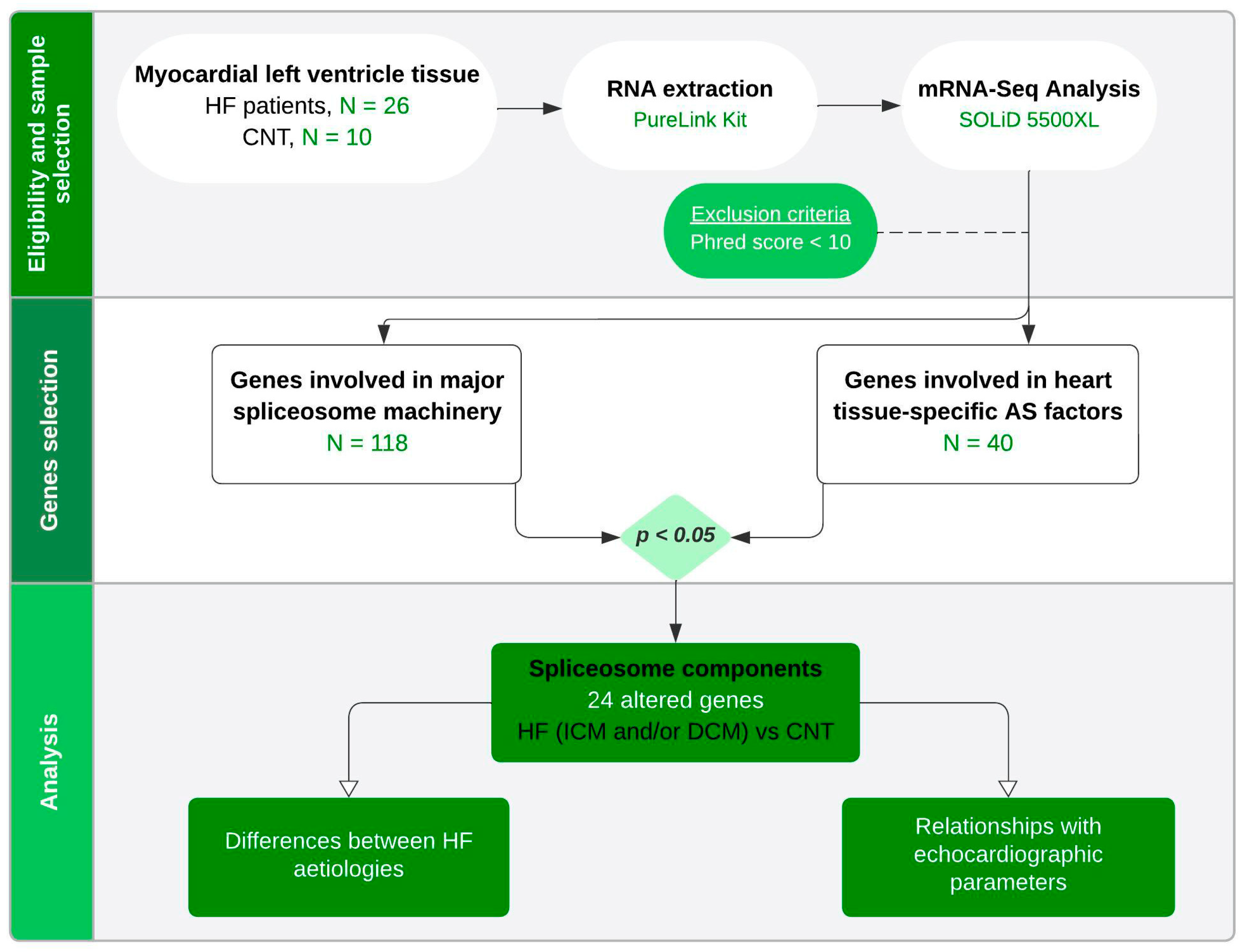
4. Materials and Methods

4.1. Tissue Sample Collection

A total of 36 myocardial left ventricular tissue samples were collected from the ventricle’s apex of explanted human hearts belonging to CNT (n = 10) donors and patients diagnosed with ICM (n = 13) and DCM (n = 13) receiving heart transplantation (Figure 3). After extraction, the samples were maintained in 0.9% NaCl at 4 °C for a maximum of 4.4 ± 3 h after loss of coronary circulation and stored at −80 °C until use.
Clinical history, electrocardiogram, echocardiographic parameters, hemodynamic studies and coronary angiography data were available for all patients. DCM was diagnosed when patients had a LVEF < 40% with a LVEDD > 55 mm. History of documented acute myocardial infarction episodes, the existence of normal contractility segments co-existing with other akinetic or dyskinetic segments shown using echocardiography and the emergence of myocardial necrosis or ischaemia signs on electrocardiography were used to diagnose ICM. Every patient was given medical treatment in accordance with the standards set out by the European Society of Cardiology [21] and was functionally categorised using the NYHA functional criteria.
The CNT samples were collected from non-diseased hearts that were ineligible for transplantation because of blood incompatibility or surgical reasons. Motor vehicle accidents or cerebrovascular incidents were the donors’ reasons of death. Every one of the CNT hearts had a normal LVEF (≥50%). In compliance with the Spanish Organic Law on Data Protection 15/1999, the only available data were age and gender.

4.2. RNA Extraction and Integrity

The cardiac samples (CNT, n = 10; ICM n = 13; DCM, n = 13) were homogenised with TRIzol reagent in a TissueLyser LT (Qiagen, Hilden, Germany). RNA extractions were conducted using a PureLink™ Kit in accordance with the instructions provided by the manufacturer (Ambion Life Technologies, Vilnus, Lithuania). The quantification of RNA was performed using a NanoDrop1000 spectrophotometer (Thermo Fisher Scientific, Waltham, MA, USA), while the purity and integrity of the RNA samples were assessed using an Agilent 2100 Bioanalyser with an RNA 6000 Nano LabChip kit (Agilent Technologies, Santa Clara, CA, USA). All samples had a 260/280 ratio greater than 2.0 and an RNA integrity number of 9 or above.

4.3. mRNA-Seq Analysis

Using a MicroPoly (A) Purist Kit™ (Ambion Life Technologies, Vilnus, Lithuania), 25 micrograms of total RNA was used to isolate the polyA-RNA. Following the manufacturer’s instructions (Ambion Life Technologies, Vilnus, Lithuania), whole-transcriptome libraries generated from the total polyA-RNA samples were sequenced using the SOLiD 5500XL platform. The Qubit 2.0 Fluorometer (Invitrogen, Waltham, MA, USA) was used to quantify the amplified cDNA, and the Bioanalyser 2100 DNA 1000 kit (Agilent Technologies, Santa Clara, CA, USA) was used to analyse the quality of the cDNA. By using the SOLiD Templated Bead Preparation guide, the whole-transcriptome libraries were utilised to generate SOLiD templated beads. Based on the parameters of the workflow analysis (WFA), bead quality was evaluated. The 50,625 paired-end procedure was used to sequence the samples, producing 75 nt + 35 nt (paired-end) + 5 nt (barcode) sequences. The program SETS parameters (SOLiD Experimental Tracking System) were used to measure the quality of the data.
The first set of whole-transcriptome and paired-end reads from the sequencing process were mapped using the Life Technologies mapping algorithm (http://www.lifetechnologies.com/, accessed on 15 May 2020), version 1.3, against the most recent version of the human genome (Version GRchr37/hg19). For both whole-transcriptome and paired-ends analysis, we employed the standard Bioscope settings of version 1.3. For both forward and reverse reads, the seed consisted in the first 25 nucleotides with a maximum of 2 mismatches permitted. The BAM/SAM format [61] was used to report aligned records. Using the Picard Tools software, version 1.83 (http://broadinstitute.github.io/picard/, accessed on 15 May 2020) [62], low-quality readings (Phred score < 10) were removed.

4.4. Statistical Methods

The mean ± standard deviation (SD) was used to express data for continuous variables, whereas percentage values were used for discrete variables. The Kolmogorov–Smirnov test was used to analyse the distribution of the data. The Mann–Whitney U test and the Student’s t-test were used to compare continuous variables with normal and non-normal distributions, respectively. For discrete variables, Fisher’s exact test was applied. To examine the relationships between variables having a normal distribution, Pearson’s correlation coefficients were computed.
The DESeq2 algorithm (version 3.4, retrieved on 15 May 2020) was utilised to evaluate the differential RNA expression between conditions [63]. To prevent false positives from being identified across the differential expression data, we regarded as differentially expressed those RNAs with a p value (p adj) adjusted by FDR < 0.05 [64]. Gene predictions were subsequently estimated using the cufflinks method [65], and the expression levels were calculated using the HTSeq software, version 0.5.4p3 [66]. Only unique readings were considered for the assessment of gene expression after the multimapped reads were removed using this approach. The differential expression between conditions was analysed using the edgeR technique, version 3.2.4 [67]. This approach is predicated on distinct normalisation procedures according to in-depth global samples, the composition of the CG and gene length. This approach uses a Poisson model to estimate the variance of the RNA-seq data for differential expressions during the differential expression process. The statistical analyses were conducted using the R statistic (version R-4.3.1) and SPSS software (version 20.0) for Windows (IBM SPSS Inc., Armonk, NY, USA). The STRING v12.0 program (accessible at https://string-db.org/, retrieved on 23 July 2024) was also used to explore protein–protein interactions. Databases, co-expression and experiments were among the criteria that were assessed.

Supplementary Materials

The following supporting information can be downloaded at: https://www.mdpi.com/article/10.3390/ijms25179590/s1.

Author Contributions

Conceptualisation, M.P., E.T. and E.R.-L.; methodology, I.G.-E. and L.P.-C.; formal analysis, M.P.; investigation, I.G.-E.; resources, E.T. and E.R.-L.; data curation, L.P.-C. and I.G.-T.; writing—original draft preparation, I.G.-E., L.P.-C., E.T. and E.R.-L.; writing—review and editing, M.D.-A., I.G.-T. and C.B.; supervision, E.T. and E.R.-L.; funding acquisition, E.T. and E.R.-L. All authors have read and agreed to the published version of the manuscript.

Funding

This work was supported by the National Institute of Health “Fondo de Investigaciones Sanitarias del Instituto de Salud Carlos III” (Projects: PI20/01469 and PI20/00071) co-funded by the European Union; Miguel Servet contract CP21/00041 co-funded by the European Union; contracts FI21/00186 and FI21/00034; “Consorcio Centro de Investigación Biomédica en Red” [CIBERCV, under Grant CB16/11/00261]; Ministry of Science and Innovation (MCIN, 10.13039/501100011033) and State Investigation Agency (AEI) (Project CNS2022-135769) co-funded by the European Union “Next Generation EU” and the European Recovery, Transformation and Resilience Plan (PRTR); and Conselleria de Educación, Universidades y Empleo (Project CIAICO/2022/246, contract CIACIF/2022/429).

Institutional Review Board Statement

This study was approved by the Ethics Committee (Biomedical Investigation Ethics Committee of La Fe University Hospital of Valencia, Spain; Code of Protocol 2016/0320, 15 November 2016) and was conducted in accordance with the principles described in the Declaration of Helsinki [68].

Informed Consent Statement

Informed consent was obtained from all subjects involved in the study.

Data Availability Statement

The data presented in this manuscript have been deposited in the NCBI’s Gene Expression Omnibus [69] (GEO) database and are accessible through the GEO series accession number GSE55296.

Conflicts of Interest

The authors declare no conflicts of interest.

References

  1. Bozkurt, B.; Coats, A.J.S.; Tsutsui, H.; Abdelhamid, C.M.; Adamopoulos, S.; Albert, N.; Anker, S.D.; Atherton, J.; Bohm, M.; Butler, J.; et al. Universal definition and classification of heart failure: A report of the Heart Failure Society of America, Heart Failure Association of the European Society of Cardiology, Japanese Heart Failure Society and Writing Committee of the Universal Definition of Heart Failure: Endorsed by the Canadian Heart Failure Society, Heart Failure Association of India, Cardiac Society of Australia and New Zealand, and Chinese Heart Failure Association. Eur. J. Heart Fail. 2021, 23, 352–380. [Google Scholar]
  2. Bozkurt, B.; Colvin, M.; Cook, J.; Cooper, L.T.; Deswal, A.; Fonarow, G.C.; Francis, G.S.; Lenihan, D.; Lewis, E.F.; McNamara, D.M.; et al. Current Diagnostic and Treatment Strategies for Specific Dilated Cardiomyopathies: A Scientific Statement From the American Heart Association. Circulation 2016, 134, e579–e646. [Google Scholar] [CrossRef]
  3. Baldasseroni, S.; Opasich, C.; Gorini, M.; Lucci, D.; Marchionni, N.; Marini, M.; Campana, C.; Perini, G.; Deorsola, A.; Masotti, G.; et al. Left bundle-branch block is associated with increased 1-year sudden and total mortality rate in 5517 outpatients with congestive heart failure: A report from the Italian network on congestive heart failure. Am. Heart J. 2002, 143, 398–405. [Google Scholar] [CrossRef]
  4. Herrer, I.; Rosello-Lleti, E.; Rivera, M.; Molina-Navarro, M.M.; Tarazon, E.; Ortega, A.; Martinez-Dolz, L.; Trivino, J.C.; Lago, F.; Gonzalez-Juanatey, J.R.; et al. RNA-sequencing analysis reveals new alterations in cardiomyocyte cytoskeletal genes in patients with heart failure. Lab. Investig. 2014, 94, 645–653. [Google Scholar] [CrossRef] [PubMed]
  5. Tarazon, E.; Rosello-Lleti, E.; Rivera, M.; Ortega, A.; Molina-Navarro, M.M.; Trivino, J.C.; Lago, F.; Gonzalez-Juanatey, J.R.; Orosa, P.; Montero, J.A.; et al. RNA sequencing analysis and atrial natriuretic peptide production in patients with dilated and ischemic cardiomyopathy. PLoS ONE 2014, 9, e90157. [Google Scholar] [CrossRef] [PubMed]
  6. Gimenez-Escamilla, I.; Benedicto, C.; Perez-Carrillo, L.; Delgado-Arija, M.; Gonzalez-Torrent, I.; Vilchez, R.; Martinez-Dolz, L.; Portoles, M.; Tarazon, E.; Rosello-Lleti, E. Alterations in Mitochondrial Oxidative Phosphorylation System: Relationship of Complex V and Cardiac Dysfunction in Human Heart Failure. Antioxidants 2024, 13, 285. [Google Scholar] [CrossRef]
  7. Rosello-Lleti, E.; Alonso, J.; Cortes, R.; Almenar, L.; Martinez-Dolz, L.; Sanchez-Lazaro, I.; Lago, F.; Azorin, I.; Juanatey, J.R.; Portoles, M.; et al. Cardiac protein changes in ischaemic and dilated cardiomyopathy: A proteomic study of human left ventricular tissue. J. Cell. Mol. Med. 2012, 16, 2471–2486. [Google Scholar] [CrossRef] [PubMed]
  8. Lara-Pezzi, E.; Dopazo, A.; Manzanares, M. Understanding cardiovascular disease: A journey through the genome (and what we found there). Dis. Models Mech. 2012, 5, 434–443. [Google Scholar] [CrossRef] [PubMed]
  9. Jiang, W.; Chen, L. Alternative splicing: Human disease and quantitative analysis from high-throughput sequencing. Comput. Struct. Biotechnol. J. 2021, 19, 183–195. [Google Scholar] [CrossRef]
  10. Wahl, M.C.; Luhrmann, R. SnapShot: Spliceosome Dynamics I. Cell 2015, 161, 1474. [Google Scholar] [CrossRef]
  11. Cao, J.; Wei, Z.; Nie, Y.; Chen, H.Z. Therapeutic potential of alternative splicing in cardiovascular diseases. EBioMedicine 2024, 101, 104995. [Google Scholar] [CrossRef] [PubMed]
  12. Badr, E.; ElHefnawi, M.; Heath, L.S. Computational Identification of Tissue-Specific Splicing Regulatory Elements in Human Genes from RNA-Seq Data. PLoS ONE 2016, 11, e0166978. [Google Scholar] [CrossRef]
  13. Tao, Y.; Zhang, Q.; Wang, H.; Yang, X.; Mu, H. Alternative splicing and related RNA binding proteins in human health and disease. Signal Transduct. Target. Ther. 2024, 9, 26. [Google Scholar] [CrossRef] [PubMed]
  14. Ito, K.; Patel, P.N.; Gorham, J.M.; McDonough, B.; DePalma, S.R.; Adler, E.E.; Lam, L.; MacRae, C.A.; Mohiuddin, S.M.; Fatkin, D.; et al. Identification of pathogenic gene mutations in LMNA and MYBPC3 that alter RNA splicing. Proc. Natl. Acad. Sci. USA 2017, 114, 7689–7694. [Google Scholar] [CrossRef] [PubMed]
  15. Guo, W.; Schafer, S.; Greaser, M.L.; Radke, M.H.; Liss, M.; Govindarajan, T.; Maatz, H.; Schulz, H.; Li, S.; Parrish, A.M.; et al. RBM20, a gene for hereditary cardiomyopathy, regulates titin splicing. Nat. Med. 2012, 18, 766–773. [Google Scholar] [CrossRef]
  16. Nutter, C.A.; Jaworski, E.A.; Verma, S.K.; Deshmukh, V.; Wang, Q.; Botvinnik, O.B.; Lozano, M.J.; Abass, I.J.; Ijaz, T.; Brasier, A.R.; et al. Dysregulation of RBFOX2 Is an Early Event in Cardiac Pathogenesis of Diabetes. Cell Rep. 2016, 15, 2200–2213. [Google Scholar] [CrossRef]
  17. Varga, Z.V.; Pipicz, M.; Baan, J.A.; Baranyai, T.; Koncsos, G.; Leszek, P.; Kusmierczyk, M.; Sanchez-Cabo, F.; Garcia-Pavia, P.; Brenner, G.J.; et al. Alternative Splicing of NOX4 in the Failing Human Heart. Front. Physiol. 2017, 8, 935. [Google Scholar] [CrossRef]
  18. Cvitkovic, I.; Jurica, M.S. Spliceosome database: A tool for tracking components of the spliceosome. Nucleic Acids Res. 2013, 41, D132–D141. [Google Scholar] [CrossRef]
  19. Seal, R.L.; Braschi, B.; Gray, K.; Jones, T.E.M.; Tweedie, S.; Haim-Vilmovsky, L.; Bruford, E.A. Genenames.org: The HGNC resources in 2023. Nucleic Acids Res. 2023, 51, D1003–D1009. [Google Scholar] [CrossRef]
  20. van den Hoogenhof, M.M.; Pinto, Y.M.; Creemers, E.E. RNA Splicing: Regulation and Dysregulation in the Heart. Circ. Res. 2016, 118, 454–468. [Google Scholar] [CrossRef]
  21. Ponikowski, P.; Voors, A.A.; Anker, S.D.; Bueno, H.; Cleland, J.G.; Coats, A.J.; Falk, V.; Gonzalez-Juanatey, J.R.; Harjola, V.P.; Jankowska, E.A.; et al. 2016 ESC Guidelines for the diagnosis and treatment of acute and chronic heart failure: The Task Force for the diagnosis and treatment of acute and chronic heart failure of the European Society of Cardiology (ESC). Developed with the special contribution of the Heart Failure Association (HFA) of the ESC. Eur. J. Heart Fail 2016, 18, 891–975. [Google Scholar]
  22. Rogalska, M.E.; Vivori, C.; Valcarcel, J. Regulation of pre-mRNA splicing: Roles in physiology and disease, and therapeutic prospects. Nat. Rev. Genet. 2023, 24, 251–269. [Google Scholar] [CrossRef] [PubMed]
  23. Li, X.; Liu, S.; Zhang, L.; Issaian, A.; Hill, R.C.; Espinosa, S.; Shi, S.; Cui, Y.; Kappel, K.; Das, R.; et al. A unified mechanism for intron and exon definition and back-splicing. Nature 2019, 573, 375–380. [Google Scholar] [CrossRef] [PubMed]
  24. Zhang, Z.; Bae, B.; Cuddleston, W.H.; Miura, P. Coordination of alternative splicing and alternative polyadenylation revealed by targeted long read sequencing. Nat. Commun. 2023, 14, 5506. [Google Scholar] [CrossRef]
  25. Qiu, F.; Fu, Y.; Lu, C.; Feng, Y.; Wang, Q.; Huo, Z.; Jia, X.; Chen, C.; Chen, S.; Xu, A. Small Nuclear Ribonucleoprotein Polypeptide A-Mediated Alternative Polyadenylation of STAT5B during Th1 Cell Differentiation. J. Immunol. 2017, 199, 3106–3115. [Google Scholar] [CrossRef]
  26. Bansal, S.S.; Ismahil, M.A.; Goel, M.; Patel, B.; Hamid, T.; Rokosh, G.; Prabhu, S.D. Activated T Lymphocytes are Essential Drivers of Pathological Remodeling in Ischemic Heart Failure. Circ. Heart Fail. 2017, 10, e003688. [Google Scholar] [CrossRef] [PubMed]
  27. Hu, Z.; Li, M.; Huo, Z.; Chen, L.; Liu, S.; Deng, K.; Lu, X.; Chen, S.; Fu, Y.; Xu, A. U1 snRNP proteins promote proximal alternative polyadenylation sites by directly interacting with 3′ end processing core factors. J. Mol. Cell Biol. 2022, 14, mjac054. [Google Scholar] [CrossRef]
  28. Ribera, J.; Portoles, I.; Cordoba-Jover, B.; Rodriguez-Vita, J.; Casals, G.; Gonzalez-de la Presa, B.; Graupera, M.; Solsona-Vilarrasa, E.; Garcia-Ruiz, C.; Fernandez-Checa, J.C.; et al. The loss of DHX15 impairs endothelial energy metabolism, lymphatic drainage and tumor metastasis in mice. Commun. Biol. 2021, 4, 1192. [Google Scholar] [CrossRef]
  29. Maul-Newby, H.M.; Amorello, A.N.; Sharma, T.; Kim, J.H.; Modena, M.S.; Prichard, B.E.; Jurica, M.S. A model for DHX15 mediated disassembly of A-complex spliceosomes. RNA 2022, 28, 583–595. [Google Scholar] [CrossRef]
  30. Zhou, Y.; Alimohamadi, S.; Wang, L.; Liu, Z.; Wall, J.B.; Yin, C.; Liu, J.; Qian, L. A Loss of Function Screen of Epigenetic Modifiers and Splicing Factors during Early Stage of Cardiac Reprogramming. Stem Cells Int. 2018, 2018, 3814747. [Google Scholar] [CrossRef]
  31. Laflamme, M.A.; Murry, C.E. Heart regeneration. Nature 2011, 473, 326–335. [Google Scholar] [CrossRef] [PubMed]
  32. Atkinson, R.; Georgiou, M.; Yang, C.; Szymanska, K.; Lahat, A.; Vasconcelos, E.J.R.; Ji, Y.; Moya Molina, M.; Collin, J.; Queen, R.; et al. PRPF8-mediated dysregulation of hBrr2 helicase disrupts human spliceosome kinetics and 5 -splice-site selection causing tissue-specific defects. Nat. Commun. 2024, 15, 3138. [Google Scholar] [CrossRef]
  33. Gonzalez-Rodriguez, P.; Klionsky, D.J.; Joseph, B. Autophagy regulation by RNA alternative splicing and implications in human diseases. Nat. Commun. 2022, 13, 2735. [Google Scholar] [CrossRef]
  34. Xu, G.; Li, T.; Chen, J.; Li, C.; Zhao, H.; Yao, C.; Dong, H.; Wen, K.; Wang, K.; Zhao, J.; et al. Autosomal dominant retinitis pigmentosa-associated gene PRPF8 is essential for hypoxia-induced mitophagy through regulating ULK1 mRNA splicing. Autophagy 2018, 14, 1818–1830. [Google Scholar] [CrossRef] [PubMed]
  35. Chang, X.; Toan, S.; Li, R.; Zhou, H. Therapeutic strategies in ischemic cardiomyopathy: Focus on mitochondrial quality surveillance. EBioMedicine 2022, 84, 104260. [Google Scholar] [CrossRef]
  36. Ye, J.; Ying, J.; Chen, H.; Wu, Z.; Huang, C.; Zhang, C.; Chen, Z.; Chen, H. PPIH acts as a potential predictive biomarker for patients with common solid tumors. BMC Cancer 2024, 24, 681. [Google Scholar] [CrossRef] [PubMed]
  37. Wang, Z.; Wang, S.; Qin, J.; Zhang, X.; Lu, G.; Liu, H.; Guo, H.; Wu, L.; Shender, V.O.; Shao, C.; et al. Splicing factor BUD31 promotes ovarian cancer progression through sustaining the expression of anti-apoptotic BCL2L12. Nat. Commun. 2022, 13, 6246. [Google Scholar] [CrossRef]
  38. Sales-Lee, J.; Perry, D.S.; Bowser, B.A.; Diedrich, J.K.; Rao, B.; Beusch, I.; Yates, J.R., 3rd; Roy, S.W.; Madhani, H.D. Coupling of spliceosome complexity to intron diversity. Curr. Biol. 2021, 31, 4898–4910.e4. [Google Scholar] [CrossRef]
  39. Hamann, F.; Enders, M.; Ficner, R. Structural basis for RNA translocation by DEAH-box ATPases. Nucleic Acids Res. 2019, 47, 4349–4362. [Google Scholar] [CrossRef]
  40. Dreyer, J.; Schleicher, M.; Tappe, A.; Schilling, K.; Kuner, T.; Kusumawidijaja, G.; Muller-Esterl, W.; Oess, S.; Kuner, R. Nitric oxide synthase (NOS)-interacting protein interacts with neuronal NOS and regulates its distribution and activity. J. Neurosci. 2004, 24, 10454–10465. [Google Scholar] [CrossRef]
  41. Rosello-Lleti, E.; Carnicer, R.; Tarazon, E.; Ortega, A.; Gil-Cayuela, C.; Lago, F.; Gonzalez-Juanatey, J.R.; Portoles, M.; Rivera, M. Human Ischemic Cardiomyopathy Shows Cardiac Nos1 Translocation and its Increased Levels are Related to Left Ventricular Performance. Sci. Rep. 2016, 6, 24060. [Google Scholar] [CrossRef] [PubMed]
  42. Coltri, P.; Effenberger, K.; Chalkley, R.J.; Burlingame, A.L.; Jurica, M.S. Breaking up the C complex spliceosome shows stable association of proteins with the lariat intron intermediate. PLoS ONE 2011, 6, e19061. [Google Scholar] [CrossRef] [PubMed]
  43. Brauch, K.M.; Karst, M.L.; Herron, K.J.; de Andrade, M.; Pellikka, P.A.; Rodeheffer, R.J.; Michels, V.V.; Olson, T.M. Mutations in ribonucleic acid binding protein gene cause familial dilated cardiomyopathy. J. Am. Coll. Cardiol. 2009, 54, 930–941. [Google Scholar] [CrossRef] [PubMed]
  44. Montanes-Agudo, P.; Pinto, Y.M.; Creemers, E.E. Splicing factors in the heart: Uncovering shared and unique targets. J. Mol. Cell. Cardiol. 2023, 179, 72–79. [Google Scholar] [CrossRef]
  45. Montaville, P.; Dai, Y.; Cheung, C.Y.; Giller, K.; Becker, S.; Michalak, M.; Webb, S.E.; Miller, A.L.; Krebs, J. Nuclear translocation of the calcium-binding protein ALG-2 induced by the RNA-binding protein RBM22. Biochim. Biophys. Acta 2006, 1763, 1335–1343. [Google Scholar] [CrossRef]
  46. Janowicz, A.; Michalak, M.; Krebs, J. Stress induced subcellular distribution of ALG-2, RBM22 and hSlu7. Biochim. Biophys. Acta 2011, 1813, 1045–1049. [Google Scholar] [CrossRef]
  47. Guo, J.; Liu, H.B.; Sun, C.; Yan, X.Q.; Hu, J.; Yu, J.; Yuan, Y.; Du, Z.M. MicroRNA-155 Promotes Myocardial Infarction-Induced Apoptosis by Targeting RNA-Binding Protein QKI. Oxid. Med. Cell Longev. 2019, 2019, 4579806. [Google Scholar] [CrossRef]
  48. Chen, X.; Liu, Y.; Xu, C.; Ba, L.; Liu, Z.; Li, X.; Huang, J.; Simpson, E.; Gao, H.; Cao, D.; et al. QKI is a critical pre-mRNA alternative splicing regulator of cardiac myofibrillogenesis and contractile function. Nat. Commun. 2021, 12, 89. [Google Scholar] [CrossRef]
  49. Montanes-Agudo, P.; Aufiero, S.; Schepers, E.N.; van der Made, I.; Cocera-Ortega, L.; Ernault, A.C.; Richard, S.; Kuster, D.W.D.; Christoffels, V.M.; Pinto, Y.M.; et al. The RNA-binding protein QKI governs a muscle-specific alternative splicing program that shapes the contractile function of cardiomyocytes. Cardiovasc. Res. 2023, 119, 1161–1174. [Google Scholar] [CrossRef]
  50. Zhang, Y.; Si, Y.; Ma, N.; Mei, J. The RNA-binding protein PCBP2 inhibits Ang II-induced hypertrophy of cardiomyocytes though promoting GPR56 mRNA degeneration. Biochem. Biophys. Res. Commun. 2015, 464, 679–684. [Google Scholar] [CrossRef]
  51. Ortiz-Sanchez, P.; Villalba-Orero, M.; Lopez-Olaneta, M.M.; Larrasa-Alonso, J.; Sanchez-Cabo, F.; Marti-Gomez, C.; Camafeita, E.; Gomez-Salinero, J.M.; Ramos-Hernandez, L.; Nielsen, P.J.; et al. Loss of SRSF3 in Cardiomyocytes Leads to Decapping of Contraction-Related mRNAs and Severe Systolic Dysfunction. Circ. Res. 2019, 125, 170–183. [Google Scholar] [CrossRef] [PubMed]
  52. Akerberg, A.A.; Trembley, M.; Butty, V.; Schwertner, A.; Zhao, L.; Beerens, M.; Liu, X.; Mahamdeh, M.; Yuan, S.; Boyer, L.; et al. RBPMS2 Is a Myocardial-Enriched Splicing Regulator Required for Cardiac Function. Circ. Res. 2022, 131, 980–1000. [Google Scholar] [CrossRef]
  53. Ye, J.; Beetz, N.; O’Keeffe, S.; Tapia, J.C.; Macpherson, L.; Chen, W.V.; Bassel-Duby, R.; Olson, E.N.; Maniatis, T. hnRNP U protein is required for normal pre-mRNA splicing and postnatal heart development and function. Proc. Natl. Acad. Sci. USA 2015, 112, E3020–E3029. [Google Scholar] [CrossRef] [PubMed]
  54. Aslan, G.S.; Jae, N.; Manavski, Y.; Fouani, Y.; Shumliakivska, M.; Kettenhausen, L.; Kirchhof, L.; Gunther, S.; Fischer, A.; Luxan, G.; et al. Malat1 deficiency prevents neonatal heart regeneration by inducing cardiomyocyte binucleation. JCI Insight 2023, 8, e162124. [Google Scholar] [CrossRef]
  55. Soubise, B.; Jiang, Y.; Douet-Guilbert, N.; Troadec, M.B. RBM22, a Key Player of Pre-mRNA Splicing and Gene Expression Regulation, Is Altered in Cancer. Cancers 2022, 14, 643. [Google Scholar] [CrossRef] [PubMed]
  56. Gao, C.; Ren, S.; Lee, J.H.; Qiu, J.; Chapski, D.J.; Rau, C.D.; Zhou, Y.; Abdellatif, M.; Nakano, A.; Vondriska, T.M.; et al. RBFox1-mediated RNA splicing regulates cardiac hypertrophy and heart failure. J. Clin. Investig. 2016, 126, 195–206. [Google Scholar] [CrossRef]
  57. Love, S.L.; Emerson, J.D.; Koide, K.; Hoskins, A.A. Pre-mRNA splicing-associated diseases and therapies. RNA Biol. 2023, 20, 525–538. [Google Scholar] [CrossRef]
  58. Zong, F.Y.; Fu, X.; Wei, W.J.; Luo, Y.G.; Heiner, M.; Cao, L.J.; Fang, Z.; Fang, R.; Lu, D.; Ji, H.; et al. The RNA-binding protein QKI suppresses cancer-associated aberrant splicing. PLoS Genet. 2014, 10, e1004289. [Google Scholar] [CrossRef] [PubMed]
  59. Griffin, C.; Saint-Jeannet, J.P. Spliceosomopathies: Diseases and mechanisms. Dev. Dyn. 2020, 249, 1038–1046. [Google Scholar] [CrossRef]
  60. Martin, S.S.; Aday, A.W.; Almarzooq, Z.I.; Anderson, C.A.M.; Arora, P.; Avery, C.L.; Baker-Smith, C.M.; Barone Gibbs, B.; Beaton, A.Z.; Boehme, A.K.; et al. 2024 Heart Disease and Stroke Statistics: A Report of US and Global Data From the American Heart Association. Circulation 2024, 149, e347–e913. [Google Scholar]
  61. Li, H.; Handsaker, B.; Wysoker, A.; Fennell, T.; Ruan, J.; Homer, N.; Marth, G.; Abecasis, G.; Durbin, R.; Genome Project Data Processing, S. The Sequence Alignment/Map format and SAMtools. Bioinformatics 2009, 25, 2078–2079. [Google Scholar] [CrossRef] [PubMed]
  62. McKenna, A.; Hanna, M.; Banks, E.; Sivachenko, A.; Cibulskis, K.; Kernytsky, A.; Garimella, K.; Altshuler, D.; Gabriel, S.; Daly, M.; et al. The Genome Analysis Toolkit: A MapReduce framework for analyzing next-generation DNA sequencing data. Genome Res. 2010, 20, 1297–1303. [Google Scholar] [CrossRef]
  63. Love, M.I.; Huber, W.; Anders, S. Moderated estimation of fold change and dispersion for RNA-seq data with DESeq2. Genome Biol. 2014, 15, 550. [Google Scholar] [CrossRef] [PubMed]
  64. Benjamini, Y.; Hochberg, Y. Controlling the False Discovery Rate: A Practical and Powerful Approach to Multiple Testing. J. R. Stat. Soc. Ser. B 1995, 57, 289–300. [Google Scholar] [CrossRef]
  65. Trapnell, C.; Williams, B.A.; Pertea, G.; Mortazavi, A.; Kwan, G.; van Baren, M.J.; Salzberg, S.L.; Wold, B.J.; Pachter, L. Transcript assembly and quantification by RNA-Seq reveals unannotated transcripts and isoform switching during cell differentiation. Nat. Biotechnol. 2010, 28, 511–515. [Google Scholar] [CrossRef] [PubMed]
  66. Anders, S.; Pyl, P.T.; Huber, W. HTSeq—A Python framework to work with high-throughput sequencing data. Bioinformatics 2015, 31, 166–169. [Google Scholar] [CrossRef]
  67. Robinson, M.D.; McCarthy, D.J.; Smyth, G.K. edgeR: A Bioconductor package for differential expression analysis of digital gene expression data. Bioinformatics 2010, 26, 139–140. [Google Scholar] [CrossRef] [PubMed]
  68. Macrae, D.J. The Council for International Organizations and Medical Sciences (CIOMS) guidelines on ethics of clinical trials. Proc. Am. Thorac. Soc. 2007, 4, 176–178, discussion 178–179. [Google Scholar] [CrossRef]
  69. Edgar, R.; Domrachev, M.; Lash, A.E. Gene Expression Omnibus: NCBI gene expression and hybridization array data repository. Nucleic Acids Res. 2002, 30, 207–210. [Google Scholar] [CrossRef]
Figure 1. Spliceosome components status in advanced heart failure patients. (A) Sequential formation of spliceosome complexes: E complex is formed upon U1 snRNP’s recognition of 5′ splicing site. Once U2 snRNP replaces SF1, it transforms into the A complex. Subsequently, the union of U4, U5 and U6 snRNPs gives rise to inactive complex B. A major compositional and conformational remodelling forms Bact and B* complexes, becoming catalytically active. After the first transesterification reaction, the C complex is formed. The altered molecules are listed for each complex alongside the alternative splicing factors (dotted box). (B) Venn diagram highlighting similarities and differences in genes of major spliceosome, with altered expression between ischaemic (ICM) and dilated (DCM) cardiomyopathies. (C,D) Heat map summarising mRNA expression levels of spliceosome components from control (CNT) subjects and ICM and DCM patients, respectively. Each row represents a sample, and each column represents an altered mRNA. Colours depict the relative expression level of each molecule, with green being the lowest and red the highest. (E,F) Schematic string interaction analysis showing protein–protein interactions of spliceosome components and heart-related splicing factors differentially expressed in ICM and DCM, respectively. Edges represent protein–protein associations including known interactions from curated databases (light blue), experimentally determined (pink) and co-expression (black).
Ijms 25 09590 g001
Figure 2. Altered mRNA expression levels of spliceosome components and splicing factors in advanced heart failure patients. (A) mRNA expression levels of E complex components. (B) Relationship between mRNA relative expression levels of SNRPD2 and left ventricular mass index (LVMI) in ischaemic cardiomyopathy patients. (C) mRNA expression levels of A complex components. (D) mRNA expression levels of B complex components. (E) mRNA expression levels of C complex components. (F) mRNA expression levels of alternative splicing factors. Data are presented as the mean ± SEM; a.u., arbitrary units. Statistical differences found between ischaemic cardiomyopathy, control and dilated cardiomyopathy groups: * p < 0.05, ** p < 0.01 and *** p < 0.001.
Ijms 25 09590 g002
Figure 3. Gene expression study flowchart. Summary of analysis approach and findings on gene expression analysis of the spliceosome components. HF, heart failure; CNT, controls; AS, alternative splicing; ICM, ischaemic cardiomyopathy; DCM, dilated cardiomyopathy.
Ijms 25 09590 g003
Table 1. Clinical characteristics of patients.
mRNA-Seq
ICM (n = 13)DCM (n = 13)
Gender male (%)10092
Age (years)54 ± 851 ± 11
NYHA classIII–IVIII–IV
BMI (kg/m2)27 ± 427 ± 5
Haemoglobin (g/dL)14 ± 313 ± 3
Haematocrit (%)41 ± 639 ± 7
Total cholesterol (mg/dL)162 ± 41147 ± 37
NT-proBNP (pg/mL)3684 ± 22854071 ± 3584
Prior hypertension (%)3317
Prior smoking (%)92 *50
Diabetes mellitus (%)4217
Echocardiographic study
 LVEF (%)25 ± 5 *17 ± 8
 LVESD (mm)57 ± 8 ***74 ± 10
 LVEDD (mm)65 ± 8 ***81 ± 8
 LVMI (g/cm2)139 ± 26 ***245 ± 64
Duration of disease (months)45 ± 4075 ± 68
Treatment (%)
 Renin–angiotensin system inhibitors71100
 B-blokers5773
 Aldosterone antagonists10091
 Digoxin4364
 Diuretics71100
ICM, ischaemic cardiomyopathy; DCM, dilated cardiomyopathy; NYHA, New York Heart Association; BMI, body mass index; LVEF, left ventricular ejection fraction; LVESD, left ventricular end-systolic diameter; LVEDD, left ventricular end-diastolic diameter; LVMI, left ventricular mass index; Duration of disease, from heart failure diagnosis until heart transplant. For differences between ICM and DCM in mRNA-seq study, * p < 0.05 and *** p < 0.001. Qualitative data are presented as percentages and quantitative data as mean values ± standard deviation.
Disclaimer/Publisher’s Note: The statements, opinions and data contained in all publications are solely those of the individual author(s) and contributor(s) and not of MDPI and/or the editor(s). MDPI and/or the editor(s) disclaim responsibility for any injury to people or property resulting from any ideas, methods, instructions or products referred to in the content.

