Article

Antimicrobial Activity of Chalcones with a Chlorine Atom and Their Glycosides

by
Agnieszka Krawczyk-Łebek
1,*,
Barbara Żarowska
2,
Tomasz Janeczko
1 and
Edyta Kostrzewa-Susłow
1
1
Department of Food Chemistry and Biocatalysis, Faculty of Biotechnology and Food Science, Wrocław University of Environmental and Life Sciences, 50-375 Wrocław, Poland
2
Department of Biotechnology and Food Microbiology, Faculty of Biotechnology and Food Science, Wrocław University of Environmental and Life Sciences, 51-630 Wrocław, Poland
*
Author to whom correspondence should be addressed.
Int. J. Mol. Sci. 2024, 25(17), 9718; https://doi.org/10.3390/ijms25179718
Submission received: 7 August 2024 / Revised: 3 September 2024 / Accepted: 5 September 2024 / Published: 8 September 2024
(This article belongs to the Special Issue Biocatalysis and Bioactive Molecules: Future and Development)

Abstract

:
Chalcones, secondary plant metabolites, exhibit various biological properties. The introduction of a chlorine and a glucosyl substituent to the chalcone could enhance its bioactivity and bioavailability. Such compounds can be obtained through a combination of chemical and biotechnological methods. Therefore, 4-chloro-2′-hydroxychalcone and 5′-chloro-2′-hydroxychalcone were obtained by synthesis and then glycosylated in two filamentous fungi strains cultures, i.e., Isaria fumosorosea KCH J2 and Beauveria bassiana KCH J1.5. The main site of the glycosylation of both compounds by I. fumosorosea KCH J2 was C-2′ and C-3 when the second strain was utilized. The pharmacokinetics of these compounds were predicted using chemoinformatics tools. Furthermore, antimicrobial activity tests were performed. Compounds significantly inhibited the growth of the bacteria strains Escherichia coli 10536, Staphylococcus aureus DSM 799, and yeast Candida albicans DSM 1386. Nevertheless, the bacterial strain Pseudomonas aeruginosa DSM 939 exhibited significant resistance to their effects. The growth of lactic acid bacteria strain Lactococcus acidophilus KBiMZ 01 bacteria was moderately inhibited, but strains Lactococcus rhamnosus GG and Streptococcus thermophilus KBM-1 were completely inhibited. In summary, chalcones substituted with a chlorine demonstrated greater efficacy in inhibiting the microbial strains under examination compared to 2′-hydroxychalcone, while aglycones and their glycosides exhibited similar effectiveness.

1. Introduction

Chalcones, secondary plant metabolites, exhibit various biological properties, including anticancer activity (e.g., isoliquiritigenin, butein, and xantohumol), anti-inflammatory activity (e.g., sappanchalcone, butein, and licochalcone A), α-Glucosidase inhibitory activity (e.g., isobavachalcone), and antibacterial activity (e.g., bavachalcone, xanthumol, and isobavachalcone) [1]. These compounds have a shared chemical scaffold, comprising two aromatic rings linked by a three-carbon α,β-unsaturated carbonyl system. Dihydrochalcones, which are α,β-hydrogenated derivatives, are also commonly found in nature, with most famous phlorizin being ubiquitous in Malus species [2,3,4]. Chalcones and dihydrochalcones may occur in the form of glycosides, making them more stable and soluble in water, which improves their biodistribution and storage in plants [5,6,7]. Introducing a chlorine atom into the chalcone structure may enhance its bioactivity., including antibacterial properties [8,9,10]. When a chlorine atom is introduced onto ring B of 2′-hydroxychalcone, it enhances its antituberculosis activity against strain Mycobacterium tuberculosis H37Rv [9]. Similarly, another flavonoid substituted with halogen, i.e., chlorflavonin, exhibited strong antituberculosis potential with (MIC90 1.56 μM) and was superior to streptomycin treatment [11]. Halogenated chalcones, while not naturally occurring, can be synthesized through the Claisen–Schmidt condensation [8,9,12].
The promising strategy for the glycosylation of chalcones can be the use of microbial enzymes—among others—as well as whole-cell biotransformation using filamentous fungi as biocatalysts [13,14]. Xie and coworkers employed genome mining and heterologous expression techniques to discover functional modules of glycosyltransferase-methyltransferase (GT-MT) in these fungi. These modules exhibit substrate promiscuity and regiospecificity, allowing them to methylglucosylate flavonoids, as shown in Figure 1 [15].
The lack of in vivo data makes it difficult to generalize the influence of glycosylation on flavonoids bioactivity. Based on some reports, it appears that O-glycosylation may reduce anti-inflammation activity, antioxidant activity, and antimicrobial activity. Simultaneously, some data show that O-glycosylation can enhance specific bioactivity, including anti-HIV activity, tyrosinase inhibition, antirotavirus activity, and antiallergic activity. Furthermore, glycosylation positively affects their bioavailability [16]. The impact of glycosylation on flavonoid bioactivity in vitro may not necessarily mirror the effects observed in vivo. Consequently, additional research in this field is essential. Therefore, the purpose of this work was to obtain two chalcones with a chlorine atom, i.e., 4-chloro-2′-hydroxychalcone and 5′-chloro-2′-hydroxychalcone, and afterward glycosylate them in entomopathogenic filamentous fungi I. fumosorosea KCH J2 and B. bassiana KCH J1.5 cultures. As a result, we obtained novel flavonoid derivatives. Subsequently, we evaluated the pharmacokinetics of the received compounds using computer-aided simulations. Furthermore, the antimicrobial activities of the chalcones with chlorine atoms, their main biotransformation products, and 2′-hydroxychalcone used for comparison were assessed to explore how chlorine substitution and glucose attachment influence their effectiveness.

2. Results and Discussion

2.1. Synthesis of Biotransformation Substrates 4-Chloro-2′-Hydroxychalcone (3) and 5′-Chloro-2′-Hydroxychalcone (6)

The initial phase of the research was a synthesis of two chalcones with a chlorine atom 4-chloro-2′-hydroxychalcone (Figure 2) and 5′-chloro-2′-hydroxychalone (Figure 3) by Claisen–Schmidt condensation reactions.
The structures of synthetic products 3 and 6 were determined based on NMR spectroscopy (Table 1, Table 2, Table 3 and Table 4, Supplementary Information: Figures S3–S14 and S18–S29, respectively), and their molecular masses were verified using LC-MS spectroscopy (Supplementary Materials: Figures S1 and S16, respectively).
In the next step, entomopathogenic filamentous fungi I. fumosorosea KCH J2 and B. bassiana KCH J1.5 were applied to biotransform 4-chloro-2′-hydroxychalcone (3) and 5′-chloro-2′-hydroxychalcone (6) into their 4″-O-methylglucosylated derivatives. The biotransformation products were extracted from the reaction mixture and subsequently purified via preparative thin-layer chromatography (TLC). Product yields were calculated based on the quantities isolated. Nuclear magnetic resonance (NMR) spectroscopy was used to elucidate the chemical structures, with confirmation provided by liquid chromatography–mass spectrometry (LC-MS). The antimicrobial activity was evaluated for biotransformation substrates (3 and 6), their primary products (3a, 6a, 6b), together with a compound without chlorine substituent – 2′-hydroxychalcone (7).

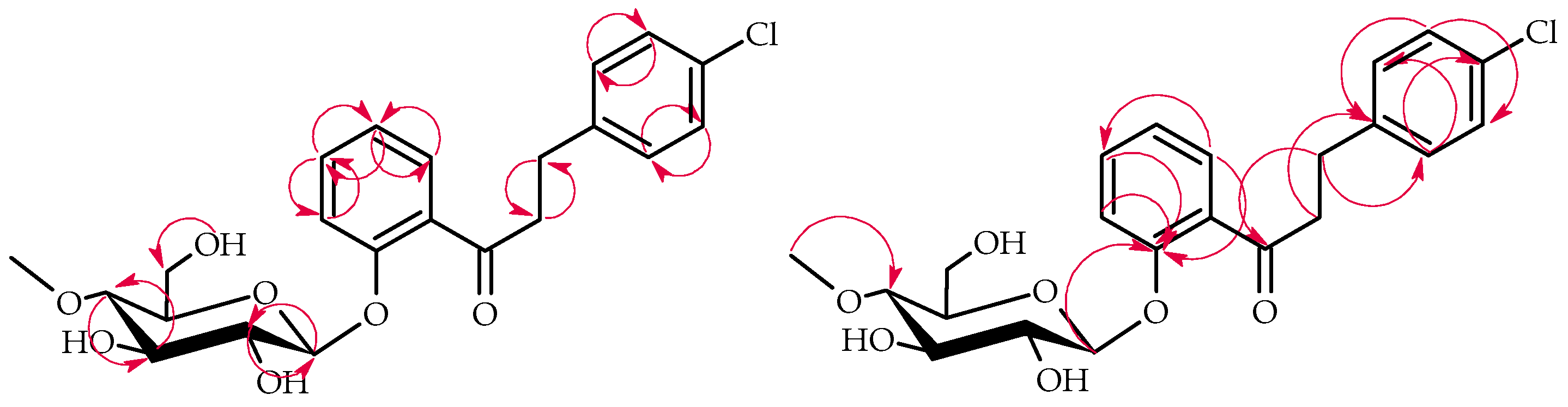
2.2. Biotransformation of 4-Chloro-2′-Hydroxychalcone (3) in Culture of I. Fumosorosea KCH J2

The 4-Chloro-2′-hydroxychalcone (3) compund was biotransformed by enzymatic systems of I. fumosorosea KCH J2 into 4-chlorodihydrochalcone 2′-O-β-D-(4″-O-methyl)-glucopyranoside (3a), yielding 60.1% (50.7 mg), and 4-chloro-2′-hydroxydihydrochalcone 5′-O-β-D-(4″-O-methyl)-glucopyranoside (3b), yielding 4.9% (4.3 mg) (Figure 4).
The products 3a3b structures were elucidated via NMR spectroscopy (Table 1 and Table 2, Figure 5 and Figure 6 below showing key COSY and HMBC correlations) and confirmed using LC-MS (section Materials and Methods and Supplementary Materials: Figures S31 and S52).
Five distinctive proton signals, with δH values between 3.22 and 3.84 ppm, verified the presence of a glucose moiety in biotransformation product 3a in the 1H-NMR spectrum (Supplementary Materials: Figure S35), as well as five carbon signals in the region from δ = 80.0 ppm to δ = 62.0 ppm in the 13C-NMR spectrum (Supplementary Materials: Figure S38). In the 1H-NMR spectrum, a one-proton doublet from the proton at the anomeric carbon atom was present at δ = 5.08 ppm, with the coupling constant (J = 7.7 Hz) evidencing a β-configuration of the glucose (Supplementary Materials: Figure S35). The glucose molecule was also O-methylated at C-4″ because, in the 1H-NMR spectrum, a three-proton singlet at δ = 3.56 ppm, with the corresponding signal at δ = 60.6 ppm in the 13C-NMR spectra, was observed (Supplementary Materials: Figures S35 and S38). The correlation in the HMBC experiment between the three-proton singlet and the signal of C-4″ (δ = 80.0 ppm) proved the substitution site with the -O-CH3 group in the attached glucose group (Supplementary Materials: Figures S49). Furthermore, in product 3a, a double bond between C-α and C-β was reduced, which shifted the protons at C-α from δ = 8.09 ppm (in 3) to δ = 3.45 ppm (in 3a) and at C-β from δ = 7.92 ppm (in 3) to δ = 2.97 ppm (in 3a) (Supplementary Materials: Figures S4 and S35). Moreover, these protons were correlated with the carbonyl group and signal of C-1 in the HMBC experiment (Supplementary Materials: Figures S47 and S50). The 1H-NMR spectrum of the compound 3 at δ = 12.83 ppm shows the signal from the hydroxyl group at C-2′, which was absent in the 1H-NMR spectrum of its microbial transformation product 3a (Supplementary Materials: Figures S3 and S33), which indicates substitution with the 4″-O-methylglucopyranose. In the HMBC experiment, the shifted signal from C-2′ δ = 157.2 ppm correlated with the signal from H-1′’of the glucose moiety at δ = 5.08 ppm (Supplementary Materials: Figure S47) and also with H-6′ (δ = 7.58 ppm), H-4′ (δ = 7.48 ppm), and H-3′ (δ = 7.30 ppm (Supplementary Materials: Figure S46).
Product 3b featured a 4″-O-methylglucose moiety in a β-configuration attached to the chalcone aglycone, alongside a reduction in the C-α and C-β double bond (Supplementary Materials: Figures S56 and S59). A shift in the 1H-NMR signal for the C-2′ hydroxyl group from δ = 12.83 ppm (substrate 3) to δ = 11.89 ppm (product 3b) indicated a 4″-O-methylglucosyl substitution on ring A of the chalcone (Supplementary Materials: Figures S3 and S54). The 1H-NMR spectrum of 3b showed shifts in ring A proton signals compared to substrate 3: H-6′ (δ 8.28 → 7.67 ppm), H-4′ (δ 7.58 → 7.29 ppm), and H-3′ (δ 7.00 → 6.87 ppm) (Supplementary Materials: Figures S4 and S55). The 1H-NMR spectrum showed the disappearance of the H-5′ signal, indicating C-5′ glucosyl substitution (Supplementary Materials: Figures S4 and S55). Furthermore, HMBC experiments revealed a correlation between H-1″ (δ 4.84 ppm) and a shifted signal at δ 150.8 ppm, which was assigned as C-5′. This C-5′ signal also correlated with H-6′ (δ 7.67 ppm), H-4′ (δ 7.29 ppm), and H-3′ (δ 6.87 ppm), confirming its assignment (Supplementary Materials: Figures S70 and S68).

2.3. Biotransformation of 4-Chloro-2′-Hydroxychalcone (3) in the Culture of B. Bassiana KCH J1.5

B. bassiana KCH J1.5 biotransformed 4-chloro-2′-hydroxychalcone (3) less efficiently than I. fumosorosea KCH J2. The primary product, 4-chloro-2′-hydroxydihydrochalcone 3-O-β-D-(4″-O-methyl)-glucopyranoside (3c), was isolated, yielding 9.7% (8.5 mg) (Figure 7).
NMR spectroscopy elucidated the structure of product 3c (Table 1 and Table 2, key COSY and HMBC correlations in Figure 8). LC-MS confirmed its molecular mass (Materials and Methods, Supplementary Materials: Figure S73).
Product 3c was identified as a dihydrochalcone due to a C-α and C-β double-bond reduction, which was evidenced by 1H-NMR signal shifts: H-α (δ 8.09 → 3.48 ppm) and H-β (δ 7.92 → 3.02 ppm) (Supplementary Materials: Figures S4 and S92). A 4″-O-methylglucopyranose substitution was confirmed by characteristic 1H-NMR and 13C-NMR signals, which were similar to products 3a and 3b (Supplementary Materials: Figures S77 and S80). The attachment at C-3 was evidenced by B ring proton shifts: H-2 (δ = 7.93 → 7.26 ppm), H-6 (δ = 7.93 → 6.96 ppm), H-5 (δ = 7.52 → 7.30 ppm), and H-3 signal absence (Supplementary Materials: Figures S4 and S76). HMBC correlations between H-5 (δ = 7.30 ppm), H-2 (δ = 7.26 ppm), and shifted C-3 (δ = 153.8 ppm)—plus glucose H-1″ (δ = 5.05 ppm) to C-3 correlation—further confirmed this attachment (Supplementary Materials: Figures S89 and S92).

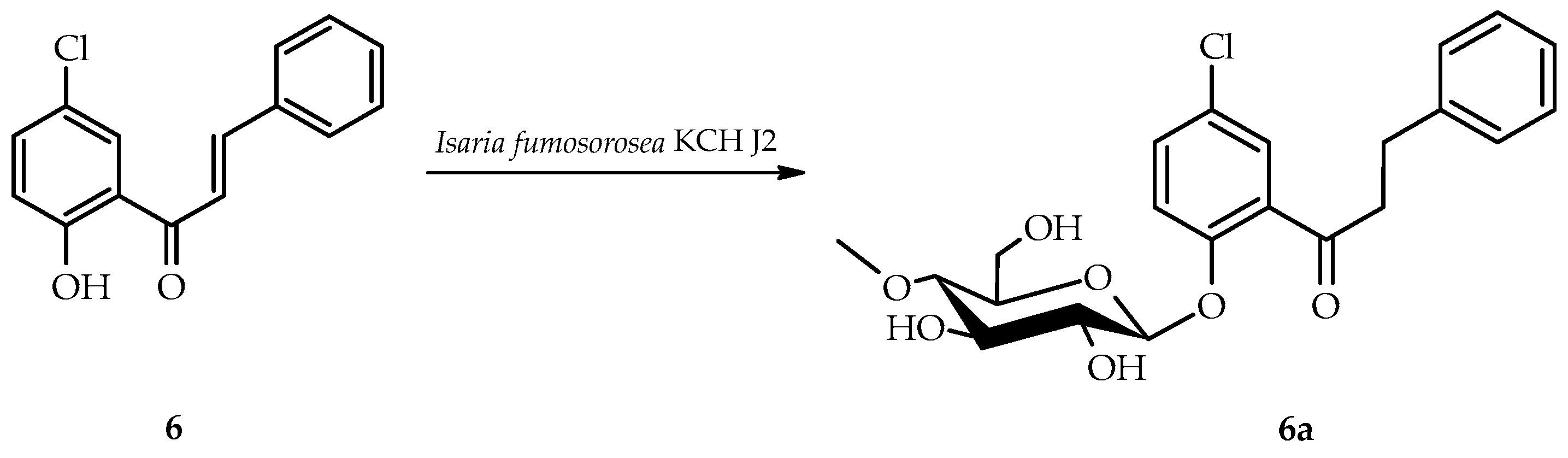
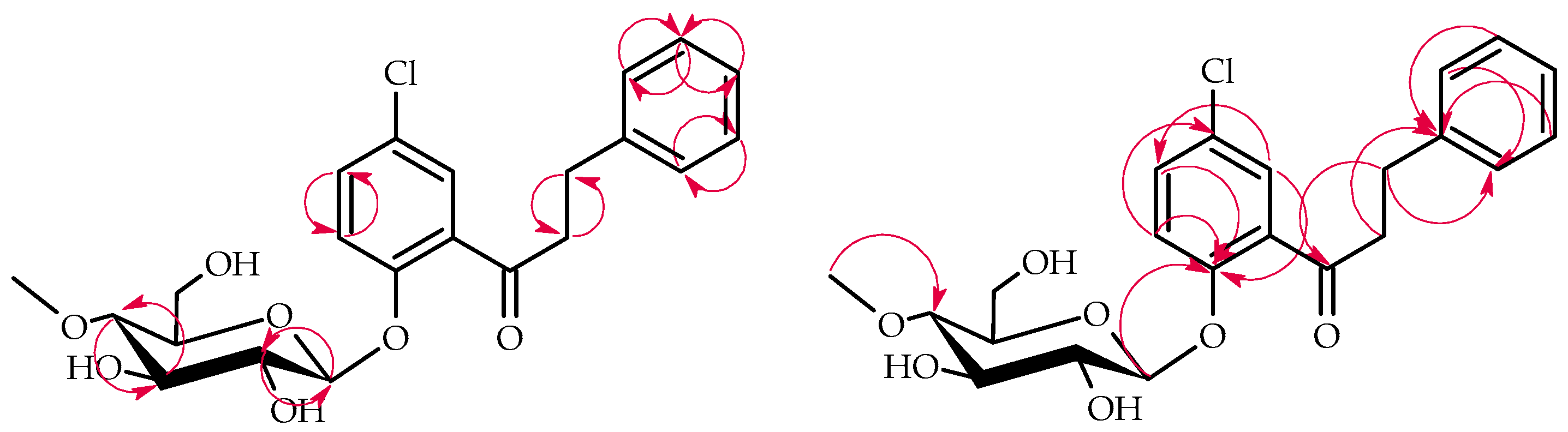
2.4. Biotransformation of 5′-Chloro-2′-Hydroxychalcone (6) in the Culture of I. Fumosorosea KCH J2

I. fumosorosea KCH J2 biotransformed 5′-chloro-2′-hydroxychalcone (6) into 5′-chlorodihydrochalcone 2′-O-β-D-(4″-O-methyl)-glucopyranoside (6a), yielding 76.1% (64.2 mg) (Figure 9).
The structure of product 6a was elucidated via NMR spectroscopy (Table 3 and Table 4 in section, key COSY and HMBC correlations in Figure 10) and confirmed using LC-MS (section Materials and Methods and Supplementary Materials: Figure S94).
Product 6a, analogous to 3a but with a different chlorine positioning, featured 4″-O-methylglucopyranose in a β-configuration at C-2′. This was evidenced by the disappearance of the 2′-OH signal and the presence of characteristic glucose signals in 1H-NMR (Supplementary Materials: Figures S96, S98 and S101). Additionally, a C-α and C-β double-bond reduction occurred, similarly to 3a, which was indicated by characteristic shifts in the 1H-NMR and 13C-NMR spectra (Supplementary Materials: Figures S19, S21, S22, S98 and S101).

2.5. Biotransformation of 5′-Chloro-2′-Hydroxychalcone (6) in the Culture of B. Bassiana KCH J1.5

B. bassiana KCH J1.5 biotransformed 5′-chloro-2′-hydroxychalcone (6) into a single product known as 5′-chloro-2′-hydroxychlorochalcone 3-O-β-D-(4″-O-methyl)-glucopyranoside (6b), yielding 40.5% (35.3 mg) (Figure 11).
NMR spectroscopy elucidated the structure of product 6b (Table 3 and Table 4, key COSY and HMBC correlations in Figure 12). LC-MS confirmed its molecular mass (Materials and Methods, Supplementary Materials: Figure S115).
The glycosylation of product 6b was confirmed by characteristic 4″-O-methylglucosyl signals in the 1H-NMR and 13C-NMR spectra (Supplementary Materials: Figures S119 and S122). In contrast to other biotransformation products, 6b retained its α,β-unsaturated double bond. C-3 glucosyl substitution in ring B was evidenced by shifts in proton signals: H-2 (δ 7.93 → 7.63 ppm), H-4 (δ 7.49 → 7.18 ppm), H-5 (δ 7.49 → 7.40 ppm), H-6 (δ 7.93 → 7.55 ppm), and the absence of H-3 signal in the 1H NMR spectrum (Supplementary Materials: Figures S19 and S76). In addition, the HMBC experiment indicated that shifted signal from C-3 (δ = 159.2 ppm) was correlated only with the H-1′’signal of the attached glucose moiety (δ = 5.04 ppm) and H-5 (δ = 7.40 ppm) (Supplementary Materials: Figure S132).
To summarize, compounds 4-chloro-2′-hydroxychalcone (3) and 5′-chloro-2′-hydroxychalcone (6) were glycosylated in cultures of both entomopathogenic fungi strains. The resulting compounds revealed different regioselectivity of the glycosyltransferase-methyltransferase functional modules of the two strains. In I. fumosorosea KCH J2, the primary products (3a and 6a) resulted from attaching a 4″-O-methylglucosyl group to the C-2′ hydroxyl moiety in the A ring and reducing the C-α and C-β double bond. For 4-chloro-2′-hydroxychalcone (3), glycosylation also occurred at C-5′, yielding 3b. Compound 3b is analogous to the products obtained earlier in the cultures of this fungal strain, i.e., 2-chloro-2′-hydroxydihydrochalcone 5′-O-β-d-(4″-O-methyl)-glucopyranoside from 2-chloro-2′-hydroxychalcone and 3-chloro-2′-hydroxydihydrochalcone 5′-O-β-d-(4″-O-methyl)-glucopyranoside from 3-chloro-2′hydroxychalcone [17], and also 2′-hydroxy-4-methyldihydrochalcone 5′-O-β-D-(4″-O-methyl)-glucopyranoside from 2′-hydroxy-4-methylchalcone [18]. These results indicate that oxidation and subsequent glycosylation occurred in ring A when ring B was already substituted with a chlorine atom or a methyl group, and this may be related to some steric hindrance of the enzyme action. Conversely, B. bassiana KCH J1.5 attached the 4″-O-methylglucosyl moiety at C-3 in ring B of both biotransformation substrates 3 and 6 in a very similar reaction. It should also be emphasized that the introduction of the sugar unit was most likely preceded by hydroxylation of the C-3 carbon. However, substrate 3 was also hydrogenated to form 3c, while an α,β-unsaturated double bond in 6b remained intact. These results show that the enzyme systems of B. bassiana KCH J1.5 and I. fumosrosoea KCH J2 differ, with the former being able to introduce a 4″-O-methylglucopyranose unit, despite the presence of a substituent in the B ring. Previous studies on methylchalcones [18,19] showed less efficient glycosylation of the 2′-hydroxyl group by I. fumosorosea KCH J2, suggesting a positive influence of the chlorine substituent on the glycosylation site. Earlier work on chalcone glycosylation with a C-5′ methyl group in both B. bassiana KCH J1.5 and I. fumosorosea KCH J2 cultures yielded a product similar to 6b but with a methyl group instead of chlorine [20].
Previous research on chalcone glycosylation by filamentous fungi primarily yielded 4′-O-β-D-glucopyranoside derivatives of xanthohumol. These were produced using Penicillium chrysogenum 6933 [21], Absidia coerulea AM93, and Rhizopus nigricans UPF701 [22]. However, certain B. bassiana strains (AM278 [23] and AM446 [22]) formed 4′-O-β-D-(4″-O-methyl)-glucopyranoside derivatives instead. The literature on chlorochalcone glycosylation is scarce. Our prior work demonstrated that B. bassiana KCH J1.5 could biotransform 3′-bromo-5′-chloro-2′-hydroxychalcone into 8-bromo-6-chloroflavanone 3′-O-β-D-(4″-O-methyl)-glucopyranoside [12]. In contrast, I. fumosorosea biotransformed 2′-hydroxychalcones with chlorine at C-2 or C-3, producing C-2′ glycosylated derivatives [17].

2.6. SwissADME Analysis: Pharmacokinetics and Drug-Likeness Prediction of 4-Chloro-2′-Hydroxychalcone (3), 5′-Chloro-2′-Hydroxychalcone (6) and Their Derivatives (3a3c, 6a6b)

The SwissADME online tool (http://www.swissadme.ch/), developed by the Swiss Institute of Bioinformatics’ Molecular Modeling Group (SIB) [24], was used to evaluate the pharmacokinetics, water solubility, and drug likeness of compounds 3, 6, their biotransformation products (3a3c, 6a6b), and 2′-hydroxychalcone (7). The BOILED-Egg predictive model [25] predicted high gastrointestinal absorption for all tested molecules. Glycosylated derivatives (3a3c, 6a6b) showed 8–18 times higher estimated aqueous solubility (ESOL method) than their aglycones (3 and 6). However, their estimated lipophilicity (consensus Log Po/w) decreased, potentially reducing their affinity for biological membranes and passive permeation in the bloodstream due to increased hydrophilicity [25]. Despite this, our previous studies on methylflavanone 4″-O-methylglucosides demonstrated their ability to bind in the hydrophilic region of phosphatidylcholine and erythrocyte membranes without disrupting their structure [26]. Simulations revealed that the glycosylated chalcones (3a3c, 6a6b) lost their ability to passively permeate the blood–brain barrier and may instead have been actively transported by P-glycoprotein, unlike their aglycones (3 and 6). Compounds 3 and 6 may inhibit certain cytochrome P450 enzymes (CYP1A2, CYP2C9, CYP2C19) but not others (CYP2D6, CYP3A4). Conversely, their glycosylated derivatives likely do not inhibit CYP1A2, CYP2C9, and CYP2C19, but they may inhibit CYP2D6 (3a and 6a) and CYP3A4 (3a, 3b, 3c, 6a, and 6b). All compounds passed the drug likeness (Lipinski, Ghose, Veber, Egan, and Muegge) estimators with zero violations. The Abbott bioavailability score (ABS) was 0.55 for all compounds, indicating a 55% probability of >10% bioavailability in rats or measurable Caco-2 permeability. In medicinal chemistry simulations, all compounds showed zero PAINS alerts. Detailed results are presented in Table 5.

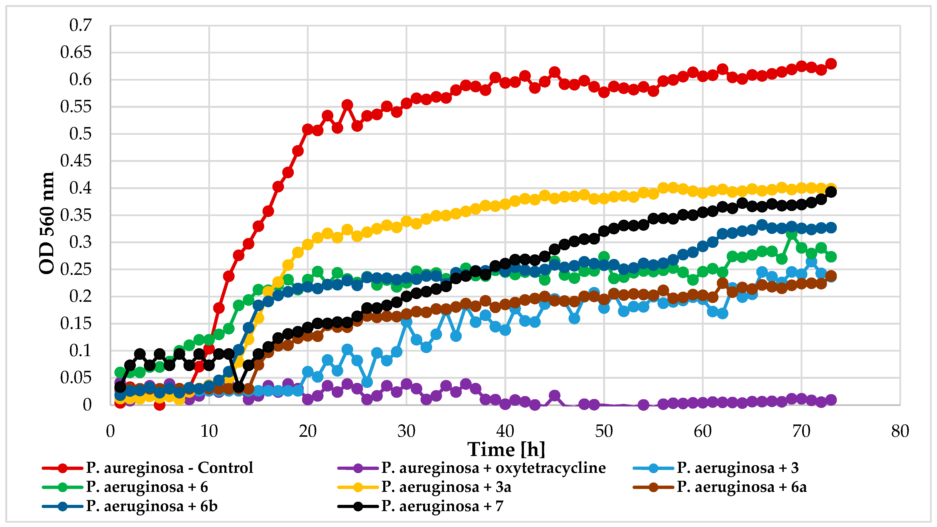
2.7. Antimicrobial Effects of 2′-Hydroxychalcone and Its Derivatives (3, 3a3c, 6, 6a6b, and 7)

Antimicrobial activity tests were conducted using a Bioscreen C device (Growth Curves USA, Piscataway, NJ, USA) to evaluate the impact of chlorine atom introduction, its position, and 4′-O-methylglucopyranose attachment on 2′-hydroxychalcone activity. The following compounds were tested: 4-chloro-2′-hydroxychalcone (3), 5′-chloro-2′-hydroxychalcone (6), 4-chlorodihydrochalcone 2′-O-β-D-(4″-O-methyl)-glucopyranoside (3a), 5′-chlorodihydrochalcone 2′-O-β-D-(4″-O-methyl)-glucopyranoside (6a), 5′-chloro-2′-hydroxy-chlorochalcone 3-O-β-D-(4″-O-methyl)-glucopyranoside (6b), and 2′-hydroxychalcone (7) for comparison. The compounds were tested against bacteria Escherichia coli 10536 (Gram-), Pseudomonas aeruginosa DSM 939(Gram-), Staphylococcus aureus DSM 799 (Gram+), Lactococcus acidophilus KBiMZ 01 (Gram+), Lactococcus rhamnosus GG (Gram+), Streptococcus thermophilus KBM-1 (Gram+), and one strain of yeast Candida albicans DSM 1386. Table 6 and Table 7 present data on lag phase duration and biomass increase (ΔOD) for control, and compound-treated cultures were collected in Table 6 and Table 7.
Flavonoid aglycone 3 and glycoside 6a exhibited the strongest inhibition against bacteria E. coli 10536 (ΔOD = 0). Compounds 3a and 6b (ΔOD = 0.15), as well as 6 (ΔOD = 0.12), also extended the microbial lag phase and significantly inhibited growth. Unsubstituted 2′-hydroxychalcone (7) showed slightly less effectiveness (ΔOD = 0.25). Figure 13 illustrates the E. coli growth in response to these compounds.
All tested compounds exhibited some inhibitory effect on the relatively resistant bacteria P. aeruginosa DSM 939. Compounds 3, 6, and 6a were most effective (ΔOD = 0.21), followed by 6b (ΔOD = 0.31), 7 (ΔOD = 0.36), and 3a (ΔOD = 0.39), compared to the control (ΔOD = 0.63). Figure 14 depicts P. aeruginosa growth in response to these compounds.
On the other hand, the bacteria growth of S. aureus DSM 799 was completely inhibited by compound 6a. Other flavonoids with a chlorine atom also prolonged the microbial lag phase and significantly inhibited their growth. The least effective was the unsubstituted chlorine 2′-hydroxychalcone (ΔOD = 0.32). Figure 15 depicts the S. aureus growth in response to compounds 3, 6, 3a, 6a, 6b, and 7.
C. albicans DSM 1386 yeast strain was highly sensitive to the tested compounds. Except for compounds 6a and 7, which allowed slight growth (ΔOD = 0.35 and ΔOD = 0.11, respectively), all others completely inhibited yeast growth. The C. albicans growth patterns under exposure to the tested compounds are presented in Figure 16.
The effect of the tested compounds on the lactic acid bacteria varied by species. L. rhamnosus GG and S. thermophilus KBM-1 growth were totally inhibited by all compounds. However, complete growth inhibition of the L. acidophilus KBiMZ 01 bacteria occurred because of the action of compounds 3 and 7, and significant inhibition occurred when molecule 6 was used (ΔOD = 0.12). The glycosylated flavonoids with a chlorine atom (3a, 6a, and 6b) prolonged the microbial lag phase and limited this lactic bacteria growth. The lowest level of inhibition was observed in the case of compound 6b. Figure 17, Figure 18 and Figure 19 detail the growth of these bacteria in response to the compounds used.
Antimicrobial activity tests of the obtained compounds 3, 6, 3a, 6a, 6b, and 7 showed that the substitution of a chlorine atom in the chalcone structure had a positive effect on their activity against the tested microorganisms: E. coli 10536, S. aureus DSM 799, P. aeruginosa DSM 939, and C. albicans DSM 1386. Prasad and coworkers also showed a positive effect of pharmacophores like chloro-, dichloro-, and fluoro-moiety on antibacterial activity against the used strain of E. coli bacteria [8]. On the other hand, antimicrobial activity tests against the S. aureus AM-176 strain performed by Alcaraz and coworkers showed that 4-chlorochalcone inhibited its growth less effectively than 2′-hydroxychalcone and chalcone (PID (Percent Inhibition Degree) = 34.7, 98.3, and 38.3 respectively) [27]. These results indicate that the hydroxyl group within the chalcone structure strongly influences its antimicrobial activity. However, researchers have not investigated how the combined effects of a 2′-hydroxyl moiety and a chlorine atom impact the antimicrobial properties of the resulting compound. In the presented work, chlorinated 2′-hydroxychalcones showed significantly stronger antibacterial activity against S. aureus DSM 799. Furthermore, their derivatives with the blocked 2′-hydroxyl group by the attached glucosyl moiety were not less effective. Konečná and coworkers also proved that introducing a bromine or chlorine atom into pyrazine-based chalcones yielded receiving compounds with strong antistaphylococcal and antienterococcal activity [10]. The impact of glucosyl groups on chalcones’ antimicrobial properties remains poorly understood. However, in the antimicrobial activity tests of another group of flavonoids with this moiety, i.e., flavonol 3-O-glycosides, researchers observed a potent suppression of Gram-positive bacteria and a weaker inhibition of Gram-negative bacteria [16]. In our studies, E. coli 10536 were exceptionally susceptible to the actions of chlorinated aglycones and the glycosides of 2′-hydroxychalcone. However, P. aeruginosa DSM 939 was quite opposing. In our previous studies with 2-chloro-2′-hydroxychalcone, 3-chloro-2′hydroxychalcones, and their glycosides, we also observed that chlorinated chalcones were more active as inhibitors of the tested microbial strains’ growth compared to their unchlorinated counterparts. However, aglycones showed slightly greater efficacy than their glycoside forms [17]. By comparing all the obtained results, one can observe differences in the inhibition of microbial growth depending on the chlorine atom substitution position in the tested chalcones. Comparing all of the results obtained, differences in microbial growth inhibition can be observed depending on a chlorine atom substitution position in chalcones tested. Interestingly, 5′-chlorodihydrochalcone 2′-O-β-D-(4‴-O-methyl)-glucopyranoside (6a) showed significant activity against all tested microbial strains but was the least effective against the C. albicans DSM 1386 strain.

3. Materials and Methods

3.1. General Procedure for the Synthesis of Biotransformation Substrates 3 and 6

An amount of 2′-Hydroxychalcones with a chlorine atom, i.e., 4-chloro-2′-hydroxychalcone (3) and 5′-chloro-2′-hydroxychalcone (6), were received in the Claisen–Schmidt condensation reaction in alkaline conditions, as previously described [17,28,29,30,31,32]. The Results and Discussion section above presents the chemical reaction schemes in Figures S1 and S2.
The physical data of compounds 3 and 6 (color, form, molecular ion mass, molecular formula, melting point (°C), retention time tR (min), retardation factor Rf, and NMR spectral data) are presented below, in Table 1, Table 2, Table 3 and Table 4 in the Results and Discussion section, and in the Supplementary Materials.
The 4-Chloro-2′-hydroxychalcone (3): Yellow crystals (73.6%, 7.6 g); ESI/MS m/z 259.0 ([M + H]+, C15H11ClO2); mp = 147–149 °C; tR = 18.58; Rf = 0.93; 1H-NMR, see Table 1, 13C-NMR, see Table 2, Supplementary Materials: Figures S3–S14.
The 5′-Chloro-2′-hydroxychalcone (6): Yellow crystals (90.5%, 9.4 g), ESI/MS m/z 259.0 ([M + H]+, C15H11ClO2), mp = 106–108 °C, tR = 18.71, Rf = 0.93, 1H-NMR, see Table 3, 13C-NMR, see Table 4, Supplementary Materials: Figures S18–S29.

3.2. Microorganisms

Microbial glycosylation of chlorochalcones 3 and 6, obtained by chemical synthesis, was achieved in cultures of I. fumosorosea KCH J2 and B. bassiana KCH J1.5 filamentous fungi belonging to the collection of the Faculty of Biotechnology and Food Microbiology of the Wrocław University of Environmental and Life Sciences in Poland. Our previous studies detailed the genetic identification, collection methods, and reproduction of these fungi [13,33].

3.3. Analysis

Thin-layer chromatography (TLC) and high-performance liquid chromatography (HPLC) were used to monitor biotransformation progress, specifically substrate transformation [18]. All compounds were 95%-98% pure according to HPLC analysis. The separation of biotransformation products on a semi-preparative scale was achieved using preparative silica gel TLC plates with thicknesses of 500 µm and 1000 µm (Supelco, Darmstadt, Germany) and mixture of chloroform and methanol (9:1 volume ratio) [17,19].
NMR analyses (1H-NMR, 13C-NMR, COSY, HMQC, and HMBC) were carried out using a DRX AvanceTM 600 MHz NMR spectrometer (Bruker, Billerica, MA, USA). All samples were dissolved in deuterated acetone for analysis.
The molecular formulas of all products (3, 3a, 3b, 3c, 6, 6a, and 6b) were confirmed through mass spectrometry analyses using a LC-MS 8045 SHIMADZU Triple Quadrupole Liquid Chromatograph Mass Spectrometer with electrospray ionization (ESI) source (Shimadzu, Kyoto, Japan), as described in our previous works [17,19].

3.4. Screening Procedure

A biotransformation screening procedure was conducted to determine the time required for the complete conversion of substrates 3 and 6 in preparation for subsequent experiments at a semi-preparative scale. Entomopathogenic fungi were cultivated using a modified Sabouraud medium. Fungal strains were initially grown for 72 h and then transferred to fresh medium. Substrates 3 or 6 (10 mg) were added to flasks containing either Isaria fumosorosea KCH J2 or Beauveria bassiana KCH J1.5, with a final concentration of 0.39 mM. Samples were collected after 3, 6, and 8 days. Biotransformation products were extracted with ethyl acetate, which were then dried and concentrated. The experiments were concluded after 8 days or when complete substrate conversion was confirmed. Controls included substrate stability and cultivation without substrates [17,19].

3.5. The Semi-Preparative Biotransformation

Semi-preparative biotransformation was conducted in 2 L flasks with 500 mL of modified Sabouraud medium to produce sufficient product for NMR analyses, structural determination, and antimicrobial testing. The process began with transferring 1 mL of a preincubated culture of I. fumosorosea KCH J2 or B. bassiana KCH J1.5 to the flasks, followed by a 72-h incubation. Then, 50 mg of substrate 3 or 6 (dissolved in 2.0 mL of dimethyl sulfoxide) was added, maintaining a final concentration of 0.39 mM. The flasks were incubated on a rotary shaker for 8 days. Post-reaction mixtures were extracted with ethyl acetate, which were then dried, filtered, and evaporated. The biotransformation products were separated and purified using preparative TLC plates, which were then visualized under UV light and extracted with ethyl acetate. The chemical structures were analyzed via spectroscopic methods, and yields were determined based on the mass of the isolated products [17,19].

3.6. Fungal Biotransformation Products

The physical data of compounds 3a3c and 6a6b (color, form, molecular ion mass, molecular formula, melting point (°C), retention time tR (min), retardation factor Rf, and NMR spectral data) are presented below, in Table 1, Table 2, Table 3 and Table 4 in the Results and Discussion section, and in the Supplementary Materials.
The 4-Chlorodihydrochalcone 2′-O-β-D-(4‴-O-methyl)-glucopyranoside (3a): white crystals; ESI/MS m/z 435.1 ([M − H], C22H25ClO7); mp = 132–134 °C; tR = 11.94, Rf = 0.39; 1H-NMR, see Table 1, 13C-NMR, see Table 2, Supplementary Information: Figures S31–S50.
The 4-Chloro-2′-hydroxydihydrochalcone 5′-O-β-D-(4‴-O-methyl)-glucopyranoside (3b): white crystals; ESI/MS m/z 451.1 ([M − H], C22H25ClO8); mp = 133–135 °C; tR = 13.31, Rf = 0.33; 1H-NMR, see Table 1, 13C-NMR, see Table 2, Supplementary Information: Figures S52–S71.
The 4-Chloro-2′-hydroxydihydrochalcone 3-O-β-D-(4‴-O-methyl)-glucopyranoside (3c): light-yellow crystals; ESI/MS m/z 451.1 ([M − H], C22H25ClO8); mp = 89–91 °C; tR = 11.24, Rf = 0.42; 1H-NMR, see Table 1, 13C-NMR, see Table 2, Supplementary Information: Figures S73–S92.
The 5′-Chlorodihydrochalcone 2′-O-β-D-(4‴-O-methyl)-glucopyranoside (6a): white crystals; ESI/MS m/z 435.1 ([M − H], C22H25ClO7); mp = 154–156 °C; tR = 12.79, Rf = 0.26; 1H-NMR, see Table 3, 13C-NMR, see Table 4, Supplementary Information: Figures S94–S113.
The 5′-Chloro-2′-hydroxy-chlorochalcone 3-O-β-D-(4‴-O-methyl)-glucopyranoside (6b): light-yellow crystals; ESI/MS m/z 451.1 ([M + H]+, C22H23ClO8); mp = 207–209 °C; tR = 13.47, Rf = 0.23; 1H-NMR, see Table 3, 13C-NMR, see Table 4, Supplementary Information: Figures S115–S133.

3.7. Pharmacokinetics and Drug Nature Predictions

The pharmacokinetic predictions, physicochemical features, and drug likeness of chalcone derivatives 3, 3a-3c, 6, 6a, 6b, and 7 were computed using SwissADME (available online: http://www.swissadme.ch, accessed on 12 January and 14 March 2024). The molecular structures were constructed using ACD Chemsketch 2021.2.0 and then imported into this SwissADME online tool [18]. The detailed prediction results are presented in the Supplementary Materials: Figures S15 (3), S30 (6), S51 (3a), S72 (3b), S93 (3c), S114 (6a), S134 (6b), S135 (7).

3.8. Antimicrobial Activity Assays

Antimicrobial activity tests of compounds 3, 3a, 6, 6a, 6b, and 7 (Sigma-Aldrich, Sant Louis, MO, USA) were conducted using a Bioscreen C (Automated Microbiology Growth Curve Analysis System, Helsinki, Finland) against the following strains of bacteria: E. coli 10536 (Gram-negative), P. aeruginosa DSM 939 (Gram-negative), S. aureus DSM 799 (Gram-positive), Gram-positive lactic bacteria S. thermophilus KBM-1, L. acidophilus KBiMZ 01, L. rhamnosus GG, and yeast strain C. albicans DSM 1386 belonging to the collection of the Faculty of Biotechnology and Food Microbiology of Wrocław University of Environmental and Life Sciences. The microbiological cultures were prepared 48 h prior to the assays in media purchased from Merck, Darmstadt, Germany. Bacteria E. coli 10536, P. aeruginosa DSM 939, and S. aureus DSM 799 were cultured in LB broth. Yeast C. albicans DSM 1386 was grown in YPG medium. Bacterium S. thermophilus KBM-1 was cultured in M17 medium. Additionally, L. acidophilus KBiMZ 01 and L. rhamnosus GG were grown in MRS medium [17,34,35,36,37,38].
Assays were conducted using 100-well microtiter plates with a working volume of 300 µL per well. Each well contained 280 µL of culture medium, 10 µL of microorganism suspension (final density 1 × 10⁶ cells/mL), and 10 µL of flavonoids dissolved in dimethyl sulfoxide (final flavonoid concentration 0.1% (m/v)). The dimethyl sulfoxide final concentration in each well was 3.3% (v/v). The plates were incubated at 30 °C with optical density measurements at 560 nm taken every 60 min for 72 h for all microorganisms except lactic acid bacteria, for which the temperature was 37 °C, and measurements were taken every 30 min for 70 h. Each test was performed in triplicate with continuous shaking. Oxytetracycline (10 mg/mL) and cycloheximide (0.1% (m/v) purchased from Sigma-Aldrich (Saint Louis, MO, USA) were used as positive controls. Data were analyzed using Microsoft Excel, and growth curves were created based on the mean absorbance values over time. Antimicrobial activity was assessed by comparing the increase in optical density (ΔOD) of treated cultures to controls with only dimethyl sulfoxide [17].

4. Conclusions

In this study, we showed the ability of fungi strains I. fumosorosea KCH J2 and B. bassiana KCH J1.5 to yield 4-O-methylglucosyl derivatives of 4-chloro-2′-hydroxychalcone (3) and 5′-chloro-2′-hydroxychalcone (6). The main biotransformation product of 3 in cultures of I. fumosorosea KCH J2-4-chlorodihydrochalcone 2′-O-β-D-(4‴-O-methyl)-glucopyranoside (3a) was obtained with a good isolated yield of 60.1%. Similarly, enzymatic systems of this strain very effectively biotransformed 5′-chloro-2′-hydroxychalcone into only one product-5′-chlorodihydrochalcone 2′-O-β-D-(4‴-O-methyl)-glucopyranoside (6a), yielding 76.1%. This biotransformation substrate was also effectively biotransformed by B. bassiana KCH J1.5 into 5′-chloro-2′-hydroxy-chlorochalcone 3-O-β-D-(4‴-O-methyl)-glucopyranoside (6b), with a yield of 40.5%. All the resulting compounds have not been described in the literature until now. The techniques outlined in this study enable the efficient and cost-effective synthesis of substantial quantities of glycoside derivatives of chalcones containing a chlorine atom. These derivatives can then be investigated further for their biological activity and bioavailability. Simulation results performed in the SwissADME online tool showed changes in the pharmacokinetics and water solubility of glycosylated flavonoids concerning their aglycones. On the other hand, antimicrobial activity tests showed that the introduction of a chlorine atom and glucosyl moiety into the structure of 2′-hydroxychalcone affects its bioactivity. All chlorinated chalcones were more effective in inhibiting the tested microbial strains than 2′-hydroxychalcone. In contrast, chalcone aglycones with a chlorine atom and their glycosides were similarly effective. The highest antibacterial potential against the tested strain was demonstrated by 5′-chlorodihydrochalcone 2′-O-β-D-(4‴-O-methyl)-glucopyranoside. Further experiments are required to elucidate their mechanisms of action.

Supplementary Materials

The following Supplementary Materials can be downloaded at https://www.mdpi.com/article/10.3390/ijms25179718/s1.

Author Contributions

Conceptualization: A.K.-Ł.; Methodology: A.K.-Ł., B.Ż. and E.K.-S.; Validation: A.K.-Ł.; Formal analysis: A.K.-Ł., B.Ż., T.J. and E.K.-S.; Investigation: A.K.-Ł.; Resources: A.K.-Ł.; Data curation: A.K.-Ł.; Writing—Original Draft Preparation: A.K.-Ł.; Writing—Review and Editing: B.Ż., T.J. and E.K.-S.; Visualization: A.K.-Ł.; Supervision: E.K.-S.; Project administration: A.K.-Ł.; Funding acquisition: A.K.-Ł. All authors have read and agreed to the published version of the manuscript.

Funding

This research was funded in whole by the National Science Center, Poland under the project Preludium 20, grant number 2021/41/N/NZ9/01195. For the purpose of Open Access, the author has applied a CC-BY public copyright license to any Author Accepted Manuscript (AAM) version arising from this submission. The APC is co-financed by Wrocław University of Environmental and Life Sciences.

Data Availability Statement

The original data presented in the study are included in the article and Supplementary Materials, and they are also openly available in the Wrocław University of Environmental and Life Sciences Repository at https://doi.org/10.57755/8dms-8k94 (accessed on 2 September 2024).

Conflicts of Interest

The authors declare no conflicts of interest. The funders had no role in the design of the study; in the collection, analyses, or interpretation of data; in the writing of the manuscript; or in the decision to publish the results.

References

  1. Zhuang, C.; Zhang, W.; Sheng, C.; Zhang, W.; Xing, C.; Miao, Z. Chalcone: A Privileged Structure in Medicinal Chemistry. Chem. Rev. 2017, 117, 7762–7810. [Google Scholar] [CrossRef] [PubMed]
  2. Ehrenkranz, J.R.L.; Lewis, N.G.; Kahn, C.R.; Roth, J. Phlorizin: A review. Diabetes/Metabolism Res. Rev. 2004, 21, 31–38. [Google Scholar] [CrossRef]
  3. Gosch, C.; Halbwirth, H.; Stich, K. Phloridzin: Biosynthesis, distribution and physiological relevance in plants. Phytochemistry 2010, 71, 838–843. [Google Scholar] [CrossRef] [PubMed]
  4. Tian, L.; Cao, J.; Zhao, T.; Liu, Y.; Khan, A.; Cheng, G. The Bioavailability, Extraction, Biosynthesis and Distribution of Natural Dihydrochalcone: Phloridzin. Int. J. Mol. Sci. 2021, 22, 962. [Google Scholar] [CrossRef] [PubMed]
  5. Londzin, P.; Siudak, S.; Cegieła, U.; Pytlik, M.; Janas, A.; Waligóra, A.; Folwarczna, J. Phloridzin, an Apple Polyphenol, Exerted Unfavorable Effects on Bone and Muscle in an Experimental Model of Type 2 Diabetes in Rats. Nutrients 2018, 10, 1701. [Google Scholar] [CrossRef]
  6. Guvenalp, Z.; Ozbek, H.; Karadayi, M.; Gulluce, M.; Kuruuzum-Uz, A.; Salih, B.; Demirezer, O. Two antigenotoxic chalcone glycosides from Mentha longifolia subsp. longifolia. Pharm. Biol. 2014, 53, 888–896. [Google Scholar] [CrossRef]
  7. Itoh, T.; Ninomiya, M.; Nozawa, Y.; Koketsu, M. Chalcone glycosides isolated from aerial parts of Brassica rapa L. ‘hidabeni’ suppress antigen-stimulated degranulation in rat basophilic leukemia RBL-2H3 cells. Bioorganic Med. Chem. 2010, 18, 7052–7057. [Google Scholar] [CrossRef] [PubMed]
  8. Prasad, Y.R.; Rao, A.L.; Rambabu, R. Synthesis and Antimicrobial Activity of Some Chalcone Derivatives. J. Chem. 2007, 5, 461–466. [Google Scholar] [CrossRef]
  9. Lin, Y.-M.; Zhou, Y.; Flavin, M.T.; Zhou, L.-M.; Nie, W.; Chen, F.-C. Chalcones and flavonoids as anti-Tuberculosis agents. Bioorganic Med. Chem. 2002, 10, 2795–2802. [Google Scholar] [CrossRef]
  10. Konečná, K.; Diepoltová, A.; Holmanová, P.; Jand’ourek, O.; Vejsová, M.; Voxová, B.; Bárta, P.; Maixnerová, J.; Trejtnar, F.; Kučerová-Chlupáčová, M. Comprehensive insight into anti-staphylococcal and anti-enterococcal action of brominated and chlorinated pyrazine-based chalcones. Front. Microbiol. 2022, 13, 912467. [Google Scholar] [CrossRef]
  11. Rehberg, N.; Akone, H.S.; Ioerger, T.R.; Erlenkamp, G.; Daletos, G.; Gohlke, H.; Proksch, P.; Kalscheuer, R. Chlorflavonin Targets Acetohydroxyacid Synthase Catalytic Subunit IlvB1 for Synergistic Killing of Mycobacterium tuberculosis. ACS Infect. Dis. 2017, 4, 123–134. [Google Scholar] [CrossRef] [PubMed]
  12. Perz, M.; Krawczyk-Łebek, A.; Dymarska, M.; Janeczko, T.; Kostrzewa-Susłow, E. Biotransformation of Flavonoids with -NO2, -CH3 Groups and -Br, -Cl Atoms by Entomopathogenic Filamentous Fungi. Int. J. Mol. Sci. 2023, 24, 9500. [Google Scholar] [CrossRef] [PubMed]
  13. Dymarska, M.; Grzeszczuk, J.; Urbaniak, M.; Janeczko, T.; Pląskowska, E.; Stępień, Ł.; Kostrzewa-Susłow, E. Glycosylation of 6-methylflavone by the strain Isaria fumosorosea KCH J2. PLoS ONE 2017, 12, e0184885. [Google Scholar] [CrossRef]
  14. Dou, F.; Wang, Z.; Li, G.; Dun, B. Microbial Transformation of Flavonoids by Isaria fumosorosea ACCC 37814. Molecules 2019, 24, 1028. [Google Scholar] [CrossRef]
  15. Xie, L.; Zhang, L.; Wang, C.; Wang, X.; Xu, Y.-M.; Yu, H.; Wu, P.; Li, S.; Han, L.; Gunatilaka, A.A.L.; et al. Methylglucosylation of aromatic amino and phenolic moieties of drug-like biosynthons by combinatorial biosynthesis. Proc. Natl. Acad. Sci. USA 2018, 115, E4980–E4989. [Google Scholar] [CrossRef] [PubMed]
  16. Xiao, J. Dietary Flavonoid Aglycones and Their Glycosides: Which Show Better Biological Significance? Crit. Rev. Food Sci. Nutr. 2015, 57, 1874–1905. [Google Scholar] [CrossRef]
  17. Krawczyk-Łebek, A.; Żarowska, B.; Dymarska, M.; Janeczko, T.; Kostrzewa-Susłow, E. Synthesis, fungal biotransformation, and evaluation of the antimicrobial potential of chalcones with a chlorine atom. Sci. Rep. 2024, 14, 1–22. [Google Scholar] [CrossRef]
  18. Krawczyk-Łebek, A.; Dymarska, M.; Janeczko, T.; Kostrzewa-Susłow, E. Glycosylation of Methylflavonoids in the Cultures of Entomopathogenic Filamentous Fungi as a Tool for Obtaining New Biologically Active Compounds. Int. J. Mol. Sci. 2022, 23, 5558. [Google Scholar] [CrossRef]
  19. Krawczyk-Łebek, A.; Dymarska, M.; Janeczko, T.; Kostrzewa-Susłow, E. New Glycosylated Dihydrochalcones Obtained by Biotransformation of 2′-Hydroxy-2-methylchalcone in Cultures of Entomopathogenic Filamentous Fungi. Int. J. Mol. Sci. 2021, 22, 9619. [Google Scholar] [CrossRef]
  20. Krawczyk-Łebek, A.; Dymarska, M.; Janeczko, T.; Kostrzewa-Susłow, E. Entomopathogenic Filamentous Fungi as Biocatalysts in Glycosylation of Methylflavonoids. Catalysts 2020, 10, 1148. [Google Scholar] [CrossRef]
  21. Kim, H.J.; Lee, I.-S. Microbial Metabolism of the Prenylated Chalcone Xanthohumol. J. Nat. Prod. 2006, 69, 1522–1524. [Google Scholar] [CrossRef] [PubMed]
  22. Tronina, T.; Bartmańska, A.; Milczarek, M.; Wietrzyk, J.; Popłoński, J.; Rój, E.; Huszcza, E. Antioxidant and antiproliferative activity of glycosides obtained by biotransformation of xanthohumol. Bioorganic Med. Chem. Lett. 2013, 23, 1957–1960. [Google Scholar] [CrossRef] [PubMed]
  23. Huszcz, E.; Bartmanska, A.; Tronin, T. Glycosylation of Xanthohumol by Fungi. Z. Fur Naturforschung Sect. C-A J. Biosci. 2008, 63, 557–560. [Google Scholar] [CrossRef]
  24. Daina, A.; Michielin, O.; Zoete, V. SwissADME: A free web tool to evaluate pharmacokinetics, drug-likeness and medicinal chemistry friendliness of small molecules. Sci. Rep. 2017, 7, 42717. [Google Scholar] [CrossRef]
  25. Daina, A.; Zoete, V. A BOILED-Egg to Predict Gastrointestinal Absorption and Brain Penetration of Small Molecules. ChemMedChem 2016, 11, 1117–1121. [Google Scholar] [CrossRef] [PubMed]
  26. Cyboran-Mikołajczyk, S.; Bonarska-Kujawa, D.; Męczarska, K.; Krawczyk-Łebek, A.; Kostrzewa-Susłow, E. Novel O-Methylglucoside Derivatives of Flavanone in Interaction with Model Membrane and Transferrin. Membranes 2022, 12, 978. [Google Scholar] [CrossRef]
  27. Alcaráz, L.; Blanco, S.; Puig, O.; Tomás, F.; Ferretti, F. Antibacterial Activity of Flavonoids Against Methicillin-resistant Staphylococcus aureus strains. J. Theor. Biol. 2000, 205, 231–240. [Google Scholar] [CrossRef]
  28. Silva, A.M.S.; Tavares, H.R.; Barros, A.I.N.R.A.; Cavaleiro, J.A.S. NMR and Structural and Conformational Features of 2′-Hydroxychalcones and Flavones. Spectrosc. Lett. 1997, 30, 1655–1667. [Google Scholar] [CrossRef]
  29. Rapposelli, S.; Da Settimo, F.; Digiacomo, M.; La Motta, C.; Lapucci, A.; Sartini, S.; Vanni, M. Synthesis and Biological Evaluation of 2′-Oxo-2,3-dihydro-3′H-spiro[chromene-4,5′-[1,3]oxazolidin]-3′yl]acetic Acid Derivatives as Aldose Reductase Inhibitors. Arch. der Pharm. 2011, 344, 372–385. [Google Scholar] [CrossRef]
  30. Yelve, N.P.; Mitra, M.; Mujumdar, P.M. Detection of delamination in composite laminates using Lamb wave based nonlinear method. Compos. Struct. 2017, 159, 126. [Google Scholar] [CrossRef]
  31. Ibrahim, N.S.; Ahmed, F. Antimicrobial Activities of Some Synthetic Flavonoids. IOSR J. Appl. Chem. 2014, 7, 01–06. [Google Scholar] [CrossRef]
  32. Gutam, M.; Mokenapelli, S.; Yerrabelli, J.R.; Banerjee, S.; Roy, P.; Chitneni, P.R. Synthesis and cytotoxicity of novel (E)-2-phenylchroman-4-one-O-((1-substituted-1H-1,2,3-triazol-4-yl)methyl) oxime derivatives. Synth. Commun. 2020, 50, 1883–1891. [Google Scholar] [CrossRef]
  33. Kozłowska, E.; Urbaniak, M.; Hoc, N.; Grzeszczuk, J.; Dymarska, M.; Stępień, Ł.; Pląskowska, E.; Kostrzewa-Susłow, E.; Janeczko, T. Cascade biotransformation of dehydroepiandrosterone (DHEA) by Beauveria species. Sci. Rep. 2018, 8, 13449. [Google Scholar] [CrossRef]
  34. Wu, Y.; Griffiths, M.; McKellar, R. A comparison of the Bioscreen method and microscopy for the determination of lag times of individual cells of Listeria monocytogenes. Lett. Appl. Microbiol. 2000, 30, 468–472. [Google Scholar] [CrossRef]
  35. Stephens, P.; Joynson, J.; Davies, K.; Holbrook, R.; Lappin-Scott, H.; Humphrey, T. The use of an automated growth analyser to measure recovery times of single heat-injured Salmonella cells. J. Appl. Microbiol. 1997, 83, 445–455. [Google Scholar] [CrossRef] [PubMed]
  36. Kozłowska, J.; Potaniec, B.; Żarowska, B.; Anioł, M. Synthesis and Biological Activity of Novel O-Alkyl Derivatives of Naringenin and Their Oximes. Molecules 2017, 22, 1485. [Google Scholar] [CrossRef] [PubMed]
  37. Grabarczyk, M.; Wińska, K.; Mączka, W.; Żarowska, B.; Maciejewska, G.; Dancewicz, K.; Gabryś, B.; Anioł, M. Synthesis, biotransformation and biological activity of halolactones obtained from β-ionone. Tetrahedron 2016, 72, 637–644. [Google Scholar] [CrossRef]
  38. Antonić, B.; Dordević, D.; Jančíková, S.; Kushkevych, I. Antimicrobial activity of natural soaps tested by Bioscreen methodology. Stud. Biol. 2020, 14, 23–32. [Google Scholar] [CrossRef]
Figure 1. Functional modules of glycosyltransferase-methyltransferase (GT-MT) in fungi such as B. bassiana (developed based on Xie and coworkers [15]).
Ijms 25 09718 g001
Figure 2. Synthesis of biotransformation substrate 4-chloro-2′-hydroxychalcone (3).
Ijms 25 09718 g002
Figure 3. Synthesis of biotransformation substrate 5′-chloro-2′-hydroxychalcone (6).
Ijms 25 09718 g003
Figure 4. Biotransformation of 4-chloro-2′-hydroxychalcone (3) in I. fumosorosea KCH J2 culture.
Ijms 25 09718 g004
Figure 5. Key COSY (on the left) and HMBC (on the right) correlations of product 3a.
Ijms 25 09718 g005
Figure 6. Key COSY (on the left) and HMBC (on the right) correlations of product 3b.
Ijms 25 09718 g006
Figure 7. Biotransformation of 4-chloro-2′-hydroxychalcone (3) in B. bassiana KCH J1.5 culture.
Ijms 25 09718 g007
Figure 8. Key COSY (on the left) and HMBC (on the right) correlations of product 3c.
Ijms 25 09718 g008
Figure 9. Biotransformation of 5′-chloro-2′-hydroxychalcone (6) in I. fumosorosea KCH J2 culture.
Ijms 25 09718 g009
Figure 10. Key COSY (on the left) and HMBC (on the right) correlations of product 6a.
Ijms 25 09718 g010
Figure 11. Biotransformation of 5′-chloro-2′-hydroxychalcone (6) in B. bassiana KCH J1.5 culture.
Ijms 25 09718 g011
Figure 12. Key COSY (on the left) and HMBC (on the right) of product 6b.
Ijms 25 09718 g012
Figure 13. The impact of compounds 3, 6, 3a, 6a, 6b, 7 on the growth of E. coli 10536.
Ijms 25 09718 g013
Figure 14. The impact of compounds 3, 6, 3a, 6a, 6b, 7 on the growth of P. aeruginosa DSM 939.
Ijms 25 09718 g014
Figure 15. The impact of compounds 3, 6, 3a, 6a, 6b, 7 on the growth of S. aureus DSM 799.
Ijms 25 09718 g015
Figure 16. The impact of compounds 3, 6, 3a, 6a, 6b, 7 on the growth of C. albicans DSM 1386.
Ijms 25 09718 g016
Figure 17. The impact of compounds 3, 6, 3a, 6a, 6b, 7 on the growth of L. acidophilus KBiMZ 01.
Ijms 25 09718 g017
Figure 18. The impact of compounds 3, 6, 3a, 6a, 6b, 7 on the growth of L. rhamnosus GG.
Ijms 25 09718 g018
Figure 19. The impact of compounds 3, 6, 3a, 6a, 6b, 7 on the growth of S. thermophilus KBM-1.
Ijms 25 09718 g019
Table 1. The 1H-NMR chemical shifts δ (ppm) and coupling constants J (Hz) of 4-chloro-2′-hydroxychalcone (3) and their biotransformation products 3a–3c in Acetone-d6, 600 MHz (Supplementary Materials: Figures S3, S4, S33–S35, S54–S56 and S75–S77).
ProtonCompound
33a3b3c
H-α8.09 (d)
J = 15.5
3.45 (m)3.48 (t)
J = 7.3
3.48 (m)
H-β7.92 (d)
J = 15.5
2.97 (m)3.04 (t)
J = 7.4
3.02 (t)
J = 7.6
H-27.93 (m)7.30 (m)7.36 (m)7.26 (d)
J = 1.8
H-37.52 (m)7.30 (m)7.31 (m)-
H-57.52 (m)7.30 (m)7.31 (m)7.30 (d)
J =8.1
H-67.93 (m)7.30 (m)7.36 (m)6.96 (m)
H-3′7.00 (m)7.30 (m)6.87 (d)
J = 9.0
6.96 (m)
H-4′7.58 (m)7.48 (ddd)
J = 9.0,
J = 7.3,
J = 1.8
7.29 (dd)
J = 9.0,
J = 2.8
7.53 (m)
H-5′7.00 (m)7.10 (td)
J = 7.7,
J = 0.9
-6.96 (m)
H-6′8.28 (dd)
J = 8.3,
J = 1.4
7.58 (dd)
J = 7.7,
J = 1.6
7.67 (d)
J = 2.9
8.00 (dd)
J = 8.4,
J = 1.6
H-1″-5.08 (d)
J = 7.7
4.84 (d)
J = 7.8
5.05 (d)
J = 7.6
H-2″-3.51 (m)3.43 (m)3.52 (m)
H-3″-3.63 (dd)
J = 8.9,
J = 3.7
3.59 (m)3.61 (m)
H-4″-3.22 (m)3.13 (dd)
J = 9.6,
J = 9.0
3.20 (dd)
J = 9.6,
J = 8.9
H-5″-3.51 (m)3.43 (m)3.48 (m)
H-6″-3.84 (m)
3.69 (m)
3.85 (m)
3.85 (m)
3.82 (m)
3.67 (m)
4″-OCH3-3.56 (s)3.54 (s)3.55 (s)
C2′-OH12.83 (s)-11.89 (s)12.24 (s)
2″-OH-4.68 (d)
J = 3.4
4.64 (d)
J = 4.0
4.61 (d)
J = 4.3
3″-OH-4.52 (d)
J = 4.0
4.42 (d)
J = 4.2
4.46 (d)
J = 4.1
6″-OH-3.78 (m)3.67 (m)3.82 (m)
Table 2. The 13C-NMR chemical shifts δ (ppm) and coupling constants J (Hz) of 4-chloro-2′-hydroxychalcone (3) and their biotransformation products 3a–3c in Acetone-d6, 151 MHz (Supplementary Materials: Figures S5–S7, S36–S38, S57–S59 and S78–S80).
CarbonCompound
33a3b3c
C-α122.345.440.040.2
C-β144.630.229.530.1
C-1134.6141.7141.0142.5
C-2131.5131.1131.2117.4
C-3130.0129.1129.2153.8
C-4137.1131.8130.6121.0
C-5130.0129.1129.2130.6
C-6131.5131.1131.2123.7
C-1′120.8130.4119.8120.2
C-2′164.5157.2158.4163.1
C-3′119.0117.1119.4118.8
C-4′137.6134.1127.6137.3
C-5′119.9123.0150.8119.3
C-6′131.5130.4118.1131.6
C-1″-102.1102.8101.3
C-2″-75.074.974.8
C-3″-78.178.078.2
C-4″-80.080.480.1
C-5″-77.277.277.1
C-6″-62.062.362.1
4″-OCH3-60.660.660.6
C=O194.9201.9206.4206.7
Table 3. The 1H-NMR chemical shifts δ (ppm) and coupling constants J (Hz) of 5′-chloro-2′-hydroxychalcone (6) and their biotransformation products 6a, 6b in Acetone-d6, 600 MHz (Supplementary Materials: Figures S18, S19, S96–S98 and S117–S119).
ProtonCompound
66a6b
H-α8.12 (d)
J = 15.4
3.44 (t)
J = 7.5
8.07 (d)
J = 15.4
H-β7.98 (d)
J = 15.4
2.98 (t)
J = 7.5
7.93 (d)
J = 15.5
H-27.93 (m)7.26 (m)7.63 (m)
H-37.49 (m)7.26 (m)-
H-47.49 (m)7.16 (m)7.18 (ddd)
J = 8.2,
J = 2.4,
J = 0.9
H-57.49 (m)7.26 (m)7.40 (t)
J = 7.9
H-67.93 (m)7.26 (m)7.55 (d)
J = 7.7
H-3′7.03 (d)
J = 8.9
7.34 (d)
J = 8.8,
7.03 (d)
J = 8.9
H-4′7.57 (dd)
J = 8.9,
J = 2.6
7.48 (dd)
J = 8.8,
J = 2.8
7.58 (dd)
J = 8.9,
J = 2.6,
H-6′8.32 (d)
J = 2.6
7.51 (d)
J = 2.7
8.35 (d)
J = 2.6
H-1″-5.09 (d)
J = 7.7
5.04 (d)
J = 7.8
H-2″-3.51 (m)3.49 (m)
H-3″-3.63 (m)3.61 (m)
H-4″-3.21 (m)3.21 (dd)
J = 9.7,
J = 8.9
H-5″-3.51 (m)3.55 (m)
H-6″-3.83 (m)
3.68 (m)
3.90 (m)
3.90 (m)
4″-OCH3-3.55 (s)3.57 (s)
C2′-OH12.85 (s)-12.82 (s)
2″-OH-4.68 (d)
J = 4.3
4.71 (d)
J = 3.8
3″-OH-4.54 (d)
J = 4.4
4.49 (d)
J = 3.7
6″-OH-3.78 (m)3.72 (m)
Table 4. The 13C-NMR chemical shifts δ (ppm) of 5′-chloro-2′-hydroxychalcone (6) and their biotransformation products 6a, 6b in Acetone-d6, 151 MHz (Supplementary Materials: Figures S20–S22, S99–S101 and S12–S122).
CarbonCompound
66a6b
C-α121.145.6121.5
C-β147.230.7146.9
C-1135.6142.4136.9
C-2130.2129.3117.3
C-3129.9129.1159.2
C-4132.1126.7120.3
C-5129.9129.1130.8
C-6130.2129.3124.3
C-1′124.2131.9124.2
C-2′163.1155.8163.0
C-3′120.9119.1120.8
C-4′137.1133.4137.2
C-5′121.7127.7121.6
C-6′130.4129.7130.6
C-1″-102.2101.5
C-2″-74.974.9
C-3″-78.078.2
C-4″-80.080.3
C-5″-77.377.1
C-6″-62.062.2
4″-OCH3-60.660.6
C=O194.3200.9194.3
Table 5. SwissADME online tool analysis of pharmacokinetic properties and drug-likeness for compounds 3, 3a3c, 6, 6a6b, and 7.
Activity/Parameter33a3b3c66a6b7
Lipophilicity consensus Log Po/w3.702.141.971.893.701.951.783.13
Water solubility [mg/mL]0.00680.1250.07860.07860.00680.1250.05250.0221
Gastrointestinal absorptionHighHighHighHighHighHighHighHigh
BBB permeantYesNoNoNoYesNoNoYes
P-gp substrateNoYesYesYesNoYesYesNo
CYP1A2 inhibitorYesNoNoNoYesNoNoNo
CYP2C9 inhibitorYesNoNoNoYesNoNoYes
CYP2C19 inhibitorYesNoNoNoYesNoNoYes
CYP2D6 inhibitorNoYesNoNoNoYesNoNo
CYP3A4 inhibitorNoYesYesYesNoYesYesNo
Log Kp (skin permeation) [cm/s]−4.68−7.58−7.54 −7.54 −4.68−7.58−7.39−4.91
Drug-likeness (Lipinski, Ghose, Veber, Egan, and Muegge)YesYesYesYesYesYesYesYes
Abbott bioavailability score (ABS)0.550.550.550.550.550.550.550.55
PAINS0 alert0 alert0 alert0 alert0 alert0 alert0 alert0 alert
Table 6. Antimicrobial activity of compounds 3, 6, 3a, 6a, 6b, 7 against E. coli 10536, S. aureus DSM 799, P. aeruginosa DSM 939, C. albicans DSM 1386.
Compounds and Standard DrugsE. coli 10536 (Gram-)P. aeruginosa DSM 939 (Gram-)S. aureus DSM 799 (Gram+)C. albicans DSM 1386 (Yeast)
Lag-Phase [h]ΔODLag-Phase [h]ΔODLag-Phase [h]ΔODLag-Phase [h]ΔOD
Control1.50.516.00.632.00.78100.96
Oxytetracycline-0-0-0--
Cycloheximide-------0
3-018.00.2116.00.13-0
3a7.00.158.00.394.00.11-0
64.00.123.00.214.00.21-0
6a-014.00.21-0-0.35
6b12.00.156.00.317.00.17-0
75.00.259.00.36100.32-0.11
Table 7. Antimicrobial activity 3, 6, 3a, 6a, 6b, 7 against lactic acid bacteria strains L. acidophilus KBiMZ 01, L. rhamnosus GG, S. thermophilus KBM-1.
CompoundsL. acidophilus KBiMZ 01 (Gram+)L. rhamnosus GG (Gram+)S. thermophilus KBM-1 (Gram+)
Lag-Phase [h]ΔODLag-Phase [h]ΔODLag-Phase [h]ΔOD
Control8.01.732.01.836.01.61
3-0-0-0
3a70.74-0-0
6-0.12-0-0.12
6a50.68-0-0
6b81.02-0-0
7-0-0-0
Disclaimer/Publisher’s Note: The statements, opinions and data contained in all publications are solely those of the individual author(s) and contributor(s) and not of MDPI and/or the editor(s). MDPI and/or the editor(s) disclaim responsibility for any injury to people or property resulting from any ideas, methods, instructions or products referred to in the content.

