Article

Glucose-Dependent Insulinotropic Polypeptide Inhibits AGE-Induced NADPH Oxidase-Derived Oxidative Stress Generation and Foam Cell Formation in Macrophages Partly via AMPK Activation †

1
Division of Diabetes, Metabolism, and Endocrinology, Department of Medicine, Graduate School of Medicine, Showa University, 1-5-8 Shinagawa, Tokyo 142-8666, Japan
2
Anti-Glycation Research Section, Division of Diabetes, Metabolism, and Endocrinology, Department of Medicine, Graduate School of Medicine, Showa University, 1-5-8 Shinagawa, Tokyo 142-8666, Japan
3
Department of Bioscience and Biotechnology, Fukui Prefectural University, Eiheiji, Fukui 910-1195, Japan
*
Author to whom correspondence should be addressed.
This study was presented in part at the 67th Annual Meeting of the Japan Diabetes Society, Tokyo, Japan, 17–19 May 2024.
Int. J. Mol. Sci. 2024, 25(17), 9724; https://doi.org/10.3390/ijms25179724
Submission received: 3 August 2024 / Revised: 2 September 2024 / Accepted: 6 September 2024 / Published: 8 September 2024

Abstract

:
Glucose-dependent insulinotropic polypeptide (GIP) of the incretin group has been shown to exert pleiotropic actions. There is growing evidence that advanced glycation end products (AGEs), senescent macromolecules formed at an accelerated rate under chronic hyperglycemic conditions, play a role in the pathogenesis of atherosclerotic cardiovascular disease in diabetes. However, whether and how GIP could inhibit the AGE-induced foam cell formation of macrophages, an initial step of atherosclerosis remains to be elucidated. In this study, we address these issues. We found that AGEs increased oxidized low-density-lipoprotein uptake into reactive oxygen species (ROS) generation and Cdk5 and CD36 gene expressions in human U937 macrophages, all of which were significantly blocked by [D-Ala2]GIP(1–42) or an inhibitor of NADPH oxidase activity. An inhibitor of AMP-activated protein kinase (AMPK) attenuated all of the beneficial effects of [D-Ala2]GIP(1–42) on AGE-exposed U937 macrophages, whereas an activator of AMPK mimicked the effects of [D-Ala2]GIP(1–42) on foam cell formation, ROS generation, and Cdk5 and CD36 gene expressions in macrophages. The present study suggests that [D-Ala2]GIP(1–42) could inhibit the AGE-RAGE-induced, NADPH oxidase-derived oxidative stress generation in U937 macrophages via AMPK activation and subsequently suppress macrophage foam cell formation by reducing the Cdk5-CD36 pathway.

1. Introduction

Glucose-dependent insulinotropic polypeptide (GIP), a 42-amino-acid hormone, is one of the incretins released from K-cells in the small intestine with response to the ingestion of nutrients, such as sugar and fat and has been revealed to promote insulin secretion in a glucose-dependent manner [1,2,3,4]. Because of its insulin-secreting property, GIP could be a potential therapeutic strategy for the treatment of type 2 diabetes patients [5,6]. The biological effects of GIP on β-cells in the pancreas are mainly regulated by the high-affinity receptor for GIP (GIPR) [7,8] and a novel dual GIP and glucagon-like peptide-1 (GLP-1) receptor agonist, tirzepatide, has been developed for the treatment of patients with type 2 diabetes [9,10,11]. Since the GIP receptor is expressed in numerous non-pancreatic tissues, including vasculature, blood cells, adipose tissues, and the brain [12,13], GIP may have pleiotropic effects on the cardiovascular system [14,15,16,17]. Indeed, we have previously shown that the continuous infusion of active GIP(1–42) attenuates the progression of macrophage-derived atherosclerosis in the aortae of both diabetic and non-diabetic apolipoprotein E-null (Apoe−/−) mice, whose effects were independent of food intake, body weight, blood pressure, and plasma glucose or lipid levels [14]. Furthermore, in vivo-treatment with GIP inhibits the foam cell formation of macrophages extracted from diabetic and non-diabetic apolipoprotein E-null (Apoe−/−) mice [14]. In addition, we recently found that although foam cell formation was inhibited when macrophages were extracted from Gipr(+/+) mice subcutaneously infused with a dipeptidyl peptidase-4-resistant GIP analog, [D-Ala2]GIP(1–42), it was not observed in macrophages from similarly treated GIP receptor-deficient (Gipr−/−) mice [18]. These findings suggest that GIP may play a protective role against atherosclerotic cardiovascular disease by suppressing macrophage foam cell formation via the GIP receptor.
According to the report of the International Diabetes Federation Diabetes Atlas in 2021, the number of patients with diabetes aged 20–79 is increasing worldwide and is estimated to be 537 million. Diabetes mellitus accelerates the progression of atherosclerosis and the macrovascular complications of diabetes, such as coronary heart disease, peripheral artery disease, and stroke, which are a leading cause of death in patients with diabetes; about 50% of patients with diabetes mellitus die from atherosclerotic cardiovascular disease [19,20]. Various biochemical pathways evoked by diabetic conditions could contribute to the progression of atherosclerosis [21,22]. Among them, advanced glycation end products (AGEs), whose formation and storage are enhanced under chronic hyperglycemic and oxidative stress conditions [23,24,25], have been known to play a principal role in the pathogenesis of atherosclerotic cardiovascular disease [26,27,28]. AGEs have been reported to stimulate oxidative stress generation, inflammatory responses, and thrombotic reactions in various types of cells and tissues, including macrophages, via engagement with their cell surface receptor for AGEs (RAGEs) [23,29]. Indeed, AGEs are localized in macrophage-derived foam cells within atherosclerotic plaque lesions and could induce the foam cell formation of macrophages, thereby showing their involvement in atherosclerotic plaque instability and the resultant increase in the risk of atherosclerotic cardiovascular disease in diabetes patients [30,31,32,33,34]. Moreover, circulating and tissue-accumulated levels of AGEs are a biomarker to predict future cardiovascular events and death in patients with diabetes, thus suggesting that the blockade of the AGE-RAGE axis in macrophages is a novel therapeutic target for preventing atherosclerotic cardiovascular disease in diabetes [35,36,37,38]. However, whether and how GIP could inhibit the AGE-induced foam cell formation of macrophages remains to be elucidated. Therefore, in this study, we investigated the effect of [D-Ala2]GIP(1–42) on the foam cell formation of macrophages exposed to AGEs by evaluating oxidized low-density-lipoprotein (ox-LDL) uptake into AGE-exposed human U937 macrophages and further examining the underlying molecular mechanism for the anti-atherosclerotic effects of [D-Ala2]GIP(1–42) on macrophages.

2. Results

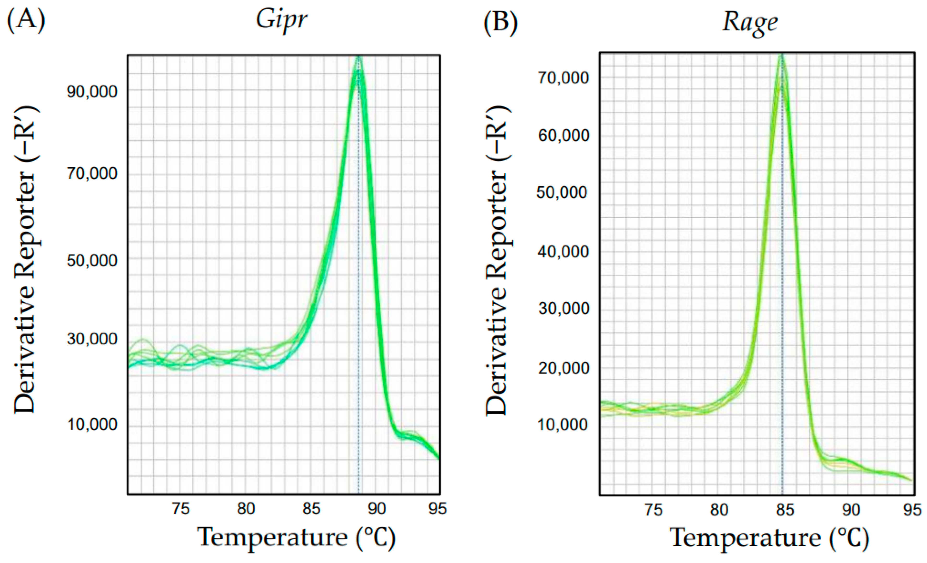
2.1. Gene Expression of Gipr and Rage in Human Monocyte-Derived U937 Cells

We investigated whether the Gipr and Rage genes were expressed in human U937 macrophages. As shown in Figure 1, the Gipr (A) and Rage (B) genes were actually expressed in U937 cells.

2.2. [D-Ala2]GIP(1–42) Inhibited the AGE-Induced Foam Cell Formation of and Intracellular Reactive Oxygen Species (ROS) Generation in Human U937 Cells

We first investigated the effect of [D-Ala2]GIP(1–42) on the AGE-induced foam cell formation of macrophages. Macrophage foam cell formation was evaluated by 1,1-dioctadecyl-3,3,3,3-tetamethylindocarbocyanine perchlorate (Dil)-labeled oxidized-LDL (Dil-ox-LDL) uptake into U937 cells. As shown in Figure 2, immunofluorescence staining showed that [D-Ala2]GIP(1–42) significantly inhibited the increase in Dil-ox-LDL uptake into AGE-exposed U937 cells, whose effect was attenuated by dorsomorphin, which is an inhibitor of AMP-activated protein kinase (AMPK). Furthermore, an activator of AMPK, 5-aminoimidazole-4-carboxamide1-B-D-ribofuranoside (AICAR), mimicked the inhibitory effect of [D-Ala2]GIP(1–42) on macrophage foam cell formation, while an inhibitor of NADPH oxidase, diphenylene iodonium (DPI), significantly inhibited the stimulatory effect of AGEs on Dil-ox-LDL uptake into U937 cells.
When intracellular oxygen species (ROS) generation was measured by fluorescence intensity of 2′,7′-dichlorodihydrofluorescein (DCF) using a cell-permeable probe, 2′,7′-dichlorodihydrofluorescein diacetate (DCFH-DA), as in the case of the foam cell formation of U937 cells, AGEs increased ROS generation in U937 cells, which was significantly inhibited by [D-Ala2]GIP(1–42) or DPI. The anti-oxidative effect of [D-Ala2]GIP(1–42) on AGE-exposed U937 cells was partially attenuated by co-treatment with dorsomorphin, while AICAR mimicked the effect of [D-Ala2]GIP(1–42) on ROS generation in U937 cells exposed to AGEs.
As shown in Figure 2, some Dil-ox-LDL-positive cells had DCF intensity. There was a significant correlation of fluorescence intensity between Dil-ox-LDL and DCF, thus suggesting that intracellular ROS generation occurred in Dil-ox-LDL-uptake macrophages (Figure 2U).

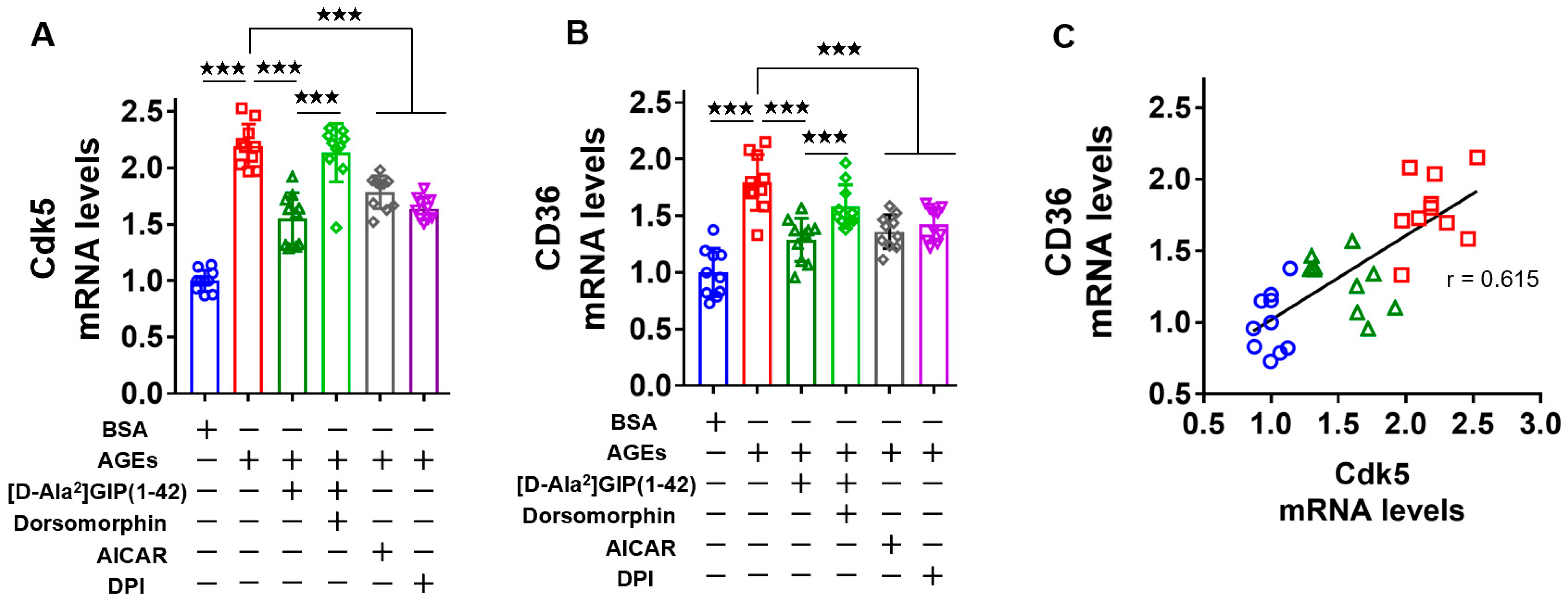
2.3. [D-Ala2]GIP(1–42) Inhibited Cdk5 and CD36 Gene Expression Levels in AGE-Exposed U937 Cells

We then elucidated the effects of [D-Ala2]GIP(1–42) on Cdk5 and CD36 gene expression in AGE-exposed U937 macrophages. As shown in Figure 3A,B, both Cdk5 and CD36 gene expression levels were increased in U937 cells by the treatment with AGEs. [D-Ala2]GIP(1–42) inhibited the AGE-induced up-regulation of Cdk5 and CD36 mRNA levels in U937 cells, which were partly attenuated by dorsomorphin. AICAR mimicked the effects of [D-Ala2]GIP(1–42) on Cdk5 and CD36 gene expression in AGE-exposed U937 cells. DPI significantly inhibited the AGE-induced up-regulation of Cdk5 and CD36 mRNA levels in U937 cells. The levels of Cdk5 and CD36 gene expression were highly correlated with each other (Figure 3C).

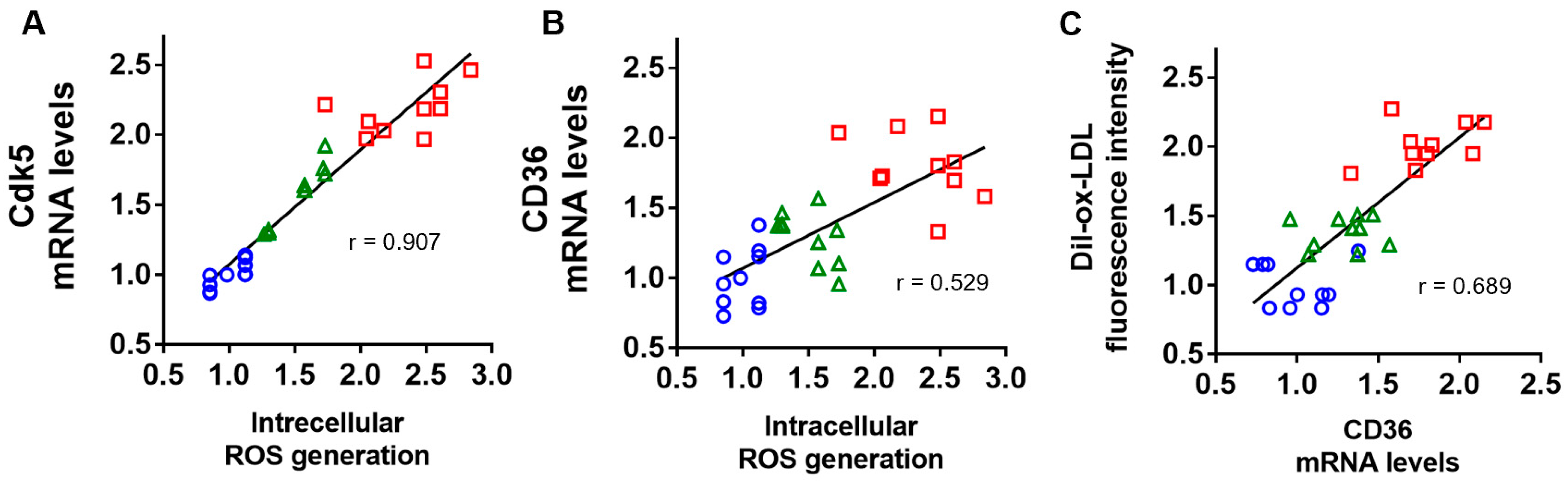
2.4. Correlation of Intracellular ROS Generation with Cdk5 and CD36 Gene Expression, and Association of CD36 mRNA Levels with Dil-ox-LDL Uptake into U937 Cells

We then studied the correlation of intracellular ROS generation with Cdk5 and CD36 mRNA levels and the association of CD36 gene expression levels with ox-LDL uptake. As shown in Figure 4A,B, intracellular ROS generation was correlated with Cdk5 and CD36 gene expression levels. CD36 mRNA levels were also positively associated with Dil-ox-LDL uptake into U937 cells (Figure 4C).

3. Discussion

A growing body of evidence has shown that the interaction of AGEs, senescent macromolecules formed at an accelerated rate under diabetes, with the cell surface receptor RAGE, evokes oxidative stress generation and inflammatory reactions in numerous kinds of cells, thereby being involved in the development and progression of atherosclerotic cardiovascular disease [23,34,39]. Indeed, AGE accumulation was increased in macrophage foam cells within human atherosclerotic plaque, whose levels were associated with plaque instability [32,33,34]. Foam cell formation and CD36 gene expression were enhanced in monocyte-driven macrophages extracted from patients with diabetes mellitus in comparison to those from non-diabetic healthy subjects [40]. In addition, we have shown that AGEs significantly enhance macrophage foam cell formation, which was evaluated by ox-LDL uptake into human monocyte-derived macrophages [40,41]. In this previous report, we found that (1) an anti-oxidant, N-acetylcysteine, significantly inhibited the AGE-induced foam cell formation and up-regulation of Cdk5 and CD36 mRNA levels in U937 cells; (2) a selective inhibitor of Cdk5 suppressed the AGE-induced increase in CD36 gene expression as well as the foam cell formation of macrophages, (3) the anti-CD36 neutralizing antibody inhibited the foam cell formation of AGE-exposed macrophages, and (4) the blockade of the AGE-RAGE interaction by RAGE-aptamer inhibited the AGE-induced ROS generation, and Cdk5 and CD36 gene expression in, and foam cell formation of, U937 macrophages [41]. These observations suggest that the engagement of RAGE with AGEs could contribute to macrophage foam cell formation by stimulating the CD36-dependent ox-LDL uptake into U937 cells via the ROS-Cdk5 signaling pathway.
Tirzepatide, a dual GIP and GLP-1 receptor agonist, has recently been approved for the treatment of patients with diabetes [9,10,11]; it has resulted in superior HbA1c reduction in diabetes patients and exhibited unprecedented weight loss in obese individuals compared with GLP-1 receptor agonists alone. Although several clinical trials have revealed that GLP-1 receptor agonists reduce the risk of cardiovascular disease in patients with diabetes [42,43,44], the effect of the GIP receptor agonist, including tirzepatide, on atherosclerotic cardiovascular disease remains unclear. Therefore, in the present study, we investigated the effect of GIP on AGE-induced macrophage foam cell formation, an initial step of atherosclerosis [21,22,45,46]. The salient findings in the present study were as follows: (1) the Gipr and Rage gene were expressed in human-cultured U937 macrophages; (2) AGEs increased oxidative stress generation and the foam cell formation of U937 macrophages, both of which were significantly inhibited by their treatment with [D-Ala2]GIP(1–42); (3) the anti-atherosclerotic and anti-oxidative effects of [D-Ala2]GIP(1–42) on AGE-exposed U937 cells were partly attenuated by an AMPK inhibitor, dorsomorphin; (4) an activator of AMPK, AICAR, mimicked the effects of [D-Ala2]GIP(1–42) on foam cell formation and oxidative stress generation in AGE-exposed U937 cells, (5) an inhibitor of NADPH oxidase, DPI, significantly inhibited the AGE-induced increase in foam cell formation and ROS generation in U937 cells; (6) foam cell formation and oxidative stress generation was correlated with each other; (7) AGEs increased Cdk5 and CD36 gene expression levels, both of which were significantly blocked by [D-Ala2]GIP(1–42); (8) AICAR mimicked the inhibitory effects of [D-Ala2]GIP(1–42) on Cdk5 and CD36 mRNA levels in AGE-exposed U937 cells, while dorsomorphin partly attenuated the effects of [D-Ala2]GIP(1–42) on the AGE-induced up-regulation of these gene levels in U937 cells; (9) DPI significantly inhibited the AGE-induced increase in Cdk5 and CD36 gene expression levels in U937 cells; and (10) there was a significant correlation between Cdk5 and CD36 gene expression levels in U937 cells. Taken together, the present findings suggest that [D-Ala2]GIP(1–42) blocks the AGE-induced foam cell formation of macrophages by inhibiting the Cdk5-CD36 pathway via the suppression of NADPH oxidase-derived ROS generation.
In this study, we showed, for the first time, that an inhibitor of NADPH oxidase, DPI, inhibited the AGE-induced ROS generation of U937 macrophages, which was associated with the downregulation of Cdk5 and CD36 mRNA levels and the subsequent foam cell formation of U937 macrophages. The present observations with our previous findings suggest that NADPH oxidase-derived ROS generation, enhanced by the AGE-RAGE interaction, could stimulate ox-LDL uptake into U937 cells and play a principal role in macrophage foam cell formation through the induction of CD36, one of the major scavenger receptors that can mediate the uptake of ox-LDL, through the activation of Cdk5. We, along with others, have previously reported that NADPH oxidase-derived ROS generation is required for the signaling pathway of the AGE-RAGE axis in various types of cells, including macrophages [47,48], thus suggesting that NADPH oxidase activity and ROS generation may contribute to atherosclerotic foam cell formation in diabetes.
In the present study, we found that an AMPK inhibitor, dorsomorphin, significantly attenuated the effects of [D-Ala2]GIP(1–42) on foam cell formation, ROS generation, and Cdk5 and CD36 gene expressions in AGE-exposed U937 cells, whereas an activator of AMPK, AICAR, mimicked the anti-oxidative and anti-atherosclerotic effects of [D-Ala2]GIP(1–42). We have previously shown that metformin inhibited AGE-RAGE-induced proliferation and vascular endothelial growth factor gene expression in breast cancer cells, both of which were blocked by compound C, an inhibitor of AMPK [49]. Moreover, metformin has been shown to exert anti-oxidative effects on lipopolysaccharide-exposed macrophages via the activation of AMPK [50]. In addition, globular adiponectin has been reported to inhibit ethanol-induced ROS generation in macrophages by suppressing NADPH oxidase activity via the AMPK pathway [51]. These findings suggest that NADPH oxidase-derived ROS generation may be a molecular target of the anti-atherosclerotic effects of [D-Ala2]GIP(1–42) on U937 macrophages via AMPK activation. In other words, [D-Ala2]GIP(1–42) may inhibit the AGE-RAGE-induced oxidative stress-mediated macrophage foam cell formation by suppressing NADPH oxidase activity through its interaction with the GIP receptor via the activation of the AMPK pathway. Several studies have shown that the binding of GIP with the GIP receptor activates AMPK through phospholipase C and calcium/calmodulin-dependent protein kinase signaling pathways [15,52,53,54], thus supporting the concept that AMPK could play a role in the anti-atherosclerotic effects of [D-Ala2]GIP(1–42) observed here.
NADPH oxidase activity in macrophages was inhibited by the cyclic AMP–protein kinase A pathway [55]. We previously showed that GIP, a stimulator of cyclic AMP or an inhibitor of NADPH oxidase, inhibits AGE-RAGE-induced oxidative stress generation and inflammatory and thrombotic reactions in endothelial cells [16]. Given that GIP stimulates cyclic AMP production in a variety of cells [16,56,57], [D-Ala2]GIP(1–42) may inhibit AGE-induced macrophage foam cell formation by stimulating the cyclic AMP-protein kinase A pathway via the suppression of NADPH oxidase activity. The effects of [D-Ala2]GIP(1–42) on foam cell formation, ROS generation, and Cdk5 and CD36 gene expressions in U937 cells exposed to AGEs were not completely inhibited by the co-treatment of dorsomorphin, thus suggesting that components other than the AMPK pathway may also be involved in the anti-atherosclerotic action of [D-Ala2]GIP(1–42).
We have recently found that Stachybotrys microspore triprenyl phenol-44D inhibits atherosclerotic plaque lesions in Apoe−/− mice by attenuating AGE-RAGE-induced macrophage foam cell formation through the Cdk5-CD36 pathway suppression [58]. The observations suggest that accelerated macrophage foam cell formation within atherosclerotic plaques may be a therapeutic strategy for preventing atherosclerotic cardiovascular disease. Therefore, our present study suggests that the GIP-GIP receptor axis may exert anti-atherosclerotic actions by suppressing AGE-RAGE-induced foam cell formation in diabetes. Further clinical studies are needed to clarify whether GIP-based therapy could actually reduce the risk of cardiovascular events in diabetes patients.
There were several limitations in the present experiments. First, we did not evaluate the protein expression levels of RAGE, Cdk5, and CD36 in this study. To examine these protein expression levels would be helpful to support the findings of this study. However, CD36 protein expression levels are reported to be functionally correlated with ox-LDL uptake and the foam cell formation of macrophages [59]. We have previously found that the gene expression levels of Cdk5 and CD36 are correlated with each other in AGE-exposed U937 macrophages [41]. Moreover, we have shown that the blockade of the AGE-RAGE signaling pathway by N-acetyl-l-cysteine or RAGE-aptamer reduces Cdk5 mRNA levels and suppresses CD36 gene expression and the foam cell formation of macrophages, the latter of which is also suppressed by an inhibitor of Cdk5 [41]. Therefore, the present and previous observations suggest that gene expressions of RAGE, Cdk5, CD36 can be correlated with each protein expression, respectively. Second, we did not exactly know how Cdk5 regulated CD36 expression in U937 macrophage cells. However, Cdk5 might increase the phosphorylation of peroxisome proliferator-activated receptor γ and subsequently enhance its transcriptional activity, which could cause CD36 overexpression and macrophage foam cell formation [41,60,61]. Third, we chose the concentration of AGE-BSA at 100 μg/mL in this study because its levels were comparable with those of diabetic patients [62]. Fourth, in this study, we evaluated macrophage foam cell formation by Dil-ox-LDL uptake. We examined the involvement of NADPH oxidase in the macrophage foam cell formation exposed by AGEs because we previously showed that the AGE-RAGE interaction stimulates NADPH oxidase activity in endothelial cells, the pathological effect of which was blocked by an inhibitor of NADPH oxidase activity, DPI [47]. Therefore, we did not examine intracellular levels of NADPH, endothelial nitric oxide synthase, and other anti-oxidant systems in the present study. Fifth, although NADPH oxidase activation may have dual effects in diabetes, there is a growing body of evidence that the inhibition of NADPH oxidase activity ameliorates vascular injury and metabolic derangement in diabetes [63,64]. Sixth, we did not investigate the tissue-specific distribution of various NAD(P)H oxidase subunits and distinctive functions of Nox isoforms. However, we have previously shown that the AGE-RAGE axis up-regulates the gene expression of all the components of NADPH oxidase, including gp91phox, Nox1, and Nox4, in diabetic kidneys [39]. It would be interesting to examine the effects of [D-Ala2]GIP(1–42) on the expression of these NADPH oxidase components in macrophages exposed to AGEs (Figure 5).

4. Materials and Methods

4.1. Chemical Regents and Materials

[D-Ala2]GIP(1–42) was purchased from Phoenix Pharmaceuticals. Inc. (Burlingame, CA, USA). Dil-ox-LDL was acquired from Highland Technology Center (Frederick, MD, USA), and an OxiSelect intracellular ROS assay kit and DCFH-DA probe were acquired from Cell Biolabs. Inc. (San Diego, CA, USA). A human cell line of monocytes, U937 cells, were obtained from the Japanese Collection of Research Bioresources Cell Bank (JCRB9021; Osaka, Japan); the AMPK inhibitor, dorsomorphin, was obtained from FUJIFILM Wako Pure Chemical Corporation (Osaka, Japan); and an inhibitor of NADPH-oxidase, DPI, an AMPK stimulator AICAR, phorbol 12-myristate 13-acetate (PMA), D-glyceraldehyde, Roswell Park Memorial Institute (RPMI) 1640 medium, BSA, and fetal bovine serum (FBS) were obtained from Sigma Aldrich (St. Louis, MO, USA). A Vibrance Antifade Mounting Medium with DAPI was acquired from Vector Laboratories (Burlingame, CA, USA).

4.2. Preparation of AGEs of Bovine Serum Albumin (BSA)

BSA (25 mg/mL) was incubated with 0.1 mol/L D-glyceraldehyde in a 0.2 M NaPO4 buffer at 37 °C under sterile conditions for 7 days, as described previously [40,41,47,49,58]. Then, unincorporated sugars were removed using dialysis against phosphate-buffered saline (PBS) to prepare AGEs [40,41,47,49,58]. In the same conditions, except for the absence of D-glyceraldehyde, non-glycated BSA was prepared and used as a control.

4.3. Cell Culture of U937 Monocyte-Derived Macrophages

U937 cells were maintained in the RPMI 1640 medium with 10% FBS, as described previously [18,40,41]. The cells were seeded onto 12 well plates at a concentration of 1.0 × 10⁶ cells/dish and cultured with PMA (40 ng/mL) in the same medium. After washing with PBS, adherent cells were used as differentiated U937 macrophages [18,41].

4.4. Measurement of Fluorescence Intensity of Dil-ox-LDL and ROS Generation in U937 Cells

U937 macrophage cells were incubated with 100 μg/mL AGE-BSA or 100 μg/mL non-glycated BSA in the presence or absence of 1 nmol/L [D-Ala2]GIP(1–42) [18], 50 nmol/L DPI, an inhibitor of NADPH oxidase [47], 10 μmol/L dorsomorphin, an inhibitor of AMPK [15], and 500 μmol/L AICAR, an activator of AMPK [15] in RPMI 1640 medium supplemented with 10% FBS for 24 h [18,40,41]. After removing the cell medium, cells were treated with 10 μg/mL Dil-ox-LDL for 18 h and with a 10 μmol/L DCFH-DA probe [65,66] for another 15 min. Then, adherent U937 macrophages were mounted in a Vibrance Antifade Mounting Medium with DAPI, and immunofluorescence images were obtained using the All-in-One Fluorescence Microscope BZ-X710 and Analysis Software BZ-X800 (Keyence; Osaka, Japan). The fluorescence intensity area, of red or green color, per the cells was calculated as described previously [18,40,41,58].

4.5. Quantitative Real-Time Reverse Transcription–Polymerase Chain Reaction (RT-PCR)

Total RNA was extracted from the adherent human U937 cells to synthesize cDNA. The SYBR Green or TaqMan-based gene expression assay and StepOnePlusTM sequence detection system (Life Technologies Japan, Tokyo, Japan) were used for quantitative real-time RT-PCR. The expression levels of target genes were initialized by the values of glyceraldehyde 3-phosphate dehydrogenase (GAPDH) and then related to the controls with BSA, as described previously [18,40,41,58]. The primers and probes used for the analysis of quantitative RT-PCR were as follows: Gipr, Hs00609201_g1; Rage, Hs00542584_g1; Cdk5, NM_001164410.3, NM_004935.4; CD36, Hs00169627_ml; and Gapdh, Hs99999905_ml.

4.6. Statistical Analysis
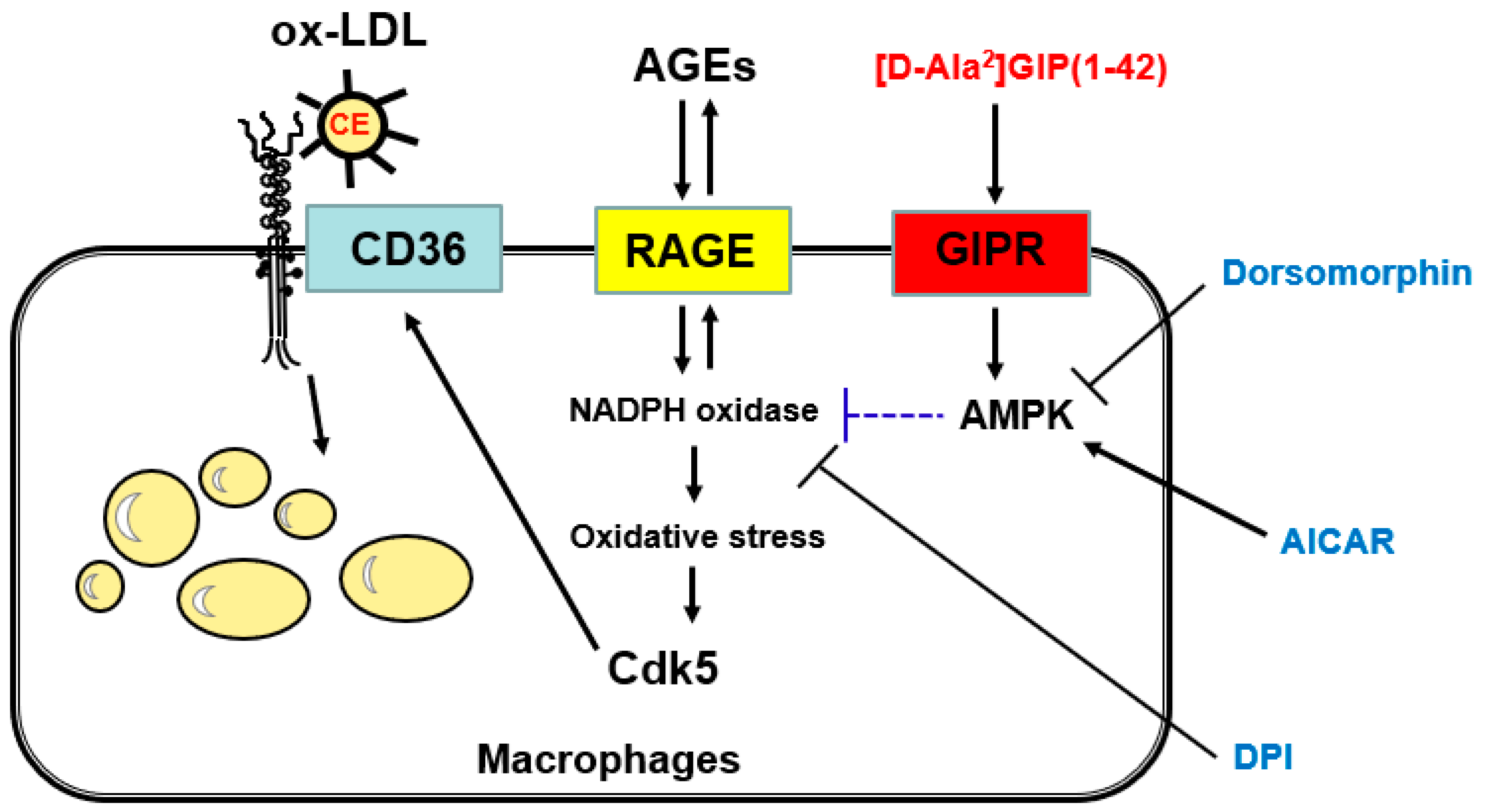
All data are expressed as the mean ± standard deviation. The parametric coefficient of Pearson’s correlation was used to analyze the correlation between the two groups. GraphPad PRISM ver. 7.05 software (GraphPad Inc., San Diego, CA, USA) was used for statistical analysis. The significance level was defined as a p-value < 0.05.

5. Conclusions

The present study suggests that [D-Ala2]GIP(1–42) could inhibit AGE-RAGE-induced, NADPH oxidase-derived oxidative stress generation in U937 macrophages via AMPK activation and subsequently suppress macrophage foam cell formation by reducing the Cdk5-CD36 pathway.

Author Contributions

Conceptualization, M.T. and S.-i.Y.; methodology, M.T. and T.S.; software, M.T. and T.S.; validation, M.T. and T.S.; formal analysis, M.T.; investigation, M.T., H.Y., Y.M., T.S., N.I. and T.M.; resources, M.T. and S.-i.Y.; data curation, M.T.; writing—original draft preparation, M.T. and S.-i.Y.; writing—review and editing, Y.M., H.Y., T.S., M.O., N.O., T.F. and N.I.; visualization, M.T.; supervision, T.M.; project administration, M.T. and S.-i.Y.; funding acquisition, M.T. All authors have read and agreed to the published version of the manuscript.

Funding

This study was funded by Grants for Young Researchers from the Japan Association for Diabetes Education and Care (grant number 2021-YNG-002 to M.T.), Research Grant Award 2022 from the Japanese Society of Anti-Aging Medicine (M.T.), and Grants-in-Aid for Scientific Research from Japan Society for the Promotion of Science (JSPS KAKENHI grant numbers: 20K07075 and 24K09316 to M.T.).

Institutional Review Board Statement

Not applicable.

Informed Consent Statement

Not applicable.

Data Availability Statement

Data are contained within the article.

Conflicts of Interest

The authors declare no conflicts of interest.

References

  1. Drucker, D.J. The biology of incretin hormones. Cell Metab. 2006, 3, 153–165. [Google Scholar] [CrossRef] [PubMed]
  2. Seino, Y.; Fukushima, M.; Yabe, D. GIP and GLP-1, the two incretin hormones: Similarities and differences. J. Diabetes Investig. 2010, 1, 8–23. [Google Scholar] [CrossRef]
  3. Fukami, A.; Seino, Y.; Ozaki, N.; Yamamoto, M.; Sugiyama, C.; Sakamoto-Miura, E.; Himeno, T.; Takagishi, Y.; Tsunekawa, S.; Ali, S.; et al. Ectopic expression of GIP in pancreatic β-cells maintains enhanced insulin secretion in mice with complete absence of proglucagon-derived peptides. Diabetes 2013, 62, 510–518. [Google Scholar] [CrossRef] [PubMed]
  4. Baggio, L.L.; Drucker, D.J. Biology of incretins: GLP-1 and GIP. Gastroenterology 2007, 132, 2131–2157. [Google Scholar] [CrossRef]
  5. Campbell, J.E.; Drucker, D.J. Pharmacology, physiology, and mechanisms of incretin hormone action. Cell Metab. 2013, 17, 819–837. [Google Scholar] [CrossRef]
  6. Seino, Y.; Yabe, D. Glucose-dependent insulinotropic polypeptide and glucagon-like peptide-1: Incretin actions beyond the pancreas. J. Diabetes Investig. 2013, 4, 108–130. [Google Scholar] [CrossRef] [PubMed]
  7. Winzell, M.S.; Ahrén, B. G-protein-coupled receptors and islet function-implications for treatment of type 2 diabetes. Pharmacol. Ther. 2007, 116, 437–448. [Google Scholar] [CrossRef] [PubMed]
  8. Siegel, E.G.; Creutzfeldt, W. Stimulation of insulin release in isolated rat islets by GIP in physiological concentrations and its relation to islet cyclic AMP content. Diabetologia 1985, 28, 857–861. [Google Scholar] [CrossRef]
  9. Coskun, T.; Sloop, K.W.; Loghin, C.; Alsina-Fernandez, J.; Urva, S.; Bokvist, K.B.; Cui, X.; Briere, D.A.; Cabrera, O.; Roell, W.C.; et al. LY3298176, a novel dual GIP and GLP-1 receptor agonist for the treatment of type 2 diabetes mellitus: From discovery to clinical proof of concept. Mol. Metab. 2018, 18, 3–14. [Google Scholar] [CrossRef]
  10. Inagaki, N.; Takeuchi, M.; Oura, T.; Imaoka, T.; Seino, Y. Efficacy and safety of tirzepatide monotherapy compared with dulaglutide in Japanese patients with type 2 diabetes (SURPASS J-mono): A double-blind, multicentre, randomised, phase 3 trial. Lancet Diabetes Endocrinol. 2022, 10, 623–633. [Google Scholar] [CrossRef]
  11. Huang, X.; Liu, J.; Peng, G.; Lu, M.; Zhou, Z.; Jiang, N.; Yan, Z. Gut hormone multi-agonists for the treatment of type 2 diabetes and obesity: Advances and challenges. J. Endocrinol. 2024, 262, e230404. [Google Scholar] [CrossRef] [PubMed]
  12. Usdin, T.B.; Mezey, E.; Button, D.C.; Brownstein, M.J.; Bonner, T.I. Gastric inhibitory polypeptide receptor, a member of the secretin-vasoactive intestinal peptide receptor family, is widely distributed in peripheral organs and the brain. Endocrinology 1993, 133, 2861–2870. [Google Scholar] [CrossRef]
  13. Drucker, D.J.; Holst, J.J. The expanding incretin universe: From basic biology to clinical translation. Diabetologia 2023, 66, 1765–1779. [Google Scholar] [CrossRef] [PubMed]
  14. Nogi, Y.; Nagashima, M.; Terasaki, M.; Nohtomi, K.; Watanabe, T.; Hirano, T. Glucose-dependent insulinotropic polypeptide prevents the progression of macrophage-driven atherosclerosis in diabetic apolipoprotein E-null mice. PLoS ONE 2012, 7, e35683. [Google Scholar] [CrossRef] [PubMed]
  15. Mori, Y.; Kushima, H.; Koshibu, M.; Saito, T.; Hiromura, M.; Kohashi, K.; Terasaki, M.; Seino, Y.; Yamada, Y.; Hirano, T. Glucose-dependent insulinotropic polypeptide suppresses peripheral arterial remodeling in male mice. Endocrinology 2018, 159, 2717–2732. [Google Scholar] [CrossRef]
  16. Ojima, A.; Matsui, T.; Maeda, S.; Takeuchi, M.; Yamagishi, S. Glucose-dependent insulinotropic polypeptide (GIP) inhibits signaling pathways of advanced glycation end products (AGEs) in endothelial cells via its antioxidative properties. Horm. Metab. Res. 2012, 44, 501–505. [Google Scholar] [CrossRef]
  17. Kahles, F.; Liberman, A.; Halim, C.; Rau, M.; Möllmann, J.; Mertens, R.W.; Rückbeil, M.; Diepolder, I.; Walla, B.; Diebold, S.; et al. The incretin hormone GIP is upregulated in patients with atherosclerosis and stabilizes plaques in ApoE−/− mice by blocking monocyte/macrophage activation. Mol. Metab. 2018, 14, 150–157. [Google Scholar] [CrossRef]
  18. Terasaki, M.; Yashima, H.; Mori, Y.; Saito, T.; Shigara, Y.; Kawakami, R.; Ohara, M.; Fukui, T.; Hirano, T.; Yamada, Y.; et al. Glucose-dependent insulinotropic polypeptide suppresses foam cell formation of macrophages through inhibition of the cyclin-dependent kinase 5-CD36 pathway. Biomedicines 2021, 9, 832. [Google Scholar] [CrossRef]
  19. Rao Kondapally Seshasai, S.; Kaptoge, S.; Thompson, A.; Di Angelantonio, E.; Gao, P.; Sarwar, N.; Whincup, P.H.; Mukamal, K.J.; Gillum, R.F.; Holme, I.; et al. Diabetes mellitus, fasting glucose, and risk of cause-specific death. N. Engl. J. Med. 2011, 364, 829–841. [Google Scholar]
  20. Damaskos, C.; Garmpis, N.; Kollia, P.; Mitsiopoulos, G.; Barlampa, D.; Drosos, A.; Patsouras, A.; Gravvanis, N.; Antoniou, V.; Litos, A.; et al. Assessing Cardiovascular Risk in Patients with Diabetes: An Update. Curr. Cardiol. Rev. 2020, 16, 266–274. [Google Scholar] [CrossRef]
  21. Lusis, A.J. Atherosclerosis. Nature 2000, 407, 233–241. [Google Scholar] [CrossRef] [PubMed]
  22. Glass, C.K.; Witztum, J.L. Atherosclerosis: The road ahead. Cell 2001, 104, 503–516. [Google Scholar] [CrossRef] [PubMed]
  23. Yamagishi, S.I. Role of advanced glycation endproduct (AGE)-receptor for advanced glycation endproduct (RAGE) axis in cardiovascular disease and its therapeutic intervention. Circ. J. 2019, 83, 1822–1828. [Google Scholar] [CrossRef]
  24. Razaei, M.; Rabizadeh, S.; Mirahmad, M.; Hajmiri, M.S.; Nakhjavani, M.; Hemmatabadi, M.; Shirzad, N. The association between advanced glycation end products (AGEs) and ABC (hemoglobin A1C, blood pressure, and low-density lipoprotein cholesterol) control parameters among patients with type 2 diabetes mellitus. Diabetol. Metab. Syndr. 2022, 14, 122. [Google Scholar] [CrossRef] [PubMed]
  25. Jud, P.; Sourij, H. Therapeutic options to reduce advanced glycation end products in patients with diabetes mellitus: A review. Diabetes Res. Clin. Pract. 2019, 148, 54–63. [Google Scholar] [CrossRef] [PubMed]
  26. Goldin, A.; Beckman, J.A.; Schmidt, A.M.; Creager, M.A. Advanced glycation end products: Sparking the development of diabetic vascular injury. Circulation 2006, 114, 597–605. [Google Scholar] [CrossRef]
  27. Kilhovd, B.K.; Juutilainen, A.; Lehto, S.; Rönnemaa, T.; Torjesen, P.A.; Hanssen, K.F.; Laakso, M. Increased serum levels of methylglyoxal-derived hydroimidazolone-AGE are associated with increased cardiovascular disease mortality in nondiabetic women. Atherosclerosis 2009, 205, 590–594. [Google Scholar] [CrossRef]
  28. Kilhovd, B.K.; Juutilainen, A.; Lehto, S.; Rönnemaa, T.; Torjesen, P.A.; Hanssen, K.F.; Laakso, M. Increased serum levels of advanced glycation endproducts predict total, cardiovascular and coronary mortality in women with type 2 diabetes: A population-based 18 year follow-up study. Diabetologia 2007, 50, 1409–1417. [Google Scholar] [CrossRef]
  29. Schmidt, A.M.; Stern, D. Atherosclerosis and diabetes: The RAGE connection. Curr. Atheroscler. Rep. 2000, 2, 430–436. [Google Scholar] [CrossRef]
  30. Bijnen, M.; Beelen, N.; Wetzels, S.; Gaar, J.V.; Vroomen, M.; Wijnands, E.; Scheijen, J.L.; van de Waarenburg, M.P.H.; Gijbels, M.J.; Cleutjens, J.P.; et al. RAGE deficiency dose not affect non-alcholic steatohepatitis and atherosclerosis in Western type diet-fed Ldlr(−/−) mice. Sci. Rep. 2018, 8, 15256. [Google Scholar] [CrossRef]
  31. Han, X.; Ma, W.; Zhu, Y.; Sun, X.; Liu, N. Advanced glycation end products enhance macrophage polarization to the Ma phenotype via the HIF-1α/PDK4 pathway. Mol. Cell. Endocrinol. 2020, 514, 110878. [Google Scholar] [CrossRef]
  32. Hassen, N.M.; Wouters, K.; Hujiberts, M.S.; Gijbels, M.J.; Sluimer, J.C.; Scheijen, J.L.; Heeneman, S.; Biessen, E.A.; Daemen, M.J.; Brownlee, M.; et al. Higher levels of advanced glycation endproducts in human carotid atherosclerotic plaques are associated with a repture-prone phenotype. Eur. Heart J. 2014, 35, 1137–1146. [Google Scholar] [CrossRef] [PubMed]
  33. Wang, Z.Q.; Jing, L.L.; Yan, J.C.; Sun, Z.; Bao, Z.Y.; Shao, C.; Pang, Q.W.; Geng, Y.; Zhang, L.L.; Li, L.H. Role of AGEs in the progression and regression of atherosclerotic plaques. Glycoconj. J. 2018, 35, 443–450. [Google Scholar] [CrossRef] [PubMed]
  34. Kume, S.; Takeya, M.; Mori, T.; Araki, N.; Suzuki, H.; Horiuchi, S.; Kodama, T.; Miyauchi, Y.; Takahashi, K. Immunohistochemical and ultrastructural detection of advanced glycation end products in atherosclerotic lesions of human aorta with a novel specific monoclonal antibody. Am. J. Pathol. 1995, 147, 654–667. [Google Scholar] [PubMed]
  35. Nin, J.W.; Jorsal, A.; Ferreira, I.; Schalkwijk, C.G.; Prins, M.H.; Parving, H.H.; Tarnow, L.; Rossing, P.; Stehouwer, C.D. Higher plasma soluble receptor for advanced glycation end products (sRAGE) levels are associated with incident cardiovascular disease and all-cause mortality in type 1 diabetes: A 12-year follow-up study. Diabetes 2010, 59, 2027–2032. [Google Scholar] [CrossRef]
  36. Nin, J.W.; Jorsal, A.; Ferreira, I.; Schalkwijk, C.G.; Prins, M.H.; Parving, H.H.; Tarnow, L.; Rossing, P.; Stehouwer, C.D. Higher plasma levels of advanced glycation end products are associated with incident cardiovascular disease and all-cause mortality in type 1 diabetes: A 12-year follow-up study. Diabetes Care 2011, 34, 442–447. [Google Scholar] [CrossRef]
  37. Semba, R.D.; Bandineli, S.; Sun, K.; Guralnik, J.M.; Ferrucci, L. Plasma carboxymethyl-lysine, an advanced glycation end product, and all-cause and cardiovascular disease mortality in older community-dwelling adults. J. Am. Geriatr. Soc. 2009, 57, 1874–1880. [Google Scholar] [CrossRef]
  38. Van Waateringe, R.P.; Fokkens, B.T.; Slagter, S.N.; van der Klauw, M.M.; van Vliet-Ostaptchouk, J.V.; Graaff, R.; Paterson, A.D.; Smit, A.J.; Lutgers, H.L.; Wolffenbuttel, B.H.R. Skin autofluorescence predicts incident type 2 diabetes, cardiovascular disease and mortality in the general population. Diabetologia 2019, 62, 269–280. [Google Scholar] [CrossRef]
  39. Matsui, T.; Higashimoto, Y.; Nishino, Y.; Nakamura, N.; Fukami, K.; Yamagishi, S. RAGE-aptamer blocks the development and progression of experimental diabetic nephropathy. Diabetes 2017, 66, 1683–1695. [Google Scholar] [CrossRef]
  40. Terasaki, M.; Yashima, H.; Mori, Y.; Saito, T.; Matsui, T.; Hiromura, M.; Kushima, H.; Osaka, N.; Ohara, M.; Fukui, T.; et al. A dipeptidyl peptidase-4 inhibitor inhibits foam cell formation of macrophages in type 1 diabetes via suppression of CD36 expression. Int. J. Mol. Sci. 2020, 21, 4811. [Google Scholar] [CrossRef]
  41. Yashima, H.; Terasaki, M.; Sotokawauchi, A.; Matsui, T.; Mori, Y.; Saito, T.; Osaka, N.; Kushima, H.; Hiromura, M.; Ohara, M.; et al. AGE-RAGE axis stimulates oxidized LDL uptake into macrophages through cyclin-dependent kinase 5-CD36 pathway via oxidative stress generation. Int. J. Mol. Sci. 2020, 21, 9263. [Google Scholar] [CrossRef] [PubMed]
  42. Kelly, M.; Lewis, J.; Rao, H.; Carter, J.; Portillo, I.; Beuttler, R. Effects of GLP-1 receptor agonists on cardiovascular outcomes in patients with type 2 diabetes and chronic kidney disease: A systematic review and meta-analysis. Pharmacotherapy 2022, 42, 921–928. [Google Scholar] [CrossRef]
  43. Kristensen, S.L.; Rørth, R.; Jhund, P.S.; Docherty, K.F.; Sattar, N.; Preiss, D.; Køber, L.; Petrie, M.C.; McMurray, J.J.V. Cardiovascular, mortality, and kidney outcomes with GLP-1 receptor agonists in patients with type 2 diabetes: A systematic review and meta-analysis of cardiovascular outcome trials. Lancet Diabetes Endocrinol. 2019, 7, 776–785. [Google Scholar] [CrossRef]
  44. Sattar, N.; Lee, M.M.Y.; Kristensen, S.L.; Branch, K.R.H.; Del Prato, S.; Khurmi, N.S.; Lam, C.S.P.; Lopes, R.D.; McMurray, J.J.V.; Pratley, R.E.; et al. Cardiovascular, mortality, and kidney outcomes with GLP-1 receptor agonists in patients with type 2 diabetes: A systematic review and meta-analysis of randomised trials. Lancet Diabetes Endocrinol. 2021, 9, 653–662. [Google Scholar] [CrossRef]
  45. Wen, S.Y.; Zhi, X.; Liu, H.X.; Wang, X.; Chen, Y.Y.; Wang, L. Is the suppression of CD36 a promising way for atherosclerosis therapy? Biochem. Pharmacol. 2024, 219, 115965. [Google Scholar] [CrossRef]
  46. Duan, H.; Song, P.; Li, R.; Su, H.; He, L. Attenuating lipid metabolism in atherosclerosis: The potential role of Anti-oxidative effects on low-density lipoprotein of herbal medicines. Front. Pharmacol. 2023, 14, 1161657. [Google Scholar] [CrossRef] [PubMed]
  47. Yamagishi, S.; Nakamura, K.; Matsui, T.; Inagaki, Y.; Takenaka, K.; Jinnouchi, Y.; Yoshida, Y.; Matsuura, T.; Narama, I.; Motomiya, Y.; et al. Pigment epithelium-derived factor inhibits advanced glycation end product-induced retinal vascular hyperpermeability by blocking reactive oxygen species mediated vascular endothelial growth factor expression. J. Biol. Chem. 2006, 281, 20213–20220. [Google Scholar] [CrossRef] [PubMed]
  48. Ping, M.; Xiao, W.; Mo, L.; Xiao, X.; Song, S.; Tang, W.; Yang, X. Paeonol attenuates advanced oxidation protein product-induced oxidative stress injury in THP-1 macrophages. Pharmacology 2014, 93, 286–295. [Google Scholar] [CrossRef]
  49. Ishibashi, Y.; Matsui, T.; Takeuchi, M.; Yamagishi, S. Metformin inhibits advanced glycation end products (AGEs)-induced growth and VEGF expression in MCF-7 breast cancer cells by suppressing AGEs receptor expression via AMP-activated protein kinase. Horm. Metab. Res. 2013, 45, 387–390. [Google Scholar] [CrossRef]
  50. Nassif, R.M.; Chalhoub, E.; Chedid, P.; Hurtado-Nedelec, M.; Raya, E.; Dang, P.M.; Marie, J.C.; El-Benna, J. Metformin Inhibits ROS Production by Human M2 Macrophages via the Activation of AMPK. Biomedicines 2022, 10, 319. [Google Scholar] [CrossRef]
  51. Kim, M.J.; Nagy, L.E.; Park, P.H. Globular adiponectin inhibits ethanol-induced reactive oxygen species production through modulation of NADPH oxidase in macrophages: Involvement of liver kinase B1/AMP-activated protein kinase pathway. Mol. Pharmacol. 2014, 86, 284–296. [Google Scholar] [CrossRef] [PubMed]
  52. Ewart, M.A.; Kennedy, S. AMPK and vasculoprotection. Pharmacol. Ther. 2011, 131, 242–253. [Google Scholar] [CrossRef]
  53. Gao, F.; Chen, J.; Zhu, H. A potential strategy for treating atherosclerosis: Improving endothelial function via AMP-activated protein kinase. Sci. China Life Sci. 2018, 61, 1024–1029. [Google Scholar] [CrossRef]
  54. Carling, D.; Sanders, M.J.; Woods, A. The regulation of AMP-activated protein kinase by upstream kinases. Int. J. Obes. 2008, 32 (Suppl. S4), S55–S59. [Google Scholar] [CrossRef]
  55. Bourdonnay, E.; Serezani, C.H.; Aronoff, D.M.; Peters-Golden, M. Regulation of alveolar macrophage p40phox: Hierarchy of activating kinases and their inhibition by PGE2. J. Leukoc. Biol. 2012, 92, 219–231. [Google Scholar] [CrossRef] [PubMed]
  56. Khan, R.; Tomas, A.; Rutter, G.A. Effects on pancreatic Beta and other Islet cells of the glucose-dependent insulinotropic polypeptide. Peptides 2020, 125, 170201. [Google Scholar] [CrossRef] [PubMed]
  57. Mohammad, S.; Ramos, L.S.; Buck, J.; Levin, L.R.; Rubino, F.; McGraw, T.E. Gastric inhibitory peptide controls adipose insulin sensitivity via activation of cAMP-response element-binding protein and p110β isoform of phosphatidylinositol 3-kinase. J. Biol. Chem. 2011, 286, 43062–43070. [Google Scholar] [CrossRef]
  58. Terasaki, M.; Shibata, K.; Mori, Y.; Saito, T.; Matsui, T.; Ohara, M.; Fukui, T.; Hasumi, K.; Higashimoto, Y.; Nobe, K.; et al. SMTP-44D inhibits atherosclerotic plaque formation in apolipoprotein-E null mice partly by suppressing the AGEs-RAGE axis. Int. J. Mol. Sci. 2023, 24, 6505. [Google Scholar] [CrossRef]
  59. Huh, H.Y.; Pearce, S.F.; Yesner, L.M.; Schindler, J.L.; Silerstein, R.L. Regulated expression of CD36 during monocyte-to-macrophage differentiation: Potential role of CD36 in foam cell formation. Blood 1996, 87, 2020–2028. [Google Scholar] [CrossRef]
  60. Choi, J.H.; Banks, A.S.; Estall, J.L.; Kajimura, S.; Bostrom, P.; Laznik, D.; Ruas, J.L.; Chalmers, M.J.; Kamenecka, T.M.; Bluher, M.; et al. Anti-diabetic drugs inhibit obesity-linked phosphorylation of PPARgamma by Cdk5. Nature 2010, 466, 451–456. [Google Scholar] [CrossRef]
  61. Leonarduzzi, G.; Gamba, P.; Gargiulo, S.; Sottero, B.; Kadl, A.; Biasi, F.; Chiarpotto, E.; Leitinger, N.; Vendemiale, G.; Serviddio, G.; et al. Oxidation as a crucial reaction for cholesterol to induce tissue degeneration: CD36 overexpression in human promonocytic cells treated with a biologically relevant oxysterol mixture. Aging Cell 2008, 7, 375–382. [Google Scholar] [CrossRef] [PubMed]
  62. Takeuchi, M.; Makita, Z.; Yanagisawa, K.; Kameda, K.; Koike, T. Detection of noncarboxymethyllysine and carboxymethyllysine advanced glycation end products (AGE) in serum of diabetic patients. Mol. Med. 1999, 5, 393–405. [Google Scholar] [CrossRef]
  63. Sylvester, A.L.; Zhang, D.X.; Ran, S.; Zinkevich, N.S. Inhibiting NADPH Oxidases to Target Vascular and Other Pathologies: An Update on Recent Experimental and Clinical Studies. Biomolecules 2022, 12, 823. [Google Scholar] [CrossRef]
  64. Yamagishi, S.; Matsui, T. Pigment Epithelium-Derived Factor: A Novel Therapeutic Target for Cardiometabolic Diseases and Related Complications. Curr. Med. Chem. 2018, 25, 1480–1500. [Google Scholar] [CrossRef] [PubMed]
  65. Wang, H.; Li, Y.; Zhang, X.; Xu, Z.; Zhou, J.; Shang, W. SPP-4 inhibitor linagliptin ameliorates oxidized LDL-induced THP-1 macrophage foam cell formation and inflammation. Drug Des. Dev. Ther. 2020, 14, 3929–3940. [Google Scholar] [CrossRef]
  66. Zhang, X.; Yang, Z.; Xu, Q.; Xu, C.; Shi, W.; Pang, R.; Zhang, K.; Liang, X.; Li, H.; Li, Z.; et al. Dexamethasone induced osteocyte apoptosis in steroid-induced femoral head osteonecrosis through ROS-mediated oxidative stress. Orthop. Surg. 2024, 16, 733–744. [Google Scholar] [CrossRef]
Figure 1. Melt curve of gene expression of Gipr (A) and Rage (B) in human U937 cells.
Ijms 25 09724 g001
Figure 2. Effects of [D-Ala2]GIP(1–42), dorsomorphin, AICAR, and DPI on Dil-ox-LDL uptake into, and ROS generation in, AGE-exposed U937 macrophages. U937 cells were treated with 100 μg/mL AGE-BSA or 100 μg/mL non-glycated control BSA in the presence or absence of 1 nmol/L [D-Ala2]GIP(1–42), 50 nmol/L dorsomorphin, 500 μmol/L AICAR, or 50 nmol/L DPI in Roswell Park Memorial Institute (RPMI) 1640 medium supplemented with 10% fetal bovine serum (FBS) at 37 °C for 24 h. Then, the adherent cells were incubated with 10 μg/mL Dil-ox-LDL for 18 h and further treated with a 10 μmol/L DCFH-DA probe for another 15 min. The fluorescence intensity of Dil-ox-LDL and DCF were measured. (AR) Representative images of immunofluorescence staining. Dil-ox-LDL-positive cells were stained in red, and ROS-generating cells are represented in green. Scale bars show 20 μm. (SU) Quantitative data of fluorescence intensity for Dil-ox-LDL and DCF were normalized for each control value with BSA-treated cells, respectively. The correlation of fluorescence intensity between Dil-ox-LDL and DCF was used by Pearson’s correlation test. Number = 10 for each group. Values are shown as mean ± standard deviation. ★★★ p < 0.005. (U) Blue circles for BSA-treated cells, red squares for AGE-treated cells, and green triangles for AGE-treated cells with [D-Ala2]GIP(1–42).
Ijms 25 09724 g002
Figure 3. Effects of [D-Ala2]GIP(1–42), dorsomorphin, AICAR, and DPI on Cdk5 and CD36 gene expression levels in AGE-exposed U937 macrophages. Gene expression levels of Cdk5 (A) or CD36 (B) and their correlation (C). The correlation of Cdk5 with CD36 gene expression levels was determined by Pearson’s correlation test. Total RNAs were transcribed and amplified by quantitative real-time RT-PCR. The levels of the target gene were initialized by the value of GAPDH mRNA-derived signals and then relatively calculated by the controls with BSA. Number = 10 for each group. Values are presented as mean ± standard deviation. ★★★ p < 0.005. (C) Blue circles for BSA-treated cells, red squares for AGE-treated cells, and green triangles for AGE-treated cells with [D-Ala2]GIP(1–42).
Ijms 25 09724 g003
Figure 4. Correlation of intracellular ROS generation with Cdk5 (A) or CD36 (B) mRNA levels and the association of CD36 gene expression levels with Dil-ox-LDL uptake (C) into U937 cells. The correlation was determined by Pearson’s correlation test. Number = 10 for each group. Blue circles for BSA-treated cells, red squares for AGE-treated cells, and green triangles for AGE-treated cells with [D-Ala2]GIP(1–42).
Ijms 25 09724 g004
Figure 5. Possible mechanisms of the inhibitory effect of [D-Ala2]GIP(1–42) on foam cell formation of macrophages. AGEs, advanced glycation end products; RAGE, receptor for AGE; GIPR, receptor of glucose-dependent insulinotropic polypeptide; AMPK, AMP-activated protein kinase; Cdk5, cyclin-dependent kinase 5; CE, cholesterol ester; DPI, diphenylene iodonium; AICAR, 5-aminoimidazole-4-carboxamide1-B-D-ribofuranoside; and ox-LDL, oxidized low-density lipoprotein.
Ijms 25 09724 g005
Disclaimer/Publisher’s Note: The statements, opinions and data contained in all publications are solely those of the individual author(s) and contributor(s) and not of MDPI and/or the editor(s). MDPI and/or the editor(s) disclaim responsibility for any injury to people or property resulting from any ideas, methods, instructions or products referred to in the content.

