Review

Pathophysiology of Arginases in Cancer and Efforts in Their Pharmacological Inhibition

by
Patrycja Marzęta-Assas
1,
Damian Jacenik
1,2 and
Zbigniew Zasłona
1,*
1
Molecure S.A., 101 Żwirki i Wigury St., 02-089 Warsaw, Poland
2
Department of Cytobiochemistry, Faculty of Biology and Environmental Protection, University of Lodz, 141/143 Pomorska St., 90-236 Lodz, Poland
*
Author to whom correspondence should be addressed.
Int. J. Mol. Sci. 2024, 25(18), 9782; https://doi.org/10.3390/ijms25189782
Submission received: 5 August 2024 / Revised: 2 September 2024 / Accepted: 5 September 2024 / Published: 10 September 2024
(This article belongs to the Section Molecular Oncology)

Abstract

:
Arginases are key enzymes that hydrolyze L-arginine to urea and L-ornithine in the urea cycle. The two arginase isoforms, arginase 1 (ARG1) and arginase 2 (ARG2), regulate the proliferation of cancer cells, migration, and apoptosis; affect immunosuppression; and promote the synthesis of polyamines, leading to the development of cancer. Arginases also compete with nitric oxide synthase (NOS) for L-arginine, and their participation has also been confirmed in cardiovascular diseases, stroke, and inflammation. Due to the fact that arginases play a crucial role in the development of various types of diseases, finding an appropriate candidate to inhibit the activity of these enzymes would be beneficial for the therapy of many human diseases. In this review, based on numerous experimental, preclinical, and clinical studies, we provide a comprehensive overview of the biological and physiological functions of ARG1 and ARG2, their molecular mechanisms of action, and affected metabolic pathways. We summarize the recent clinical trials’ advances in targeting arginases and describe potential future drugs.

1. Introduction

Arginases (known as amidinohydrolases, EC 3.5.3.1) are binuclear metalloenzymes that were discovered in 1904 by Kossel and Dakin in the mammalian liver [1,2,3]. Arginases participate in many key enzymatic reactions, including the urea cycle, where they catalyze the conversion of L-arginine to L-ornithine and urea [1,2,3,4]. So far, two isoforms of arginases have been identified in mammals, i.e., arginase 1 (ARG1) and arginase 2 (ARG2) [1,2,5]. Although these two isozymes have a homologous enzymatic function, their tissue and subcellular distribution, genes, and chromosomal locations are different [1,6,7]. ARG1 is a much more explored isoform of arginase localized in the cytosol and is expressed mainly in the liver. ARG1 consists of 322 amino acids and is placed in the chromosome 6q23 region. ARG2, also called mitochondrial arginase, is expressed in extrahepatic tissues, i.e., the kidneys, small intestine, prostate, and mammary gland. ARG2 consists of 354 amino acids and is positioned in the chromosome 14q24 region [1,2,8,9].
ARG1, known as hepatic arginase, plays a major role in the urea cycle by removing toxic ammonia, while ARG2, in addition to regulating arginine homeostasis, is associated with the biosynthesis of polyamines, proline, citrulline, and glutamate, as well as nitric oxide (NO) (Table 1) [6,10,11]. Polyamines as organic compounds, together with proline, are necessary for controlling cell differentiation, proliferation, and viability, collagen synthesis, and tissue repair, as well as for regulating the immune response and immune system function [12,13,14,15,16]. The tumor microenvironment (TME) alters the immune response and modifies immune cell composition, resulting in immunosuppression [17,18]. The induced metabolism of arginases weakens the immune anti-tumor response by L-arginine depletion. In fact, arginine is the most depleted amino acid in the TME [17,19]. Myeloid-derived suppressor cells (MDSCs), tumor-associated macrophages (TAMs), and neutrophils are the main sources of ARG1 in the TME [17,18]. By contrast, ARG2 is mainly expressed in T regulatory (Treg) cells, where signaling through the mammalian target of the rapamycin (mTOR) pathway can improve their metabolic efficiency and regulate inflammation [17,20,21]. ARG2, depending on the access of L-arginine, is also able to regulate CD8+ T cells, influencing their functions of memory formation for self-renewal and transformation into effector T cells and regulating their anti-cancer function [17,21].
In recent years, both ARG1 and ARG2 have been linked to many diseases, such as inflammatory, cardiovascular, and neurodegenerative diseases, as well as various cancers, as described in detail in this review [22,23,24,25,26,27,28,29,30,31,32]. The accumulating evidence regarding the impact of arginases in many pathophysiological stages is linked with a growing interest in these enzymes in the area of drug discovery. Moreover, arginases are biomarkers and can be used to track disease progression [33]. Therefore, therapy targeting ARG1 and ARG2 has been proposed for various diseases. Currently, a dual inhibitor of arginases is under validation in clinical trials in patients with solid tumors; OATD-02 targets both extracellular and intracellular ARG1 and ARG2 [17,34]. In this review, we elaborate on the main physiological functions of ARG1 and ARG2, their metabolic pathways, and molecular mechanisms of action [1,35,36,37]. Additionally, based on recent experimental, preclinical, and clinical studies, we highlight how all these factors participate in the pathophysiology of many diseases. Lastly, we indicate the therapeutic candidates targeting arginases, their progress in clinical trials, and potential benefits for patients.

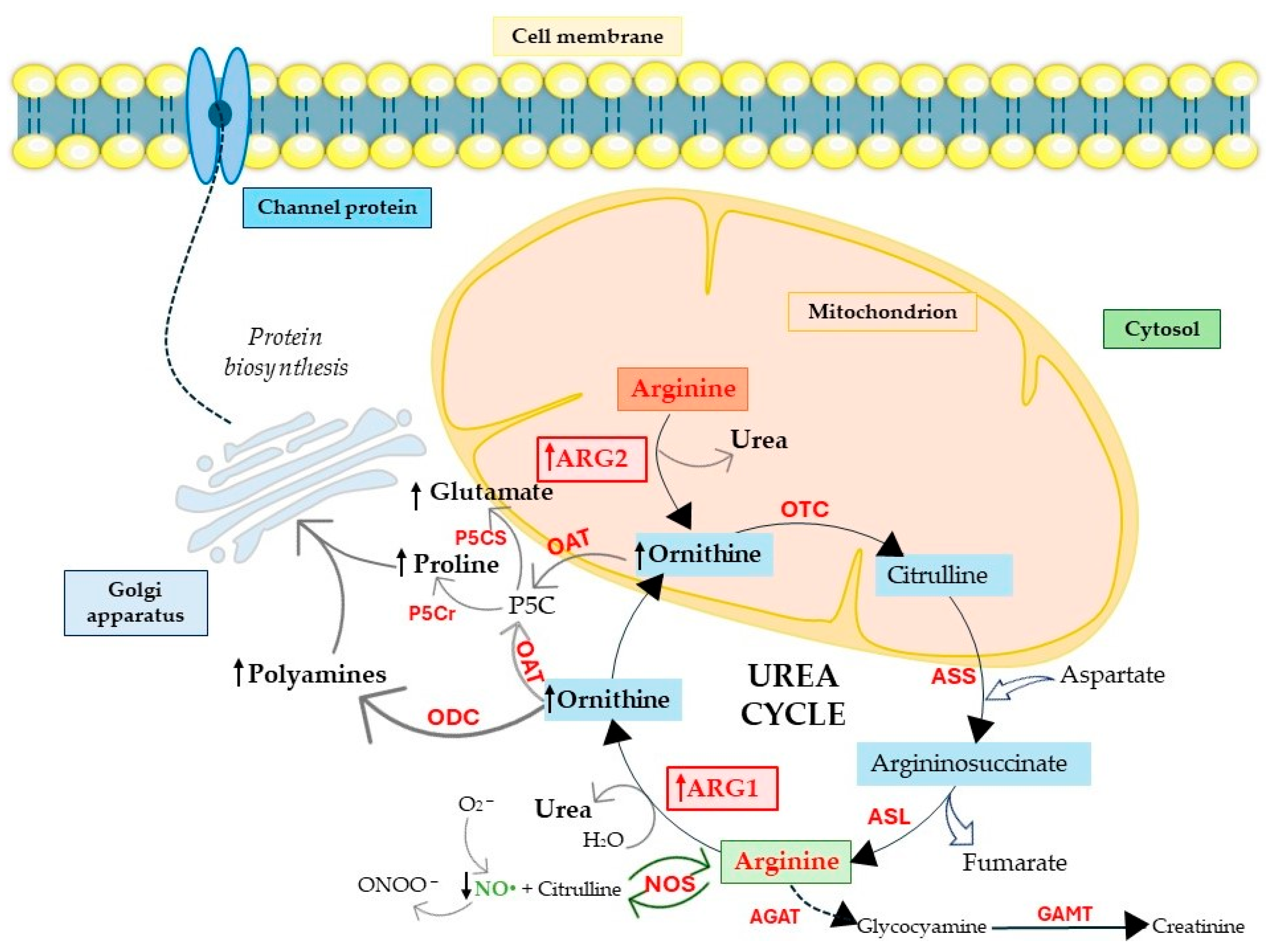
2. Arginases as Crucial Players in Physiology and Cancer Pathophysiology

2.1. Arginine Biosynthesis and Polyamine Production

Arginases, as enzymes of the urea cycle taking place in the liver, are primarily responsible for the catalysis of amino acids. In detail, ARG1 cleaves L-arginine into L-ornithine and urea, and this phenomenon allows for the removal of toxic ammonia in the last stage of the urea cycle [1,36,37,38]. However, in extrahepatic tissues, arginases are responsible for the hydrolysis of exo- and endogenous L-arginine [38]. L-ornithine obtained from the hydrolysis process can be metabolized in the decarboxylation reaction by ornithine decarboxylase (ODC, 4.1.1.17) to putrescine [37,38,39]. Putrescine is a substrate necessary for the synthesis of higher polyamines, such as spermine and spermidine [1,40,41]. Polyamines can also be produced from agmatine (endogenous guanido amine) through the activity of arginine decarboxylase (ADC, 4.1.1.4) [38]. In fact, polyamines are involved in the proper course of many metabolic processes; they are primarily responsible for cell growth and proliferation, as well as tissue repair [36,38,42]. As biogenic compounds, they stimulate the synthesis of phospholipids, ribonucleic acids (RNAs), deoxyribonucleic acids (DNAs), and proteins. Polyamines are components of cytoplasmic membranes and participate in the transport of metabolites and the regulation of ion channels [38,43]. Polyamines may also play an important role in carcinogenesis [38]. They are overexpressed in rapidly dividing cells such as cancer cells, e.g., in lung, breast, and colon cancer. This is related to the increased concentration of not only polyamines themselves but also genes and enzymes involved in their biosynthesis—including arginases [44,45,46]. López-Contreras et al. observed that polyamine levels were increased in non-small-cell lung cancer (NSCLC) with a change in arginine metabolism [47]. In the study conducted by Srivastava et al., stimulation of the ARG1 pathway was observed in head and neck squamous-cell carcinoma (HNSCC), as well as increased expression of ODC, which is essential for polyamine biosynthesis and promotes tumor growth [48]. The above studies indicate the pro-cancer characteristics of the increased activity of arginases and polyamine synthesis in various types of cancer [47,48,49].

2.2. Arginases and Proline and Glutamate Production

Further studies have indicated that arginases participate in the synthesis of proline and glutamate [1,38,50]. Mitochondrial arginase, i.e., ARG2, catalyzes reactions to produce L-ornithine, which in the mitochondria is transaminated to pyrroline-5-carboxylate with the participation of ornithine aminotransferase (OAT, 2.6.1.13) [38,50]. Proline is an important amino acid in collagen and is formed from pyrroline-5-carboxylate in the cytosol [38,50], playing an active role in shaping the TME [1,41]. Another amino acid produced from pyrroline-5-carboxylate is glutamate. Glutamate is a neurotransmitter and participates in many biochemical transformations. Its amide, i.e., glutamine, plays an important role in the body’s nitrogen metabolism [38,51]. L-ornithine, necessary for the synthesis of polyamines, is also a precursor for the synthesis of proline and glutamate. The inhibition of ARG2 activity would result in blocking the formation of L-ornithine from L-arginine in the arginase pathway and its transport from the cytosol to the mitochondrion—thus inhibiting the synthesis of both amino acids [38,50].

2.3. Arginases Control NO Production

Arginases regulate NO levels since L-arginine is the substrate for both ARG1, and ARG2, and can thus inhibit NO—a well-known endothelium-derived relaxing factor. NO modulates vascular tone, blood pressure, and hemodynamics; therefore, the dysregulation of NO signaling pathways contributes to the development of cardiovascular diseases but is also very relevant in cancer [8,36]. Increased levels of one of the arginases may cause nitric oxide synthase (NOS, EC 1.14.13.39) uncoupling, and this phenomenon is known as superoxide generation in cells. This event is related to reduction in NO generation and the enhanced production of O2 and ONOO [37,52,53,54,55,56]. Small amounts of L-arginine may also cause NOS uncoupling and the increased release of O2 [38,52,53,54,55,56]. Such damage caused by reactive nitrogen and oxygen species (RNOS) may contribute to cancer progression, which highlights the importance of a balance between these two enzymes [57]. The competition between the two catalytic enzymes, NOS and ARG1, is driven by the same cellular location in the cytosol. By contrast, ARG2 has a different intracellular localization compared to ARG1 and is manifested by a lower affinity for L-arginine than NOS [38,58,59]—important characteristics discriminating between the functions of ARG2 and ARG1. Finally, L-arginine can also serve as the sole substrate for NOS, which metabolizes arginine to NO and L-citrulline [36,38,60,61].
There are three different isoforms of NOS: inducible (iNOS), endothelial (eNOS), and neuronal (nNOS) [36,38]. To achieve full NO synthase activity, many cofactors are needed, such as flavin adenine dinucleotide (FAD), flavin mononucleotide (FMN), reduced nicotinamide adenine dinucleotide phosphate (NADPH2), molecular oxygen, tetrahydrobiopterin (BH4), and heme. The constitutive isoforms of NOS, i.e., nNOS and eNOS, produce small and short-lived amounts of NO, while iNOS is activated by various pro-inflammatory factors like interleukin (IL)-1, IL-6, interferon γ (INF-γ), macrophage migration inhibitory factor (MIF), and tumor necrosis factor α (TNF-α), as well as bacterial lipopolysaccharide (LPS), all of which produce large and long-lasting amounts of NO [5,51,60,61,62,63,64,65,66]. NO participates in many processes such as inflammation, thrombosis, regulates blood pressure, and neurotransmission, among others [62,67,68,69]. Enhanced arginase expression leads to a dysregulated production of NO and cancer progression. High levels of ARG2 and reduced levels of NO and iNOS were observed in prostate cancer cells, compared to non-cancerous tissues [70]. Inhibited NO production enhances angiogenesis and metastasis. Low NO concentrations result in the prevention of apoptosis and, subsequently, stimulation of the progression of cancer cells. At the molecular level NO stimulates signaling pathways such as mTOR, protein kinase B (PKB know as v-akt murine thymoma viral oncogene homolog 1, AKT), and extracellular-signal-regulated kinases (ERKs), which are necessary for the survival of cancer cells [41]. Conversely, a high concentration of NO causes cellular cytotoxicity and regulates the expression of one of the main DNA repair enzymes—DNA-dependent protein kinase catalytic subunit (DNA-PKcs)—and the expression of the p53 gene, which is critical for the apoptosis process and induces oxidative stress [71].

3. Cellular Consequences of Induced Arginase Expression

High levels of polyamines are observed in tissues that are characterized by high cell division activity, reflected by their presence in rapidly dividing cancer cells [72]. The TME consists of stromal and immune cells dependent on arginases, namely MDSCs, T and B cells, TAMs, and tumor-associated fibroblasts (CAFs) [1,73]. The microenvironment is important for cancer development and progression by interacting with nearby cells through the lymphatic and circulatory systems [74,75]. The overexpression of ARG1 and ARG2 in cancer impairs T cell function involving L-arginine, which is necessary for T cell differentiation and proliferation [76]. Vonderhaar et al. showed that increased ARG2 activity in the renal cell carcinoma (RCC) cell line depletes L-arginine and reduces CD3ζ expression. Moreover, the RCC cell line expressing ARG2 can modulate the L-arginine pathway and influence the function of T cells [77]. ARG1 is able to activate immunosuppressive cells such as MDSCs, which have the ability to suppress T cell activity and cause the escape of cancer cells from the immune response [78]. In melanoma, the overexpression of ARG2 in Treg increases their accumulation in tissues and suppressive capacity for the inhibition of mTOR signaling by the modulation of the PI3K/AKT/mTOR signaling pathway [1,20,79]. Enhanced activation of this pathway causes disturbances in cell proliferation and viability, which contributes to the formation of cancer and its further metastases (Figure 1). The regulation of arginases is quite complex. Chen et al. showed that ARG2 activation in pulmonary artery smooth muscle cells (PASMCs) was activated by the PI3K-AKT pathway [80]. AKT activation is followed by the phosphorylation of proteins such as caspase 9, Bad, glycogen synthase kinase-3 (GSK-3), nuclear factor-κB (NF-κB), p21Cip1, and p27 Kip1, which act as regulators of differentiation, proliferation, and apoptosis, as well as cell migration [81,82]. Similar to the AKT signaling pathway, mTOR kinase, which is a threonine-serine protein kinase, seems to modulate cell proliferation, growth, movement, and autophagy [1,81]. Yang et al. observed that ARG2 mediates the activation of mTORC1-S6K1 by myo-sin-IB (Myo1b), leading to lysosomal positioning, spatial separation, and the subsequent overactivation of the mTORC1-S6K1 signal, linking this phenomenon with aging and cell apoptosis [83]. In the most malignant thyroid tumors, ARG2 expression was increased compared to healthy tissues, and silencing the Arg2 gene decreased AKT expression, causing apoptosis and reducing the expression of cell proliferation [84]. The overexpression of ARG1 in neuroblastoma cells increases the phosphorylated levels of ERKs and AKT [1,85]. IL-6 and IL-8 stimulate the production and subsequent secretion of ARG1 from MDSCs by stimulating the PI3K-AKT pathway, causing the suppression of T cells, which contributes to tumor development (Table 1) [86]. STAT3 and MAPK can also be linked to arginase action and modulate tumor growth and progression [1,14,87]. STAT3 is involved in tumor proliferation, invasiveness, angiogenesis, and migration [88]. Phosphorylated STAT3 can bind to the human ARG1 promoter region in MDSCs and thus regulate the immunosuppression of these cells [34]. STAT3 inhibition also causes a reduction in ARG1 expression in patients with acute myeloid leukemia (AML), partially restoring T cell proliferation [89]. Moreover, ARG2 can promote cancer metastasis through the mitochondrial H2O2-STAT3 pathway in melanoma [90].

3.1. Apoptosis

Arginases can regulate the process of apoptosis in various cells [36,91]. Métayer et al. conducted a study in which they evaluated the mechanism of arginase-induced cell death in precursor B-cell lymphoblasts using the pre-B ALL 697 cell line. They observed that arginase induced the rapid apoptotic death of B-ALL leukemic cells through arginine depletion, without killing normal human lymphocytes. Additionally, they described that arginase induced the activation of caspases, leading to increased cleavage of the endogenous substrate poly(ADP-ribose) polymerase (PARP) and caspase-3; chromatin condensations and exposure to phosphatidylserine were observed, which indicated the process of apoptosis. The overexpression of BCL-2 inhibited arginase-induced cell death but did not affect arginase-induced cytostasis [92]. In vascular smooth muscle cells (VSMCs), ARG2 induces apoptosis through mitochondrial dysfunction by complex positive crosstalk among p66Shc, S6K1-JNK, ERKs, and p53 [14]. In cells defective in argininosuccinate synthase (ASS, EC 6.3.4.5) and ornithine transcarbamylase (OTC, EC 2.1.3.3), pegylated human recombinant arginase (rhARG) induced intracellular apoptosis by depleting L-arginine and generating reactive oxygen species (ROS) [93]. Additionally, ARG2 in intestinal endothelial cells (ECs) can alleviate extracellular apoptosis induced by inflammatory cytokines through the competitive depletion of L-arginine and, consequently, reduce NO production, where excess NO can also lead to apoptosis (Table 1) [94].

3.2. Cell Senescence

Cellular senescence is a state of permanent and irreversible cell cycle arrest that occurs in proliferating cells subjected to stress [36]. We can distinguish two types of senescence: accelerated and replicative [95]. In human umbilical vein endothelial cells (HUVECs), Zhu et al. observed that ARG1 promoted eNOS uncoupling and induced the expression of senescence markers p21Cip1 and p53-S15, as well as the number of senescence-associated beta-galactosidase (SA-β-gal)-positive cells without S6K1 activation. The above-mentioned data indicating the involvement of arginases in the process of cellular senescence may be related to ROS generation during the uncoupling of NOS (Table 1) [96]. Yepuri et al. revealed a mechanism of reciprocal positive regulation between ARG2 and S6K1 in endothelial inflammation and aging. In fact, silencing Arg2 in ECs suppressed senescence markers such as p53-S15, p21, and β-galactosidase, and it activated the expression of intercellular adhesion molecule-1 (ICAM1) and vascular adhesion molecule-1 (VCAM1) [13]. Xiong et al. demonstrated that ARG2, together with Myo1b, mediates the activation of mTORC1-S6K1, leading to lysosome redistribution and subsequent cell senescence in VSMCs [83]. Notably, in non-cancerous cells, ARG2 overexpression in keratinocytes increased β-Gal expression, which is associated with cellular senescence [97].

3.3. Autophagy

Autophagy is recognized as the controlled breakdown of chemical molecules and cell fragments to maintain intracellular homeostasis under conditions of stress [98]. Autophagy in ECs is important for maintaining their plasticity and redox homeostasis and for the regulation of NO production and angiogenesis [98]. ARG2, independently from L-arginine-urea hydrolase in ECs, can suppress cellular autophagy through the activation of mTORC2 and AKT-mTORC1-S6K1 signaling and can cause the inhibition of AMPK [12]. Zhang et al. observed in their study that rhARG in bladder cancer cells induced autophagy by activating the ROS-mediated AKT/mTOR signaling pathway [99]. In another study, Wang et al. reported that rhARG impairs macrophage immune functions, such as TNF-α and IL-6 production, phagocytosis, and the surface expression of MHC-II, which results in the suppression of immune functions in activated macrophages through an autophagy-mediated mechanism [100].

3.4. Vascularization

The arginase-driven conversion of L-arginine into L-ornithine creates polyamines, necessary for cancer proliferation [36], but also structural cells such as fibroblasts and endothelium. Induced ARG1 expression accelerated rat VSMC proliferation and neointima formation through increased polyamine synthesis [101], while polyamine overproduction in macrophages increased cardiac fibroblast (CF) proliferation and collagen production [102]. The increased production of ARG1 and ARG2 may also influence EC proliferation linked to wound healing and angiogenesis [103]. Hypoxia in human pulmonary microcirculation ECs through ARG2 overexpression accelerates the proliferation of these cells by the epidermal growth factor (EGF)–epidermal growth factor receptor (EGFR)–extracellular signal pathway [104]. L-arginine is also necessary for cell growth and for the proliferation of brain microvascular ECs [36]. Hepatic ischemia–reperfusion causes increased arginase activity, which depletes the amount of L-arginine, subsequently causing cell cycle arrest and, consequently, inhibition of the proliferation [105].

3.5. Immune Response and Inflammation

Arginases are also significantly involved in the inflammation process [36], caused by physical, chemical, or biological factors [106]. Chronic inflammation is involved in changes in blood vessels, leading to cardiovascular diseases such as myocardial infarction, atherosclerosis, or exacerbated heart failure (HF) [107]. As a result of this process, blood vessels dilate and become more permeable so that various plasma proteins can reach the affected tissue. When harmful factors stimulate the external barrier, dendritic cells (DCs), mast cells, and macrophages are activated, which phagocytize to the side of inflammation and then secrete inflammatory mediators [106,107]. ARG1 is considered an anti-inflammatory marker of the M2 phenotype of macrophages, suppressing their inflammatory response. ARG1 expression in macrophages can be downregulated by Fos-related antigen 1 or upregulated by lipoproteins, and, as shown, activated macrophages challenged with rhARG1 can inhibit their immune response [100,108]. Conversely, ARG2 is associated with the M1 phenotype of macrophages and therefore may have pro-inflammatory properties compared to ARG1 [36]. T cells are the major cell components of the adaptive immune system responsible for the cellular immune response. Their proliferation is impaired in the presence of low L-arginine concentrations, so the depletion of this amino acid is associated with impaired immune response to tumor-associated neoantigens. Gołąb et al. described that ovarian cancer cells (OvCa) release ARG1 into the TME by extracellular vesicles (EVs) and then by DCs acquire suppressive properties and inhibit T-cell proliferation. Blocking arginase activity can restore appropriate L-arginine levels and T cell proliferative and anti-tumor potential [109].
ARG2 in senescent ECs triggers the release of inflammatory cytokines and chemokines such as IL-6 and IL-8 and the induction of ICAM-1 and VCAM-1 through the activation of p38MAPK and S6K1 (Table 1) [13,87]. In line with this, Huang et al. observed in their study that ARG2 induced age-related adipose tissue inflammation and inflammatory cell recruitment by secreting IL-6 and activation of p38 MAPK pathway [110]. Xiong and colleagues observed ARG2-induced TNF-α release in acinar cells mediated by p38 MAPK signaling, leading to β-cell apoptosis and subsequent decreased insulin secretion and glucose intolerance [111]. Liu et al. also observed the reduced secretion of pro-inflammatory cytokines such as IL-6 and TNF-α in Arg2 knockout mice in bone marrow-derived macrophages, compared to WT macrophages [112]. Furthermore, Ming et al. found that the overexpression of ARG2 in macrophages promotes their pro-inflammatory response through mitochondrial ROS generation, increasing the expression of monocyte chemoattractant protein-1 and the production of TNF-α, as well as IL6, contributing to atherogenesis and insulin resistance [16]. However, in myeloid cells, L-arginine depletion suppresses the immune response of T cells through the action of arginases [60]. T cells require an appropriate amount of extracellular arginases for proper proliferation and functioning. An L-arginine deficiency abolishes the formation of an immunological synapse, which inhibits T cell receptor (TCR) signaling and blockage of the cell cycle of T cells in the G0–G1 phase [60]. T cells are unable to up-regulate cyclin kinase 4 (CDK4) and cyclin D3, which is associated with reduced phosphorylation of the Rb protein and the low binding of transcription factor E2F1 [113].
Taken together, we can conclude that ARG1 and ARG2 play an often opposite role in the regulation of immune cell functions and the inflammatory response, which has consequences in the pathogenesis of metabolic and cardiovascular diseases.

4. Arginases Regulate Metabolic and Mitochondria Machinery

4.1. ARG2 Regulates Oxidative Metabolism

Mitochondria are responsible for producing adenosine triphosphate (ATP) through oxidative phosphorylation [114]. These organelles are involved in the production of ROS, the regulation of Ca2+ homeostasis, and amino acid metabolism. Mitochondria maintain homeostasis through their dynamic cycles like biogenesis, fusion, and fission, as well as mitophagy, as a response to unfolded proteins [114,115]. Due to the fact that ARG2 is located in mitochondria, it plays the main role in mitochondrial functions. Mitochondrial ARG2 present in ECs can be activated by various stimuli such as thrombin, TNFα, H2O2, the EGF, and LPSs, and its expression can be reduced by compounds such as simvastatin, cocoa flavanols, and genistein [116]. Pandey et al. showed that oxidized low-density lipoprotein (oxLDL) can induce the activation of ARG2, which is regulated by mitochondrial processing peptidase (MPP). MPP cleaves N-terminal cleavable presequences and plays a role in the transport of mitochondrial proteins; therefore, it is responsible for the reverse translocation from mitochondria to the cytoplasm. In mitochondria, ARG2 reduces L-arginine production, causing eNOS uncoupling [116,117,118,119,120,121]. Subsequently, reduced synthase activity leads to reduced NO production and the overproduction of ROS and superoxides, which in turn lead to oxidative stress, which continuously activates mitochondrial-to-cytoplasmic translocation by MPP and ARG2 activation (Table 1).
ROS also play a key role in regulating the oxidation of lipoproteins, including oxLDL, which in turn induce ARG2 by ROCK and MPP, and thus forms a feedback loop [116,117,118,119,120,121]. Hwang et al. described how eNOS can be regulated by various extracellular signals, e.g., by Ca2+, which in mitochondria regulates ATP synthesis and the rate of oxidative phosphorylation. It has been reported that ARG2 can regulate both mitochondrial and cytolytic Ca2+ concentration. ARG2, together with the p32 protein in endothelial mitochondria, played an important role in modulating intracellular NO, ROS, and Ca2+ concentrations [122]. Zhang et al. in their study specified that ARG2 present in hepatocytes induced mitochondrial ureahydrolysis and drove hepatic oxidative metabolism, leading to glucose oxidation in mammals [123]. ARG2 present in hepatocytes enhances peripheral and hepatic AKT phosphorylation, which increases peripheral energy expenditure and inhibits hepatic steatosis, regardless of the hydrolytic activity. Mitochondrial ARG2 is able to control glycemia in the obese state driving glucose oxidation. Lim et al., in their study, confirmed that in murine endothelium, ARG2 was mainly confined to mitochondria regulating NO production and vascular endothelial function by modulating eNOS activity [124]. Mitochondria are also key organelles for the polarization of macrophages. M1 macrophages, through the expression of NOS, are responsible for the synthesis of arginine into NO and citrulline. By contrast, M2 macrophages express more ARG1, which hydrolyzes arginine to urea and ornithine [125]. Uchida et al. used mice with acute kidney injury (AKI) induced by cisplatin to test whether ARG2 could be a therapeutic target for AKI. Cisplastin-treated Arg2 knockout mice had significant improvement in renal dysfunction through a characteristic process of apoptosis and an apparent reduction in acute tubular injury compared to WT mice [126]. LPS administration increases the mitochondrial membrane potential, reducing ROS secretion dependent on ARG2 [126]. In summary, ARG2 expression correlates with the inflammatory response of macrophages through the secretion of ROS and, consequently, the exacerbation of AKI [126].

4.2. ARG2’s Specific Role in Mitochondrial Function

Accumulating evidence has highlighted the essential role of ARG2 in the regulation of cancer progression, suggesting that the inhibition of ARG2 may lead to clinical benefits for patients with cancer. The role of ARG2 in pancreatic cancer was suggested by Zaytouni et al., who observed that the silencing or loss of Arg2 suppresses obesity-driven pancreatic cancer development [127]. Líndez et al. demonstrated how mitochondrial ARG2 determines the activity of CD8+ T cells. The adoptive transfer of CD8+ T cells with Arg2 knockout reduced tumor growth in murine model of melanoma and colon cancer. The anti-cancer function of CD8+ T cells with Arg2 knockout is related to enhanced cytotoxicity, memory formation, and the persistence of anti-cancer CD8+ T cell responses [21]. The above-mentioned events are independent from ARG1 presence. The significance of ARG2 is fundamental not only for cancers but also for inflammation, especially for macrophage function. Hardbower et al. reported that the knockout of Arg2 in Helicobacter pylori-infected mice led to increased regulation of polyamine metabolism and the activation of M1 macrophages, which is associated with increased gastritis. ARG2 may play an inflammasome function in macrophages, as pro-IL-1β cleavage into mature IL-1β [128]. Data obtained by Dowling et al. documented that ARG2 expression is mediated by IL-10/microRNA-155 axis in inflammatory macrophages, and this phenomenon affects the dynamics and oxidative respiration of mitochondria and determines the inflammatory stage of macrophages [129]. The functions of arginases in metabolic and mitochondrial mechanisms indicate that both enzymes play key roles in proper cellular functioning and are involved in the pathophysiology of many diseases; therefore, it is important to maintain an appropriate balance, especially between arginases and NO, which compete together for arginine (Figure 2).

5. Significance of Arginases in Non-Cancer Diseases

The function of arginases is well documented in human pathophysiology, and their disturbed expression and action are observed in many diseases. Numerous studies have observed the link between arginases, NOS, and NO in cardiovascular diseases [37]. Decreased NO production contributes to the increased adhesion potential of leukocytes to the endothelium, impaired vasorelaxation, and the deposition of L-ornithine, which is related to fibrosis and the stiffening of vessels [37,130]. In the case of hypertension, increased arginase activity, decreased amounts of NO, L-arginine, and BH4, and increased O2 production cause impaired endothelium-dependent vascular relaxation [131,132]. Increased ARG2 activity has also been observed in pulmonary arterial hypertension (PAH). The increased protein level of ARG2 attenuates the endothelium-dependent dilation of pulmonary segments in the presented pulmonary embolism model [133,134].
Increased ARG1 and a decrease level of NO is observed in sickle cell disease (SCD), where during hemolysis, red blood cells (RBCs) release large amounts of ARG1, which contributes to reduction in NO and L-arginine and increased levels of L-ornithine [135,136,137]. The activation of ECs, reduced amount of NO, and the deposition of sickled RBCs in capillaries causes the pathology to deepen.
It has been shown that arginases may also be key factors causing cardiovascular problems in diabetic patients [37]. Chandra et al. presented data showing that diabetes or hyperglycemia may increase the level of arginases in aortic ECs through the Ras homolog family member A (RhoA)/ Rho-associated protein kinase (ROCK)/MAPK pathway [138]. The activation of ARG1 in RBCs in patients with type 2 diabetes causes dysfunction in the endothelium of healthy arteries through a ROS-dependent mechanism [139].
Most studies have linked the increased activation of arginases with neurological diseases such as AD, multiple sclerosis (MS), Parkinson’s disease (PD), stroke, and traumatic brain injury [37,140]. Both ARG1 and ARG2 are expressed in neurons and alter NO signaling, which is the main cause of neurological disorders and brain damage [2]. In the case of long-term cerebral ischemia, NO is synthesized with the contribution of iNOS, which is activated by released cytokines [38]. NO, which enters synapses, is responsible for the increased secretion of glutamate and Ca2+, which then leads to the promotion of apoptosis or necrosis of nerve cells [38,141].
Zimmerman et al. noted a particular increase of the Arg1 gene expression in lung tissues of mice with an experimental asthma model [142]. Nevertheless, Ceylan et al. observed that the L-arginine/NO pathway is involved in the pathophysiology of asthma [143]. Pawliczak and Lewandowicz reported that both IL-4 and IL-13 strongly induced arginase expression in the lungs of patients with asthma [144]. Arginases as precursors of polyamines and proline modulate the activity of NO synthase, and this event is involved in the remodeling of the airways in inflammation-induced asthma [38,145]. Additionally, it is worth noting that phenotypes of mice with liver-specific KO of Arg1 and phenotypes of mice with total-body KO of Arg1 are similar to each other and in both cases are lethal [146,147]. The increased activity of ARG1 and its enhanced expression is associated with the release of cytokines, catecholamines, and the transforming growth factor β (TGF-β), and this results in the down-regulation of citrulline, arginine, and NO production [38,148]. Increased levels of arginases and L-ornithine, which are necessary for the synthesis of polyamines and proline, may play a significant role in the wound-healing process, so the overexpression of arginases can also be observed in injuries, exactly in macrophages, which occur in increased amounts in the area of damaged tissues [1,38,51,148].

6. Clinical Perspectives of Arginase Inhibitors

6.1. Arginase Vaccines and Pegylated rhARG in Clinical Trials

For some time now, scientists have begun to develop immunomodulatory vaccines targeting arginases to improve the anti-cancer response in cancer immunotherapy [1]. Andersen and other colleagues conducted preclinical trials where immunogenic peptides derived from the ARG1 protein sequence were used and noted that these peptides are able to activate T cell-specific ARG1 and suppress ARG1-expressing malignant cells [149,150,151]. In phase I of a clinical trial (NCT03689192), a vaccine containing ARG1 peptides was tested in combination with immune checkpoint inhibitors, i.e., anti-programmed death receptor 1 (PD-1). Vaccines were administered subcutaneously to patients every third week. The study used enzyme-linked immunosorbent assays and intracellular cytokine staining to assess the vaccine-specific immune response. It was documented that co-administration of ARG1 vaccine with anti-PD-1 antibodies created a pro-inflammatory microenvironment and changed the M1/M2 macrophage ratio in the tumor, reducing tumor suppressive immunity [152,153]. Adverse events reported in the study included injection site reactions and shoulder arthralgia [152,153]. There was a risk of inducing autoimmune reactions in patients due to the fact that ARG1 is expressed in the liver and other non-cancerous tissues. However, increased transaminase levels were noted only in patients with progressing metastatic lesions in the liver [152,153]. The same group developed a study using 34 peptides covering the entire ARG2 sequence and assessed the reactivity of these peptides in peripheral blood mononuclear cells (PBMCs) obtained from healthy donors and cancer patients. In a Lewis lung cancer tumor model, they found that vaccines with these peptides significantly inhibited tumor growth by activating ARG2-specific T cell response, which may constitute a future potential for a new therapeutic candidate in immunotherapy [154]. Chan et al. showed that the growth of hepatocellular carcinoma (HCC) depends on exogenous arginine, which cannot be hydrolyzed due to the weak expression of ASS and OTC in these cells. This group in phase II of the clinical trial (NCT01092091) verified pegylated rhARG1 (PEG-BCT-100) as an arginine-depleting drug, which has potential in the treatment of advanced HCC [155]. PEG-BCT-100 was administered intravenously to patients at a dose of 2.7 mg/kg weekly [155]. Adverse events after the administration of PEG-BCT-100 reported by patients included limb edema, fatigue, constipation, and anemia [155]. Chiang et al. also conducted a study (NCT01551628) in patients with relapsed or refractory leukemia or lymphoma using rhARG1 to show that the drug was safe and effective, but the final report from the study is currently unavailable. Zhang et al. conducted a study on bladder cancer cell lines, establishing that rhARG1 (BCT-100) induced autophagy and apoptosis through the ROS-activated AKT/mTOR signaling pathway, exhibiting anticancer effects [99]. The use of rhARG1 may prove to be a therapy for some types of cancer in the future.

6.2. Naturally Occurring Arginase Inhibitors

Inhibitors of arginases can be divided into two classes, naturally occurring compounds and synthetic compounds. Naturally occurring arginase inhibitors include chlorogenic acid (CA) and piceatannol (PIC), and synthetic compounds include N(ω)-hydroxy-l-arginine (NOHA) and its analogs, such as S-(2-boronoethyl)-L-cysteine (BEC), 2(S)-amino-6-boronohexanoic acid (ABH), L-norvaline, and CB-1158 [1]. PIC is a polyphenol and belongs to the natural inhibitors of arginases [156]. Its effect on mammalian ARG1 was determined to be an IC50 of 12.1 µM [157]. Song et al. reported that PIC, through pathways related to p38, p-STAT3, NF-ĸB, HIF-1α, and c-Myc, induced apoptosis and limited the growth and metastasis of breast cancer [158]. From the cellular point of view, PIC induces apoptosis and arrests the cell cycle in the G0/G1 phase by inhibiting cyclin-dependent kinase (CDK) activity. This event led to reduction in bladder cancer cell proliferation [159]. Its anti-cancer properties have also been reported in lung cancer, apart from and in combination with gemcitabine [160,161]. CA inhibited ARG1 in mammals with IC50 of 10.6 μM [157]. CA is able to induce apoptosis, and its anti-cancer function is linked to antioxidant properties [162,163,164]. These compounds, however, show low potential for arginase inhibition and a lack of specificity. Nevertheless, the CA structure may be a promising starting point for creating new and more potent inhibitors of arginases.

6.3. Synthetic Arginase Inhibitors

Previously identified and developed inhibitors of arginases, such as norvaline as a substrate for aminotransferases, had low potency and, therefore, side effects due to the high concentrations required [165]. α-difluoromethylornithine (DFMO) is a potent irreversible inhibitor of ODC; however, no significant effect on arginase activity has been determined [166]. NOHA and nor-NOHA belong to the group of hydroxy arginine derivatives; both are competitive and reversible inhibitors of arginases. NOHA is produced during the breakdown of L-arginine into NO and L-citrulline using NOS. nor-NOHA is an inhibitor with a longer half-life than NOHA; the Kd of nor-NOHA is 0.517 µM, while for NOHA it is 3.6 µM for human ARG1. The Ki for nor-NOHA against ARG2 is 51 nM, and for NOHA the Ki is 1.6 μM [167,168]. The inhibition of arginases by NOHA has pro-apoptotic and anti-proliferative effects in colorectal and breast cancer cells [169,170]. Zhu et al. showed that the use of nor-NOHA inhibited ARG1 in HepG2 cells, induced the apoptosis process, and inhibited the migratory and invasive abilities of these cells [171]. Ludwig et al. demonstrated that nor-NOHA, by reversing the tumor-growth-promoting effect of ARG1 by TAM-derived exosomes, may play a significant therapeutic role in the treatment of glioblastoma [172]. Alonso et al., using murine ovarian cancer, reported that the application of nor-NOHA blocked ARG1 activity and reversed the immunosuppression of vascular leukocytes (VLCs) in ovarian cancer [79]. In line with this, Javrushyan and his colleagues reported that the administration of nor-NOHA to rats with a 7,12-dimethylbenzathracene-induced mammary tumor reduced the size and number of these tumors [49]. In phase II of clinical trials, nor-NOHA was tested in the treatment of cardiovascular diseases. The use of nor-NOHA improved the function of the microvascular endothelium in patients with type 2 diabetes mellitus (T2DM, NCT02687152) and in patients with coronary artery disease (CAD). Additionally, it protected against endothelial dysfunction after ischemia–reperfusion (NCT02009527, Table 2) [173,174]. Finally, nor-NOHA reduced the development of atherosclerosis and hypertension [175]; however, poor specificity for the isoforms of both enzymes still limits its use [1,168]. Currently, no clinical trials using NOHA or nor-NOHA are ongoing, and the last clinical trial data on these compounds were reported in 2023.
ABH and BEC are further boronic acid derivatives of arginase inhibitors that can bind to the active manganese site of arginases. Both inhibitors can competitively inhibit arginase activity. The IC50 of ABH is 1.54 µM for human ARG1 and 2.55 µM for human ARG2 [176]. BEC is a cysteine-based ABH analog, its Kd for human ARG1 is determined to be 270 nM, and the Kd for ARG2 is determined to be 220 nM [58,177,178]. It has been reported that the inhibition value of both these inhibitors is dependent on the pH level, and the affinity for both arginases is different [179]. The selectivity of ABH is lower towards ARG1 compared to ARG2 [180]. Abdelkawy et al. determined that both compounds are characterized by low bioavailability, and only 5% of BEC and ABH is detectable after single oral administration, while after intraperitoneal multiple-dose administration, bioavailability increased to 50% [181]. In Fischer rats, oral administration of ABH at a dose of 400 mg per day for 25 days did not lead to toxicity and side effects [182]. However, it was reported that compounds with a boronic acid head group achieved unacceptable toxicity to normal cells [182]. The above-mentioned data strongly suggest that further research is needed to determine the appropriate safe dose, as well as the structural optimization of ABH and BEC to make them promising candidates for clinical use in cancer therapy.
The arginase inhibitor, CB-280, was tested in phase I of clinical trial in patients with cystic fibrosis (CF) (NCT04279769, Table 2). In this study, the authors examined the safety, pharmacokinetics, pharmacodynamics, and biological activity of CB-280. They predicted that CB-280 will be able to inhibit arginases, thereby restoring endogenous NO production, leading to improved respiratory function, as well as antimicrobial response in CF patients, but the results are still not available [183]. Numidargistat (CB-1158) developed by Calithera Biosciences is a small molecule and ARG1 inhibitor, administrated orally, with an IC50 of 98 nM (Table 2) [184,185]. Steggerda et al. showed that CB-1158 inhibited ARG1, creating a pro-inflammatory environment, reducing tumor growth and reversing the immunosuppression of TME [33]. CB-1158, in combination with other chemotherapeutics and/or immunotherapies, may provide clinical benefits for patients. NCT03314935 and NCT02903914 were clinical trials where CB-1158 was investigated as a single agent, as well as in combination with immune checkpoint inhibitors, i.e., pembrolizumab or other chemotherapeutic agents, in the treatment of advanced and metastatic solid tumors [186,187,188].
Currently, in phase I of clinical trials is OATD-02, developed by Molecure S.A. (NCT05759923, Table 2). OATD-02 is a boronic acid derivative and a potent dual inhibitor of ARG1 and ARG2, with an IC50 of 20 nM and 48 nM, respectively (Figure 3) [189]. Additionally, this is the first study to confirm the benefits of ARG2 inhibition in cancer treatment. Błaszczyk et al. and Borek et al. have demonstrated the anti-cancer and immunomodulatory efficacy of OATD-02 in animal models, as well as its moderate bioavailability and long half-life after oral administration [17,189]. OATD-02 differs primarily from CB-1158 in potency, is characterized by better arginase selectivity, and showed greater anti-tumor activity in in vivo studies [17]. In a study using the syngeneic Renca model, OATD-02 reduced regulatory T cells (Treg) infiltration, thereby improving the CD8+/Treg ratio [17], and inhibited the proliferation of K562 human leukemic cells [17]. The anti-cancer efficiency of OATD-02 has also been reported in xenografts induced by GL261 glioma, B16F10 melanoma, CT26 colorectal adenocarcinoma, Lewis lung cancer, and ID8 ovarian cell administration [17,189,190,191,192]. Błaszczyk et al. observed that OATD-02 reduced glioma growth by modifying the TME and increased the anti-cancer effect of anti-PD-1 antibodies, affecting the response of NK and myeloid cells [17,191]. Grzybowski et al. conducted a study using an OATD-02 inhibitor in combination therapy with a STING agonist (DMXAA) and immune checkpoint inhibitors, thereby improving the anti-tumor response [17,190]. OATD-02 may demonstrate effectiveness in the treatment of immune-related cancers and in cancers with enhanced ARG2 expression, such as pancreatic cancer, chronic and acute myeloid leukemia, kidney cancer, and in patients with prostate cancer [17,77,127,193,194,195]. It can therefore be concluded that OATD-02, as the dual arginase inhibitor, may improve anti-cancer therapy, which seems to be linked to the inhibition of cellular arginases, ARG2-dependent metabolic adaptations, and ARG1 located in EVs.

7. Conclusions

Arginases, as enzymes involved in the hydrolytic breakdown of arginine to ornithine and urea, serve important functions in cellular metabolism. The deregulation of ARG1 and ARG2 activity are key in the initiation and development of many human diseases. From a functional point of view, the competition of arginases with NOS for L-arginine, as well as their participation in proline synthesis, may play a crucial role in diseases such as injuries, inflammation, stroke, and cardiovascular diseases. Moreover, arginase-mediated synthesis of polyamines is directly associated with tumorigenesis and cancer development.
Arginases regulate cancer cell proliferation, apoptosis, and migration and affect immunosuppression involving not only L-arginine metabolism but also numerous other signaling pathways. Accumulating studies indicate that the finding of an appropriate candidate that inhibits the activity of ARG1 and ARG2 may be crucial in the therapy for many human diseases. Recently, numerous clinical trials have been developed addressing inhibitors of arginases for the treatment of cancer, such as CB-1158 and OATD-02. Based on the homology of the enzymatic active sites of human ARG1 and ARG2, which is quite high, finding a specific inhibitor targeting a specific isoform is challenging. Moreover, further clinical trials have to be conducted to help determine the effects of the long-term use of inhibitors of arginases in patients.

Author Contributions

Conceptualization, P.M.-A., D.J. and Z.Z.; writing—original draft preparation, P.M.-A. and D.J.; writing—review and editing, P.M.-A., D.J. and Z.Z.; visualization, P.M.-A.; supervision, Z.Z. All authors have read and agreed to the published version of the manuscript.

Funding

This research was funded by the project DIMUNO: “Development of new cancer therapies based on selective antitumor immunomodulators” (STRATEGMED2/265503/3/NCBR/15)—co-financed by the National Centre for Research and Development in the framework of STRATEGMED Program and Pre-clinical and clinical development of arginase inhibitor for application in anticancer immunotherapy (POIR.01.01.01-00-0415/17), acronym ARG, co-financed by the European Union in the framework of European Funds Smart Growth and European Regional Development Fund.

Conflicts of Interest

The authors are employers of the biotech company Molecure S.A.

References

  1. Niu, F.; Yu, Y.; Li, Z.; Ren, Y.; Li, Z.; Ye, Q.; Liu, P.; Ji, C.; Qian, L.; Xiong, Y. Arginase: An emerging and promising therapeutic target for cancer treatment. Biomed. Pharmacother. 2022, 149, 112840. [Google Scholar] [CrossRef]
  2. Toque, H.A.; Narayanan, S.P.; Caldwell, R.W. Arginase: An old enzyme with new tricks. Trends Pharmacol. Sci. 2015, 36, 395–405. [Google Scholar] [CrossRef]
  3. Das, P.; Lahiri, A.; Lahiri, A.; Chakravortty, D. Modulation of the Arginase Pathway in the Context of Microbial Pathogenesis: A Metabolic Enzyme Moonlighting as an Immune Modulator. PLoS Pathog. 2010, 6, e1000899. [Google Scholar] [CrossRef] [PubMed]
  4. Ash, D.E. Structure and Function of Arginases 1,2. J. Nutr. 2004, 134, 2760S–2764S. [Google Scholar] [CrossRef] [PubMed]
  5. Wu, G.; Morris, S.M., Jr. Arginine metabolism: Nitric oxide and beyond. Biochem. J. 1998, 336 Pt 1, 1–17. [Google Scholar] [CrossRef] [PubMed]
  6. Jenkinson, C.P.; Grody, W.W.; Cederbaum, S.D. Comparative properties of arginases. Comp. Biochem. Physiol. Part B Biochem. Mol. Biol. 1996, 114, 107–132. [Google Scholar] [CrossRef] [PubMed]
  7. Cederbaum, S.D.; Yu, H.; Grody, W.W.; Kern, R.M.; Yoo, P.; Iyer, R.K. Arginases I and II: Do their functions overlap? Mol. Genet. Metab. 2004, 81 (Suppl 1), S38–S44. [Google Scholar] [CrossRef]
  8. Bhatta, A.; Yao, L.; Xu, Z.; Toque, H.A.; Chen, J.; Atawia, R.T.; Fouda, A.Y.; Bagi, Z.; Lucas, R.; Caldwell, R.B.; et al. Obesity-induced vascular dysfunction and arterial stiffening requires endothelial cell arginase 1. Cardiovasc. Res. 2017, 113, 1664–1676. [Google Scholar] [CrossRef]
  9. Savelieva, O.N.; Karunas, A.S.; Fedorova, Y.Y.; Murzina, R.R.; Savelieva, A.N.; Gatiyatullin, R.F.; Etkina, E.I.; Khusnutdinova, E.K. The role of polymorphic variants of arginase genes (ARG1, ARG2) involved in beta-2-agonist metabolism in the development and course of asthma. Vavilov J. Genet. Breed. 2020, 24, 391–398. [Google Scholar] [CrossRef]
  10. Marselli, L.; Bosi, E.; De Luca, C.; Del Guerra, S.; Tesi, M.; Suleiman, M.; Marchetti, P. Arginase 2 and Polyamines in Human Pancreatic Beta Cells: Possible Role in the Pathogenesis of Type 2 Diabetes. Int. J. Mol. Sci. 2021, 22, 12099. [Google Scholar] [CrossRef]
  11. Tian, Y.; Du, W.; Cao, S.; Wu, Y.; Dong, N.; Wang, Y.; Xu, Y. Systematic analyses of glutamine and glutamate metabolisms across different cancer types. Chin. J. Cancer 2017, 36, 88. [Google Scholar] [CrossRef] [PubMed]
  12. Xiong, Y.; Yepuri, G.; Forbiteh, M.; Yu, Y.; Montani, J.-P.; Yang, Z.; Ming, X.-F. ARG2 impairs endothelial autophagy through regulation of MTOR and PRKAA/AMPK signaling in advanced atherosclerosis. Autophagy 2014, 10, 2223–2238. [Google Scholar] [CrossRef] [PubMed]
  13. Yepuri, G.; Velagapudi, S.; Xiong, Y.; Rajapakse, A.G.; Montani, J.; Ming, X.; Yang, Z. Positive crosstalk between arginase-II and S6K1 in vascular endothelial inflammation and aging. Aging Cell 2012, 11, 1005–1016. [Google Scholar] [CrossRef] [PubMed]
  14. Xiong, Y.; Yu, Y.; Montani, J.-P.; Yang, Z.; Ming, X.-F. Arginase-II Induces Vascular Smooth Muscle Cell Senescence and Apoptosis Through p66Shc and p53 Independently of Its l -Arginine Ureahydrolase Activity: Implications for Atherosclerotic Plaque Vulnerability. J. Am. Hear. Assoc. 2013, 2, e000096. [Google Scholar] [CrossRef] [PubMed]
  15. Monticelli, L.A.; Buck, M.D.; Flamar, A.L.; Saenz, S.A.; Wojno, E.D.T.; Yudanin, N.A.; Osborne, L.C.; Hepworth, M.R.; Tran, S.V.; Rodewald, H.R.; et al. Arginase 1 is an innate lymphoid-cell-intrinsic metabolic checkpoint controlling type 2 inflammation. Nat. Immunol. 2016, 17, 656–665. [Google Scholar] [CrossRef]
  16. Ming, X.; Rajapakse, A.G.; Yepuri, G.; Xiong, Y.; Carvas, J.M.; Ruffieux, J.; Scerri, I.; Wu, Z.; Popp, K.; Li, J.; et al. Arginase II Promotes Macrophage Inflammatory Responses through Mitochondrial Reactive Oxygen Species, Contributing to Insulin Resistance and Atherogenesis. J. Am. Hear. Assoc. 2012, 1, e000992. [Google Scholar] [CrossRef]
  17. Grzybowski, M.M.; Stańczak, P.S.; Pomper, P.; Błaszczyk, R.; Borek, B.; Gzik, A.; Nowicka, J.; Jędrzejczak, K.; Brzezińska, J.; Rejczak, T.; et al. OATD-02 Validates the Benefits of Pharmacological Inhibition of Arginase 1 and 2 in Cancer. Cancers 2022, 14, 3967. [Google Scholar] [CrossRef]
  18. Groth, C.; Hu, X.; Weber, R.; Fleming, V.; Altevogt, P.; Utikal, J.; Umansky, V. Immunosuppression mediated by myeloid-derived suppressor cells (MDSCs) during tumour progression. Br. J. Cancer 2018, 120, 16–25. [Google Scholar] [CrossRef]
  19. Sullivan, M.R.; Danai, L.V.; Lewis, C.A.; Chan, S.H.; Gui, D.Y.; Kunchok, T.; Dennstedt, E.A.; Vander Heiden, M.G.; Muir, A. Quantification of microenvironmental metabolites in murine cancers reveals determinants of tumor nutrient availability. eLife 2019, 8, e44235. [Google Scholar] [CrossRef]
  20. Lowe, M.M.; Boothby, I.; Clancy, S.; Ahn, R.S.; Liao, W.; Nguyen, D.N.; Schumann, K.; Marson, A.; Mahuron, K.M.; Kingsbury, G.A.; et al. Regulatory T cells use arginase 2 to enhance their metabolic fitness in tissues. J. Clin. Investig. 2019, 4. [Google Scholar] [CrossRef]
  21. Líndez, A.-A.M.; Dunand-Sauthier, I.; Conti, M.; Gobet, F.; Núñez, N.; Hannich, J.T.; Riezman, H.; Geiger, R.; Piersigilli, A.; Hahn, K.; et al. Mitochondrial arginase-2 is a cell-autonomous regulator of CD8+ T cell function and antitumor efficacy. J. Clin. Investig. 2019, 4. [Google Scholar] [CrossRef] [PubMed]
  22. Berkowitz, D.E.; White, R.; Li, D.; Minhas, K.M.; Cernetich, A.; Kim, S.; Burke, S.; Shoukas, A.A.; Nyhan, D.; Champion, H.C.; et al. Arginase Reciprocally Regulates Nitric Oxide Synthase Activity and Contributes to Endothelial Dysfunction in Aging Blood Vessels. Circulation 2003, 108, 2000–2006. [Google Scholar] [CrossRef] [PubMed]
  23. Wiesinger, H. Arginine metabolism and the synthesis of nitric oxide in the nervous system. Prog. Neurobiol. 2001, 64, 365–391. [Google Scholar] [CrossRef]
  24. Liu, P.; Fleete, M.S.; Jing, Y.; Collie, N.D.; Curtis, M.A.; Waldvogel, H.J.; Faull RAbraham, W.C.; Zhang, H. Altered arginine metabolism in Alzheimer’s disease brains. Neurobiol. Aging 2014, 35, 1992–2003. [Google Scholar] [CrossRef] [PubMed]
  25. Grzywa, T.M.; Sosnowska, A.; Matryba, P.; Rydzynska, Z.; Jasinski, M.; Nowis, D.; Golab, J. Myeloid Cell-Derived Arginase in Cancer Immune Response. Front. Immunol. 2020, 11, 938. [Google Scholar] [CrossRef] [PubMed]
  26. Polis, B.; Samson, A.O. Arginase as a Potential Target in the Treatment of Alzheimer’s Disease. Adv. Alzheimer’s Dis. 2018, 7, 119–140. [Google Scholar] [CrossRef]
  27. Roci, I.; Watrous, J.D.; Lagerborg, K.A.; Lafranchi, L.; Lindqvist, A.; Jain, M.; Nilsson, R. Mapping Metabolic Events in the Cancer Cell Cycle Reveals Arginine Catabolism in the Committed SG2M Phase. Cell Rep. 2019, 26, 1691–1700.e5. [Google Scholar] [CrossRef]
  28. Wu, C.W.; Chi, C.W.; Lin, E.C.; Lui, W.Y.; P’eng, F.K.; Wang, S.R. Serum arginase level in patients with gastric cancer. J. Clin. Gastroenterol. 1994, 18, 84–85. [Google Scholar] [CrossRef]
  29. Leu, S.Y.; Wang, S.R. Clinical significance of arginase in colorectal cancer. Cancer 1992, 70, 733–736. [Google Scholar] [CrossRef]
  30. Sovová, V.; Sloncová; Fric, P. Differences of alkaline phosphatase and arginase activities in human colorectal carcinoma cell lines. Folia Biol. 1997, 43, 101–104. [Google Scholar]
  31. Chrzanowska, A.; Graboń, W.; Mielczarek-Puta, M.; Barańczyk-Kuźma, A. Significance of arginase determination in body fluids of patients with hepatocellular carcinoma and liver cirrhosis before and after surgical treatment. Clin. Biochem. 2014, 47, 1056–1059. [Google Scholar] [CrossRef] [PubMed]
  32. Clemente, G.S.; van Waarde, A.; Antunes, I.F.; Dömling, A.; Elsinga, P.H. Arginase as a Potential Biomarker of Disease Progression: A Molecular Imaging Perspective. Int. J. Mol. Sci. 2020, 21, 5291. [Google Scholar] [CrossRef] [PubMed]
  33. Steggerda, S.M.; Bennett, M.K.; Chen, J.; Emberley, E.; Huang, T.; Janes, J.R.; Li, W.; MacKinnon, A.L.; Makkouk, A.; Marguier, G.; et al. Inhibition of arginase by CB-1158 blocks myeloid cell-mediated immune suppression in the tumor microenvironment. J. Immunother. Cancer 2017, 5, 101. [Google Scholar] [CrossRef] [PubMed]
  34. Vasquez-Dunddel, D.; Pan, F.; Zeng, Q.; Gorbounov, M.; Albesiano, E.; Fu, J.; Blosser, R.L.; Tam, A.J.; Bruno, T.; Zhang, H.; et al. STAT3 regulates arginase-I in myeloid-derived suppressor cells from cancer patients. J. Clin. Investig. 2013, 123, 1580–1589. [Google Scholar] [CrossRef]
  35. Toque, H.A.; Romero, M.J.; Tostes, R.C.; Shatanawi, A.; Chandra, S.; Carneiro, Z.N.; Inscho, E.W.; Webb, R.C.; Caldwell, R.B.; Caldwell, R.W. p38 Mitogen-Activated Protein Kinase (MAPK) Increases Arginase Activity and Contributes to Endothelial Dysfunction in Corpora Cavernosa from Angiotensin-II-Treated Mice. J. Sex. Med. 2010, 7, 3857–3867. [Google Scholar] [CrossRef]
  36. Li, Z.; Wang, L.; Ren, Y.; Huang, Y.; Liu, W.; Lv, Z.; Qian, L.; Yu, Y.; Xiong, Y. Arginase: Shedding light on the mechanisms and opportunities in cardiovascular diseases. Cell Death Discov. 2022, 8, 1–14. [Google Scholar] [CrossRef]
  37. Caldwell, R.W.; Rodriguez, P.C.; Toque, H.A.; Narayanan, S.P. Arginase: A Multifaceted Enzyme Important in Health and Disease. Physiol. Rev. 2018, 98, 641–665. [Google Scholar] [CrossRef]
  38. Mielczarek-Puta, M.; Chrzanowska, A.; Graboń, W.; Barańczyk-Kuźma, A. Nowe oblicza arginazy. Część II. Rola w fizjologii i patologii. Postepy Hig. Med. Dosw. 2008, 62, 214–221. [Google Scholar]
  39. Igarashi, K.; Kashiwagi, K. Polyamines: Mysterious Modulators of Cellular Functions. Biochem. Biophys. Res. Commun. 2000, 271, 559–564. [Google Scholar] [CrossRef]
  40. Pegg, A.E.; McCann, P.P. Polyamine metabolism and function. Am. J. Physiol. 1982, 243, C212–C221. [Google Scholar] [CrossRef]
  41. Satriano, J. Arginine pathways and the inflammatory response: Interregulation of nitric oxide and polyamines: Review article. Amino Acids 2004, 26, 321–329. [Google Scholar] [CrossRef]
  42. Kusano, T.; Berberich, T.; Tateda, C.; Takahashi, Y. Polyamines: Essential factors for growth and survival. Planta 2008, 228, 367–381. [Google Scholar] [CrossRef] [PubMed]
  43. Dhara, M.; Matta, J.A.; Lei, M.; Knowland, D.; Yu, H.; Gu, S.; Bredt, D.S. Polyamine regulation of ion channel assembly and implications for nicotinic acetylcholine receptor pharmacology. Nat. Commun. 2020, 11, 1–12. [Google Scholar] [CrossRef] [PubMed]
  44. Delage, B.; Fennell, D.A.; Nicholson, L.; McNeish, I.; Lemoine, N.R.; Crook, T.; Szlosarek, P.W. Arginine deprivation and argininosuccinate synthetase expression in the treatment of cancer. Int. J. Cancer 2010, 126, 2762–2772. [Google Scholar] [CrossRef]
  45. Cervelli, M.; Pietropaoli, S.; Signore, F.; Amendola, R.; Mariottini, P. Polyamines metabolism and breast cancer: State of the art and perspectives. Breast Cancer Res. Treat. 2014, 148, 233–248. [Google Scholar] [CrossRef] [PubMed]
  46. Gerner, E.W.; Bruckheimer, E.; Cohen, A. Cancer pharmacoprevention: Targeting polyamine metabolism to manage risk factors for colon cancer. J. Biol. Chem. 2018, 293, 18770–18778. [Google Scholar] [CrossRef]
  47. López-Contreras, F.; Muñoz-Uribe, M.; Pérez-Laines, J.; Ascencio-Leal, L.; Rivera-Dictter, A.; Martin-Martin, A.; Burgos, R.A.; Alarcon, P.; López-Muñoz, R. Searching for Drug Synergy Against Cancer Through Polyamine Metabolism Impairment: Insight into the Metabolic Effect of Indomethacin on Lung Cancer Cells. Front. Pharmacol. 2020, 10, 1670. [Google Scholar] [CrossRef]
  48. Srivastava, S.; Ghosh, S.K. Modulation of L-Arginine-Arginase Metabolic Pathway Enzymes: Immunocytochemistry and mRNA Expression in Peripheral Blood and Tissue Levels in Head and Neck Squamous Cell Carcinomas in North East India. Asian Pac. J. Cancer Prev. 2015, 16, 7031–7038. [Google Scholar] [CrossRef]
  49. Avtandilyan, N.; Javrushyan, H.; Mamikonyan, A.; Grigoryan, A.; Trchounian, A. The potential therapeutic effect of NG-hydroxy-nor-L-arginine in 7,12-dimethylbenz(a)anthracene-induced breast cancer in rats. Exp. Mol. Pathol. 2019, 111, 104316. [Google Scholar] [CrossRef]
  50. Caldovic, L.; Tuchman, M. N-Acetylglutamate and its changing role through evolution. Biochem. J. 2003, 372, 279–290. [Google Scholar] [CrossRef]
  51. Scibior, D.; Czeczot, H. Arginina—Metabolizm i funkcje w organizmie człowieka. Post. Hig. Med. Dośw. 2004, 58, 321–332. [Google Scholar]
  52. Romero, M.J.; Platt, D.H.; Tawfik, H.E.; Labazi, M.; El-Remessy, A.B.; Bartoli, M.; Caldwell, R.B.; Caldwell, R.W. Diabetes-induced Coronary Vascular Dysfunction Involves Increased Arginase Activity. Circ. Res. 2008, 102, 95–102. [Google Scholar] [CrossRef] [PubMed]
  53. White, A.R.; Ryoo, S.; Li, D.; Champion, H.C.; Steppan, J.; Wang, D.; Nyhan, D.; Shoukas, A.A.; Hare, J.M.; Berkowitz, D.E. Knockdown of Arginase I Restores NO Signaling in the Vasculature of Old Rats. Hypertension 2006, 47, 245–251. [Google Scholar] [CrossRef] [PubMed]
  54. Beckman, J.S.; Beckman, T.W.; Chen, J.; Marshall, P.A.; Freeman, B.A. Apparent hydroxyl radical production by peroxynitrite: Implications for endothelial injury from nitric oxide and superoxide. Proc. Natl. Acad. Sci. USA 1990, 87, 1620–1624. [Google Scholar] [CrossRef]
  55. Katusic, Z.S.; d’Uscio, L.; Nath, K.A. Vascular Protection by Tetrahydrobiopterin: Progress and Therapeutic Prospects. Trends Pharmacol. Sci. 2009, 30, 48–54. [Google Scholar] [CrossRef] [PubMed]
  56. Pacher, P.; Beckman, J.S.; Liaudet, L. Nitric Oxide and Peroxynitrite in Health and Disease. Physiol. Rev. 2007, 87, 315–424. [Google Scholar] [CrossRef] [PubMed]
  57. Weidinger, A.; Kozlov, A.V. Biological Activities of Reactive Oxygen and Nitrogen Species: Oxidative Stress versus Signal Transduction. Biomolecules 2015, 5, 472–484. [Google Scholar] [CrossRef]
  58. Di Costanzo, L.; Sabio, G.; Mora, A.; Rodriguez, P.C.; Ochoa, A.C.; Centeno, F.; Christianson, D.W. Crystal structure of human arginase I at 1.29-A resolution and exploration of inhibition in the immune response. Proc. Natl. Acad. Sci. USA 2005, 102, 13058–13063. [Google Scholar] [CrossRef]
  59. Gerber, N.C.; Nishida, C.R.; de Montellano, P.R.O. Characterization of Human Liver Inducible Nitric Oxide Synthase Expressed inEscherichia coli. Arch. Biochem. Biophys. 1997, 343, 249–253. [Google Scholar] [CrossRef]
  60. Munder, M. Arginase: An emerging key player in the mammalian immune system. Br. J. Pharmacol. 2009, 158, 638–651. [Google Scholar] [CrossRef]
  61. Knowles, R.G.; Moncada, S. Nitric oxide synthases in mammals. Biochem. J. 1994, 298, 249–258. [Google Scholar] [CrossRef]
  62. Lechner, M.; Lirk, P.; Rieder, J. Inducible nitric oxide synthase (iNOS) in tumor biology: The two sides of the same coin. Semin. Cancer Biol. 2005, 15, 277–289. [Google Scholar] [CrossRef] [PubMed]
  63. Siddhanta, U.; Wu, C.; Abu-Soud, H.M.; Zhang, J.; Ghosh, D.K.; Stuehr, D.J. Heme Iron Reduction and Catalysis by a Nitric Oxide Synthase Heterodimer Containing One Reductase and Two Oxygenase Domains. J. Biol. Chem. 1996, 271, 7309–7312. [Google Scholar] [CrossRef]
  64. Stuehr, D.J.; Kwon, N.S.; Nathan, C.F.; Griffith, O.W.; Feldman, P.L.; Wiseman, J. N omega-hydroxy-L-arginine is an intermediate in the biosynthesis of nitric oxide from L-arginine. J. Biol. Chem. 1991, 266, 6259–6263. [Google Scholar] [CrossRef] [PubMed]
  65. Liew, F.Y. Regulation of nitric oxide synthesis in infectious and autoimmune diseases. Immunol. Lett. 1994, 43, 95–98. [Google Scholar] [CrossRef]
  66. Morris, S.M.; Billiar, T.R. New insights into the regulation of inducible nitric oxide synthesis. Am. J. Physiol. Metab. 1994, 266, E829–E839. [Google Scholar] [CrossRef] [PubMed]
  67. Kanazawa, H.; Hirata, K.; Yoshikawa, J. Role of endogenous nitric oxide in exercise-induced airway narrowing in patients with bronchial asthma. J. Allergy Clin. Immunol. 2000, 106, 1081–1087. [Google Scholar] [CrossRef]
  68. Romero-Grimaldi, C.; Gheusi, G.; Lledo, P.-M.; Estrada, C. Chronic inhibition of nitric oxide synthesis enhances both subventricular zone neurogenesis and olfactory learning in adult mice. Eur. J. Neurosci. 2006, 24, 2461–2470. [Google Scholar] [CrossRef]
  69. Rao, C.V. Nitric oxide signaling in colon cancer chemoprevention. Mutat. Res. 2004, 555, 107–119. [Google Scholar] [CrossRef]
  70. Heller, A. Apoptosis-inducing high (.)NO concentrations are not sustained either in nascent or in developed cancers. ChemMedChem 2008, 3, 1493–1499. [Google Scholar] [CrossRef]
  71. Vahora, H.; Khan, M.A.; Alalami, U.; Hussain, A. The Potential Role of Nitric Oxide in Halting Cancer Progression Through Chemoprevention. J. Cancer Prev. 2016, 21, 1–12. [Google Scholar] [CrossRef] [PubMed]
  72. Phillips, R.; Press, M.; Eason, A. Polyamines in Relation to Cell Division and Xylogenesis in Cultured Explants ofHelianthus tuberosus: Lack of Evidence for Growth-Regulatory Action. J. Exp. Bot. 1987, 38, 164–172. [Google Scholar] [CrossRef]
  73. Ferguson, L.P.; Diaz, E.; Reya, T. The Role of the Microenvironment and Immune System in Regulating Stem Cell Fate in Cancer. Trends Cancer 2021, 7, 624–634. [Google Scholar] [CrossRef]
  74. Mittal, D.; Gubin, M.M.; Schreiber, R.D.; Smyth, M.J. New insights into cancer immuno-editing and its three component phases—Elimination, equilibrium and escape. Curr. Opin. Immunol. 2014, 27, 16–25. [Google Scholar] [CrossRef] [PubMed]
  75. Riva, A.; Chokshi, S. Immune checkpoint receptors: Homeostatic regulators of immunity. Hepatol. Int. 2018, 12, 223–236. [Google Scholar] [CrossRef]
  76. Munder, M.; Engelhardt, M.; Knies, D.; Medenhoff, S.; Wabnitz, G.; Luckner-Minden, C.; Feldmeyer, N.; Voss, R.-H.; Kropf, P.; Müller, I.; et al. Cytotoxicity of Tumor Antigen Specific Human T Cells Is Unimpaired by Arginine Depletion. PLoS ONE 2013, 8, e63521. [Google Scholar] [CrossRef]
  77. Tate, D.J., Jr.; Vonderhaar, D.J.; Caldas, Y.A.; Metoyer, T.; Patterson, J.R.; Aviles, D.H.; Zea, A.H. Effect of arginase II on L-arginine depletion and cell growth in murine cell lines of renal cell carcinoma. J. Hematol. Oncol. 2008, 25, 14. [Google Scholar] [CrossRef]
  78. Ostrand-Rosenberg, S.; Sinha, P. Myeloid-Derived Suppressor Cells: Linking Inflammation and Cancer. J. Immunol. 2009, 182, 4499–4506. [Google Scholar] [CrossRef]
  79. Porta, C.; Paglino, C.; Mosca, A. Targeting PI3K/Akt/mTOR Signaling in Cancer. Front. Oncol. 2014, 4, 64. [Google Scholar] [CrossRef]
  80. Chen, B.; Xue, J.; Meng, X.; Slutzky, J.L.; Calvert, A.E.; Chicoine, L.G. Resveratrol prevents hypoxia-induced arginase II expression and proliferation of human pulmonary artery smooth muscle cells via Akt-dependent signaling. Am. J. Physiol. Cell. Mol. Physiol. 2014, 307, L317–L325. [Google Scholar] [CrossRef]
  81. Bjornsti, M.-A.; Houghton, P.J. The tor pathway: A target for cancer therapy. Nat. Rev. Cancer 2004, 4, 335–348. [Google Scholar] [CrossRef] [PubMed]
  82. Kumar, G.; Dange, P.; Kailaje, V.; Vaidya, M.M.; Ramchandani, A.G.; Maru, G.B. Polymeric black tea polyphenols modulate the localization and activity of 12-O-tetradecanoylphorbol-13-acetate-mediated kinases in mouse skin: Mechanisms of their anti-tumor-promoting action. Free. Radic. Biol. Med. 2012, 53, 1358–1370. [Google Scholar] [CrossRef]
  83. Yu, Y.; Xiong, Y.; Montani, J.-P.; Yang, Z.; Ming, X.-F. Arginase-II activates mTORC1 through myosin-1b in vascular cell senescence and apoptosis. Cell Death Dis. 2018, 9, 1–16. [Google Scholar] [CrossRef] [PubMed]
  84. Sousa, M.S.A.; Latini, F.R.M.; Monteiro, H.P.; Cerutti, J.M. Arginase 2 and nitric oxide synthase: Pathways associated with the pathogenesis of thyroid tumors. Free Radic. Biol. Med. 2010, 49, 997–1007. [Google Scholar] [CrossRef]
  85. Hackett, C.S.; Quigley, D.A.; Wong, R.A.; Chen, J.; Cheng, C.; Song, Y.K.; Wei, J.S.; Pawlikowska, L.; Goldenberg, D.D.; Nguyen, K.; et al. Expression quantitative trait loci and receptor pharmacology implicate Arg1 and the GABA-A receptor as therapeutic targets in neuroblastoma. Cell Rep. 2014, 9, 1034–1046. [Google Scholar] [CrossRef] [PubMed]
  86. Mao, F.Y.; Zhao, Y.L.; Lv, Y.P.; Teng, Y.S.; Kong, H.; Liu, Y.G.; Wu, X.L.; Hao, C.J.; Chen, W.; Duan, M.B.; et al. CD45+CD33lowCD11bdim myeloid-derived suppressor cells suppress CD8+ T cell activity via the IL-6/IL-8-arginase I axis in human gastric cancer. Cell Death Dis. 2018, 9, 763. [Google Scholar] [CrossRef]
  87. Wu, Z.; Yu, Y.; Liu, C.; Xiong, Y.; Montani, J.P.; Yang, Z.; Ming, X.F. Role of p38 mito-gen-activated protein kinase in vascular endothelial aging: Interaction with Arginase-II and S6K1 signaling pathway. Aging 2015, 7, 70–81. [Google Scholar] [CrossRef]
  88. Shahmarvand, N.; Nagy, A.; Shahryari, J.; Ohgami, R.S. Mutations in the signal transducer and activator of transcription family of genes in cancer. Cancer Sci. 2018, 109, 926–933. [Google Scholar] [CrossRef]
  89. Zhang, Q.; Hossain, D.M.S.; Duttagupta, P.; Moreira, D.; Zhao, X.; Won, H.; Buettner, R.; Nechaev, S.; Majka, M.; Zhang, B.; et al. Serum-resistant CpG-STAT3 decoy for targeting survival and immune checkpoint signaling in acute myeloid leukemia. Blood 2016, 127, 1687–1700. [Google Scholar] [CrossRef]
  90. Yu, Y.; Ladeiras, D.; Xiong, Y.; Boligan, K.F.; Liang, X.; von Gunten, S.; Hunger, R.E.; Ming, X.; Yang, Z. Arginase-II promotes melanoma migration and adhesion through enhancing hydrogen peroxide production and STAT3 signaling. J. Cell. Physiol. 2020, 235, 9997–10011. [Google Scholar] [CrossRef]
  91. Zhang, Y.; Wang, S.; Li, H.; Xu, X. miR-495 reduces neuronal cell apoptosis and relieves acute spinal cord injury through inhibiting PRDM5. Histochem. J. 2021, 52, 385–396. [Google Scholar] [CrossRef]
  92. Métayer, L.E.; Brown, R.D.; Carlebur, S.; Burke, G.A.A.; Brown, G.C. Mechanisms of cell death induced by arginase and asparaginase in precursor B-cell lymphoblasts. Apoptosis 2018, 24, 145–156. [Google Scholar] [CrossRef] [PubMed]
  93. Xu, S.; Lam, S.K.; Cheng, P.N.M.; Ho, J.C.M. Recombinant human arginase induces apoptosis through oxidative stress and cell cycle arrest in small cell lung cancer. Cancer Sci. 2018, 109, 3471–3482. [Google Scholar] [CrossRef] [PubMed]
  94. Talavera, M.M.; Nuthakki, S.; Cui, H.; Jin, Y.; Liu, Y.; Nelin, L.D. Immunostimulated arginase II expression in intestinal epithelial cells reduces nitric oxide production and apoptosis. Front. Cell Dev. Biol. 2017, 5, 15. [Google Scholar] [CrossRef] [PubMed]
  95. Byun, H.O.; Lee, Y.K.; Kim, J.M.; Yoon, G. From cell senescence to age-related diseases: Differential mechanisms of action of senescence-associated secretory phenotypes. BMB Rep. 2016, 49, 641–650. [Google Scholar] [CrossRef] [PubMed]
  96. Zhu, C.; Yu, Y.; Montani, J.P.; Ming, X.F.; Yang, Z. Arginase-I enhances vascular endothelial inflammation and senescence through eNOS-uncoupling. BMC Res. Notes 2017, 10, 82. [Google Scholar] [CrossRef]
  97. Kim, N.H.; Choi, S.H.; Yi, N.; Lee, T.R.; Lee, A.Y. Arginase-2, a miR-1299 target, enhances pigmentation in melasma by reducing melanosome degradation via senescence-induced autophagy inhibition. Pigment Cell Melanoma Res. 2017, 30, 521–530. [Google Scholar] [CrossRef]
  98. Levine, B.; Kroemer, G. Autophagy in the Pathogenesis of Disease. Cell 2008, 132, 27–42. [Google Scholar] [CrossRef]
  99. Zhao, Z.; Zhang, P.; Li, W.; Wang, D.; Ke, C.; Liu, Y.; Ho, J.C.-M.; Cheng, P.N.-M.; Xu, S. Pegylated Recombinant Human Arginase 1 Induces Autophagy and Apoptosis via the ROS-Activated AKT/mTOR Pathway in Bladder Cancer Cells. Oxidative Med. Cell. Longev. 2021, 2021, 1–13. [Google Scholar] [CrossRef]
  100. Wang, Z.; Song, P.; Li, Y.; Wang, S.; Fan, J.; Zhang, X.; Luan, J.; Chen, W.; Wang, Y.; Liu, P.; et al. Recombinant human arginase I elicited immunosuppression in activated macrophages through inhibiting autophagy. Appl. Microbiol. Biotechnol. 2019, 103, 4825–4838. [Google Scholar] [CrossRef]
  101. Wei, L.H.; Wu, G.; Morris, S.M., Jr.; Ignarro, L.J. Elevated arginase I expression in rat aortic smooth muscle cells increases cell proliferation. Proc. Natl. Acad. Sci. USA 2001, 98, 9260–9264. [Google Scholar] [CrossRef] [PubMed]
  102. Yang, Y.; Ma, L.; Song, M.; Li, X.; He, F.; Wang, C.; Chen, M.; Zhou, J.; Mei, C. The role of the complement factor B—Arginase—Polyamine molecular axis in uremia-induced cardiac remodeling in mice. Eur. J. Immunol. 2019, 50, 220–233. [Google Scholar] [CrossRef] [PubMed]
  103. Li, H.; Meininger, C.J.; Kelly, K.A.; Hawker, J.J.R.; Morris, S.M.; Wu, G. Activities of arginase I and II are limiting for endothelial cell proliferation. Am. J. Physiol. Integr. Comp. Physiol. 2002, 282, R64–R69. [Google Scholar] [CrossRef]
  104. Pool, C.M.; Jin, Y.; Chen, B.; Liu, Y.; Nelin, L.D. Hypoxic-induction of arginase II requires EGF-mediated EGFR activation in human pulmonary microvascular endothelial cells. Physiol. Rep. 2018, 6, e13693. [Google Scholar] [CrossRef] [PubMed]
  105. Zhu, L.; Zhou, H.; Xu, F.; Yang, H.; Li, P.; Sheng, Y.; Liu, P.; Kong, W.; Liu, X.; Yang, L.; et al. Hepatic Ischemia-Reperfusion Impairs Blood-Brain Barrier Partly Due to Release of Arginase from Injured Liver. Front. Pharmacol. 2021, 12. [Google Scholar] [CrossRef]
  106. Chen, L.; Deng, H.; Cui, H.; Fang, J.; Zuo, Z.; Deng, J.; Li, Y.; Wang, X.; Zhao, L. Inflammatory responses and inflammation-associated diseases in organs. Oncotarget 2017, 9, 7204–7218. [Google Scholar] [CrossRef]
  107. Tasneem, S.; Liu, B.; Li, B.; Choudhary, M.I.; Wang, W. Molecular pharmacology of inflammation: Medicinal plants as anti-inflammatory agents. Pharmacol. Res. 2019, 139, 126–140. [Google Scholar] [CrossRef]
  108. Hannemann, N.; Cao, S.; Eriksson, D.; Schnelzer, A.; Jordan, J.; Eberhardt, M.; Schleicher, U.; Rech, J.; Ramming, A.; Uebe, S.; et al. Transcription factor Fra-1 targets arginase-1 to enhance macrophage-mediated inflammation in arthritis. J. Clin. Investig. 2019, 129, 2669–2684. [Google Scholar] [CrossRef]
  109. Sosnowska, A.; Czystowska-Kuzmicz, M.; Golab, J. Extracellular vesicles released by ovarian carcinoma contain arginase 1 that mitigates antitumor immune response. OncoImmunology 2019, 8, e1655370. [Google Scholar] [CrossRef] [PubMed]
  110. Huang, J.; Liu, C.; Ming, X.-F.; Yang, Z. Inhibition of p38mapk Reduces Adipose Tissue Inflammation in Aging Mediated by Arginase-II. Pharmacology 2020, 105, 491–504. [Google Scholar] [CrossRef]
  111. Xiong, Y.; Yepuri, G.; Necetin, S.; Montani, J.-P.; Ming, X.-F.; Yang, Z. Arginase-II Promotes Tumor Necrosis Factor-α Release from Pancreatic Acinar Cells Causing β-Cell Apoptosis in Aging. Diabetes 2017, 66, 1636–1649. [Google Scholar] [CrossRef] [PubMed]
  112. Liu, C.; Rajapakse, A.G.; Riedo, E.; Fellay, B.; Bernhard, M.-C.; Montani, J.-P.; Yang, Z.; Ming, X.-F. Targeting arginase-II protects mice from high-fat-diet-induced hepatic steatosis through suppression of macrophage inflammation. Sci. Rep. 2016, 6, 20405. [Google Scholar] [CrossRef]
  113. Rodriguez, P.C.; Quiceno, D.G.; Ochoa, A.C. L-arginine availability regulates T-lymphocyte cell-cycle progression. Blood 2007, 109, 1568–1573. [Google Scholar] [CrossRef] [PubMed]
  114. Brand, M.D.; Orr, A.L.; Perevoshchikova, I.V.; Quinlan, C.L. The role of mitochondrial function and cellular bioenergetics in ageing and disease. Br. J. Dermatol. 2013, 169 (Suppl. S2), 1–8. [Google Scholar] [CrossRef]
  115. Ding, Q.; Qi, Y.; Tsang, S.-Y. Mitochondrial Biogenesis, Mitochondrial Dynamics, and Mitophagy in the Maturation of Cardiomyocytes. Cells 2021, 10, 2463. [Google Scholar] [CrossRef]
  116. Touyz, R.M. Linking LOX-1 and Arginase II Through Mitochondria. Circ. Res. 2014, 115, 412–414. [Google Scholar] [CrossRef] [PubMed]
  117. Gakh, O.; Cavadini, P.; Isaya, G. Mitochondrial processing peptidases. Biochim. Biophys. Acta 2002, 1592, 63–77. [Google Scholar] [CrossRef]
  118. Chandra, S.; Romero, M.J.; Shatanawi, A.; Alkilany, A.M.; Caldwell, R.B.; Caldwell, R.W. Oxidative species increase arginase activity in endothelial cells through the RhoA/Rho kinase pathway. Br. J. Pharmacol. 2012, 165, 506–519. [Google Scholar] [CrossRef]
  119. Matthiesen, S.; Lindemann, D.; Warnken, M.; Juergens, U.R.; Racké, K. Inhibition of NADPH oxidase by apocynin inhibits lipopolysaccharide (LPS) induced up-regulation of arginase in rat alveolar macrophages. Eur. J. Pharmacol. 2008, 579, 403–410. [Google Scholar] [CrossRef]
  120. Ryoo, S.; Lemmon, C.A.; Soucy, K.G.; Gupta, G.; White, A.R.; Nyhan, D.; Shoukas, A.; Romer, L.H.; Berkowitz, D.E. Oxidized Low-Density Lipoprotein–Dependent Endothelial Arginase II Activation Contributes to Impaired Nitric Oxide Signaling. Circ. Res. 2006, 99, 951–960. [Google Scholar] [CrossRef]
  121. Pandey, D.; Bhunia, A.; Oh, Y.J.; Chang, F.; Bergman, Y.; Kim, J.H.; Serbo, J.; Boronina, T.N.; Cole, R.N.; Van Eyk, J.; et al. OxLDL Triggers Retrograde Translocation of Arginase2 in Aortic Endothelial Cells via ROCK and Mitochondrial Processing Peptidase. Circ. Res. 2014, 115, 450–459. [Google Scholar] [CrossRef]
  122. Koo, B.H.; Hwang, H.M.; Yi, B.G.; Lim, H.K.; Jeon, B.H.; Hoe, K.L.; Kwon, Y.G.; Won, M.H.; Kim, Y.M.; Berkowitz, D.E.; et al. Arginase II Contributes to the Ca2+/CaMKII/eNOS Axis by Regulating Ca2+ Concentration Between the Cytosol and Mitochondria in a p32-Dependent Manner. J. Am. Heart Assoc. 2018, 7, e009579. [Google Scholar] [CrossRef] [PubMed]
  123. Zhang, Y.; Sun, J.; Wasserman, H.D.; Adams, J.A.; Higgins, C.B.; Kelly, S.C.; Lantier, L.; DeBosch, B.J. A Structure-function Analysis of Hepatocyte Arginase 2 Reveals Mitochondrial Ureahydrolysis as a Determinant of Glucose Oxidation. Cell. Mol. Gastroenterol. Hepatol. 2024, 17, 801–820. [Google Scholar] [CrossRef]
  124. Lim, H.K.; Ryoo, S.; Benjo, A.; Shuleri, K.; Miriel, V.; Baraban, E.; Camara, A.; Soucy, K.; Nyhan, D.; Shoukas, A.; et al. Mitochondrial arginase II constrains endothelial NOS-3 activity. Am. J. Physiol. Circ. Physiol. 2007, 293, H3317–H3324. [Google Scholar] [CrossRef] [PubMed]
  125. Rath, M.; Müller, I.; Kropf, P.; Closs, E.I.; Munder, M. Metabolism via Arginase or Nitric Oxide Synthase: Two Competing Arginine Pathways in Macrophages. Front. Immunol. 2014, 5, 532. [Google Scholar] [CrossRef]
  126. Uchida, Y.; Torisu, K.; Aihara, S.; Imazu, N.; Ooboshi, H.; Kitazono, T.; Nakano, T. Arginase 2 Promotes Cisplatin-Induced Acute Kidney Injury by the Inflammatory Response of Macrophages. Mod. Pathol. 2023, 103, 100227. [Google Scholar] [CrossRef]
  127. Zaytouni, T.; Tsai, P.-Y.; Hitchcock, D.S.; DuBois, C.D.; Freinkman, E.; Lin, L.; Morales-Oyarvide, V.; Lenehan, P.J.; Wolpin, B.M.; Mino-Kenudson, M.; et al. Critical role for arginase 2 in obesity-associated pancreatic cancer. Nat. Commun. 2017, 8, 1–12. [Google Scholar] [CrossRef] [PubMed]
  128. Hardbower, D.M.; Asim, M.; Murray-Stewart, T.; Casero, R.A.; Verriere, T.; Lewis, N.D.; Chaturvedi, R.; Piazuelo, M.B.; Wilson, K.T. Arginase 2 deletion leads to enhanced M1 macrophage activation and upregulated polyamine metabolism in response to Helicobacter pylori infection. Amino Acids 2016, 48, 2375–2388. [Google Scholar] [CrossRef] [PubMed]
  129. Dowling, J.K.; Afzal, R.; Gearing, L.J.; Cervantes-Silva, M.P.; Annett, S.; Davis, G.M.; De Santi, C.; Assmann, N.; Dettmer, K.; Gough, D.J.; et al. Mitochondrial arginase-2 is essential for IL-10 metabolic reprogramming of inflammatory macrophages. Nat. Commun. 2021, 12, 1–14. [Google Scholar] [CrossRef]
  130. Koshland, D.E. The molecule of the year. Science 1992, 258, 1861. [Google Scholar] [CrossRef]
  131. Toque, H.A.; Caldwell, R.W. New approaches to the design and discovery of therapies to prevent erectile dysfunction. Expert Opin. Drug Discov. 2014, 9, 1447–1469. [Google Scholar] [CrossRef]
  132. Bagnost, T.; Ma, L.; da Silva, R.F.; Rezakhaniha, R.; Houdayer, C.; Stergiopulos, N.; André, C.; Guillaume, Y.; Berthelot, A.; Demougeot, C. Cardiovascular effects of arginase inhibition in spontaneously hypertensive rats with fully developed hypertension. Cardiovasc. Res. 2010, 87, 569–577. [Google Scholar] [CrossRef]
  133. Chen, B.; Calvert, A.E.; Cui, H.; Nelin, L.D. Hypoxia promotes human pulmonary artery smooth muscle cell proliferation through induction of arginase. Am. J. Physiol. Cell. Mol. Physiol. 2009, 297, L1151–L1159. [Google Scholar] [CrossRef]
  134. Watts, J.A.; Gellar, M.A.; Fulkerson, M.-B.K.; Das, S.K.; Kline, J.A. Arginase depletes plasma l-arginine and decreases pulmonary vascular reserve during experimental pulmonary embolism. Pulm. Pharmacol. Ther. 2012, 25, 48–54. [Google Scholar] [CrossRef]
  135. Potoka, K.P.; Gladwin, M.T. Vasculopathy and pulmonary hypertension in sickle cell disease. Am. J. Physiol. Cell. Mol. Physiol. 2015, 308, L314–L324. [Google Scholar] [CrossRef] [PubMed]
  136. Bakshi, N.; Morris, C.R. The role of the arginine metabolome in pain: Implications for sickle cell disease. J. Pain Res. 2016, 9, 167–175. [Google Scholar]
  137. Kato, G.J.; Steinberg, M.H.; Gladwin, M.T. Intravascular hemolysis and the pathophysiology of sickle cell disease. J. Clin. Investig. 2017, 127, 750–760. [Google Scholar] [CrossRef] [PubMed]
  138. Chandra, S.; Fulton, D.J.; Caldwell, R.W.; Toque, H.A. Hyperglycemia-impaired aortic vasorelaxation mediated through arginase elevation: Role of stress kinase pathways. Eur. J. Pharmacol. 2018, 844, 26–37. [Google Scholar] [CrossRef] [PubMed]
  139. Zhou, Z.; Mahdi, A.; Tratsiakovich, Y.; Zahorán, S.; Kövamees, O.; Nordin, F.; Uribe Gonzalez, A.E.; Alvarsson, M.; Östenson, C.-G.; Andersson, D.C.; et al. Erythrocytes from Patients with Type 2 Diabetes Induce Endothelial Dysfunction Via Arginase I. J. Am. Coll. Cardiol. 2018, 72, 769–780. [Google Scholar] [CrossRef]
  140. Yu, H.; Iyer, R.K.; Kern, R.M.; Rodriguez, W.I.; Grody, W.W.; Cederbaum, S.D. Expression of arginase isozymes in mouse brain. J. Neurosci. Res. 2001, 66, 406–422. [Google Scholar] [CrossRef]
  141. Lee, J.; Ryu, H.; Ferrante, R.J.; Morris, S.M.; Ratan, R.R. Translational control of inducible nitric oxide synthase expression by arginine can explain the arginine paradox. Proc. Natl. Acad. Sci. USA 2003, 100, 4843–4848. [Google Scholar] [CrossRef] [PubMed]
  142. Zimmermann, N.; King, N.E.; Laporte, J.; Yang, M.; Mishra, A.; Pope, S.M.; Muntel, E.E.; Witte, D.P.; Pegg, A.A.; Foster, P.S.; et al. Dissection of experimental asthma with DNA microarray analysis identifies arginase in asthma pathogenesis. J. Clin. Investig. 2003, 111, 1863–1874. [Google Scholar] [CrossRef] [PubMed]
  143. Ceylan, E.; Aksoy, N.; Gencer, M.; Vural, H.; Keles, H.; Selek, S. Evaluation of oxidative–antioxidative status and the l-arginine–nitric oxide pathway in asthmatic patients. Respir. Med. 2005, 99, 871–876. [Google Scholar] [CrossRef] [PubMed]
  144. Lewandowicz, A.M.; Pawliczak, R. Znaczenie metabolizmu argininy w astmie oskrzelowej. Postepy Hig. Med. Dosw. 2007, 61, 156–166. [Google Scholar]
  145. Benson, R.C.; Hardy, K.A.; Morris, C.R. Arginase and Arginine Dysregulation in Asthma. J. Allergy 2011, 2011, 1–12. [Google Scholar] [CrossRef]
  146. Ballantyne, L.L.; Sin, Y.Y.; Al-Dirbashi, O.Y.; Li, X.; Hurlbut, D.J.; Funk, C.D. Liver-specific knockout of arginase-1 leads to a profound phenotype similar to inducible whole body arginase-1 deficiency. Mol. Genet. Metab. Rep. 2016, 9, 54–60. [Google Scholar] [CrossRef]
  147. Ballantyne, L.L.; Sin, Y.Y.; Amand, T.S.; Si, J.; Goossens, S.; Haenebalcke, L.; Haigh, J.J.; Kyriakopoulou, L.; Schulze, A.; Funk, C.D. Strategies to Rescue the Consequences of Inducible Arginase-1 Deficiency in Mice. PLoS ONE 2015, 10, e0125967. [Google Scholar] [CrossRef]
  148. Ochoa, J.B.; Bernard, A.C.; Mistry, S.K.; Morris, S.M., Jr.; Figert, P.L.; Maley, M.E.; Tsuei, B.J.; Boulanger, B.R.; Kearney, P.A. Trauma increases extrahepatic arginase activity. Surgery 2000, 127, 419–426. [Google Scholar] [CrossRef]
  149. Martinenaite, E.; Ahmad, S.M.; Svane, I.M.; Andersen, M.H. Peripheral memory T cells specific for Arginase-1. Cell. Mol. Immunol. 2019, 16, 718–719. [Google Scholar] [CrossRef]
  150. Martinenaite, E.; Ahmad, S.M.; Bendtsen, S.K.; Jørgensen, M.A.; Weis-Banke, S.E.; Svane, I.M.; Andersen, M.H. Arginase-1-based vaccination against the tumor microenvironment: The identification of an optimal T-cell epitope. Cancer Immunol. Immunother. 2019, 68, 1901–1907. [Google Scholar] [CrossRef]
  151. Martinenaite, E.; Mortensen, R.E.J.; Hansen, M.; Holmström, M.O.; Ahmad, S.M.; Jørgensen, N.G.D.; Met, O.; Donia, M.; Svane, I.M.; Andersen, M.H. Frequent adaptive immune responses against arginase-1. OncoImmunology 2017, 7, e1404215. [Google Scholar] [CrossRef]
  152. Andersen, M.H. The targeting of tumor-associated macrophages by vaccination. Cell Stress 2019, 3, 139–140. [Google Scholar] [CrossRef]
  153. Jørgensen, M.A.; Ugel, S.; Hübbe, M.L.; Carretta, M.; Perez-Penco, M.; Weis-Banke, S.E.; Martinenaite, E.; Kopp, K.; Chapellier, M.; Adamo, A.; et al. Arginase 1–Based Immune Modulatory Vaccines Induce Anticancer Immunity and Synergize with Anti–PD-1 Checkpoint Blockade. Cancer Immunol. Res. 2021, 9, 1316–1326. [Google Scholar] [CrossRef]
  154. Weis-Banke, S.E.; Hübbe, M.L.; Holmström, M.O.; Jørgensen, M.A.; Bendtsen, S.K.; Martinenaite, E.; Carretta, M.; Svane, I.M.; Ødum, N.; Pedersen, A.W.; et al. The metabolic enzyme arginase-2 is a potential target for novel immune modulatory vaccines. OncoImmunology 2020, 9, 1771142. [Google Scholar] [CrossRef] [PubMed]
  155. Chan, S.L.; Cheng, P.N.M.; Liu, A.M.; Chan, L.L.; Li, L.; Chu, C.M.; Chong, C.C.N.; Lau, Y.M.; Yeo, W.; Ng, K.K.C.; et al. A phase II clinical study on the efficacy and predictive biomarker of pegylated recombinant arginase on hepatocellular carcinoma. Investig. New Drugs 2021, 39, 1375–1382. [Google Scholar] [CrossRef]
  156. Seyed, M.A.; Jantan, I.; Bukhari, S.N.A.; Vijayaraghavan, K. A Comprehensive Review on the Chemotherapeutic Potential of Piceatannol for Cancer Treatment, with Mechanistic Insights. J. Agric. Food Chem. 2016, 64, 725–737. [Google Scholar] [CrossRef]
  157. Bordage, S.; Pham, T.-N.; Zedet, A.; Gugglielmetti, A.-S.; Nappey, M.; Demougeot, C.; Girard-Thernier, C. Investigation of Mammal Arginase Inhibitory Properties of Natural Ubiquitous Polyphenols by Using an Optimized Colorimetric Microplate Assay. Planta Medica 2016, 83, 647–653. [Google Scholar] [CrossRef]
  158. Lucas, J.; Hsieh, T.C.; Halicka, H.D.; Darzynkiewicz, Z.; Wu, J.M. Upregulation of PD-L1 expression by resveratrol and piceatannol in breast and colorectal cancer cells occurs via HDAC3/p300-mediated NF-κB signaling. Int. J. Oncol. 2018, 53, 1469–1480. [Google Scholar] [CrossRef] [PubMed]
  159. Sáez, V.; Pastene, E.; Vergara, C.; Mardones, C.; Hermosín-Gutiérrez, I.; Gómez-Alonso, S.; Gómez, M.V.; Theoduloz, C.; Riquelme, S.; von Baer, D. Oligostilbenoids in Vitis vinifera L. Pinot Noir grape cane extract: Isolation, characterization, in vitro antioxidant capacity and anti-proliferative effect on cancer cells. Food Chem. 2018, 265, 101–110. [Google Scholar] [CrossRef] [PubMed]
  160. Dhanapal, J.; Ravindrran, M.B. Chitosan/poly (lactic acid)-coated piceatannol nanoparticles exert an in vitro apoptosis activity on liver, lung and breast cancer cell lines. Artif. Cells Nanomed. Biotechnol. 2018, 46 (Suppl. 1), 274–282. [Google Scholar] [CrossRef]
  161. Kimura, Y.; Baba, K.; Okuda, H. Inhibitory effects of active substances isolated from Cassia garrettiana heartwood on tumor growth and lung metastasis in Lewis lung carcinoma-bearing mice (Part 2). Anticancer Res. 2000, 20, 2923–2930. [Google Scholar]
  162. Tajik, N.; Tajik, M.; Mack, I.; Enck, P. The potential effects of chlorogenic acid, the main phenolic components in coffee, on health: A comprehensive review of the literature. Eur. J. Nutr. 2017, 56, 2215–2244. [Google Scholar] [CrossRef]
  163. Zeng, A.; Liang, X.; Zhu, S.; Liu, C.; Wang, S.; Zhang, Q.; Zhao, J.; Song, L. Chlorogenic acid induces apoptosis, inhibits metastasis and improves antitumor immunity in breast cancer via the NF-κB signaling pathway. Oncol Rep. 2021, 45, 717–727. [Google Scholar] [CrossRef] [PubMed]
  164. Huang, S.; Wang, L.L.; Xue, N.N.; Li, C.; Guo, H.H.; Ren, T.K.; Zhan, Y.; Li, W.B.; Zhang, J.; Chen, X.G.; et al. Chlorogenic acid effectively treats cancers through induction of cancer cell differentiation. Theranostics 2019, 9, 6745–6763. [Google Scholar] [CrossRef] [PubMed]
  165. Samardzic, K.; Rodgers, K.J. Cytotoxicity and mitochondrial dysfunction caused by the dietary supplement l-norvaline. Toxicol. In Vitro 2019, 56, 163–171. [Google Scholar] [CrossRef]
  166. Qu, N.; Ignatenko, N.A.; Yamauchi, P.; Stringer, D.E.; Levenson, C.; Shannon, P.; Perrin, S.; Gerner, E.W. Inhibition of human ornithine decarboxylase activity by enantiomers of difluoromethylornithine. Biochem. J. 2003, 375, 465–470. [Google Scholar] [CrossRef] [PubMed]
  167. Di Costanzo, L.; Ilies, M.; Thorn, K.J.; Christianson, D.W. Inhibition of human arginase I by substrate and product analogues. Arch. Biochem. Biophys. 2010, 496, 101–108. [Google Scholar] [CrossRef]
  168. Colleluori, D.M.; Ash, D.E. Classical and slow-binding inhibitors of human type II arginase. Biochemistry 2001, 40, 9356–9362. [Google Scholar] [CrossRef]
  169. Lee, H.; Jeong, A.J.; Ye, S.-K. Highlighted STAT3 as a potential drug target for cancer therapy. BMB Rep. 2019, 52, 415–423. [Google Scholar] [CrossRef]
  170. Buga, G.M.; Wei, L.H.; Bauer, P.M.; Fukuto, J.M.; Ignarro, L.J. NG-hydroxy-l-arginine and nitric oxide inhibit Caco-2 tumor cell proliferation by distinct mechanisms. Am. J. Physiol. Integr. Comp. Physiol. 1998, 275, R1256–R1264. [Google Scholar] [CrossRef]
  171. Li, X.; Zhu, F.; He, Y.; Luo, F. Arginase inhibitor nor-NOHA induces apoptosis and inhibits invasion and migration of HepG2 cells. Xi Bao Yu Fen Zi Mian Yi Xue Za Zhi 2017, 33, 477–482. [Google Scholar] [PubMed]
  172. Azambuja, J.H.; Ludwig, N.; Yerneni, S.S.; Braganhol, E.; Whiteside, T.L. Arginase-1+ Exosomes from Reprogrammed Macrophages Promote Glioblastoma Progression. Int. J. Mol. Sci. 2020, 21, 3990. [Google Scholar] [CrossRef] [PubMed]
  173. Kövamees, O.; Shemyakin, A.; Checa, A.; Wheelock, C.E.; Lundberg, J.O.; Östenson, C.-G.; Pernow, J. Arginase Inhibition Improves Microvascular Endothelial Function in Patients with Type 2 Diabetes Mellitus. J. Clin. Endocrinol. Metab. 2016, 101, 3952–3958. [Google Scholar] [CrossRef] [PubMed]
  174. Kövamees, O.; Shemyakin, A.; Pernow, J. Effect of Arginase Inhibition on Ischemia-Reperfusion Injury in Patients with Coronary Artery Disease with and without Diabetes Mellitus. PLoS ONE 2014, 9, e103260. [Google Scholar] [CrossRef]
  175. Olivon, V.C.; Fraga-Silva, R.A.; Segers, D.; Demougeot, C.; de Oliveira, A.M.; Savergnini, S.S.; Berthelot, A.; de Crom, R.; Krams, R.; Stergiopulos, N.; et al. Arginase inhibition prevents the low shear stress-induced development of vulnerable atherosclerotic plaques in ApoE-/- mice. Atherosclerosis 2013, 227, 236–243. [Google Scholar] [CrossRef]
  176. Van Zandt, M.C.; Jagdmann, G.E.; Whitehouse, D.L.; Ji, M.; Savoy, J.; Potapova, O.; Cousido-Siah, A.; Mitschler, A.; Howard, E.I.; Pyle, A.M.; et al. Discovery of N-Substituted 3-Amino-4-(3-boronopropyl)pyrrolidine-3-carboxylic Acids as Highly Potent Third-Generation Inhibitors of Human Arginase I and II. J. Med. Chem. 2019, 62, 8164–8177. [Google Scholar] [CrossRef]
  177. Cama, E.; Colleluori, D.M.; Emig, F.A.; Shin, H.; Kim, S.W.; Kim, N.N.; Traish, A.M.; Ash, D.E.; Christianson, D.W. Human arginase II: Crystal structure and physiological role in male and female sexual arousal. Biochemistry 2003, 42, 8445–8451. [Google Scholar] [CrossRef]
  178. Cox, J.D.; Kim, N.N.; Traish, A.M.; Christianson, D.W. Arginase-boronic acid complex highlights a physiological role in erectile function. Nat. Struct. Biol. 1999, 6, 1043–1047. [Google Scholar]
  179. Pudlo, M.; Demougeot, C.; Girard-Thernier, C. Arginase Inhibitors: A Rational Approach Over One Century. Med. Res. Rev. 2016, 37, 475–513. [Google Scholar] [CrossRef]
  180. Golebiowski, A.; Beckett, R.P.; Van Zandt, M.; Ji, M.K.; Whitehouse, D.; Ryder, T.R.; Jagdmann, E.; Andreoli, M.; Mazur, A.; Padmanilayam, M.; et al. 2-Substituted-2-amino-6-boronohexanoic acids as arginase inhibitors. Bioorganic Med. Chem. Lett. 2013, 23, 2027–2030. [Google Scholar] [CrossRef]
  181. Abdelkawy, K.S.; Lack, K.; Elbarbry, F. Pharmacokinetics and Pharmacodynamics of Promising Arginase Inhibitors. Eur. J. Drug Metab. Pharmacokinet. 2016, 42, 355–370. [Google Scholar] [CrossRef] [PubMed]
  182. Ivanenkov, Y.A.; Chufarova, N.V. Small-molecule arginase inhibitors. Pat. Anal. 2014, 3, 65–85. [Google Scholar] [CrossRef] [PubMed]
  183. Moreno, R.M.G.; García-Clemente, M.; Diab-Cáceres, L.; Martínez-Vergara, A.; Martínez-García, M.; Gómez-Punter, R.M. Treatment of Pulmonary Disease of Cystic Fibrosis: A Comprehensive Review. Antibiotics 2021, 10, 486. [Google Scholar] [CrossRef]
  184. Blaszczyk, R.; Brzezinska, J.; Dymek, B.; Stanczak, P.S.; Mazurkiewicz, M.; Olczak, J.; Nowicka, J.; Dzwonek, K.; Zagozdzon, A.; Golab, J.; et al. Discovery and Pharmacokinetics of Sulfamides and Guanidines as Potent Human Arginase 1 Inhibitors. ACS Med. Chem. Lett. 2020, 11, 433–438. [Google Scholar] [CrossRef]
  185. Works, M.; Bennett, M.; Chen, J.; Emberley, E.; Huang, T.; Janes, J.; Li, W.; Mackinnon, A.; Marguier, G.; Neou, S.; et al. Abstract 552: Immuno-oncology agent CB-1158 is a potent and selective arginase inhibitor and causes an immune-mediated anti-tumor response. Cancer Res. 2016, 76, 552. [Google Scholar] [CrossRef]
  186. Javle, M.M.; Bridgewater, J.A.; Gbolahan, O.B.; Jungels, C.; Cho, M.T.; Papadopoulos, K.P.; Thistlethwaite, F.C.; Canon, J.-L.R.; Cheng, L.; Ioannidis, S.; et al. A phase I/II study of safety and efficacy of the arginase inhibitor INCB001158 plus chemotherapy in patients (Pts) with advanced biliary tract cancers. J. Clin. Oncol. 2021, 39, 311. [Google Scholar] [CrossRef]
  187. Rodriguez, P.C.; Quiceno, D.G.; Zabaleta, J.; Ortiz, B.; Zea, A.H.; Piazuelo, M.B.; Delgado, A.; Correa, P.; Brayer, J.; Sotomayor, E.M.; et al. Arginase I Production in the Tumor Microenvironment by Mature Myeloid Cells Inhibits T-Cell Receptor Expression and Antigen-Specific T-Cell Responses. Cancer Res. 2004, 64, 5839–5849. [Google Scholar] [CrossRef] [PubMed]
  188. Papadopoulos, K.P.; Tsai, F.Y.-C.; Bauer, T.M.; Muigai, L.; Liang, Y.; Bennett, M.K.; Orford, K.W.; Fu, S. CX-1158-101: A first-in-human phase 1 study of CB-1158, a small molecule inhibitor of arginase, as monotherapy and in combination with an anti-PD-1 checkpoint inhibitor in patients (pts) with solid tumors. J. Clin. Oncol. 2017, 35, 3005. [Google Scholar] [CrossRef]
  189. Borek, B.; Gajda, T.; Golebiowski, A.; Blaszczyk, R. Boronic acid-based arginase inhibitors in cancer immunotherapy. Bioorganic Med. Chem. 2020, 28, 115658. [Google Scholar] [CrossRef]
  190. Sosnowska, A.; Chlebowska-Tuz, J.; Matryba, P.; Pilch, Z.; Greig, A.; Wolny, A.; Grzywa, T.M.; Rydzynska, Z.; Sokolowska, O.; Rygiel, T.P.; et al. Inhibition of arginase modulates T-cell response in the tumor microenvironment of lung carcinoma. OncoImmunology 2021, 10, 1956143. [Google Scholar] [CrossRef]
  191. Pilanc, P.; Wojnicki, K.; Roura, A.J.; Cyranowski, S.; Ellert-Miklaszewska, A.; Ochocka, N.; Gielniewski, B.; Grzybowski, M.M.; Błaszczyk, R.; Stańczak, P.S.; et al. A Novel Oral Arginase 1/2 Inhibitor Enhances the Antitumor Effect of PD-1 Inhibition in Murine Experimental Gliomas by Altering the Immunosuppressive Environment. Front. Oncol. 2021, 11, 703465. [Google Scholar] [CrossRef] [PubMed]
  192. Czystowska-Kuzmicz, M.; Sosnowska, A.; Nowis, D.; Ramji, K.; Szajnik, M.; Chlebowska-Tuz, J.; Wolinska, E.; Gaj, P.; Grazul, M.; Pilch, Z.; et al. Small extracellular vesicles containing arginase-1 suppress T-cell responses and promote tumor growth in ovarian carcinoma. Nat. Commun. 2019, 10, 3000. [Google Scholar] [CrossRef] [PubMed]
  193. Mussai, F.; De Santo, C.; Abu-Dayyeh, I.; Booth, S.; Quek, L.; McEwen-Smith, R.M.; Qureshi, A.; Dazzi, F.; Vyas, P.; Cerundolo, V. Acute myeloid leukemia creates an arginase-dependent immunosuppressive microenvironment. Blood J. Am. Soc. Hematol. 2013, 122, 749–758. [Google Scholar] [CrossRef] [PubMed]
  194. Ng, K.P.; Manjeri, A.; Lee, L.M.; Chan, Z.E.; Tan, C.Y.; Tan, Q.D.; Majeed, A.; Lee, K.L.; Chuah, C.; Suda, T.; et al. The arginase inhibitor Nω-hydroxy-nor-arginine (nor-NOHA) induces apoptosis in leukemic cells specifically under hypoxic conditions but CRISPR/Cas9 excludes arginase 2 (ARG2) as the functional target. PLoS ONE. 2018, 13, e0205254. [Google Scholar] [CrossRef] [PubMed]
  195. Ino, Y.; Yamazaki-Itoh, R.; Oguro, S.; Shimada, K.; Kosuge, T.; Zavada, J.; Kanai, Y.; Hiraoka, N. Arginase II Expressed in Cancer-Associated Fibroblasts Indicates Tissue Hypoxia and Predicts Poor Outcome in Patients with Pancreatic Cancer. PLoS ONE 2013, 8, e55146. [Google Scholar] [CrossRef] [PubMed]
Figure 1. Cellular and molecular consequences of the imbalance of NO and L-arginine action in cancer. NOS metabolizes L-arginine to generate both L-citrulline and NO. At high NO concentrations, cellular apoptosis, oxidative stress, DNA damage, and cellular toxicity occur. Moderate and low amounts of NO prevent cells from apoptosis through the ERK, mTOR, and AT signaling pathways, and they promote the development of cancers and the invasion of cancer cells. Arginases hydrolyze L-arginine to generate L-ornithine and urea. In the reaction catalyzed by OAT, L-ornithine is transformed to L-proline and glutamate. ODC catalyzes the reaction of polyamine generation from L-ornithine. L-proline participates in the production of collagen. In the case of cancer, the balance between NO and L-arginine is disturbed. There is increased arginase activity, which depletes L-arginine to a greater extent, causing reduced NO production and thus increasing L-ornithine levels. Then, L-ornithine catalyzed by OAT and ODC increases the proline, glutamate, and, most importantly, polyamine generation. In this way, cancer cell migration, invasion, and proliferation occur. Because the NO level is low, there is no cellular apoptosis by the AKT, ERK, and mTOR pathways but further proliferation of these cells. OAT, ornithine aminotransferase; NOS, nitric oxide synthase; ARG, arginase; ODC, ornithine decarboxylase; NO, nitric oxide; AKT, protein kinase B; CAT, cationic amino acid transporter; mTOR, mammalian target of rapamycin; ERK, extracellular signal-regulated kinase; NPCs, neural progenitor cells.
Ijms 25 09782 g001
Figure 2. The role of arginase in cancer metabolism. In the urea cycle, the carbonyl group of citrulline, together with the amino group of aspartate, forms argininosuccinate in a condensation reaction. This reaction is catalyzed by ASS and is ATP-dependent. Argininosuccinate is then cleaved by ASL to form fumarate and arginine. Arginine and glycine in the urea cycle are catalyzed by glycine amidinotransferase AGAT (EC 2.1.4.1) to produce glycocyamine, which is further methylated to creatinine by guanidinoacetate methyltransferase GAMT (EC 2.1.1.2). Arginine in the urea cycle is also cleaved by ARG to ornithine and urea, and by NOS to NO and citrulline. Ornithine is necessary for the further synthesis of polyamines, proline, and glutamate by ODC and OAT synthases, respectively. Ornithine is further transported back to the mitochondria where it is converted to citrulline by OTC to begin the urea cycle again. In the case of cancer, ARG1 and ARG2 become activated, so arginine is fully depleted. Arginine is not hydrolyzed by NOS, and, as a result, the amount of NO is reduced, which disturbs the synthesis of chemokines and pro-inflammatory cytokines responsible for the activation of T cells. Activated arginases lead to the increased production of ornithine, which is a direct substrate for the synthesis of polyamines, glutamate, and proline. This effect leads to increased protein biosynthesis and the proliferation and migration of cancer cells. ARG, arginase; ODC, ornithine decarboxylase; NOS, nitric oxide synthase; ASL (EC 4.3.2.1), argininosuccinate lyase; ASS, argininosuccinate synthase; OTC, ornithine transcarbamylase; OAT, ornithine aminotransferase; P5Cr (EC 1.5.1.2), P5C reductase; P5CS (EC 2.7.2.11), P5C synthase; P5C, Δ1-pyrroline-5-carboxylate; AGAT, glycine amidinotransferase; GAMT, guanidinoacetate methyltransferase, → direct path; −→ multistep path.
Ijms 25 09782 g002
Figure 3. OATD-02 inhibits intracellular ARG1 and ARG2. The ARG1/ARG2 inhibitor OATD-02 inhibits the activity of cytosolic ARG1 and mitochondrial ARG2 by preventing the degradation of arginine by arginase. Arginine can therefore use NOS to stimulate NO synthesis and the production of chemokines and pro-inflammatory cytokines, which leads to T cell activation and proliferation. The inhibition of both arginases causes reduced production of L-ornithine. ODC does not catalyze the decarboxylation of L-ornithine to form putrescine, a substrate necessary for the synthesis of polyamines, which translates into reduced polyamine generation, thus inhibiting further proliferation of cancer cells. Additionally, OAT does not catalyze the reaction of converting L-ornithine to 5PC, which translates into reduced proline and glutamate generation. ARG, arginase; ODC, ornithine decarboxylase; NOS, nitric oxide synthase; ASL, argininosuccinate lyase; ASS, argininosuccinate synthase; OTC, ornithine transcarbamylase; OAT, ornithine aminotransferase; P5Cr, P5C reductase; P5CS, P5C synthase; P5C, Δ1-pyrroline-5-carboxylate; AGAT, glycine amidinotransferase; GAMT, guanidinoacetate methyltransferase, → direct path; −→ multistep path.
Ijms 25 09782 g003
Table 1. ARG1 and ARG2 regulate essential processes involving several mediators.
Arginase IsoformSubstrateMediatorProcess
ARG1L-arginineNOApoptosis
ROSCell senescence, apoptosis, inflammation
IL-6
IL-8
Suppression
of T cells
ARG2NOApoptosis
ROSCell senescence, apoptosis, inflammation
IL-6
IL-8
TNF-α
Inflammation
Table 2. Inhibitors of arginases in clinical trials.
CompoundClinical Trial IDPhase of Clinical TrialCondition or Disease
N(ω)-hydroxy-nor-L-arginine
(nor-NOHA)
Ijms 25 09782 i001
NCT02687152Phase I
(completed)
Type 2 Diabetes Mellitus
N(ω)-hydroxy-nor-L-arginine
(nor-NOHA)
Ijms 25 09782 i002
NCT02009527Phase I
(completed)
Ischemia–Reperfusion
Injury
Numidargistat (CB-1158)
Ijms 25 09782 i003
INCB001158 Phase II
(completed)
Metastatic Cancer; Colorectal Cancer; Lung Cancer; Solid Tumors; Gastric Cancer; Head and Neck Cancer; Bladder Cancer; Mesothelioma; Renal Cell Carcinoma; Urothelial Cancer
CB-280
(chemical structure of the compound undisclosed)
NCT04279769Phase I
(completed)
Cystic Fibrosis
OATD-02
Ijms 25 09782 i004
NCT05759923 Phase I
(active)
Advanced/Metastatic Ovarian Carcinoma; Advanced/Metastatic Colorectal Carcinoma; Advanced/Metastatic Renal Cell Carcinoma; Advanced /Metastatic Pancreatic Carcinoma
Disclaimer/Publisher’s Note: The statements, opinions and data contained in all publications are solely those of the individual author(s) and contributor(s) and not of MDPI and/or the editor(s). MDPI and/or the editor(s) disclaim responsibility for any injury to people or property resulting from any ideas, methods, instructions or products referred to in the content.

