Article

Lacticaseibacillus paracsei HY7207 Alleviates Hepatic Steatosis, Inflammation, and Liver Fibrosis in Mice with Non-Alcoholic Fatty Liver Disease

R&BD Center, hy Co., Ltd., 22, Giheungdanji-ro 24beon-gil, Giheung-gu, Yongin-si 17086, Republic of Korea
*
Author to whom correspondence should be addressed.
Int. J. Mol. Sci. 2024, 25(18), 9870; https://doi.org/10.3390/ijms25189870
Submission received: 19 August 2024 / Revised: 10 September 2024 / Accepted: 11 September 2024 / Published: 12 September 2024

Abstract

:
Non-alcoholic fatty acid disease (NAFLD) is caused by a build-up of fat in the liver, inducing local inflammation and fibrosis. We evaluated the effects of probiotic lactic acid-generating bacteria (LAB) derived from a traditional fermented beverage in a mouse model of NAFLD. The LAB isolated from this traditional Korean beverage were screened using the human hepatic cell line HepG2, and Lactocaseibacillus paracasei HY7207 (HY7207), which was the most effective inhibitor of fat accumulation, was selected for further study. HY7207 showed stable productivity in industrial-scale culture. Whole-genome sequencing of HY7207 revealed that the genome was 2.88 Mbp long, with 46.43% GC contents and 2778 predicted protein-coding DNA sequences (CDSs). HY7207 reduced the expression of lipogenesis and hepatic apoptosis-related genes in HepG2 cells treated with palmitic acid. Furthermore, the administration of 109 CFU/kg/day of HY7207 for 8 weeks to mice fed an NAFLD-inducing diet improved their physiologic and serum biochemical parameters and ameliorated their hepatic steatosis. In addition, HY7207 reduced the hepatic expression of genes important for lipogenesis (Srebp1c, Fasn, C/ebpa, Pparg, and Acaca), inflammation (Tnf, Il1b, and Ccl2), and fibrosis (Col1a1, Tgfb1, and Timp1). Finally, HY7207 affected the expression of the apoptosis-related genes Bax (encoding Bcl2 associated X, an apoptosis regulator) and Bcl2 (encoding B-cell lymphoma protein 2) in the liver. These data suggest that HY7207 consumption ameliorates NAFLD in mice through effects on liver steatosis, inflammation, fibrosis, and hepatic apoptosis. Thus, L. paracasei HY7207 may be suitable for use as a functional food supplement for patients with NAFLD.

1. Introduction

Non-alcoholic fatty liver disease (NAFLD) is a disease in which excessive fat accumulates in the liver in the absence of excessive alcohol intake or viral hepatitis. NAFLD is characterized by triglyceride accumulation in ≥5% of hepatocytes and is a leading cause of chronic liver disorders. It comprises steatosis, non-alcoholic steatohepatitis (NASH), liver fibrosis, and cirrhosis [1,2,3]. The global prevalence of NAFLD is estimated to be approximately 32%, and it is more prevalent in men than in women (33% in men and 20% in women) [4,5]. Treatment strategies for NAFLD and NASH are being researched worldwide, but appropriate treatments are currently lacking [6,7]. Accordingly, it is necessary to study the key mechanisms of NAFLD to help develop superior methods of diagnosing and treating the constituent diseases.
De novo lipogenesis (DNL) increases the transport and accumulation of fatty acids in the liver [8]. The lipogenesis pathway is principally regulated by sterol regulatory element-binding protein-1 (SREBP-1C) and peroxisome proliferator-activated receptor γ (PPARγ) [9,10]. SREBP-1c induces the synthesis of triglycerides and cholesterol by promoting the activities of acetyl-CoA carboxylase (ACC) and fatty acid synthase (FAS), resulting in fat accumulation in the liver [11,12]. Moreover, greater triglyceride synthesis in the liver increases low-density lipoprotein (LDL) release from the liver into the blood, resulting in dyslipidemia [13,14]. Therefore, to prevent the progression of NAFLD, it is important to diagnose the disease early and treat the hyperlipidemia. In addition, hepatocyte apoptosis, one of key components of NAFLD, is considered to be the primary cause of liver inflammation and fibrosis [15,16,17]. This is mediated through the upregulation of Bcl-2 family proteins and caspase-induced hepatocyte apoptosis [18]. Furthermore, the progression of NAFLD to NASH induces an increase in the production of pro-inflammatory substances, such as tumor necrosis factor (TNF) and CC-chemokine ligand (CCL-2) [19,20]. In addition, liver inflammation is a major precursor of fibrosis, which is largely mediated by transforming growth factor-β (TGF-β) [21].
Probiotics were defined as “Live microorganisms that when administered in adequate amounts confer a health benefit on the host” by the World Health Organization (WHO) in 2001 [22]. Most probiotics are known to have beneficial effects on the health of the host, as well as to have beneficial effects on the gastrointestinal tract and intestinal microbial balance [23,24]. Lacticaseibacillus paracasei (L. paracasei) is a probiotic strain that has been identified in dairy products and fermented foods and has been used safely in the food industry for a long time. L. paracasei has various health-promoting effects, such as improving intestinal health, reducing inflammation, acting as an antioxidant, and ameliorating allergic respiratory disease and inflammatory bowel disease (IBD) [25,26,27,28]. In addition, recent studies have shown that L. paracasei ameliorates obesity and NAFLD by suppressing NAFLD-related inflammation and reducing liver fat accumulation in high-fat-diet-fed rodents [29,30]. This suggests that probiotics may be one of the most effective treatments for these conditions.
The purpose of this study was to determine whether the new potential probiotic strain Lacticaseibacillus paracasei HY7207 (HY7207), isolated from Korean fermented beverages, ameliorates NAFLD. Fifteen strains of L. paracasei were screened for their effects to inhibit lipid accumulation in palmitic acid (PA)-treated HepG2 cells, and HY7202 was found to be the most effective. Subsequently, the industrial growth curve of this strain was evaluated for use in the food industry, and whole-genome sequencing was performed. We also evaluated the anti-lipogenic and anti-apoptotic effects of HY7207 in vitro and in mice fed an NAFLD-inducing diet. Finally, we performed a histopathologic analysis to evaluate the amelioration of the NAFLD activity score (NAS) in L. paracasei HY7207-treated mice with NAFLD.

2. Results

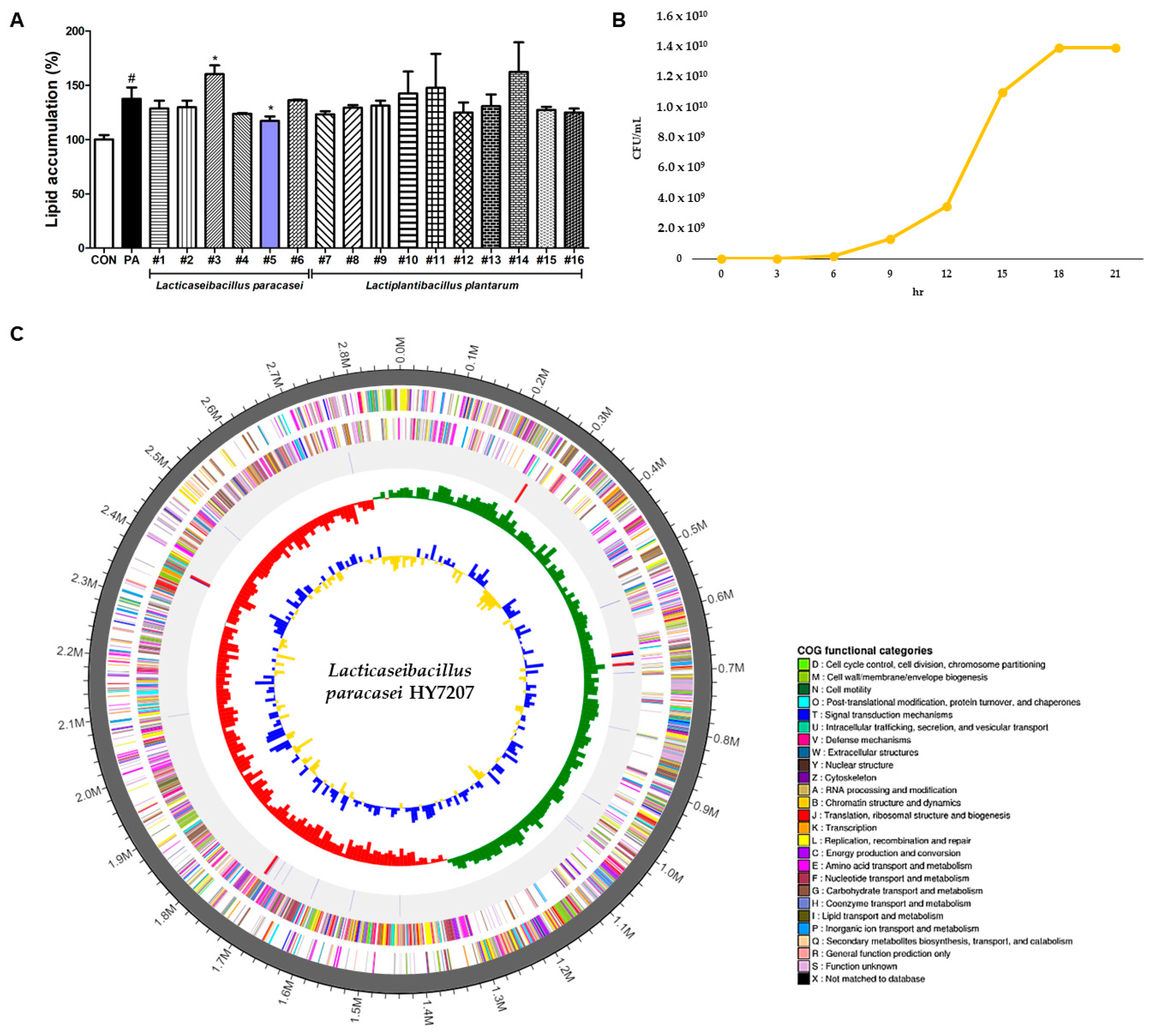
2.1. Screening of LAB Strains Isolated from Traditional Fermented Beverages

We isolated a total of 15 lactic acid-generating bacterial (LAB) strains from a Korean traditional fermented beverage (six strains of Lacticaseibacillus paracasei and ten strains of Lactiplantibacillus plantarum). The isolated LAB strains were then evaluated with respect to their effects on lipid accumulation in PA-treated HepG2 cells. As shown in Figure 1A, lipid accumulation in the PA-treated cells (137.4% ± 10.57%) was significantly more substantial than in the control cells (p < 0.01). The #5 strain (114.63% ± 4.22%) was the only one to significantly reduce lipid accumulation in these HepG2 cells (p < 0.05). Therefore, we selected L. paracasei #5 for further study and renamed this strain Lacticaseibacillus paracasei HY7207 (HY7207).

2.2. Growth Curve of HY7207 in Industrial Culture

To determine whether HY7207 is suitable for commercial use, the strain was cultured using an industrial fermenter, then we measured bacterial growth in terms of colony-forming units (CFUs)/mL. In addition, the growth curve for HY7207 reached the stationary phase after 21 h in a large fermentation volume (1200 L).

2.3. Whole-Genome Sequencing of HY7207

Whole-genome sequencing of L. paracasei HY7207 showed that it consisted of a circular chromosome of 2,877,365 bp, with a G+C content of 46.43% (Table 1). This genome contained a predicted 2778 CDSs, 15 rRNA-encoding genes, and 58 tRNA-encoding genes. The predicted CDSs were divided into clusters of orthologous gene (COG) functional categories as follows: T (56), U (19), V (74), C (98), D (26), E (190), F (79), G (264), H (42), I (49), J (148), K (183), L (190), M (126), N (1), O (59), P (115), Q (17), and S (694). The A, B, R, W, X, Y, and Z categories were not classified. Furthermore, the sequencing of HY7207 confirmed no antibiotic resistance genes. The HY7207 genome map is shown in Figure 1C.

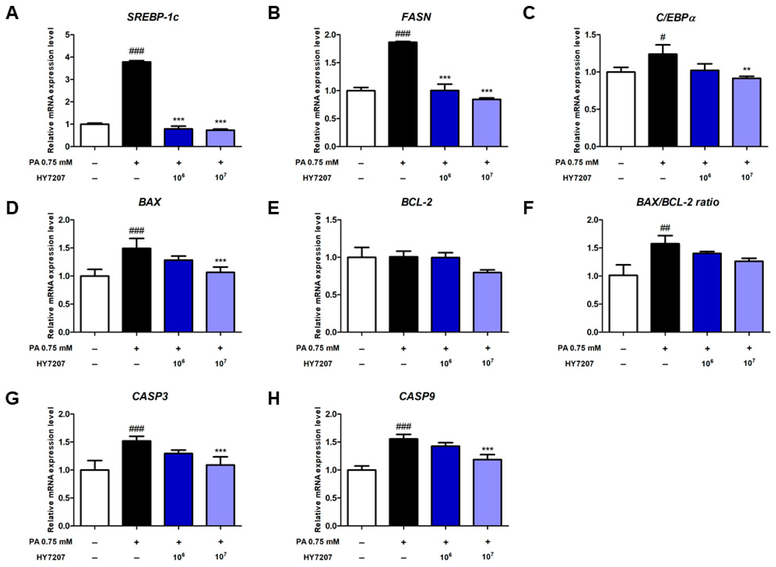
2.4. Effects of HY7207 on the Expression of Genes Involved in Lipogenesis and Apoptosis by PA-Treated HepG2 Cells

We determined the effects of HY7207 on the expression of genes related to lipogenesis in PA-treated HepG2 cells. For in vitro experiments, 106 and 107 CFU/mL of HY7207 were added to the lipid accumulation-induced hepatic cell models. As shown in Figure 2A–C, the expression of SREBP-1c (3.78-fold, p < 0.001), FASN (1.87-fold, p < 0.001), and C/EBPα (1.23-fold, p < 0.05) in PA-treated cells was significantly higher than that of the untreated cells. However, the expression of SREBP-1c and FASN in the HY7207-treated group was significantly lower than that of the PA-treated group (p < 0.001). The mRNA expression of C/EBPα tended to be lower in the 106 CFU/mL HY7207-treated group, but this difference was not significant. However, 107 CFU/mL HY7207 treatment significantly reduced the mRNA expression of C/EBPα vs. the PA-treated cells. Overall, HY7207 treatment reduced the expression of lipogenesis-related genes in a dose-dependent manner.
Next, we measured the expression of the apoptosis-related genes BAX and BCL-2 (Figure 2D,E). The expression of BAX in the PA-treated group was significantly higher than that in the untreated group (p < 0.001), but that in the HY7207-treated group was significantly lower than that of the PA-induced group. By contrast, the mRNA expression of BCL-2 did not differ between the groups. In Figure 2F, the BAX/BCL-2 ratio was significantly higher in cells treated with PA than in untreated cells (p < 0.01), but HY7207 treatment reduced this. As illustrated in Figure 2G,H, the mRNA expression of CASP3 and CASP9 in the PA-treated group was significantly elevated compared to that untreated group (p < 0.001). Treatment of HY7207 showed a dose-dependent manner reduction effect. These results indicate that HY7207 reduces the expression of lipogenesis-related genes and ameliorates the abnormal expression of apoptosis-related genes.

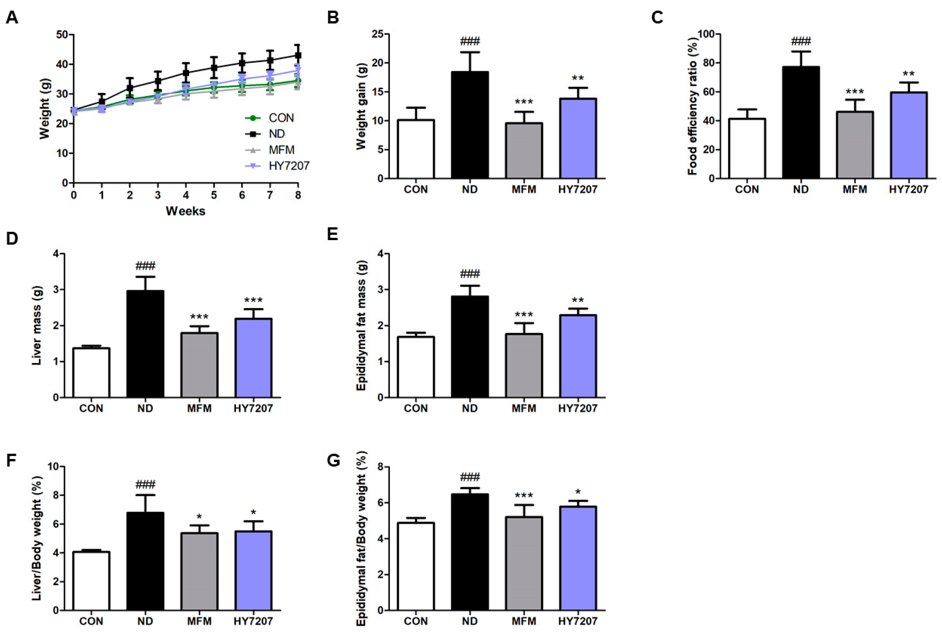
2.5. Effects of HY7207 on Physiologic Parameters in Mice Fed an NAFLD-Inducing Diet

To investigate the effects of HY7207 on NAFLD, we fed mice with an NAFLD-inducing diet and used metformin (MFM) as a positive control, because it is known to reduce hepatic fat synthesis and accumulation and to suppress glucose production [31,32]. In animal experiments, the MFM and HY7207-treated group orally ingested 250 mg/kg/day of metformin and 109 CFU/kg/day of L. paracasei HY7207, respectively. We measured the changes in body mass and food intake during the animal experiment (Figure 3 and Figure S1).
As shown in Figure 3B, the body mass gain of the mice in the NAFLD-inducing diet (ND)-fed group was significantly higher (18.43 ± 3.44 g, p < 0.001) than that of the control group (10.13 ± 2.13 g). Both MFM and HY7207 significantly reduced this body mass gain, to 9.59 ± 1.94 g (p < 0.001) and 13.82 ± 1.88 g (p < 0.01), respectively, vs. the ND group.
As illustrated in Figure 3C, the food efficiency ratio (FER) for the ND group was also significantly higher (77.19% ± 10.81%, p < 0.001) than that of the control group (CON, 41.38% ± 6.47%). However, the FERs for the positive control and HY7207-treated groups were significantly lower (46.17% ± 8.40% (p < 0.001) and 59.66% ± 6.86% (p < 0.01), respectively) than that of the ND group.
Figure 3D,F shows the liver and epididymal fat masses of the mouse groups. The ND group had significantly higher masses than the CON group (p < 0.001), but these were significantly lower in the MFM and HY7207 groups than in the ND group.
In addition, the liver/body mass ratio (L/B ratio) of the ND group was significantly higher (6.78% ± 1.23%, p < 0.001) than that of the control group (4.07% ± 0.14%). The L/B ratios of the MFM and HY7207 groups were significantly lower (5.37% ± 0.54% (p < 0.05) and 5.50% ± 0.70% (p < 0.05)) than that of the ND group (Figure 3E). The epididymal fat/body mass ratio (E/B ratio) was significantly increased (6.47% ± 0.34%, p < 0.001) by the ND, and this was reduced to 5.21% ± 0.66% (p < 0.001) and 5.78% ± 0.32% (p < 0.05) by MFM and HY7207 treatment, respectively (Figure 3G).

2.6. Effects of HY7207 on Blood Biochemistry

Next, we measured levels of liver function markers (AST, ALT, ALP, and γ-GPT) in the serum of the mice (Figure 4A–D). The activities in the ND group were significantly higher than those in the CON group (AST, ALT, ALP, p < 0.001; γ-GPT, p < 0.001). MFM treatment restored these activities to levels similar to those of the control group, and HY7207 significantly reduced these activities vs. those of the ND group.
As shown in Figure 4E–G, the serum TG, T-CHO, and LDL-C concentrations were significantly higher in the ND group than in the CON group, and MFM and HY7207 significantly reduced the concentrations of all three vs. the ND group. HY7207 treatment also tended to reduce these concentrations, but there were no significant differences. In addition, the HDL-C concentration was slightly higher in the ND, MFM, and HY7207 groups than in the CON group, but the differences were not significant (Figure 4H). Finally, the GLU concentration of the ND group was slightly higher than that of the control group (Figure 4I), and MFM significantly reduced this vs. the ND group (p < 0.01).

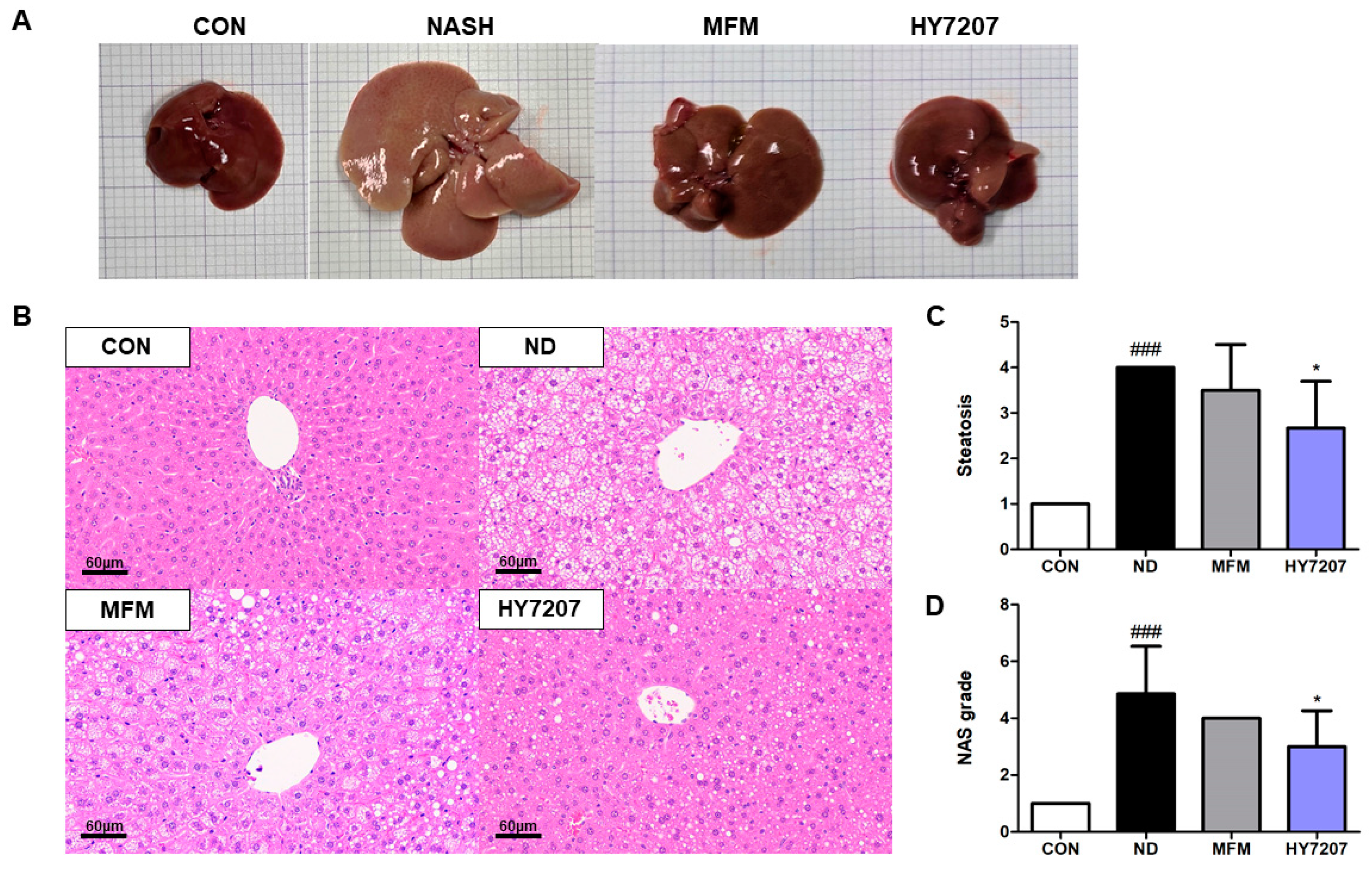
2.7. Effects of HY7207 on the Histology of the Liver

To analyze the effect of HY7207 on hepatic morphology and histology, we collected the liver from each mouse. The morphology of the livers is shown in Figure 5A. We then examined sections of each liver after hematoxylin and eosin (H&E) staining, and as shown in Figure 5B, the ND group showed hepatic steatosis, but MFM and HY7207 treatment reduced this. Figure 5C shows that the high hepatic steatosis score of the ND group was ameliorated by MFM and HY7207. In particular, the administration of HY7207 significantly reduced the degree of steatosis induced by the NALFD diet (p < 0.05). The score of lobular inflammation and hepatocyte ballooning in the MFM and HY7207 ingestion group was also reduced compared to the ND group (Figure S2). In addition, as illustrated in Figure 5D, the NAS grade of the ND group was significantly higher than that of the CON group (p < 0.001), but that of the HY7207 group was significantly lower than that of the ND group (p < 0.05). Thus, HY7207 ameliorates NAFLD in mice.

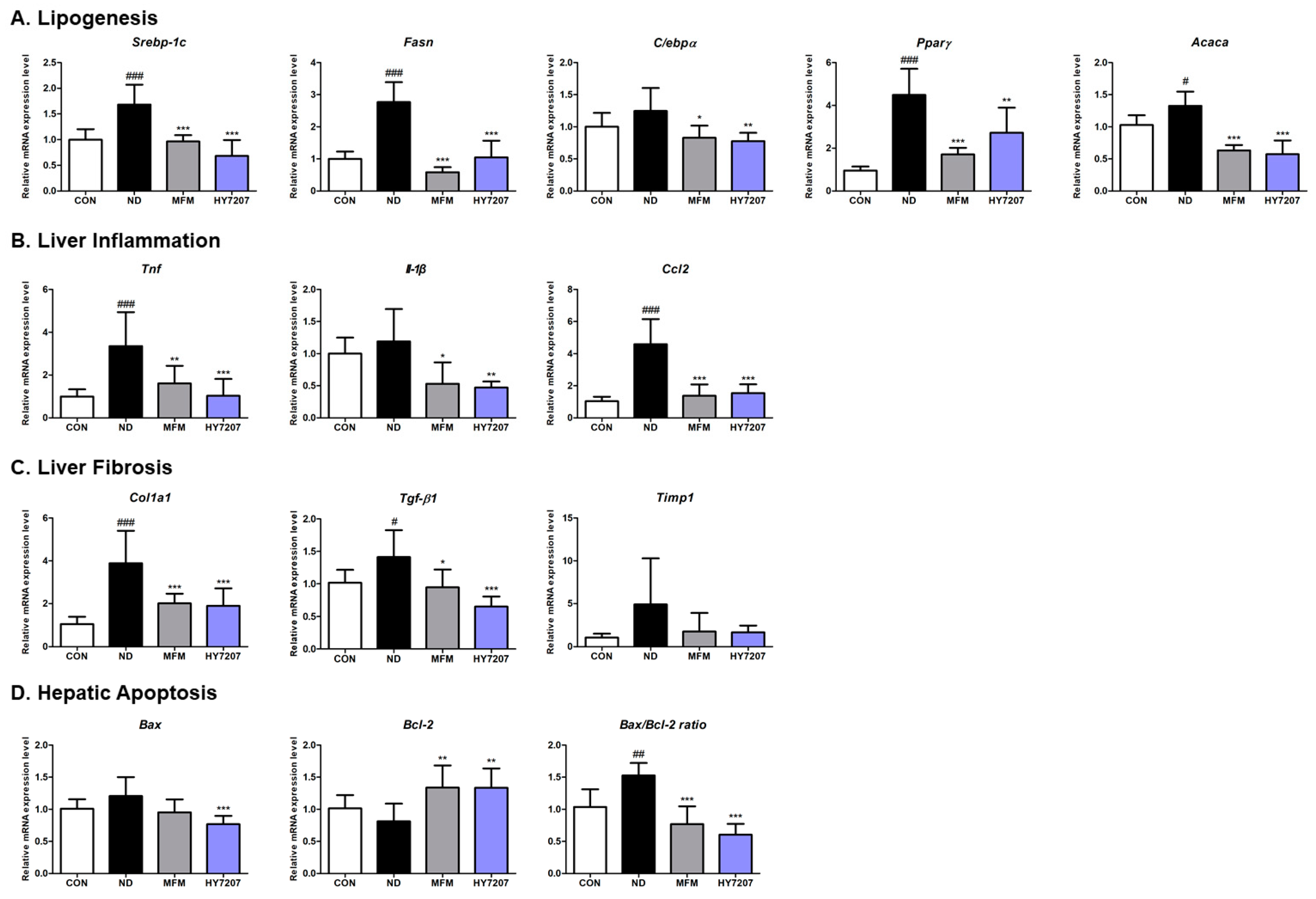
2.8. Effects of HY7207 on the Hepatic Gene Expression of Mice with NAFLD

We next evaluated the effects of HY7207 on the mRNA expression of genes related to lipid synthesis, inflammation, and fibrosis in NAFLD-inducing diet-fed mice. First, we measured the expression of the lipogenesis-related genes Srebp-1c, Fasn, C/ebpα, Pparγ, and Acaca. As shown in Figure 6A, the expression of Srebp-1c (2.77-fold, p < 0.001), Fasn (2.77-fold, p < 0.001), Pparγ (4.49-fold, p < 0.001), and Acaca (1.33-fold, p < 0.05) was significantly higher in the ND group than in the control group. The expression of C/ebpα was also high in the ND group (1.24-fold), but the difference was not significant. However, the mRNA expression of Srebp-1c, Fasn, Pparγ, Acaca (p < 0.001), and C/ebpα (p < 0.05) was significantly lower in the MFM group. HY7207 also significantly reduced the expression of lipogenesis-related genes vs. the ND group (Srebp-1c, Fasn, Acaca, p < 0.001; C/ebpα, Pparγ, p < 0.01). In particular, HY7207 reduced the mRNA expression of Srebp-1c (0.68-fold), C/ebpα (0.78-fold), and Acaca (0.67-fold) to levels similar to those achieved by MFM (Srebp-1c, 0.96-fold; C/ebpα, 0.83-fold; Acaca, 0.63-fold).
In addition, we measured the expression of inflammation-related genes, such as those encoding pro-inflammatory cytokines (Tnf and Il-1β), and a chemokine (Ccl2) (Figure 6B). The ND group showed significantly higher expression of these genes than the control group, and these differences were significantly different with respect to Tnf and Ccl2 (p < 0.001). The positive control, MFM, significantly altered the expression of Tnf, Il-1β, and Ccl2 by 1.61-fold (p < 0.01), 0.53-fold (p < 0.05), and 1.38-fold (p < 0.001), respectively, vs. the ND group (Tnf, 3.35-fold; Il-1β, 1.19-fold; and Ccl2, 4.59-fold). In addition, the HY7207 group had significantly lower expression of the three genes (1.03-fold (p < 0.001), 0.47-fold (p < 0.01), and 1.54-fold (p < 0.001), respectively, vs. the ND group).
We next measured the mRNA expression of the fibrosis-related genes Col1a1, Tgf-β1, and Timp1 (Figure 6C). The expression of all three was higher in the ND group than in the normal group (Col1a1, p < 0.001; Tgf-β1, p < 0.05; Timp1, p > 0.05). However, MFM and HY7207 significantly reduced the expression of Col1a1 and Tgf-β1 vs. the ND group. Treatment with MFM or HY7207 also tended to reduce Timp1 expression, but there was no significant difference vs. the ND group. Thus, HY7207 reduces the expression of genes related to lipogenesis, inflammation, and fibrosis in NAFLD-inducing diet-fed mice.
In addition, we assessed the effects of HY7207 on the mRNA expression of apoptosis-related genes in the liver (Figure 6D) and found that mRNA expression of Bax was 1.21-fold higher in ND mice, but was reduced 0.96-fold and 0.77-fold by treatment with MFM (p > 0.05) or HY7207 (p < 0.001). The mRNA expression of Bcl-2 in the ND group (0.81-fold) tended to be lower than that of the CON group. Treatment with MFM or HY7207 led to a significant recovery in Bcl-2 expression, to 1.34-fold (p < 0.01) and 1.33-fold (p < 0.01), respectively, vs. the ND group. Furthermore, in NAFLD-inducing diet-fed mice, the Bax/Bcl-2 ratio was significantly higher (1.53-fold, p < 0.01) than that of the control group. By contrast, the Bax/Bcl-2 ratio of the MFM and HY7207 groups was significantly lower (0.77-fold and 0.60-fold (p < 0.001), respectively) than that of the ND group. Taken together, these results show that HY7207 limits apoptosis in the livers of mice fed an ND.

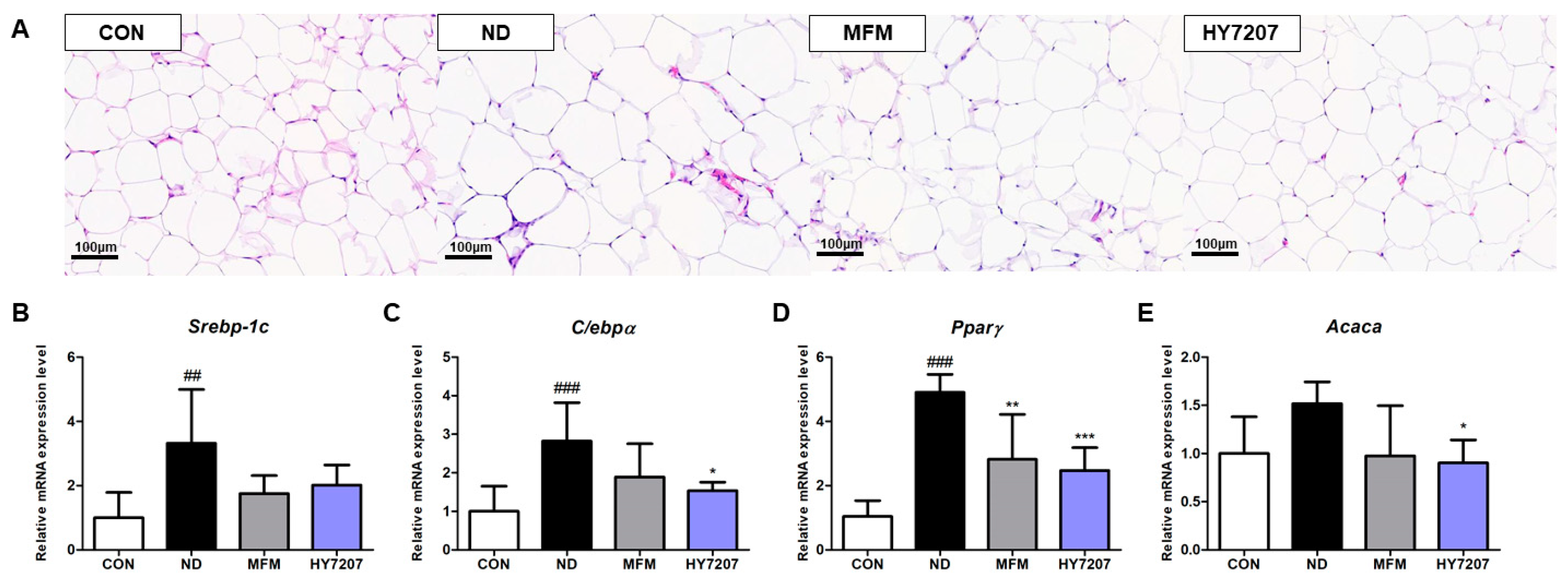
2.9. Effects of HY7207 on the Histology of Epididymal Fat

To analyze the effect of HY7207 on adipose tissue histology, we collected epididymal fat samples from the mice and examined sections of tissue after H&E staining. As shown in Figure 7A, the adipocytes of the ND group were larger than those of the control group. By contrast, the MFM and HY7207 groups had smaller adipocytes than the ND group.

2.10. Effects of HY7207 on the Expression of Lipogenesis-Related Genes in the Epididymal Fat of Mice with NAFLD

Finally, we determined the effects of HY7207 on genes related to lipid synthesis in epididymal fat (Figure 7B–E). The mRNA expression of Srebp-1c, C/ebpα, Pparγ, Acaca, and Lipe was significantly increased by the ND (Srebp-1c, p < 0.01; C/ebpα, Pparγ, Lipe, p < 0.001; Acaca, p < 0.05). The expression of Srebp-1c tended to be reduced by MFM and HY7207 treatment, but there were no significant differences. The mRNA expression of C/ebpα, Acaca, and Lipe in the MFM and HY7207 groups was lower than that in the ND group, but only significantly so in the case of HY7207 (C/ebpα and Lipe, p < 0.01; Acaca, p < 0.05). The expression of Pparγ in the MFM group was significantly lower than that of the ND group (p < 0.01). HY7207 treatment also significantly reduced the expression of this gene vs. the ND group (p < 0.001). Thus, HY7207 reduces the expression of lipid synthesis-related genes in epididymal fat, as well as in the livers, of NAFLD-inducing diet-fed mice.

3. Discussion

NAFLD is a common liver disease, characterized by hepatic steatosis, liver inflammation, and fibrosis [1,3]. The prevalence of NAFLD is estimated to be 20–30% worldwide, but there are no U.S. Food and Drug Administration or European Medicines Agency-approved therapies [33,34]. However, several recent studies have shown that probiotics may prevent and/or ameliorate NAFLD [30,35,36].
L. paracasei HY7207, isolated from a traditional Korean beverage, was selected from among 15 LAB strains because it caused significant inhibition of lipid accumulation in PA-treated HepG2 cells. We next tested the potential of HY7207 for industrial use through culture in a large-scale fermenter. In general, the bacterial growth curve is divided into four phases: the lag, exponential, stationary, and death phases [37]. The HY7207 strain growth curve could be divided into these phases as follows: 0–6 h, lag phase; 6–18 h, exponential phase; and 18–21 h, stationary phase. The growth curve of the HY7207 cells showed a typical growth curve as far as the stationary phase in industrial-scale culture. In addition, we performed whole-genome sequencing to determine the probiotic potential of HY7207, and we analyzed genetic data related to metabolic activity, antibiotic resistance, and virulence factors. The complete genome of L. paracasei HY7207 was 2.88 Mbp in length, with 46.43% GC content, 2778 CDSs, 15 rRNA-encoding genes, and 58 tRNA-encoding genes. In addition, no virulence factors or antibiotic resistance genes were predicted in the DNA sequence of HY7207. Taken together, these results suggest that HY7207 is a potentially useful strain that can be mass-produced.
Excessive DNL in hepatocytes is known to be a major cause of NAFLD [8]. SREBP-1c is a transcription factor that regulates the expression of genes encoding proteins in the DNL pathway and thereby increases fatty acid synthesis by FASN from acetyl-CoA and malonyl-CoA [11,38]. Previous studies have shown that hepatic DNL is upregulated in patients with NAFLD vs. healthy people, alongside higher hepatic expression of FASN [39,40]. In addition, C/EBPα plays an important role in the regulation of adipogenesis, and high expression of this protein is also associated with NAFLD [41,42]. We determined the effect of HY7207 on the expression of lipogenesis-related genes in PA-treated HepG2 cells, finding that this strain significantly increases the expression of SREBP-1c, FASN, and C/EBPα mRNA. Furthermore, hepatocyte apoptosis has been demonstrated to be a common feature of patients with NAFLD. An imbalance in BAX (a pro-apoptotic protein) and BCL-2 (an anti-apoptotic protein) expression is known to be involved in hepatocyte apoptosis during NAFLD [18,43]. Here, we found that HY7207 alters the expression of these apoptosis-related genes and the BAX/BCL-2 ratio in PA-treated HepG2 cells. These data suggest that HY7207 reduces hepatic fatty acid synthesis and apoptosis in hepatocytes.
Next, we showed that HY7207 improves physiologic and serum biochemical parameters, hepatic histology, and the gene expression in the liver and epididymal fat tissues of mice fed an ND, using MFM as a positive control. MFM is widely used for the treatment of diabetes and is known to reduce fat accumulation and ameliorate NAFLD [44]. During the experimental period, the ND induced significant increases in body mass, FER, and the masses of the liver and epididymal fat in the mice. However, treatment with HY7207 reduced these effects, and in the case of the liver and epididymal fat/body mass ratios, these were reduced to levels similar to those of the MFM group.
The circulating AST and ALT activities are markers of liver injury, are high in NAFLD/NASH, and indicate the progression of the disease [45]. The serum ALP activity may also be a predictor of significant fibrosis in patients with obesity and NAFLD [46]. In addition, γ-GPT is an indicator of liver injury, and a decrease in γ-GPT activity is associated with an improvement in liver histology [47]. The serum activities of AST, ALT, ALP, and γ-GPT were high in mice fed the ND, whereas MFM and HY7207 significantly reduced these. This is further evidence that treatment with HY7207 ameliorates NAFLD. High serum TG, T-CHOL, and LDL-C concentrations are associated with NAFLD, and these defects were present in the ND group [48,49]. However, both MFM and HY7207 reduced their concentrations. NAFLD is associated with excessive hepatic glucose production, resulting in hyperglycemia, and therefore the circulating glucose concentration is also a potential marker of the development of NAFLD [50]. This was high in the ND group, but slightly lower in the HY7207 group. However, the serum HDL-C concentration did not show significant differences between the groups. In addition, analysis of the hepatic histology of the mice showed that HY7207 reduces the steatosis and NAS score (steatosis + lobular inflammation + hepatocyte ballooning) of mice with NAFLD [51]. These results imply that HY7207 improves the serum lipid profile and ameliorates the hepatic steatosis of mice, suggesting that this probiotic reduces liver fat accumulation and may alleviate the symptoms of NAFLD.
Hepatic fat accumulation induces the development of inflammation and leads to the production of pro-inflammatory cytokines and chemokines [19]. This liver inflammation is an important precursor to liver fibrosis in NAFLD [52]. The hepatic mRNA expression of the lipogenic genes Srebp1c, Fasn, Cebpa, Pparg, and Acaca of the mice was lower in the HY7207 group than in the ND group. In addition, there was high expression of genes encoding pro-inflammatory cytokines (Tnfa and Il1b) and the chemokine CCL2 in the ND group, while treatment with HY7207 significantly reduced their expression. Furthermore, the expression of the fibrosis-related genes Col1a1, Tgfb1, and Timp1 was lower in the HY7207 group than in the ND group These data imply that HY7207 ameliorates hepatic lipid accumulation, steatosis, inflammation, and fibrosis in mice with NAFLD. Hepatic apoptosis is considered to be a major cause of the liver inflammation and fibrosis. Similar to the in vitro experiments, HY7207 treatment had positive effects on the expression of the apoptosis-related genes Bax and Bcl2 and the Bax/Bcl2 ratio in the animal model. Histopathologically, hepatocyte ballooning, a form of hepatocyte death, is generally considered to indicate apoptosis [53,54]. In the present study, the effects of HY7207 on the expression of apoptosis-related genes were consistent with its effects on hepatocyte ballooning on H&E-stained sections.
NAFLD is closely associated with excessive calorie intake in the form of carbohydrates or fats and obesity [55]. The long-term excessive intake of either can lead to hyperinsulinemia and hyperglycemia. This is associated with greater breakdown of triglyceride in adipose tissue, which increases the amount of free fatty acids entering the liver [56,57,58]. Therefore, a better understanding of the liver–adipose tissue crosstalk in NAFLD may be necessary for the development of improved approaches to the treatment and prevention of NAFLD. In the present study, the hypertrophy of adipocytes and lipid synthesis related gene expression in the epididymal fat in the ND group was reduced by HY7207 administration (Figure 7). This may suggest that HY7207 may also ameliorate NAFLD through a distinct mechanism. However, the molecular mechanism whereby HY7027 affects crosstalk between the liver and adipose tissue and thereby influences NAFLD should be the subject of further in-depth studies.

4. Materials and Methods

4.1. Sample Collection and Isolation of LAB Strains

LAB strains were obtained from Makgeolli, a traditional Korean fermented beverage. Samples were purchased from traditional Korean markets, collected in sterile containers, and stored in a −80 °C freezer until used. For strain isolation, liquid samples were mixed with sterile phosphate-buffered saline (PBS) in a ratio of 1:9 (v/v). The mixed samples were serially diluted with sterile PBS at 1/10 ratio, spread on MRS agar plates (BD Difco, Sparks, MD, USA), and incubated under anaerobic conditions at 37 °C for 48 h. Fifteen colonies were randomly selected and streaked onto fresh MRS agar plates to obtain pure isolates. These strains were then stored at −80 °C in glycerol (20%, v/v).

4.2. Culture of HepG2 Cells

HepG2 human liver cancer cells were purchased from the American Type Culture Collection (Manassas, VA, USA). The cells were cultured at 37 °C in a 5% CO2 incubator in Minimum Essential Medium (MEM; Welgene, Gyeongsan, Republic of Korea) containing 10% fetal bovine serum (Gibco, Waltham, MA, USA) and 1% penicillin/streptomycin (Gibco, Waltham, MA, USA). The HepG2 cells used in the experiment had undergone ≤10 passages.

4.3. Assessment of the Inhibition of Lipid Accumulation in HepG2 Cells

To evaluate the abilities of the 15 selected strains to inhibit lipid accumulation, Oil-red-O (ORO) staining was performed on PA-treated HepG2 cells. HepG2 cells were seeded onto 12-well plates at a density of 1 × 10⁵ cells/well and acclimated for 24 h, then 0.75 mM PA-containing antibiotic-free MEM was added to all the wells except the control wells, and each LAB strain (107 CFU/mL) was added. The control wells were treated with antibiotic-free MEM containing saline, and negative controls were treated with 0.75 mM PA alone. After 24 h, each well was washed twice with PBS, and 1 mL of 10% formalin was added, followed by incubation for 1 h. The wells were then rinsed with distilled water, and the cells were treated with 60% isopropanol and incubated for 5 min. After removing the isopropanol, 400–500 µL of 60% ORO solution was added, and the cells were incubated for 10 min. After that, the wells were washed with tap water and dried, then 100% isopropanol was added, and the cells were further incubated for 5 min. The isopropanol was then transferred to a microcentrifuge tube and centrifuged at 10,000× g for 2 min. The absorbance of the supernatant was measured at 480 nm using a microplate reader.

4.4. Industrial Culture and Assessment of the Growth Curve of HY7207 Cells

HY7207 cells were cultured in a 2000-L plant-scale fermenter (CNS, Daejeon, Republic of Korea) to assess the possibility of industrial scale-up and to confirm appropriate bacterial growth. A 1200-L culture was prepared at 37 °C and pH 5.5, with rotation at 100 rpm, for 21 h. The culture medium was sampled every 1 h, and the number of bacteria present was measured by two methods. First, the culture medium was serially diluted in saline for a standard plate count to be performed. The dilutions were incubated with MRS in petri dishes for 48 h, then 30–300 colonies were checked and expressed as CFU/mL.

4.5. Whole Genome Sequencing of HY7207

Genomic DNA (gDNA) of HY7207 was isolated by the Qiagen MagAttract HMW DNA Kit (Qiagen, Hilden, Germany), and quality and quantity of gDNA was measured by the Qubit 2.0 fluorometer (Invitrogen, Carlsbad, CA, USA). Sequencing of purified gDNA was performed at CJ bioscience (Seoul, Republic of Korea) using Illumina Miseq (Illumina Inc., San Diego, CA, USA) and PacBio RS Ⅱ (Pacific Biosciences Inc., Menlo Park, CA, USA) sequencing platforms. DNA libraries were prepared by the TruSeq DNA Library LT Kit (Illumina Inc., San Diego, CA, USA) and the SMRTbell Template Preparation Kit (Pacific Biosciences Inc., Menlo Park, CA, USA). Gene prediction was performed by Prodigal 2.6.2, and Cluster of Orthologous (COG) of predicted gene was annotated by EggNOG 4.5 (http://eggnogdb.embl.de (accessed on 8 June 2024)). Antibiotic resistance genes were predicted using The Comprehensive Antibiotics Resistance Database (CARD; https://card.mcmaster.ca/ (accessed on 8 June 2024)).

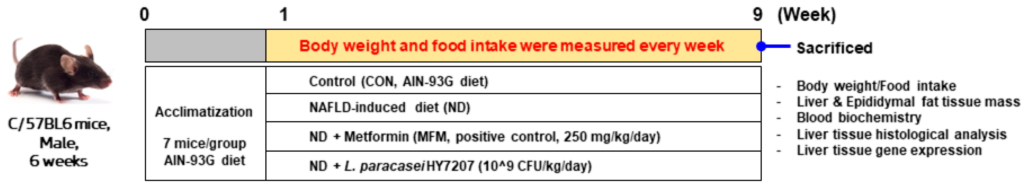
4.6. Animal Experiments

Six-week-old male C57BL/6 mice were purchased from Dooyeol Biotech (Gyeonggi, Republic of Korea) and acclimated for 1 week. The mice were housed under constant humidity and temperature conditions (55% ± 10%, 22 ± 1 °C) under a 12 h light/dark cycle. After acclimation, the mice were randomly assigned to one of four groups, each containing seven mice: control (CON, AIN-93G diet), NAFLD diet only (ND), NAFLD diet with metformin (MFM, 250 mg/kg/day), and NAFLD diet with HY7207 (HY7207, 109 CFU/kg/day). The ND comprised a rodent diet providing 40 kcal% as fat (principally palm oil), 20 kcal% as fructose, and 2% as cholesterol (D09100310, Research Diets, Inc., New Brunswick, NJ, USA). The MFM and probiotic were dissolved in 200 uL of saline and orally administered for 8 weeks, and the Control and ND groups were administered an equal volume of saline over the same period. The body masses and food intakes of the mice were measured weekly. After 8 weeks, the mice were sacrificed, and blood, liver, and epididymal fat tissue samples were obtained. The blood samples were allowed to stand at room temperature and then centrifuged at 3000× g for 20 min to separate the serum. The separated serum samples were then used for biochemical analysis. The other tissue samples were rinsed with PBS, weighed, and stored at −80 °C until subsequent analysis. The animal studies were approved by the Institutional Animal Care and Committee of the hy Co., Ltd. R&D Center, Seoul, Republic of Korea (approval number: AEC-2024-00001-Y). Figure 8 shows a flowchart of the animal experiments performed in the present study.

4.7. Blood Biochemical Analysis

The serum activities/concentrations of AST, ALT, ALP, γ-GTP, T-CHOL, TG, GLU, HDL, and LDL were analyzed at T&P Bio (Gyeonggi, Republic of Korea).

4.8. Histological Analysis

Liver and epididymal fat samples were collected and fixed in 10% (v/v) formalin solution. The fixed tissues were embedded in paraffin, and sections were cut and mounted on slides. Images were then obtained using a Zeiss Axiovert 200M microscope (Carl Zeiss AG, Thornwood, NY, USA) after H&E staining. The NAS of the liver samples, comprising hepatic steatosis, lobular inflammation, and hepatocyte ballooning, was evaluated [51]. The degree of hepatic steatosis was graded 1–4 as follows: 1, <5%; 2, 5–33%; 3, 34–65%; and 4, >66%. The degree of lobular inflammation was graded 0–3 as follows: 0, no foci; 1, <2 foci/×200 field; 2, 2–4 foci/×200 field; and 3, >4 foci/×200 field. The degree of hepatocyte ballooning was graded 0–2 as follows: 0, none; 1, few ballooned cells; 2, many ballooned cells/prominent ballooning. Histopathologic analysis, including H&E staining and NAS scoring, were performed by DooYeol Biotech.
NAS = Hepatic steatosis + Lobular inflammation + Hepatocyte ballooning

4.9. Isolation of RNA, cDNA Synthesis, and Real-Time PCR

RNA was extracted from HepG2 cells and mouse livers using an Easy-spin Total RNA Extraction Kit (iNtRON Biotechnology, Seoul, Republic of Korea), then cDNA was synthesized from 2 μg RNA using an Omniscript RT Kit (Qiagen, Hilden, Germany) at 37 °C for 60 min. The cDNA template was analyzed using the QuantStudio 6 Flex Real-time PCR System (Applied Biosystems, Foster City, CA, USA), and real-time PCR was performed using a TaqMan™ Gene Expression Master Mix (Applied Biosystems, Waltham, MA, USA). Table 2 lists the target genes and assay catalog numbers. The expression levels of the target genes were normalized to that of GAPDH (HepG2 cells, Hs99999905_m1; Mice, Mm99999915_g1) using the comparative C τ method.

4.10. Statistical Analysis

Data are presented as the mean ± standard deviation (SD). Groups were compared using one-way ANOVA, followed by Tukey’s post hoc test. All analyses were conducted using Prism v.6.0 (GraphPad Software, San Diego, CA, USA), and p < 0.05 was considered to represent statistical significance.

5. Conclusions

Lacticaseibacillus paracasei HY7207, isolated from a traditional Korean fermented beverage, can be cultured at an industrial scale. The data presented herein suggest that HY7207 has potential for use as a functional food supplement for the amelioration of NAFLD. HY7207 ameliorates hepatic fat accumulation in PA-treated HepG2 cells and mice with NAFLD. In addition, HY7207 may ameliorate the symptoms of NAFLD by reducing the expression of lipogenesis and apoptosis-related genes. Furthermore, HY7207 has anti-inflammatory and anti-fibrotic effects by reducing the expression of genes encoding cytokines, chemokines, and mediators of liver fibrosis. Therefore, we propose that HY7207 could be produced by the food industry as a functional supplement for patients with NAFLD.

Supplementary Materials

The supporting information can be downloaded at: https://www.mdpi.com/article/10.3390/ijms25189870/s1.

Author Contributions

Conceptualization, writing—original draft preparation and writing—review and editing, H.-J.K., H.-J.J. and J.-Y.K.; software, validation, resources, data curation, formal analysis, H.-J.K.; visualization, H.-J.K. and D.-G.K.; methodology and investigation, H.-J.K. and H.-J.J.; supervision, J.-J.S. and J.-H.L.; project administration, J.-Y.K. and J.-H.L. All authors have read and agreed to the published version of the manuscript.

Funding

This research received no external funding.

Institutional Review Board Statement

The animal study protocol was approved by the Ethics Review Committee of R&BD Center, hy Co., Ltd., Republic of Korea (approval number: AEC-2022-0003-Y).

Informed Consent Statement

Not applicable.

Data Availability Statement

The data presented in this study are available in the article and Supplementary Materials.

Conflicts of Interest

The authors declare that this study received funding from hy Co., Ltd. The funder was not involved in the study design, collection, analysis, or interpretation of data, the writing of the article, or the decision to submit it for publication.

References

  1. Abd El-Kader, S.M.; El-Den Ashmawy, E.M.S. Non-alcoholic fatty liver disease: The diagnosis and management. World J. Hepatol. 2015, 7, 846. [Google Scholar] [CrossRef] [PubMed]
  2. Buss, C.; Valle-Tovo, C.; Miozzo, S.; de Mattos, A.A. Probiotics and synbiotics may improve liver aminotransferases levels in non-alcoholic fatty liver disease patients. Ann. Hepatol. 2014, 13, 482–488. [Google Scholar] [CrossRef] [PubMed]
  3. Paolella, G.; Mandato, C.; Pierri, L.; Poeta, M.; Di Stasi, M.; Vajro, P. Gut-liver axis and probiotics: Their role in non-alcoholic fatty liver disease. World J. Gastroenterol. WJG 2014, 20, 15518. [Google Scholar] [CrossRef] [PubMed]
  4. Bellentani, S.; Scaglioni, F.; Marino, M.; Bedogni, G. Epidemiology of non-alcoholic fatty liver disease. Dig. Dis. 2010, 28, 155–161. [Google Scholar] [CrossRef] [PubMed]
  5. Vernon, G.; Baranova, A.; Younossi, Z. Systematic review: The epidemiology and natural history of non-alcoholic fatty liver disease and non-alcoholic steatohepatitis in adults. Aliment. Pharmacol. Ther. 2011, 34, 274–285. [Google Scholar] [CrossRef]
  6. Friedman, S.L.; Neuschwander-Tetri, B.A.; Rinella, M.; Sanyal, A.J. Mechanisms of NAFLD development and therapeutic strategies. Nat. Med. 2018, 24, 908–922. [Google Scholar] [CrossRef]
  7. Yin, X.; Guo, X.; Liu, Z.; Wang, J. Advances in the diagnosis and treatment of non-alcoholic fatty liver disease. Int. J. Mol. Sci. 2023, 24, 2844. [Google Scholar] [CrossRef]
  8. Ipsen, D.H.; Lykkesfeldt, J.; Tveden-Nyborg, P. Molecular mechanisms of hepatic lipid accumulation in non-alcoholic fatty liver disease. Cell. Mol. Life Sci. 2018, 75, 3313–3327. [Google Scholar] [CrossRef]
  9. Li, N.; Li, X.; Ding, Y.; Liu, X.; Diggle, K.; Kisseleva, T.; Brenner, D.A. SREBP regulation of lipid metabolism in liver disease, and therapeutic strategies. Biomedicines 2023, 11, 3280. [Google Scholar] [CrossRef]
  10. Souza-Mello, V. Peroxisome proliferator-activated receptors as targets to treat non-alcoholic fatty liver disease. World J. Hepatol. 2015, 7, 1012. [Google Scholar] [CrossRef]
  11. Ruiz, R.; Jideonwo, V.; Ahn, M.; Surendran, S.; Tagliabracci, V.S.; Hou, Y.; Gamble, A.; Kerner, J.; Irimia-Dominguez, J.M.; Puchowicz, M.A. Sterol regulatory element-binding protein-1 (SREBP-1) is required to regulate glycogen synthesis and gluconeogenic gene expression in mouse liver. J. Biol. Chem. 2014, 289, 5510–5517. [Google Scholar] [CrossRef] [PubMed]
  12. Zhou, Y.; Tao, J.; Calvisi, D.F.; Chen, X. Role of lipogenesis rewiring in hepatocellular carcinoma. In Seminars in Liver Disease; Thieme Medical Publishers, Inc.: New York, NY, USA, 2022; Volume 42, pp. 77–86. [Google Scholar]
  13. Chatrath, H.; Vuppalanchi, R.; Chalasani, N. Dyslipidemia in patients with nonalcoholic fatty liver disease. In Seminars in Liver Disease; Thieme Medical Publishers: New York, NY, USA, 2012; Volume 32, pp. 22–29. [Google Scholar]
  14. Martin, A.; Lang, S.; Goeser, T.; Demir, M.; Steffen, H.-M.; Kasper, P. Management of dyslipidemia in patients with non-alcoholic fatty liver disease. Curr. Atheroscler. Rep. 2022, 24, 533–546. [Google Scholar] [CrossRef] [PubMed]
  15. Alkhouri, N.; Carter-Kent, C.; Feldstein, A.E. Apoptosis in nonalcoholic fatty liver disease: Diagnostic and therapeutic implications. Expert Rev. Gastroenterol. Hepatol. 2011, 5, 201–212. [Google Scholar] [CrossRef]
  16. Lei, Z.; Yu, J.; Wu, Y.; Shen, J.; Lin, S.; Xue, W.; Mao, C.; Tang, R.; Sun, H.; Qi, X. CD1d protects against hepatocyte apoptosis in non-alcoholic steatohepatitis. J. Hepatol. 2024, 80, 194–208. [Google Scholar] [CrossRef] [PubMed]
  17. Kim, C.W.; Lee, C.D. Apoptosis and diagnosis of nonalcoholic steatohepatitis. Korean J. Hepatol. 2011, 17, 247. [Google Scholar] [CrossRef]
  18. Kanda, T.; Matsuoka, S.; Yamazaki, M.; Shibata, T.; Nirei, K.; Takahashi, H.; Kaneko, T.; Fujisawa, M.; Higuchi, T.; Nakamura, H. Apoptosis and non-alcoholic fatty liver diseases. World J. Gastroenterol. 2018, 24, 2661. [Google Scholar] [CrossRef]
  19. Braunersreuther, V.; Viviani, G.L.; Mach, F.; Montecucco, F. Role of cytokines and chemokines in non-alcoholic fatty liver disease. World J. Gastroenterol. WJG 2012, 18, 727. [Google Scholar] [CrossRef]
  20. Nagata, N.; Chen, G.; Xu, L.; Ando, H. An update on the chemokine system in the development of NAFLD. Medicina 2022, 58, 761. [Google Scholar] [CrossRef]
  21. Dooley, S.; Ten Dijke, P. TGF-β in progression of liver disease. Cell Tissue Res. 2012, 347, 245–256. [Google Scholar] [CrossRef]
  22. Sánchez, B.; Delgado, S.; Blanco-Míguez, A.; Lourenço, A.; Gueimonde, M.; Margolles, A. Probiotics, gut microbiota, and their influence on host health and disease. Mol. Nutr. Food Res. 2017, 61, 1600240. [Google Scholar] [CrossRef]
  23. Hemarajata, P.; Versalovic, J. Effects of probiotics on gut microbiota: Mechanisms of intestinal immunomodulation and neuromodulation. Ther. Adv. Gastroenterol. 2013, 6, 39–51. [Google Scholar] [CrossRef]
  24. Kim, S.-K.; Guevarra, R.B.; Kim, Y.-T.; Kwon, J.; Kim, H.; Cho, J.H.; Kim, H.B.; Lee, J.-H. Role of probiotics in human gut microbiome-associated diseases. J. Microbiol. Biotechnol. 2019, 29, 1335–1340. [Google Scholar] [CrossRef] [PubMed]
  25. Bengoa, A.A.; Dardis, C.; Garrote, G.L.; Abraham, A.G. Health-promoting properties of Lacticaseibacillus paracasei: A focus on kefir isolates and exopolysaccharide-producing strains. Foods 2021, 10, 2239. [Google Scholar] [CrossRef] [PubMed]
  26. Yang, T.; Fan, X.; Li, D.; Zhao, T.; Wu, D.; Liu, Z.; Long, D.; Li, B.; Huang, X. High Antioxidant Capacity of Lacticaseibacillus paracasei TDM-2 and Pediococcus pentosaceus TCM-3 from Qinghai Tibetan Plateau and Their Function towards Gut Modulation. Foods 2023, 12, 1814. [Google Scholar] [CrossRef] [PubMed]
  27. Lin, E.-K.; Chang, W.-W.; Jhong, J.-H.; Tsai, W.-H.; Chou, C.-H.; Wang, I.-J. Lacticaseibacillus paracasei GM-080 ameliorates allergic airway inflammation in children with allergic rhinitis: From an animal model to a double-blind, randomized, placebo-controlled trial. Cells 2023, 12, 768. [Google Scholar] [CrossRef] [PubMed]
  28. Guo, W.; Tang, X.; Zhang, Q.; Xiong, F.; Yan, Y.; Zhao, J.; Mao, B.; Zhang, H.; Cui, S. Lacticaseibacillus paracasei CCFM1222 Ameliorated the Intestinal Barrier and Regulated Gut Microbiota in Mice with Dextran Sulfate Sodium-Induced Colitis. Probiotics Antimicrob. Proteins 2024, 1–13. [Google Scholar] [CrossRef]
  29. Ji, Y.; Xie, Q.; Meng, X.; Wang, W.; Li, S.; Lang, X.; Zhao, C.; Yuan, Y.; Ye, H. Lactobacillus paracasei improves dietary fatty liver by reducing insulin resistance and inflammation in obese mice model. J. Funct. Foods 2022, 95, 105150. [Google Scholar] [CrossRef]
  30. Wang, W.; Li, Q.; Chai, W.; Sun, C.; Zhang, T.; Zhao, C.; Yuan, Y.; Wang, X.; Liu, H.; Ye, H. Lactobacillus paracasei Jlus66 extenuate oxidative stress and inflammation via regulation of intestinal flora in rats with non alcoholic fatty liver disease. Food Sci. Nutr. 2019, 7, 2636–2646. [Google Scholar] [CrossRef]
  31. Fulgencio, J.-P.; Kohl, C.; Girard, J.; Pégorier, J.-P. Effect of metformin on fatty acid and glucose metabolism in freshly isolated hepatocytes and on specific gene expression in cultured hepatocytes. Biochem. Pharmacol. 2001, 62, 439–446. [Google Scholar] [CrossRef]
  32. Zheng, J.; Woo, S.-L.; Hu, X.; Botchlett, R.; Chen, L.; Huo, Y.; Wu, C. Metformin and metabolic diseases: A focus on hepatic aspects. Front. Med. 2015, 9, 173–186. [Google Scholar] [CrossRef]
  33. Kumar, R.; Priyadarshi, R.N.; Anand, U. Non-alcoholic fatty liver disease: Growing burden, adverse outcomes and associations. J. Clin. Transl. Hepatol. 2020, 8, 76. [Google Scholar] [CrossRef]
  34. Negi, C.K.; Babica, P.; Bajard, L.; Bienertova-Vasku, J.; Tarantino, G. Insights into the molecular targets and emerging pharmacotherapeutic interventions for nonalcoholic fatty liver disease. Metabolism 2022, 126, 154925. [Google Scholar] [CrossRef] [PubMed]
  35. Meroni, M.; Longo, M.; Dongiovanni, P. The role of probiotics in nonalcoholic fatty liver disease: A new insight into therapeutic strategies. Nutrients 2019, 11, 2642. [Google Scholar] [CrossRef] [PubMed]
  36. Kim, H.; Lee, K.; Kim, J.-Y.; Shim, J.-J.; Lim, J.; Kim, J.-Y.; Lee, J.-L. Lactobacillus helveticus Isolated from Raw Milk Improves Liver Function, Hepatic Steatosis, and Lipid Metabolism in Non-Alcoholic Fatty Liver Disease Mouse Model. Microorganisms 2023, 11, 2466. [Google Scholar] [CrossRef] [PubMed]
  37. Garrison, A.T.; Huigens, R.W., III. Eradicating bacterial biofilms with natural products and their inspired analogues that operate through unique mechanisms. Curr. Top. Med. Chem. 2017, 17, 1954–1964. [Google Scholar] [CrossRef]
  38. Dewulf, J.P.; Gerin, I.; Rider, M.H.; Veiga-da-Cunha, M.; Van Schaftingen, E.; Bommer, G.T. The synthesis of branched-chain fatty acids is limited by enzymatic decarboxylation of ethyl-and methylmalonyl-CoA. Biochem. J. 2019, 476, 2427–2447. [Google Scholar] [CrossRef] [PubMed]
  39. Mitsuyoshi, H.; Yasui, K.; Harano, Y.; Endo, M.; Tsuji, K.; Minami, M.; Itoh, Y.; Okanoue, T.; Yoshikawa, T. Analysis of hepatic genes involved in the metabolism of fatty acids and iron in nonalcoholic fatty liver disease. Hepatol. Res. 2009, 39, 366–373. [Google Scholar] [CrossRef]
  40. Dorn, C.; Riener, M.-O.; Kirovski, G.; Saugspier, M.; Steib, K.; Weiss, T.S.; Gäbele, E.; Kristiansen, G.; Hartmann, A.; Hellerbrand, C. Expression of fatty acid synthase in nonalcoholic fatty liver disease. Int. J. Clin. Exp. Pathol. 2010, 3, 505. [Google Scholar]
  41. Matsusue, K.; Gavrilova, O.; Lambert, G.; Brewer, H.B., Jr.; Ward, J.M.; Inoue, Y.; LeRoith, D.; Gonzalez, F.J. Hepatic CCAAT/enhancer binding protein α mediates induction of lipogenesis and regulation of glucose homeostasis in leptin-deficient mice. Mol. Endocrinol. 2004, 18, 2751–2764. [Google Scholar] [CrossRef]
  42. Lakhani, H.V.; Sharma, D.; Dodrill, M.W.; Nawab, A.; Sharma, N.; Cottrill, C.L.; Shapiro, J.I.; Sodhi, K. Phenotypic alteration of hepatocytes in non-alcoholic fatty liver disease. Int. J. Med. Sci. 2018, 15, 1591. [Google Scholar] [CrossRef]
  43. Pang, L.; Liu, K.; Liu, D.; Lv, F.; Zang, Y.; Xie, F.; Yin, J.; Shi, Y.; Wang, Y.; Chen, D. Differential effects of reticulophagy and mitophagy on nonalcoholic fatty liver disease. Cell Death Dis. 2018, 9, 90. [Google Scholar] [CrossRef] [PubMed]
  44. Pinyopornpanish, K.; Leerapun, A.; Pinyopornpanish, K.; Chattipakorn, N. Effects of metformin on hepatic steatosis in adults with nonalcoholic fatty liver disease and diabetes: Insights from the cellular to patient levels. Gut Liver 2021, 15, 827. [Google Scholar] [CrossRef]
  45. Khosravi, S.; Alavian, S.M.; Zare, A.; Daryani, N.E.; Fereshtehnejad, S.-M.; Daryani, N.E.; Keramati, M.R.; Abdollahzade, S.; Taba Vakili, S.T. Non-alcoholic fatty liver disease and correlation of serum alanin aminotransferase level with histopathologic findings. Hepat. Mon. 2011, 11, 452. [Google Scholar] [PubMed]
  46. Ali, A.H.; Petroski, G.F.; Diaz-Arias, A.A.; Al Juboori, A.; Wheeler, A.A.; Ganga, R.R.; Pitt, J.B.; Spencer, N.M.; Hammoud, G.M.; Rector, R.S. A model incorporating serum alkaline phosphatase for prediction of liver fibrosis in adults with obesity and nonalcoholic fatty liver disease. J. Clin. Med. 2021, 10, 3311. [Google Scholar] [CrossRef] [PubMed]
  47. Chen, L.W.; Huang, M.S.; Shyu, Y.C.; Chien, R.N. Gamma-glutamyl transpeptidase elevation is associated with metabolic syndrome, hepatic steatosis, and fibrosis in patients with nonalcoholic fatty liver disease: A community-based cross-sectional study. Kaohsiung J. Med. Sci. 2021, 37, 819–827. [Google Scholar] [CrossRef]
  48. Tu, L.N.; Showalter, M.R.; Cajka, T.; Fan, S.; Pillai, V.V.; Fiehn, O.; Selvaraj, V. Metabolomic characteristics of cholesterol-induced non-obese nonalcoholic fatty liver disease in mice. Sci. Rep. 2017, 7, 6120. [Google Scholar] [CrossRef]
  49. Lee, J.-H.; Lee, H.S.; Cho, A.-R.; Lee, Y.-J.; Kwon, Y.-J. Non-alcoholic fatty liver disease is an independent risk factor for LDL cholesterol target level. Int. J. Environ. Res. Public Health 2021, 18, 3442. [Google Scholar] [CrossRef]
  50. Simental-Mendía, L.E.; Simental-Mendía, E.; Rodríguez-Hernández, H.; Rodríguez-Morán, M.; Guerrero-Romero, F. The product of triglycerides and glucose as biomarker for screening simple steatosis and NASH in asymptomatic women. Ann. Hepatol. 2017, 15, 715–720. [Google Scholar]
  51. Lee, N.Y.; Yoon, S.J.; Han, D.H.; Gupta, H.; Youn, G.S.; Shin, M.J.; Ham, Y.L.; Kwak, M.J.; Kim, B.Y.; Yu, J.S. Lactobacillus and Pediococcus ameliorate progression of non-alcoholic fatty liver disease through modulation of the gut microbiome. Gut Microbes 2020, 11, 882–899. [Google Scholar] [CrossRef]
  52. Koyama, Y.; Brenner, D.A. Liver inflammation and fibrosis. J. Clin. Investig. 2017, 127, 55–64. [Google Scholar] [CrossRef]
  53. Singla, T.; Muneshwar, K.N.; Pathade, A.G.; Yelne, S. Hepatocytic Ballooning in Non-alcoholic Steatohepatitis: Bridging the Knowledge Gap and Charting Future Avenues. Cureus 2023, 15, e45884. [Google Scholar] [CrossRef] [PubMed]
  54. Machado, M.V.; Cortez-Pinto, H. Cell death and nonalcoholic steatohepatitis: Where is ballooning relevant? Expert Rev. Gastroenterol. Hepatol. 2011, 5, 213–222. [Google Scholar] [CrossRef] [PubMed]
  55. Dharmalingam, M.; Yamasandhi, P.G. Nonalcoholic fatty liver disease and type 2 diabetes mellitus. Indian J. Endocrinol. Metab. 2018, 22, 421–428. [Google Scholar] [CrossRef] [PubMed]
  56. Vancells Lujan, P.; Vinas Esmel, E.; Sacanella Meseguer, E. Overview of non-alcoholic fatty liver disease (NAFLD) and the role of sugary food consumption and other dietary components in its development. Nutrients 2021, 13, 1442. [Google Scholar] [CrossRef]
  57. Basaranoglu, M.; Basaranoglu, G.; Bugianesi, E. Carbohydrate intake and nonalcoholic fatty liver disease: Fructose as a weapon of mass destruction. Hepatobiliary Surg. Nutr. 2015, 4, 109. [Google Scholar]
  58. Gowda, D.; Shekhar, C.; Gowda, S.G.B.; Chen, Y.; Hui, S.P. Crosstalk between Lipids and Non-Alcoholic Fatty Liver Disease. Livers 2023, 3, 687–708. [Google Scholar] [CrossRef]
Figure 1. (A) Screening of 15 LAB strains for their ability to reduce lipid accumulation in PA-treated HepG2 cells. (B) Growth curve of HY7207 in industrial-scale culture. (C) Genome map of Lactocaseibacilllus paracasei HYY7207. From the outer circle to the inner circle, the genomic features are indicated as follows: genome size, gray color; forward strand and reverse strand CDSs, different colors, according to the COG classification; tRNA, blue line; rRNA, red line; GC skew, green and red peaks; GC ratio, blue and yellow peaks. Data are presented as the mean ± SD. # p < 0.05 vs. the control group, * p < 0.05 vs. the PA group; LAB: lactic acid bacteria; PA: palmitic acid; CDS: protein-coding DNA sequence; COG: clusters of orthologous groups; G: guanine, C: cytosine.
Ijms 25 09870 g001
Figure 2. Effect of HY7207 on the expression of lipogenesis and apoptosis-related genes in PA-treated HepG2 cells. (A) SREBP-1c, (B) FASN, (C) C/EBPα, (D) BAX, (E) BCL-2, (F) BAX/BCL-2 ratio (G) CASP3, and (H) CASP9. The results are presented as the mean ± SD. # p < 0.05, ## p < 0.01, and ### p < 0.001 vs. the control group; ** p < 0.01 and *** p < 0.001 vs. the PA group. PA: palmitic acid; HY7207: Lacticaseibacillus paracasei HY7207; SREBP-1c: sterol regulatory element-binding protein 1; FASN: fatty acid synthase; C/EBPα: CCAAT/enhancer-binding protein alpha; BAX: BCL2-associated X, apoptosis regulator; BCL-2: BCL2, apoptosis regulator; CASP3: Caspase 3; CASP9: Caspase 9.
Ijms 25 09870 g002
Figure 3. Effect of HY7207 on mice fed an NAFLD-inducing diet. (A) Body mass changes, (B) body mass gain, (C) food efficiency ratio, (D) liver tissue mass, (E) liver/body mass ratio, (F) epididymal fat mass, and (G) epididymal fat/body mass ratio. The results are presented as the mean ± SD. ### p < 0.001 vs. the CON group; * p < 0.05, ** p < 0.01, and *** p < 0.001 vs. the ND group. CON: Untreated group; ND: NAFLD-inducing diet group; MFM: metformin; HY7207: Lacticaseibacillus paracasei HY7207; FER: food efficiency ratio.
Ijms 25 09870 g003
Figure 4. Effect of HY7207 on the blood biochemistry of NAFLD-inducing diet-fed mice. (A) AST, (B) ALT, (C) ALP, and (D) γ-GPT activities; (E) TG, (F) T-CHO, (G) LDL-C, (H) HDL-C, and (I) GLU concentrations. The results are presented as the mean ± SD. ## p < 0.01 and ### p < 0.001 vs. the CON group; * p < 0.05, ** p < 0.01, and *** p < 0.001 vs. the ND group. CON: Untreated group; ND: NAFLD-inducing diet group; MFM: Metformin group; HY7207: Lacticaseibacillus paracasei HY7207 group; AST: aspartate aminotransferase; ALT: alanine aminotransferase; ALP: alkaline phosphatase; γ-GTP: gamma-glutamyl transferase; TG: triglyceride; T-CHO: total cholesterol; LDL-C: low-density lipoprotein-cholesterol; HDL-C: high-density lipoprotein-cholesterol; GLU: glucose.
Ijms 25 09870 g004
Figure 5. Effect of HY7207 on the hepatic histology of NAFLD-inducing diet-fed mice. (A) Liver morphology, (B) histology of the liver (hematoxylin and eosin-stained sections; 100× magnification), and (C) steatosis grade of the mice. (D) NAS grade of the mice. Results are presented as the mean ± SD. ### p < 0.001 vs. the CON group; * p < 0.05 vs. the ND group. CON: Untreated group; ND: NAFLD-inducing diet-fed group; MFM: Metformin group; HY7207: Lacticaseibacillus paracasei HY7207 group; NAS: NAFLD activity score.
Ijms 25 09870 g005
Figure 6. Effect of HY7207 on the hepatic gene expression of NAFLD-inducing diet-fed mice. The expression of genes related to (A) lipogenesis, (B) inflammation, (C) fibrosis, and (D) hepatic apoptosis is shown. Results are presented as the mean ± SD. # p < 0.05, ## p < 0.01, and ### p < 0.001 vs. the CON group; * p < 0.05, ** p < 0.01, and *** p < 0.001 vs. the ND group. CON: Untreated group; ND: NAFLD-inducing diet-fed group; MFM: Metformin group; HY7207: Lacticaseibacillus paracasei HY7207 group.
Ijms 25 09870 g006
Figure 7. (A) Effects of HY7207 on the histology of the epididymal fat of NAFLD-inducing diet-fed mice. (H&E staining; 100× magnification). Effect of HY7207 on the expression of lipogenesis related genes in NAFLD-inducing diet-fed mice. (B) Srebp-1c, (C) C/ebpα, (D) Pparγ and (E) Acaca. Results are presented as the mean ± SD. ## p < 0.01, and ### p < 0.001 vs. the CON group; * p < 0.05, ** p < 0.01, and *** p < 0.001 vs. the ND group. CON: Untreated group; ND: NAFLD-inducing diet-fed group; MFM: Metformin group; HY7207: Lacticaseibacillus paracasei HY7207 group.
Ijms 25 09870 g007
Figure 8. A flow chart of animal experiments.
Ijms 25 09870 g008
Table 1. Genomic information of Lacticaseibacillus paracasei HY7207.
FeaturesTerms
Sequencing platformsIllumina MiSeq
PacBio RSII
Libraries usedTruSeq DNA Library LT Kit
SMRTbell® Prep Kit
Genome size (bp)2,877,365
G+C contents (%)46.43
rRNA genes15
tRNA genes58
Table 2. Lists of TaqMan probes used for gene expression analysis.
GeneGene NameCatalog Number
In vitro
GAPDHGlyceraldehyde-3-phosphate dehydrogenaseHs99999905_m1
SREBP-1cSterol regulatory element-binding protein 1Hs01088691_m1
FASNFatty acid synthaseHs00188012_m1
C/EBPαCCAAT/enhancer-binding protein alphaHs00269972_s1
BAXBCL2 associated X, apoptosis regulatorHs00180269_m1
Bcl-2BCL2, apoptosis regulatorHs04986394_s1
CASP3Caspase 3Hs00234387_m1
CASP9Caspase 9Hs00962278_m1
In vivo
GapdhGlyceraldehyde-3-phosphate dehydrogenaseMm99999915_g1
Srebp-1cSterol regulatory element-binding protein 1Mm00550338_m1
FasnFatty acid synthaseMm00433237_m1
C/ebpαCCAAT/enhancer-binding protein alphaMm00514283_m1
PparγPeroxisome proliferator-activated receptor gammaMm00440945_m1
AcacaAcetyl-CoA carboxylase alphaMm01304257_m1
TnfTumor necrosis factorMm00443258_m1
Il-1βInterleukin 1 betaMm00434228_m1
Ccl2C-C motif chemokine ligand 2Mm00441242_m1
Col1a1Collagen type I alpha 1Mm00801666_g1
Tgf- β1Transforming growth factor beta 1Mm01178820_m1
Timp1tissue inhibitor of metalloproteinase 1Mm01341361_m1
BaxBCL2 associated X, apoptosis regulatorMm00432051_m1
Bcl-2BCL2, apoptosis regulatorMm00477631_m1
Disclaimer/Publisher’s Note: The statements, opinions and data contained in all publications are solely those of the individual author(s) and contributor(s) and not of MDPI and/or the editor(s). MDPI and/or the editor(s) disclaim responsibility for any injury to people or property resulting from any ideas, methods, instructions or products referred to in the content.

