Review

Defenders of the Transcriptome: Guard Protein-Mediated mRNA Quality Control in Saccharomyces cerevisiae

Abteilung für Molekulare Genetik, Institut für Mikrobiologie und Genetik, Göttinger Zentrum für Molekulare Biowissenschaften (GZMB), Georg-August Universität Göttingen, 37077 Göttingen, Germany
*
Author to whom correspondence should be addressed.
Int. J. Mol. Sci. 2024, 25(19), 10241; https://doi.org/10.3390/ijms251910241
Submission received: 21 August 2024 / Revised: 17 September 2024 / Accepted: 19 September 2024 / Published: 24 September 2024

Abstract

:
Cell survival depends on precise gene expression, which is controlled sequentially. The guard proteins surveil mRNAs from their synthesis in the nucleus to their translation in the cytoplasm. Although the proteins within this group share many similarities, they play distinct roles in controlling nuclear mRNA maturation and cytoplasmic translation by supporting the degradation of faulty transcripts. Notably, this group is continuously expanding, currently including the RNA-binding proteins Npl3, Gbp2, Hrb1, Hrp1, and Nab2 in Saccharomyces cerevisiae. Some of the human serine–arginine (SR) splicing factors (SRSFs) show remarkable similarities to the yeast guard proteins and may be considered as functional homologues. Here, we provide a comprehensive summary of their crucial mRNA surveillance functions and their implications for cellular health.

1. Introduction

To ensure the export of only correctly generated mRNAs, a series of quality-control mechanisms occurs in the nucleus. As different mRNA features are essential for trouble-free gene expression, pre-mRNAs are already surveilled at different time points during transcription. At an early stage of transcription, when approximately 13–20 nucleotides are transcribed, various proteins mediate 5′-capping. This process ensures that a cap-binding complex (CBC)-bound 7-methylguanosine (m7G) cap is synthesized, which helps pre-mRNAs withstand nucleolytic attacks [1,2]. Subsequently, noncoding introns are excised from the open reading frame (ORF) by the spliceosome [3]. Importantly, for splicing to work efficiently, a properly capped 5′-end of the pre-mRNA must exist [4], already revealing the importance of functional quality-control mechanisms. Upon arrival of the RNA polymerase II (RNAPII) containing transcription machinery at the 3′-end of the pre-mRNA, the termination machinery is recruited, and the transcript is released from the DNA. Correct termination requires the cleavage and polyadenylation complex (CPF-CF), which is directed by cis-acting elements located in the 3′ untranslated region (UTR) [5]. If termination fails, transcripts are terminated downstream of the CPF-CF site, mostly by the Nrd1, Nab3, and Sen1 (NNS)-mediated termination system, which leads to the elimination of the readthrough transcript [6,7,8]. Finally, a poly(A) tail is attached to the 3′-end of the mRNA to protect the transcript from exonucleolytic attacks from this RNA end [9].
These regulatory steps are overseen by a group of proteins called the guard proteins (Table 1). In the context of Saccharomyces cerevisiae, several guard proteins have been discovered, including Npl3, Gbp2, Hrb1, Nab2, and the recently identified Hrp1. Each of these proteins is characterized by the presence of several RNA-binding motifs, notably the RNA-recognition motif (RRM) or zinc-finger (ZF) domain, as well as a serine–arginine/arginine–glycine–glycine (SR/RGG) domain. Among the guard proteins, Npl3 contains the most canonical SR/RGG domain at its C-terminus, along with two central RRMs and an N-terminal APQE domain still awaiting characterization (Figure 1) [10]. Interestingly, the two RRMs are not equivalent, as the second RRM is able to form stronger interactions with guanine/uracil (G/U)-rich sequences on RNA [11]. The paralogues Gbp2 and Hrb1 both feature an N-terminal SR/RGG domain mainly containing SR repeats and three RRMs each. While RRM1 and RRM2 preferentially recognize RNAs containing the core motif GGUG, the atypical RRM3 at the C-terminus does not interact with nucleic acids but is crucial for the interaction with the THO/TREX complex [12]. Hrp1 exhibits a C-terminal SR/RGG domain rich in single-serine and arginine residues, in addition to two centrally located RRMs. As evident by the lack of common amino acid identity with the other guard proteins, Nab2 varies in its domain composition (Figure 1). Instead of RRMs, it contains seven ZF motifs within its C-terminal ZF domain [13]. The ZF domain is critical for its interaction with poly(A) RNA, with ZF 5-7 being crucial for this function [13]. Additionally, Nab2 harbors an N-terminal domain essential for the nuclear export of poly(A) RNAs and Nab2 itself, a nonessential glutamine (Q)-rich domain of currently unknown function, and the SR/RGG domain, which is important for the reimport of Nab2 [13]. These structural features play a crucial role in facilitating the interactions of all guard proteins with the nascent mRNA during the various stages of its maturation [14].
At their core, all of the guard proteins share a common function as retention factors for mRNAs, which they conduct until the recruitment of the export receptor Mex67-Mtr2 (short Mex67) after the successful completion of a step in the mRNA-maturation pathway (Figure 2) [15,16,17,18]. Generally, the more Mex67 is bound to an mRNA, the more it is shielded from the hydrophobic interior of the nuclear pore complex (NPC), enabling better passage into the cytoplasm [19]. Coverage with the export receptor is controlled by the interaction of the guard proteins with Mlp1, the gatekeeper of the NPC. In this way, Mlp1 prevents the passage of immature pre-mRNAs when not sufficiently Mex67-covered. All of the guard proteins are nuclear at a steady state, and their export depends on active transcription and the export of mRNAs in general. Upon correct maturation, they accompany the mRNA into the cytoplasm, where translation occurs. Here, many of them serve a second purpose in cytoplasmic mRNA surveillance, as most are either involved in translation or participate in nonsense-mediated decay (NMD) [20,21,22]. The release of the guard proteins from mRNA occurs through distinct mechanisms. For Npl3, Gbp2, and Hrb1, dissociation is primarily facilitated through binding with the import receptor Mtr10, while for Hrp1 and Nab2, dissociation occurs via interaction with the import receptor Kap104 [23,24,25].
All currently known guard proteins share the common trait of interacting physically and genetically with the export receptor Mex67 and the NPC gatekeeper Mlp1 [16,18,26,27,28,29]. Additionally, they exhibit mutual physical and genetic interactions among themselves (https://www.yeastgenome.org/ (accessed on 1 August 2024)) (Figure 3). While typically responsible for retaining only faulty transcripts, it is notable that the overexpression of any guard protein results in the widespread retention of poly(A)+ RNAs in the nucleus. This phenomenon is likely due to excessive binding of the proteins to the same transcripts, possibly overwhelming the capacity of Mex67 for coverage or not being situated in the correct context to allow Mex67 binding, therefore leading to retention, which proves detrimental to cellular health [16,27]. Lastly, a shared characteristic among the guard proteins is their interaction with the cellular degradation machinery, ensuring the timely elimination of faulty transcripts [15,16,17,18,30].

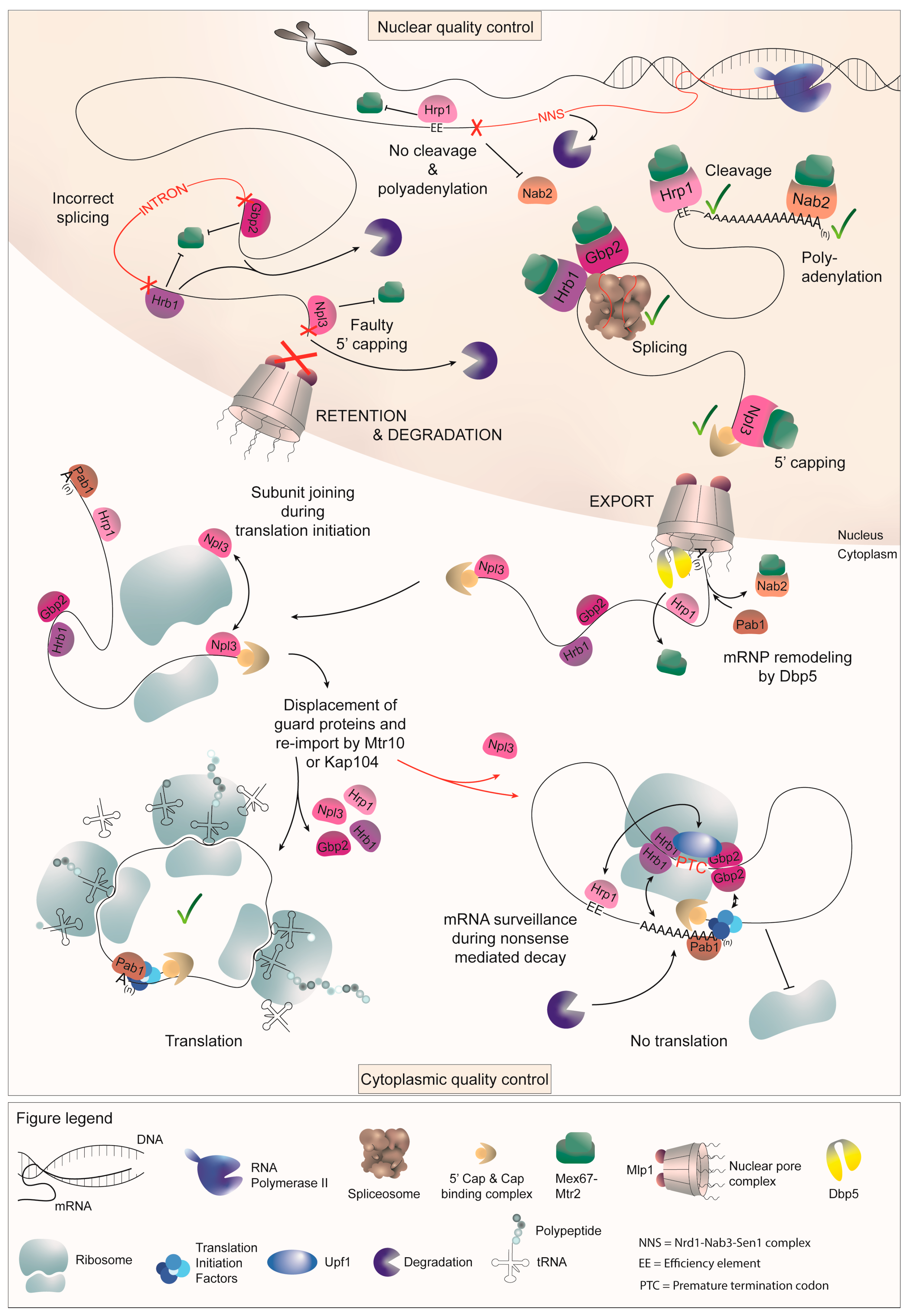
2. Maturation of Pre-mRNAs

mRNAs are continuously synthesized through transcription by RNAP II from their 5′- to 3′-ends, with the initial modification being mRNA capping. During transcription initiation, an inverted 7-methylguanosine (m7G) cap is synthesized to protect the 5′-end from exonucleolytic degradation, promote proper splicing and nuclear export, and facilitate efficient translation initiation [1,2,31]. The capping process involves three key enzymes: The 5′ RNA triphosphatase Cet1 converts the 5′ triphosphate into a diphosphate, the RNA guanylyltransferase Ceg1 adds a guanosine-7 cap, and the methyltransferase Abd1 methylates the cap [32,33,34,35]. The 5′ cap is immediately recognized and bound by the CBC, which facilitates subsequent processing steps, including splicing, nuclear export, and translation initiation in the cytoplasm [36,37].
Similar to those in higher eukaryotes, pre-mRNAs in yeast may contain noncoding introns, which are excised during splicing to create a mature ORF. The splicing process is mediated by the spliceosome, a large ribonucleoprotein complex composed of small nuclear RNA (snRNA) and protein counterparts. Splicing factors are recruited throughout transcription elongation to determine the proper splice sites and facilitate the assembly of the spliceosome [3,38,39]. The yeast spliceosome comprises five snRNAs, termed U1, U2, U4, U5, and U6, along with approximately 70 binding proteins, which together form small nuclear ribonucleoprotein particles (snRNPs) [40,41].
Once splicing is completed, the mature mRNA undergoes further processing steps, including 3′ end cleavage and polyadenylation, which may occur in close proximity to the nuclear pores [42]. This process is crucial for stabilizing the mRNA, regulating its nuclear export and facilitating efficient translation in the cytoplasm. For 3′ polyadenylation and cleavage, numerous proteins form the cleavage and polyadenylation complex (CPF-CF). Specific sequences within the 3′ UTR of the pre-mRNA serve as recognition sites for the cleavage and polyadenylation machinery. Sequences such as the positioning element (PE) or efficiency element (EE) upstream of the cleavage site help tether the complex to the mRNA, while downstream sequence elements flanking the cleavage site may help make the poly(A) site (PAS) more accessible. The recognition of the PAS initiates the cleavage of the pre-mRNA by the endonuclease Ysh1, leaving a free 3′ hydroxyl group. Finally, the poly(A) polymerase Pap1 adds a 60–80 nucleotide long poly(A) tail to the 3′-end of the mRNA to protect it from exonucleolytic attacks from this RNA-end [8,9,43]. The mature mRNA is then ready for export from the nucleus to the cytoplasm, where it can be translated into a functional protein.

2.1. Guard Protein-Mediated 5′ Capping Requires Npl3

The process of 5′ capping is monitored through the shuttling guard protein Npl3, which is recruited to the nascent pre-mRNA by RNAP II during early transcription [44]. It serves as an adaptor for Mex67 and modulates the switch between export and degradation, surveilling the proper capping of transcripts due to its proximity to the 5′ end. Notably, Npl3 associates with pre-mRNAs bound by the 5′ end degradation factor Rai1 and recruits the export receptor Mex67 only after successful capping. In that case, the CBC binds the 5′ end of the mRNA, and Rai1 dissociates, leaving Npl3 to be able to recruit the export receptor. However, in the absence of correct CBC attachment, Npl3 fails to sense proper capping, allowing for the 5′ exonuclease Rat1 to interact with Rai1 and degrade the faulty transcript. In cases where immediate degradation is not possible, Npl3 retains the transcript in the nucleus, while the absence of Npl3 can lead to the leakage of uncapped transcripts into the cytoplasm (Figure 4) [15].
Unlike other Mex67-recruiting proteins that remain in the nucleus and are displaced from the mRNA prior to export, the guard proteins continue to stay bound to the mRNA after export. They shuttle with the messenger ribonucleoprotein (mRNP) into the cytoplasm, enabling them to fulfill additional tasks. Likewise, Npl3 also participates in the nuclear export of large ribosomal subunits, in addition to its function in mRNA surveillance and export [45]. Similar to its attachment to mRNAs, which persists even after export, Npl3 remains bound to the large ribosomal subunit after shuttling as well. Being bound to both, the mRNA on which the small subunit is positioned at the AUG start codon and the large ribosomal subunit, Npl3 further assists the ribosomal monosome formation during translation initiation. As the small 40S ribosomal subunit first binds to the mRNA upon translation initiation, the 60S subunit-bound Npl3 contributes to successful subunit joining by self-dimerizing with its counterparts bound to the mRNA (Figure 4) [21]. This cytoplasmic function of Npl3 promotes the assembly of the translation machinery only onto properly capped mRNAs. This is crucial for further RNA protection but also for subsequent translation, because circularized RNAs are more efficiently translated, as the ribosomal subunits are recycled to the 5′-end for another round of translation after reaching the stop codon on the mRNA 3′ end [46,47]. In this way, Npl3 helps to make translation as efficient as possible, since mRNAs that have failed quality control are rather degraded than those allowed to occupy the translation machinery.

2.2. Intron Splicing Is Surveilled through the Guard Proteins Gbp2 and Hrb1

Among the guard proteins, Gbp2 and Hrb1 stand out as key players in regulating proper splicing. During splicing, these paralogues are co-transcriptionally recruited to pre-mRNAs by the THO complex [24,48,49,50]. They function as a switch between export and degradation, as they retain transcripts in the nucleus until splicing is completed successfully or until a faulty transcript is marked for degradation. Hence, Gbp2 and Hrb1 contact the spliceosome in late splicing and either recruit the export receptor Mex67-Mtr2 to signal export competence or recruit the TRAMP complex, which subsequently assists in the degradation of the transcript [18]. The TRAMP complex, comprising the RNA helicase Mtr4, zinc-finger mRNA-binding subunits Air1 or Air2, and either Trf4 or Trf5 polymerases, assists in degrading transcripts by adding a short oligo(A) tail to the 3′ end, marking them for nuclear exosome-mediated degradation [51,52]. Conversely, coverage of the guard proteins with Mex67 signals proper splicing to the NPC gatekeeper protein Mlp1, allowing their export into the cytoplasm as a result of correct processing. When Gbp2 and Hrb1 are absent from cells, unspliced transcripts can be observed leaking into the cytoplasm [16,18].
Gbp2 and Hrb1 remain bound to spliced mRNAs until translation, where they assist in the degradation of faulty transcripts containing a PTC [20]. Both proteins exhibit a preference for binding to the 5′ end of mRNAs, presumably due to the prevalence of yeast introns in this region [53,54]. Upon recognition of a PTC, a complex containing Upf1-2-3 is formed and Gbp2 and Hrb1 facilitate the remodeling of the RNP. The mRNA is circularized to ensure fast and efficient rounds of translation, and upon PTC detection, Gbp2 and Hrb1 are able to restructure the mRNA, conveying the PTC signal to both ends of the transcript, ultimately triggering RNA degradation. Their ability to form dimers with themselves, as well as with each other, suggests that their interactions may bring the PTC recognition site close to the transcript ends through necessary mRNA folding. Additionally, interactions with 5′ or 3′ end-associating proteins such as Pab1 or some of the transcription initiation factors (TIFs) may further assist the RNA remodeling. This way, further translation initiation is inhibited and degradation is initiated (Figure 4) [20].
While both Gbp2 and Hrb1 mediate translational inhibition, the degradation process differs between them. Hrb1 recruits the decapping factor Dcp1 [20], which allows the degradation of the aberrant mRNA by the cytoplasmic 5′-3′ exonuclease Xrn1 after decapping [55,56]. The minor degradation pathway from the 3′ end, on the other hand, is propagated by Gbp2, which recruits the Ski complex and thus designates the mRNA for degradation by the cytoplasmic exosome [20].

2.3. Proper 3′ Cleavage Is Monitored by Hrp1

Hrp1 has recently emerged as the candidate to fill the role of a guard protein for 3′ cleavage, completing the set of guard proteins for each primary mRNA-processing step. In the nucleus, Hrp1 binds to the UAUAUA-containing efficiency element (EE) located in the 3′ UTR of the mRNA and shuttles into the cytoplasm along with the transcript [57,58,59]. It is a component of the cleavage and polyadenylation factor (CPF) and cleavage factor (CF) complex, which is crucial for the 3′ end cleavage and release of pre-mRNA from RNAPII. Hrp1 binds to the EE located upstream of the mRNA cleavage site, recruits the export receptor Mex67-Mtr2 upon correct cleavage, and also interacts with the nuclear gatekeeper Mlp1 [9,27,60]. Interestingly, it is the only component of the CPF-CF that shuttles into the cytoplasm alongside transcripts [57,58,61]. Hrp1 is able to sense successful cleavage through its contact with Rna14, which is a scaffold component of the CPF-CF and the primary interaction partner for Hrp1. This contact allows Mex67 to bind the guard protein, while the absence of the Rna14-Hrp1 interaction, possibly due to an improper assembly of the CPF-CF and, therefore, readthrough of the termination site, leads to the retention of the transcript [27]. Interestingly, Hrp1 stands out as the only guard protein lacking direct contact with the nuclear-degradation machinery. Instead, contact with nuclear-degradation factors depends on the NNS fail-safe termination system, which detects transcription readthrough and initiates the subsequent degradation of the faulty transcript [6]. The NNS complex consists of two RNA-binding proteins, Nrd1 and Nab3, which specifically recognize the RNA sequences GUAA/G and UCUU downstream of the CPF-CF termination site [62,63]. The RNA-DNA helicase Sen1 serves as the third component of the complex, likely recruited by Nab3 [64]. Together, the NNS complex facilitates termination by displacing RNAPII, and the interaction of Nrd1 with the exosome and the TRAMP component Trf4 results in the recruitment of the degradation machinery [65,66].
In the absence of Hrp1, elongated transcripts are no longer retained in the nucleus and leak into the cytoplasm [27]. Most importantly, mutation of HRP1 results in 3′ extended transcripts for the majority of all mRNAs already after a 1 h shift to the non-permissive temperature, suggesting that Hrp1 has a broad function as a nuclear and cytoplasmic quality-control factor. Again, such elongated transcripts are suboptimal for translation, as long 3′ UTRs lower the half life of an mRNA and may trigger NMD, as the ribosome recycling process becomes more difficult. In addition to its role in nuclear quality control, Hrp1 has been implicated in the process of NMD. Hrp1 has been reported to interact with the NMD factor Upf1, and it supposedly associates with so-called downstream sequence elements (DSEs) [22]. Initially, these sequences were believed to be located in coding regions, where they were bound by RNA-binding proteins that were subsequently replaced by the translating ribosome. In the presence of a PTC, the presence of these proteins would then trigger NMD. Although this model has not been extensively explored due to the fact that the DSE sequences have proven to be highly degenerate and because alternative NMD models in yeast have emerged, the notion that PTCs might be recognized through marks downstream of the termination codon remains relevant in current hypotheses. Transcriptome-wide studies have revealed that Hrp1 is widely present in most mRNAs [27]. Thus, the EE might be the main region to which Hrp1 binds in the nucleus and remains bound until translation.

2.4. Nab2 Surveils the Proper Attachment of a Poly(A) Tail

In yeast, Nab2 is the nuclear poly(A) tail-binding protein that monitors the tail length and interacts with the export receptor Mex67 to facilitate mRNA transport through the NPC [30,67,68]. Similar to the other guard proteins, Nab2 also interacts with Mlp1 at the NPC, creating another checkpoint for nuclear export [68]. Moreover, mutations in Nab2 have been observed to result in the leakage of faulty transcripts into the cytoplasm [16]. Notably, Nab2 is distinct among the guard proteins as it is displaced from the mRNA at the cytoplasmic side of the NPC by the DEAD-box RNA helicase Dbp5 [69]. Consequently, Nab2 is the first guard to leave the mRNA, which is also reflected in its absence from polysomes [50]. While the other guards remain bound until translation commences and, in the case of Gbp2, Hrb1, and Hrp1, are also involved in NMD, Nab2 departs prior to the start of translation. Nab2 is most likely replaced by Pab1, which enables the circularization of the mRNA, occurring through the interaction of Pab1 and the 5′ cap-binding complex, including eIF4G [70]. The circularization of the transcript is thought to enhance ribosome cycling for more efficient translation [71].
Furthermore, beyond its role in maintaining optimal poly(A) tail length, Nab2 likely serves an additional function in facilitating the formation of correctly folded, export-competent mRNPs. Due to its documented propensity for dimerization via the ZF domain and reported binding along the entire transcript length, it is hypothesized that Nab2 contributes to the correct folding of mRNPs [72,73]. Moreover, mutations within the ZF domain have been shown to promote an accelerated disassembly of the mRNP by Dbp5 in the cytoplasm, presumably due to substandard packaging [74].

2.5. Npl3 Participates in 3′ End Quality Control

Interestingly, Npl3 may emerge as an additional key player in nuclear 3′ end quality control, fulfilling a function during the final stages of transcription. It appears to rival several 3′ end processing factors for pre-mRNA binding, offering protection against premature termination and facilitating the coordination of transcription termination and the packaging of finalized transcripts for export [11]. Notably, the functional dynamics of Npl3 are influenced by phosphorylation mediated by CKII during these crucial stages, which may finetune the termination process by enabling the binding of termination factors through a reduced RNA-binding capacity of Npl3 [75,76]. Additionally, Rat1 and Rai1, which cooperate with Npl3 during 5′ quality control in order to ensure proper degradation, have been implicated in poly(A)-dependent transcription termination as well [32,77]. Moreover, the absence of Npl3 leads to a widespread 3′ extension of transcripts transcribed by RNAPII, indicating the possibility of anomalies in pre-mRNA packaging events that may lead to termination readthrough. This widespread readthrough has also been linked to the downregulation of neighboring genes, underscoring the significance of Npl3 in maintaining proper termination and gene expression [78]. While the precise mechanism of the involvement of Npl3 in nuclear 3′ end quality control remains to be fully elucidated, these observations strongly indicate a role for Npl3 in this process.

3. The Guard Proteins Are Subjected to Post-Translational Modification

Post-translational modifications (PTMs) play a crucial role in modulating the functions and activities of a plethora of proteins. Modifications such as methylation or phosphorylation are also observed across the spectrum of the previously described guard proteins. Although numerous sites for a variety of PTMs have been reported for all of them, our understanding of the functional implications of many of these modifications remains incomplete. Currently, the best-described modifications are methylation and phosphorylation.
Among the guard proteins, Npl3 stands out as the cornerstone of our understanding of PTMs. As such, unphosphorylated Npl3 is recruited co-transcriptionally by the phosphorylated Ser2 of RNAPII, promoting RNAPII elongation and preventing premature termination by competing with Rna15 for RNA binding at the 3′ end. Release of Npl3 from the CTD is achieved through phosphorylation by casein kinase 2 (CK2) at several serine residues, most of which are located within the SR/RGG domain [75]. In the nucleus, Glc7-mediated dephosphorylation promotes the association of Npl3 with the export receptor Mex67 [79]. It has been suggested that Sky1-mediated phosphorylation aids Npl3 dissociation from mRNA in the cytoplasm, with the process resembling phosphorylation in the nucleus, as many of the same residues targeted by CK2 in the nucleus are also targeted by Sky1 [75,80]. However, gradient analysis of polysomes revealed that the import receptor Mtr10 is involved in the dissociation of Npl3 from the mRNA [50]. This interaction with the import receptor then enables phosphorylated Npl3 to reimport into the nucleus [81]. Additionally, Npl3 undergoes methylation by the methyltransferase Hmt1, a modification potentially regulating its export by modulating interactions with nuclear proteins and promoting splicing [80]. Regulation of methylation may be facilitated by Air1 and Air2 through Hmt1 inhibition [82]. While numerous phosphorylation and methylation sites have been documented for Npl3, reports also suggest succinylation and acetylation, although their functional significance remains elusive [83].
For the remaining guard proteins, methylation predominates in our understanding of PTMs, as all of them are also methylated by Hmt1. Consequently, Hmt1-catalyzed methylation plays a significant role in modulating the function of Hrp1 [80]. Interestingly, methylated Hrp1 binds to the EE of mRNA with the same efficiency as its unmethylated counterpart, but the presence of RNA itself inhibits Hrp1 methylation, particularly if the RNA displays a strong binding affinity to Hrp1 [84]. Deletion of Hmt1 itself results in only a minor growth defect [85] but leads to substantial alterations in Hrp1 binding to the gene. Thus, methylation appears to be crucial for ensuring the proper association of Hrp1 with genomic targets [86]. Furthermore, methylation reportedly facilitates the export of Hrp1 from the nucleus [87]. This export mechanism may be intertwined with the methylation state of Npl3, as the exchange of methylated arginine residues in Hrp1 is insufficient to fully block export, but instead, methylated Npl3, as part of the mature mRNP complex, likely plays a pivotal role [83]. In addition to multiple reported methylation sites, a large number of Hrp1 phosphorylation sites have been identified [83,88,89,90]. Moreover, Hrp1 is subject to sumoylation, succinylation, acetylation, and ubiquitylation, expanding the repertoire of PTMs possibly implicated in its regulation [89,91,92,93].
The poly(A)-binding protein Nab2 undergoes arginine modification by Hmt1 as well, with methylation primarily being focused on the SR/RGG domain. Similar to Npl3 and Hrp1, Nab2 methylation is required for the export of the protein, indicating a conserved role of methylation in regulating the localization of guard proteins [30]. Moreover, the absence of Hmt1 leads to significant alterations in the genome-wide binding profile of Nab2, underscoring the importance of methylation in modulating the Nab2 function [86]. Alongside reported methylation sites, several phosphorylation sites have also been identified for Nab2 [83,89,90].
In contrast to Npl3, Hrp1, and Nab2, the export of the splicing guards Gbp2 and Hrb1 appears to be unaffected by a lack of methylation, although methylation sites have been reported for both proteins [49,83,87]. For Gbp2, methylation by Hmt1 at seven methylation sites has been documented, yet this modification does not significantly impact protein–protein interactions, as evident by an unchanged binding between Gbp2 and Npl3 [94]. Additionally, several phosphorylation sites have been described for both Gbp2 and Hrb1 [89,90].
The complex interplay of PTMs among the guard proteins underscores the intricacy of the mRNA export machinery, with methylation and phosphorylation emerging as crucial regulatory mechanisms essential for proper protein shuttling. However, the diverse responses of the guard proteins to these modifications highlight the nuanced dynamics of mRNA metabolism, with many aspects still remaining unclear. While research has primarily focused on methylation and, in the context of Npl3, phosphorylation, exploring different modifications could also present an interesting topic for investigation. Unraveling the complex network of PTMs may lead to a more comprehensive understanding of the mRNA life cycle and its regulation.

4. Human Guard Proteins

Potential homologues of the SR/RGG containing guard proteins also exist in humans. Notable human counterparts can be found within the large group of heterogenous nuclear ribonucleoproteins (hnRNPs), some of which shuttle with the mRNA into the cytoplasm [95]. Among the class of RNA-binding proteins, those containing an RGG domain are the second-most common [96,97]. In humans, the yeast guards are probably most closely related to the shuttling SR-motif containing RNA-binding proteins SRSF1-12, several of which shuttle with the mRNA from the nucleus into the cytoplasm and interact with the Mex67-Mtr2 homologue TAP-p15 [98,99,100,101]. The human SR proteins are structurally related, as all of them contain at least one RRM in addition to the C-terminal SR domain [102]. Currently, proteins are categorized as canonical SR proteins if they meet specific criteria: They have to possess one or two conserved RRMs and a C-terminal SR domain containing at least 50 amino acids with an SR content of more than 40% [103]. All of the human SR proteins are predominantly nuclear, but SRSF1, 3, 4, 6, 7, and 10 are known to shuttle into the cytoplasm, with SRFS1, 3, 7, and 10 shuttling at a faster rate than SRSF4 and 6 [104,105,106,107]. SRSF2 and 5 have also been reported to shuttle in cells that are not yet differentiated [108]. SR proteins are mainly involved in the highly intricate process of alternative splicing, as they often recruit other splicing factors to pre-mRNAs. They also appear to be essential for other processes such as transcriptional activation, NMD, mRNA export, or translation, but the exact involvement in these processes remains to be elucidated [109,110,111]. Arginine methylation has been identified as a regulator of the nucleo-cytoplasmic distribution of certain SR proteins [100,112]. Moreover, SRSF3 has been implicated in the degradation of intronless viral transcripts by recruiting the nuclear exosome through interactions with both the exosome and its adapter complex, NEXT [113]. These discoveries highlight striking parallels to the functions of the yeast guard proteins. However, much remains unknown regarding the specific functions of SR proteins, particularly in the context of mRNA quality control. Implementing systematic leakage studies of faulty transcripts could represent a promising avenue for further investigation into the intricacies of their quality-control functions. Dysregulation of SR/RGG proteins has been linked to various human diseases, including cancer and neurodegenerative diseases [114,115,116,117,118,119,120,121] (https://www.cbioportal.org/ (accessed on 1 August 2024)). In yeast, the lack of classical SR proteins meeting the human classification criteria can be attributed to the inexistence of alternative splicing. Thus, while there are no direct homologies in humans, considering the functional similarities, the notion of human SR proteins serving as potential mRNA quality-control factors and guard proteins for transcript maturation appears plausible.
While Npl3 shows a direct relation to the human SR proteins SRSF4, SRSF5, and SRSF6, the other yeast guards have more close orthologues among the hnRNPs. Nab2, for instance, is related to the human hnRNP ZC3H14, which plays a role in controlling poly(A) tail length in neuronal cells and contains a conserved zinc finger motif [122]. ZC3H14 has even been shown to be able to functionally substitute for Nab2 in Drosophila neurons, highlighting their functional similarity in regulating RNA processing [123]. The closest orthologue for Hrb1 is human MYEF2, a nuclear protein with three RNA-recognition motifs (RRMs) involved in neuronal differentiation [124]. Similarly, Gbp2 is most closely related to human HNRNPM, a paralog of MYEF2, which binds pre-mRNAs and participates in splicing [125]. Additionally, Hrp1 has orthologs in human hnRNPs such as HNRNPDL, HNRNPA1, HNRNPD, HNRNPA3, and HNRNPAB, further emphasizing the functional parallels between these proteins.
Another intriguing perspective arises from the possibility that the yeast guard proteins could serve as precursors to the human exon junction complex (EJC). In higher eukaryotes, the multi-protein EJC plays a pivotal role in mRNA metabolism. During splicing, EJCs associate with mRNA approximately 20–24 nucleotides upstream of the exon-exon junction, independent of sequence, and accompany the mRNA throughout its lifecycle [126]. Interacting with a myriad of proteins, EJCs influence almost every step of gene expression [127]. Notably, the EJC shuttles with the mRNA into the cytoplasm and is only displaced by the translating ribosome [128]. Moreover, its presence downstream of a premature stop codon typically triggers NMD [129,130]. Since the yeast guard proteins also interact with mRNA in a similar manner, it is intriguing to speculate that they might be precursors of EJCs in higher eukaryotes. One of the EJC key components, RBM8A (also known as Y14), further exhibits remarkable structural similarities to the smallest SR protein SRSF3. RBM8A not only contains a central RRM but also a C-terminal RS-rich, SR protein-like sequence. This sequence, which undergoes modifications such as phosphorylation and methylation, is crucial for the protein’s localization within the cell [131,132]. EJCs have been reported to interact with a multitude of SR proteins, and their interaction even stabilizes the association of SR proteins with poly(A)+ RNA [133]. The interaction of EJCs and SR proteins likely enhances correct mRNP packaging, showing an additional parallel to how the yeast guard proteins contribute to proper mRNP processing. Further exploration of this concept could shed light on the functional conservation of mRNA quality-control systems.
Undoubtedly, the transcriptomic organization in humans is considerably more complex than in yeast. In yeast, despite high transcriptional activity, intron-containing genes are relatively rare (<5%), and introns themselves are small, with an average length of 154 base pairs for non-ribosomal protein-coding genes [134,135]. Moreover, most intron-containing genes have only one intron, with very few exceptions having more [135]. Alternative splicing is also uncommon, and while yeast does have many long non-coding RNAs, their numbers are modest compared to humans. In contrast, human RNA processing involves a vast majority of intron-containing genes (>97%), with introns often spanning several thousand base pairs and a median length of over 1700 bp for protein-coding genes [136]. More than 40% of human genes contain at least three introns [137], and alternative splicing is a widespread phenomenon, affecting approximately 95% of multi-exon genes and generating numerous mRNA and protein isoforms [138]. Additionally, only 1–2% of the human genome consists of coding DNA, while there are over 60,000 lncRNAs alone [139] and there are multiple RNA species that are lacking in yeast, such as miRNAs and siRNAs. This complexity demands a more intricate RNA quality control system, which must also regulate the abundant ncRNAs. Despite these differences, the simpler RNA surveillance mechanisms in yeast may provide valuable insights. Yeast guard proteins could very well represent evolutionary precursors to analogous human RNA-binding proteins, such as SR proteins, hnRNPs, and components of the EJC. Understanding the fundamental processes in yeast may guide future research into uncovering analogous pathways in humans, offering directions for elucidating how RNA integrity is maintained in more complex transcriptomes.

5. Outlook

RNA transcripts come in various forms, and mRNAs represent just one category within the vast spectrum of the transcriptome. In addition to coding RNAs, many noncoding (nc)RNAs exist. As such, recent studies have shown that antisense (as)RNAs boost the expression of their sense mRNA counterpart by forming a double strand [140]. The involvement of guard proteins in controlling the process of the double-strand formation remains to be elucidated. Additionally, other non-coding RNAs, such as the previously mentioned snRNAs, which are important for splicing, but also snoRNAs, tRNAs, or ribozyme RNAs, such as the telomerase, may be influenced by guard protein-mediated quality control. All of these transcripts depend on properly processed 3′ and 5′ ends as well, but so far, limited information is available about the surveillance of these mechanisms. Interestingly, the maturation pathway of ncRNAs such as snRNAs or the telomerase involves nucleo-cytoplasmic shuttling [37,141,142], and both of these transcripts also interact with some of the guard proteins [53,54]. Therefore, it is tempting to speculate that the known guard proteins may potentially be involved in the quality control of shuttling ncRNAs as well.
Moreover, Npl3, Gbp2, Hrb1, Hrp1, and Nab2 might not be the only guard proteins that surveil pre-mRNA maturation. Other factors might exist since quality control during processes such as RNA modifications has not yet been analyzed. For mRNAs, there is a wide array of post-translational modifications as well, including but not limited to methylation, acetylation, or pseudouridinylation. However, since 5′ capping, splicing, cleavage, and polyadenylation are the most common mRNA processing steps, these proteins represent the main facilitators of mRNA quality control.
It would be intriguing to explore whether similar mRNA quality control mechanisms exist in human cells. However, investigating these processes in humans presents significant technical challenges. In yeast, nuclear degradation can be blocked by knocking out a component of the nuclear exosome, and when combined with the knockout or mutation of a guard protein, this allows the study of faulty transcript leakage [16]. To our knowledge, a method that allows the specific inhibition of nuclear RNA degradation while enabling the investigation of RNA leakage has not yet been established in human systems. This lack of a suitable model for studying nuclear RNA surveillance limits our ability to directly explore these mechanisms in humans. As such, while insights from yeast studies provide a valuable foundation, further methodological advancements are necessary before analogous RNA quality-control systems in humans can be fully understood.

Author Contributions

Writing—original draft preparation, L.Q. and H.K.; writing—review and editing, L.Q. and H.K.; visualization, L.Q.; supervision, H.K.; project administration, H.K.; funding acquisition, H.K. All authors have read and agreed to the published version of the manuscript.

Funding

This research was funded by the Deutsche Forschungsgemeinschaft (DFG) via SFB 1565 (project number 469281184) and KR1779/12-1 awarded to H.K.

Acknowledgments

We thank all past and present members of our lab for their contributions to the research summarized here and for reviewing this manuscript. In particular Jan-Philipp Lamping for critically reading the manuscript.

Conflicts of Interest

The authors declare no conflicts of interest. The funders had no role in the design of the study; in the collection, analyses, or interpretation of data; in the writing of the manuscript; or in the decision to publish the results.

References

  1. Ghosh, A.; Lima, C.D. Enzymology of RNA cap synthesis. Wiley Interdiscip. Rev. RNA 2010, 1, 152–172. [Google Scholar] [CrossRef] [PubMed]
  2. Martinez-Rucobo, F.W.; Kohler, R.; van de Waterbeemd, M.; Heck, A.J.; Hemann, M.; Herzog, F.; Stark, H.; Cramer, P. Molecular Basis of Transcription-Coupled Pre-mRNA Capping. Mol. Cell 2015, 58, 1079–1089. [Google Scholar] [CrossRef] [PubMed]
  3. Will, C.L.; Luhrmann, R. Spliceosomal UsnRNP biogenesis, structure and function. Curr. Opin. Cell Biol. 2001, 13, 290–301. [Google Scholar] [CrossRef] [PubMed]
  4. Gonatopoulos-Pournatzis, T.; Cowling, V.H. Cap-binding complex (CBC). Biochem. J. 2014, 457, 231–242. [Google Scholar] [CrossRef]
  5. Tian, B.; Graber, J.H. Signals for pre-mRNA cleavage and polyadenylation. Wiley Interdiscip. Rev. RNA 2012, 3, 385–396. [Google Scholar] [CrossRef]
  6. Rondón, A.G.; Mischo, H.E.; Kawauchi, J.; Proudfoot, N.J. Fail-safe transcriptional termination for protein-coding genes in S. cerevisiae. Mol. Cell 2009, 36, 88–98. [Google Scholar] [CrossRef]
  7. Singh, P.; Chaudhuri, A.; Banerjea, M.; Marathe, N.; Das, B. Nrd1p identifies aberrant and natural exosomal target messages during the nuclear mRNA surveillance in Saccharomyces cerevisiae. Nucleic Acids Res. 2021, 49, 11512–11536. [Google Scholar] [CrossRef]
  8. Porrua, O.; Libri, D. Transcription termination and the control of the transcriptome: Why, where and how to stop. Nat. Rev. Mol. Cell Biol. 2015, 16, 190–202. [Google Scholar] [CrossRef]
  9. Mandel, C.R.; Bai, Y.; Tong, L. Protein factors in pre-mRNA 3′-end processing. Cell Mol. Life Sci. 2008, 65, 1099–1122. [Google Scholar] [CrossRef]
  10. Singleton, D.R.; Chen, S.; Hitomi, M.; Kumagai, C.; Tartakoff, A.M. A yeast protein that bidirectionally affects nucleocytoplasmic transport. J. Cell Sci. 1995, 108 Pt 1, 265–272. [Google Scholar] [CrossRef]
  11. Deka, P.; Bucheli, M.E.; Moore, C.; Buratowski, S.; Varani, G. Structure of the yeast SR protein Npl3 and Interaction with mRNA 3′-end processing signals. J. Mol. Biol. 2008, 375, 136–150. [Google Scholar] [CrossRef] [PubMed]
  12. Martínez-Lumbreras, S.; Taverniti, V.; Zorrilla, S.; Séraphin, B.; Pérez-Cañadillas, J.M. Gbp2 interacts with THO/TREX through a novel type of RRM domain. Nucleic Acids Res. 2016, 44, 437–448. [Google Scholar] [CrossRef] [PubMed]
  13. Marfatia, K.A.; Crafton, E.B.; Green, D.M.; Corbett, A.H. Domain analysis of the Saccharomyces cerevisiae heterogeneous nuclear ribonucleoprotein, Nab2p. Dissecting the requirements for Nab2p-facilitated poly(A) RNA export. J. Biol. Chem. 2003, 278, 6731–6740. [Google Scholar] [CrossRef] [PubMed]
  14. Busch, A.; Hertel, K.J. Evolution of SR protein and hnRNP splicing regulatory factors. Wiley Interdiscip. Rev. RNA 2012, 3, 1–12. [Google Scholar] [CrossRef] [PubMed]
  15. Klama, S.; Hirsch, A.G.; Schneider, U.M.; Zander, G.; Seel, A.; Krebber, H. A guard protein mediated quality control mechanism monitors 5′-capping of pre-mRNAs. Nucleic Acids Res. 2022, 50, 11301–11314. [Google Scholar] [CrossRef]
  16. Zander, G.; Hackmann, A.; Bender, L.; Becker, D.; Lingner, T.; Salinas, G.; Krebber, H. mRNA quality control is bypassed for immediate export of stress-responsive transcripts. Nature 2016, 540, 593–596. [Google Scholar] [CrossRef]
  17. Zander, G.; Krebber, H. Quick or quality? How mRNA escapes nuclear quality control during stress. RNA Biol. 2017, 14, 1642–1648. [Google Scholar] [CrossRef]
  18. Hackmann, A.; Wu, H.; Schneider, U.M.; Meyer, K.; Jung, K.; Krebber, H. Quality control of spliced mRNAs requires the shuttling SR proteins Gbp2 and Hrb1. Nat. Commun. 2014, 5, 3123. [Google Scholar] [CrossRef] [PubMed]
  19. Soheilypour, M.; Mofrad, M.R.K. Regulation of the affinity between RNA-binding proteins and the export receptor enables nuclear basket proteins to distinguish and retain aberrant mRNAs. Sci. Rep. 2016, 6, 35380. [Google Scholar] [CrossRef]
  20. Grosse, S.; Lu, Y.Y.; Coban, I.; Neumann, B.; Krebber, H. Nuclear SR-protein mediated mRNA quality control is continued in cytoplasmic nonsense-mediated decay. RNA Biol. 2021, 18, 1390–1407. [Google Scholar] [CrossRef]
  21. Baierlein, C.; Hackmann, A.; Gross, T.; Henker, L.; Hinz, F.; Krebber, H. Monosome formation during translation initiation requires the serine/arginine-rich protein Npl3. Mol. Cell Biol. 2013, 33, 4811–4823. [Google Scholar] [CrossRef] [PubMed]
  22. Gonzalez, C.I.; Ruiz-Echevarria, M.J.; Vasudevan, S.; Henry, M.F.; Peltz, S.W. The yeast hnRNP-like protein Hrp1/Nab4 marks a transcript for nonsense-mediated mRNA decay. Mol. Cell 2000, 5, 489–499. [Google Scholar] [CrossRef]
  23. Senger, B.; Simos, G.; Bischoff, F.R.; Podtelejnikov, A.; Mann, M.; Hurt, E. Mtr10p functions as a nuclear import receptor for the mRNA-binding protein Npl3p. Embo J. 1998, 17, 2196–2207. [Google Scholar] [CrossRef]
  24. Hacker, S.; Krebber, H. Differential Export Requirements for Shuttling Serine/Arginine-type mRNA-binding Proteins. J. Biol. Chem. 2004, 279, 5049–5052. [Google Scholar] [CrossRef] [PubMed]
  25. Lee, D.C.; Aitchison, J.D. Kap104p-mediated nuclear import. Nuclear localization signals in mRNA- binding proteins and the role of Ran and Rna. J. Biol. Chem. 1999, 274, 29031–29037. [Google Scholar] [CrossRef]
  26. Iglesias, N.; Stutz, F. Regulation of mRNP dynamics along the export pathway. FEBS Lett. 2008, 582, 1987–1996. [Google Scholar] [CrossRef] [PubMed]
  27. Li, J.; Querl, L.; Coban, I.; Salinas, G.; Krebber, H. Surveillance of 3′ mRNA cleavage during transcription termination requires CF IB/Hrp1. Nucleic Acids Res. 2023, 51, 8758–8773. [Google Scholar] [CrossRef]
  28. Wilmes, G.M.; Bergkessel, M.; Bandyopadhyay, S.; Shales, M.; Braberg, H.; Cagney, G.; Collins, S.R.; Whitworth, G.B.; Kress, T.L.; Weissman, J.S.; et al. A genetic interaction map of RNA-processing factors reveals links between Sem1/Dss1-containing complexes and mRNA export and splicing. Mol. Cell 2008, 32, 735–746. [Google Scholar] [CrossRef]
  29. Vinciguerra, P.; Iglesias, N.; Camblong, J.; Zenklusen, D.; Stutz, F. Perinuclear Mlp proteins downregulate gene expression in response to a defect in mRNA export. Embo J. 2005, 24, 813–823. [Google Scholar] [CrossRef]
  30. Green, D.M.; Marfatia, K.A.; Crafton, E.B.; Zhang, X.; Cheng, X.; Corbett, A.H. Nab2p Is Required for Poly(A) RNA Export in Saccharomyces cerevisiae and Is Regulated by Arginine Methylation via Hmt1p. J. Biol. Chem. 2002, 277, 7752–7760. [Google Scholar] [CrossRef]
  31. Furuichi, Y.; LaFiandra, A.; Shatkin, A.J. 5′-Terminal structure and mRNA stability. Nature 1977, 266, 235–239. [Google Scholar] [CrossRef] [PubMed]
  32. Kim, M.; Krogan, N.J.; Vasiljeva, L.; Rando, O.J.; Nedea, E.; Greenblatt, J.F.; Buratowski, S. The yeast Rat1 exonuclease promotes transcription termination by RNA polymerase II. Nature 2004, 432, 517–522. [Google Scholar] [CrossRef] [PubMed]
  33. Schwer, B.; Saha, N.; Mao, X.; Chen, H.W.; Shuman, S. Structure-function analysis of yeast mRNA cap methyltransferase and high-copy suppression of conditional mutants by AdoMet synthase and the ubiquitin conjugating enzyme Cdc34p. Genetics 2000, 155, 1561–1576. [Google Scholar] [CrossRef]
  34. Shatkin, A.J. Capping of eucaryotic mRNAs. Cell 1976, 9, 645–653. [Google Scholar] [CrossRef]
  35. Takagi, T.; Cho, E.J.; Janoo, R.T.; Polodny, V.; Takase, Y.; Keogh, M.C.; Woo, S.A.; Fresco-Cohen, L.D.; Hoffman, C.S.; Buratowski, S. Divergent subunit interactions among fungal mRNA 5′-capping machineries. Eukaryot. Cell 2002, 1, 448–457. [Google Scholar] [CrossRef] [PubMed]
  36. Ramanathan, A.; Robb, G.B.; Chan, S.H. mRNA capping: Biological functions and applications. Nucleic Acids Res. 2016, 44, 7511–7526. [Google Scholar] [CrossRef]
  37. Becker, D.; Hirsch, A.G.; Bender, L.; Lingner, T.; Salinas, G.; Krebber, H. Nuclear Pre-snRNA Export Is an Essential Quality Assurance Mechanism for Functional Spliceosomes. Cell Rep. 2019, 27, 3199–3214 e3. [Google Scholar] [CrossRef]
  38. Hsin, J.-P.; Manley, J.L. The RNA polymerase II CTD coordinates transcription and RNA processing. Genes Dev. 2012, 26, 2119–2137. [Google Scholar] [CrossRef]
  39. Carrillo Oesterreich, F.; Bieberstein, N.; Neugebauer, K.M. Pause locally, splice globally. Trends Cell Biol. 2011, 21, 328–335. [Google Scholar] [CrossRef]
  40. Brody, E.; Abelson, J. The “spliceosome”: Yeast pre-messenger RNA associates with a 40S complex in a splicing-dependent reaction. Science 1985, 228, 963–967. [Google Scholar] [CrossRef]
  41. Plaschka, C.; Newman, A.J.; Nagai, K. Structural Basis of Nuclear pre-mRNA Splicing: Lessons from Yeast. Cold Spring Harb. Perspect. Biol. 2019, 11, a032391. [Google Scholar] [CrossRef] [PubMed]
  42. Tutucci, E.; Stutz, F. Keeping mRNPs in check during assembly and nuclear export. Nat. Rev. Mol. Cell Biol. 2011, 12, 377–384. [Google Scholar] [CrossRef] [PubMed]
  43. Nino, C.A.; Herissant, L.; Babour, A.; Dargemont, C. mRNA Nuclear Export in Yeast. Chem. Rev. 2013, 113, 8523–8545. [Google Scholar] [CrossRef] [PubMed]
  44. Lei, E.P.; Krebber, H.; Silver, P.A. Messenger RNAs are recruited for nuclear export during transcription. Genes Dev. 2001, 15, 1771–1782. [Google Scholar] [CrossRef]
  45. Hackmann, A.; Gross, T.; Baierlein, C.; Krebber, H. The mRNA export factor Npl3 mediates the nuclear export of large ribosomal subunits. EMBO Rep. 2011, 12, 1024–1031. [Google Scholar] [CrossRef] [PubMed]
  46. Hellen, C.U.T. Translation Termination and Ribosome Recycling in Eukaryotes. Cold Spring Harb. Perspect. Biol. 2018, 10, a032656. [Google Scholar] [CrossRef]
  47. Sherlock, M.E.; Baquero Galvis, L.; Vicens, Q.; Kieft, J.S.; Jagannathan, S. Principles, mechanisms, and biological implications of translation termination-reinitiation. RNA 2023, 29, 865–884. [Google Scholar] [CrossRef]
  48. Hurt, E.; Luo, M.-J.; Röther, S.; Reed, R.; Strässer, K. Cotranscriptional recruitment of the serine-arginine-rich (SR)-like proteins Gbp2 and Hrb1 to nascent mRNA via the TREX complex. Proc. Natl. Acad. Sci. USA 2004, 101, 1858–1862. [Google Scholar] [CrossRef]
  49. Windgassen, M.; Krebber, H. Identification of Gbp2 as a novel poly(A)+ RNA-binding protein involved in the cytoplasmic delivery of messenger RNAs in yeast. EMBO Rep. 2003, 4, 278–283. [Google Scholar] [CrossRef]
  50. Windgassen, M.; Sturm, D.; Cajigas, I.J.; Gonzalez, C.I.; Seedorf, M.; Bastians, H.; Krebber, H. Yeast shuttling SR proteins Npl3p, Gbp2p, and Hrb1p are part of the translating mRNPs, and Npl3p can function as a translational repressor. Mol. Cell Biol. 2004, 24, 10479–10491. [Google Scholar] [CrossRef]
  51. Houseley, J.; Tollervey, D. The Many Pathways of RNA Degradation. Cell 2009, 136, 763–776. [Google Scholar] [CrossRef] [PubMed]
  52. LaCava, J.; Houseley, J.; Saveanu, C.; Petfalski, E.; Thompson, E.; Jacquier, A.; Tollervey, D. RNA degradation by the exosome is promoted by a nuclear polyadenylation complex. Cell 2005, 121, 713–724. [Google Scholar] [CrossRef] [PubMed]
  53. Baejen, C.; Torkler, P.; Gressel, S.; Essig, K.; Söding, J.; Cramer, P. Transcriptome Maps of mRNP Biogenesis Factors Define Pre-mRNA Recognition. Mol. Cell 2014, 55, 745–757. [Google Scholar] [CrossRef] [PubMed]
  54. Tuck, A.C.; Tollervey, D. A Transcriptome-wide Atlas of RNP Composition Reveals Diverse Classes of mRNAs and lncRNAs. Cell 2013, 154, 996–1009. [Google Scholar] [CrossRef]
  55. Muhlrad, D.; Parker, R. Premature translational termination triggers mRNA decapping. Nature 1994, 370, 578–581. [Google Scholar] [CrossRef]
  56. He, F.; Jacobson, A. Nonsense-Mediated mRNA Decay: Degradation of Defective Transcripts Is Only Part of the Story. Annu. Rev. Genet. 2015, 49, 339–366. [Google Scholar] [CrossRef]
  57. Henry, M.; Borland, C.Z.; Bossie, M.; Silver, P.A. Potential RNA binding proteins in Saccharomyces cerevisiae identified as suppressors of temperature-sensitive mutations in NPL3. Genetics 1996, 142, 103–115. [Google Scholar] [CrossRef]
  58. Kessler, M.M.; Henry, M.F.; Shen, E.; Zhao, J.; Gross, S.; Silver, P.A.; Moore, C.L. Hrp1, a sequence-specific RNA-binding protein that shuttles between the nucleus and the cytoplasm, is required for mRNA 3′-end formation in yeast. Genes Dev. 1997, 11, 2545–2556. [Google Scholar] [CrossRef]
  59. Henry, M.F.; Silver, P.A. A novel methyltransferase (Hmt1p) modifies poly(A)+-RNA-binding proteins. Mol. Cell Biol. 1996, 16, 3668–3678. [Google Scholar] [CrossRef]
  60. Amrani, N.; Minet, M.; Le Gouar, M.; Lacroute, F.; Wyers, F. Yeast Pab1 interacts with Rna15 and participates in the control of the poly(A) tail length in vitro. Mol. Cell Biol. 1997, 17, 3694–3701. [Google Scholar] [CrossRef]
  61. Hammell, C.M.; Gross, S.; Zenklusen, D.; Heath, C.V.; Stutz, F.; Moore, C.; Cole, C.N. Coupling of termination, 3′ processing, and mRNA export. Mol. Cell Biol. 2002, 22, 6441–6457. [Google Scholar] [CrossRef]
  62. Carroll, K.L.; Pradhan, D.A.; Granek, J.A.; Clarke, N.D.; Corden, J.L. Identification of cis elements directing termination of yeast nonpolyadenylated snoRNA transcripts. Mol. Cell Biol. 2004, 24, 6241–6252. [Google Scholar] [CrossRef]
  63. Creamer, T.J.; Darby, M.M.; Jamonnak, N.; Schaughency, P.; Hao, H.; Wheelan, S.J.; Corden, J.L. Transcriptome-wide binding sites for components of the Saccharomyces cerevisiae non-poly(A) termination pathway: Nrd1, Nab3, and Sen1. PLoS Genet. 2011, 7, e1002329. [Google Scholar] [CrossRef]
  64. Nedea, E.; Nalbant, D.; Xia, D.; Theoharis, N.T.; Suter, B.; Richardson, C.J.; Tatchell, K.; Kislinger, T.; Greenblatt, J.F.; Nagy, P.L. The Glc7 phosphatase subunit of the cleavage and polyadenylation factor is essential for transcription termination on snoRNA genes. Mol. Cell 2008, 29, 577–587. [Google Scholar] [CrossRef]
  65. Vasiljeva, L.; Buratowski, S. Nrd1 interacts with the nuclear exosome for 3′ processing of RNA polymerase II transcripts. Mol. Cell 2006, 21, 239–248. [Google Scholar] [CrossRef] [PubMed]
  66. Lemay, J.F.; Bachand, F. Fail-safe transcription termination: Because one is never enough. RNA Biol. 2015, 12, 927–932. [Google Scholar] [CrossRef] [PubMed]
  67. Hector, R.E.; Nykamp, K.R.; Dheur, S.; Anderson, J.T.; Non, P.J.; Urbinati, C.R.; Wilson, S.M.; Minvielle-Sebastia, L.; Swanson, M.S. Dual requirement for yeast hnRNP Nab2p in mRNA poly(A) tail length control and nuclear export. Embo J. 2002, 21, 1800–1810. [Google Scholar] [CrossRef]
  68. Green, D.M.; Johnson, C.P.; Hagan, H.; Corbett, A.H. The C-terminal domain of myosin-like protein 1 (Mlp1p) is a docking site for heterogeneous nuclear ribonucleoproteins that are required for mRNA export. Proc. Natl. Acad. Sci. USA 2003, 100, 1010–1015. [Google Scholar] [CrossRef]
  69. Tran, E.J.; Zhou, Y.; Corbett, A.H.; Wente, S.R. The DEAD-box protein Dbp5 controls mRNA export by triggering specific RNA:protein remodeling events. Mol. Cell 2007, 28, 850–859. [Google Scholar] [CrossRef]
  70. Kessler, S.H.; Sachs, A.B. RNA recognition motif 2 of yeast Pab1p is required for its functional interaction with eukaryotic translation initiation factor 4G. Mol. Cell Biol. 1998, 18, 51–57. [Google Scholar] [CrossRef]
  71. Rajkowitsch, L.; Vilela, C.; Berthelot, K.; Ramirez, C.V.; McCarthy, J.E. Reinitiation and recycling are distinct processes occurring downstream of translation termination in yeast. J. Mol. Biol. 2004, 335, 71–85. [Google Scholar] [CrossRef] [PubMed]
  72. Batisse, J.; Batisse, C.; Budd, A.; Bottcher, B.; Hurt, E. Purification of nuclear poly(A)-binding protein Nab2 reveals association with the yeast transcriptome and a messenger ribonucleoprotein core structure. J. Biol. Chem. 2009, 284, 34911–34917. [Google Scholar] [CrossRef]
  73. Aibara, S.; Gordon, J.M.; Riesterer, A.S.; McLaughlin, S.H.; Stewart, M. Structural basis for the dimerization of Nab2 generated by RNA binding provides insight into its contribution to both poly(A) tail length determination and transcript compaction in Saccharomyces cerevisiae. Nucleic Acids Res. 2017, 45, 1529–1538. [Google Scholar] [CrossRef]
  74. Brockmann, C.; Soucek, S.; Kuhlmann, S.I.; Mills-Lujan, K.; Kelly, S.M.; Yang, J.C.; Iglesias, N.; Stutz, F.; Corbett, A.H.; Neuhaus, D.; et al. Structural basis for polyadenosine-RNA binding by Nab2 Zn fingers and its function in mRNA nuclear export. Structure 2012, 20, 1007–1018. [Google Scholar] [CrossRef]
  75. Dermody, J.L.; Dreyfuss, J.M.; Villen, J.; Ogundipe, B.; Gygi, S.P.; Park, P.J.; Ponticelli, A.S.; Moore, C.L.; Buratowski, S.; Bucheli, M.E. Unphosphorylated SR-like protein Npl3 stimulates RNA polymerase II elongation. PLoS ONE 2008, 3, e3273. [Google Scholar] [CrossRef]
  76. Bucheli, M.E.; Buratowski, S. Npl3 is an antagonist of mRNA 3′ end formation by RNA polymerase II. Embo J. 2005, 24, 2150–2160. [Google Scholar] [CrossRef]
  77. Bernstein, J.; Toth, E.A. Yeast nuclear RNA processing. World J. Biol. Chem. 2012, 3, 7–26. [Google Scholar] [CrossRef]
  78. Holmes, R.K.; Tuck, A.C.; Zhu, C.; Dunn-Davies, H.R.; Kudla, G.; Clauder-Munster, S.; Granneman, S.; Steinmetz, L.M.; Guthrie, C.; Tollervey, D. Loss of the Yeast SR Protein Npl3 Alters Gene Expression Due to Transcription Readthrough. PLoS Genet. 2015, 11, e1005735. [Google Scholar] [CrossRef]
  79. Gilbert, W.; Guthrie, C. The glc7p nuclear phosphatase promotes mRNA export by facilitating association of mex67p with mRNA. Mol. Cell 2004, 13, 201–212. [Google Scholar] [CrossRef]
  80. Weiss, V.H.; McBride, A.E.; Soriano, M.A.; Filman, D.J.; Silver, P.A.; Hogle, J.M. The structure and oligomerization of the yeast arginine methyltransferase, Hmt1. Nat. Struct. Biol. 2000, 7, 1165–1171. [Google Scholar] [CrossRef]
  81. Gilbert, W.; Siebel, C.W.; Guthrie, C. Phosphorylation by Sky1p promotes Npl3p shuttling and mRNA dissociation. RNA 2001, 7, 302–313. [Google Scholar] [CrossRef] [PubMed]
  82. Inoue, K.; Mizuno, T.; Wada, K.; Hagiwara, M. Novel RING finger proteins, Air1p and Air2p, interact with Hmt1p and inhibit the arginine methylation of Npl3p. J. Biol. Chem. 2000, 275, 32793–32799. [Google Scholar] [CrossRef] [PubMed]
  83. Hamey, J.J.; Nguyen, A.; Wilkins, M.R. Discovery of Arginine Methylation, Phosphorylation, and Their Co-occurrence in Condensate-Associated Proteins in Saccharomyces cerevisiae. J. Proteome Res. 2021, 20, 2420–2434. [Google Scholar] [CrossRef]
  84. Valentini, S.R.; Weiss, V.H.; Silver, P.A. Arginine methylation and binding of Hrp1p to the efficiency element for mRNA 3′-end formation. RNA 1999, 5, 272–280. [Google Scholar] [CrossRef]
  85. Giaever, G.; Chu, A.M.; Ni, L.; Connelly, C.; Riles, L.; Véronneau, S.; Dow, S.; Lucau-Danila, A.; Anderson, K.; André, B.; et al. Functional profiling of the Saccharomyces cerevisiae genome. Nature 2002, 418, 387–391. [Google Scholar] [CrossRef] [PubMed]
  86. Yu, M.C.; Bachand, F.; McBride, A.E.; Komili, S.; Casolari, J.M.; Silver, P.A. Arginine methyltransferase affects interactions and recruitment of mRNA processing and export factors. Genes Dev. 2004, 18, 2024–2035. [Google Scholar] [CrossRef]
  87. Shen, E.C.; Henry, M.F.; Weiss, V.H.; Valentini, S.R.; Silver, P.A.; Lee, M.S. Arginine methylation facilitates the nuclear export of hnRNP proteins. Genes Dev. 1998, 12, 679–691. [Google Scholar] [CrossRef]
  88. Xu, C.; Henry, M.F. Nuclear export of hnRNP Hrp1p and nuclear export of hnRNP Npl3p are linked and influenced by the methylation state of Npl3p. Mol. Cell Biol. 2004, 24, 10742–10756. [Google Scholar] [CrossRef]
  89. Swaney, D.L.; Beltrao, P.; Starita, L.; Guo, A.; Rush, J.; Fields, S.; Krogan, N.J.; Villén, J. Global analysis of phosphorylation and ubiquitylation cross-talk in protein degradation. Nat. Methods 2013, 10, 676–682. [Google Scholar] [CrossRef] [PubMed]
  90. Lanz, M.C.; Yugandhar, K.; Gupta, S.; Sanford, E.J.; Faça, V.M.; Vega, S.; Joiner, A.M.N.; Fromme, J.C.; Yu, H.; Smolka, M.B. In-depth and 3-dimensional exploration of the budding yeast phosphoproteome. EMBO Rep. 2021, 22, e51121. [Google Scholar] [CrossRef] [PubMed]
  91. Bhagwat, N.R.; Owens, S.N.; Ito, M.; Boinapalli, J.V.; Poa, P.; Ditzel, A.; Kopparapu, S.; Mahalawat, M.; Davies, O.R.; Collins, S.R.; et al. SUMO is a pervasive regulator of meiosis. Elife 2021, 10, e57720. [Google Scholar] [CrossRef] [PubMed]
  92. Weinert, B.T.; Schölz, C.; Wagner, S.A.; Iesmantavicius, V.; Su, D.; Daniel, J.A.; Choudhary, C. Lysine succinylation is a frequently occurring modification in prokaryotes and eukaryotes and extensively overlaps with acetylation. Cell Rep. 2013, 4, 842–851. [Google Scholar] [CrossRef]
  93. Henriksen, P.; Wagner, S.A.; Weinert, B.T.; Sharma, S.; Bacinskaja, G.; Rehman, M.; Juffer, A.H.; Walther, T.C.; Lisby, M.; Choudhary, C. Proteome-wide analysis of lysine acetylation suggests its broad regulatory scope in Saccharomyces cerevisiae. Mol. Cell Proteom. 2012, 11, 1510–1522. [Google Scholar] [CrossRef] [PubMed]
  94. Erce, M.A.; Abeygunawardena, D.; Low, J.K.; Hart-Smith, G.; Wilkins, M.R. Interactions affected by arginine methylation in the yeast protein-protein interaction network. Mol. Cell Proteom. 2013, 12, 3184–3198. [Google Scholar] [CrossRef]
  95. Geuens, T.; Bouhy, D.; Timmerman, V. The hnRNP family: Insights into their role in health and disease. Hum. Genet. 2016, 135, 851–867. [Google Scholar] [CrossRef] [PubMed]
  96. Thandapani, P.; O’Connor, T.R.; Bailey, T.L.; Richard, S. Defining the RGG/RG motif. Mol. Cell 2013, 50, 613–623. [Google Scholar] [CrossRef]
  97. Ozdilek, B.A.; Thompson, V.F.; Ahmed, N.S.; White, C.I.; Batey, R.T.; Schwartz, J.C. Intrinsically disordered RGG/RG domains mediate degenerate specificity in RNA binding. Nucleic Acids Res. 2017, 45, 7984–7996. [Google Scholar] [CrossRef]
  98. Slišković, I.; Eich, H.; Müller-McNicoll, M. Exploring the multifunctionality of SR proteins. Biochem. Soc. Trans. 2022, 50, 187–198. [Google Scholar] [CrossRef]
  99. Jeong, S. SR Proteins: Binders, Regulators, and Connectors of RNA. Mol. Cells 2017, 40, 1–9. [Google Scholar] [CrossRef]
  100. Lu, Y.Y.; Krebber, H. Nuclear mRNA Quality Control and Cytoplasmic NMD Are Linked by the Guard Proteins Gbp2 and Hrb1. Int. J. Mol. Sci. 2021, 22, 11275. [Google Scholar] [CrossRef]
  101. Huang, Y.; Gattoni, R.; Stevenin, J.; Steitz, J.A. SR splicing factors serve as adapter proteins for TAP-dependent mRNA export. Mol. Cell 2003, 11, 837–843. [Google Scholar] [CrossRef] [PubMed]
  102. Zahler, A.M.; Lane, W.S.; Stolk, J.A.; Roth, M.B. SR proteins: A conserved family of pre-mRNA splicing factors. Genes Dev. 1992, 6, 837–847. [Google Scholar] [CrossRef] [PubMed]
  103. Manley, J.L.; Krainer, A.R. A rational nomenclature for serine/arginine-rich protein splicing factors (SR proteins). Genes Dev. 2010, 24, 1073–1074. [Google Scholar] [CrossRef] [PubMed]
  104. Cáceres, J.F.; Misteli, T.; Screaton, G.R.; Spector, D.L.; Krainer, A.R. Role of the modular domains of SR proteins in subnuclear localization and alternative splicing specificity. J. Cell Biol. 1997, 138, 225–238. [Google Scholar] [CrossRef] [PubMed]
  105. Caceres, J.F.; Screaton, G.R.; Krainer, A.R. A specific subset of SR proteins shuttles continuously between the nucleus and the cytoplasm. Genes Dev. 1998, 12, 55–66. [Google Scholar] [CrossRef]
  106. Cazalla, D.; Zhu, J.; Manche, L.; Huber, E.; Krainer, A.R.; Cáceres, J.F. Nuclear export and retention signals in the RS domain of SR proteins. Mol. Cell Biol. 2002, 22, 6871–6882. [Google Scholar] [CrossRef]
  107. Sapra, A.K.; Ankö, M.L.; Grishina, I.; Lorenz, M.; Pabis, M.; Poser, I.; Rollins, J.; Weiland, E.M.; Neugebauer, K.M. SR protein family members display diverse activities in the formation of nascent and mature mRNPs in vivo. Mol. Cell 2009, 34, 179–190. [Google Scholar] [CrossRef]
  108. Botti, V.; McNicoll, F.; Steiner, M.C.; Richter, F.M.; Solovyeva, A.; Wegener, M.; Schwich, O.D.; Poser, I.; Zarnack, K.; Wittig, I.; et al. Cellular differentiation state modulates the mRNA export activity of SR proteins. J. Cell Biol. 2017, 216, 1993–2009. [Google Scholar] [CrossRef]
  109. Li, D.; Yu, W.; Lai, M. Towards understandings of serine/arginine-rich splicing factors. Acta Pharm. Sin. B 2023, 13, 3181–3207. [Google Scholar] [CrossRef]
  110. Sanford, J.R.; Ellis, J.; Cáceres, J.F. Multiple roles of arginine/serine-rich splicing factors in RNA processing. Biochem. Soc. Trans. 2005, 33 Pt 3, 443–446. [Google Scholar] [CrossRef]
  111. Zhong, X.Y.; Wang, P.; Han, J.; Rosenfeld, M.G.; Fu, X.D. SR proteins in vertical integration of gene expression from transcription to RNA processing to translation. Mol. Cell 2009, 35, 1–10. [Google Scholar] [CrossRef] [PubMed]
  112. Sinha, R.; Allemand, E.; Zhang, Z.; Karni, R.; Myers, M.P.; Krainer, A.R. Arginine methylation controls the subcellular localization and functions of the oncoprotein splicing factor SF2/ASF. Mol. Cell Biol. 2010, 30, 2762–2774. [Google Scholar] [CrossRef] [PubMed]
  113. Mure, F.; Corbin, A.; Benbahouche, N.E.H.; Bertrand, E.; Manet, E.; Gruffat, H. The splicing factor SRSF3 is functionally connected to the nuclear RNA exosome for intronless mRNA decay. Sci. Rep. 2018, 8, 12901. [Google Scholar] [CrossRef] [PubMed]
  114. Corbo, C.; Orrù, S.; Salvatore, F. SRp20: An overview of its role in human diseases. Biochem. Biophys. Res. Commun. 2013, 436, 1–5. [Google Scholar] [CrossRef]
  115. Das, S.; Krainer, A.R. Emerging functions of SRSF1, splicing factor and oncoprotein, in RNA metabolism and cancer. Mol. Cancer Res. 2014, 12, 1195–1204. [Google Scholar] [CrossRef]
  116. de Bruin, R.G.; Rabelink, T.J.; van Zonneveld, A.J.; van der Veer, E.P. Emerging roles for RNA-binding proteins as effectors and regulators of cardiovascular disease. Eur. Heart J. 2017, 38, 1380–1388. [Google Scholar] [CrossRef] [PubMed]
  117. Fasken, M.B.; Corbett, A.H. Links between mRNA Splicing, mRNA Quality Control, and Intellectual Disability. RNA Dis. 2016, 3, e1448. [Google Scholar]
  118. Zheng, X.; Peng, Q.; Wang, L.; Zhang, X.; Huang, L.; Wang, J.; Qin, Z. Serine/arginine-rich splicing factors: The bridge linking alternative splicing and cancer. Int. J. Biol. Sci. 2020, 16, 2442–2453. [Google Scholar] [CrossRef]
  119. Liu, J.; Huang, B.; Xiao, Y.; Xiong, H.M.; Li, J.; Feng, D.Q.; Chen, X.M.; Zhang, H.B.; Wang, X.Z. Aberrant expression of splicing factors in newly diagnosed acute myeloid leukemia. Onkologie 2012, 35, 335–340. [Google Scholar] [CrossRef]
  120. Wong, J.; Garner, B.; Halliday, G.M.; Kwok, J.B. Srp20 regulates TrkB pre-mRNA splicing to generate TrkB-Shc transcripts with implications for Alzheimer’s disease. J. Neurochem. 2012, 123, 159–171. [Google Scholar] [CrossRef]
  121. Wagner, R.E.; Frye, M. Noncanonical functions of the serine-arginine-rich splicing factor (SR) family of proteins in development and disease. Bioessays 2021, 43, e2000242. [Google Scholar] [CrossRef] [PubMed]
  122. Kelly, S.M.; Leung, S.W.; Pak, C.; Banerjee, A.; Moberg, K.H.; Corbett, A.H. A conserved role for the zinc finger polyadenosine RNA binding protein, ZC3H14, in control of poly(A) tail length. RNA 2014, 20, 681–688. [Google Scholar] [CrossRef] [PubMed]
  123. Corgiat, E.B.; List, S.M.; Rounds, J.C.; Corbett, A.H.; Moberg, K.H. The RNA-binding protein Nab2 regulates the proteome of the developing Drosophila brain. J. Biol. Chem. 2021, 297, 100877. [Google Scholar] [CrossRef] [PubMed]
  124. Wang, Y.; Zhang, Y.; Zhang, S.; Kim, B.; Hull, V.L.; Xu, J.; Prabhu, P.; Gregory, M.; Martinez-Cerdeno, V.; Zhan, X.; et al. PARP1-mediated PARylation activity is essential for oligodendroglial differentiation and CNS myelination. Cell Rep. 2021, 37, 109695. [Google Scholar] [CrossRef] [PubMed]
  125. Llères, D.; Denegri, M.; Biggiogera, M.; Ajuh, P.; Lamond, A.I. Direct interaction between hnRNP-M and CDC5L/PLRG1 proteins affects alternative splice site choice. EMBO Rep. 2010, 11, 445–451. [Google Scholar] [CrossRef]
  126. Sauliere, J.; Murigneux, V.; Wang, Z.; Marquenet, E.; Barbosa, I.; Le Tonqueze, O.; Audic, Y.; Paillard, L.; Roest Crollius, H.; Le Hir, H. CLIP-seq of eIF4AIII reveals transcriptome-wide mapping of the human exon junction complex. Nat. Struct. Mol. Biol. 2012, 19, 1124–1131. [Google Scholar] [CrossRef] [PubMed]
  127. Boehm, V.; Gehring, N.H. Exon Junction Complexes: Supervising the Gene Expression Assembly Line. Trends Genet. 2016, 32, 724–735. [Google Scholar] [CrossRef]
  128. Lejeune, F.; Ishigaki, Y.; Li, X.; Maquat, L.E. The exon junction complex is detected on CBP80-bound but not eIF4E-bound mRNA in mammalian cells: Dynamics of mRNP remodeling. Embo J. 2002, 21, 3536–3545. [Google Scholar] [CrossRef] [PubMed]
  129. Colombo, M.; Karousis, E.D.; Bourquin, J.; Bruggmann, R.; Mühlemann, O. Transcriptome-wide identification of NMD-targeted human mRNAs reveals extensive redundancy between SMG6- and SMG7-mediated degradation pathways. RNA 2017, 23, 189–201. [Google Scholar] [CrossRef]
  130. Nagy, E.; Maquat, L.E. A rule for termination-codon position within intron-containing genes: When nonsense affects RNA abundance. Trends Biochem. Sci. 1998, 23, 198–199. [Google Scholar] [CrossRef]
  131. Kataoka, N.; Yong, J.; Kim, V.N.; Velazquez, F.; Perkinson, R.A.; Wang, F.; Dreyfuss, G. Pre-mRNA splicing imprints mRNA in the nucleus with a novel RNA-binding protein that persists in the cytoplasm. Mol. Cell 2000, 6, 673–682. [Google Scholar] [CrossRef] [PubMed]
  132. Tatsuno, T.; Ishigaki, Y. C-terminal short arginine/serine repeat sequence-dependent regulation of Y14 (RBM8A) localization. Sci. Rep. 2018, 8, 612. [Google Scholar] [CrossRef] [PubMed]
  133. Singh, G.; Kucukural, A.; Cenik, C.; Leszyk, J.D.; Shaffer, S.A.; Weng, Z.; Moore, M.J. The cellular EJC interactome reveals higher-order mRNP structure and an EJC-SR protein nexus. Cell 2012, 151, 750–764. [Google Scholar] [CrossRef] [PubMed]
  134. Neuveglise, C.; Marck, C.; Gaillardin, C. The intronome of budding yeasts. Comptes Rendus Biol. 2011, 334, 662–670. [Google Scholar] [CrossRef]
  135. Parenteau, J.; Durand, M.; Véronneau, S.; Lacombe, A.A.; Morin, G.; Guérin, V.; Cecez, B.; Gervais-Bird, J.; Koh, C.S.; Brunelle, D.; et al. Deletion of many yeast introns reveals a minority of genes that require splicing for function. Mol. Biol. Cell 2008, 19, 1932–1941. [Google Scholar] [CrossRef] [PubMed]
  136. Piovesan, A.; Antonaros, F.; Vitale, L.; Strippoli, P.; Pelleri, M.C.; Caracausi, M. Human protein-coding genes and gene feature statistics in 2019. BMC Res. Notes 2019, 12, 315. [Google Scholar] [CrossRef] [PubMed]
  137. Dvorak, P.; Hanicinec, V.; Soucek, P. The position of the longest intron is related to biological functions in some human genes. Front. Genet. 2022, 13, 1085139. [Google Scholar] [CrossRef]
  138. Pan, Q.; Shai, O.; Lee, L.J.; Frey, B.J.; Blencowe, B.J. Deep surveying of alternative splicing complexity in the human transcriptome by high-throughput sequencing. Nat. Genet. 2008, 40, 1413–1415. [Google Scholar] [CrossRef] [PubMed]
  139. Iyer, M.K.; Niknafs, Y.S.; Malik, R.; Singhal, U.; Sahu, A.; Hosono, Y.; Barrette, T.R.; Prensner, J.R.; Evans, J.R.; Zhao, S.; et al. The landscape of long noncoding RNAs in the human transcriptome. Nat. Genet. 2015, 47, 199–208. [Google Scholar] [CrossRef]
  140. Coban, I.; Lamping, J.P.; Hirsch, A.G.; Wasilewski, S.; Shomroni, O.; Giesbrecht, O.; Salinas, G.; Krebber, H. dsRNA formation leads to preferential nuclear export and gene expression. Nature 2024, 631, 432–438. [Google Scholar] [CrossRef]
  141. Wu, H.; Becker, D.; Krebber, H. Telomerase RNA TLC1 Shuttling to the Cytoplasm Requires mRNA Export Factors and Is Important for Telomere Maintenance. Cell Rep. 2014, 8, 1630–1638. [Google Scholar] [CrossRef] [PubMed]
  142. Hirsch, A.G.; Becker, D.; Lamping, J.P.; Krebber, H. Unraveling the stepwise maturation of the yeast telomerase including a Cse1 and Mtr10 mediated quality control checkpoint. Sci. Rep. 2021, 11, 22174. [Google Scholar] [CrossRef] [PubMed]
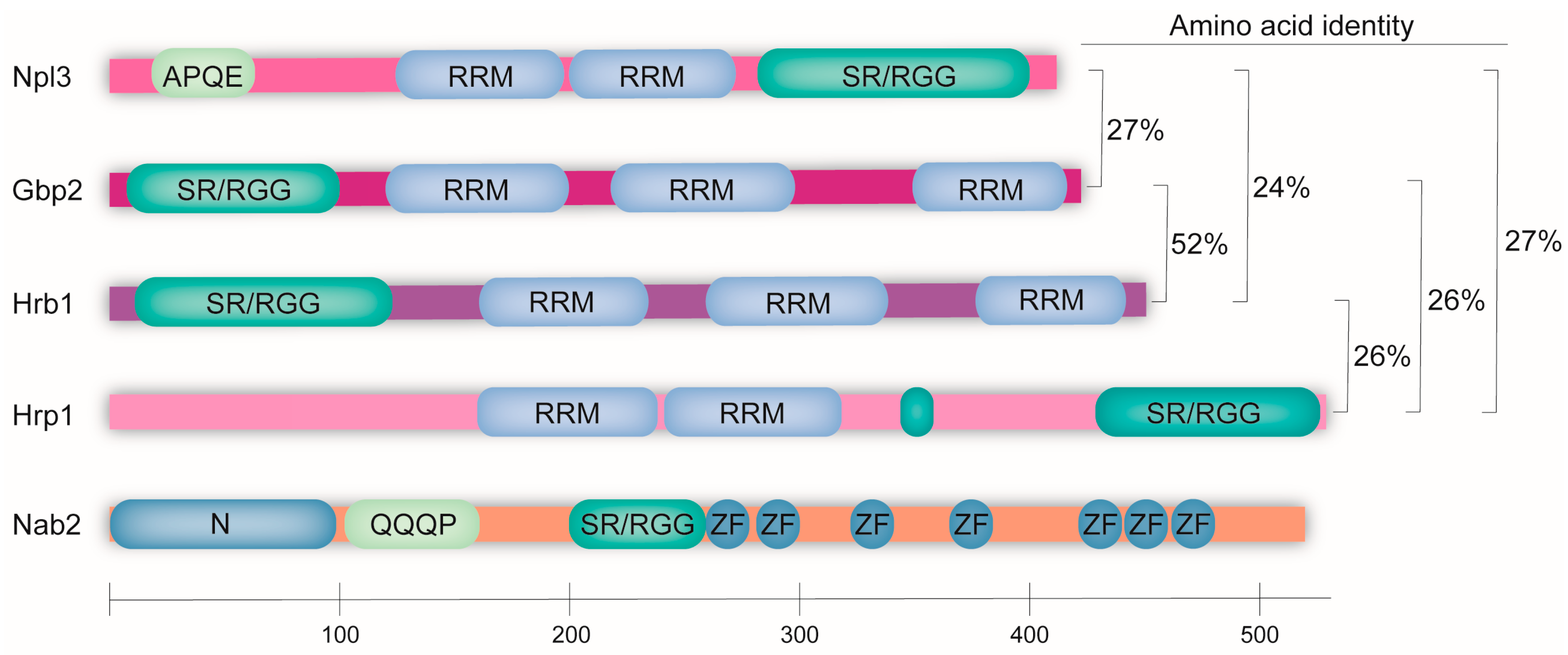
Figure 1. The SR/RGG-rich guard proteins share domains and participate in several quality-control steps during pre-mRNA maturation. The domain structure of the yeast guard proteins and their level of amino acid identity is shown. The five homologous proteins all contain a serine–arginine or arginine–glycine–glycine-rich domain (SR/RGG) that is important for shuttling and protein modifications. They also contain RNA-binding domains, either as RNA-recognition motifs (RRMs) or as a zinc-finger domain (ZF). Npl3 additionally contains a domain with the repeatedly occurring amino acid residues APQE and Nab2, and it contains a Q-rich domain at the N-terminus. Currently, both domains are of unknown function.
Ijms 25 10241 g001
Figure 2. The stepwise mRNA maturation is controlled by the guard proteins. 5′ capping is recognized by Npl3, which identifies the cap-binding complex (CBC)-bound m7G cap. Completed splicing is recognized by the interaction of Gbp2 and Hrb1 with the late spliceosome. Then, 3′ cleavage is monitored by Hrp1, which detects the binding of the CPF-CF protein Rna14. Nab2 recognizes and binds the poly(A) tail at the 3′ end. An involvement of Npl3 in 3′ processing has been proposed. However, the underlying mechanism is currently unclear. In the cytoplasm, the guard proteins support the formation of the ribosome and their positioning in the proper context signals the presence of an intact open reading frame.
Ijms 25 10241 g002
Figure 3. The guard proteins interact with each other. Interaction map showing physical and genetic interactions between each of the guard proteins. Known physical interactions are indicated by a solid line, while genetic interactions are visualized with a dashed line.
Ijms 25 10241 g003
Figure 4. The SR-rich guard proteins mediate mRNA quality control in the nucleus and the cytoplasm. In the nucleus, the SR/RGG motif containing guard proteins is loaded onto the nascent pre-mRNA co-transcriptionally. Npl3 is recruited during early transcription and surveils 5′ capping, Gbp2 and Hrb1 are loaded during splicing and control proper intron excision, while Hrp1 and Nab2 bind during 3′ processing and surveil cleavage and polyadenylation, respectively. All of the guard proteins recruit the export receptor Mex67-Mtr2 upon correct mRNA processing and their coverage signals the export competence of the mRNP to the NPC gatekeeper Mlp1. While Mex67-Mtr2 is depleted from the mRNA by the RNA helicase Dbp5 in the cytoplasm, all of the guard proteins except for Nab2 remain bound to the transcript to facilitate further tasks during translation. Nab2 is replaced by the poly(A) tail-binding protein Pab1. Npl3 assists in monosome formation during translation initiation, where it may assist in subunit joining through interactions between mRNA-bound Npl3 and Npl3 bound to the large ribosomal subunit. Gbp2 and Hrb1 assist in nonsense-mediated decay by signaling the presence of a PTC to the ends of the folded mRNA, thereby inhibiting further translation initiation and recruiting cytoplasmic decapping and degradation factors. Hrp1 may signal the presence of a PTC by associating with the 3′ end of an mRNA.
Ijms 25 10241 g004
Table 1. Overview of the currently established guard proteins 1.
Npl3Gbp2Hrb1Hrp1Nab2
Alternative namesNab1Rlf7Tom34Nab4-
Mtr13 Nab5
Mts1 CFIB
Nop3
Length414 aa427 aa454 aa534 aa525 aa
Molecular weight45.4 kDa48.7 kDa52.1 kDa59.6 kDa58.3 kDa
Median abundance (mol./cell)34,936 ± 26,2913171 ± 18705191 ± 112613,717 ± 83548716 ± 2946
Export receptorMex67Mex67Mex67Mex67Mex67
Import receptorMtr10Mtr10Mtr10Kap104Kap104
Essential in yeast?NoNoNoYesYes
1 aa: amino acids, mol./cell: molecules per cell.
Disclaimer/Publisher’s Note: The statements, opinions and data contained in all publications are solely those of the individual author(s) and contributor(s) and not of MDPI and/or the editor(s). MDPI and/or the editor(s) disclaim responsibility for any injury to people or property resulting from any ideas, methods, instructions or products referred to in the content.

