Article

Gonadal Development and Differentiation of Hybrid F1 Line of Ctenopharyngodon idella (♀) × Squaliobarbus curriculus (♂)

by
Qiaolin Liu
1,2,
Shitao Hu
1,
Xiangbei Tang
1,
Chong Wang
1,
Le Yang
1,
Tiaoyi Xiao
1,2,* and
Baohong Xu
1,2,*
1
Fisheries College, Hunan Agricultural University, Changsha 410128, China
2
Yuelushan Laboratory, Changsha 410128, China
*
Authors to whom correspondence should be addressed.
Int. J. Mol. Sci. 2024, 25(19), 10566; https://doi.org/10.3390/ijms251910566
Submission received: 1 September 2024 / Revised: 23 September 2024 / Accepted: 27 September 2024 / Published: 30 September 2024
(This article belongs to the Special Issue Aquatic Biotechnology and Its Application in Genetic Breeding)

Abstract

:
The hybrid F1 offspring of Ctenopharyngodon idella (♂) and Squaliobarbus curriculus (♀) exhibit heterosis in disease resistance and also show abnormal sex differentiation. To understand the mechanism behind gonadal differentiation in the hybrid F1, we analyzed the transcriptomes of C. idella, S. curriculus, and the hybrid F1; screened for genes related to gonad development in these samples; and measured their expression levels. Our results revealed that compared to either C. idella or S. curriculus, the gene expressions in most sub-pathways of the SNARE interactions in the vesicular transport pathway in the hypothalamus, pituitary, and gonadal tissues of their hybrid F1 offspring were significantly up-regulated. Furthermore, insufficient transcription of genes involved in oocyte meiosis may be the main reason for the insufficient reproductive ability of the hybrid F1 offspring. Through transcriptome screening, we identified key molecules involved in gonad development, including HSD3B7, HSD17B1, HSD17B3, HSD20B2, CYP17A2, CYP1B1, CYP2AA12, UGT2A1, UGT1A1, and FSHR, which showed significant differences in expression levels in the hypothalamus, pituitary, and gonads of these fish. Notably, the expression levels of UGT1A1 in the gonads of the hybrid F1 were significantly higher than those in C. idella and S. curriculus. These results provide a scientific basis for further research on the gonadal differentiation mechanism of hybrid F1 offspring.

1. Introduction

China is known for having some of the most abundant fish resources in the world, making it the leading aquaculture country globally. According to Yue et al. [1], there are over 800 species, and 240 improved varieties have been used in Chinese aquaculture. However, recently, the water quality in China has deteriorated, resulting in an increase in diseases among the main aquaculture species. As a result, breeding improved fish varieties has become a crucial task in promoting the sustainable and healthy development of fisheries. This includes the breeding of high-yield, high-quality, disease-resistant, and stress-resistant varieties, as emphasized by Du et al. [2] and Hu et al. [3].
Hybrid breeding is a widely used method in animal and plant breeding, known for its effectiveness in improving growth rates, meat quality, and disease resistance in fish [4,5]. Through artificial hybridization technology, fish can produce offspring with significant advantages [2]. For instance, a study on a hybrid of Megalobrama amblycephala (♀) × Erythroculter mongolicus (♂) showed that the average weight of the offspring was significantly higher than that of either parent species [6]. Similarly, a study on the crossbreeding of Platichthys bicoloratus (♂) × Paralichthys olivaceus (♀) found that the body height of the offspring increased by more than 4% compared to the parents, demonstrating clear growth advantages [7].
Reproductive fertility is one of the advantages of hybrid fish, although the fertility of hybrid offspring varies. For instance, while the hybrid offspring of Erythroculter ilishaeformis × Carassius auratus [8] and C. auratus × M. amblycephala [9] were fertile, the hybrid offspring of Hypophthalmichthys nobilis × Squaliobarbus curriculus [10] were found to be sterile. The mechanisms of sex determination and differentiation in fish are complex and can be influenced by both genetics and the environment [11,12]. Sex-determining genes are activated during the embryonic period and undergo a series of developmental processes to form either a sperm nest or an ovary [13,14]. Sex differentiation and gonadal development in fish are also closely linked to changes in the levels of sex steroid hormones [15]. These hormones are synthesized by steroid synthetase, which includes the cytochrome P450 gene family (CYP), the hydroxysteroid deoxygenase family (HSD), and other steroid oxidoreductases. Initially, StAR transports cholesterol from the cytosol to the mitochondria, where it is then converted to testosterone by steroid synthetase. This testosterone can then be further converted to either 17β-estradiol (E2) or 11-testosterone (11-KT), ultimately resulting in the production of estrogen and androgen [16].
Grass carp (Ctenopharyngodon idella) is the main species used in freshwater aquaculture in China. To improve the disease resistance and survival rate of C. idella, Jin et al. [17] utilized distant hybridization technology to hybridize C. idella and S. curriculus. They discovered that the hybrid offspring exhibited similar quantitative traits to their female parent. He et al. [18] also reported that the hybrid F1 showed a combination of fast growth inherited from female C. idella and strong resistance to GCRV inherited from male S. curriculus. Transcriptome sequencing of the gonadal tissues of 12-month-old C. idella revealed that dmrt1 and Amh were highly expressed genes in the C. idella sperm nest, whereas CYP19A1A and foxl2 were highly expressed in the C. idella ovary, playing a role in the early gonadal development of C. idella [19]. Our preliminary study showed that the gonadal differentiation of the hybrid F1 offspring of C. idella (♀) × S. curriculus (♂) varied, with some females being partially fertile and others being infertile. The fertility performance of individuals also varied greatly, with diverse degrees of ovarian differentiation. Males were able to produce sperm cells, whereas no mature sperm or normal ejaculation were observed [20]. Anatomical, histological, and production results showed that whereas some females of the hybrid F1 line of C. idella (♀) × S. curriculus (♂) were partially fertile, others were infertile. Similarly, among fertile individuals, some were partially infertile. Males were able to produce sperm cells, but no mature sperm or normal sperm excretion were observed. To uncover the molecular regulatory mechanism of gonadal development in the hybrid F1 line of C. idella (♀) × S. curriculus (♂), transcriptome sequencing was used to screen and detect the expression levels of genes related to C. idella, S. curriculus, and their hybrid F1 offspring. Our results provide valuable data for understanding the gonadal differentiation characteristics of C. idella, S. curriculus, and their hybrid F1 offspring, as well as for the future breeding of parental and hybrid populations.

2. Results

2.1. Transcript Expression Analysis

The transcriptome sequences and corresponding amino acid sequences were predicted using TransDecoder, and a total of 159,917 open reading frames (ORFs) were obtained, of which 142,391 were complete. A total of 8636 transcription factors were predicted using animalTFDB 2.0 (Figure 1A). In total, 43,776, 123,331, 93,639, and 110,049 isoforms were obtained based on the COG, GO, KEGG, and KOG databases, respectively (Table S2).
The homologous species were identified through sequence alignment using the NR database. The main homologous species were Danio rerio, Astyanax mexicanus, Ctenopharyngodon idella, Oncorhynchus mykiss, Cyprinus carpio, Esox luclus, Stegastes partitus, Lepisosteus oculatus, Oreochromis niloticus, and Carassius auratus (Figure 1B). These species are all fish, indicating the reliability of the sequencing data and the absence of contamination from other species. The decision to compare multiple fish species may have been influenced by the differences in data and parameter settings among the species in the database.
The results of the COG function classification showed that the two highest transcriptional groups were general function prediction (18.26%) and signal transduction mechanisms (14.87%) (Figure 1C). Additionally, the results of the eggnog functional classification indicated that 45.3% of the transcripts were classified as having an unknown function (Figure 1D). This suggested that the eggnog functional classification may not be suitable for classifying the transcriptomes of the studied fish.

2.2. Transcriptional Differences between C. idella, S. curriculus, and Their Hybrid F1 Offspring

Cluster analysis of the transcriptome data revealed that the transcriptomes of S. curriculus were more similar to those of their hybrid F1 offspring in the hypothalamus and pituitary tissue compared to C. idella (Figure 2A). Additionally, the hypothalamus transcriptomes of C. idella showed greater similarity to those of the hybrid F1 offspring compared to other samples (Figure 2A). Similarly, the pituitary and gonad transcriptomes of C. idella were also more similar to each other than to other samples (Figure 2A). When comparing the transcriptome of S. curriculus to that of C. idella, the numbers of up- and down-regulated genes were relatively balanced (37,293 vs. 37,197 in the hypothalamus, 41,293 vs. 38,881 in pituitary tissue, and 40,083 vs. 39,446 in gonadal tissue; Figure 2B,E,H). However, the number of up-regulated genes in the transcriptome of the hybrid F1 offspring was significantly higher than that in C. idella and S. curriculus (Figure 2C,D,F,G,I,J). The GO classification results indicated significant differences in the transcription levels of genes involved in cellular components, molecular functions, and biological processes. Specifically, for the cellular component, the transcription levels of genes involved in cells, membranes, macromolecular complexes, organelles, organelle parts, membrane parts, and cell parts accounted for more than 10%. In terms of molecular function, the transcription levels of genes involved in catalytic activity and binding accounted for more than 10%. For biological processes, the transcription levels of genes involved in metabolic processes, cellular processes, signaling, multicellular organismal processes, developmental processes, single-organism processes, response to stimuli, localization, biological regulation, and cellular component organization of biogenesis accounted for more than 10% (Figure 1D).

2.3. Screening and Mining of Gonadal-Development-Related Pathways

The pathways associated with reproduction that were screened via transcriptome analysis mainly include the oxytocin signaling pathway (ko04921), the GnRH signaling pathway, SNARE interactions in vesicular transport (ko04130), renin secretion (ko04924), and oocyte meiosis (ko04114) (Table 1).
Compared to C. idella, only the gene expression in the VAMP7 sub-pathway of the SNARE interactions in the vesicular transport pathway in the hypothalamus, pituitary, and gonadal tissues of S. curriculus was significantly reduced. All other sub-pathways showed a mix of up- and down-regulated genes (Figure S1). However, when compared to either C. idella or S. curriculus, the gene expressions in most sub-pathways of the SNARE interactions in the vesicular transport pathway in the hypothalamus, pituitary, and gonadal tissues of their hybrid F1 offspring were significantly up-regulated. The remaining significantly different sub-pathways also showed a mix of up- and down-regulated genes, with no sub-pathways containing significantly down-regulated genes (Figure 3).
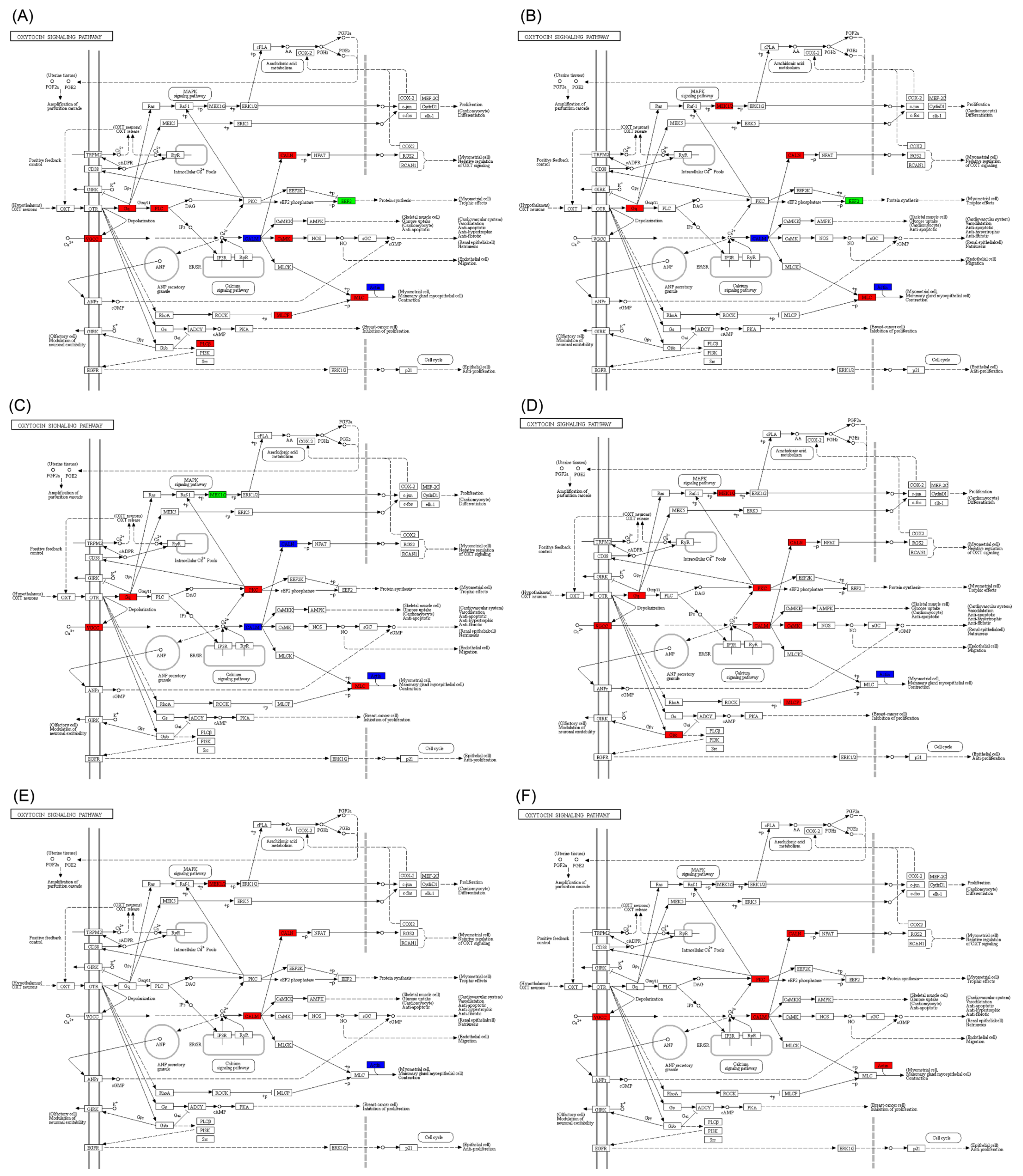
In the oxytocin signaling pathway, the number of up-regulated genes in S. curriculus was lower compared to that in C. idella in the hypothalamus, pituitary, and gonadal tissues. Additionally, the number of sub-pathways with differentially expressed genes was also lower (Figure S2). In comparison to S. curriculus, the expressions of the EEF2 gene in the hypothalamus and pituitary were significantly reduced, whereas the other differential genes showed significant up-regulation or both up- and down-regulation (Figure 4A,B). However, the distribution of these significantly differential genes in sub-pathways across different organs was not consistent (Figure 4A–C). Furthermore, in the hybrid F1 offspring, the significantly different genes were either significantly up-regulated or showed both up- and down-regulation simultaneously (action genes) (Figure 4D–F).
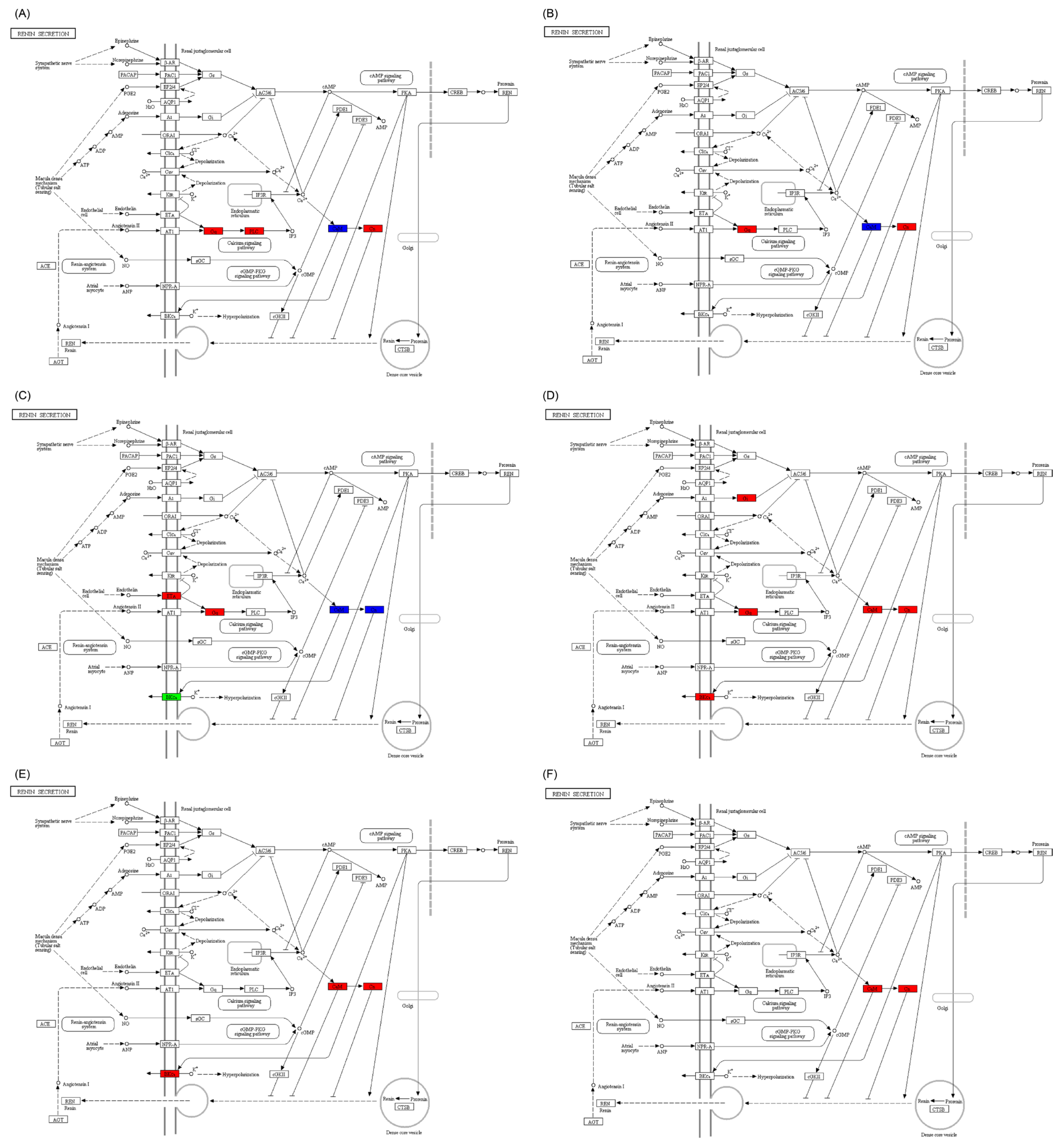
In the renin secretion pathway, the expressions of only a few genes in the three organs of S. curriculus were significantly different compared to those for C. idella (Figure S3). However, in the hybrid F1 offspring, the expressions of different genes were significantly up-regulated or both up- and down-regulated (such as CaM in the hypothalamus and pituitary gland and Cn in the gonadal organs) compared to both S. curriculus and C. idella. The only exception was the BKCa gene, which showed significant down-regulation in the gonadal organ of S. curriculus (Figure 5).
In the oocyte meiosis pathway, there were significant differences in the expression of genes between C. idella and S. curriculus in the hypothalamus, pituitary, and gonadal tissues. Whereas most of the differentially expressed genes in S. curriculus were both up- and down-regulated simultaneously, only a few genes showed significant up- or down-regulation (Figure S4). Interestingly, in the hybrid F1 offspring, the expressions of differentially expressed genes in the hypothalamus and pituitary were either up-regulated or both up- and down-regulated simultaneously (Figure 6A,B), whereas a large number of genes were significantly down-regulated in the gonadal tissue (Figure 6C). Although the number of down-regulated genes in the gonadal tissue of the hybrid F1 offspring was lower than that in C. idella, there was a similar trend to that seen in S. curriculus (Figure 6D–F). These results suggest that insufficient transcription of genes involved in oocyte meiosis may be the main factor contributing to the reduced reproductive ability of the hybrid F1 offspring.
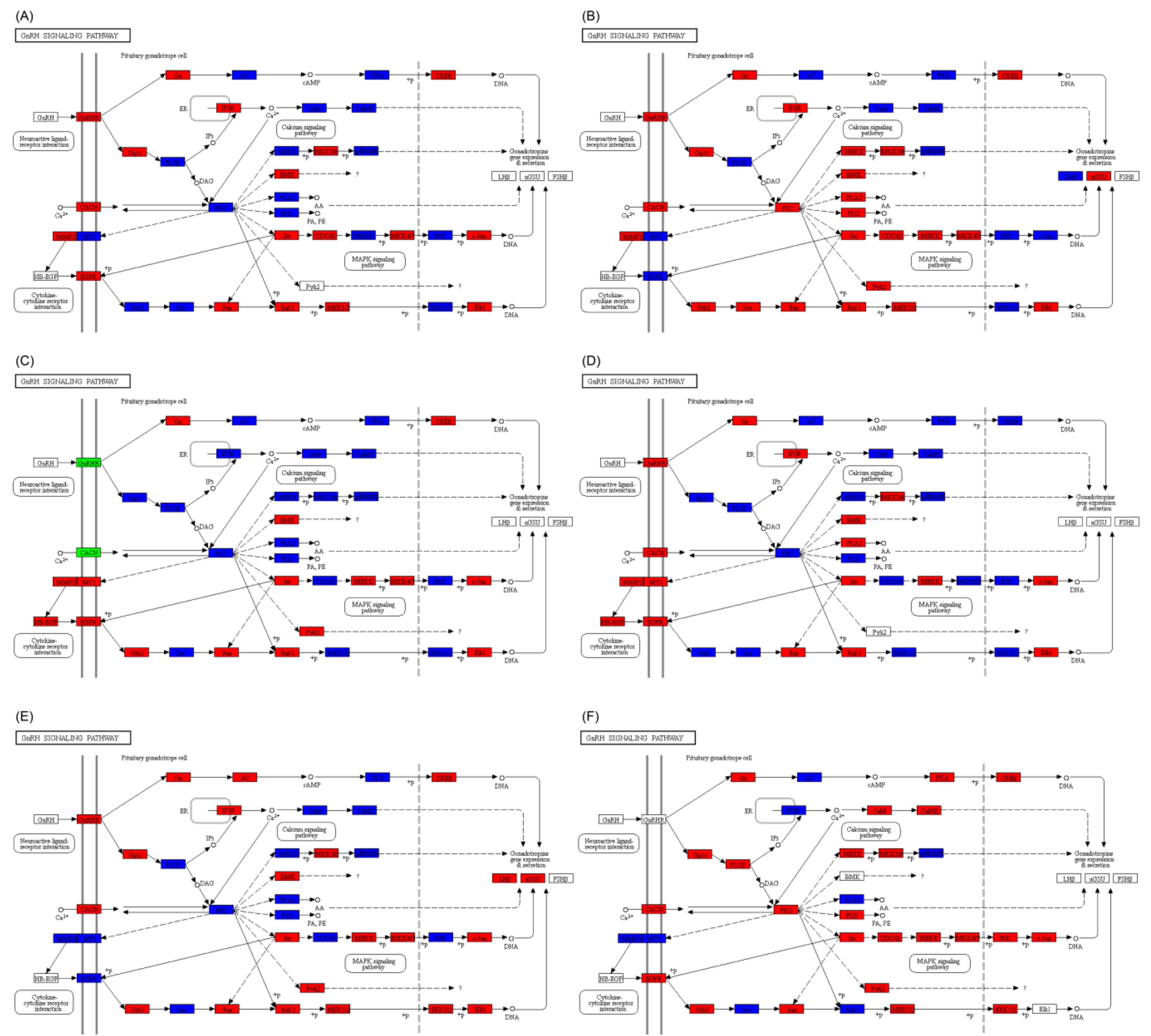
In the GnRH signaling pathway, there were significant differences in the expression of genes between C. idella and S. curriculus in the hypothalamus, pituitary, and gonadal tissues. These differences were observed in both the up- and down-regulation of genes simultaneously (Figure S5). Furthermore, compared with S. curriculus or C. idella, the expressions of differentially expressed genes in the hybrid F1 offspring’s hypothalamus, pituitary, and gonadal tissues were up-regulated and up- and down-regulated simultaneously (Figure 7A–F).

2.4. Expression of Gonadal-Development-Related Genes in C. idella, S. curriculus, and Hybrid F1 Offspring

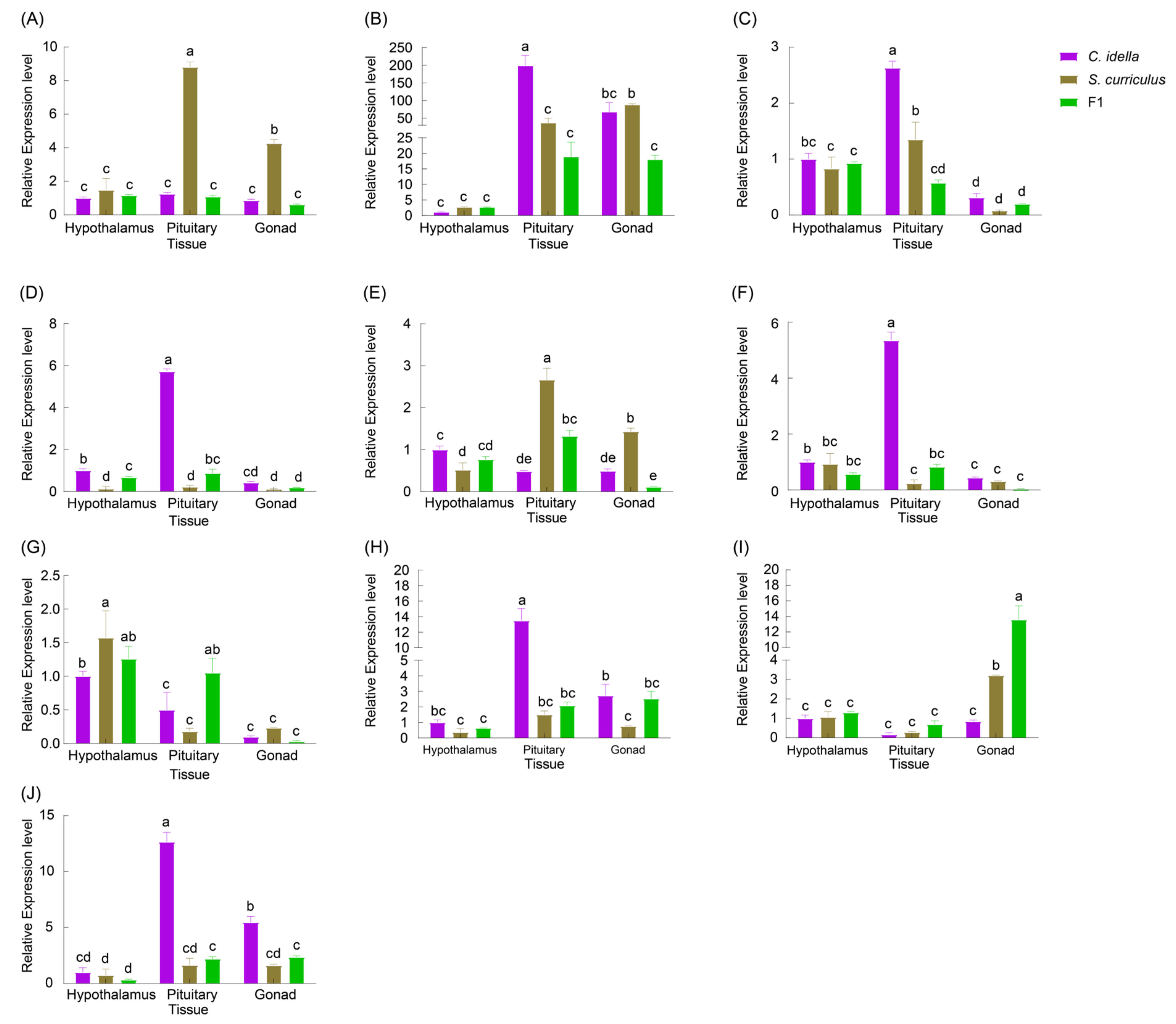
The expressions of the HSD3B7 gene did not show significant differences in the hypothalamus, pituitary, and gonads between C. idella and the hybrid F1 offspring (p > 0.05). However, in S. curriculus, the expression of the HSD3B7 gene was significantly higher in the pituitary gland compared to the gonads, and the expression in the gonads was significantly higher than that in the hypothalamus (Figure 8A). The expression of the HSD3B7 gene in the hypothalamus of C. idella did not significantly differ from that of S. curriculus and the hybrid F1 offspring (p > 0.05; Figure 8A). In the pituitary gland, the expression of the HSD3B7 gene in S. curriculus was significantly higher than that in C. idella and the hybrid F1 offspring (p < 0.05; Figure 8A), whereas the expression in C. idella did not significantly differ from that in the hybrid F1 offspring (p > 0.05; Figure 8A). In the gonads, the expression of the HSD3B7 gene in S. curriculus was significantly higher than that in C. idella and the hybrid F1 offspring (p < 0.05; Figure 8A), whereas the expression in C. idella did not significantly differ from that in the hybrid F1 offspring (p > 0.05; Figure 8A).
The expression of HSD17B1 in the pituitary gland of C. idella was significantly higher than that in the hypothalamus and gonads (p < 0.05; Figure 8B). However, there was no significant difference in expression between the hypothalamus and gonads. In S. curriculus, the expression of the HSD17B1 gene in the gonads was the highest, significantly higher than that in the hypothalamus and pituitary gland. The expression of the HSD17B1 gene in the hypothalamus did not significantly differ from that in the pituitary gland. In the hybrid F1 offspring, there was no significant difference in expression between the hypothalamus, pituitary, and gonadal tissues (p > 0.05; Figure 8B). Additionally, there was no significant difference in expression of the HSD17B1 gene between C. idella, S. curriculus, and the hybrid F1 offspring in the hypothalamus (p > 0.05; Figure 8B). In the pituitary gland, there was no significant difference in expression between S. curriculus and the hybrid F1 offspring (p > 0.05; Figure 8B), whereas the expression for both was significantly lower than that in C. idella (p < 0.05; Figure 8B). In the gonads, there was no significant difference in expression between C. idella and the in the hybrid F1 offspring (p > 0.05; Figure 8B), whereas the expression of the HSD17B1 gene in S. curriculus was significantly higher than that in the hybrid F1 offspring (p < 0.05; Figure 8B).
The expression of the HSD17B3 gene in the pituitary of C. idella was significantly higher than that in the hypothalamus and gonads (p < 0.05; Figure 8C). Additionally, the relative expression level in the hypothalamus was significantly higher than that in the gonads (p < 0.05; Figure 8C). Similarly, in S. curriculus, the expression of the HSD17B3 gene in the pituitary gland was significantly higher than in the hypothalamus and gonads (p < 0.05; Figure 8C), with the expression in the hypothalamus also being significantly higher than in the gonads (p < 0.05; Figure 8C). In the hybrid F1 offspring, the expression of the HSD17B3 gene in the hypothalamus was significantly higher than that in the gonads (p < 0.05; Figure 8C), whereas the expression in the pituitary gland did not differ significantly from that in the hypothalamus and gonads (p > 0.05; Figure 8B). Furthermore, there were no significant differences in the expression of the HSD17B3 gene between C. idella, S. curriculus, and the hybrid F1 offspring in the hypothalamus and gonads (p > 0.05; Figure 8B). However, in the pituitary gland, the expression of the HSD17B3 gene was the highest in C. idellus, followed by that in S. curriculus, and lowest in the hybrid F1 offspring, with all three having significant differences (p < 0.05; Figure 8C).
The expression of the HSD20B2 gene was the highest in the pituitary gland of C. idella, followed by the hypothalamus, and the lowest expression was found in the gonads. All three tissues showed significant differences (p < 0.05; Figure 8D). In contrast, there was no significant difference in the expressions of the HSD20B2 gene in the three tissues of S. curriculus. Additionally, the expression of the HSD20B2 gene in the hypothalamus of the hybrid F1 offspring was not significantly different from that in the pituitary gland (p > 0.05; Figure 8D). However, the expression of this gene in the pituitary gland was significantly higher than that in the gonads (p < 0.05; Figure 8D). Furthermore, the expression of the HSD20B2 gene in the hypothalamus of C. idella was significantly higher than that in S. curriculus and the hybrid F1 offspring, and the expression of this gene in the hypothalamus of the hybrid F1 offspring was significantly higher than that in S. curriculus (p < 0.05; Figure 8D). Furthermore, the expression of the HSD20B2 gene in the hypothalamus of C. idella was significantly higher than that in S. curriculus and the hybrid F1 offspring, and the expression of this gene in the hypothalamus of the hybrid F1 offspring was significantly higher than that in S. curriculus (p < 0.05; Figure 8D). Interestingly, there was no significant difference in the expression of the HSD20B2 gene in the gonads between C. idella, S. curriculus, and the hybrid F1 offspring (p > 0.05; Figure 8D).
The expression of CYP17A2 gene in the hypothalamus of C. idella was significantly higher than that in the pituitary gland and gonads (p < 0.05; Figure 8E), and there was no significant difference in the expressions between the pituitary gland and gonads (p > 0.05; Figure 8E). The expression of the CYP17A2 gene in the pituitary gland of S. curriculus was significantly higher than that in the hypothalamus and gonads, and the expression of this gene in the gonads was significantly higher than that in the hypothalamus (p < 0.05; Figure 8E). There was no significant difference in the expression of the CYP17A2 gene in the hypothalamus and pituitary gland of the hybrid F1 offspring, but both showed significantly higher expression of this gene than that in the gonads (p < 0.05; Figure 8E). There was no significant difference in the expressions of the CYP17A2 gene in the hypothalamus of C. idella and the hybrid F1 offspring, whereas the expressions were significantly higher than that in S. curriculus. In the pituitary gland, the expression of the CYP17A2 gene was the highest in S. curriculus, followed by that in the hybrid F1 offspring, and the lowest in the C. idella, and all of them had significant differences (p < 0.05; Figure 8E). In the gonads, the expression of the CYP17A2 gene was the highest in S. curriculus, being significantly higher than that in C. idella and the hybrid F1 offspring, and there was no significant difference in the expressions of this gene in C. idella and hybrid F1 offspring (p > 0.05; Figure 8E).
The expression of the CYP1B1 gene was the highest in the pituitary gland of C. idella, followed by that in the hypothalamus, and the lowest in the gonads, and all of them had significant differences (p < 0.05; Figure 8F). There was no significant difference in the expressions of the CYP1B1 gene in the hypothalamus, pituitary, and gonads between S. curriculus and the hybrid F1 offspring (p > 0.05; Figure 8F). The expressions of the CYP1B1 gene in the hypothalamus and gonads of C. idella were not significantly different from those of S. curriculus and the hybrid F1 offspring (p > 0.05; Figure 8F). The expression of the CYP1B1 gene was the highest in the pituitary gland of C. idella, being significantly higher than that in S. curriculus and the hybrid F1 offspring (p < 0.05; Figure 8F), and there was no significant difference in the expression of this gene in S. curriculus and the hybrid F1 offspring in the pituitary gland (p > 0.05; Figure 8F).
The expression of the CYP2AA12 gene in the hypothalamus of C. idella was significantly higher than that in the pituitary gland and gonads, and the expression in the pituitary was significantly higher than that in the gonads (p < 0.05; Figure 8G). The expression of the CYP2AA12 gene in the hypothalamus of S. curriculus was significantly higher than that in the pituitary gland and gonads, and there was no significant difference in the expressions of this gene in the pituitary gland and gonads of S. curriculus. The expression of the CYP2AA12 gene in the hypothalamus of the hybrid F1 offspring was not significantly different from that in the pituitary gland (p > 0.05; Figure 8G), whereas it was significantly higher than that in the gonads (p < 0.05; Figure 8G). The expression of the CYP2AA12 gene in the hypothalamus of S. curriculus was significantly higher than that in C. idella (p < 0.05; Figure 8G), whereas there was no significant difference from that in the hybrid F1 offspring (p > 0.05; Figure 8G), and the expressions of this gene in the hypothalamus of C. idella and the hybrid F1 offspring were not significantly different (p > 0.05; Figure 8G). The expression of the CYP2AA12 gene in the pituitary gland of C. idella was not significantly different from that in S. curriculus (p > 0.05; Figure 8G), whereas it was significantly lower than that in the hybrid F1 offspring (p < 0.05; Figure 8G). The expressions of the CYP2AA12 gene in the gonads of C. idella, S. curriculus, and the hybrid F1 offspring were not significantly different (p > 0.05; Figure 8G).
The expression of the UGT2A1 gene in the hypothalamus of C. idella was not significantly different to that in the gonads (p > 0.05; Figure 8H), whereas it was significantly lower than that in the pituitary gland (p < 0.05; Figure 8H). There was no significant difference in the expression of the UGT2A1 gene in the hypothalamus, pituitary gland, and gonads of S. curriculus (p > 0.05; Figure 8H) or in the hybrid F1 offspring (p > 0.05; Figure 8H). There was no significant difference in the expression of the UGT2A1 gene in the hypothalamus of C. idella, S. curriculus, or the hybrid F1 offspring (p > 0.05; Figure 8H). The expression of the UGT2A1 gene in the pituitary gland of S. curriculus and the hybrid F1 offspring showed no significant differences (p > 0.05; Figure 8H), but it was significantly lower than that in C. idella (p < 0.05; Figure 8H). The expression of the UGT2A1 gene in the gonads of the hybrid F1 offspring was not significantly different from that of C. idella or S. curriculus (p > 0.05; Figure 8H), whereas the expression of this gene in the gonads of C. idella was significantly higher than that in the gonads of S. curriculus (p < 0.05; Figure 8H).
There was no difference in the expression of the UGT1A1 gene in the hypothalamus, pituitary gland, or gonads of C. idella (p > 0.05; Figure 8I). The expression of the UGT1A1 gene in the hypothalamus of S. curriculus was not significantly different from that in the pituitary gland (p > 0.05; Figure 8I), while it was significantly lower than that in the gonads (p < 0.05; Figure 8I). The expression of the UGT1A1 gene in the hypothalamus of the hybrid F1 offspring was not significantly different from that in the pituitary gland (p > 0.05; Figure 8I), whereas the expression in both was significantly lower than that in the gonads (p < 0.05; Figure 8I). There was no significant difference in the expression of the UGT1A1 gene in the hypothalamus and pituitary gland between C. idella, S. curriculus, and the hybrid F1 offspring (p > 0.05; Figure 8I). The expression of the UGT1A1 gene in the gonads of C. idella was significantly lower than that in the gonads of S. curriculus and the hybrid F1 offspring, and the expression of this gene in the gonads of S. curriculus was significantly lower than that in the hybrid F1 offspring’s gonads (p < 0.05; Figure 8I).
The expression of the FSHR gene was the highest in the pituitary gland of C. idella, followed by that in the gonads, and lowest in the hypothalamus, and all of the expression levels had significant differences (p < 0.05; Figure 8J). There was no significant difference in the expressions of the FSHR gene in the hypothalamus, pituitary, and gonad of S. curriculus (p > 0.05; Figure 8J). There was no significant difference in the expression of the FSHR gene in the hybrid F1 offspring’s pituitary gland and gonads (p > 0.05; Figure 8J), whereas it was significantly higher in the hypothalamus (p < 0.05; Figure 8J). There was no significant difference in the expressions of the FSHR gene in the hypothalamus of the three fishes (p > 0.05; Figure 8J). There was no significant difference in the expressions in the pituitary gland in S. curriculus and the hybrid F1 offspring (p > 0.05; Figure 8J), whereas they were significantly lower than the expression in the pituitary gland in C. idella (p < 0.05; Figure 8J). There was no significant difference in the expression of the FSHR gene in the gonads of S. curriculus and the hybrid F1 offspring (p > 0.05; Figure 8J), whereas it was significantly lower in the gonads of C. idella (p < 0.05; Figure 8J).

3. Discussion

Fish sex hormones are classified into three categories: protein hormones, glycoprotein hormones, and sex steroid hormones. Protein hormones include adrenocorticotropic hormone (ACTH), gonadotrophic growth hormone (GtH), thyroid-stimulating hormone (TSH), prolactin (PRL), growth hormone (GH), and melanocyte-stimulating hormone (MSH) [21]. Glycoprotein hormones mainly consist of follicle-stimulating hormone (FSH), luteinizing hormone (LH), GtH, and human chorionic gonadotropin (HCG) [22]. Sex steroids, such as 17β-estradiol (E2), 11-testosterone (11-KT), testosterone, estrone (E1), estriol (E3), progesterone, 17α-20β-dihydroxy-4-pregnen-3-one (DHP), and 17α-hydroxyprogesterone (17α-OHP), play a crucial role in inducing oocyte development and promoting maturation (meiosis) [23,24]. 11-KT is particularly important in spermatogenesis, sperm fertilization, and sperm storage [25,26]. The expression of serum 11-ketotestosterone reflects the degree of sperm nest development [27,28]. In fish, external stimulation triggers the hypothalamus to secrete GnRH, which then stimulates the pituitary gland to produce and release GtH (including FSH and LH). GtH travels through the bloodstream to the gonads, where it prompts the production of sex steroid hormones. These hormones, in turn, affect the development of eggs/sperm and regulate reproductive behavior [24,29]. GtH also regulates the secretion of sex hormones (mainly testosterone and E2), the initiation of the reproductive cycle, and the differentiation of germ cells. Sex hormones, in turn, regulate the synthesis and secretion of GnRH and GtH through negative feedback [30]. Fish have the ability to undergo sexual reversal by blocking the synthesis of exogenous hormones [31]. For instance, providing androgen to Oncorhynchus mykiss inhibits the synthesis of estrogen and results in virilization [32]. Sex steroids also play a crucial role in the process of sex reversal in species such as Epinephelus akaara [33], Oreochromis mossambicus [34], and Oryzias latipes [35]. Therefore, sex steroid hormones are essential for the development of gonadal differentiation. Our results suggested that insufficient transcription of genes involved in oocyte meiosis may be the main reason for the reduced reproductive ability of hybrid F1 offspring.
Transcriptome-sequencing technology has been widely applied to research on fish reproduction. In a study by Lin et al. [36], 12 key candidate genes related to sex determination and gonadal differentiation were identified through the transcriptome sequencing of mature gonadal tissues of Symphysodon haraldi. Similarly, He [37] used transcriptome sequencing to screen 19 genes related to the sex steroid hormone synthesis pathway and its receptor genes in stage III sperm nest and ovarian tissues of Scatophagus argus. The results showed that CYP11A1, CYP11B2, CYP19A1B, HSD11B2, HSD3B1, and HSD3B7 genes were overexpressed in the sperm nest, whereas CYP19A1A, HSD17B1, HSD17B8, HSD17B12, and HSD17B14 genes were overexpressed in the ovaries. In a study by Tao et al. [38], transcriptome sequencing was performed on male and female Oreochromis niloticus specimens that were exposed to high temperatures. The results showed that the expressions of genes related to androgen synthesis, such as HSD17B7 and 3β-HSD, increased in the male high-temperature treatment group, whereas the expression of estrogen synthesis genes, such as CYP19A1A, decreased. This suggests that the synthesis of sex hormones may play a role in the process of sexual reversal in O. niloticus under high-temperature treatment. In a study by Qin [39], transcriptome sequencing was performed on the brain and gonadal tissues of pseudomale, gynogenetic, and normal male and female Nibea albiflora. The results showed that male-related genes, such as dmrt1, Gsdf, Amh, and Ar, and female-related genes, such as CYP19A, zp3, zp4, and foxl2, were identified. Interestingly, dmrt1 was only expressed in the sperm nest, whereas CYP19A was only expressed in the ovary.
The hypothalamic–pituitary–gonadal (HPG) axis is a crucial reproductive axis for studying sexual maturation and development. The neural and endocrine systems, with HPG as the core, primarily regulate gonadal development and gamete maturation in fish [40,41]. By conducting a differential expression analysis of the HPG axis in C. idellus, S. curriculus, and the hybrid F1 offspring, we accumulated enough data to analyze the molecular mechanisms underlying the difference in sexual maturity times between these two species and the reproductive disorders in the hybrid F1 offspring. Our study analyzed the HPG transcriptomes of these three fish species and identified several pathways associated with reproduction, including the oxytocin signaling pathway (ko04921), the GnRH signaling pathway, SNARE interactions in vesicular transport (ko04130), renin secretion (ko04924), and oocyte meiosis (ko04114). These results provide valuable omics information on C. idellus, S. curriculus, and their hybrid F1 offspring and serve as a reference for the further analysis of reproductive development in cyprinid fishes.
HSD3B7, HSD17B1, HSD17B3, and HSD20B2 belong to the short-chain dehydrogenation/reductase (SDR) superfamily, which plays key roles in steroid hormones, biological metabolism, and redox sensing mechanisms. HSD3βs is involved in the oxidation and reduction of steroid hormones, and the expression pattern of HSD3βs is closely related to the growth-and-development period of animals. Among HSD3βs, HSD3B7 plays a crucial role in the biosynthesis of all hormonal steroids. In the tilapia genome, two HSD3β genes have been identified, which may have an important impact on gonadal differentiation in tilapia [42]. HSD17βs affect the function of sex steroid hormones by regulating the binding of sex steroid hormones to receptors and controlling the expression of sex steroid hormones [43]. In Osteichthyes, HSD17B1 catalyzes the transition between estrogen ketone and estrogen [44] and may also be involved in gonadal differentiation and development through sex steroid hormones [45].
CYP11A1 is the first step in the synthesis of sex steroids and catalyzes the conversion of progesterone to pregnenolone. This gene has been cloned and identified in several fish species, including Oryzias latipes [46], Odontesthes bonariensis [47], Danio rerio [48], and Anguilla japonica [49], and is primarily expressed in the ovaries. It is believed to play a crucial role in oocyte development [50]. CYP17 is a microsomal cytochrome P450 enzyme that promotes the production of sex steroid hormones and cortisol. This gene has been cloned and identified in Sebastods schlegelii, Paralichthys olivaceus, Verasper moseri, and Cynoglossus semilaevis [10,51,52,53]. CYP19A1 has been identified as an early biomarker of ovarian differentiation in fish [50] and isolated in various bony fish species [54,55]. 3β-HSD is a gene that encodes 3β-hydroxysteroid dehydrogenase, which is involved in the conversion of sterol hormones in hormone-producing tissues. This gene has been cloned in the genomes of Danio rerio, Oreochromis mossambicus, and Oryzias latipes [56,57]. In bony fishes, Ad4BP/sf1 binds to the CYP19A promoter and affects the expression characteristics of aromatase genes, thereby regulating the synthesis of sex hormones [58]. sf1 binding sites in the promoter region of CYP19A1A have been found in Gobiocypris rarus, Oreochromis niloticus, and Lateolabrax japonicus [59,60]. In tilapia, the expression of CYP19A1A in the gonads of females decreases with the inhibition of foxl2 expression, resulting in a decrease in serum E2 levels and ultimately leading to the induction of male characteristics [61]. Mutations in foxl2 or CYP19A1A can cause sexual reversal from females to males [62]. In zebrafish, double mutants of the two subtypes of foxl2a and foxl2b can result in complete female sex reversal in the early stages [63].
In this study, we observed significant differences in the expression of HSD3B7, HSD17B1, HSD17B3, HSD20B2, CYP17A2, CYP1B1, CYP2AA12, UGT2A1, UGT1A1, and FSHR in the hypothalamus, pituitary, and gonadal tissues of C. idella, S. curriculus, and their hybrid F1 offspring. Specifically, the expression of UGT1A1 was significantly higher in the gonads of the hybrid F1 offspring compared to that in C. idella and S. curriculus. Additionally, the expressions of HSD3B7 and CYP17A2 in the pituitary gland and gonads of S. curriculus were significantly higher than those in C. idella and the hybrid F1 offspring. Furthermore, the expression of CYP2AA12 in the hypothalamus of S. curriculus was significantly higher than that in C. idella and the hybrid F1 offspring, with the most significant expression observed in the pituitary gland of the hybrid F1 offspring. With the exception of UGT1A1, the expressions of the remaining nine genes in the pituitary tissues of the three species were significantly different. Specifically, the expressions of HSD17B1, HSD17B3, HSD20B2, CYP1B1, UGT2A1, and FSHR in the pituitary tissues of C. idella were significantly higher than in S. curriculus and the hybrid F1 offspring. Further research is needed to determine which of these genes are the key factors in regulating F1 gonadal development in the hybrid C. idella (♀) × S. curriculus (♂) and the related molecular mechanisms that regulate the synthesis of steroid hormones and affect the differentiation and development of gonadal differentiation.

4. Materials and Methods

4.1. Experimental Design and Sample Collection

The animal experiments were conducted in accordance with the guidelines approved by the Animal Care and Use Committee of Hunan Agricultural University (Changsha, China; Approval Code: 201903295; Approval Date: 13 September 2019). Based on the results of our previous histological study on hybrid F1 gonadal development [20], 150-day-old specimens of C. idella (Gc), S. curriculus (Sc), and their hybrid F1 offspring (Zj) were collected from Xiangyin Institute of Fishery Sciences in Hunan Province in October 2019. The fish were cultured in different cages in an indoor circulating water culture system with an average water temperature of 26.0 °C and containing 6.5 mg/L of dissolved oxygen. The fish were fed 3% of their average body weight in the same commercial feed twice daily (8:00 and 18:00). Fifteen larvae from each species were randomly collected from the cages and anesthetized with 200 mg/L of tricaine methanesulfonate (MS-222). Due to the small size of the fish and the small quantity of tissue samples, five fish tissues of each species were mixed to create a sample for transcriptome sequencing. Three samples were collected for each species, with separate samples for the hypothalamus (xqn), pituitary (ct), and gonadal (xx) tissues. The samples were quickly frozen using liquid nitrogen and stored at −80 °C (Table S1).

4.2. Total RNA Extraction and Transcriptome Sequencing

Total RNA was extracted from fish tissues using a TRIzol RNA extraction kit (Omega Bio-tek, Norcross, GA, USA). To enrich for mRNA, magnetic beads containing OligodT (Yeasen Biotechnology, Shanghai, China) were used, followed by random fragmentation using reagents (Thermo Fisher Scientific, Waltham, MA, USA). The resulting eukaryotic mRNA was then used as a template for cDNAs synthesis and purified using AMPure XP beads (Beckman Coulter, Brea, CA, USA). Transcriptome sequencing was performed using the Illumina HiSeq platform at Biomarker Technologies Co., Ltd. (Beijing, China), following the previously described method [64].
After checking for redundancy, the integrity of deredundant transcriptome was evaluated using BUSCO [65]. Single-copy gene sets from multiple evolutionary branches were then constructed using the BUSCO-referenced OrthoDB database to assess the accuracy and completeness of the transcripts. The coding sequences (CDSs) were analyzed using TransDecoder. The LncRNAs of the transcriptomes were analyzed and predicted using CPC [66], CNCI, pfam protein domain, and CPAT. The transcript sequences were then compared to the animalTFDB 2.0 database [67] to identify any potential transcription factors. Non-redundant transcript sequences were also compared to the NR [68], Swissprot [69], GO [70], COG [71], KOG, Pfam [72], and KEGG [73] databases using BLAST [74] to obtain functional annotations for the transcripts. Transcript expression analysis was performed using RSEM [75], and differential expression analysis was conducted using DESeq [76].

4.3. RT-qPCR

The expression levels of 10 gonadal developmental genes (HSD3B7, HSD17B1, HSD17B3, HSD20B2, CYP17A2, CYP1B1, CYP2AA12, UGT2A1, UGT1A1, and FSHR; Table 2) in the hypothalamus, pituitary, and gonadal tissues were detected using real-time qPCR as previously described [77].

4.4. Data Analysis

Data were presented as means ± standard deviation (SD). To analyze the data, a one-way ANOVA was conducted using R 4.2.3 [78]. Statistical significance was set at a p-value of less than 0.05.

5. Conclusions

After conducting transcriptome analysis on C. idella, S. curriculus, and their hybrid F1 offspring, several pathways related to reproduction were identified. These mainly included the oxytocin signaling pathway (ko04921), the GnRH signaling pathway, SNARE interactions in vesicular transport (ko04130), renin secretion (ko04924), and oocyte meiosis (ko04114). The insufficient transcription of genes involved in oocyte meiosis was found to be the main factor contributing to the inadequate reproductive ability of the hybrid F1 offspring. Through transcriptome analysis, a total of 10 key genes responsible for gonadal development were identified, including HSD3B7, HSD17B1, HSD17B3, HSD20B2, CYP17A2, CYP1B1, CYP2AA12, UGT2A1, UGT1A1, and FSHR. These genes showed varying expression patterns in different tissues of C. idella, S. curriculus, and their hybrid F1 offspring.

Supplementary Materials

The following supporting information can be downloaded at https://www.mdpi.com/article/10.3390/ijms251910566/s1.

Author Contributions

Conceptualization, Q.L., T.X. and B.X.; methodology, Q.L., S.H., C.W. and T.X.; software, Q.L. and T.X.; validation, X.T. and L.Y.; formal analysis, Q.L., S.H. and C.W.; investigation, Q.L., S.H., C.W., X.T. and L.Y.; resources, Q.L. and T.X.; data curation, X.T. and T.X.; writing-original draft preparation, Q.L.; writing-review and editing, T.X. and B.X.; visualization, Q.L. and S.H.; project administration, T.X. and B.X. All authors have read and agreed to the published version of the manuscript.

Funding

This work was supported by the National Key Research and Development Program of China (2023YFD2401602) and the Natural Science Foundation of Hunan Province (2022JJ30289).

Institutional Review Board Statement

The animal experiments were conducted in accordance with the guidelines approved by the Animal Care and Use Committee of Hunan Agricultural University (Changsha, China; Approval Code: 201903295; Approval Date: 13 September 2019).

Informed Consent Statement

Not applicable.

Data Availability Statement

The data that support the findings of this study are available in the Sequence Read Archive (SRA) under the accession number PRJNA1146714.

Acknowledgments

We thank an anonymous technician at Guangdong Meilikang Bio-Science Ltd., China, for their assistance with visualization.

Conflicts of Interest

The authors declare no conflicts of interest.

References

  1. Yue, G.H.; Tay, Y.X.; Wong, J.; Shen, Y.; Xia, J. Aquaculture Species Diversification in China. Aquac. Fish. 2024, 9, 206–217. [Google Scholar] [CrossRef]
  2. Du, Z.; Nie, P.; Liu, J. Genetic Improvement for Aquaculture Species: A Promising Approach for Aquaculture Challenges and Development. Rev. Aquac. 2021, 13, 1756–1757. [Google Scholar] [CrossRef]
  3. Hu, F.; Zhong, H.; Wu, C.; Wang, S.; Guo, Z.; Tao, M.; Zhang, C.; Gong, D.; Gao, X.; Tang, C.; et al. Development of Fisheries in China. Reprod. Breed. 2021, 1, 64–79. [Google Scholar] [CrossRef]
  4. Liu, Q.; Liu, J.; Liang, Q.; Qi, Y.; Tao, M.; Zhang, C.; Qin, Q.; Zhao, R.; Chen, B.; Liu, S. A Hybrid Lineage Derived from Hybridization of Carassius cuvieri and Carassius auratus Red Var. and a New Type of Improved Fish Obtained by Back-Crossing. Aquaculture 2019, 505, 173–182. [Google Scholar] [CrossRef]
  5. Labroo, M.R.; Studer, A.J.; Rutkoski, J.E. Heterosis and Hybrid Crop Breeding: A Multidisciplinary Review. Front. Genet. 2021, 12, 643761. [Google Scholar] [CrossRef] [PubMed]
  6. Fan, J. Study on the Biological Characteristics of Offspring of Megalobrama amblycephala × Culter mongolianus. Master Thesis, Hunan Normal University, Changsha, China, 2020. [Google Scholar]
  7. Wang, X.; You, F.; Ni, G.; Zhang, Q.; Li, S. Hybridization Betweed Stone Flounder Kareius bicoloratus and Olive Flounder Paralichthys olivaceus. Mar. Sci. 2003, 27, 4–9. [Google Scholar]
  8. Yan, J.; Liu, L.; Liu, S.; Guo, X.; Liu, Y. Comparative Analysis of Mitochondrial Control Region in Polyploid Hybrids of Red Crucian Carp (Carassius auratus) x Blunt Snout Bream (Megalobrama amblycephala). Fish Physiol. Biochem. 2010, 36, 263–272. [Google Scholar] [CrossRef] [PubMed]
  9. He, W.; Qin, Q.; Liu, S.; Li, T.; Wang, J.; Xiao, J.; Xie, L.; Zhang, C.; Liu, Y. Organization and Variation Analysis of 5S RDNA in Different Ploidy-Level Hybrids of Red Crucian Carp × Topmouth Culter. PLoS ONE 2012, 7, e38976. [Google Scholar] [CrossRef]
  10. Jin, W.; Yu, L.; Yang, J.; Gao, Y.; Zhu, Z.; Zhao, Y. Biological Characteristics of F1 Hybrid Generations from Squaliobarbus curriculus (♀) × Aristichthys nobilis (♂). J. Fish. Sci. China 2012, 19, 611–619. [Google Scholar] [CrossRef]
  11. Stelkens, R.B.; Wedekind, C. Environmental Sex Reversal, Trojan Sex Genes, and Sex Ratio Adjustment: Conditions and Population Consequences. Mol. Ecol. 2010, 19, 627–646. [Google Scholar] [CrossRef]
  12. Wang, T.; Yu, Y.; Li, S.; Li, F. Molecular Mechanisms of Sex Determination and Differentiation in Decapod Crustaceans for Potential Aquaculture Applications: An Overview. Rev. Aquac. 2024, 16, 1819–1839. [Google Scholar] [CrossRef]
  13. Matson, C.K.; Zarkower, D. Sex and the Singular DM Domain: Insights into Sexual Regulation, Evolution and Plasticity. Nat. Rev. Genet. 2012, 13, 163–174. [Google Scholar] [CrossRef] [PubMed]
  14. Wagner, S.; Whiteley, S.L.; Castelli, M.; Patel, H.R.; Deveson, I.W.; Blackburn, J.; Holleley, C.E.; Marshall Graves, J.A.; Georges, A. Gene Expression of Male Pathway Genes Sox9 and Amh during Early Sex Differentiation in a Reptile Departs from the Classical Amniote Model. BMC Genom. 2023, 24, 243. [Google Scholar] [CrossRef] [PubMed]
  15. Hayashida, T.; Soma, S.; Nakamura, Y.; Higuchi, K.; Kazeto, Y.; Gen, K. Transcriptome Characterization of Gonadal Sex Differentiation in Pacific Bluefin Tuna, Thunnus orientalis (Temminck et Schlegel). Sci. Rep. 2023, 13, 13867. [Google Scholar] [CrossRef] [PubMed]
  16. Hammes, S.R.; Levin, E.R. Impact of Estrogens in Males and Androgens in Females. J. Clin. Investig. 2019, 129, 1818–1826. [Google Scholar] [CrossRef]
  17. Jin, X.; Jin, H.; Wang, M.; Zheng, T. Comparison of Genetic Characteristics between the F1 Hybrid ( Ctenopharyngodon idella × Squaliobarbus cursiculus) and Its Parents. Life Sci. Res. 1999, 3, 316–320. [Google Scholar]
  18. He, M.; Xiao, T.; Liu, Q.; Li, D.; Li, W.; Deng, Y. Morphological Characteristics Analysis of Ctenopharyngodon idellus, Squaliobarbus curriculus and Their Reciprocal Hybrids F1. J. Hunan Univ. Arts Sci. (Sci. Technol.) 2015, 27, 36–42,47. [Google Scholar]
  19. Yao, W.; Jiang, P.; Bai, J.; Ma, D. Analysis of Differential Expressed Genes between Male and Female Gonads of Grass Carp (Ctenopharyngodon idellus) Based on High Throughput Transcriptome Group Sequencing. Genomics Appl. Biol. 2019, 38, 3901–3911. [Google Scholar]
  20. Tang, X. Study on the Characteristics of Gonadal Differentiation of Hybrid F1 of Grass Carp (♀) × Barbel Chub (♂). Ph.D. Thesis, Hunan Agricultural University, Changsha, China, 2021. [Google Scholar]
  21. Lin, J.; Zhan, J.; Shuai, D.; Wang, T.; Wang, Q.; Wang, L.; Yu, X.; Liu, L. Quantification of 6 Sexual Steroid Hormones in the Ovary of Marbled Eel Anguilla marmorata during Artificial Induced Maturation. J. Fish. China 2015, 39, 1341–1349. [Google Scholar]
  22. Cahoreau, C.; Klett, D.; Combarnous, Y. Structure-Function Relationships of Glycoprotein Hormones and Their Subunits’ Ancestors. Front. Endocrinol. 2015, 6, 26. [Google Scholar] [CrossRef] [PubMed]
  23. Rajakumar, A.; Senthilkumaran, B. Steroidogenesis and Its Regulation in Teleost—A Review. Fish Physiol. Biochem. 2020, 46, 803–818. [Google Scholar] [CrossRef]
  24. Kumar, P.; Behera, P.; Christina, L.; Kailasam, M. Sex Hormones and Their Role in Gonad Development and Reproductive Cycle of Fishes. In Recent Updates in Molecular Endocrinology and Reproductive Physiology of Fish; Springer: Singapore, 2021; pp. 1–22. [Google Scholar]
  25. Walker, W.H. Testosterone Signaling and the Regulation of Spermatogenesis. Spermatogenesis 2011, 1, 116–120. [Google Scholar] [CrossRef] [PubMed]
  26. Witherspoon, L.; Flannigan, R. It Puts the T’s in Fertility: Testosterone and Spermatogenesis. Int. J. Impot. Res. 2022, 34, 669–672. [Google Scholar] [CrossRef]
  27. Imamichi, Y.; Yuhki, K.; Orisaka, M.; Kitano, T.; Mukai, K.; Ushikubi, F.; Taniguchi, T.; Umezawa, A.; Miyamoto, K.; Yazawa, T. 11-Ketotestosterone Is a Major Androgen Produced in Human Gonads. J. Clin. Endocrinol. Metab. 2016, 101, 3582–3591. [Google Scholar] [CrossRef]
  28. Zhang, Q.; Ye, D.; Wang, H.; Wang, Y.; Hu, W.; Sun, Y. Zebrafish Cyp11c1 Knockout Reveals the Roles of 11-Ketotestosterone and Cortisol in Sexual Development and Reproduction. Endocrinology 2020, 161, bqaa048. [Google Scholar] [CrossRef] [PubMed]
  29. Casati, L.; Ciceri, S.; Maggi, R.; Bottai, D. Physiological and Pharmacological Overview of the Gonadotropin Releasing Hormone. Biochem. Pharmacol. 2023, 212, 115553. [Google Scholar] [CrossRef]
  30. RE, P.; KL, Y. Neuroendocrine Regulation of Ovulation in Fishes: Basic and Applied Aspects. Reviews in Fish Biology and Fisheries. Rev. Fish Biol. Fish. 1997, 7, 173–197. [Google Scholar]
  31. Li, M.; Sun, L.; Wang, D. Roles of Estrogens in Fish Sexual Plasticity and Sex Differentiation. Gen. Comp. Endocrinol. 2019, 277, 9–16. [Google Scholar] [CrossRef]
  32. Vizziano, D.; Baron, D.; Randuineau, G.; Mahè, S.; Cauty, C.; Guiguen, Y. Rainbow Trout Gonadal Masculinization Induced by Inhibition of Estrogen Synthesis Is More Physiological than Masculinization Induced by Androgen Supplementation. Biol. Reprod. 2008, 78, 939–946. [Google Scholar] [CrossRef]
  33. Li, G.; Liu, X.; Lin, H. Effects of 17α-Methyltestosterone on Sex Reversal in Red-Spotted Grouper, Epinephelus akaara. J. Fish. China 2006, 30, 145–150. [Google Scholar]
  34. Ruksana, S.; Pandit, N.P.; Nakamura, M. Efficacy of Exemestane, a New Generation of Aromatase Inhibitor, on Sex Differentiation in a Gonochoristic Fish. Comp. Biochem. Physiol. C. Toxicol. Pharmacol. 2010, 152, 69–74. [Google Scholar] [CrossRef]
  35. Paul-Prasanth, B.; Bhandari, R.K.; Kobayashi, T.; Horiguchi, R.; Kobayashi, Y.; Nakamoto, M.; Shibata, Y.; Sakai, F.; Nakamura, M.; Nagahama, Y. Estrogen Oversees the Maintenance of the Female Genetic Program in Terminally Differentiated Gonochorists. Sci. Rep. 2013, 3, 2862. [Google Scholar] [CrossRef] [PubMed]
  36. Lin, R.; Gao, J.; Jin, S.; Zhao, Y.; Chen, Z. Cloning and Expression Analysis of Vasa Gene in Symphysodon haraldi. J. Shanghai Ocean Univ. 2017, 26, 330–338. [Google Scholar]
  37. He, F. Transcriptome Analysis of Male and Female Gonads and Study on Sex Steroid Hormone in Spotted Scat (Scatophagus argus). Master’s Thesis, Guangdong Ocean University, Zhanjing, China, 2019. [Google Scholar]
  38. Tao, W.; Yuan, J.; Zhou, L.; Sun, L.; Sun, Y.; Yang, S.; Li, M.; Zeng, S.; Huang, B.; Wang, D. Characterization of Gonadal Transcriptomes from Nile Tilapia (Oreochromis niloticus) Reveals Differentially Expressed Genes. PLoS ONE 2013, 8, e63604. [Google Scholar] [CrossRef] [PubMed]
  39. Qin, Z. Induction of Neo-Male and Screening Analysis of Sex Related Genes in Yellow Drum. Master’s Thesis, Zhejiang Ocean University, Hangzhou, China, 2020. [Google Scholar]
  40. Nishimura, T.; Tanaka, M. Gonadaldevelopment in Fish. Sex. Dev. 2014, 8, 252–261. [Google Scholar] [CrossRef] [PubMed]
  41. Tenugu, S.; Pranoty, A.; Mamta, S.-K.; Senthilkumaran, B. Development and Organisation of Gonadal Steroidogenesis in Bony Fishes—A Review. Aquac. Fish. 2021, 6, 223–246. [Google Scholar] [CrossRef]
  42. Tao, W.; Xu, L.; Zhao, L.; Zhu, Z.; Wu, X.; Min, Q.; Wang, D.; Zhou, Q. High-quality Chromosome-level Genomes of Two Tilapia Species Reveal Their Evolution of Repeat Sequences and Sex Chromosomes. Mol. Ecol. Resour. 2021, 21, 543–560. [Google Scholar] [CrossRef]
  43. Proaño, S.B.; Miller, C.K.; Krentzel, A.A.; Dorris, D.M.; Meitzen, J. Sex Steroid Hormones, the Estrous Cycle, and Rapid Modulation of Glutamatergic Synapse Properties in the Striatal Brain Regions with a Focus on 17β-Estradiol and the Nucleus Accumbens. Steroids 2024, 201, 109344. [Google Scholar] [CrossRef] [PubMed]
  44. Sinreih, M.; Gjorgoska, M.; Möller, G.; Adamski, J.; Rižner, T.L. 17β-Hydroxysteroid Dehydrogenases Types 1 and 2: Enzymatic Assays Based on Radiometric and Mass-Spectrometric Detection. Methods Enzym. 2023, 689, 201–234. [Google Scholar]
  45. Rajakumar, A.; Senthilkumaran, B. Molecular Cloning and Expression Analysis of 17b-Hydroxysteroid Dehydrogenase 1 and 12 during Gonadal Development, Recrudescence and after in Vivo HCG Induction in Catfish Clarias batrachus. Steroids 2014, 92, 81–89. [Google Scholar] [CrossRef] [PubMed]
  46. Nakamoto, M.; Fukasawa, M.; Orii, S.; Shimamori, K.; Maeda, T.; Suzuki, A.; Matsuda, M.; Kobayashi, T.; Nagahama, Y.; Shibata, N. Cloning and Expression of Medaka Cholesterol Side Chain Cleavage Cytochrome P450 during Gonadal Development. Dev. Growth Differ. 2010, 52, 385–395. [Google Scholar] [CrossRef] [PubMed]
  47. Blasco, M.; Fernandino, J.I.; Guilgur, L.G.; Strüssmann, C.A.; Somoza, G.M.; Vizziano-Cantonnet, D. Molecular Characterization of cyp11a1 and cyp11b1 and Their Gene Expression Profile in Pejerrey (Odontesthes bonariensis) during Early Gonadal Development. Comp. Biochem. Physiol. Part A Mol. Integr. Physiol. 2010, 156, 110–118. [Google Scholar] [CrossRef]
  48. Hsu, H.-J.; Hsiao, P.; Kuo, M.-W.; Chung, B. Expression of Zebrafish cyp11a1 as a Maternal Transcript and in Yolk Syncytial Layer. Gene Expr. Patterns 2002, 2, 219–222. [Google Scholar] [CrossRef] [PubMed]
  49. Kazeto, Y.; Ijiri, S.; Adachi, S.; Yamauchi, K. Cloning and Characterization of a CDNA Encoding Cholesterol Side-Chain Cleavage Cytochrome P450 (CYP11A1): Tissue-Distribution and Changes in the Transcript Abundance in Ovarian Tissue of Japanese Eel, Anguilla japonica, during Artificially Induced Sexual. J. Steroid Biochem. Mol. Biol. 2006, 99, 121–128. [Google Scholar] [CrossRef]
  50. Guiguen, Y.; Fostier, A.; Piferrer, F.; Chang, C.-F. Ovarian Aromatase and Estrogens: A Pivotal Role for Gonadal Sex Differentiation and Sex Change in Fish. Gen. Comp. Endocrinol. 2010, 165, 352–366. [Google Scholar] [CrossRef] [PubMed]
  51. Chen, C.F.; Wen, H.S.; Wang, Z.P.; He, F.; Zhang, J.R.; Chen, X.Y.; Jin, G.X.; Shi, B.; Shi, D.; Yang, Y.P.; et al. Cloning and Expression of P450c17-I (17α-Hydroxylase/17,20-Lyase) in Brain and Ovary during Gonad Development in Cynoglossus semilaevis. Fish Physiol. Biochem. 2010, 36, 1001–1012. [Google Scholar] [CrossRef] [PubMed]
  52. Ding, Y.; He, F.; Wen, H.; Li, J.; Qian, K.; Chi, M.; Ni, M.; Yin, X.; Bu, Y.; Zhao, Y.; et al. Polymorphism in Exons CpG Rich Regions of the cyp17-II Gene Affecting Its MRNA Expression and Reproductive Endocrine Levels in Female Japanese Flounder (Paralichthys olivaceus). Gen. Comp. Endocrinol. 2012, 179, 107–114. [Google Scholar] [CrossRef]
  53. Mu, W.J.; Wen, H.S.; He, F.; Li, J.F.; Liu, M.; Ma, R.Q.; Zhang, Y.Q.; Hu, J.; Qi, B.X. Cloning and Expression Analysis of the Cytochrome P450c17s Enzymes during the Reproductive Cycle in Ovoviviparous Korean Rockfish (Sebastes schlegeli). Gene 2013, 512, 444–449. [Google Scholar] [CrossRef] [PubMed]
  54. Wu, G.-C.; Tomy, S.; Nakamura, M.; Chang, C.-F. Dual Roles of cyp19a1a in Gonadal Sex Differentiation and Development in the Protandrous Black Porgy, Acanthopagrus schlegeli1. Biol. Reprod. 2008, 79, 1111–1120. [Google Scholar] [CrossRef] [PubMed]
  55. Ijiri, S.; Kaneko, H.; Kobayashi, T.; Wang, D.-S.; Sakai, F.; Paul-Prasanth, B.; Nakamura, M.; Nagahama, Y. Sexual Dimorphic Expression of Genes in Gonads during Early Differentiation of a Teleost Fish, the Nile Tilapia Oreochromis niloticus. Biol. Reprod. 2008, 78, 333–341. [Google Scholar] [CrossRef]
  56. Ding, Y.; He, F.; Wen, H.; Li, J.; Ni, M.; Chi, M.; Qian, K.; Bu, Y.; Zhang, D.; Si, Y.; et al. DNA Methylation Status of cyp17-II Gene Correlated with Its Expression Pattern and Reproductive Endocrinology during Ovarian Development Stages of Japanese Flounder (Paralichthys olivaceus). Gene 2013, 527, 82–88. [Google Scholar] [CrossRef] [PubMed]
  57. Liu, S.; Wang, L.; Qin, F.; Zheng, Y.; Li, M.; Zhang, Y.; Yuan, C.; Wang, Z. Gonadal Development and Transcript Profiling of Steroidogenic Enzymes in Response to 17α-Methyltestosterone in the Rare Minnow Gobiocypris rarus. J. Steroid Biochem. Mol. Biol. 2014, 143, 223–232. [Google Scholar] [CrossRef] [PubMed]
  58. Zhang, W.; Li, X.; Zhang, Y.; Zhang, L.; Tian, J.; Ma, G. CDNA Cloning and MRNA Expression of a FTZ-F1 Homologue from the Pituitary of the Orange-Spotted Grouper, Epinephelus coioides. J. Exp. Zool. Part A Comp. Exp. Biol. 2004, 301, 691–699. [Google Scholar] [CrossRef]
  59. Wang, J.; Liu, X.; Wang, H.; Wu, T.; Hu, X.; Qin, F.; Wang, Z. Expression of Two Cytochrome P450 Aromatase Genes Is Regulated by Endocrine Disrupting Chemicals in Rare Minnow Gobiocypris rarus Juveniles. Comp. Biochem. Physiol. Part C Toxicol. Pharmacol. 2010, 152, 313–320. [Google Scholar] [CrossRef] [PubMed]
  60. Navarro-Martín, L.; Viñas, J.; Ribas, L.; Díaz, N.; Gutiérrez, A.; Di Croce, L.; Piferrer, F. DNA Methylation of the Gonadal Aromatase (cyp19a) Promoter Is Involved in Temperature-Dependent Sex Ratio Shifts in the European Sea Bass. PLoS Genet. 2011, 7, e1002447. [Google Scholar] [CrossRef] [PubMed]
  61. Wang, D.-S.; Kobayashi, T.; Zhou, L.-Y.; Paul-Prasanth, B.; Ijiri, S.; Sakai, F.; Okubo, K.; Morohashi, K.; Nagahama, Y. Foxl2 Up-Regulates Aromatase Gene Transcription in a Female-Specific Manner by Binding to the Promoter as Well as Interacting with Ad4 Binding Protein/Steroidogenic Factor 1. Mol. Endocrinol. 2007, 21, 712–725. [Google Scholar] [CrossRef] [PubMed]
  62. Zhang, X.; Li, M.; Ma, H.; Liu, X.; Shi, H.; Li, M.; Wang, D. Mutation of foxl2 or cyp19a1a Results in Female to Male Sex Reversal in XX Nile Tilapia. Endocrinology 2017, 158, 2634–2647. [Google Scholar] [CrossRef]
  63. Yang, Y.-J.; Wang, Y.; Li, Z.; Zhou, L.; Gui, J.-F. Sequential, Divergent, and Cooperative Requirements of Foxl2a and Foxl2b in Ovary Development and Maintenance of Zebrafish. Genetics 2017, 205, 1551–1572. [Google Scholar] [CrossRef]
  64. Li, R.; Song, W.; Qu, J.; Liu, H.; Qi, J.; He, Y.; Niu, J. Transcriptome Sequencing Reveals Ovarian Immune Response and Development during Female Sperm Storage in Viviparous Black Rockfish (Sebastes schlegelii). Comp. Biochem. Physiol. Part D Genomics Proteomics 2023, 45, 101050. [Google Scholar] [CrossRef]
  65. Kong, L.; Zhang, Y.; Ye, Z.-Q.; Liu, X.-Q.; Zhao, S.-Q.; Wei, L.; Gao, G. CPC: Assess the Protein-Coding Potential of Transcripts Using Sequence Features and Support Vector Machine. Nucleic Acids Res. 2007, 35, W345–W349. [Google Scholar] [CrossRef]
  66. Wang, L.; Park, H.J.; Dasari, S.; Wang, S.; Kocher, J.-P.; Li, W. CPAT: Coding-Potential Assessment Tool Using an Alignment-Free Logistic Regression Model. Nucleic Acids Res. 2013, 41, e74. [Google Scholar] [CrossRef]
  67. Altschul, S. Gapped BLAST and PSI-BLAST: A New Generation of Protein Database Search Programs. Nucleic Acids Res. 1997, 25, 3389–3402. [Google Scholar] [CrossRef]
  68. Apweiler, R.; Bairoch, A.; Wu, C.H.; Barker, W.C.; Boeckmann, B.; Ferro, S.; Gasteiger, E.; Huang, H.; Lopez, R.; Magrane, M.; et al. UniProt: The Universal Protein Knowledgebase. Nucleic Acids Res. 2004, 32, D115–D119. [Google Scholar] [CrossRef]
  69. Ashburner, M.; Ball, C.A.; Blake, J.A.; Botstein, D.; Butler, H.; Cherry, J.M.; Davis, A.P.; Dolinski, K.; Dwight, S.S.; Eppig, J.T.; et al. Gene Ontology: Tool for the Unification of Biology. The Gene Ontology Consortium. Nat. Genet. 2000, 25, 25–29. [Google Scholar] [CrossRef]
  70. Tatusov, R.L.; Galperin, M.Y.; Natale, D.A.; Koonin, E. V The COG Database: A Tool for Genome-Scale Analysis of Protein Functions and Evolution. Nucleic Acids Res. 2000, 28, 33–36. [Google Scholar] [CrossRef]
  71. Koonin, E.V.; Fedorova, N.D.; Jackson, J.D.; Jacobs, A.R.; Krylov, D.M.; Makarova, K.S.; Mazumder, R.; Mekhedov, S.L.; Nikolskaya, A.N.; Rao, B.S.; et al. A Comprehensive Evolutionary Classification of Proteins Encoded in Complete Eukaryotic Genomes. Genome Biol. 2004, 5, R7. [Google Scholar] [CrossRef]
  72. Finn, R.D.; Bateman, A.; Clements, J.; Coggill, P.; Eberhardt, R.Y.; Eddy, S.R.; Heger, A.; Hetherington, K.; Holm, L.; Mistry, J.; et al. Pfam: The Protein Families Database. Nucleic Acids Res. 2014, 42, D222–D230. [Google Scholar] [CrossRef]
  73. Kanehisa, M.; Goto, S.; Kawashima, S.; Okuno, Y.; Hattori, M. The KEGG Resource for Deciphering the Genome. Nucleic Acids Res. 2004, 32, D277–D280. [Google Scholar] [CrossRef]
  74. Deng, Y.Y.; Li, J.Q.; Wu, S.F.; Zhu, Y.; Chen, Y.W.; He, F.C. Integrated NR Database in Protein Annotation System and Its Localization. Comput. Eng. 2006, 32, 71–73,76. [Google Scholar]
  75. Li, B.; Dewey, C.N. RSEM: Accurate Transcript Quantification from RNA-Seq Data with or without a Reference Genome. BMC Bioinformatics 2011, 12, 323. [Google Scholar] [CrossRef]
  76. Anders, S.; Huber, W. Differential Expression Analysis for Sequence Count Data. Genome Biol. 2010, 11, R106. [Google Scholar] [CrossRef]
  77. Jiang, H.; Liu, S.; Xiao, T.Y.; Cao, Y.K.; Xie, M.; Yin, Z.F. Cellular Biological and Eumelanin-Related Gene Expressional Bases of Pigment Deviation of Leptobotia taeniops. Appl. Ecol. Environ. Res. 2019, 17, 12181–12189. [Google Scholar] [CrossRef]
  78. R Core Team. R: A Language and Environment for Statistical Computing; R Foundation for Statistical Computing: Vienna, Austria, 2017. [Google Scholar]
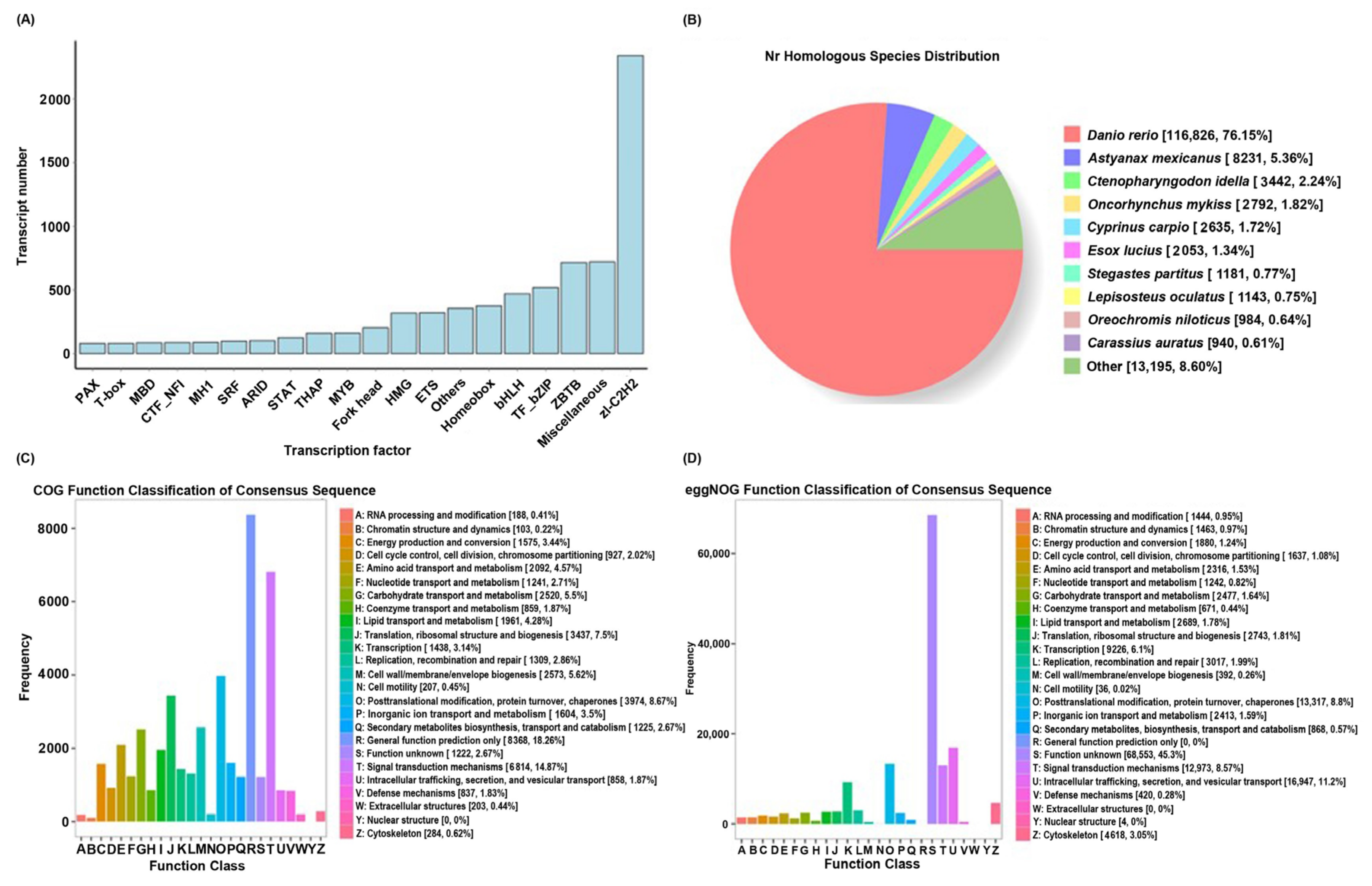
Figure 1. Basic transcriptome results. (A) Distribution of transcription factor types. The horizontal axis represents the predicted transcription factor type, and the vertical axis represents the predicted number. (B) Species classification of transcripts annotated with NR. (C) Statistical chart regarding COG annotation classification of transcripts. The horizontal axis represents the classification content of COG, and the vertical axis represents the number of transcripts. (D) Statistical chart regarding eggnog annotation classification of transcripts.
Ijms 25 10566 g001
Figure 2. Heatmap profile (A) and volcano plots (BJ) displaying the variations in gene transcriptional levels between C. idella, S. curriculus, and their hybrid F1 offspring; (B) number of genes with significant differences in hypothalamus transcription between C. idella and S. curriculus; (C) number of genes with significant differences in hypothalamus transcription between their hybrid F1 offspring and S. curriculus; (D) number of genes with significant differences in hypothalamus transcription between their hybrid F1 offspring and C. idella; (E) number of genes with significant differences in pituitary transcription between C. idella and S. curriculus; (F) number of genes with significant differences in pituitary transcription between their hybrid F1 offspring and S. curriculus; (G) number of genes with significant differences in pituitary transcription between their hybrid F1 offspring and C. idella; (H) number of genes with significant differences in gonadal transcription between C. idella and S. curriculus; (I) number of genes with significant differences in gonadal transcription between their hybrid F1 offspring and S. curriculus; (J) number of genes with significant differences in gonadal transcription between their hybrid F1 offspring and C. idella; (K) GO classification of differential genes.
Ijms 25 10566 g002
Figure 3. Differences in gonadal-development-related pathways in hypothalamus (A,D), pituitary (B,E), and gonadal (C,F) tissues between S. curriculus and hybrid F1 offspring (AC) and between C. idella and hybrid F1 offspring (DF). Red indicates pathways that contain significantly up-regulated genes in hybrid F1 offspring compared with S. curriculus or C. idella, green indicates pathways that contain significantly down-regulated genes in hybrid F1 offspring compared with S. curriculus or C. idella, and blue indicates pathways that contain significantly down- and up-regulated genes in hybrid F1 offspring compared with S. curriculus or C. idella.
Ijms 25 10566 g003
Figure 4. Differences in oxytocin signaling pathway in hypothalamus (A,D), pituitary (B,E), and gonadal (C,F) tissues between S. curriculus and hybrid F1 offspring (AC) and between C. idella and hybrid F1 offspring (DF). Red indicates pathways that contain significantly up-regulated genes in hybrid F1 offspring compared with S. curriculus or C. idella, green indicates pathways that contain significantly down-regulated genes in hybrid F1 offspring compared with S. curriculus or C. idella, and blue indicates pathways that contain significantly down- and up-regulated genes in hybrid F1 offspring compared with S. curriculus or C. idella.
Ijms 25 10566 g004
Figure 5. Differences in renin secretion pathway in hypothalamus (A,D), pituitary (B,E), and gonadal (C,F) tissues between S. curriculus and hybrid F1 offspring (AC) and between C. idella and hybrid F1 offspring (DF). Red indicates pathways that contain significantly up-regulated genes in hybrid F1 offspring compared with S. curriculus or C. idella, green indicates pathways that contain significantly down-regulated genes in hybrid F1 offspring compared with S. curriculus or C. idella, and blue indicates pathways that contain significantly down- and up-regulated genes in hybrid F1 offspring compared with S. curriculus or C. idella.
Ijms 25 10566 g005
Figure 6. Differences in oocyte meiosis pathway in hypothalamus (A,D), pituitary (B,E), and gonadal (C,F) tissues between S. curriculus and hybrid F1 offspring (AC) and between C. idella and hybrid F1 offspring (DF). Red indicates pathways that contain significantly up-regulated genes in hybrid F1 offspring compared with S. curriculus or C. idella, green indicates pathways that contain significantly down-regulated genes in hybrid F1 offspring compared with S. curriculus or C. idella, and blue indicates pathways that contain significantly down- and up-regulated genes in hybrid F1 offspring compared with S. curriculus or C. idella.
Ijms 25 10566 g006
Figure 7. Differences in GnRH signaling pathway in hypothalamus (A,D), pituitary (B,E), and gonadal (C,F) tissues between S. curriculus and hybrid F1 offspring (AC) and between C. idella and hybrid F1 offspring (DF). Red indicates pathways that contain significantly up-regulated genes in hybrid F1 offspring compared with S. curriculus or C. idella, green indicates pathways that contain significantly down-regulated genes in hybrid F1 offspring compared with S. curriculus or C. idella, and blue indicates pathways that contain significantly down- and up-regulated genes in hybrid F1 offspring compared with S. curriculus or C. idella.
Ijms 25 10566 g007
Figure 8. Expression of gonadal-development-related genes in C. idella, S. curriculus, and the hybrid F1 offspring. (A) Relative expression level of HSD3B7; (B) relative expression level of HSD17B1; (C) relative expression level of HSD17B3; (D) relative expression level of HSD20B2; (E) relative expression level of CYP17A2; (F) relative expression level of CYP1B1; (G) relative expression level of CYP2AA12; (H) relative expression level of UGT2A1; (I) relative expression level of UGT1A1; (J) relative expression level of FSHR. Different lowercase letters above the bars indicate there are significant differences between the groups.
Ijms 25 10566 g008
Table 1. Gene enrichment of gonadal-development-related pathways.
FishPathway Termko IDRich Factorq-ValueGene Number
Scct_vs_Zjf1ctSNARE interactions in vesicular transportko041301.4173490.44530564
Oxytocin signaling pathwayko049211.499386134
Renin secretionko049241.635953120
Scxqn_vs_Zjf1xqnOocyte meiosisko041141.2104910.001366413
GnRH signaling pathwayko049121.2212230.001727371
SNARE interactions in vesicular transportko041301.3491690.60779176
Oxytocin signaling pathwayko049211.414003140
Renin secretionko049241.573648124
Gcct_vs_ScctSNARE interactions in vesicular transportko041301.3432096.61 × 10−5150
Gcxqn_vs_ScxqnOocyte meiosisko041141.1799192.62 × 10−10846
GnRH signaling pathwayko049121.1215130.001189716
SNARE interactions in vesicular transportko041301.2417670.025536147
Gcxqn_vs_Zjf1xqnOocyte meiosisko041141.232360.000173416
GnRH signaling pathwayko049121.2210120.001951367
Gcxx_vs_ScxxSNARE interactions in vesicular transportko041301.2844990.0018150
Table 2. The RT-qPCR primers used in this study.
Target GenePrimer NamePrimer Sequence 5′-3′
HSD3B7HSD3B7-FACAAAGTGTGGCAACTTGGC
HSD3B7-RTCACACCAATAGGCTGCTTG
HSD17B1HSD17B1-FTGGACCAGTCAACACAGACTTC
HSD17B1-RTGAGCTGCATTCTGGAACAC
HSD17B3HSD17B3-FATTCTGCCCAGCCAAATACC
HSD17B3-RTTTGCTGCATTCCTGGTAGC
HSD20B2HSD20B2-FGCGACAGACACATGTGATTCAG
HSD20B2-RTCCATGCCCATTAGCTGTTG
CYP17A2CYP17A2-FACGCCGTTCTTTGTGAAGTG
CYP17A2-RTTGTGTCCTGCATAGCAACG
CYP1B1CYP1B1-FTCGCTTCATTTCGGTTCGTG
CYP1B1-RTGTTTGGTGTGGATGTTGGC
CYP2AA12CYP2AA12-FACCCAGATGTACAAGAGCGATG
CYP2AA12-RTTGCCAAAGCGCTGAAACTC
UGT2A1UGT2A1-FTGCCTTACACAAAGCAGGAC
UGT2A1-RTGGAAGCCGTGATGATGTTG
UGT1A1UGT1A1-FTTCCCCAAACCTCAAATGCC
UGT1A1-RTGAAGACCACAAAGCCATGC
FSHRFSHR-FTTCTCACGCCAAAGTCTTGC
FSHR-RTGTTTTGAAGCAGCCGAACC
Disclaimer/Publisher’s Note: The statements, opinions and data contained in all publications are solely those of the individual author(s) and contributor(s) and not of MDPI and/or the editor(s). MDPI and/or the editor(s) disclaim responsibility for any injury to people or property resulting from any ideas, methods, instructions or products referred to in the content.