Article

Role of Genetic Polymorphisms -238 G>A and -308 G>A, and Serum TNF-α Levels in a Cohort of Mexican Pediatric Neuroblastoma Patients: Preliminary Study

by
Arturo Ramírez-Pacheco
1,†,
Silvia Selene Moreno-Guerrero
1,†,
Luz María Rocha-Ramírez
2,*,
Gabriela Hernández-Pliego
1,
María Argelia Escobar-Sánchez
3,
Alfonso Reyes-López
4,
Juan José Luis Sienra-Monge
5,‡ and
Luis Enrique Juárez-Villegas
1,*,‡
1
Departamento de Hemato-Oncología, Hospital Infantil de México Federico Gómez, Dr. Márquez No. 162, Col Doctores, Ciudad de México 06720, Mexico
2
Unidad de Investigación en Enfermedades Infecciosas, Hospital Infantil de México Federico Gómez, Dr. Márquez No. 162, Col Doctores, Ciudad de México 06720, Mexico
3
Departamento de Patología Clínica y Experimental, Hospital Infantil de México Federico Gómez, Dr. Márquez No. 162, Col Doctores, Ciudad de México 06720, Mexico
4
Centro de Estudios Económicos y Sociales en Salud, Hospital Infantil de México Federico Gómez, Dr. Márquez No. 162, Col Doctores, Ciudad de México 06720, Mexico
5
Subdirección de Pediatría Ambulatoria, Hospital Infantil de México Federico Gómez, Dr. Márquez No. 162, Col Doctores, Ciudad de México 06720, Mexico
*
Authors to whom correspondence should be addressed.
These authors contributed equally to this paper.
They are co-last authors.
Int. J. Mol. Sci. 2024, 25(19), 10590; https://doi.org/10.3390/ijms251910590
Submission received: 30 July 2024 / Revised: 5 September 2024 / Accepted: 27 September 2024 / Published: 1 October 2024
(This article belongs to the Special Issue State-of-the-Art Molecular Oncology in Mexico, 2nd Edition)

Abstract

:
The results of in vitro and in vivo studies have shown the pro-tumor effects of TNF-α, and this cytokine’s increased expression is associated with poor prognosis in patients with some types of cancer. Our study objective was to evaluate the possible association of TNF-α genetic polymorphisms and serum levels with susceptibility and prognosis in a cohort of Mexican patients with NB. We performed PCR-RFLP and ELISA methods to analyze the genetics of these SNPs and determine serum concentrations, respectively. The distribution of the -308 G>A and -238 G>A polymorphisms TNFα genotypes was considerably different between patients with NB and the control group. The SNP rs1800629 GG/GA genotypes were associated with a decreased risk of NB (OR = 0.1, 95% CI = 0.03–0.393, p = 0.001) compared with the AA genotype, which was associated with susceptibility to NB (OR = 2.89, 95% CI = 1.45–5.76, p = 0.003) and related to unfavorable histology and high-risk NB. The rs361525 polymorphism GG genotype was associated with a lower risk of developing NB compared with the GA and AA genotypes (OR = 0.2, 95% CI = 0.068–0.63, p = 0.006). Circulating TNF-α serum concentrations were significantly different (p < 0.001) between patients with NB and healthy controls; however, we found no relationship between the analyzed TNF-α serum levels and SNP genotypes. We found associations between the rs1800629AA genotype and lower event-free survival (p = 0.026); SNP rs361525 and TNF-α levels were not associated with survival in patients with NB. Our results suggest the TNF-α SNP rs1800629 as a probable factor of NB susceptibility. The -308 G/A polymorphism AA genotype has a probable role in promoting NB development and poor prognosis associated with unfavorable histology, high-risk tumors, and lower EFS in Mexican patients with NB. It should be noted that it is important to conduct research on a larger scale, through inter-institutional studies, to further evaluate the contribution of TNF-α genetic polymorphisms to the risk and prognosis of NB.

1. Introduction

Neuroblastic tumors (NB) are the third most common solid tumors in children, accounting for about 10% of malignant neoplasms in pediatric patients and representing 10–15% of mortality from pediatric tumors [1,2]. In Mexico, its incidence is around 2.9–3.6% per one million children/year [3,4,5]. Most patients are diagnosed in the advanced stages of the disease, decreasing disease-free survival [3,4,6,7]. In addition, metastasis is another factor related to poor clinical results, with bone metastasis occurring most frequently [3,4,6,7,8]. This tumor presents considerable histopathological, morphological, biological, genetic, and clinical heterogeneity, which can decisively influence prognosis [3,8,9,10].
Cytokines produced by cell interactions in the tumor microenvironment play an important role in the pathogenesis of cancer [11]. Non-malignant cells in the tumor microenvironment can positively or negatively affect the growth, survival, and metastatic potential of tumor cells, including NB [12,13]. Alternatively, malignant cells may respond to host-derived cytokines that promote growth, attenuate apoptosis, and facilitate invasion and metastasis [11]. Tumor necrosis factor α (TNF-α) is a pro-inflammatory cytokine produced by both malignant and immune cells within the tumor-associated microenvironment and contributes to the development and progression of malignant disease by creating an inflammatory niche that supports the tumor [12]. TNF-α is a powerful immunomodulator implicated in growth factor and cytokine activation, such as in IL-8 and MCP-1, which display chemo-attractant activity that affects the synthesis and alteration of adhesion molecules. Furthermore, TNF-α promotes inflammatory response and is critical in the pathogenesis of inflammatory, autoimmune, and malignant diseases [14]. TNF-α displays pro-tumor activity by stimulating the growth, proliferation, invasion, angiogenesis, and metastasis of cancer cells by activating nuclear factor kB (NF-kB), an important cell survival signal that inhibits the apoptotic process [12,15]. On the other hand, TNF-α suppresses tumor growth through its anti-proliferative, cytostatic, and cytolytic effects against various malignant cells; however, many of these cells are resistant to TNF-α-induced cytotoxicity [16]. TNF-α acts through its two distinct receptors: TNFR1 (TNF receptor 1) is expressed on all cells in the body, and TNFR2 (TNF receptor 2) is expressed primarily on immune suppressor and neoplastic cells. TNFR1 has an important role in apoptotic cell death and TNFR2 seems to have a tumor-promoting effect [17]. Both receptors transduce their signals cooperatively [18]. Recently, the interaction between TNFR2 of the tumor cell and the TNF-α bound to the monocyte membrane was reported, triggering tumorigenic inflammation in NB [19].
The TNF-α coding gene is located on chromosome 6p21.3. It is 3 kb in size; contains four exons; has several single nucleotide polymorphisms (SNPs), two of which are in the gene’s promoter region; and can affect the production of TNF-α [20]. The -308 G/A (rs1800629) and -238 G/A (rs361525) SNPs are found in a region close to the transcription site, resulting in differences in gene expression and TNF-α protein secretion [21]. The -308 G/A polymorphism is related to an increase in TNF-α gene transcription supported by gene reporter assays [14,21]. Substituting a guanine (G) base for adenine (A) in position 308 generates two allelic forms: the wild-type -308G and the variant-type -308A. The presence of the -308A allele correlates with increased production of TNF-α in vitro and in vivo [21,22]. It has been documented that transcriptional activity increases severalfold and secretion of this cytokine increases when the A allele is present [21,22]. The GA and AA genotypes have been associated with higher mRNA levels and elevated serum TNF-α concentrations compared with individuals with the GG genotype [23,24,25]. The -308A allele is linked to clinical susceptibility to various diseases. In vitro research results reported its links to increased TNF-α production [26]. However, SNP -238 G/A is produced from substituting a guanine (G) base for adenine (A) in position 238, generating the wild- and variant-type alleles, -238G and -238A. This SNP is associated with reductions in TNF-α gene transcription [27]. The G allele of the -238 polymorphism has been shown to be associated with elevated TNF-α production compared with the A allele [28]. However, reports are limited, and they still do not provide clear evidence of this SNP’s role in regulating TNF-α gene transcription. The -238 A allele is a protective factor in disease susceptibility [27,29,30], and based on reports from meta-analyses, both polymorphisms are associated with susceptibility and clinical outcomes in various inflammatory diseases, infections, and cancer [21,26,27,28,29]. In addition, bioinformatic analyses of online databases describe positive associations of SNPs in genes encoding various cytokines with several human diseases, including the polymorphisms -238 G/A (rs361525) and -308 G/A (rs1800629) of TNF-α [31].
It has been reported that the distribution of polymorphisms in genes of several cytokines such as TNF-α distinguishes the Mexican population from other groups, including Caucasians, Asians, and Africans, showing a different distribution for the AG haplotype, made up of the polymorphisms -238 G/A and -308 G/A of TNF-α, in the Mexican population (7.2%), Asian population including Chinese (8.8%) and Koreans (8.2%), and two European populations Romanians (13.2%) and Macedonians (14%) [32]. Likewise, published data on frequencies of the A allele of SNP rs1800629 shows differences between European (5–10%), African (0.8–14%), South African (5%), British White Caucasian (30%), Asian (0–2%), and Mexican (0–8%) populations [14,33,34].
The A allele for the rs361525 variant in heterozygous genotype has been reported to be present with a frequency of around 0.1–0.2% in different populations [35,36]. Since these TNF-α polymorphisms are associated with the development of various diseases, knowledge of the distribution of these SNPs in the Mexican population may be useful to understand their role as possible genetic factors of susceptibility or prognosis in our population.
TNFα genetic polymorphisms can influence the levels of this cytokine in vivo in various conditions. The modulation of immune responses, as in TNF-α-dependent cell death processes, has long been the subject of intense investigation. However, its role in cancer progression is complex and controversial because of this cytokine’s involvement in cancer development, which may have opposite effects on tumor growth [37,38,39].
There are no reported studies on the implications of circulating concentrations and TNF-α genetic polymorphisms on NB. Therefore, the purpose of our study was to evaluate the association of the -238 G/A rs361525 and -308 G/A rs1800629 genetic polymorphisms and serum levels of TNF-α with the clinical pathological factors and disease susceptibility in a cohort of children with NB.

2. Results

Our study population was 27 pediatric patients with NB, the majority (59.3%) were aged <18 months at diagnosis, and 37.03% were men and 62.9% were women. More than half of the subjects were classified as high risk (55.6%), and 48.1% were at INSS stage 4 (Table 1). The most frequent primary tumor location was in the retroperitoneal space (40.7%), and the majority presented partially differentiated tumors (59.3%) (Table 1). A total of 55.5% presented metastases at diagnosis, only 3.7% had amplification of the MYCN gene, and 25.9% of the patients relapsed, with 22.2% dying during the follow-up. In the control group of 38 healthy pediatric subjects, who were matched with the NB cases by age and sex, none presented amplification of the MYCN gene (Table 1).

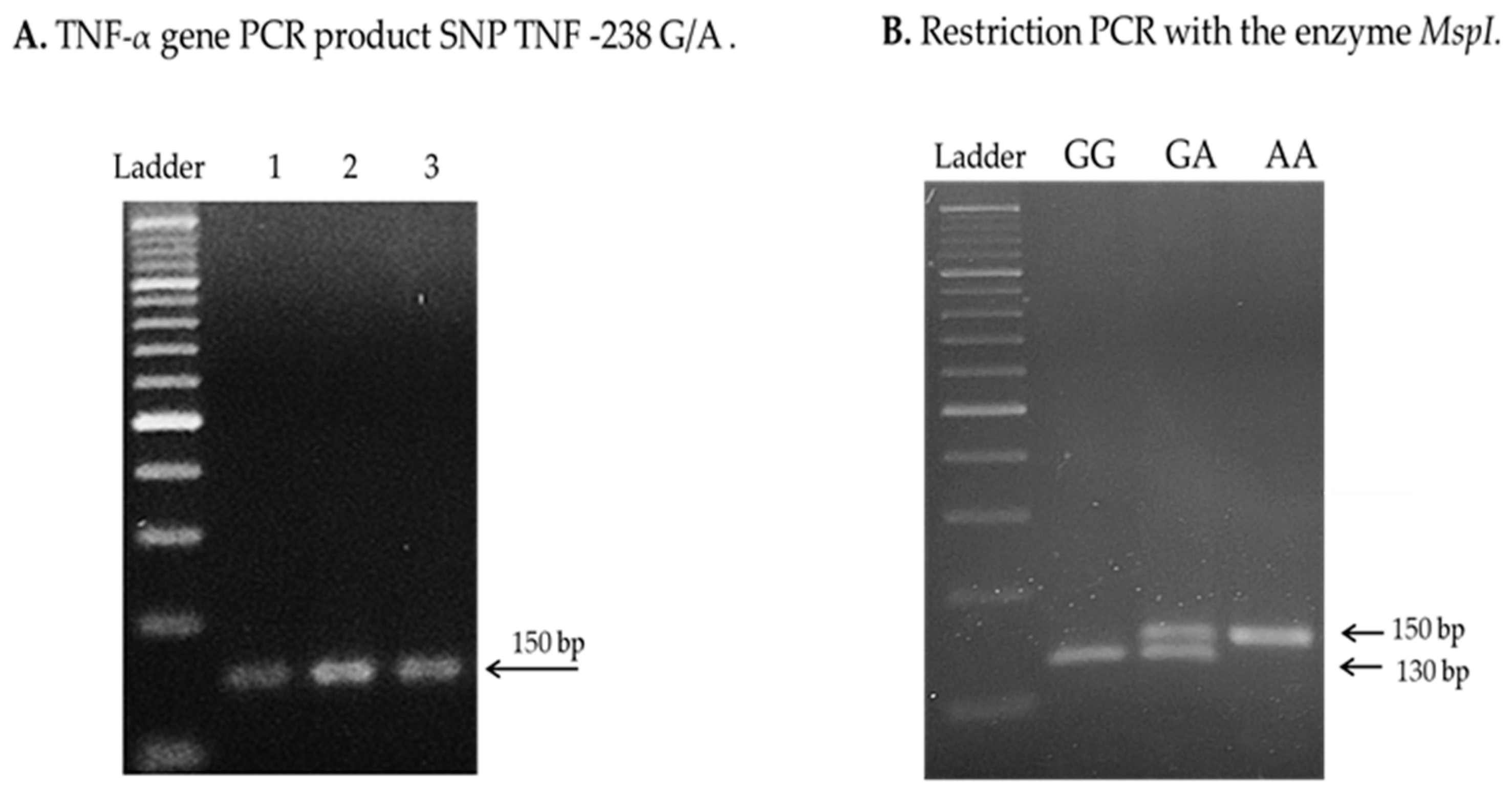
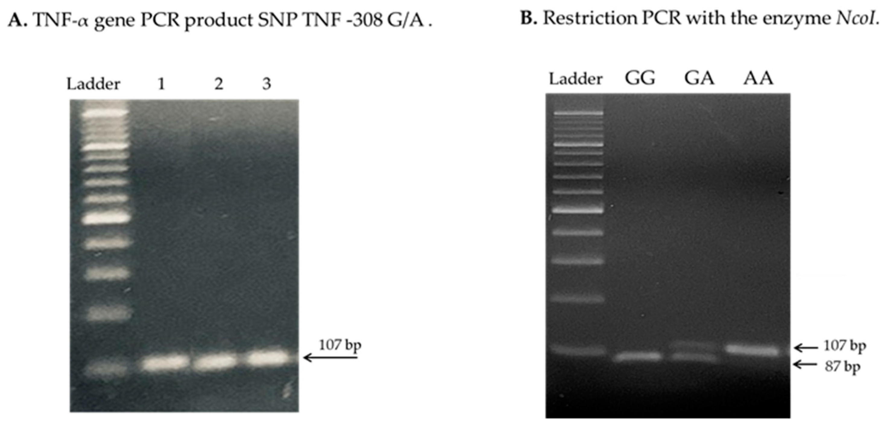
2.1. Distribution of TNF-α Genetic Polymorphisms between NB Cases and Control Group

The genotype and allele frequencies presented important differences between cases and controls for the analyzed SNPs (Table 2). For the SNP rs361525, the homozygous wild-type GG genotype occurred more frequently in healthy controls compared with NB cases (81.6% vs. 48.1%); on the contrary, the heterozygous GA and homozygous variable AA genotypes occurred more frequently in NB cases in relation to controls (18.4% vs. 40.7%) and (0% vs. 11.1%), respectively (Table 2). Furthermore, the AA genotype was not found in the analyzed healthy controls. For SNP rs1800629, the homozygous wild-type GG was the most frequent in controls and cases with NB but with a marked difference in percentage, being much higher for controls compared with cases (86.8% vs. 48.2%). We observed that the heterozygous GA (2.6% vs. 14.8%) and homozygous wild-type AA (10.5% vs. 37%) genotypes were more frequent in patients with NB compared with the control group. Our logistic regression analysis revealed that the rs361525 polymorphism GA genotype was associated with (p = 0.004) increased risk of presenting NB (OR 4.52; 95% C.I. 1.61–12.6) compared with the wild-type homozygous GG genotype (Table 2). For the rs1800629 variant, the AA genotype may increase the risk of disease compared with the homozygous wild-type GG genotype (p = 0.003) (OR 2.89; 95% C.I. 1.452–5.765) (Table 2).

2.2. Analysis of NB Patient Genotypes and Prognostic Factors

We observed that most of the patients who presented high-risk prognostic factors had at least one G allele for rs361525 TNF-α polymorphism; however, our analysis did not show a significant association with any of the analyzed prognostic factors (Table A1). For the rs1800629 TNF-α polymorphism, the GA + AA genotypes showed a significant association (p = 0.038) with the presence of unfavorable histology and were at the limit of significance (p = 0.05) with high-risk category and INSS stage 4; however, there were no associations with other prognostic factors (Table A1).
A simple logistic regression analysis was performed to evaluate the association between TNF-α genetic polymorphisms and prognostic factors. For SNP rs361525, we found that carriers of the GG and GA genotypes have intermediate/high-risk NB (OR 1.5, 95% CI 0.20–10.8); however, this association is at the limit of significance (p = 0.05). In addition, the AA genotype was found to be significantly associated (p = 0.017) with a decreased risk of developing neuroblastic tumors with INSS stages 3 and 4 (OR 0.043, 95% CI 0.003–0.57) (Table 3). The remaining prognostic factors we evaluated did not present an important relationship with this SNP (Table 3).
For SNP rs1800629, a high frequency of patients with the GG genotype had favorable histology; however, this association was not significant. On the contrary, the patients with the GA/AA genotype were more likely to display high-risk NB (p = 0.04) and unfavorable histology (p = 0.03) (Table 4). With these findings, we can suggest that the A allele of this polymorphism may probably be a genetic biomarker of poor prognosis in Mexican patients, associated with unfavorable advanced-stage neuroblastic tumors.

2.3. Relationship of TNF-α Serum Levels Between Cases of NB and Control Group

Our analysis of TNF-α serum levels between the NB patients (n = 27) and control group (n = 27) showed significant differences (p < 0.001), indicating that the serum concentrations of this cytokine may probably have implications in the development of NB, due to the pro-tumoral role of TNF that has been reported in previous studies (Table 5). However, we considered low ≤ 20 pg/mL and high levels ≥ 21 pg/mL in the stratification of TNF-α serum levels. Based on this, 88.9% of patients with NB had low levels of TNF-α. All healthy pediatric subjects showed low levels of TNF-α (Table 5).

2.4. Association of Genotypes and Serum Levels

Our evaluation of the distribution of the genotypes rs1800629 and rs361525 of TNF-α with respect to serum levels in NB patients did not reveal a significant association of any genotype for both polymorphisms studied with circulating levels of TNF-α (Table A2). We observed that more than 40% of NB patients with homozygous wild-type (GG) genotypes for these polymorphisms presented low TNF-α serum levels compared with patients with heterozygous and homozygous-variant genotypes (Table A2).

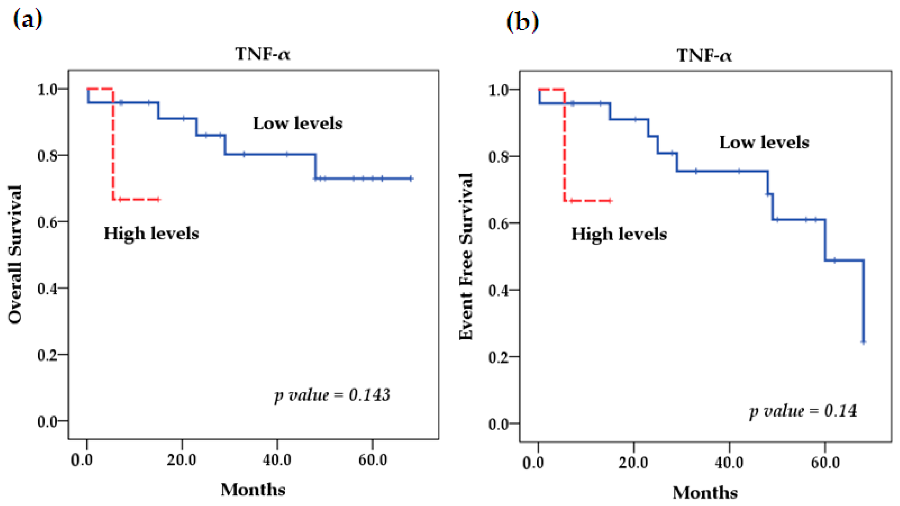
2.5. Survival Analysis

Our survival analysis using the Kaplan–Meier method showed that for the rs361525 TNF-α polymorphism, patients with NB and the AA genotype had better overall (OS) and event-free survival (EFS) rates. It was not significant (Figure 1). However, the TNF-α SNP rs1800629 had a significant effect (p = 0.026) on EFS because patients with the homozygous-variant AA genotype had lower EFS compared with patients with homozygous wild-type GG (92.35% vs. 20%) and heterozygous GA (75% vs. 20%) (Figure 2). On the other hand, OS and EFS in relation to the stratification of patients with high and low TNF-α serum levels did not show significant differences either (p = 0.14 and p = 0.143) (Figure 3). In addition, we performed multivariate Cox regression analysis, which was not significant; further details on the variables included and the results obtained can be found in Table A3.
We performed multivariate Cox regression analysis, which included the variables of age at diagnosis, MYCN status, INSS stage, and TNF-α serum concentrations and genetic variants rs1800629 and rs361525. It was not possible to adjust for MYCN amplification due to the reduced sample size. Our analyses showed no significant effects for these variants in conjunction with the risk of death Table A3

3. Discussion

TNF-α is a mediator of the immune response in various immunological diseases, infections, and types of cancer. The -238G/A and -308G/A polymorphisms are in the promoter region of the TNF-α gene and are associated with alterations in the levels of TNF-α expression, which may be related to the clinical outcomes of various diseases. In our study, we investigated the association of the genetic polymorphisms -238G/A rs361525 and 308 G/A rs1800629 and TNF-α serum concentrations with clinical outcomes in patients with NB. Previous studies analyzed the relationship of these genetic variants with various diseases and types of cancer, presenting contradictory results that largely depended on the ethnicity of the population and the type of disease [40,41,42]. Until now, there were no published studies about the role of TNF-α polymorphisms in NB.
In our study, for the -238 G/A rs361525 polymorphism, we found a significant difference in the frequency of the homozygous GG genotype (81.6% vs. 48.1%) in the control group compared with patients with NB. We observed the G allele an association with decrease in the risk of NB in the analyzed population. Likewise, we found an association between the GA heterozygous genotype of this variant and the risk of presenting NB. Our findings are consistent with previous reports in various meta-analysis studies that associated the -238 G/A polymorphism with the risk of various types of cancer, specifically the GA and AA genotypes with the risk of colorectal cancer in the Caucasian population [43,44,45,46]. The A allele and the GA and AA genotypes were associated with the risk of gastric cancer in Asian populations [41,42,43,44], the AA genotype was associated with the risk of hepatocellular carcinoma in African, Asian, and South American populations [45], and patients with genotypes GA + AA presented a higher risk of multiple myeloma in the Polish population [40]. Also, an association between the GA genotype of this polymorphism and cancer susceptibility in Polish patients with systemic sclerosis was recently reported [47]. However, multiple myeloma in the Turkish population has no association between this polymorphism and risk of disease [48], which is why ethnicity is an important factor that influences the incidences of genetic variants and their relationships with different diseases. Furthermore, our results contrast with those of other studies where the A allele of this polymorphism was presented as a biomarker of protection against diseases, such as lung cancer and idiopathic arthritis [29,30], as well as with reports where no association of SNP -238 G/A with the risk of diseases such as osteosarcoma, ischemic heart disease, and acute pancreatitis, among others [34,42,43,44,48]. In addition, results from studies on the Mexican population found no relationship between this polymorphism and the risk or severity of colorectal cancer [45].
The -308 G/A rs1800629 polymorphism regulates the expression of TNF-α, which may be associated with clinical outcomes in patients with NB. However, previous studies on this genetic polymorphism have produced contradictory results regarding the genotypes associated with the prognosis or development of various diseases. The results of several studies found no correlations between SNP -308 G/A and colorectal cancer, head and neck cancer, idiopathic arthritis, and acute pancreatitis, among others [30,42,44,45,46,48,49,50]. Contrary to these reports, in our study we found a significant difference in the frequency of the homozygous GG genotype (81.6% vs. 48.1%) in the control group compared with patients with NB. In addition, the G allele in its homozygous (GG) or heterozygous (GA) forms showed a decreased risk of NB, which indicates that this allele may be a probable biomarker of protection in this disease. On the contrary, we found an association between the AA genotype and the risk of NB, and for carriers of the A allele, it may be a probable biomarker of risk for this disease. This is consistent with a study showing that carriers of at least one A allele of this polymorphism are significantly associated with a two-fold increased risk of developing relapsing-remitting multiple sclerosis [51]. We also observed the GA/AA genotypes showed considerable associations with poor prognostic factors, such as high risk, INSS stage 4, and unfavorable histology. Likewise, we found the variant AA homozygous genotype to be considerably associated with lower EFS in patients with NB. Some of our findings are consistent with reports from other research groups, such as in studies of breast cancer, which also reported the GA and AA genotypes of the -308 G>A polymorphism contributed considerably to the risk of this disease in the Mexican population [34]; likewise, the GA genotype is associated with lower overall survival and EFS in Lithuanian women with breast cancer [49]. Similarly, in several reports and meta-analysis studies, the AA or GA genotypes of this variant are associated with the risk of various malignant diseases, such as osteosarcoma, gastric or colorectal cancer, and hepatocellular carcinoma, and the A allele is also associated with the development and progression of lung cancer, as well as the development of metastasis in rectal cancer [29,34,44,45]. However, there is a report that associates the homozygous wild-type GG genotype with the risk of multiple myeloma compared with a group of healthy controls [41]. These reported differences may be due to the sample size, as well as the genetic differences of the populations studied. The Mexican population is ethnically heterogeneous, and the results of reports on the distribution of cytokine polymorphisms, such as TNF-α, are different in the Mexican population compared with other ethnic groups, including Caucasians, Asians, and Africans [32]. This is why knowledge of the distribution of these polymorphisms in the Mexican population may be relevant in understanding their participation as genetic markers of susceptibility in Mexico.
In our study in the analysis of the polymorphisms evaluated with the clinicopathologic factors of the patients, we did not obtain results related to MYCN status, due to the low frequency of occurrence of this oncogene. On the other hand, we observed that most patients carrying the G allele for SNP-238 G/A rs361525 presented tumors in disseminated stages (INSS3 and INSS4) (p = 0.017) (Table 3); the G allele of this polymorphism is associated with elevated TNF-α production compared with the A allele [28]. Therefore, we suppose that this cytokine could probably be involved in the progression of this type of tumors; however, reports are limited and do not yet provide clear evidence of the role of this SNP in the regulation of TNF-α gene transcription. On the other hand, analysis of the SNP -308 G/A rs1800629 TNF-α SNP with clinicopathologic factors of patients with NB, we only observed that the majority of patients with A allele presented high-risk NB (p = 0.04) and unfavorable tumor histology (p = 0.03) (Table 4); it has been reported that the presence of the -308A allele correlates with increased TNF-α production in vitro and in vivo [21,22]. These associations related to tumor histology and staging based on the INSS staging system are important because NB treatment should be performed based on risk groups; among the most important clinical factors are age, tumor location and histology, and stage based on INSS criteria. However, these findings should be taken with caution because, from a statistical point of view, it would be important to increase the power of the tests to obtain associations with better significance values, so it is necessary to increase the sample size.
The clear distinction that we observed between the levels of TNF-α in patients with NB versus the control group may indicate that high serum levels of this cytokine may have an implication in the development of NB. This can be explained by the fact that TNF-α is a potent immunostimulatory cytokine with local effects in the tumor microenvironment and possible systemic effects; it can also contribute to the maintenance of a pro-inflammatory environment [11]. However, there are still discrepancies in this regard because it has been reported that TNF-α acts as an autocrine growth factor for NB cells; on the other hand, an in vitro study reported that TNF-α inhibited NB cell growth and DNA synthesis [52]. In addition, the increased TNF-α serum levels in cancer patients are associated with adverse disease outcomes [50]. We found a significant difference (p < 0.001) in TNF-α serum levels between patients with neuroblastoma and the control group. Other investigations produced similar results in other diseases such as lung and colorectal cancer and osteosarcoma, among others [53,54,55]. However, when stratifying the cytokine’s serum concentrations into low and high levels, considering the previously established cut-off values, we observed no considerable differences between the group with NB and the healthy individuals. It is important to mention that the population considered for the estimation of the cut-off values was between the ages of 0 and 17 years, which is the criterion for the pediatric population in our country. On the other hand, the control group used in this work was matched by age with the patients with NB, so the age range was from 0 to 8 years; for this reason, we consider that the differences between the control group and the cases with NB are important because they are within the same age group and the fact that when performing the stratification at low and high levels considering the cut-off values, this difference was not significant, may be because the group to obtain the cut-off values included children with a higher age range than the patients included in this study. This is related to what was previously reported by authors who indicate that age clearly influences the cytokine expression profiles in healthy children [56,57], reporting that several cytokines, including TNF-α, show an increase associated with age, suggesting that cytokine expression dynamics in infancy should be taken into account when evaluating them in diagnostic assays or as immunological biomarkers [57].
TNF-α expression is mainly regulated at the transcriptional level and some polymorphisms in the gene that codes for this protein are related to the production of TNF-α. We found no significant associations between the genotypes of polymorphisms -238 G/A rs361525 and -308 G/A rs1800629 and low and high TNF-α levels in patients with NB. Our results agree with those of other studies that also found no relationship between TNF-α serum concentrations and these SNPs [45]. However, our results contrast with several trials that found a significant relationship between elevated TNF-α serum levels and patients carrying the -308 A allele of the rs1800629 variant [38,58,59,60]; in the case of SNP rs361525, the -238A allele is associated with decreased TNF-α serum levels [20,21,60]. Probably, in our study, we did not observe a relationship between polymorphisms analyzed and serum concentrations of TNF-α due to the size of the sample included. It would also be productive to analyze other genetic variants of the TNF-α promoter gene that could affect transcription, such as SNP -863 C/A, which has been associated with reduced levels of TNF-α [61]. On the other hand, high TNF-α expression has been described as a predictor of poor survival in cancer patients [62,63]. In our case, the levels of this cytokine showed no association with survival in the studied population. Our results may be due to the small sample size. In addition, we only performed a measurement of the TNF-α concentration in serum at the time of diagnosis; however, it is important to evaluate the serum levels at various stages of the disease evolution to examine whether the increase in cytokine circulation can affect the development and prognosis of NB.
It is also important to consider larger cohort studies in the future, as well as additional prospective and retrospective studies. In addition, it is necessary to evaluate the serum levels of TNF-α during the disease and to analyze the expression of the TNF-α receptors (TNFR-1 and TNFR-2). These determinations, together with the genotyping of genetic polymorphisms, could contribute to improving our understanding of the role of TNF-α in NB.

4. Materials and Methods

In this study, we included a total of 27 pediatric patients diagnosed with neuroblastoma from the Hospital Infantil de México Federico Gómez from December 2015 to December 2019. The staging of Neuroblastoma tumors in this study was performed based on clinical, radiographic, and surgical evaluation, according to the criteria of the International Neuroblastoma Staging System (INSS) and the International Neuroblastoma Risk Group (INRG) [64,65]. The degree of differentiation of neuroblastic tumors was determined based on histopathological criteria following the classification parameters of the International Neuroblastoma Pathology Classification (INCP), for which histopathological and immunohistochemical studies were performed on the tumor material, such as the detection of the NB84 marker, morphometric evaluation to study ploidy, and molecular analysis to determine MYCN amplification [66,67]. The histology according to the Shimada system, was based on the amount of Schwannian stroma, the degree of differentiation, the mitosis–karyorrhexis index (MKI), and age at diagnosis, classifying the tumors into two groups of favorable and unfavorable histological prognosis [9,64]. We collected paraffin-embedded tissue samples and peripheral blood at the time of diagnosis. We obtained the characteristics of each patient, such as age at diagnosis, risk, stage, and site of the primary tumor, histology, clinical follow-up time, and prognostic factors, from clinical records. The control group was age- and sex-matched with NB cases; 38 healthy children were included, without presenting any infectious or inflammatory processes or immune diseases at the time of sample collection.

4.1. Ethical Considerations

Our research was evaluated and approved by the Ethics Committee of the Institute (number CONBIOÉTICA-09-CEI-010-20160627). Informed consent was provided by the legal guardians of all patients and healthy controls. We carried out this project in accordance with the principles established by the Helsinki World Medical Assembly and all the applicable modifications established by the World Medical Assembly and the ICH guidelines for Good Clinical Practice (GCP), as well as the regulations of the General Law of Health in Research for the Health of Mexico.

4.2. Determination of MYCN Gene Amplification

To detect MYCN gene amplification, we obtained DNA from formalin-fixed, paraffin-embedded samples of NB using the QIAamp DNA FFPE Tissue kit (Qiagen, Hilden, Germany) according to the manufacturer’s instructions. For healthy controls, DNA was obtained from peripheral blood samples with the DNA Blood Midi kit (Qiagen, Hilden, Germany) according to the manufacturer’s instructions. We analyzed the MYCN copy number by real-time PCR and used the commercial TaqMan copy number assay for the MYCN gene (ID Hs00658058) from Thermo Fisher Scientific Applied Biosystems (Thermo Fisher Scientific, Foster, CA, USA) according to the manufacturer’s instructions.

4.3. Analysis of TNF-α Genetic Polymorphisms

We obtained peripheral blood samples (2 mL) in EDTA tubes to obtain genomic DNA. We extracted blood with the DNA Blood Midi Kit (Qiagen, Hilden, Germany) according to the manufacturer’s instructions. We used the PCR-RFLP (polymerase chain reaction–restriction fragment length polymorphisms) method to determine the TNF-α genetic polymorphisms. The PCR mixture contained: 0.1 µg DNA, 2.5 mM dNTPs, 3 mM MgCl2, 1X buffer (20 mM Tris-HCl and 2 mM MgSO4), 10 pm oligonucleotides, and 0.5 U DNA Taq polymerase (New England Biolabs, Beverley, MA, USA). For restriction fragment length polymorphism analysis, the PCR products were purified before restriction using the MinElute PCR Purification Kit (Qiagen, Hilden, Germany). We digested the PCR products with specific restriction enzymes. We analyzed the -238 G>A polymorphism using the forward and reverse primers 5′-ATCTGGAGGAAGCGGTAGTG-3′ and 5′-AGAAGACCCCCCTCGGAACC-3′. We used a PCR protocol of 94 °C for 3 min, 35 cycles of 94 °C for 30 s, 57 °C for 30 s, 72 °C for 30 s, and a final extension at 72 °C for 5 min, and we cut 10 µL of the 150 bp PCR product using 2U of the MspI restriction enzyme (New England Biolabs, Beverley, MA, USA) at 37 °C for 4 h. The restriction fragments for the genotypes included GG: 130 pb and 20 pb; GA: 150, 130, and 20 pb; and AA: 150 pb Figure A1. For the -308 G>A polymorphism, we used the forward and reverse primers 5′-AGGCAATAGGTTTTGAGGGCCAT-3′ and 5′-TCCTCCCTGCTCCGATTCCG-3′. We used a PCR protocol of 94 °C for 3 min, 35 cycles of 94 °C for 30 s, 61 °C for 30 s, 72 °C for 30 s, and a final extension at 72 °C for 5 min, and we cut the 107 bp PCR product using 2 U of the NcoI restriction enzyme (New England Biolabs, Beverley, MA, USA) at 37 °C for 4 h. The restriction fragments for the genotypes included GG: 87 bp and 20 bp; GA: 107, 87, and 20 bp; and AA: 107 bp Figure A2. We analyzed the PCR and enzyme restriction products by electrophoresis in 2.5% agarose gels stained with ethidium bromide, and we visualized them with UV light to detect each genotype.

4.4. Measurement of TNF-α Serum Levels

Of the 38 healthy children, we selected 27 to evaluate serum cytokine levels; these were age- and gender-matched with Neuroblastoma patients to avoid any bias because serum cytokine levels may vary by age and gender. We obtained the blood samples in tubes with separating gel for serum analysis. The blood remained for 1 h to complete the clot retraction. Later, we centrifuged for 15 min at 3500 rpm. We stored serum at −70 °C in 500 µL aliquots and subsequently analyzed the immunological markers. We quantified TNF-α in the serum of NB patients and controls by the ELISA method using the BD OptEIA ELISA Kit (BD Biosciences, Franklin Lakes, NJ, USA) according to the manufacturer’s instructions. We analyzed the samples in duplicate, expressed the concentration in pg/mL, calculated by interpolation to a TNF-α standard curve, and considered the dilution factor 1:5. To establish cut-off lines for TNF-α, we previously performed a preliminary analysis that included 30 serum samples from the pediatric population from 0 to 17 years in which inflammatory history and chronic degenerative disorders were ruled out, and 3 negative controls to obtain the value of the mean of the determinations ± 2 standard deviations (SD), to establish the cut-off values of the serum levels of TNF-alpha in the pediatric population. TNF-alpha concentrations were quantified from the logistic curve of the TNF-α standard; the specificity of the test is 98%, according to the manufacturer’s instructions. The cut-off point of TNF-α obtained was 21 pg/mL; in this way, the values for the stratification of serum levels of TNF-α were obtained, considering low levels ≤ 20 pg/mL and high levels ≥ 21 pg/mL, which coincide with what was reported by Noori et al. 2017 [68].

4.5. Statistical Analysis

We performed the statistical and graphical analyses of this study using STATA version 13 and SPSS version 19 programs. Descriptive data were represented in terms of frequencies. Differences between clinical data categorical variables of patients and genotypes were performed using Fisher’s exact test. To assess the association between genetic polymorphisms with the presence of the disease and prognostic factors, we estimated the odds ratios (OR) by simple logistic regression analysis. The Hardy–Weinberg genetic equilibrium (EH-W) was determined using the χ2 test. The comparison of serum concentrations of TNF-α between the group of patients with NB and the control group was performed using the Wilcoxon test. Survival analysis was performed using the Kaplan–Meier method, and the differences between groups were estimated by the log rank test; for overall survival (OS), it was calculated from the date of diagnosis to the date of death or last contact with patient, and event-free survival (EFS) was considered from the date of diagnosis to the date of relapse or death. The Cox model was used to identify prognostic variables that affected overall survival. The level of statistical significance considered for all tests was a value of p < 0.05.

5. Conclusions

We identified a possible association between the TNF-α genetic polymorphisms -238 G/A rs361525 and -308 G/A rs1800629 and the risk of neuroblastoma. Patients with NB carrying the GG genotype for SNP rs1800629 had a better prognosis than those with the AA genotype, who had low EFS, for which the A allele may play an important role in the progression of NB as probable factors associated with, and useful for, identifying high-risk patients and following up the disease in Mexican patients with neuroblastoma. Circulating TNF-α serum concentrations were different between patients with NB and healthy controls; however, we found no relationship between the analyzed TNF-α serum levels and SNP genotypes. It should be noted that this is a preliminary study, and it is important to conduct research on a larger scale, through inter-institutional studies, to further evaluate the contribution of TNF-α genetic polymorphisms as potential markers of treatment response, disease progression, and severity.

Author Contributions

Conceptualization, S.S.M.-G., A.R.-P., L.E.J.-V. and L.M.R.-R.; methodology, S.S.M.-G., A.R.-P., L.M.R.-R., M.A.E.-S., G.H.-P. and L.E.J.-V.; formal analysis, S.S.M.-G., A.R.-P., L.E.J.-V. and L.M.R.-R.; investigation, S.S.M.-G., A.R.-P. and L.M.R.-R.; data curation and analysis, A.R.-L., A.R.-P. and S.S.M.-G.; writing—original draft preparation, S.S.M.-G., A.R.-P., L.E.J.-V. and L.M.R.-R.; writing—review and editing, S.S.M.-G., A.R.-P., L.E.J.-V. and L.M.R.-R.; visualization, S.S.M.-G., A.R.-P., L.E.J.-V. and L.M.R.-R.; writing S.S.M.-G., A.R.-P., L.E.J.-V. and L.M.R.-R.; supervision, J.J.L.S.-M., L.E.J.-V. and L.M.R.-R.; resources, J.J.L.S.-M., L.E.J.-V. and L.M.R.-R.; project administration, J.J.L.S.-M., L.E.J.-V., S.S.M.-G., A.R.-P. and L.M.R.-R. All authors have read and agreed to the published version of the manuscript.

Funding

This research was funded with Fondos Sectoriales del Consejo Nacional de Humanidades, Ciencias, y Tecnologías (CONAHCYT) with project number 272420, and HIM/2016/098.

Institutional Review Board Statement

The study was conducted according to the guidelines of the Declaration of Helsinki and approved by the investigation committee with number 18CI09015010 and Ethics Committee with number CONBIOÉTICA-09-CEI-010-20160627 of the Hospital Infantil de México Federico Gómez (protocol code HIM-2016-098 date of approval 15 November 2016).

Informed Consent Statement

Informed consent was obtained from all subjects involved in the study. Written informed consent has been obtained from the patient(s) to publish this paper.

Data Availability Statement

The data presented in this study are available on request from the corresponding author. The data are not publicly available due to confidentiality of patient data.

Acknowledgments

We thank the patients who agreed to participate in the study.

Conflicts of Interest

The authors declare no conflicts of interest.

Appendix A

Table A1. Genotype frequencies of -238 G/A rs361525 and -308 G/A rs1800629 TNF-α and prognostic factors of NB patients.
SNP -238 G/A rs361525
n = 27 (%)
SNP -308 G/A rs1800629
n = 27 (%)
VariablesGG + GAAAGGGA + AA
INSS stage
13 (11.1)2 (7.4)5 (18.5)----
2b2 (7.4)----1 (3.7)1 (3.7)
37 (25.9)----3 (11.1)4 (14.8)
412 (44.4)1 (3.7)4 (14.8)9 (33.3)
p value0.2180.05
INRG
L13 (11.1)2 (7.4)3 (11.1)2 (7.4)
L28 (29.6)----5 (18.5)3 (11.1)
M13 (48.1)1 (3.7)5 (18.5)9 (33.3)
MS----------
p value0.1460.51
Risk
Low3 (11.1)2 (7.4)4 (14.8)1 (3.7)
Intermediate7 (25.9)----5 (18.5)2 (7.4)
High14 (51.9)1 (3.7)4 (14.8)11 (40.7)
p value0.1260.05
Differentiation
Undifferentiated 2 (7.4)--------2 (7.4)
Partially differentiated14 (51.9)2 (7.4)9 (33.3)7 (25.9)
Differentiated4 (14.8)1 (3.7)3 (11.1)2 (7.4)
Not specified4 (14.8)----1 (3.7)3 (11.1)
p value10.414
Histology (Shimada)
Favorable13 (48.1)2 (7.4)10 (37)5 (18.5)
Unfavorable11 (40.7)1 (3.7)3 (11.1)9 (33.3)
p value0.5860.038
MYCN
Amplified1 (3.7)---------1 (3.7)
Not amplified23 (85.2)3 (11.1)13 (48.1)13 (48.1)
p value0.2960.3
p value obtained by X analysis and Fisher exact test
Table A2. Association of polymorphism rs1800629 and rs361525 genotypes with serum levels of TNF-α in patients with NB.
Low Serum
TNF-α Levels
High Serum TNF-α Levels Value pOR (95% CI)
TNF-α -308 G/A rs1800629
GG12 (44.44)1 (3.7)Ref----
GA4 (14.8)0------
AA8 (29.6)2 (7.4)0.1452.63 (0.71–9.70)
GG + GA vs. AA----------0.1197.27 (0.60–87.84)
GA + AA vs. GG----------0.2140.20 (0.017–2.47)
TNF-α -238 G/A rs361525
GG13 (48.14)0Ref----
GA8 (29.6)3 (11.1)0.1183.87 (0.71–21.12)
AA3 (11.1)0-----------
GG + GA vs. AA------------NC
GA + AA vs. GG-------------NC
CI = Confidence interval; OR = odds ratio; ref = reference genotype; vs. = versus; NC = not calculable.
Table A3. Results of the multivariate Cox proportional hazards model.
ModelHR (95% CI)p Value
A
TNF-α rs361525 (GG + GA vs. AA)NCNC
Age (≥18 months vs. <18 months)1.16 (0.16–8.39)0.87
INSS stage (4 vs. 1, 2, 3)5.62 (0.51–61.6)0.15
MYCN (amplified vs. not amplified)NCNC
B
TNF-α rs1800629 (GG + GA vs. AA)2.08 (0.35–12.30)0.41
Age (≥18 months vs. <18 months)1.04 (0.14–7.64)0.96
INSS stage (4 vs. 1, 2, 3)5.16 (0.41–64.3)0.20
MYCN (amplified vs. not amplified)NCNC
C
Serum TNF-α levels (High vs. Low)4.95 (0.30–79.5)0.25
Age (≥18 months vs. <18 months)1.74 (0.20–15.12)0.61
INSS stage (4 vs. 1, 2, 3)4.91 (0.48–49.35)0.17
MYCN (amplified vs. not amplified)NCNC
HR = hazard ratio; CI = confidence interval; vs. = versus; NC = not calculable.

Appendix B

Figure A1. Polymorphism analysis of the TNF-α -238 G/A by PCR-RFLP. (A) shows the 100 bp molecular weight marker and three shows the 150 bp PCR product. (B) shows the enzymatic digestion of the PCR product with the MspI enzyme, lane 1 was a 100 bp molecular weight marker, lanes 2 to 4 show representative samples of the GG, GA, and AA genotypes for this SNP.
Ijms 25 10590 g0a1
Figure A2. Polymorphism analysis of the TNF-α -308 G/A by PCR-RFLP. (A) shows the 100 bp molecular weight marker and three shows the 107 bp PCR product. (B) shows the enzymatic digestion of the PCR product with the NcoI enzyme, lane 1 was a 100 bp molecular weight marker, lanes 2 to 4 show representative samples of the GG, GA, and AA genotypes for this SNP.
Ijms 25 10590 g0a2

References

  1. Swift, C.C.; Eklund, M.J.; Kraveka, J.M.; Alazraki, A.L. Updates in Diagnosis, Management, and Treatment of Neuroblastoma, Radiographics: A review publication of the Radiological Society of North America. RadioGraphics 2018, 38, 566–580. [Google Scholar] [CrossRef] [PubMed]
  2. Newman, E.A.; Abdessalam, S.; Aldrink, J.H.; Austin, M.; Heaton, T.E.; Bruny, J.; Ehrlich, P.; Dasgupta, R.; Baertschiger, R.M.; Lautz, T.B.; et al. APSA Cancer committee. Update on neuroblastoma. J. Pediatr. Surg. 2019, 54, 383–389. [Google Scholar] [CrossRef] [PubMed]
  3. Juárez-Ocaña, S.; Palma-Padilla, V.; González-Miranda, G.; Siordia-Reyes, A.G.; López-Aguilar, E.; Aguilar-Martínez, M.; Mejía-Aranguré, J.M.; Carreón-Cruz, R.; Rendón-Macías, M.E.; Fajardo-Gutiérrez, A. Epidemiological and some clinical characteristics of neuroblastoma in Mexican children (1996–2005). BMC Cancer 2009, 3, 266. [Google Scholar] [CrossRef] [PubMed] [PubMed Central]
  4. Palma-Padilla, V.; Juárez-Ocaña, S.; González-Miranda, G.; Siordia-Reyes, A.G.; Mejia-Aranguré, J.M.; Carreón-Cruz, R.; Fajardo-Gutiérrez, A. Incidencia y tendencia del Neuroblastoma en niños derechohabientes del IMSS. Rev. Med. Inst. Mex. Seguro Soc. 2010, 48, 151–158. [Google Scholar] [PubMed]
  5. Pinheiro, P.S.; Callahan, K.E.; Stern, M.C.; de Vries, E. Migration from Mexico to the United States: A high-speed cancer transition. Int. J. Cancer 2018, 142, 477–488. [Google Scholar] [CrossRef] [PubMed] [PubMed Central]
  6. Tolbert, V.P.; Matthay, K.K. Neuroblastoma: Clinical and biological approach to risk stratification and treatment. Cell Tissue Res. 2018, 372, 195–209. [Google Scholar] [CrossRef]
  7. Juárez-Villegas, L.E.; Zapata-Tarrés, M.; Lezama del Valle, P. Resultados del tratamiento de niños con neuroblastoma en el Hospital Infantil de México “Federico Gómez”. Gac. Mex. Oncol. 2014, 13, 26–30. [Google Scholar]
  8. Takita, J. Molecular Basis and Clinical Features of Neuroblastoma. JMA J. 2021, 4, 321–331. [Google Scholar] [CrossRef]
  9. Liang, W.H.; Federico, S.M.; London, W.B.; Naranjo, A.; Irwin, M.S.; Volchenboum, S.L.; Cohn, S.L. Tailoring Therapy for Children With Neuroblastoma on the Basis of Risk Group Classification: Past, Present, and Future. JCO Clin. Cancer Inform. 2020, 4, 895–905. [Google Scholar] [CrossRef]
  10. Zeineldin, M.; Patel, A.G.; Dyer, M.A. Neuroblastoma: When differentiation goes awry. Neuron 2022, 110, 2916–2928. [Google Scholar] [CrossRef]
  11. Li, L.; Yu, R.; Cai, T.; Chen, Z.; Lan, M.; Zou, T.; Wang, B.; Wang, Q.; Zhao, Y.; Cai, Y. Effects of immune cells and cytokines on inflammation and immunosuppression in the tumor microenvironment. Int. Immunopharmacol. 2020, 88, 106939. [Google Scholar] [CrossRef] [PubMed]
  12. Laha, D.; Grant, R.; Mishra, P.; Nilubol, N. The Role of Tumor Necrosis Factor in Manipulating the Immunological Response of Tumor Microenvironment. Front. Immunol. 2021, 12, 656908. [Google Scholar] [CrossRef] [PubMed]
  13. Kartikasari, A.E.R.; Huertas, C.S.; Mitchell, A.; Plebanski, M. Tumor-Induced Inflammatory Cytokines and the Emerging Diagnostic Devices for Cancer Detection and Prognosis. Front. Oncol. 2021, 11, 692142. [Google Scholar] [CrossRef] [PubMed]
  14. Elahi, M.M.; Asotra, K.; Matata, B.M.; Mastana, S.S. Tumor necrosis factor alpha -308 gene locus promoter polymorphism: An analysis of association with health and disease. Biochim. Biophys. Acta 2009, 1792, 163–172. [Google Scholar] [CrossRef]
  15. Waters, J.P.; Pober, J.S.; Bradley, J.R. Tumour necrosis factor and cancer. J. Pathol. 2013, 230, 241–248. Available online: https://onlinelibrary.wiley.com/doi/pdf/10.1002/path.4188 (accessed on 22 April 2021). [CrossRef]
  16. Cereda, C.; Gagliardi, S.; Cova, E.; Diamanti, L.; Ceroni, M. The Role of TNF-Alpha in ALS: New Hypotheses for Future Therapeutic Approaches. In Amyotrophic Lateral Sclerosis; IntechOpen: London, UK, 2012. [Google Scholar] [CrossRef]
  17. de Oliveira, M.M.; da Silva, J.C.S.; Costa, J.F.; Amim, L.H.; Loredo, C.C.S.; Melo, H.; Queiroz, L.F.; Mello, F.C.Q.; e Silva, J.R.L.; Kritski, A.L.; et al. Single Nucleotide Polymorphisms (SNPs) of the TNF-alpha (-238/-308) gene among TB and nom TB patients: Susceptibility markers of TB occurrence? J. Bras. Pneumol. 2004, 30, 371–377. [Google Scholar] [CrossRef]
  18. Shih, C.M.; Lee, Y.L.; Chiou, H.L.; Chen, W.; Chang, G.C.; Chou, M.C.; Lin, L.Y. Association of TNF-alpha polymorphism with susceptibility to and severity of non-small cell lung cancer. Lung Cancer 2006, 52, 15–20. [Google Scholar] [CrossRef]
  19. Tomolonis, J.A.; Xu, X.; Dholakia, K.H.; Zhang, C.; Guo, L.; Courtney, A.N.; Wang, S.; Balzeau, J.; Barragán, G.A.; Tian, G.; et al. Interaction between tumor cell TNFR2 and monocyte membrane-bound TNF-α triggers tumorigenic inflammation in neuroblastoma. J. Immunother. Cancer 2023, 11, e005478. [Google Scholar] [CrossRef]
  20. Li, X.Y.; Liang, C.H.; Parkman, V.; Lv, Z.T. The association between TNF-α 238A/G and 308A/G polymorphisms and juvenile idiopathic arthritis: An updated PRISMA-compliant meta-analysis. Medicine 2018, 97, e12883. [Google Scholar] [CrossRef]
  21. Wilson, A.G.; Symons, J.A.; McDowell, T.L.; McDevitt, H.O.; Duff, G.W. Effects of a polymorphism in the human tumor necrosis factor alpha promoter on transcriptional activation. Proc. Natl. Acad. Sci. USA 1997, 94, 3195–3199. [Google Scholar] [CrossRef]
  22. Glossop, J.R.; Dawes, P.T.; Nixon, N.B.; Mattey, D.L. Polymorphism in the tumour necrosis factor receptor II gene is associated with circulating levels of soluble tumour necrosis factor receptors in rheumatoid arthritis. Arthritis Res. Ther. 2005, 7, R1227–R1234. [Google Scholar] [CrossRef] [PubMed]
  23. Ramírez-Bello, J.; Vargas-Alarcón, G.; Tovilla-Zárate, C.; Fragoso, J.M. Polimorfismos de un solo nucleótido (SNP): Implicaciones funcionales de los SNP reguladores (rSNP) y de los SNP-ARN estructurales (srSNP) en enfermedades complejas [Single nucleotide polymorphisms (SNPs): Functional implications of regulatory-SNP (rSNP) and structural RNA (srSNPs) in complex diseases]. Gac. Medica Mex. 2013, 149, 220–228. [Google Scholar]
  24. Jeong, P.; Kim, E.J.; Kim, E.G.; Byun, S.S.; Kim, C.S.; Kim, W.J. Association of bladder tumors and GA genotype of -308 nucleotide in tumor necrosis factor-alpha promoter with greater tumor necrosis factor-alpha expression. Urology 2004, 64, 1052–1056. [Google Scholar] [CrossRef] [PubMed]
  25. Sharma, S.; Sharma, A.; Kumar, S.; Sharma, S.K.; Ghosh, B. Association of TNF haplotypes with asthma, serum IgE levels, and correlation with serum TNF-alpha levels. Am. J. Respir. Cell Mol. Biol. 2006, 35, 488–495. [Google Scholar] [CrossRef]
  26. Zhou, P.; Lv, G.Q.; Wang, J.Z.; Li, C.W.; Du, L.F.; Zhang, C.; Li, J.P. The TNF-alpha-238 polymorphism and cancer risk: A meta-analysis. PLoS ONE 2011, 6, e22092. [Google Scholar] [CrossRef]
  27. Zhao, H.; Liu, L.; Liu, B.; Wang, Y.; Li, F.; Yu, H. An updated association between TNF-α -238G/A polymorphism and gastric cancer susceptibility in East Asians. Biosci. Rep. 2018, 38, BSR20181231. [Google Scholar] [CrossRef]
  28. Bounder, G.; Jouimyi, M.R.; Boura, H.; Touati, E.; Michel, V.; Badre, W.; Jouhadi, H.; Kadi, M.; Eljihad, M.; Benomar, H.; et al. Associations of the -238(G/A) and -308(G/A) TNF-α Promoter Polymorphisms and TNF-α Serum Levels with the Susceptibility to Gastric Precancerous Lesions and Gastric Cancer Related to Helicobacter pylori Infection in a Moroccan Population. Asian Pac. J. Cancer Prev. APJCP 2020, 21, 1623–1629. [Google Scholar] [CrossRef] [PubMed]
  29. Yuan, C.; Xu, X.H.; Xu, L.; Liu, Y.; Sun, M.; Ni, L.H.; Wang, X.L.; Chen, Z.; Zhang, K.; Zeng, G. No association of TNF-α-308G/A polymorphisms, with head and neck cancer risk: A PRISMA-compliant meta-analysis. Medicine 2017, 96, e7298. [Google Scholar] [CrossRef] [PubMed]
  30. Huang, X.; Qin, S.; Liu, Y.; Tao, L.; Jiang, H. Associations of tumor necrosis factor-α polymorphisms with the risk of colorectal cancer: A meta-analysis. Biosci. Rep. 2019, 39, BSR20181750. [Google Scholar] [CrossRef]
  31. Hollegaard, M.V.; Bidwell, J.L. Cytokine gene polymorphism in human disease: On-line databases, Supplement 3. Genes Immun. 2006, 7, 269–276. [Google Scholar] [CrossRef]
  32. Vargas-Alarcon, G.; Ramírez-Bello, J.; Juárez-Cedillo, T.; Ramírez-Fuentes, S.; Carrillo-Sánchez, S.; Fragoso, J.M. Distribution of the IL-1RN, IL-6, IL-10, INF-γ, and TNF-α Gene Polymorphisms in the Mexican Population. Genet. Test. Mol. Biomark. 2012, 16, 1246–1253. [Google Scholar] [CrossRef] [PubMed]
  33. Parra-Rojas, I.; Ruíz-Madrigal, B.; Martínez-López, E.; Panduro, A. Influence of the -308 TNF-alpha and -174 IL-6 polymorphisms on lipid profile in Mexican subjects. Hereditas 2006, 143, 167–172. [Google Scholar] [CrossRef] [PubMed]
  34. Gómez Flores-Ramos, L.; Escoto-De Dios, A.; Puebla-Pérez, A.M.; Figuera-Villanueva, L.E.; Ramos-Silva, A.; Ramírez-Patiño, R.; Delgado-Saucedo, J.I.; Salas-González, E.; Zúñiga-González, G.M.; Alonzo-Rojo, A.; et al. Association of the tumor necrosis factor-alpha -308G>A polymorphism with breast cancer in Mexican women. Genet. Mol. Res. GMR 2013, 12, 5680–5693. [Google Scholar] [CrossRef]
  35. Mohamed, A.A.; Rashed, L.A.; Shaker, S.M.; Ammar, R.I. Association of tumor necrosis factor-alpha polymorphisms with susceptibility and clinical outcomes of rheumatic heart disease. Saudi Med. J. 2010, 31, 644–649. [Google Scholar]
  36. Vázquez-Huerta, D.I.; Alvarez-Rodríguez, B.A.; Topete-Reyes, J.F.; Muñoz-Valle, J.F.; Parra-Michel, R.; Fuentes-Ramírez, F.; Salazar-López, M.A.; Valle, Y.; Reyes-Castillo, Z.; Cruz-González, A.; et al. Tumor necrosis factor alpha -238 G/A and -308 G/A polymorphisms and soluble TNF-α levels in chronic kidney disease: Correlation with clinical variables. Int. J. Clin. Exp. Med. 2014, 7, 2111–2119. [Google Scholar]
  37. Zheng, W.; Zhang, S.; Zhang, S.; Min, L.; Wang, Y.; Xie, J.; Hou, Y.; Tian, X.; Cheng, J.; Liu, K.; et al. The relationship between tumor necrosis factor-α polymorphisms and gastric cancer risk: An updated meta-analysis. Biomed. Rep. 2017, 7, 133–142. [Google Scholar] [CrossRef] [PubMed]
  38. Montfort, A.; Colacios, C.; Levade, T.; Andrieu-Abadie, N.; Meyer, N.; Ségui, B. The TNF Paradox in Cancer Progression and Immunotherapy. Front. Immunol. 2019, 10, 1818. [Google Scholar] [CrossRef]
  39. Wungu, C.; Ariyanto, F.C.; Prabowo, G.I.; Soetjipto; Handajani, R. Association between five types of Tumor Necrosis Factor-α gene polymorphism and hepatocellular carcinoma risk: A meta-analysis. BMC Cancer 2020, 20, 1134. [Google Scholar] [CrossRef]
  40. Zmorzyński, S.; Popek-Marciniec, S.; Szudy-Szczyrek, A.; Wojcierowska-Litwin, M.; Korszeń-Pilecka, I.; Chocholska, S.; Styk, W.; Hus, M.; Filip, A.A. The Association of GSTT1, GSTM1, and TNF-α Polymorphisms With the Risk and Outcome in Multiple Myeloma. Front. Oncol. 2019, 9, 1056. [Google Scholar] [CrossRef]
  41. Basmaci, C.; Pehlivan, M.; Tomatir, A.; Sever, T.; Okan, V.; Yilmaz, M.; Oguzkan-Balci, S.; Pehlivan, S. Effects of TNFα, NOS3, MDR1 Gene Polymorphisms on Clinical Parameters, Prognosis and Survival of Multiple Myeloma Cases. Asian Pac. J. Cancer Prev. APJCP 2016, 17, 1009–1014. [Google Scholar] [CrossRef]
  42. Zhao, Z.; Tang, X.; Song, K.; Li, X.; Zhang, Y. Association of -308G/A and -238G/A polymorphisms of TNF-α and osteosarcoma risk. Int. J. Clin. Exp. Pathol. 2015, 8, 4177–4181. [Google Scholar] [PubMed]
  43. Zhang, P.; Wu, X.; Li, G.; He, Q.; Dai, H.; Ai, C.; Shi, J. Tumor necrosis factor-alpha gene polymorphisms and susceptibility to ischemic heart disease: A systematic review and meta-analysis. Medicine 2017, 96, e6569. [Google Scholar] [CrossRef] [PubMed]
  44. Ozhan, G.; Yanar, H.T.; Ertekin, C.; Alpertunga, B. Polymorphisms in tumour necrosis factor alpha (TNFalpha) gene in patients with acute pancreatitis. Mediat. Inflamm. 2010, 2010, 482950. [Google Scholar] [CrossRef] [PubMed]
  45. Gutiérrez-Hurtado, I.A.; Puebla-Pérez, A.M.; Delgado-Saucedo, J.I.; Figuera, L.E.; Zúñiga-González, G.M.; Gomez-Mariscal, K.; Ronquillo-Carreón, C.A.; Gallegos-Arreola, M.P. Association between TNF-α-308G>A and -238G>A gene polymorphisms and TNF-α serum levels in Mexican colorectal cancer patients. Genet. Mol. Res. GMR 2016, 15, gmr.15028199. [Google Scholar] [CrossRef]
  46. Mandal, R.K.; Khan, M.A.; Hussain, A.; Akhter, N.; Jawed, A.; Dar, S.A.; Wahid, M.; Panda, A.K.; Lohani, M.; Mishra, B.N.; et al. A trial sequential meta-analysis of TNF-α -308G>A (rs800629) gene polymorphism and susceptibility to colorectal cancer. Biosci. Rep. 2019, 39, BSR20181052. [Google Scholar] [CrossRef]
  47. Kosałka-Węgiel, J.; Lichołai, S.; Dziedzina, S.; Milewski, M.; Kuszmiersz, P.; Rams, A.; Gąsior, J.; Matyja-Bednarczyk, A.; Kwiatkowska, H.; Korkosz, M.; et al. Genetic Association between TNFA Polymorphisms (rs1799964 and rs361525) and Susceptibility to Cancer in Systemic Sclerosis. Life 2022, 12, 698. [Google Scholar] [CrossRef]
  48. Miao, Z.; Wang, K.; Wang, X.; Zhang, C.; Xu, Y. TNF-α-308G/A polymorphism and the risk of colorectal cancer: A systematic review and an updated meta-analysis. J. BUON 2018, 23, 1616–1624. [Google Scholar]
  49. Korobeinikova, E.; Myrzaliyeva, D.; Ugenskiene, R.; Raulinaityte, D.; Gedminaite, J.; Smigelskas, K.; Juozaityte, E. The prognostic value of IL10 and TNF alpha functional polymorphisms in premenopausal early-stage breast cancer patients. BMC Genet. 2015, 16, 70. [Google Scholar] [CrossRef]
  50. Balkwill, F. Tumor necrosis factor or tumor promoting factor? Cytokine Growth Factor Rev. 2002, 13, 135–141. [Google Scholar] [CrossRef]
  51. Grigorova, A.A.; Trenova, A.G.; Stanilova, S.A. Association of polymorphism -308G/A in tumor necrosis factor-alpha gene (TNF-α) and TNF-α serum levels in patients with relapsing-remitting multiple sclerosis. Neurol. Res. 2021, 43, 291–298. [Google Scholar] [CrossRef]
  52. Pistoia, V.; Bianchi, G.; Borgonovo, G.; Raffaghello, L. Cytokines in neuroblastoma: From pathogenesis to treatment. Immunotherapy 2011, 3, 895–907. [Google Scholar] [CrossRef] [PubMed]
  53. Liu, Y.; Gao, Y.; Lin, T. Expression of interleukin-1 (IL-1), IL-6, and tumor necrosis factor-α (TNF-α) in non-small cell lung cancer and its relationship with the occurrence and prognosis of cancer pain. Ann. Palliat. Med. 2021, 10, 12759–12766. [Google Scholar] [CrossRef] [PubMed]
  54. Coşkun, Ö.; Öztopuz, Ö.; Özkan, Ö.F. Determination of IL-6, TNF-α and VEGF levels in the serums of patients with colorectal cancer. Cell. Mol. Biol. 2017, 63, 97–101. [Google Scholar] [CrossRef]
  55. Xiao, H.; Chen, L.; Luo, G.; Son, H.; Prectoni, J.H.; Zheng, W. Effect of the cytokine levels in serum on osteosarcoma. Tumour Biol. J. Int. Soc. Oncodev. Biol. Med. 2014, 35, 1023–1028. [Google Scholar] [CrossRef]
  56. Berdat, P.A.; Wehrle, T.J.; Küng, A.; Achermann, F.; Sutter, M.; Carrel, T.P.; Nydegger, U.E. Age-specific analysis of normal cytokine levels in healthy infants. Clin. Chem. Lab. Med. 2003, 41, 1335–1339. [Google Scholar] [CrossRef]
  57. Decker, M.L.; Grobusch, M.P.; Ritz, N. Influence of Age and Other Factors on Cytokine Expression Profiles in Healthy Children-A Systematic Review. Front. Pediatr. 2017, 5, 255. [Google Scholar] [CrossRef]
  58. Abraham, L.J.; Kroeger, K.M. Impact of the -308 TNF promoter polymorphism on the transcriptional regulation of the TNF gene: Relevance to disease. J. Leukoc. Biol. 1999, 66, 562–566. [Google Scholar] [CrossRef] [PubMed]
  59. Prasad, K.N.; Nyati, K.K.; Verma, A.; Rizwan, A.; Paliwal, V.K. Tumor necrosis factor-alpha polymorphisms and expression in Guillain-Barré syndrome. Hum. Immunol. 2010, 71, 905–910. [Google Scholar] [CrossRef]
  60. Kaluza, W.; Reuss, E.; Grossmann, S.; Hug, R.; Schopf, R.E.; Galle, P.R.; Maerker-Hermann, E.; Hoehler, T. Different transcriptional activity and in vitro TNF-alpha production in psoriasis patients carrying the TNF-alpha 238A promoter polymorphism. J. Investig. Dermatol. 2000, 114, 1180–1183. [Google Scholar] [CrossRef]
  61. Skoog, T.; van’t Hooft, F.M.; Kallin, B.; Jovinge, S.; Boquist, S.; Nilsson, J.; Eriksson, P.; Hamsten, A. A common functional polymorphism (C → A substitution at position -863) in the promoter region of the tumour necrosis factor-alpha (TNF-alpha) gene associated with reduced circulating levels of TNF-alpha. Hum. Mol. Genet. 1999, 8, 1443–1449. [Google Scholar] [CrossRef]
  62. Wang, H.; Liu, J.; Hu, X.; Liu, S.; He, B. Prognostic and Therapeutic Values of Tumor Necrosis Factor-Alpha in Hepatocellular Carcinoma. Med. Sci. Monit. Int. Med. J. Exp. Clin. Res. 2016, 22, 3694–3704. [Google Scholar] [CrossRef] [PubMed]
  63. Stoll, J.R.; Vaidya, T.S.; Mori, S.; Dusza, S.W.; Lacouture, M.E.; Markova, A. Association of interleukin-6 and tumor necrosis factor-α with mortality in hospitalized patients with cancer. J. Am. Acad. Dermatol. 2021, 84, 273–282. [Google Scholar] [CrossRef] [PubMed]
  64. American Cancer Society. Available online: www.cancer.org/cancer/types/neuroblastoma/detection-diagnosis-staging/staging.html (accessed on 28 April 2021).
  65. Shohet, J.M.; Nuchtern, J.G. Clinical Presentation, Diagnosis, and Staging Evaluation of Neuroblastoma, UpToDate. 2021. Available online: https://www.medilib.ir/uptodate/show/5187 (accessed on 7 April 2021).
  66. Sokol, E.; Desai, A.V. The Evolution of Risk Classification for Neuroblastoma. Children 2019, 6, 27. [Google Scholar] [CrossRef] [PubMed]
  67. Navarro, S.; Llombart-Bosch, A.; Pellin, A.; Burgués, O.; Ruiz, A.; Piqueras, M.; Noguera, R. Patología de los tumores neuroblásticos: Evaluación pronóstica. Experiencia del centro español de referencia de la SEOP para estudios biopatológicos del neuroblastoma (1992–2005). Rev. Esp. Patol. 2007, 40, 70–90. [Google Scholar] [CrossRef]
  68. Noori, N.M.; Shahramian, I.; Teimouri, A.; Keyvani, B.; Mahjoubifard, M. Serum Levels of Tumor Necrosis Factor-α and Interleukins in Children with Congenital Heart Disease. J. Tehran Heart Cent. 2017, 12, 15–22. [Google Scholar]
Figure 1. Kaplan–Meier curves of overall survival (a) and event-free survival (b) by genotype. Kaplan–Meier curves of overall survival (c) and event-free survival (d) stratifying GG + GA vs. AA genotype for SNP rs361525 TNF-α.
Ijms 25 10590 g001aIjms 25 10590 g001b
Figure 2. Kaplan–Meier curves of overall survival (a) and event-free survival (b) by genotype. Kaplan–Meier curves of overall survival (c) and event-free survival (d) stratifying GG + GA vs. AA genotype for SNP rs1800629 TNF-α.
Ijms 25 10590 g002
Figure 3. Kaplan–Meier curves comparing patients with high levels against those with low levels of TNF-α: (a) Overall survival (OS) and (b) event-free survival (EFS).
Ijms 25 10590 g003
Table 1. Characteristics and clinical features of the study population.
Characteristics Patients
n = 27 (%)
Healthy Controls
n = 38 (%)
Age<18 months16 (59.3)22 (57.9)
18 months–5 years6 (22.2)9 (23.7)
>5 years5 (18.5)7 (18.4)
SexMale10 (37.03)14 (36.85)
Female17 (62.97)24 (63.15)
INSS stage15 (18.5)-----
2a0 (0)-----
2b2 (7.5)-----
37 (25.9)-----
413 (48.1)-----
4S0 (0)-----
INRGL15 (18.5)-----
L28 (29.6)-----
M14 (51.9)-----
MS0 (0)-----
RiskLow5 (18.5)-----
Intermediate7 (25.9)-----
High15 (55.6)-----
Primary Tumor SiteAdrenal8 (29.6)-----
Retroperitoneal11 (40.7)-----
Paraspinal3 (11.1)-----
Abdomen/Pelvic1 (3.7)-----
Mediastinal4 (14.9)
DifferentiationUndifferentiated2 (7.4)-----
Partially differentiated16 (59.3)-----
Differentiated5 (18.5)-----
Not specified4 (14.8)-----
Histology
(Shimada)
Favorable15 (55.6)-----
Unfavorable12 (44.4)-----
MYCNAmplified1 (3.7)-----
Not amplified26 (96.3)38 (100)
RelapseYes7 (25.9)
DeathYes6 (22.2)
INSS = International Neuroblastoma Staging System, INRG = International Neuroblastoma Risk Group, MYCN = MYCN proto-oncogene.
Table 2. Distribution of genotypes and alleles of polymorphisms -238 G/A rs361525 and -308 G/A rs1800629 of TNF-α in NB patients and control group.
SNPControls (%)
n = 38
NB Patients (%)
n = 27
OR (95% CI)p Value
TNF-α -238 G/A rs361525
GG31 (81.6)13 (48.1)Ref
GA7 (18.4)11 (40.7)4.52 (1.61–12.67)0.004
AA0 (0)3 (11.1)--------
GA + AA vs. GG------0.20 (0.068–0.63)0.006
Allele G69 (90.79)37 (68.5)--------
Allele A7 (9.21)17 (31.5)--------
TNF-α -308 G/A rs1800629
GG33 (86.8)13(48.2)Ref
GA1 (2.6)4 (14.8)-----
AA4 (10.5)10 (37)2.89 (1.452–5.769)0.003
GG + GA vs. AA--------0.10 (0.030–0.393)0.001
GA + AA vs. GG--------5 (1.36–18.3)0.015
Allele G67 (88.15)30 (55.6)-----
Allele A9 (11.84)24 (44.4)--------
SNP: Single nucleotide polymorphisms, NB: Neuroblastoma, OR: Odds ratio, CI: Confidence interval, ref.: reference genotype, vs.: versus, HWE: Hardy–Weinberg equilibrium. HWE rs361525 controls (X2 = 0.39, p = 0.53), cases (X2 = 0.08, p = 0.77), HWE rs1800629 controls (X2 = 29.02, p < 0.05) and cases (X2 = 13.2, p < 0.05).
Table 3. The association between rs361525 TNF-α polymorphism stratified by genotype GG + GA versus AA and the clinicopathologic factors of NB patients.
VariablesGG + GA AAOR * (95% CI)p Value
Risk
Low3 (11.1) 2 (7.4)1.5 (0.20–10.8)0.05
Intermediate/High 21 (77.7)1 (3.7)
INSS stage
stage 1, 25 (18.5)3 (11.1)0.043 (0.003–0.57)0.017
stage 3, 419 (70.3)1 (3.7)
Histology (Shimada)
Favorable13 (48.1) 2 (7.4)1.69 (0.134–21.26)0.68
Unfavorable11 (40.7) 1 (3.7)
MYCN
Amplified1 (3.7) ----NCNC
Not amplified23 (85.2) 3 (11.1)
Death
Yes6 (22.2) --NCNC
No18 (66.7) 3 (11.1)
OR * = unadjusted odds ratio; CI = confidence interval; NC = not calculable; INSS = International Neuroblastoma Staging System; MYCN = MYCN proto-oncogene.
Table 4. The association between rs1800629 TNF-α polymorphism stratified by genotype GG versus GA + AA and the clinicopathologic factors of NB patients.
VariablesGGGA + AAOR * (95% CI)p Value
Risk
Low/Intermediate9 (33.3)3 (11.1)2.07 (0.39–10.8)0.04
High 4 (14.8)11 (40.7)
INSS stage
stage 1, 26 (22.2)1 (3.7)0.71 (0.07–6.53)0.76
stage 3,47 (25.9)13 (48.1)
Histology (Shimada)
Favorable10 (37)5 (18.5)6 (1.10–32.5)0.03
Unfavorable3 (11.1)9 (33.3)
MYCN
Amplified---1 (3.7)NCNC
Not amplified13 (48.1)13 (48.1)
Death
Yes1 (3.7)5 (18.5)0.15 (0.01–1.51)0.10
No12 (44.4)9 (33.3)
OR * = unadjusted odds ratio; CI = confidence interval; NC = not calculable; INSS = International Neuroblastoma Staging System; MYCN = MYCN proto-oncogene.
Table 5. Analysis of serum concentrations of TNF-α between NB cases and the control group.
TNF-α (pg/mL)Controls n = 27NB Cases n = 27
Range1.8–37.8–80.1
Mean2.47411.851
Standard deviation (SD)0.35814.86
p value<0.001
Low levels of TNF-α (≤20 pg/mL)27 (100)24 (88.9)
High levels of TNF-α (≥21 pg/mL)------3 (11.1)
Disclaimer/Publisher’s Note: The statements, opinions and data contained in all publications are solely those of the individual author(s) and contributor(s) and not of MDPI and/or the editor(s). MDPI and/or the editor(s) disclaim responsibility for any injury to people or property resulting from any ideas, methods, instructions or products referred to in the content.

