Article

Mesenchymal Stem Cells Cultured in a 3D Microgel Environment Containing Platelet-Rich Plasma Significantly Modify Their Chondrogenesis-Related miRNA Expression

by
Manuel Mata
1,2,3,*,
Rubén Salvador-Clavell
1,2,
Joaquín Ródenas-Rochina
3,
María Sancho-Tello
1,2,
Gloria Gallego Ferrer
3,4 and
José Luis Gómez Ribelles
3,4
1
Departamento de Patología, Facultad de Medicina y Odontología, Universitat de València, 46010 Valencia, Spain
2
INCLIVA Biomedical Research Institute, 46010 Valencia, Spain
3
Centro de Investigación Biomédica en Red de Bioingeniería Biomateriales y Nanomedicina, Instituto de Salud Carlos III, 28029 Madrid, Spain
4
Centre for Biomaterials and Tissue Engineering (CBIT), Universitat Politècnica de València, 46022 Valencia, Spain
*
Author to whom correspondence should be addressed.
Int. J. Mol. Sci. 2024, 25(2), 937; https://doi.org/10.3390/ijms25020937
Submission received: 14 December 2023 / Revised: 3 January 2024 / Accepted: 5 January 2024 / Published: 11 January 2024
(This article belongs to the Special Issue Advances in Molecular Research of Cartilage)

Abstract

:
The aim of this work is to study the effect of platelet factors on the differentiation of mesenchymal stem cells (MSCs) to hyaline cartilage chondrocytes in a three-dimensional environment. MSCs were cultured in a microgel environment with a chondrogenic medium. The microgel consisted of microspheres that combine gelatin and platelet-rich plasma (PRP). The gelatin/PRP microdroplets were produced by emulsion. The gelatin containing the microdroplets was enzymatically gelled, retaining PRP and, just before seeding the cells, platelets were activated by adding calcium chloride so that platelet growth factors were released into the culture media but not before. Platelet activation was analyzed before activation to rule out the possibility that the gelatin cross-linking process itself activated the platelets. The gene expression of characteristic chondrogenic markers and miRNA expression were analyzed in cells cultured in a differentiation medium and significant differences were found between gelation/PRP microgels and those containing only pure gelatin. In summary, the gelatin microspheres effectively encapsulated platelets that secreted and released factors that significantly contributed to cellular chondrogenic differentiation. At the same time, the microgel constituted a 3D medium that provided the cells with adherent surfaces and the possibility of three-dimensional cell–cell contact.

1. Introduction

Platelet-rich plasma (PRP) has been proposed as an autologous, easy-to-obtain, and cost-effective vehicle for a cocktail of growth factors, capable of releasing them with a positive effect on tissue regeneration. It is indicated for application through different procedures in various pathologies: wound healing, periodontal surgery or regeneration of bone, articular cartilage, tendon, or intervertebral disc [1,2]. However, both clinical evaluation and studies in animal models are controversial, which some authors justify by the variability in the PRP preparation procedure [2].
PRP is obtained by separating anticoagulated circulating blood by centrifugation. The PRP fraction is located between the bottom layer of red blood cells and the platelet-poor plasma fraction above. Some authors have established that the platelet concentration that can be considered the threshold for designating plasma as PRP is 1.4 × 10⁶ platelets per microliter [3]. Once the platelet-rich fraction is separated, it can be activated with thrombin and/or calcium chloride. Platelets actively release growth factors during the first 10 min after activation, and within 1 h they release more than 95% of the factors they had presynthesized, although they continue to synthesize and release factors after being transplanted [4]. Therefore, it is important to inoculate PRP immediately after its activation, although there are also studies that show its effectiveness even hours after its preparation. For all these facts, some variability can be expected in the effects found with PRP [2].
One of the important problems in the administration of recombinant growth factors in regenerative therapies is related to their rapid dispersion in the tissue and their short life in the organism. These facts mean that the doses that show effectiveness in the delivery of the growth factors are really very high, and there is concern about their systemic distribution throughout the organism [5,6]. These features should also be considered in the supply of platelet growth factors. Thus, it may be interesting to encapsulate PRP in a medium that retains growth factors and delays their release into the tissue, to increase the effectiveness of the treatment. For example, the dispersion of PRP in hydrogels such as polyethylene glycol [7] or gelatin [8,9,10,11,12] have been studied.
Regarding the regeneration of articular cartilage, the effect of PRP on the proliferation and differentiation of chondrocytes and mesenchymal stem cells (MSCs) has been studied [7,13,14,15], and its effect on osteochondral regeneration has been evaluated in animal models [16]. One of the most widespread clinical applications has been the treatment of osteoarthritis through intra-articular injections [17,18,19].
On the other hand, microRNAs (miRNAs) are small non-coding RNAs of approximately 21–22 nucleotides in length, which are key factors in the regulation of several cellular processes such as apoptosis, cell cycle, angiogenesis, aging, proliferation, etc. [20]. In recent years, the regulatory role of miRNAs has been studied in relation to the maintenance, regeneration, and degeneration of cartilage tissue. Thus, there are different studies focused on the role of these molecules in the differentiation of MSCs of diverse origin into chondrocytes, and also human-induced pluripotent stem cells [21]. Furthermore, the regulatory role of miRNAs in degenerative processes, especially osteoarthritis, has also been studied [22]. However, to the best of our knowledge, there are no studies focused on the effect of PRP on miRNAs during chondrogenic differentiation of MSCs.
Regeneration of an articular cartilage lesion requires a source of cells with chondrogenic capacity, as well as a supportive environment at the site where regeneration must take place, as well as the supply of growth factors. The role of the support biomaterial is to transmit to the cells the dynamic stresses to which the articular cartilage is subjected. This local load will be essential in the generation or maintenance of the phenotype of hyaline cartilage cells. The strategy based on the implantation of solid microspheres as mechanical support has the advantage of allowing their displacement while the formation and growth of the newly formed tissue occurs, allowing it to organize itself with the specific structure of the hyaline cartilage. On the other hand, it allows mesenchymal cells to easily migrate from the subchondral bone if the implantation of the support material is combined with an injury to the subchondral bone with techniques such as microfracture. This strategy allowed the formation of a tissue with all the characteristics of hyaline cartilage in a rabbit knee model [23].
In this study we propose the production of microspheres containing PRP for the regeneration of focal lesions of articular cartilage. The matrix used to encapsulate the PRP is gelatin, which has already been used alone [24,25] or in combination with PRP [8,9] for the regeneration of articular cartilage. The goal of our work has been to avoid platelet activation during the preparation process of the microspheres, producing them through an enzymatic process of gelatin cross-linking in an emulsion containing PRP, without platelet activation being involved in the cross-linking. We checked the level of platelet activation and the effect of platelet factors released from the microspheres on chondrocytes differentiation in vitro. To do this, we established an in vitro model of differentiation of porcine MSCs into chondrocytes, cultured in gelatin microgels with or without PRP. It allows us, on the one hand, to evaluate changes in the gene expression of chondrogenic markers such as COL2A1, ACAN, COL1A1, COL10A1, VEGF, MMP13, SOX6 and SOX9 and, on the other hand, to study the changes in the expression profile of miRNAs due to the differentiation process. The results presented here complement those obtained by other authors in relation to the chondrogenic potential of PRP and propose new regulatory molecules involved in the cartilage differentiation process.

2. Results

The schematic modification of gelatin to add a tyramine and peroxidase cross-linking reaction to produce the gelatin network, catalyzed by hydrogen peroxide, is shown in Figure 1.
Microspheres were produced using a water-in-oil emulsion. The aqueous phase consisted of the mixture of PRP with a solution of tyraminated gelatin (G-Tyr), which contained HRP. Different vegetable oils with a wide range of viscosity were tested as the oil medium. The emulsion was formed by adding the required amount of hydrogen peroxide to the aqueous phase, which was immediately added to the Falcon tube containing the oil and mixed by vortex for 12 s. This time is sufficient for the emulsion to form, which remains stable because the enzymatic reaction of gelatin cross-linking begins in seconds when using the composition described in this work [26]. The suspension was kept for 20 min at room temperature without stirring to allow the cross-linking reaction to be completed. The microspheres were then extracted from the oil suspension by centrifugation with the help of a filter that retained the microspheres while allowing the oil to pass through. Figure 2a shows the morphology of the microspheres in a stereoscopic microscopy image. Figure 2b shows the microspheres diameter distribution. The mean diameter was 76.8 ± 24.4 µm (mean ± SD). Shorter stirring times achieved larger particle size and greater size dispersion; thus, stirring for only 6 s produced particles with a size of 95.8 ± 41.2 µm.
The cross-linking of tyramine phenol groups by peroxidase action is highly selective. Thus, the coagulation of the microsphere does not imply, at least intentionally, the activation of the platelets. After this process, the microspheres will be called μGel/PRP. Just before adding the microspheres into the cell culture, a series of microspheres were immersed in calcium chloride to activate the platelets (μGel/PRP-A). Platelet activating factor (PAF) was measured to quantify platelet activation in the microspheres (Figure 3). Pure PRP, activated with CaCl2 (PRP-A), and not activated (PRP) were used as controls. Furthermore, bulk gelatin/PRP hydrogels activated with CaCl2 (bGel/PRP-A) and not activated (bGel/PRP) were tested for comparison (Table 1).
Next, a 3D cell culture system was performed to analyze gene expression changes in well-known cartilage-related genes. As described above, pMSCs were isolated and cultured in microgels manufactured from gelatin microparticles containing or not containing PRP at a density of 3 × 10⁵ cells/mL. The cells in microgels were then cultured for 6 weeks with either proliferation (PM) or chondrogenic differentiation culture medium (DM). RT-PCR was used to study changes in the relative expression of cartilage-related genes, which was calculated with respect to cells cultured in 2D with PM. The results obtained are shown in Figure 4.
Our final objective was to study whether PRP could induce changes in the expression of miRNAs during the chondrogenic differentiation process. To this end, we analyzed the miRNAs expression profiles of pMSCs cultured in gelatin microgels, in the presence or absence of PRP, cultured with PM or chondrogenic DM. We used the same total RNAs previously extracted and analyzed in the gene expression studies. Once the miRNA expression data were obtained, we performed the following comparative analyses: (i) pMSCs cultured with PM in gelatin microgels vs. those cultured in gelatin-PRP microgels, and (ii) pMSCs cultured with chondrogenic DM in gelatin microgels vs. those cultured in gelatin-PRP microgels. To detect significant changes, the t-test was used after false discovery rate (FDR) p-value correction. The results were filtered according to an absolute fold change > 1.7 and an adjusted p < 0.05.
Regarding the cells cultured in PM, we observed 34 miRNAs that passed the filtering criteria. Table 2 shows the miRNA ID, fold change, and adjusted p-value. More detailed tables containing the annotation and description of the potential targets of the miRNAs that passed the filters are included as supplementary material (Supplementary Materials Tables S1 and S2, with data from cells cultured in differentiation and proliferation medium, respectively). Hierarchical clustering analysis demonstrated that the expression values of these miRNAs efficiently discriminated between the defined experimental conditions (Figure 5A).
To study whether our scaffold could improve chondrogenesis in a more realistic in vitro model, we compared miRNA expression under the same experimental conditions but using a chondrogenic DM. In this way, we wanted to evaluate the potential of these factors to improve the effect of the biochemical molecules used for the induction of the chondral phenotype from MSCs. The samples were analyzed in the same way as for cells cultured with PM and filtered according to the same parameters. Interestingly, we found variations in the same number of miRNAs, which is nothing more than a coincidence. Therefore, 34 miRNAs passed the filtering criteria. Table 2 shows the miRNA’s ID, fold change, and adjusted p-value. Hierarchical clustering demonstrated that the expression values of these miRNAs efficiently discriminated between the defined experimental conditions (Figure 5B).

3. Discussion

The results show that gelatin/PRP microspheres, as obtained after enzymatic cross-linking (μGel/PRP), show a much lower degree of activation than when activated with CaCl2 (μGel/PRP-A; Figure 3). This result allows us to trust that the microsphere manufacturing process preserves the release of platelet growth factors, which would cause their loss in the successive washes. Therefore, platelets can be activated just before starting a cell culture or before implanting them in the body.
pMSCs cultured in gelatin microgels with PM did not show the acquisition of a chondrogenic phenotype. No significant increase in COL2A1 expression was observed and, although an increase in ACAN expression was detected, it was not significant. On the contrary, the use of chondrogenic DM induced a statistically significant increase in the expression of COL2A1 and ACAN. No induction was detected in the expression of hypertrophy markers such as COL10A1, VEGF, or MMP13. The use of PRP in the scaffold induced an appreciable differentiation of chondrocyte. Even when cultured in PM, a remarkable increase in the expression of ACAN and COL2A1 was detected, which was notably higher in the group in which PRP was used along with DM. These increases were of greater magnitude than in the groups in which gelatin microparticles without PRP were used. Likewise, no changes were observed in the expression of hypertrophy markers. In the experimental group cultured with microparticles and PRP in DM, a significant increase in the expression of SOX6 and SOX9 was also detected.
As expected, the use of chondrogenic DM induced the expression of the two main components of the chondral matrix, COL2A1 and ACAN [27]. This increase was notably enhanced by PRP, which agrees with other authors [28,29]. It is noteworthy that PRP alone was able to induce the expression of both COL2A1 and ACAN, demonstrating its potential to stimulate the secretion of certain chondrogenic factors such as TGFβ, which could stimulate their differentiation in a paracrine way [29].
On the other hand, hypertrophy encompasses a set of processes that can lead to cartilage degeneration. Different factors are involved in this process, among which the increased expression of COL10A1 (related to the acquisition of the hypertrophic phenotype), VEFG (related to angiogenesis) or MMP13 (related to chondral matrix remodeling) are well known [30]. We have not detected the induction of any of these markers, indicating that at least at the times tested, the culture environment developed fits a model of cartilage generation. Moreover, we have detected a significant increase in the expression of SOX6 and SOX9, which are two key factors that control the chondrogenic differentiation from MSCs [31].
When pMSCs were cultured in the microgel in PM, the most upregulated miRNAs were miR-182 and miR-183 (with a 6.46- and 9.24-fold change, respectively). Both miRNAs belong to the same cluster and there is experimental evidence that relates them to the induction of chondrogenesis by strengthening the regulation of the SOX5-SOX6 axis, which leads to type II collagen responses, or the HIF1α-PGF-axis, which leads to various hypoxia responses [32]. A significant induction of miR-183 was observed in the PRP-treated group compared to the untreated group (4.3-fold change). Some authors have previously reported that the level of miR-138 was significantly decreased in the chondral tissues of patients with osteoarthritis (OA) compared to those of control patients. Those authors associated the induction of this miRNA with an inhibition of OA by suppressing the effects mediated by TNF-alpha or IL6, through mechanisms regulated by NF-kB [33].
Overexpression of hsa-miR-148a inhibits hypertrophic chondrocyte differentiation by decreasing COL10A1, MMP13, and ADAMTS5, and increases type II collagen production and deposition by OA chondrocytes, accompanied by increased proteoglycan retention. Therefore, in OA, hsa-miR-148a might be a potential disease-modulatory compound since it promotes the production of hyaline cartilage [34]. We found an induction in miR-148a expression levels of 4.51-fold change in cells exposed to PRP compared to control groups, which confirms the results obtained in real-time RT-PCR experiments. Similarly, miR-30a was shown to promote chondrogenic differentiation with the deposition of characteristic chondral proteins such as type II collagen or ACAN, through mechanisms related to the inhibition of the Notch pathway [35]. We found that the expression of this miRNA increased by 3.17-fold. We also found a significant upregulation of miR182a and b, which have been shown to induce chondrogenesis through mechanisms involving downregulation of the Wnt/β-catenin pathway [36], in association with other miRNAs such as miR-185 (whose expression increased 2.08-fold).
Regarding the downregulated miRNAs, we want to highlight the downregulation of miR-92b, a member of the miR-17-92 cluster, whose downregulation has been reported through experimental evidence with the induction of the chondral matrix secretion in vitro [37].
These results, supported by those obtained in gene expression experiments, demonstrate the potential of undifferentiated MSCs to induce the chondrogenic phenotype, because of the release of growth and differentiation factors from PRP.
We observed between 2.86 and −2.88-fold changes in mRNA expression in cells cultured in the chondrogenic differentiation medium. The most upregulated miRNA was miR-503, which is significantly upregulated in MSCs differentiated into mature chondrocytes and has been related to cell cycle arrest in different cell types [38]. Another significantly upregulated miRNA is miR-140, a well-known regulator of chondrogenesis. This transcriptional regulator targets RALA and enhances SOX9 by stimulating chondrogenesis in vitro, upregulating these molecules at the protein level [39]. During chondrogenesis, there is a balance between the characteristic deposition of chondral matrix proteins and the development of the hypertrophic phenotype. In this sense, miRNAs such as miR-218 have been directly associated with targets related to hypertrophy, such as COL10A1, MEF2C, and RUNX2, causing a reduction in the accumulated MEF2C and RUNX2 proteins, with attenuation of COL10A1 expression and a significant concomitant reduction of alkaline phosphatase (ALP) activity. Additionally, upregulation of miR-218 in human MSCs attenuated hypertrophic markers (MEF2C, RUNX2, COL10A1, ALPL), although without an increase in chondrogenic markers (GAG deposition, COL2A1) due to activation of WNT/β-catenin signaling [40]. Our data support these findings and are consistent with the real-time gene expression of COL10A1 detected in our model.
Regarding the downregulated miRNAs, we distinguish miR-212 whose downregulation has been related to an increase in well-known chondrogenesis inducers, such as SOX6 [41]. Also noteworthy are the inhibitory effect of miR-222 and miR-411. The former regulates MMP13 that targets HDAC4 during osteoarthritis and its inhibition has been reported to accelerate the chondrogenic process [42]. The second one is related to the induction of chondrocyte autophagy through HID-1α-dependent mechanisms [43].
Finally, we want to point out that some caution is necessary when interpreting the data presented in this manuscript. On the one hand, in relation to the comparison between MSCs cultured in the PM, the miRNAs detected are directly related and are responsible for the differentiation of the chondral phenotype. In the comparison between MSCs cultured with DM, we are evaluating the modulatory, additive, or synergistic effect of PRP on biochemical elements that are already chondrogenic. To our knowledge, this is the first work that addresses the effects of PRP from this perspective, and reinforces, in line with the extensive existing bibliography, the beneficial effects of PRP for cartilage regeneration. In addition, the scaffold presented here allows a controlled release of the factors contained in the PRP, which increases the novelty of this work.
This is, therefore, a preliminary in vitro study that supports the use of these materials for future tests in experimental animal models more representative of what happens in humans, such as pigs. For this reason, and thinking about these future experiments, we have incorporated pig mesenchymal cells in our experiments.

4. Materials and Methods

4.1. Materials

The reagents purchased from Sigma (Madrid, Spain) were 4-Morpholineethanesulfonic acid, 2-(N-Morpholino)ethanesulfonic acid (MES; M3671), Horseradish peroxidase (HRP; P8375), N-Hydroxysuccinimide 98% (130672), Gelatin type A 300 bloom strength from porcine skin (G2500), and Calcium Chloride (223506). Sodium Chloride was purchased from Scharlab (SO0225005P, Barcelona, Spain). N-(3-Dimethylaminopropyl)-N-ethylcarbodiimide hydrochloride was purchased from Iris Biotech GmbH (RL-1022.0025, Marktredwitz, Germany), and Dulbecco’s phosphate buffered saline (DPBS) from Gibco (14200-059, Madrid, Spain). Olive oil (Aceites Toledo; Batch 394819135, Madrid, Spain) was sterilized by heating the oil at 150 °C for 90 min. The biocompatibility of all the materials used was verified in previous studies [26,44].

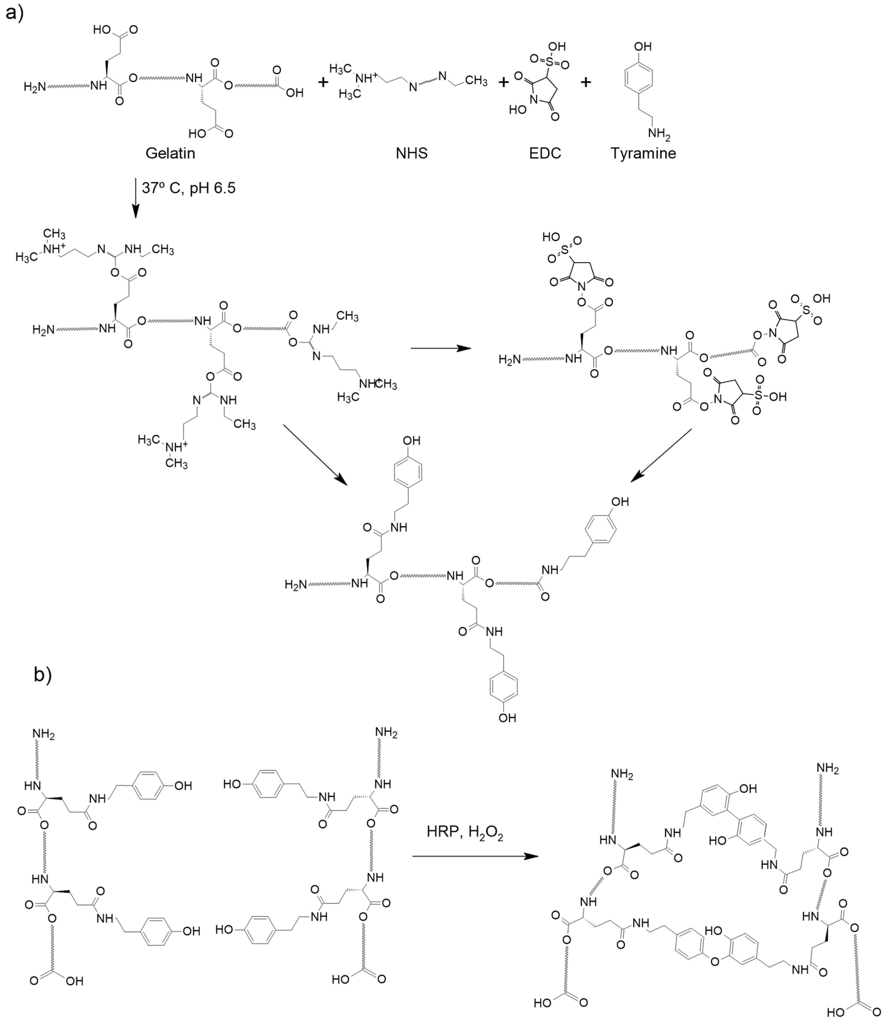
4.2. Gelatin Modification to Add Tyramine Groups

The chemical modification of gelatin with tyramine substitution was performed by reacting carbodiimide with carboxyl groups of gelatin, following previously published work [26]. Gelatin was dissolved at 2% w/v in 20 mL ultrapure water with 0.98% w/v MES at 60 °C and 0.56% w/v tyramine, before equilibrating the solution to pH 6. Cross-linking reagents (0.04% w/v N-Hydroxysuccinimide (NHS) and 0.61% w/v 1-Ethyl-3-(3-dimethylaminopropyl)carbodiimide (EDC) were added and once dissolved, the sample was incubated at 37 °C and stirred to allow the reaction. The sample was poured onto a cellulose membrane to dialyze and remove small molecules, changing the washing water every 8 h for three days. Finally, the tyramined gelatin was filtered through a filter with 0.22 µm pores and lyophilized. The degree of tyramine substitution was determined by absorbance at 275 nm.

4.3. PRP Collection

Sheep blood was donated by Instituto Universitario de Investigación de Ciencia y Tecnología Animal, Universitat Politècnica de València (ICTA; Valencia, Spain). Blood was collected into 5 mL citrate vacuum tubes and gently mixed to prevent blood clotting. The blood was centrifuged for 18 min at 1800 rpm to separate the formed blood elements from the plasma. The plasma fraction was divided into 4 portions, with the platelet-rich plasma (PRP) being the lower portion, in contact with the red fraction. Platelet-poor plasma fractions were discarded and PRP was stored frozen at −80 °C in sterile centrifuge tubes.

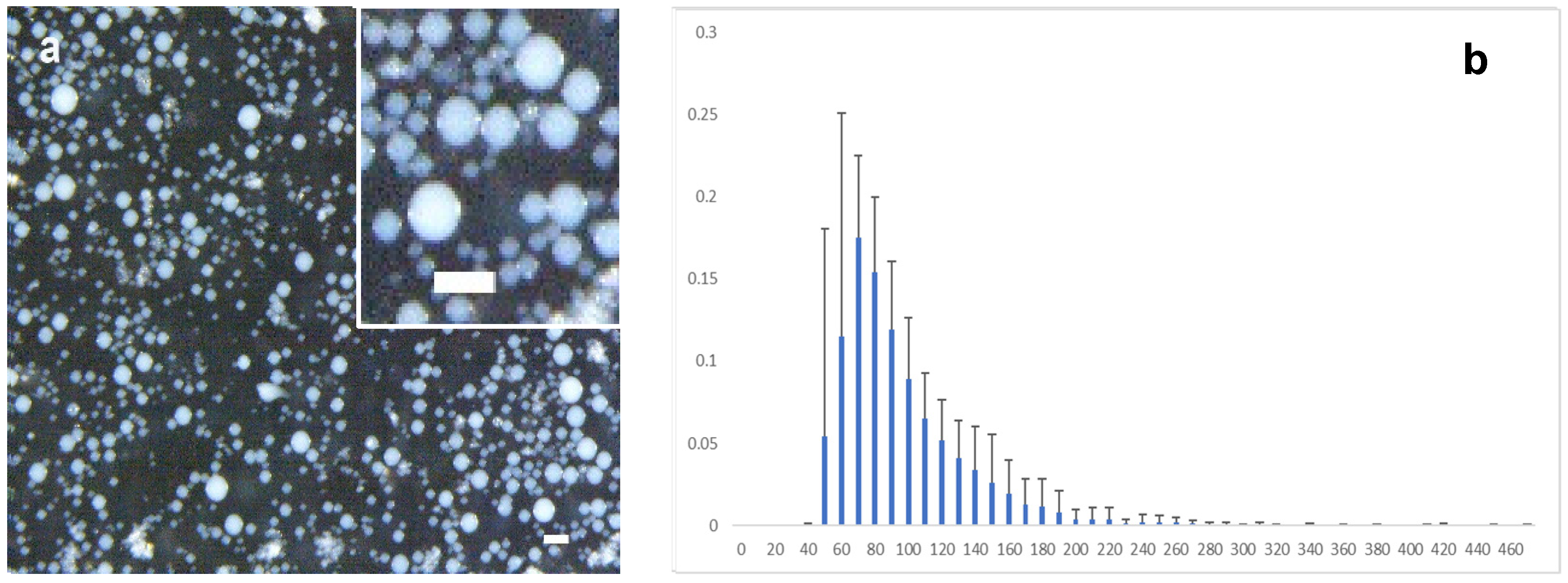
4.4. Preparation of the Microspheres

Gelatin microgels were obtained using a water-in-oil emulsion. The modified gelatin was dissolved at 4% (w/v) in calcium-free Krebbs–Ringer buffer (115 mM NaCl, 5 mM KCl, 1 mM KH2PO4 and 25 mM HEPES at pH 7.4) and sterilized by filtration. The gelatin solution was mixed with PRP and 12.5 U/mL HRP in a 4:4:1 solution. A volume of 180 µL of gelatin/PRP solution was mixed with 20 µL of 22 mM H2O2, under sterile conditions, poured into a 50 mL tube with 5 mL of sterile olive oil and emulsified for 12 s with a vortex (444–1372 (EU), VWR international, Radnor, PA, USA). The emulsion obtained was kept at room temperature for 20 min to complete the cross-linking reaction before removing the oil by filtration. The microspheres formed were then washed with DPBS, removing any remaining oil in the supernatant. Platelets were activated immediately after being added to the cell culture by immersing the microspheres in a solution of 0.5% CaCl2 with 0.9% NaCl. The diameter of the microspheres was determined in optical microscopy pictures by image analysis using Image J software v 1.53t (National Insitute of Health, Bethesda, MD, USA) from 10 pictures/batch from two different batches.

4.5. Evaluation of Platelet Activation

The platelet activation factor (PAF) quantification assay was performed on the gelatin/PRP microspheres using pure gelatin microspheres and bulk PRP, gelatin, and gelatin/PRP hydrogels as controls. Pure gelatin microspheres were prepared as described above without the addition of PRP. Control bulk gelatin hydrogels were obtained from a solution containing 4% tyramined gelatin, 12.5 U/mL HRP, and 22 mM H2O2 in a ratio of 8:1:1. Control bulk gelatin/PRP hydrogels were obtained by mixing 8% w/v tyramined gelatin, w/v PRP, 12.5 U/mL HRP and 22 mM H2O2 in a 4:4:1:1 solution. Both control bulk hydrogels were produced by pouring 200 µL of the corresponding solutions into a silicone mold and allowing them to coagulate at room temperature. Control PRP gels were obtained by mixing 200 µL of PRP with 10 µL of 1% w/v CaCl2 for 40 min at 37 °C in a glass vial. The gels were washed three times with 0.9% w/v NaCl. The microspheres and bulk hydrogel samples were seeded in a 48-well plate and incubated with 200 µL of 0.9% NaCl (non-activated platelets) or 0.5% CaCl2 (to activate platelets) for 30 min at 37 °C.

4.6. Cell Isolation and Culture

Pig mesenchymal stem cells (pMSCs) from bone marrow were used to perform all tests. Porcine mesenchymal stem cells were harvested from the femoral bone marrow. The femur bone was cut in the upper part in aseptic conditions and the gelatinous bone marrow was removed. Bone marrow was grinded and sieved with a 40 µm nylon mesh to remove tissue debris. Cells were counted with trypan blue and acetic acid and seeded at 4 × 10⁵ cells/cm in a T75 cm2 culture flask. Non-adherent cells were removed by changing the culture medium. Cells at passage three were seeded in culture flasks with a proliferation medium (PM) containing Dulbeccos’s modified minimum essential media (DMEM; Gibco, Waltham, MA, USA) supplemented with 10% fetal bovine serum (FBS; Gibco), 1% L glutamine (Gibco), 1% penicillin/streptomycin (Gibco), and 1% fungizone (Gibco), and were cultured in a humidified atmosphere incubator at 37 °C and 5% CO2. Culture media were changed every 2–3 days. Cells were detached from the flasks with a 0.25% (w/v) trypsin-0.91 mM EDTA solution (Gibco) and cultured at a density of 3 × 10⁵ cells in 30 µL of sample (µGel or µGel/PRP) in 0.5 mL microcentrifuge tubes. After 24 h, the culture media of half of the samples were changed to chondrogenic differentiation medium (DM) containing proliferation medium (1% FBS) supplemented with 1% insulin-transferrin-selenium (ITS; Gibco), 1% non-essential aminoacids (Gibco), 1% ascorbic acid (Gibco), 1% sodium pyruvate (Gibco), and 10 ng/mL transforming growth factor beta 1 (TGFβ1; Gibco).

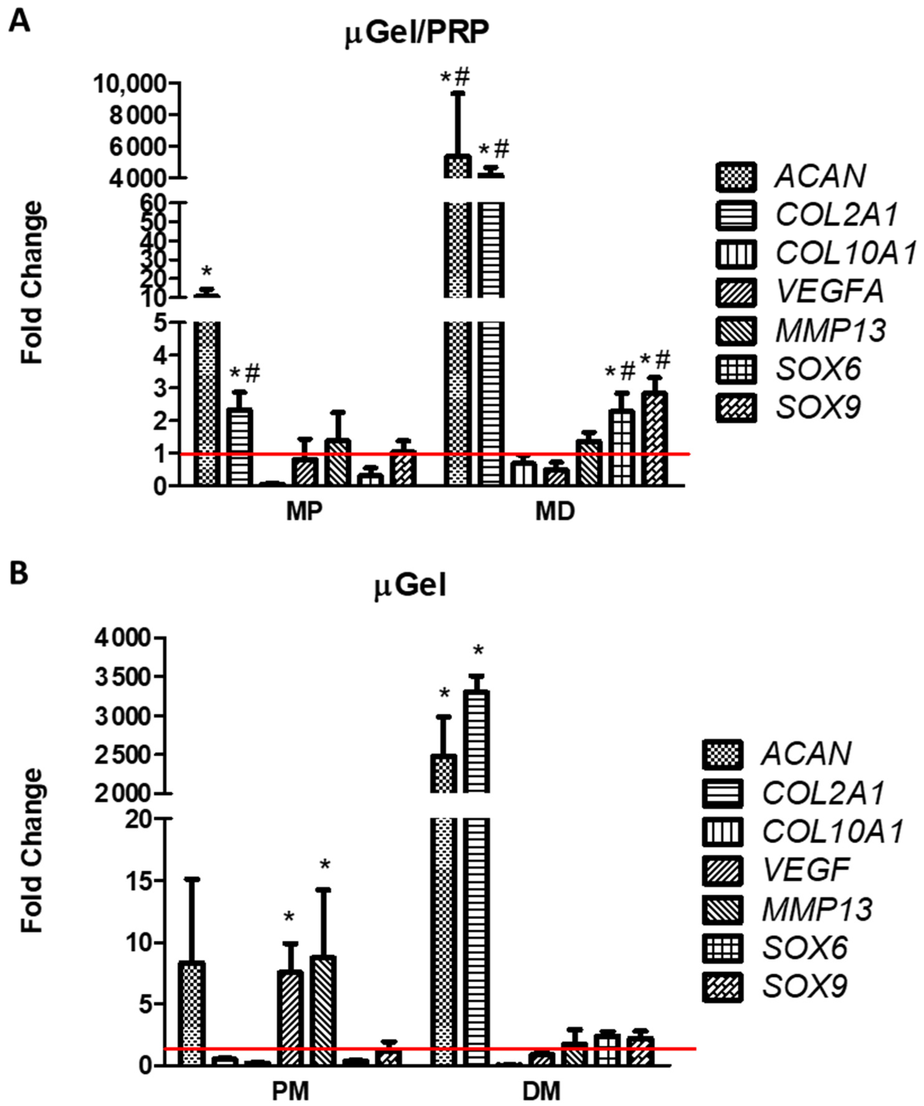
4.7. Analysis of Relative Gene Expression of Chondrocyte-Related Genes

Total RNA was extracted from 3D cultures using Trizol reagent (Thermo Fischer Scientific Inc., Waltham, MA, USA) according to the manufacturer’s instructions. RNA concentration was determined by spectrophotometry using a Nanodrop 2000 spectrophotometer (Fischer Scientific, Madrid, Spain). Only extracts with a 260/280 ratio > 1.8 were used. RNA integrity (RI) was evaluated by capillary electrophoresis using a Bioanalyzer (Agilent Technologies, Santa Clara, CA, USA). Only extracts with an RI number (RIN) of ~10 were used to determine gene expression levels. The expression levels of COL1A1, COL2A1, ACAN, COL10A1 and GAPDH genes were assayed by reverse transcriptase polymerase chain reaction (RT-PCR) using Assays on Demand (Applied Biosystems, Madrid, Spain, ID Ss03373340_m1, Ss03373344_g1, Ss03374823_m1, Ss03391766_m1 and Ss03375629_u1, respectively). The reactions were carried out in a 7900HT real-time Thermocycler (Applied Biosystems, Madrid, Spain). The comparative ΔΔCt method with glyceraldehyde 3 phosphate dehydrogenase (GAPDH) was used as an endogenous control to calculate relative gene expression levels [45].

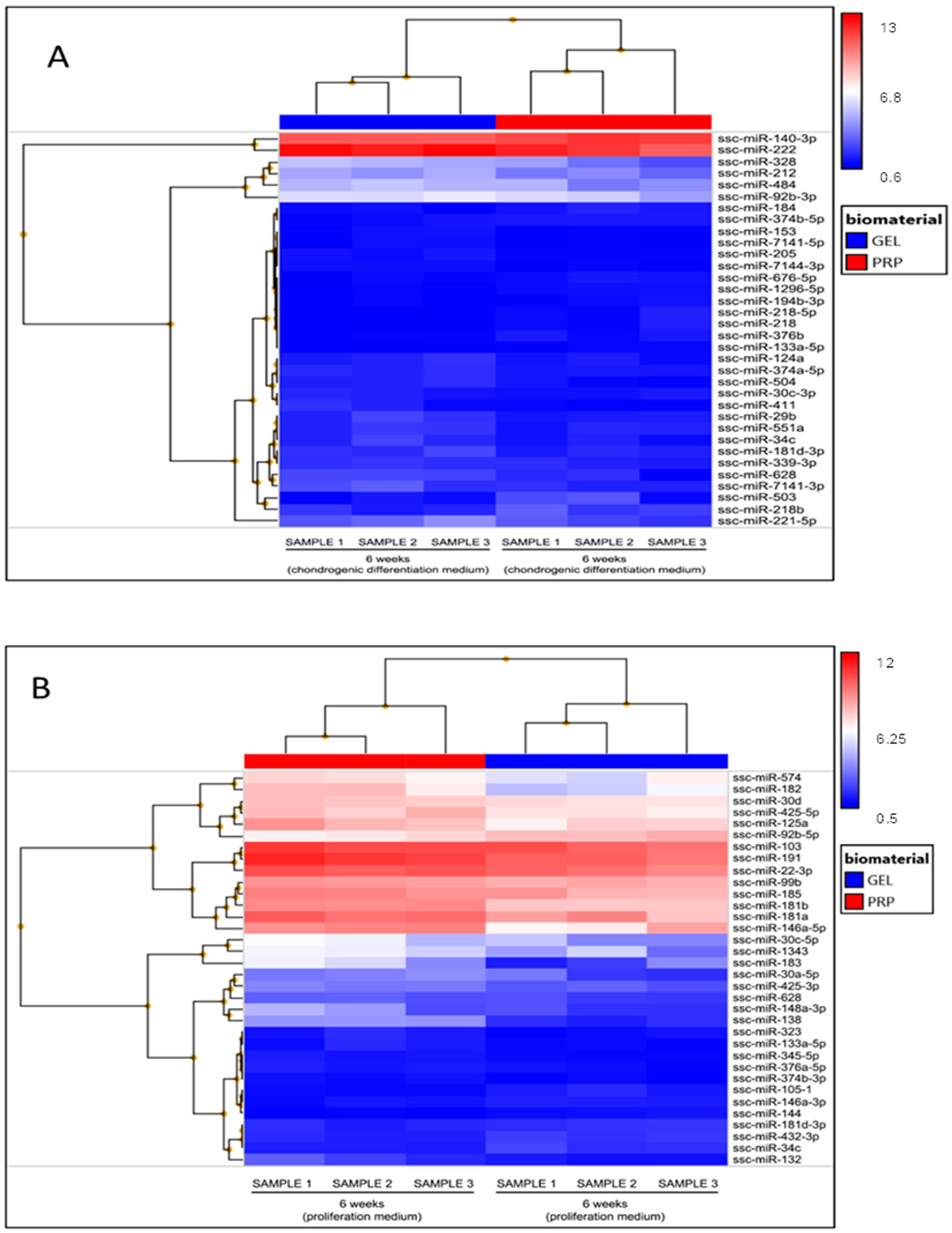
4.8. miRNAs Analysis

For analysis of global miRNA expression, Affymetrix miRNA 3.0 arrays (Affymetrix, CA, USA) were used, as previously reported [46]. The miRNA was extracted from cells using Trizol reagent as recommended by the manufacturer (Thermo Fischer Scientific Inc., Waltham, MA, USA). The miRNA extracts were labeled with biotin, using 3DNA Array Detection FlashTag™ Biotin HSR (Genisphere, Hatfield, PA, USA) and hybridized with the microarrays in a 645-hybridization oven (Affymetrix, CA, USA), following standardized protocols provided by Affymetrix. Microarray staining, washing, and scanning were then performed using a Fluidic 450 Station and a GeneChip 3000 7G confocal scanner (Affymetrix, Santa Clara, CA, USA). DAT and CEL files were acquired using GeneChip Command Console Software (version 4.3.2, AGCC, Affymetrix, CA, USA). Then, miRNA expressions were analyzed using Transcriptome Analysis Console (TAC) Software (version 4.0, Applied Biosystems). To do this, quality control of the array was performed, and data were normalized, followed by statistical tests to study the differential expression of these miRNA. Finally, the results were interpreted, and the significant differential expressions were represented using the hierarchical clustering method, comparing samples in different conditions (µGel vs. µGel/PRP, and proliferation samples vs. induced chondrogenic differentiation).

4.9. Data Presentation and Statistical Analysis

The experimental samples were replicated three times. The mean ± standard deviation (SD) was used. For statistical analysis, Tukey’s one-way ANOVA test was performed to find differences between samples groups, with a significance threshold of p-value of ≤0.05. miRNA arrays were normalized using the quartil method. ANOVA was used to select significant changes. False Discovery Rate (FDR) algorithm was used to correct the p-value.

5. Conclusions

By means of an emulsion process, cross-linked gelatin microspheres containing PRP can be prepared, avoiding at least in part, the activation of platelets during this process. Subsequent activation of platelets by calcium chloride immediately before cell seeding allows the release of platelet growth factors to be as efficient as possible. The release of these factors during the culture of MSCs in DM significantly induces chondrogenesis, as demonstrated by the expression of genes and miRNAs characteristic of the hyaline cartilage chondrocyte.

Supplementary Materials

The supporting information can be downloaded at: https://www.mdpi.com/article/10.3390/ijms25020937/s1.

Author Contributions

R.S.-C.: Cell culture experiments, gene and miRNA expression. J.R.-R.: Obtaining and characterization of materials. M.S.-T.: Conceptualization, Methodology, Cell culture experiments, Writing—Review and Editing. G.G.F.: Conceptualization, Methodology, Materials development, Writing—Review and Editing. J.L.G.R.: Conceptualization, Methodology, Supervision, Writing—Original Draft. M.M.: Conceptualization, Methodology, Cell culture experiments, Formal Analysis, Supervision, Writing—Review and Editing. All authors have read and agreed to the published version of the manuscript.

Funding

This work was supported by the projects PDC2021-121658-C21 and -C22, funded by MCIN/AEI/10.13039/501100011033 and by the European Union “NextGenerationEU”/PRTR. The research was also supported by CIBER-Consorcio Centro de Investigación Biomédica en Red—CB06/01/1026, Instituto de Salud Carlos III, Ministerio de Ciencia e Innovación.

Informed Consent Statement

Not applicable.

Data Availability Statement

Complete array data have been submitted to the GEO database (accession code ID GSE250185).

Conflicts of Interest

The authors declare no conflicts of interest. The funders had no role in the design of the study; in the collection, analyses, or interpretation of data; in the writing of the manuscript; or in the decision to publish the results.

References

  1. Anitua, E.; Sánchez, M.; Orive, G.; Andia, I. Delivering growth factors for therapeutics. Trends Pharmacol. Sci. 2008, 29, 37–41. [Google Scholar] [CrossRef] [PubMed]
  2. Alsousou, J.; Thompson, M.; Hulley, P.; Noble, A.; Willett, K. The biology of platelet-rich plasma and its application in trauma and orthopaedic surgery: A review of the literature. J. Bone Jt. Surg. Br. 2009, 91, 987–996. [Google Scholar] [CrossRef] [PubMed]
  3. Weibrich, G.; Hansen, T.; Kleis, W.; Buch, R.; Hitzler, W.E. Effect of platelet concentration in platelet-rich plasma on peri-implant bone regeneration. Bone 2004, 34, 665–671. [Google Scholar] [CrossRef] [PubMed]
  4. Marx, R.E. Platelet-rich plasma: Evidence to support its use. J. Oral Maxillofac. Surg. 2004, 62, 489–496. [Google Scholar] [CrossRef]
  5. Schmidt-Bleek, K.; Willie, B.M.; Schwabe, P.; Seemann, P.; Duda, G.N. BMPs in bone regeneration: Less is more effective, a paradigm-shift. Cytokine Growth Factor Rev. 2016, 27, 141–148. [Google Scholar] [CrossRef]
  6. Vukicevic, S.; Oppermann, H.; Verbanac, D.; Jankolija, M.; Popek, I.; Curak, J.; Brkljacic, J.; Pauk, M.; Erjavec, I.; Francetic, I.; et al. The clinical use of bone morphogenetic proteins revisited: A novel biocompatible carrier device OSTEOGROW for bone healing. Int. Orthop. 2014, 38, 635–647. [Google Scholar] [CrossRef]
  7. Jain, E.; Chinzei, N.; Blanco, A.; Case, N.; Sandell, L.J.; Sell, S.; Rai, M.F.; Zustiak, S.P. Platelet-rich plasma released from polyethylene glycol hydrogels exerts beneficial effects on human chondrocytes. J. Orthop. Res. 2019, 37, 2401–2410. [Google Scholar] [CrossRef]
  8. Ishida, K.; Kuroda, R.; Miwa, M.; Tabata, Y.; Hokugo, A.; Kawamoto, T.; Sasaki, K.; Doita, M.; Kurosaka, M. The regenerative effects of platelet-rich plasma on meniscal cells in vitro and its in vivo application with biodegradable gelatin hydrogel. Tissue Eng. 2007, 13, 1103–1112. [Google Scholar] [CrossRef]
  9. Saito, M.; Takahashi, K.A.; Arail, Y.; Inoue, A.; Sakao, K.; Tonomura, H.; Honjo, K.; Nakagawa, S.; Inoue, H.; Tabata, Y.; et al. Intraarticular administration of platelet-rich plasma with biodegradable gelatin hydrogel microspheres prevents osteoarthritis progression in the rabbit knee. Clin. Exp. Rheumatol. 2009, 27, 201–207. [Google Scholar]
  10. Nagae, M.; Ikeda, T.; Mikami, Y.; Hase, H.; Ozawa, H.; Matsuda, K.I.; Sakamoto, H.; Tabata, Y.; Kawata, M.; Kubo, T. Intervertebral disc regeneration using platelet-rich plasma and biodegradable gelatin hydrogel microspheres. Tissue Eng. 2007, 13, 147–158. [Google Scholar] [CrossRef]
  11. Xu, X.; Hu, J.; Lu, H. Histological observation of a gelatin sponge transplant loaded with bone marrow-derived mesenchymal stem cells combined with platelet-rich plasma in repairing an annulus defect. PLoS ONE 2017, 12, e0171500. [Google Scholar] [CrossRef] [PubMed]
  12. Zhang, M.; Zhen, J.; Zhang, X.; Yang, Z.; Zhang, L.; Hao, D.; Ren, B. Effect of autologous platelet-rich plasma and gelatin sponge for tendon-to-bone healing after rabbit anterior cruciate ligament reconstruction. Arthroscopy 2019, 35, 1486–1497. [Google Scholar] [CrossRef]
  13. Akeda, K.; An, H.S.; Okuma, M.; Attawia, M.; Miyamoto, K.; Thonar, E.J.M.A.; Lenz, M.; Sah, R.; Masuda, K. Platelet-rich plasma stimulates porcine articular chondrocyte proliferation and matrix biosynthesis. Osteoarthr. Cartil. 2006, 14, 1272–1280. [Google Scholar] [CrossRef] [PubMed]
  14. Jeyakumar, V.; Niculescu-Morzsa, E.; Bauer, C.; Lacza, Z.; Nehrer, S. Platelet-rich plasma supports proliferation and redifferentiation of chondrocytes during in vitro expansion. Front. Bioeng. Biotechnol. 2017, 5, 75. [Google Scholar] [CrossRef] [PubMed]
  15. Vetrano, M.; Ranieri, D.; Nanni, M.; Pavan, A.; Malisan, F.; Vulpiani, M.C.; Visco, V. Hyaluronic acid (HA), platelet-rich plasm and extracorporeal shock wave therapy (ESWT) promote human chondrocyte regeneration in vitro and ESWT-mediated increase of CD44 expression enhances their susceptibility to HA treatment. PLoS ONE 2019, 14, e0218740. [Google Scholar] [CrossRef]
  16. Zhang, Y.T.; Pan, Z.; Xia, L.H.; Liu, X.N.; Guo, X.L.; He, Y.; Zhou, J.; Qu, Z.H.; Mei, G.; Jin, D.; et al. Bilayered poly(Lactide-co-glycolide) scaffold with platelet-rich plasma and mesenchymal stem cells improves restoration of osteochondral defects. J. Biomater. Tissue Eng. 2015, 5, 757–765. [Google Scholar] [CrossRef]
  17. Filardo, G.; Kon, E.; Buda, R.; Timoncini, A.; Di Martino, A.; Cenacchi, A.; Fornasari, P.M.; Giannini, S.; Marcacci, M. Platelet-rich plasma intra-articular knee injections for the treatment of degenerative cartilage lesions and osteoarthritis. Knee Surg. Sports Traumatol. Arthrosc. 2011, 19, 528–535. [Google Scholar] [CrossRef]
  18. Kon, E.; Buda, R.; Filardo, G.; Di Martino, A.; Timoncini, A.; Cenacchi, A.; Fornasari, P.M.; Giannini, S.; Marcacci, M. Platelet-rich plasma: Intra-articular knee injections produced favorable results on degenerative cartilage lesions. Knee Surg. Sports Traumatol. Arthrosc. 2010, 18, 472–479. [Google Scholar] [CrossRef]
  19. Battaglia, M.; Guaraldi, F.; Vannini, F.; Rossi, G.; Timoncini, A.; Buda, R.; Giannini, S. Efficacy of ultrasound-guided intra-articular injections of platelet-rich plasma versus hyaluronic acid for hip osteoarthritis. Orthopedics 2013, 36, e1501–e1509. [Google Scholar] [CrossRef]
  20. Li, N.; Long, B.; Han, W.; Yuan, S.; Wang, K. MicroRNAs: Important regulators of stem cells. Stem Cell Res. Ther. 2017, 8, 110. [Google Scholar] [CrossRef]
  21. Stelcer, E.; Kulcenty, K.; Rucinski, M.; Jopek, K.; Richter, M.; Trzeciak, T.; Suchorska, W.M. The role of microRNAs in early chondrogenesis of human induced Pluripotent Stem Cells (hiPSCs). Int. J. Mol. Sci. 2019, 20, 4371. [Google Scholar] [CrossRef] [PubMed]
  22. Paganopoulos, P.K.; Lambrou, G.I. The involvement of microRNAs in osteoarthritis and recent developments: A narrative review. Mediterr. J. Theumatol. 2018, 29, 67–79. [Google Scholar] [CrossRef]
  23. Zurriaga Carda, J.; Lastra, M.L.; Antolinos-Turpin, C.M.; Morales-Román, R.M.; Sancho-Tello, M.; Perea-Ruiz, S.; Milián, L.; Fernández, J.M.; Cortizo, A.M.; Carda, C.; et al. A cell-free approach with a supporting biomaterial in the form of dispersed microspheres induces hyaline cartilage formation in a rabbit knee model. J. Biomed. Mater. Res. B Appl. Biomater. 2020, 108, 1428–1438. [Google Scholar] [CrossRef] [PubMed]
  24. Bello, A.B.; Kim, D.; Kim, D.; Park, H.; Lee, S.H. Engineering and functionalization of gelatin biomaterials: From cell culture to medical applications. Tissue Eng. Part B Rev. 2020, 26, 164–180. [Google Scholar] [CrossRef]
  25. Sulaiman, S.B.; Idrus, R.B.H.; Hwei, N.M. Gelatin microsphere for cartilage tissue engineering: Current and future strategies. Polymers 2020, 12, 2404. [Google Scholar] [CrossRef] [PubMed]
  26. Poveda-Reyes, S.; Moulisova, V.; Sanmartín-Masiá, E.; Quintanilla-Sierra, L.; Salmerón-Sánchez, M.; Ferrer, G.G. Gelatin-hyaluronic acid hydrogels with tuned stiffness to counterbalance cellular forces and promote cell differentiation. Macromol. Biosci. 2016, 16, 1311–1324. [Google Scholar] [CrossRef]
  27. Oliver-Ferrándiz, M.; Milián, L.; Sancho-Tello, M.; de Llano, J.J.M.; Roca, F.G.; Martínez-Ramos, C.; Carda, C.; Mata, M. Alginate-agarose hydrogels improve the in vitro differentiation of human dental pulp stem cells in chondrocytes. A histological study. Biomedicines 2021, 9, 834. [Google Scholar] [CrossRef]
  28. Infante, A.; Rubio-Azpeitia, E.; Sánchez, P.; Alberdi, R.; Rodriguez, C.I.; Andia, I. Platelet rich plasma and culture configuration affect the matrix forming phenotype of bone marrow stromal cells. Tissue Eng. Regen. Med. 2017, 14, 567–577. [Google Scholar] [CrossRef]
  29. Wang, Z.; Wang, Z.; Zhang, B.; Zhao, Q.; Liu, Y.; Qi, W. Effect of activated platelet-rich plasma on chondrogenic differentiation of rabbit bone marrow-derived mesenchymal stem cells. Stem Cells Int. 2021, 2021, 9947187. [Google Scholar] [CrossRef]
  30. Colnot, C.I.; Helms, J.A. A molecular analysis of matrix remodeling and angiogenesis during long bone development. Mech. Dev. 2001, 100, 245–250. [Google Scholar] [CrossRef]
  31. Lefebvre, V.; Behringer, R.R.; De Crombrugghe, B. L-Sox5, Sox6 and SOx9 control essential steps of the chondrocyte differentiation pathway. Osteoarthr. Cartil. 2001, 9, S69–S75. [Google Scholar] [CrossRef] [PubMed]
  32. Suomi, S.; Taipaleenmäki, H.; Seppänen, A.; Ripatti, T.; Väänänen, K.; Hentunen, T.; Säämänen, A.-M.; Laitala-Leinonen, T. MicroRNAs regulate osteogenesis and chondrogenesis of mouse bone marrow stromal cells. Gene Regul. Syst. Biol. 2008, 2, 177–191. [Google Scholar] [CrossRef] [PubMed]
  33. Wei, Z.J.; Liu, J.; Qin, J. MiR-138 suppressed the progression of osteoarthritis mainly through targeting p65. Eur. Rev. Med. Pharmacol. Sci. 2017, 21, 2177–2184. [Google Scholar]
  34. Vonk, L.A.; Kragten, A.H.M.; Dhert, W.J.A.; Saris, D.B.F.; Creemers, L.B. Overexpression of hsa-miR-148a promotes cartilage production and inhibits cartilage degradation by osteoarthritic chondrocytes. Osteoarthr. Cartil. 2014, 22, 145–153. [Google Scholar] [CrossRef]
  35. Tian, Y.; Guo, R.; Shi, B.; Chen, L.; Yang, L.; Fu, Q. MicroRNA-30a promotes chondrogenic differentiation of mesenchymal stem cells through inhibiting Delta-like 4 expression. Life Sci. 2016, 148, 220–228. [Google Scholar] [CrossRef] [PubMed]
  36. Melnik, S.; Hofmann, N.; Gabler, J.; Hecht, N.; Richter, W. MiR-181a targets RSPO2 and regulates bone morphogenetic protein—WNT signaling crosstalk during chondrogenic differentiation of mesenchymal stromal cells. Front. Cell. Dev. Biol. 2021, 9, 747057. [Google Scholar] [CrossRef]
  37. Xu, R.; Wei, Y.; Yin, X.; Shi, B.; Li, J. miR-20a suppresses chondrogenic differentiation of ATDC5 cells by regulating Atg7. Sci. Rep. 2019, 9, 9243. [Google Scholar] [CrossRef]
  38. Yan, C.; Wang, Y.; Shen, X.Y.; Yang, G.; Jian, J.; Wang, H.S.; Chen, G.Q.; Wu, Q. MicroRNA regulation associated chondrogenesis of mouse MSCs grown on polyhydroxyalkanoates. Biomaterials 2011, 32, 6435–6444. [Google Scholar] [CrossRef]
  39. Karlsen, T.A.; Jakobsen, R.B.; Mikkelsen, T.S.; Brinchmann, J.E. microRNA-140 targets RALA and regulates chondrogenic differentiation of human mesenchymal stem cells by translational enhancement of SOX9 and ACAN. Stem Cells Dev. 2014, 23, 290–304. [Google Scholar] [CrossRef]
  40. Melnik, S.; Gabler, J.; Dreher, S.I.; Hecht, N.; Hofmann, N.; Großner, T.; Richter, W. MiR-218 affects hypertrophic differentiation of human mesenchymal stromal cells during chondrogenesis via targeting RUNX2, MEF2C, and COL10A1. Stem Cell Res. Ther. 2020, 11, 532. [Google Scholar] [CrossRef]
  41. Yang, B.; Guo, H.; Zhang, Y.; Dong, S.; Ying, D. The microRNA expression profiles of mouse mesenchymal stem cell during chondrogenic differentiation. BMB Rep. 2011, 44, 28–33. [Google Scholar] [CrossRef] [PubMed]
  42. Song, J.; Jin, E.H.; Kim, D.; Kim, K.Y.; Chun, C.H.; Jin, E.J. MicroRNA-222 regulates MMP-13 via targeting HDAC-4 during osteoarthritis pathogenesis. BBA Clin. 2015, 3, 79–89. [Google Scholar] [CrossRef] [PubMed]
  43. Yang, F.; Huang, R.; Ma, H.; Zhao, X.; Wang, G. MiRNA-411 regulates chondrocyte autophagy in osteoarthritis by targeting hypoxia-inducible factor 1 alpha (HIF-1α). Med. Sci. Monit. 2020, 26, e921155. [Google Scholar] [CrossRef] [PubMed]
  44. Moulisová, V.; Poveda-Reyes, S.; Sanmartín-Masiá, E.; Quintanilla-Sierra, L.; Salmerón-Sánchez, M.; Gallego Ferrer, G. Hybrid Protein-Glycosaminoglycan Hydrogels Promote Chondrogenic Stem Cell Differentiation. ACS Omega 2017, 2, 7609–7620. [Google Scholar] [CrossRef]
  45. Milian, L.; Mata, M.; Alcacer, J.; Oliver, M.; Sancho-Tello, M.; de Llano, J.J.M.; Camps, C.; Galbis, J.; Carretero, J.; Carda, C. Cannabinoid receptor expression in non-small cell lung cancer. Effectiveness of tetrahydrocannabinol and cannabidiol inhibiting cell proliferation and epithelial-mesenchymal transition in vitro. PLoS ONE 2020, 15, e0228909. [Google Scholar] [CrossRef]
  46. Sarrion, I.; Milian, L.; Juan, G.; Ramon, M.; Furest, I.; Carda, C.; Gimeno, J.C.; Roig, M.M. Role of circulating miRNAs as biomarkers in idiopathic pulmonary arterial hypertension: Possible relevance of miR-23a. Oxid. Med. Cell. Longev. 2015, 2015, 792846. [Google Scholar] [CrossRef]
Figure 1. (a) Scheme of the modification of gelatin to add phenol groups. (b) Scheme of the gelatin network.
Ijms 25 00937 g001
Figure 2. (a) Stereoscopic microscope image of the gelatin/PRP microspheres obtained with 12 s of vortex mixing; the size bar corresponds to 200 μm. A representative panoramic view of the size distribution and an inset with a more detailed view are shown. Insert size bar equals to 200 μm. (b) Microsphere diameter histogram. Mean ± SD of n = 6 independent experiments are shown.
Ijms 25 00937 g002
Figure 3. Level of platelet activating factor (PAF) in the different groups studied. The results were normalized according to the PRP content. Immersion in CaCl2 activated platelets encapsulated in μGel/PRP microspheres, which showed a much lower level of PAF than without activation by CaCl2. The details of the experimental groups are shown in Table 1. The mean ± SD of three different experiments is represented as * p < 0.05 compared to the control group (black bars).
Ijms 25 00937 g003
Figure 4. Expression levels of genes related to chondrocytes. pMSCs were cultured for 6 weeks in gelatin microgels containing (A) or not containing (B) PRP with proliferation (PM) or chondrogenic differentiation media (DM). The relative gene expression levels of ACAN, COL2A1, COL10A1, VEGF, MMP13, SOX6 and SOX9 were evaluated by real-time RT-PCR. GAPDH was used as a housekeeping gene. The fold change (2−∆∆Ct) was calculated using the relative expression of cells cultured in 2D with PM as a control group (indicated by the red line). The mean ± SD of three different experiments is represented as * p < 0.05 compared to the control group (2D cultured cells), and # p < 0.05 comparing cells cultured in µgel in the presence or absence of PRP.
Ijms 25 00937 g004
Figure 5. Expression levels of genes related to chondrogenesis. pMSCs were cultured in gelatin microgels containing or not containing PRP, with (A) chondrogenic differentiation or (B) proliferation culture medium for 6 weeks. Total RNA was extracted and analyzed using Affymetrix miRNA arrays. The t-test following the false discovery rate (FDR) p-value correction was used to detect significant changes between cells culture in PRP containing microgels compared to non-PRP microgels. Hierarchical clustering was used to analyze the data obtained.
Ijms 25 00937 g005
Table 1. Experimental series.
μGel/PRPGelatin/PRP Microspheres
μGel/PRP-AGelatin/PRP microspheres activated with CaCl2
bGel/PRPBulk Gelatin/PRP
bGel/PRP-ABulk Gelatin/PRP activated with CaCl2
PRPPure PRP
PRP-APure PRP activated with CaCl2
Table 2. Differential expression of miRNAs. pMSCs were cultured in gelatin microgels containing or not PRP, in proliferation or chondrogenic differentiation culture media. Data was filtered according to a fold change > 1.7 and adjusted to p < 0.05. Means and adjusted p-value of n = 3 replicates are shown.
Proliferation MediumChondrogenic Differentiation Medium
miRNAFold ChangeAdjusted p-ValuemiRNAFold ChangeAdjusted p-Value
ssc-miR-374b-3p1.750.0706ssc-miR-5033.360.1456
ssc-miR-1321.980.0542ssc-miR-140-3p2.060.0137
ssc-miR-181b2.550.00000402ssc-miR-218b1.990.1005
ssc-miR-3231.780.0994ssc-miR-376b1.870.0612
ssc-miR-125a2.080.0653ssc-miR-133a-5p1.820.0226
ssc-miR-144−1.940.0167ssc-miR-194b-3p1.780.174
ssc-miR-105-1−1.960.0236ssc-miR-1296-5p1.780.0269
ssc-miR-432-3p−2.130.0161ssc-miR-676-5p1.740.0882
ssc-miR-1384.630.0000379ssc-miR-1841.730.0856
ssc-miR-148a-3p4.510.0889ssc-miR-2181.730.083
ssc-miR-181a2.420.041ssc-miR-218-5p1.730.083
ssc-miR-30d2.070.0098ssc-miR-374b-5p1.710.0364
ssc-miR-1031.890.0903ssc-miR-124a−1.710.1324
ssc-miR-6281.730.0598ssc-miR-7141-5p−1.710.1605
ssc-miR-146a-3p−1.910.0282ssc-miR-205−1.720.0238
ssc-miR-92b-5p−2.620.0076ssc-miR-7144-3p−1.720.008
ssc-miR-22-3p2.10.0644ssc-miR-339-3p−1.740.0988
ssc-miR-30c-5p5.880.0861ssc-miR-374a-5p−1.760.0084
ssc-miR-99b1.90.028ssc-miR-92b-3p−1.760.1694
ssc-miR-13433.730.0838ssc-miR-181d-3p−1.780.1152
ssc-miR-1852.080.058ssc-miR-30c-3p−1.780.1384
ssc-miR-133a-5p1.760.0941ssc-miR-153−1.790.1415
ssc-miR-30a-5p3.170.0763ssc-miR-34c−1.810.1241
ssc-miR-425-3p2.150.0073ssc-miR-551a−1.830.1258
ssc-miR-1839.240.0538ssc-miR-29b−1.910.0807
ssc-miR-376a-5p1.710.0444ssc-miR-504−1.980.03
ssc-miR-425-5p1.80.0188ssc-miR-628−1.990.0747
ssc-miR-345-5p1.720.0043ssc-miR-411−2.070.0789
ssc-miR-34c−1.970.0341ssc-miR-484-2.220.1198
ssc-miR-181d-3p−1.940.0561ssc-miR-221-5p−2.270.1237
ssc-miR-5742.590.0699ssc-miR-7141-3p−2.340.0945
ssc-miR-1826.460.0163ssc-miR-222−2.510.083
ssc-miR-146a-5p5.30.0401ssc-miR-212−2.60.0398
ssc-miR-1911.850.0115ssc-miR-328−3.380.0559
Disclaimer/Publisher’s Note: The statements, opinions and data contained in all publications are solely those of the individual author(s) and contributor(s) and not of MDPI and/or the editor(s). MDPI and/or the editor(s) disclaim responsibility for any injury to people or property resulting from any ideas, methods, instructions or products referred to in the content.

