Article

Serotypes, Antimicrobial Susceptibility, and Potential Mechanisms of Resistance Gene Transfer in Erysipelothrix rhusiopathiae Strains from Waterfowl in Poland

1
Department of Veterinary Prevention and Avian Diseases, Faculty of Veterinary Medicine, University of Life Sciences in Lublin, Akademicka 12, 20-033 Lublin, Poland
2
Laboratory of Molecular Biology, Vet-Lab Brudzew, Turkowska 58c, 62-720 Brudzew, Poland
3
Elizabeth Macarthur Agricultural Institute, NSW Department of Primary Industries and Regional Development, PMB 408, Narellan, NSW 2570, Australia
*
Author to whom correspondence should be addressed.
Int. J. Mol. Sci. 2024, 25(22), 12192; https://doi.org/10.3390/ijms252212192
Submission received: 10 October 2024 / Revised: 11 November 2024 / Accepted: 11 November 2024 / Published: 13 November 2024

Abstract

:
Erysipelas is a significant problem in the waterfowl farming in Poland, and information on the characteristics of the Erysipelothrix rhusiopathiae strains causing this disease is limited. In this study, we determined the serotypes, antimicrobial susceptibility, and potential mechanisms of resistance gene transfer in E. rhusiopathiae isolates (n = 60) from domestic geese and ducks. We also developed a multiplex PCR for the detection of resistance genes. The antimicrobial susceptibility of the isolates was assessed using the broth microdilution method. Resistance genes, integrative conjugative element (ICE)-specific genes, phage-specific genes, and serotype determinants were detected by PCR. Multilocus sequence typing (MLST) was performed for selected resistant strains. The comparative analyses included 260 E. rhusiopathiae strains whose whole genome sequences (WGSs) are publicly available. E. rhusiopathiae isolates represented 7 serotypes, among which serotypes 5 (38.3%) and 1b (28.3%) were the most common. All strains were susceptible to β-lactams, and the vast majority of them were resistant to tetracycline (85%) and enrofloxacin (80%). The percentages of isolates resistant to other antimicrobials used ranged from 3.3% to 16.7%. Ten isolates (16.7%) were found to be multidrug resistant (MDR). The genotypic resistance profiles of the E. rhusiopathiae strains corresponded to their phenotypic resistance, and the amplification patterns obtained using the 10-plex PCR developed in this study were fully consistent with the results of single PCRs. The most prevalent resistance gene was tetM. In enrofloxacin-resistant strains, nonsynonymous mutations in the gyrA and parC genes were identified. The presence of ICE-specific genes was confirmed in resistant strains, and in MDR isolates of serotype 8 that represented sequence type (ST) 113, prophage DNA (Javan630-like) linked to the lsaE gene was additionally detected. The results indicate that β-lactam antibiotics should be the first choice for the treatment of waterfowl erysipelas in Poland. ICEs, including a transposon from the Tn916/Tn1545 family, and bacteriophages are most likely responsible for the transfer of resistance genes in E. rhusiopathiae.

1. Introduction

Erysipelothrix rhusiopathiae (E. rhusiopathiae) is a facultative anaerobic, thin, Gram-positive, non-motile, and non-spore-forming rod-shaped bacterium that causes erysipelas in various animal species, as well as in humans [1]. It belongs to the genus Erysipelothrix, which currently includes 13 species: E. rhusiopathiae, E. tonsillarum, E. inopinata, Erysipelothrix sp. strain 1, E. piscisicarius (formerly Erysipelothrix sp. strain 2), Erysipelothrix sp. strain 3, E. larvae, E. anatis, E. urinaevulpis, E. aquatica, E. murinus, and the recently discovered E. amsterdamensis and E. enhydrae [2,3,4,5,6]. They represent class Erysipelotrichia in the phylum Bacillota (Firmicutes) [2,7]. E. rhusiopathiae strains grow on media supplemented with blood (whole or lysed), serum, or 0.1% Tween 80. They favor alkaline pH (optimum 7.2–7.6), usually produce H2S, and may be α-hemolytic [7]. Based on the structure of peptidoglycan antigens in E. rhusiopathiae strains, 15 serotypes are distinguished: 1a, 1b, 2, 4, 5, 6, 8, 9, 11, 12, 15, 16, 17, 19, and 21. Strains that do not cause the production of precipitating antibodies in immunized animals are classified as type N [2].
E. rhusiopathiae infections, both symptomatic and asymptomatic, are common in domestic pigs worldwide [8,9]. In Poland, however, erysipelas is also a serious problem in waterfowl farming [10]. Infection of birds takes the form of septicemia, leading to deaths in the flock. Anatomopathological examination shows hyperemia of internal organs, subcutaneous tissue, visceral fat, and muscles, as well as swelling of the liver, spleen, and air sacs [10,11]. Outdoor rearing is believed to increase birds’ exposure to E. rhusiopathiae bacteria, which can contaminate soil and water bodies. Prevention of erysipelas in waterfowl is limited due to the lack of vaccines for this group of animals and insufficient knowledge of predisposing factors for the disease [10]. Therefore, antibiotic therapy plays the most important role in combating E. rhusiopathiae infections in geese and ducks [12].
Currently available data on the antimicrobial susceptibility and genotypic resistance profiles of E. rhusiopathiae strains causing septicemia in waterfowl are very scarce [13,14]. Reports in this area, presenting the susceptibility of wild-type isolates to antimicrobial agents approved for the treatment of poultry diseases, are essential to developing effective infection control methods. Immediate implementation of appropriate therapy is crucial in the case of acute erysipelas, due to the risk of high mortality in the flock and thus huge economic losses. Moreover, monitoring the occurrence of antimicrobial resistant strains, including zoonotic ones, is an important task of the ‘European One Health Action Plan to combat antimicrobial resistance’ (launched in the EU in 2017, COM/2017/0339) [15].
The literature data show that E. rhusiopathiae strains can harbor various resistance genes that can be located on genomic islands or integrative and conjugative elements (ICEs), including the Tn916-like transposon. In single strains, the presence of prophage DNA-carrying resistance genes has also been demonstrated [14,16,17].
The aim of the study was to evaluate the antimicrobial susceptibility and determine the genotypic resistance profiles and serotypes of E. rhusiopathiae strains isolated from clinical cases of erysipelas in domestic geese and ducks reared in Poland, as well as to determine the potential mechanisms of resistance gene transfer occurring in these bacteria. An additional goal was to develop a multiplex PCR for detection of resistance genes and to determine the sequence type (ST) of multidrug-resistant (MDR) isolates. The comparative analyses included 260 E. rhusiopathiae strains whose whole genome sequences (WGSs) are publicly available.

2. Results

2.1. Identification of E. rhusiopathiae Isolates

Real-time PCR analysis confirmed that all 60 isolates obtained from geese and ducks with septicemia belonged to the species E. rhusiopathiae. In the case of four isolates, the affiliation to the species E. rhusiopathiae was additionally confirmed based on our previous analysis of the WGSs [14].

2.2. Serotyping

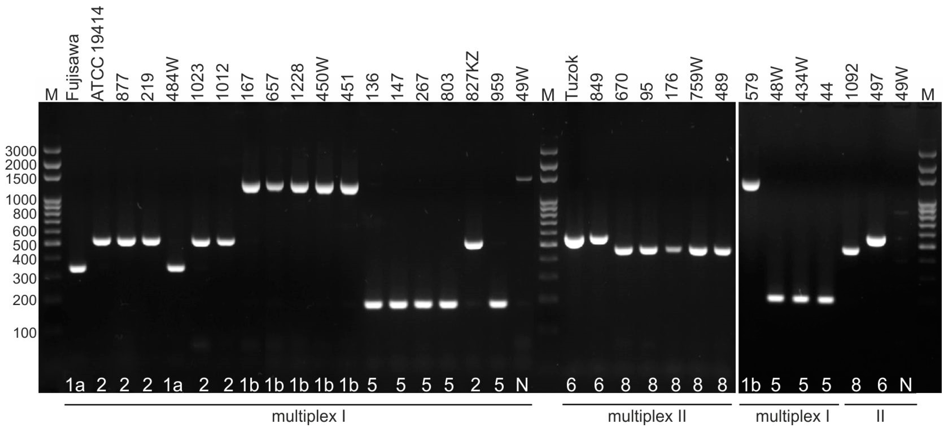
The analyses performed showed that more than 38% of E. rhusiopathiae isolates belonged to serotype 5 (23/60; 38.3%) and 28.3% to serotype 1b (17/60). The remaining isolates represented serotypes 8 (9/60; 15.0%), 2 (6/60; 10.0%), and 6 (2/60; 3.3%). Two isolates for which no PCR product was obtained in any of the four multiplex PCRs were designated serotype N (2/60; 3.3%) (Table 1, Figure A1). Strains from ducks represented serotypes 5 (1/5), 1b (2/5), and 2 (2/5). The remaining serotypes were detected only in strains from geese. Interestingly, in the case of one sample obtained from a nine-week-old goose from the reproductive flock, two strains were isolated simultaneously, one of which belonged to serotype 8 (strain 818) and the other to serotype 5 (strain 155).
Among the 260 strains whose WGSs were retrieved from the GenBank database (https://www.ncbi.nlm.nih.gov/genbank/, accessed on 26 September 2024), serotypes 1a (93/260), 1b (57/260), and 2 (56/260) were dominant (Table 1). Serotype 8 was not identified in any strain. Among the 23 genomes of strains from poultry and wild birds, serovar 1b (8/23) and 5 (7/23) were most frequently identified. Serovar 6, like 5, was found only in isolates from birds (Table 1, Table S1).

2.3. Antimicrobial Susceptibility

All E. rhusiopathiae isolates (n = 60) were susceptible to β-lactam antibiotics, i.e., penicillin, ampicillin, and ceftiofur, and were also characterized by low MIC values of amoxicillin and amoxicillin + clavulanic acid (ca), i.e., ≤0.06 µg/mL. Low MICs in the range of 2–8 µg/mL were also observed for florfenicol. Most isolates showed resistance to tetracycline (85%) and enrofloxacin (80%). The percentages of strains resistant to other antimicrobial substances ranged from 3.3% (erythromycin) to 16.7% (lincosamides) (Table 2). Very high MICs (≥512 µg/mL) of gentamicin, neomycin and trimethoprim/sulfamethoxazole were obtained for all isolates except one, for which the MIC of gentamicin was 256 µg/mL. The MICs of streptomycin and spectinomycin ranged from 16 to ≥512 µg/mL (Table 2). Ten strains (16.7%) were multidrug resistant (MDR—strains with resistance to at least one antimicrobial substance from three or more groups). Interestingly, they most often represented serotype 8 (7/10), and less frequently serotype 5 (2/10) or serotype 2 (1/10) (Table A1).
Two erythromycin-resistant strains had concurrent tylosin resistance; surprisingly, one strain (1012, serotype 2) was resistant to tylosin but susceptible to erythromycin. Nine of 10 lincosamide-resistant strains were also resistant to tiamulin (Table A1). A correlation between tetracycline and enrofloxacin resistance was noted as well, except for three strains of serotype 2, which were resistant to tetracycline but susceptible to enrofloxacin (Table A1).

2.4. Genotypic Resistance Profiles

The amplification patterns obtained using the 10-plex PCR developed in this study for the detection of tetM, lsaE, lnuB, ant(6)-Ia, spw, aph(3′)-IIIa, sat4, ermB, erm47, and int-Tn were fully consistent with the results of single PCRs and with the genotypic resistance profiles determined based on DNA sequence analysis of control strains. No nonspecific PCR products were visible on the gel. The results indicate that the new multiplex PCR protocol can be successfully used to detect resistance genes and transposons from the Tn916/Tn1545 family not only in E. rhusiopathiae strains but also in other Gram-positive bacteria, such as Ligilactobacillus salivarius, Enterococcus faecium, or Streptococcus gallolyticus (Figure 1).
The genotypic resistance profiles of E. rhusiopathiae strains were fully consistent with their phenotypic resistance. All tetracycline-resistant isolates contained the tetM gene (coding for ribosomal protection protein, which catalyzes the release of tetracycline from ribosomes in a GTP-dependent reaction). Resistance to macrolides, found in two isolates (no. 1023 and 267), whose WGSs were analyzed in our previous work [14], was caused by the erm47 and ermB genes, respectively. All isolates resistant to tiamulin and lincosamides (n = 10) contained the lsaE gene (coding for ABC transporter) and the lnuB gene (coding for lincosamide nucleotide transferase). The exception was strain 267, whose resistance to lincosamides was determined by the ermB gene (coding for ribosomal methylase, which dimethylates a single adenine in 23S rRNA of 50S ribosomal subunit). In four strains characterized by high MICs of streptomycin (>512 µg/mL) and spectinomycin (256–>512 µg/mL), the presence of the ant(6)-Ia gene (coding for aminoglycoside nucleotidyltransferase) and the spw gene (coding for aminoglycoside nucleotidyltransferase of the ANT(9) family) was confirmed (Table A1). These genes have always coexisted, like lnuB and lsaE (Table A1, Figure 1).
Sequence analysis of the gyrA and parC genes showed that resistance of E. rhusiopathiae strains to enrofloxacin is due to a mutation at position 257 in the gyrA gene (C → T or C → A) and at position 242 in the parC gene (T → G). The mutations translate into the change Thr86 → Ile or Thr86 → Lys in the gyrA gene and Ser81 → Ile in the parC gene. Interestingly, a gyrAThr86 → Lys mutation was recorded in all tested strains of serotype 5 and one strain of serotype 2, while the gyrAThr86 → Ile mutation occurred in strains of serotypes 1b, 8 and 6 (Table 3).

2.5. Detection of ICE-Specific Genes and Prophage Regions

The int-Tn gene encoding the integrase of Tn916/Tn1545 transposons was detected in all tetM-positive isolates (Table A1, Figure 1). The mobL gene, encoding MobL relaxase, which is most likely responsible for DNA transfer by conjugation, was detected in several phenotypically resistant strains (n = 7) of serotypes 2 and 5. The virB4 gene encoding the VirB4 protein specific for bacterial secretion system IV was found in only one strain, i.e., 1023, whose WGSs had been analyzed in our previous research [14] (Table A1).
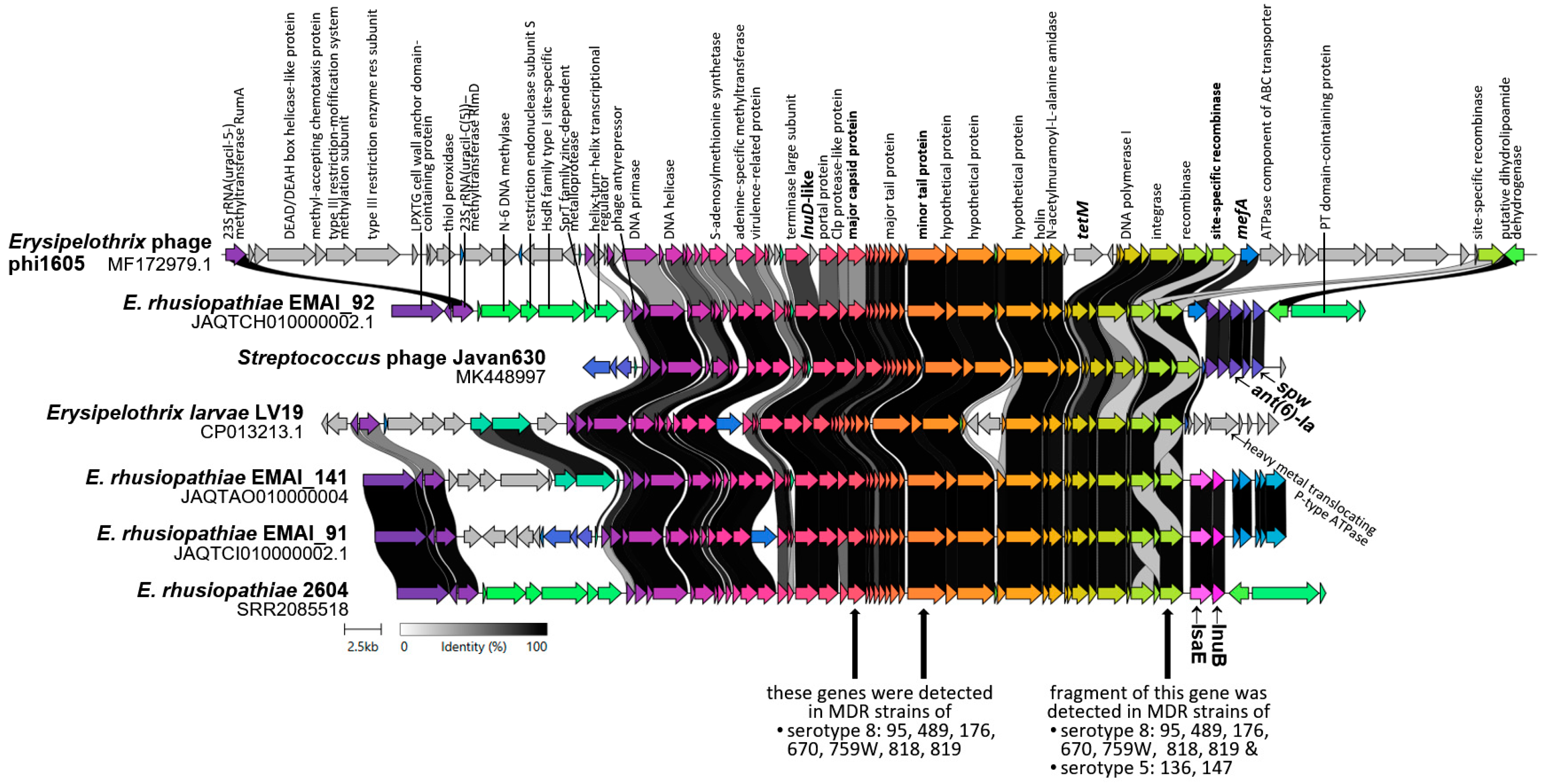
BLAST analysis showed that regions homologous to the sequences of Erysipelothrix phage phi1605 (90,000 bp, GB: MF172979.1) [16] and Streptococcus phage Javan630 (48,058 bp, GB: MK448997.1) [18] are present in 18 of the 260 analyzed E. rhusiopathiae genomes (Table S1). Homologous prophage sequences were also confirmed in E. larvae strain LV19 and two strains of Thomasclavelia ramosa (DFI.6.112 and DFI.6.30, family Erysipelotrichales), as well as in several genomes of Gram-positive bacteria outside the Erysipelotrichales family, i.e., Enterococcus, Streptococcus, Listeria, Clostridium, and Geosporobacter. Interestingly, 17 out of 18 prophage-positive E. rhusiopathiae genomes contained the resistance genes: tetM (16/18), lnuB and lsaE (9/18), ant(6)-Ia and spw (6/18), mph(B) (3/18), mef(A) (2/18), msr(D) (2/18), lnu(D)-like, and ermG (1/18) (Table S1). Further analyses using BLAST and Clinker showed that most of these resistance genes are located within the prophage DNA (Table S1, Figure 2).
The results of analyses performed using publicly available WGSs of phages (Javan630 and phi1605) and E. rhusiopathiae strains suggested that also the MDR isolates examined in this study may harbor prophage regions containing resistance genes. Our assumptions were confirmed by PCR results, where the presence of three phage-specific genes, i.e., the gene coding for site-specific recombinase, minor tail protein, and major capsid protein, was detected in all MDR serotype 8 isolates (n = 7) (Figure 2). By amplifying a DNA region encompassing the phage site-specific recombinase gene and lasE, we showed that the lsaE gene is located within the prophage DNA in all of these isolates.
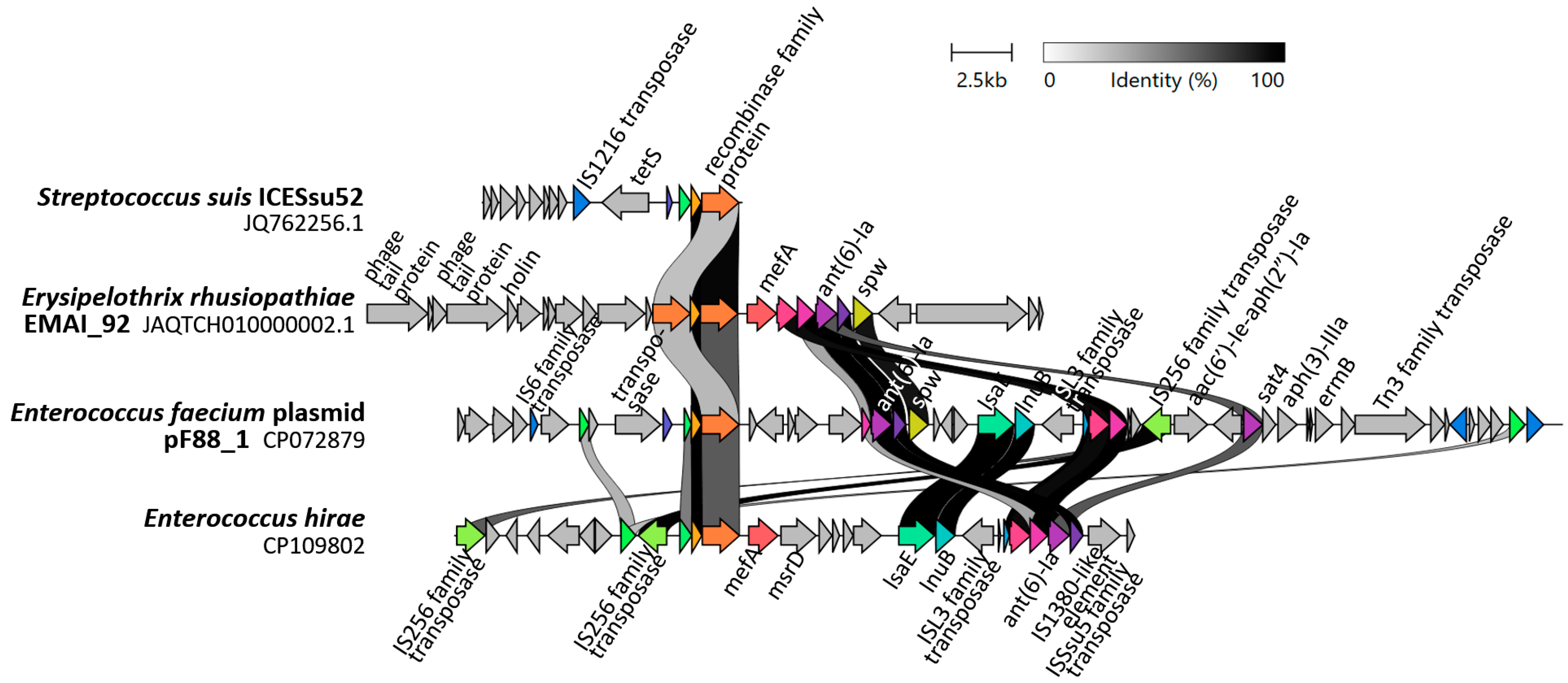
In MDR isolate nos. 136 and 147 serotype 5, only the gene coding for phage site-specific recombinase was detected (Figure 2). Additional BLAST analysis showed that a 462-bp amplified region of this gene is present in many genomes of a wide variety of Gram-positive bacteria and may be located not only in the prophage DNA but also on a plasmid or an ICE, usually next to resistance genes (Figure A2).
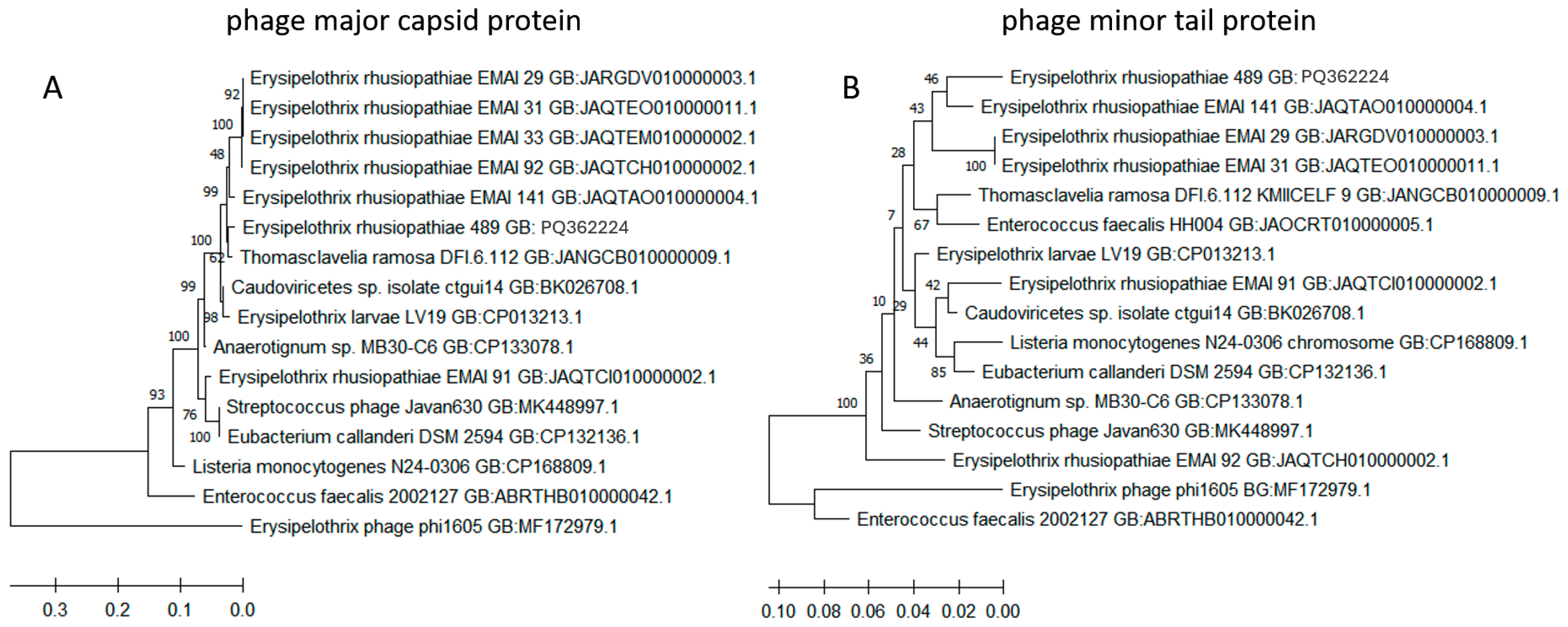
BLAST analysis showed that the sequence of the gene encoding phage major capsid protein of the E. rhusiopathiae strain 489 serotype 8 (GB: PQ362224) is 98% homologous to the prophage region of Thomasclavelia ramosa strain DFI.6.112 (GB: JANGCB010000009.1) and the 93–97% homologous gene of E. rhusiopathiae strains EMAI 29, 31, 33, 91, 92, and 141 [19]. The similarity to the sequence of Streptococcus phage Javan630 was 93%, and to phage phi1605 (found in the E. rhusiopathiae ZJ strain in China)—only 67% (at 34% coverage) (Figure 3A). The sequence of the gene encoding phage minor tail protein in strain no. 489 serotype 8 (GB: PQ362225) was highly homologous to the analogous gene found in E. rhusiopathiae strains EMAI_141 (96%), EMAI_29, 31, 33, 92 (≥92%), and phage Javan630 (95%), and it was less similar (85%) to the sequences of phage phi1605 (Figure 3B). Sequences homologous to the gene coding of phage major capsid protein (≥85%) and minor tail protein (≥90%) of isolate no. 489 were also detected in the genomes of strains representing other genera of Gram-positive bacteria, i.e., Enterococcus, Anaerotignum, Eubacterium, and Listeria (Figure 3).
The results of the analysis using two phage genes allow us to conclude that the prophage regions present in MDR serotype 8 isolates are more similar to the sequence of prophage DNA detected in E. rhusiopathiae EMAI strains from Australia and Streptococcus phage Javan630 than to the Erysipelothrix phage phi1605.

2.6. MLST Results

Among the eight E. rhusiopathiae strains subjected to MLST analysis, the occurrence of two STs was demonstrated, i.e., 113 and 4. All serotype 8 strains, both multidrug-resistant (95, 489, 176, 759 W) and non-MDR (1092), represented ST 113. The same sequence type was also determined in MDR strain 8S serotype 8 from a pig in Poland. MDR strain 136 serotype 5 and non-MDR strain 451 serotype 1b represented ST 4, similarly to MDR strain 1023, whose WGSs were analyzed in our previous work [14]. It is worth noting that ST 4 and ST 113 differ only in the galK allele (Table 4).
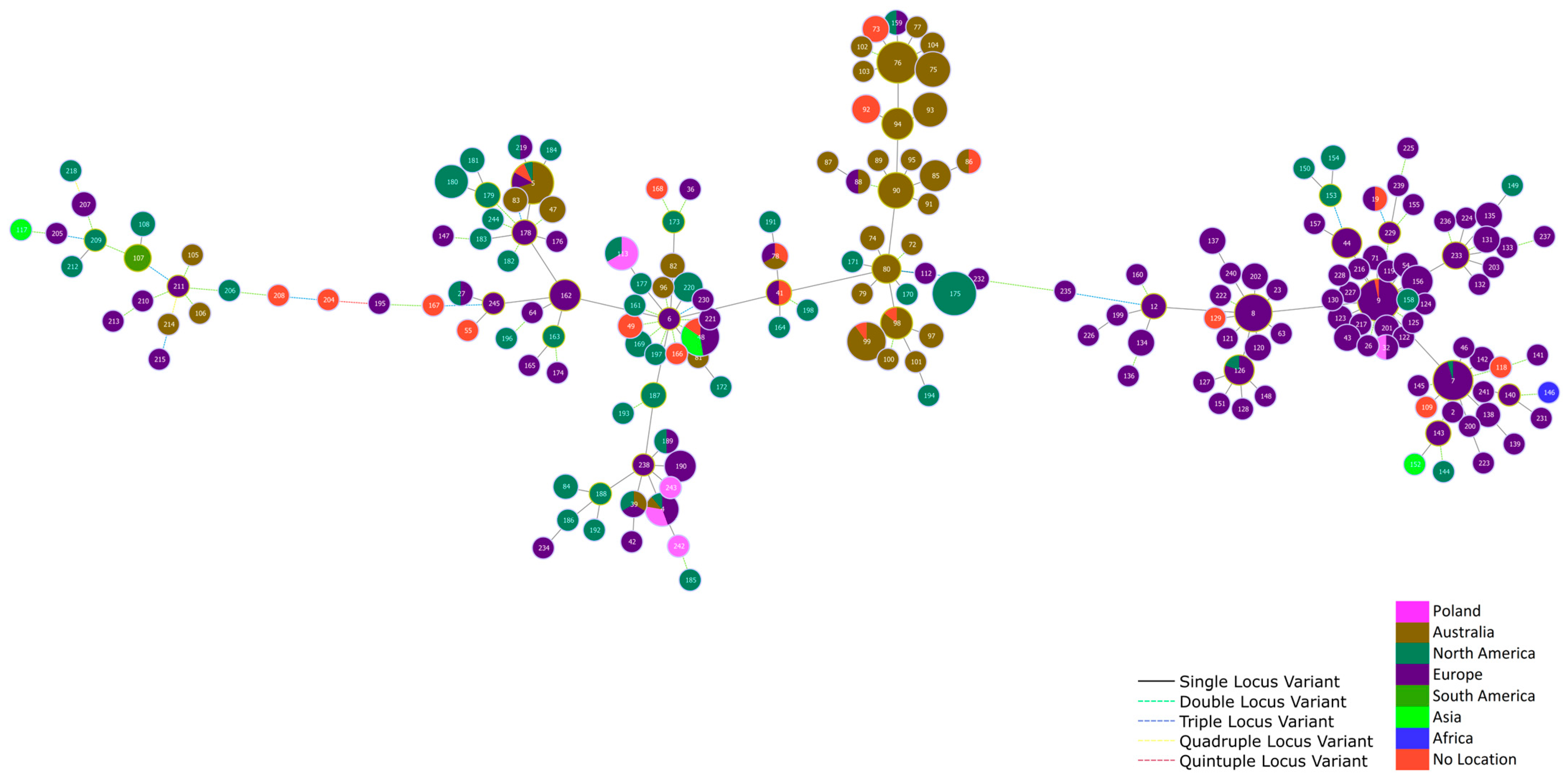
Figure 4 shows the location of the studied E. rhusiopathiae strains representing ST 4, 113, 242, and 243 within a large group comprising isolates from different continents, i.e., North America, Europe, Australia, and Asia. In contrast, goose strain no. 584 representing ST32 was located in a large single-locus variant (SLV) group comprising mainly isolates from Europe (Figure 4).

3. Discussion

3.1. Serotypes of E. rhusiopathiae Strains

The high-to-moderate frequency of E. rhusiopathiae strains of serotypes 5 (38.3%), 1b (28.3%), 8 (15%), and 2 (10%) observed in waterfowl is largely consistent with our previous results on E. rhusiopathiae strains isolated from pigs in Poland [9]. High prevalence of strains of serotype 5 (53.3%) and 1b (43.3%) and low prevalence of serotype 2 (3.3%) have also been recorded in poultry (laying hens and turkeys) in Austria [20]. The same three serotypes (1b, 2, and 5) were identified by Bobrek and Gaweł [13] in E. rhusiopathiae isolates from domestic geese in Poland, with serotype 1b clearly predominating (55.3%). Different results were obtained in Germany, where among E. rhusiopathiae strains (n = 32) from birds and mammals, serotype 1a (62.5%) predominated, while strains of serotype 2 (28.1%) and N (9.4%) were isolated less frequently [21]. Clinical cases of swine erysipelas reported in Japan, China, and the UK are usually caused by strains of serotypes 1a, 1b, or 2 [22,23,24]. Serotyping is a basic method to determine the genetic diversity of isolates and can be helpful in epidemiological studies to assess the spread of strains in the environment. At the same time, it should be remembered that strains of the same serotype may differ significantly, e.g., in terms of resistance profile.
Analyses conducted in this paper indicate that strains of serotypes 5, 6, and 8 are characteristic of birds. However, in our previous studies we have shown the occurrence of E. rhusiopathie strains of these three serotypes also in pigs in Poland [9]. These observations, as well as the fact that serotype 1a is sporadically recorded in Poland, suggest that the prevalence of strains of a given serotype is to some extent correlated with the geographical region.

3.2. Antibiotic Susceptibility and Genotypic Resistance Profiles

The widespread susceptibility of E. rhusiopathiae strains to β-lactam antibiotics demonstrated in this study is fully consistent with several previous reports [9,25,26,27]. Low MICs of ampicillin and/or penicillin (≤0.25 µg/mL) were obtained for 100% of isolates obtained from pigs with symptoms of erysipelas in Poland [9], China [25], and Japan [26], as well as in E. rhusiopathiae strains isolated from various sources in Australia [27]. Contrasting results were presented by researchers from Austria, who tested strains of E. rhusiopathiae from poultry (mainly layer hens and turkeys) and observed ampicillin resistance (20% of strains, MIC ≥ 0.5 µg/mL) and penicillin resistance (40%, MIC ≥ 0.25 µg/mL) [20]. It should be noted, however, that those authors did not attempt to explain the mechanisms of this resistance, and to determine antimicrobial susceptibility they used MICRONAUT-S Lifestock/Equines GP microplates (MERLIN Diagnostika GmbH, Bornheim-Hersel, Germany), which in the case of E. rhusiopathiae strains can yield false positive results (based on our own observations, unpublished). This study is one of the first reports showing the susceptibility of E. rhusiopathiae bacteria to amoxicillin and amoxicillin + ca, and the obtained low MIC values ≤ 0.06 µg/mL indicate the lack of resistance of the tested strains to these antimicrobial agents. These results are very encouraging, as amoxicillin is the drug of choice for treating erysipelas in waterfowl [12,28]. The susceptibility of E. rhusiopathiae isolates from geese to amoxicillin and amoxicillin + cc (MIC ≤ 0.125 µg/mL) has also recently been demonstrated by Bobrek and Gaweł [13].
Infections in poultry are also treated with florfenicol, whose MIC for the E. rhusiopathiae strains studied here can be considered low (2–8 µg/mL). Similar susceptibility to chloramphenicols has been observed in E. rhusiopathiae strains isolated from pigs (florfenicol MIC range of 2–4 µg/mL) [9] and in E. rhusiopathiae strains from humans and various animals (other than poultry) collected in Australia (chloramphenicol MIC range of 8–16 µg/mL) [27].
The sporadic occurrence of E. rhusiopathiae isolates resistant to macrolides (2/60; 5%) observed in the present study is largely consistent with the findings of other authors [9,13,25,26]. Wholly different data were obtained in Austria, where as many as 76.7% of strains isolated from cases of erysipelas in poultry (chickens and turkeys) were classified as resistant to erythromycin [20]. High prevalence of macrolide resistance (53%) was also noted in a pool of isolates (obtained in 2012–2013) from pigs in China. The genes ermT and ermA-like were detected in most phenotypically resistant strains [29]. The erythromycin resistance recorded in the present study was determined by the presence of the ermB and erm47 genes, and, as shown in our previous work, the ermB gene (in strain no. 267) was located within the prophage DNA, and the erm47 gene (in strain no. 1023) was located on 138-kp ICE Er1023 [9]. While the gene ermB is common in Gram-positive bacteria, erm47 has thus far only been confirmed in Helcococcus kunzi strain UCN99 from a patient with a diabetic foot ulcer in France [30] and in a strain of Streptococcus suis in Spain [31].
The prevalence of strains resistant to tiamulin (15.0%) is similar to that noted for E. rhusiopathiae isolates from pigs in China in 2015 (15.2%) [32]. The percentage of strains resistant to lincosamides (16.7%) was more than twice as high as in E. rhusiopathiae isolates from poultry in Austria (6.7%) [20] or from pigs in Poland (7.1%) [9]. Much higher prevalence of resistance to this group of antimicrobials (64–72%) was recorded in isolates from pigs in China [25,29]. The co-occurrence of the gene lnuB responsible for resistance to lincosamides with the gene lsaE determining resistance to pleuromutilins in the present study has also been confirmed by other authors [9,14,19]. The determinants lnuB and lsaE can be part of a resistance gene cluster, which may also include ant(6)-Ia (aadE), spw, ant(3′)-IIIa (aphA3), and sat4 genes [14,16]. Interestingly, as many as 7 of 10 strains found in our study to be resistant to lincomycin, clindamycin, and tiamulin belonged to serotype 8. In our earlier work, the lincomycin-clindamycin-tiamulin resistance profile was confirmed in one of 14 (7.1%) E. rhusiopathiae strains from pigs, and this was also a strain representing serotype 8 [9].
The very high percentage of E. rhusiopathiae strains resistant to tetracycline (85%) in the present study is consistent with the results of our previous research on E. rhusiopathiae strains from pigs (71.4% of isolates were resistant to tetracycline) [9]. Somewhat lower prevalence of resistance to this group of antibiotics has been described for E. rhusiopathiae strains isolated from domestic geese (in 2008–2018) in Poland (63.8%) [13] and from pigs in China and Japan (38–60%) [25,26,29,33]. The presence of the tetM gene in phenotypically resistant E. rhusiopathiae strains has also been confirmed by other authors [9,13,29].
The high prevalence of resistance to enrofloxacin in E. rhusiopathiae strains from waterfowl (80%) is consistent with results reported by Ding et al. [25] and Wu et al. [29], who confirmed resistance to fluoroquinolones (norfloxacin, levofloxacin, enrofloxacin, and ciprofloxacin) in ~70–91.7% of E. rhusiopathiae strains from pigs in China. A somewhat lower rate of resistance to enrofloxacin was noted among isolates from domestic geese in Poland (76.6%) [13] and from poultry in Austria (60%) [20]. The mechanism of resistance to fluoroquinolones confirmed in the present study (a mutation in the gene gyrA at position 257 and in parC at position 242) is in agreement with the findings of other authors [9,13,29]. It should be noted, however, that resistance to fluoroquinolones in these bacteria may also be due to a mutation at position 90 of gyrase subunit A [29]. The correlation observed in our study between the serotype and the type of mutation (in serotype 5 strains it was always Thr86 → Lys, while in isolates with serotypes 1b, 6, and 8 it was Thr86 → Ile86) is in agreement with the results of our previous research on E. rhusiopathiae isolates from pigs [9]. However, such a relationship was not shown in the study by Bobrek i Gaweł [13].
The correlation between the susceptibility of E. rhusiopathiae strains to tetracycline and enrofloxacin observed in the present study was also noted in isolates from pigs [9]. Due to the different molecular basis of resistance of E. rhusiopathiae to these tetracyclines, this correlation is difficult to explain.
The wide range of MIC values of streptomycin and spectinomycin (16–>512 µg/mL) noted in this study is largely consistent with data presented by Coutinho et al. [34], who showed that the MICs of spectinomycin in E. rhusiopathiae strains from pigs ranged from 8 to >64, with an MIC90 of 32 µg/mL. The authors suggested that E. rhusiopathiae should be considered spectinomycin-resistant at MIC ≥128 µg/mL. The results of our study, however, indicate that acquired resistance to streptomycin and spectinomycin determined by the presence of the genes ant(6)-Ia and spw, respectively, is correlated with higher MICs of these antibiotics, from 256 to >512 µg/mL (in the only strain with a spectinomycin MIC of 128 µg/mL, spw was not detected). Therefore, an MIC of 256 µg/mL should be considered as a possible cut-off point for spectinomycin. The same MIC value is a potential cut-off point for streptomycin (MICs of ant(6)-Ia-negative isolates ranged from 16 to 128 µg/mL). However, the validity of these suggestions requires further research on a larger number of E. rhusiopathiae strains. The presence of the determinant ant(6)-Ia encoding aminoglycoside nucleotidyltransferase has previously been confirmed in multidrug-resistant E. rhusiopathiae strains from pigs (at streptomycin MIC = 512 µg/mL) [32,34].
The high MICs noted in this study for gentamicin, neomycin, and trimethoprim/sulfamethoxazole (256–≥512 µg/mL) obtained for all E. rhusiopathiae isolates indicate the occurrence of natural resistance in these bacteria. The lack of sensitivity of E. rhusiopathiae to neomycin and kanamycin was first described by Wood [35], who developed a medium with these antibiotics for isolating E. rhusiopathiae strains (at that time Erysipelothrix insidiosa). Natural resistance of E. rhusiopathiae bacteria to aminoglycosides (neomycin, kanamycin, amikacin, and gentamicin) and folic acid inhibitors (sulfadiazine and trimethoprim) has been confirmed by several other authors [9,25,34,36].

3.3. Potential Mechanisms of Resistance Gene Transfer

Mobile genetic elements (MGEs), i.e., plasmids, integrative and conjugative elements (ICEs), and bacteriophages, are crucial for horizontal gene transfer (HGT) and the spread of antibiotic resistance in bacteria. Studies conducted so far show that in E. rhusiopathiae bacteria, transfer of resistance genes may be determined by ICEs or bacteriophages [14,16,17].
The occurrence of prophage DNA (Javan 630-like) associated with resistance genes (lsaE and most likely lnuB) in MDR isolates of serotype 8 demonstrated in this work is consistent with the studies by Gu et al. [16], who confirmed the presence of temperate phage phi1605 carrying the mef(A), mrs(D), lnu(D)-like, and tetM genes in the E. rhusiopathiae ZJ strain. The ability of this phage to infect recipient E. rhusiopathiae strain in the presence of mitomycin C and to transfer resistance genes was also demonstrated [16]. The results of our analyses showing that the lsaE and lnuB as well as ant(6)-Ia and spw determinants in most E. rhusiopathiae strains are located within the prophage DNA (Table S1) indicate that bacteriophages play a key role in the horizontal transfer of these resistance genes. The fact that prophage DNA was found only in serotype 8 isolates may be related to the presence of serotype-dependent phage receptors. In previous work, prophage regions linked to the ermB gene were detected in one of the E. rhusiopathiae isolates tested (no. 260), but they did not show homology to any of the three phage genes detected in serotype 8 isolates or to the Javan630 and phi1605 phage sequences.
The coexistence of the tetM gene and int-Tn gene (Tn916 transposon integrase gene) noted in this study is consistent with earlier analyses, which showed that the tetM gene in E. rhusiopathiae is always located within 18 kp of the Tn916-like conjugative transposon [14]. E. rusiopathiae strains may contain larger ICEs containing resistance genes [14,16]. In the previous studies, in one of the isolates from waterfowl, i.e., 1023, we demonstrated the presence of ICEEr1023 (130 kb) carrying the ant(6)-Ia–spw–lsa(E)–lnu(B) cluster and the erm47 and tetM genes. In another strain from geese, i.e., 1012, we confirmed the presence of ICEEr1012 (74 kb) containing the tetM gene. Within both ICEs, a Tn916-like transposon and the mobL gene were present. The latter encodes the MobL relaxase, which initiates bacterial conjugation through a site- and strand-specific nick in the oriT region of the conjugation element. Finally, a single-stranded DNA that is transferred from the donor to the recipient cell via the multicomponent protein pore called the type IV secretion system (T4SS) [37].
The presence of the mobL gene confirmed in these studies also in several other isolates (i.e., 877, 86, 13, 815, 136, 147) makes it highly probable that their genomes contain ICE homologous to ICEEr1012. Interestingly, these were only isolates of serotypes 5 and 2, including 3 MDR strains. The presence of ICEs or genomic islands carrying resistance genes was also demonstrated in E. rhusiopathiae strains from pigs in China [16,17]. In resistant isolates no. 136 and 147 of serotype 5, the presence of ICE is additionally indicated by the site-specific recombinase homologous to the integrase of phages Javan630 and phi1605 phage recombinase gene. ICE-encoded recombinases, which enable both integration and excision, are often homologous to phage integrases, and, like temperate phages, many ICEs insert at a specific attachment site in the bacterial chromosome (attB) [37].

3.4. Sequence Types of E. rhusiopathiae Isolates

The results of MLST analysis performed on selected E. rhusiopathiae isolates from waterfowl were compared to STs of 557 strains whose sequences are publicly available. The ST 113 assigned to six MDR serotype 8 isolates from Poland was also determined in 3 strains from pigs from Canada, which belonged to the serotype 1b. ST 4 identified in isolates no. 136, 1023, and 451 (serotype 5 and 1b) was also recorded in several E. rhusiopathiae strains from domestic and wild pigs from different countries, including Italy (3 strains of serotype 1b), Denmark (1 strain of serotype 2), Australia (1 strain of serotype 21), and Canada (1 strain of serotype 1b) (Table S2). The ST in E. rhusiopathiae strains therefore does not appear to be correlated with either the host species or the serotype.

4. Materials and Methods

4.1. Isolation of E. rhusiopathiae Strains

The research material comprised 60 E. rhusiopathiae isolates from domestic geese (n = 54) and ducks (n = 5) from 59 farms located in Poland, mainly in the Wielkopolskie Voivodeship. Dead birds were delivered to the laboratory for diagnostic purposes during the period from 2019 to 2021. E. rhusiopathiae bacteria were isolated from the internal organs (heart, spleen, and air sacs) of dead domestic geese and ducks. Before taking the smear, the organs were incised with a sterile scalpel. Swabs were inoculated onto Columbia agar with 5% sheep blood (GRASO Biotech, Owidz, Poland), and the plates were incubated for up to 48 h [10]. Small (up to 1 mm), flat colonies, some of which were hemolytic, were propagated on BHI broth with the addition of 0.1% Tween 80 (Merck, Warsaw, Poland) to obtain pure cultures. Broth suspensions of bacteria supplemented with 20% glycerol were stored at −80 °C.

4.2. Identification of E. rhusiopathiae Strains

Isolates were identified using the EXOone Erysipelothrix rhusiopathiae test (Exopol, Spain) based on the real-time PCR technique. Amplification was carried out in the Applied Biosystems® 7500 FAST Real-Time PCR System with High Resolution Melt software v3.0.1 (Thermo Fisher Scientific, Waltham, MA, USA) according to the manufacturer’s protocol, i.e., 95 °C—5 min; 40 cycles of 95 °C—15 s, 60 °C—1 min. The template included with the kit and DNA of E. rhusiopathiae strain ATCC 19414 were used as positive controls. A PCR product was detected in the FAM channel and the internal control (IC) in the HEX channel, in accordance with the kit manufacturer’s guidelines [10].

4.3. Serotyping of E. rhusiopathiae Strains

Serotyping was based on four multiplex PCR protocols [2,38]. The primer sequences, annealing temperature, and size of PCR products are listed in Table S3. PCR reactions were performed using DreamTaq Green DNA Polymerase (Thermo Fisher Scientific Baltics UAB, Vilnius, Lithuania). The following E. rhusiopathiae strains were used as positive controls: Fujisawa serotype 1a (GB: AP012027.1), ATCC 19414 (NCTC 8163) serotype 2 (GB: NZ_LR134439.1), Tuzok serotype 6 (GB: SRR11123328), Bano serotype 21, and wild-type strain 267, in the genome of which we confirmed in silico the presence of sequences determining serotype 5 (ENA: ERR12736634). Strain ATCC 19414 was obtained from Argenta (Poznań, Poland), and other reference strains were kindly provided in the form of genomic DNA by Dr. Yoshihiro Shimoji, National Institute of Animal Health, Japan.
The comparative analysis included 260 E. rhusiopathiae strains whose WGSs are publicly available (Table S1). The serotype of these strains was determined in silico using the serotype-determining sequences deposited in the GenBank database [2] and the BLAST tool (https://blast.ncbi.nlm.nih.gov/Blast.cgi, accessed on 17 September 2024).

4.4. Antimicrobial Susceptibility Testing (AST)

The antimicrobial susceptibility of E. rhusiopathiae isolates was determined by the broth microdilution method, as previously described [9]. The following antimicrobial agents were used in the AST: penicillin, ampicillin, amoxicillin, amoxicillin + ca (5:1), ceftiofur, tetracycline, erythromycin, tylosin, clindamycin, lincomycin, tiamulin, enrofloxacin, streptomycin, spectinomycin, gentamicin, neomycin, trimethoprim + sulfamethoxazole (1:19), and florfenicol. All antimicrobial agent powders were obtained from Merck (Warsaw, Poland). Ready-to-use solutions of tiamulin (Biomutin, 200 mg/mL) and spectinomycin (100 mg/mL) were purchased from BIOWET DRWALEW S.A. (Drwalew, Poland) and Merck (Warsaw, Poland), respectively. The drug Taromentin (powder for solution for infusion) (Polfa Tarchomin, Warsaw, Poland) was used as a source of amoxicillin + ca.
Antibiotic/chemotherapeutic stock solutions were prepared by dissolving the powder in appropriate solvents (according to the recommendations of CLSI or the manufacturer). The AST was performed in BHI broth (BTL, Łódź, Poland) containing 0.1% Tween 80, as previously described [9]. Plates were incubated at 37 °C in 5% CO2 for 24–26 h, and MIC values were read as the lowest concentration of an antimicrobial agent at which visible growth was inhibited. Quality control of antimicrobial agents was carried out using E. coli strain ATCC 25922 and Müller-Hinton broth [39].
Categorization of E. rhusiopathiae strains as susceptible, intermediate resistant, and resistant was carried out based on CLSI guidelines (document Vet06, 2017) [40]. In the case of tetracycline, tylosin, lincomycin, and tiamulin, the recommendations of Dec et al. [9] were used. For amoxicillin and amoxicillin + ca, the CLSI cut-off points recommended for ampicillin were adopted. The categorization did not include florfenicol, trimethoprim/sulfamethoxazole, or aminoglycoside antibiotics due to the lack of available guidelines and unimodal MIC distribution (Table 5).

4.5. Detection of Resistance Genes and Development of 10-Plex PCR

Conventional PCR was used to detect the presence of genes that confer resistance to aminoglycosides (aac(6′)-Ie-aph(2”)-Ia, aph(3′)-IIIa, ant(4′)-Ia, aph(2”)-Ib, aph(2”)-Ic, aph(2”)-Id, ant(6)-Ia, and ant(9)-Ia), spectinomycin (spw), tetracyclines (tetK, tetL, tetM, and tetO), pleurumutilins (lsaE), macrolides and/or lincosamides (ermA, ermB, ermT, and mefA/E, lnuB), and streptothricin (sat4). Primer sequences, PCR product size, and annealing temperature are shown in Table S4.
The primers for amplification of lnuB, ant(6)-Ia, ermB, erm47, spw, sat4, aph(3′)-IIIa, and int-Tn were designed using the Primer-BLAST tool (https://www.ncbi.nlm.nih.gov/tools/primer-blast/, accessed on 30 June 2024).
In order to develop a multiplex PCR protocol for detection of the lnuB, ant(6)-Ia, ermB, erm47, tetM, lsaE, spw, aph(3′)-IIIa, sat4, and int-Tn genes, the interactions between the primers, i.e., their ability to form primer dimers and self-dimers, were assessed using the Multiple Primer Analyzer (https://www.thermofisher.com, accessed on 2 July 2024). First, the primers were used in single PCRs to assess their amplification efficiency in the annealing temperature range of 55–60 °C and the possible formation of non-specific PCR products. Analyses were performed on control strains (Table 6).
Amplification was performed using DreamTaq Green DNA polymerase (Thermo Fisher Scientific Baltics UAB, Vilniaus, Lithuania), and the final composition of the reaction mixture for 10-plex PCR was as follows: 10× DreamTaq Green Buffer—1.25 µL, dNTP mix (10 mM)—0.75 µL, each primer (10 pmol/µL)—0.23 µL, template DNA (15–30 ng/µL)—0.7 µL, DreamTaq DNA polymerase (5 U/µL)—0.07 µL, and nuclease-free water—up to 12.5 µL. DNA amplification was performed under the following conditions: initial denaturation for 5 min at 94 °C, 30 cycles of 40 s at 94 °C (denaturation), 40 s at 57.3 °C (annealing), 1 min at 72 °C (extension), and 8 min at 72 °C (final extension).

4.6. Sequence Analysis of the gyrA and parC Genes

To determine the mechanism of resistance of E. rhusiopathiae strains to quinolones, the gyrA and parC genes of representative enrofloxacin-susceptible and enrofloxacin-resistant strains were amplified using the primers listed in Table S4 and sequenced using the Sanger method. Amino acid (aa) sequences were predicted using the NCBI translate tool ORF finder (https://www.ncbi.nlm.nih.gov/orffinder/, accessed on 12 March 2024). The gyrA sequence of reference strain E. rhusiopathiae ATCC 19414 (enrofloxacin-susceptible) was retrieved from the NCBI GenBank database (GB: LR134439.1).

4.7. Detection of ICE-Specific Genes and Prophage DNA

Genes specific for ICEs occurring in E. rhusiopathiae (ICEEr010, ICEEr1012, ICEEr1023, Tn916-like) [14,16] were detected by PCR using primers listed in Table S5. These were the int-Tn gene encoding the Tn916/Tn1545 transposon integrase, the virB4 gene coding for type IV secretory pathway protein VirB4, and the mobL gene coding for MobL relaxase involved in the conjugative transfer of DNA.
Prophage regions were detected by PCR using primers specific for selected phage genes present in the genome of phage Javan630 (GB: MK448997.1) [18] and phage phi1605 (GB: MF172979.1) [16]. These were gene-encoding site-specific recombinase, major capsid protein, and minor tail protein (Table S5). Primers for detection of the sequence comprising the phage recombinase gene and the lsaE resistance gene were designed on the template of the E. rhusiopathiae strain EMAI_141 (GB: JAQTAO010000004.1) [19]. Comparative analysis of phage sequences was performed using BLAST (https://www.ncbi.nlm.nih.gov/tools/primer-blast/, accessed on 13 September 2024), Clinker (https://cagecat.bioinformatics.nl, accessed on 21 September 2024) [43], and MEGA X software (https://www.megasoftware.net/, accessed on 26 November 2022) [44].
The comparative analyses (detection of ICE-specific genes and prophage DNA, analysis of the occurrence of resistance genes within phage DNA) included 260 E. rhusiopathiae strains whose WGSs are publicly available (Table S1).

4.8. Multilocus Sequence Typing

The MLST of E. rhusiopathiae strains was performed for 10 isolates, including 7 MDR ones. Amplification of the pta, galK, purA, ldhA, recA, gpsA, and prsA genes [21] was performed using primers listed in Table S6. The allele sequences were screened against the MLST scheme updated by Webster et al. [19] using mlst v.2.19.0 (https://github.com/tseemann/mlst, accessed on 16 September 2024). A minimum spanning tree based on MLST allele numbers was computed in PHYLOViZ Online (https://online.phyloviz.net/index, accessed on 16 September 2024) with the goeBURST full MST function. The analysis included a total of 557 E. rhusiopathiae strains (Table S2).

5. Conclusions

This study provides new data on the serotypes and antimicrobial susceptibility of E. rhusiopathiae strains causing erysipelas in geese and ducks in Poland. For the first time, the occurrence of E. rhusiopathiae isolates representing serotype 8 was reported in poultry. Of particular importance are the findings regarding the susceptibility of E. rhusiopathiae isolates to antimicrobials approved for the treatment of poultry diseases. These data may contribute to expanding the current CLSI recommendations with guidelines that would allow veterinary diagnosticians worldwide to uniformly categorize E. rhusiopathiae strains as susceptible or resistant to antimicrobial agents used in poultry farming.
The results of our research indicate that β-lactam antibiotics should be the first choice for the treatment of erysipelas in waterfowl, while aminoglycosides, folate inhibitors, tetracyclines, and fluoroquinolones should not be considered. The low MIC values of florfenicol also suggest that it may be effective in the treatment of E. rhusiopathiae infections, but due to its broad spectrum of activity, it should be used only when treatment with narrow-spectrum agents is not possible. As E. rhusiopathiae is a zoonotic microorganism, the data obtained on antimicrobial susceptibility may also be helpful in the treatment of erysipelas in humans.
The presence of prophage DNA and ICE-specific genes, including the Tn916/Tn1545 transposon integrase gene, in the genomes of the isolates tested indicates that bacteriophages and ICEs play a key role in the spread of resistance genes in E. rhusiopathiae. The MDR strains of serotype 8 representing ST 113 seem to be specific to the area of Poland.
Further studies based on WGS analysis will allow us to determine the sequence of prophage regions occurring in these strains and to determine the genetic environment of all resistance genes detected in MDR strain nos. 136 and 147 serotype 5.
The multiplex PCR developed for the detection of 9 resistance genes and the int-Tn gene encoding the integrase of transposons from the Tn916/Tn1545 family makes it possible to significantly reduce both the time and cost of analysis compared to single PCR. Moreover, it can be used to detect resistance genes not only in E. rhusiopathiae but also in other Gram-positive bacteria.

Supplementary Materials

The following supporting information can be downloaded at https://www.mdpi.com/article/10.3390/ijms252212192/s1 [45,46,47,48,49,50,51,52,53,54,55,56,57,58,59].

Author Contributions

Conceptualization, M.D. and T.N.; methodology, M.D.; validation, M.D.; formal analysis, M.D., T.N. and J.W.; investigation, M.D., T.N. and K.W.; resources, M.D. and K.W.; data curation, M.D. and K.W.; writing—original draft preparation, M.D.; visualization, M.D. and J.W.; supervision, M.D.; funding acquisition, M.D. and T.N. All authors have read and agreed to the published version of the manuscript.

Funding

This research received no external funding.

Institutional Review Board Statement

Not applicable. Samples for the isolation of E. rhusiopathiae bacteria were collected from dead birds delivered to the laboratory for diagnostic purposes. No procedures were performed on animals; therefore, the research did not require the consent of the local ethics committee (according to Polish law: Act of 15 January 2015 on the protection of animals used for scientific or educational purposes, Journal of Laws 2015, item 266).

Informed Consent Statement

Not applicable.

Data Availability Statement

The nucleotide sequences reported in this paper have been deposited in the NCBI GenBank database under the following accession numbers: OP921301-OP921305, PQ015304-PQ015312 (gyrA), PQ015313-PQ015323 (parC), PQ362224 (gene coding for major capsid protein), and PQ362225 (gene encoding phage minor tail protein).

Acknowledgments

The authors would like to thank Yoshihiro Shimoji (National Institute of Animal Health, Japan) for providing the DNA of E. rhusiopathiae reference strains used as positive controls in serotyping.

Conflicts of Interest

The authors declare no conflicts of interest.

Appendix A

Figure A1. Serotypic differentiation of E. rhusiopathiae strains by multiplex PCR. PCR products were separated in 1.8% agarose. The labels below each lane correspond to serotypes. The strains Fujisawa serotype 1a, ATCC 19414 serotype 2, Tuzok serotype 6, and 267 serotype 5 were used as positive controls; M—molecular size marker.
Ijms 25 12192 g0a1
Figure A2. Clinker visualization of homology of bacterial DNA regions containing the gene encoding the recombinase family protein characteristic of Streptococcus phage Javan630 and Erysipelothrix phage phi1605. Arrows represent genes; the arrow’s colors represent the gene clusters identified by Clinker; homology between genes is represented by a gray gradient (%, the scale at the top of the figure).
Ijms 25 12192 g0a2
Table A1. Detailed results of the antimicrobial susceptibility test and detection of resistance genes and ICEs-specific genes.
IsolateSer.AMPPENAMXAMCCEFERYTYLLINCLITIATETENRSTRSPEGENNEOTR/SFLOResistance GenesICE-Specific GenesProphage Encoded Genes
1484W1a≤0.06≤0.06≤0.06≤0.06≤0.060.1250.1251≤0.064≤0.25≤0.12512832>512>512>5124
22192≤0.06≤0.06≤0.06≤0.06≤0.060.1250.1250.25≤0.06232≤0.1256416>512>5125124tetMint-Tn
38772≤0.06≤0.06≤0.06≤0.06≤0.060.1250.1250.5≤0.06232≤0.12512832>512>512>5124tetMint-Tn, mobL
4827KZ2≤0.06≤0.06≤0.06≤0.06≤0.060.1250.1250.5≤0.06232812832>512>512>5124tetMint-Tn
5827NW2≤0.06≤0.06≤0.06≤0.06≤0.060.1250.1250.25≤0.0621681632>512>512>5124tetMint-Tn
6101220.125≤0.06≤0.06≤0.060.250.25210.125116≤0.12564128>512>512>5124tetMint-Tn, mobL
710232≤0.06≤0.06≤0.06≤0.06≤0.06321>642>643216>512256>512>512>5124tetM, lnuB, lsaE, ant(6)-Ia, spw, erm47int-Tn, virB, mobL
82675≤0.06≤0.06≤0.06≤0.06≤0.06>32>32>64>16232812832512>512>5124tetM, ermBint-Tn
9865≤0.06≤0.06≤0.06≤0.06≤0.060.1250.125>644>64648>512>512>512>512>5124tetM, lnuB, lsaE, ant(6)-Ia, spwint-Tn, mobLND
101365≤0.06≤0.06≤0.06≤0.06≤0.060.125≤0.0640.1250.5648>512256>512>512>5124tetM, ant(6)-Ia, spwint-Tn, mobLrec *
111475≤0.06≤0.06≤0.06≤0.06≤0.060.1250.1258≤0.060.5328>512512>512>512>5124tetM, ant(6)-Ia, spwint-Tn, mobLrec
124135≤0.06≤0.06≤0.06≤0.06≤0.060.250.1250.25≤0.0613286464>512>512>5124tetMint-Tn
135125≤0.06≤0.06≤0.06≤0.06≤0.060.1250.1250.5≤0.0623286432>512>512>5124tetMint-Tn
146525≤0.06≤0.06≤0.06≤0.06≤0.060.1250.1250.25≤0.0613283232512>512>5124tetMint-Tn
1581550.125≤0.06≤0.06≤0.06≤0.060.1250.1250.50.25132812864512>512>5124tetMint-Tn, mobL
1611545≤0.06≤0.06≤0.06≤0.06≤0.060.250.12510.12543286464512>512>5122tetMint-Tn
174350.125≤0.06≤0.06≤0.06≤0.060.1250.250.5≤0.0613286416>512>512>5124tetMint-Tn
184450.125≤0.06≤0.06≤0.06≤0.060.1250.250.5≤0.0623286464>512>512>5124tetMint-Tn
1911735≤0.06≤0.06≤0.06≤0.06≤0.060.1250.1250.25≤0.060.53283216256>512>5124tetMint-Tn
201350.125≤0.06≤0.06≤0.06≤0.060.250.1250.5≤0.06232812864>512>512>5124tetMint-Tn, mobL
2148W5≤0.06≤0.06≤0.06≤0.06≤0.060.1250.1250.25≤0.0623283216512>512>5128tetMint-Tn
2251W5≤0.06≤0.06≤0.06≤0.06≤0.060.1250.1250.250.12521683216512>512>5124tetMint-Tn
2389W5≤0.06≤0.06≤0.06≤0.06≤0.060.1250.250.5≤0.06432812832>512>512>5124tetMint-Tn
2490W5≤0.06≤0.06≤0.06≤0.06≤0.060.250.250.5≤0.0643286432>512>512>5124tetMint-Tn
25936W5≤0.06≤0.06≤0.06≤0.06≤0.060.1250.50.25≤0.0611683216512>512>5124tetMint-Tn
26434W50.125≤0.06≤0.06≤0.06≤0.060.1250.1250.25≤0.060.53283232>512>512>5124tetMint-Tn
279595≤0.06≤0.06≤0.06≤0.06≤0.060.250.1250.5≤0.0681≤0.12512832512>512>5124
285845≤0.06≤0.06≤0.06≤0.06≤0.060.250.1250.5≤0.0621≤0.1256432512512>5122
298035≤0.06≤0.06≤0.06≤0.06≤0.060.250.251≤0.0611≤0.12512864>512512>5122
301555≤0.06≤0.06≤0.06≤0.06≤0.060.250.1250.5≤0.06416163216512>512>5124tetMint-Tn
31958≤0.06≤0.06≤0.06≤0.06≤0.060.1250.125>641>6432166432512>512>5128tetM, lnuB, lsaEint-Tnrec, mcp **, mtp ***
324898≤0.06≤0.06≤0.06≤0.06≤0.060.1250.125>641643281632512>512>5124tetM, lnuB, lsaEint-Tnrec, mcp, mtp
3317680.125≤0.06≤0.06≤0.06≤0.060.250.125>644>646486432>512>512>5124tetM, lnuB, lsaEint-Tnrec, mcp, mtp
346708≤0.06≤0.06≤0.06≤0.06≤0.060.1250.125>642>6432812864>512>512>5128tetM, lnuB, lsaEint-Tnrec, mcp, mtp
35759W8≤0.06≤0.06≤0.06≤0.06≤0.060.1250.125>6426432812832>512>512>5128tetM, lnuB, lsaEint-Tnrec, mcp, mtp
3681880.125≤0.06≤0.06≤0.06≤0.060.1250.125>642>643286432>512>512>5124tetM, lnuB, lsaEint-Tnrec, mcp, mtp
3781980.125≤0.06≤0.06≤0.06≤0.060.1250.125>642>643286432>512>512>5124tetM, lnuB, lsaEint-Tnrec, mcp, mtp
3810928≤0.06≤0.06≤0.06≤0.06≤0.060.1250.1250.5≤0.06432166464>512>512>5124tetMint-Tn
395380.125≤0.06≤0.06≤0.06≤0.060.1250.1250.5≤0.0683286432>512>512>5128tetMint-Tn
405791b0.125≤0.06≤0.06≤0.06≤0.060.250.1250.5≤0.06232812832>512>512>5124tetMint-Tn
411671b0.125≤0.06≤0.06≤0.06≤0.060.1250.1250.25≤0.06232163232512>512>5124tetMint-Tn
423951b0.125≤0.06≤0.06≤0.06≤0.060.250.1250.5≤0.06432812864>512>512>5128tetMint-Tn
437841b0.125≤0.06≤0.06≤0.06≤0.060.1250.1250.25≤0.0623286432512>512>5124tetMint-Tn
4410421b0.125≤0.06≤0.06≤0.06≤0.060.250.1250.5≤0.06232812864>512>512>5124tetMint-Tn
4512281b0.125≤0.06≤0.06≤0.06≤0.060.50.1251≤0.06216812864512>512>5124tetMint-Tn
46439W1b≤0.06≤0.06≤0.06≤0.06≤0.060.1250.1250.5≤0.0623286432>512>512>5124tetMint-Tn
47450W1b≤0.06≤0.06≤0.06≤0.06≤0.060.1250.1250.25≤0.0613286416512>512>5124tetMint-Tn
481951b0.125≤0.06≤0.06≤0.06≤0.060.250.2520.12541≤0.12512864>512>512>5124
496571b≤0.06≤0.06≤0.06≤0.06≤0.060.1250.1250.5≤0.062≤0.25≤0.1256432>512>512>5124
5010831b≤0.06≤0.06≤0.06≤0.06≤0.060.1250.1250.5≤0.062≤0.25≤0.1256432>512>512>5124
512021b≤0.06≤0.06≤0.06≤0.06≤0.060.250.12510.125432812864>512>512>5128tetMint-Tn
525251b0.25≤0.06≤0.06≤0.06≤0.060.250.1250.5≤0.06432812864>512>512>5124tetMint-Tn
535261b0.125≤0.06≤0.06≤0.06≤0.060.250.1251≤0.064161612864512>512>5124tetMint-Tn
547831b0.125≤0.06≤0.06≤0.06≤0.060.250.12510.125864166432>512>512>5124tetMint-Tn
556331b≤0.06≤0.06≤0.06≤0.06≤0.06≤0.06≤0.060.25≤0.0623286432>512>512>5124tetMint-Tn
564511b0.125≤0.06≤0.06≤0.06≤0.060.250.1250.5≤0.062321612864>512>512>5124tetMint-Tn
5749WN≤0.06≤0.06≤0.06≤0.06≤0.060.1250.1250.25≤0.061≤0.25≤0.1256432512>512>5124
58320N0.125≤0.06≤0.06≤0.06≤0.060.250.1250.5≤0.06432812864>512>512>5124tetMint-Tn
5949760.125≤0.06≤0.06≤0.06≤0.060.250.1250.5≤0.062321612864>512>512>5124tetMint-Tn
608496≤0.06≤0.06≤0.06≤0.06≤0.06≤0.060.1250.25≤0.061≤0.25≤0.1253216512512>5124
000002
3.3%
3
5%
10
16.7%
10
16.7%
9
15%
51
85%
48
80%
NDNDNDNDNDND
Legend: MIC values highlighted in grey indicate resistance, and those highlighted in blue indicate medium susceptibility. Ser.—serotype; PEN—penicillin; AMP—ampicillin; AMX—amoxicillin; AMC—amoxicillin + ca; CEF—ceftiofur; TET—tetracycline; ERY—erythromycin; TYL—tylosin; CLI—clindamycin; LIN—lincomycin; TIA—tiamulin; ENR—enrofloxacin; STR—streptomycin; SPE—spectinomycin; GEN—gentamicin; NEO—neomycin; TR/S—trimethoprim/sulfamethoxazole; FLO—florfenicol; * rec—gene coding for recombinase family protein; ** mcp—gene coding for phage major capsid protein; *** mtp—gene coding for phage minor tail protein.

References

  1. Wang, Q.; Chang, B.J.; Riley, T.V. Erysipelothrix rhusiopathiae. Vet. Microbiol. 2010, 140, 405–417. [Google Scholar] [CrossRef] [PubMed]
  2. Shimoji, Y.; Shiraiwa, K.; Tominaga, H.; Nishikawa, S.; Eguchi, M.; Hikono, H.; Ogawa, Y. Development of a Multiplex PCR-Based Assay for Rapid Serotyping of Erysipelothrix Species. J. Clin. Microbiol. 2020, 58, e00315-20. [Google Scholar] [CrossRef] [PubMed]
  3. Eisenberg, T.; Mühldorfer, K.; Erhard, M.; Fawzy, A.; Kehm, S.; Ewers, C.; Semmler, T.; Blom, J.; Lipski, A.; Rau, J.; et al. Erysipelothrix anatis sp. nov., Erysipelothrix aquatica sp. nov. and Erysipelothrix urinaevulpis sp. nov., three novel species of the genus, and emended description of Erysipelothrix. Int. J. Syst. Evol. Microbiol. 2022, 72, 005454. [Google Scholar] [CrossRef] [PubMed]
  4. Zhong, J.; Medvecky, M.; Tornos, J.; Clessin, A.; Le Net, R.; Gantelet, H.; Gamble, A.; Forde, T.L.; Boulinier, T. Erysipelothrix amsterdamensis sp. nov., associated with mortalities among endangered seabirds. Int. J. Syst. Evol. Microbiol. 2024, 74, 006264. [Google Scholar] [CrossRef] [PubMed]
  5. Chang, R.K.; Miller, M.A.; Tekedar, H.C.; Rose, D.; García, J.C.; LaFrentz, B.R.; Older, C.E.; Waldbieser, G.C.; Pomaranski, E.; Shahin, K.; et al. Pathology, microbiology, and genetic diversity associated with Erysipelothrix rhusiopathiae and novel Erysipelothrix spp. infections in southern sea otters (Enhydra lutris nereis). Front. Microbiol. 2024, 14, 1303235. [Google Scholar] [CrossRef]
  6. Chen, M.; Huang, W.; Li, J. The First Report of Erysipelothrix muris sp. nov. Unpublished. 2006. Available online: https://www.ncbi.nlm.nih.gov/Taxonomy/Browser/wwwtax.cgi?id=380638 (accessed on 29 September 2024).
  7. Stackebrandt, E. Erysipelothrix. In Bergey’s Manual of Systematics of Archaea and Bacteria, 1st ed.; Whitman, W.B., Ed.; John Wiley & Sons, Inc.: Hoboken, NJ, USA; Bergey’s Manual Trust: Glasgow, UK, 2015; pp. 1–15. [Google Scholar] [CrossRef]
  8. Takahashi, T.; Zarkasie, K.; Mariana, S.; Sumadi; Ogata, M. Serological and pathogenic characterization of Erysipelothrix rhusiopathiae isolates from tonsils of slaughter pigs in Indonesia. Vet. Microbiol. 1989, 21, 165–175. [Google Scholar] [CrossRef]
  9. Dec, M.; Łagowski, D.; Nowak, T.; Pietras-Ożga, D.; Herman, K. Serotypes, Antibiotic Susceptibility, Genotypic Virulence Profiles and SpaA Variants of Erysipelothrix rhusiopathiae Strains Isolated from Pigs in Poland. Pathogens 2023, 12, 409. [Google Scholar] [CrossRef]
  10. Nowak, T.; Wódz, K.; Kwiecieński, P.; Kwieciński, A.; Dec, M. Incidence of erysipelas in waterfowl in Poland–clinical & pathological investigations. Br. Poult. Sci. 2024. [Google Scholar] [CrossRef]
  11. Bobrek, K.; Nowak, M.; Borkowska, J.; Bobusia, K.; Gaweł, A. An outbreak of erysipelas in commercial geese. Pak. Vet. J. 2016, 36, 372–374. [Google Scholar]
  12. Żbikowski, A.; Karpińska, E.; Rzewuska, M.; Szeleszczuk, P. Różyca u drobiu [Erysipelas in poultry]. Życie Weterynaryjne 2011, 86, 357–360. [Google Scholar]
  13. Bobrek, K.; Gaweł, A. Antimicrobial Resistance of Erysipelothrix rhusiopathiae Strains Isolated from Geese to Antimicrobials Widely Used in Veterinary Medicine. Antibiotics 2023, 12, 1339. [Google Scholar] [CrossRef] [PubMed]
  14. Dec, M.; Zomer, A.; Webster, J.; Nowak, T.; Stępień-Pyśniak, D.; Urban-Chmiel, R. Integrative and Conjugative Elements and Prophage DNA as Carriers of Resistance Genes in Erysipelothrix rhusiopathiae Strains from Domestic Geese in Poland. Int. J. Mol. Sci. 2024, 25, 4638. [Google Scholar] [CrossRef] [PubMed]
  15. Com/2017/0339-Communication from the Commission to the Council and the European Parliament A European One Health Action Plan Against Antimicrobial Resistance (AMR). Available online: https://eur-lex.europa.eu/legal-content/en/ALL/?uri=CELEX%3A52017DC0339 (accessed on 29 September 2024).
  16. Gu, J.; Li, Y.X.; Xu, C.W.; Xie, X.J.; Li, P.; Ma, G.X.; Lei, C.W.; Liu, J.X.; Zhang, A.Y. Genome sequence of multidrug-resistant Erysipelothrix rhusiopathiae ZJ carrying several acquired antimicrobial resistance genes. J. Glob. Antimicrob. Resist. 2020, 21, 13–15. [Google Scholar] [CrossRef] [PubMed]
  17. Zhao, D.; Hu, Y.; Wu, H.; Feng, Z.; Hu, C.; Hu, H.; Liu, Y.; Sun, W.; Yu, X. Phenotypic and Genotypic Characterization of a Highly Virulent Erysipelothrix rhusiopathiae Strain. Transboundary and Emerging Diseases. Transpoundary Emerg. Dis. 2024, 2024, 5401707. [Google Scholar] [CrossRef]
  18. Rezaei Javan, R.; Ramos-Sevillano, E.; Akter, A.; Brown, J.; Brueggemann, A.B. Prophages and satellite prophages are widespread in Streptococcus and may play a role in pneumococcal pathogenesis. Nat. Commun. 2019, 10, 4852. [Google Scholar] [CrossRef]
  19. Webster, J.; Bowring, B.; Stroud, L.; Marsh, I.; Sales, N.; Bogema, D. Population Structure and Genomic Characteristics of Australian Erysipelothrix rhusiopathiae Reveals Unobserved Diversity in the Australian Pig Industry. Microorganisms 2023, 11, 297. [Google Scholar] [CrossRef]
  20. Hess, C.; Bilic, I.; Jandreski-Cvetkovic, D.; Hess, M. Antimicrobial Dilution Susceptibility Testing of Erysipelothrix rhusiopathiae According to CLSI Document VET06 Reveals High Resistance against Penicillin G, Erythromycin and Enrofloxacin. Poultry 2023, 2, 54–62. [Google Scholar] [CrossRef]
  21. Janßen, T.; Voss, M.; Kühl, M.; Semmler, T.; Philipp, H.-C.; Ewers, C. A Combinational Approach of Multilocus Sequence Typing and Other Molecular Typing Methods in Unravelling the Epidemiology of Erysipelothrix Rhusiopathiae Strains from Poultry and Mammals. Vet. Res. 2015, 46, 84. [Google Scholar] [CrossRef]
  22. McNeil, M.; Gerber, P.F.; Thomson, J.; Williamson, S.; Opriessnig, T. Serotypes and Spa Types of Erysipelothrix Rhusiopathiae Isolates from British Pigs (1987 to 2015). Vet. J. 2017, 225, 13–15. [Google Scholar] [CrossRef]
  23. Imada, Y.; Takase, A.; Kikuma, R.; Iwamaru, Y.; Akachi, S.; Hayakawa, Y. Serotyping of 800 Strains of Erysipelothrix Isolated from Pigs Affected with Erysipelas and Discrimination of Attenuated Live Vaccine Strain by Genotyping. J. Clin. Microbiol. 2004, 42, 2121–2126. [Google Scholar] [CrossRef]
  24. Zou, Y.; Zhu, X.; Muhammad, H.M.; Jiang, P.; Li, Y. Characterization of Erysipelothrix rhusiopathiae strains isolated from acute swine erysipelas outbreaks in Eastern China. J. Vet. Med. Sci. 2015, 77, 653–660. [Google Scholar] [CrossRef] [PubMed]
  25. Ding, Y.; Zhu, D.; Zhang, J.; Yang, L.; Wang, X.; Chen, H.; Tan, C. Virulence determinants, antimicrobial susceptibility, and molecular profiles of Erysipelothrix rhusiopathiae strains isolated from China. Emerg. Microbes Infect. 2015, 4, e69. [Google Scholar] [CrossRef] [PubMed]
  26. Chuma, T.; Kawamoto, T.; Shahada, F.; Fujimoto, H.; Okamoto, K. Antimicrobial susceptibility of Erysipelothrix rhusiopathiae isolated from pigs in Southern Japan with a modified agar dilution method. J. Vet. Med. Sci. 2010, 72, 643–645. [Google Scholar] [CrossRef] [PubMed]
  27. Fidalgo, S.G.; Longbottom, C.J.; Rjley, T.V. Susceptibility of Erysipelothrix rhusiopathiae to antimicrobial agents and home disinfectants. Pathology 2002, 34, 462–465. [Google Scholar] [CrossRef]
  28. Abd El-Ghany, W. A review article on avian Erysipelas infection: An occupational disease of one health importance. Vet. Integr. Sci. 2023, 21, 481–494. [Google Scholar] [CrossRef]
  29. Wu, C.; Lv, C.; Zhao, Y.; Zhu, W.; Liu, L.; Wang, T.; Kang, C.; Yang, Y.; Sun, X.; Zhang, Q.; et al. Characterization of Erysipelothrix Rhusiopathiae Isolates from Diseased Pigs in 15 Chinese Provinces from 2012 to 2018. Microorganisms 2021, 9, 2615. [Google Scholar] [CrossRef]
  30. Guérin, F.; Isnard, C.; Bucquet, F.; Fines-Guyon, M.; Giard, J.C.; Burrus, V.; Cattoir, V. Novel chromosome-encoded erm(47) determinant responsible for constitutive MLSB resistance in Helcococcus kunzii. J. Antimicrob. Chemother. 2016, 71, 3046–3049. [Google Scholar] [CrossRef]
  31. Uruén, C.; Gimeno, J.; Sanz, M.; Fraile, L.; Marín, C.M.; Arenas, J. Invasive Streptococcus suis isolated in Spain contain a highly promiscuous and dynamic resistome. Front. Cell. Infect. Microbiol. 2024, 13, 1329632. [Google Scholar] [CrossRef]
  32. Zhang, A.; Xu, C.; Wang, H.; Lei, C.; Liu, B.; Guan, Z.; Yang, C.; Yang, Y.; Peng, L. Presence and new genetic environment of pleuromutilin-lincosamide-streptogramin A resistance gene lsa(E) in Erysipelothrix rhusiopathiae of swine origin. Vet. Microbiol. 2015, 177, 162–167. [Google Scholar] [CrossRef]
  33. Yamamoto, K.; Kijima, M.; Yoshimura, H.; Takahashi, T. Antimicrobial Susceptibilities of Erysipelothrix Rhusiopathiae Isolated from Pigs with Swine Erysipelas in Japan, 1988–1998. J. Vet. Med. B Infect. Dis. Vet. Public Health 2001, 48, 115–126. [Google Scholar] [CrossRef]
  34. Coutinho, T.A.; Imada, Y.; Barcellos, D.E.S.N.; Oliveira, S.J.; Moreno, A.M. Phenotypic and Molecular Characterization of Recent and Archived Erysipelothrix Spp. Isolated from Brazilian Swine. Diagn. Microbiol. Infect. Dis. 2011, 69, 123–129. [Google Scholar] [CrossRef] [PubMed]
  35. Wood, R.L. A selective liquid medium utilizing antibiotics for isolation of Erysipelothrix insidiosa. Am. J. Vet. Res. 1965, 26, 1303–1308. [Google Scholar] [PubMed]
  36. Takahashi, T.; Sawada, T.; Muramatsu, M.; Tamura, Y.; Fujisawa, T.; Benno, Y.; Mitsuoka, T. Serotype, antimicrobial susceptibility, and pathogenicity of Erysipelothrix rhusiopathiae isolates from tonsils of apparently healthy slaughter pigs. J. Clin. Microbiol. 1987, 25, 536–539. [Google Scholar] [CrossRef] [PubMed]
  37. Johnson, C.M.; Grossman, A.D. Integrative and Conjugative Elements (ICEs): What They Do and How They Work. Annu. Rev. Genet. 2015, 49, 577–601. [Google Scholar] [CrossRef]
  38. Shiraiwa, K.; Ogawa, Y.; Nishikawa, S.; Eguchi, M.; Shimoji, Y. Identification of serovar 1a, 1b, 2, and 5 strains of Erysipelothrix rhusiopathiae by a conventional gel-based PCR. Vet. Microbiol. 2018, 225, 101–104. [Google Scholar] [CrossRef]
  39. CLSI Supplement M100. Performance Standards for Antimicrobial Susceptibility Testing, 30th ed.; Clinical and Laboratory Standards Institute: Wayne, PA, USA, 2020. [Google Scholar]
  40. CLSI Supplement Vet06. Methods for Antimicrobial Susceptibility Testing of Infrequently Isolated or Fastidious Bacteria Isolated from Animals, 1st ed.; Clinical and Laboratory Standards Institute: Wayne, PA, USA, 2017. [Google Scholar]
  41. Dec, M.; Stępień-Pyśniak, D.; Gnat, S.; Fratini, F.; Urban-Chmiel, R.; Cerri, D.; Winiarczyk, S.; Turchi, B. Antibiotic Susceptibility and Virulence Genes in Enterococcus Isolates from Wild Mammals Living in Tuscany, Italy. Microb. Drug Resist. 2020, 26, 505–519. [Google Scholar] [CrossRef]
  42. Dec, M.; Stępien-Pyśniak, D.; Puchalski, A.; Hauschild, T.; Pietras-Ozga, D.; Ignaciuk, S.; Urban-Chmiel, R. Biodiversity of Ligilactobacillus salivarius Strains from Poultry and Domestic Pigeons. Animals 2021, 11, 972. [Google Scholar] [CrossRef]
  43. Gilchrist, C.L.M.; Chooi, Y.H. Clinker & clustermap.js: Automatic generation of gene cluster comparison figures. Bioinformatics 2021, 37, 2473–2475. [Google Scholar] [CrossRef]
  44. Kumar, S.; Stecher, G.; Li, M.; Knyaz, C.; Tamura, K. MEGA X: Molecular Evolutionary Genetics Analysis across computing platforms. Mol. Biol. Evol. 2018, 35, 1547–1549. [Google Scholar] [CrossRef]
  45. Tang, H.B.; Xie, J.; Wang, L.; Liu, F.; Wu, J. Complete genome sequence of Erysipelothrix rhusiopathiae strain GXBY-1 isolated from acute swine erysipelas outbreaks in south China. Genom. Data 2016, 8, 70–71. [Google Scholar] [CrossRef]
  46. Ogawa, Y.; Ooka, T.; Shi, F.; Ogura, Y.; Nakayama, K.; Hayashi, T.; Shimoji, Y. The genome of Erysipelothrix rhusiopathiae, the causative agent of swine erysipelas, reveals new insights into the evolution of firmicutes and the organism’s intracellular adaptations. J. Bacteriol. 2011, 193, 2959–2971. Available online: https://journals.asm.org/doi/10.1128/jb.01500-10 (accessed on 1 October 2024). [CrossRef] [PubMed]
  47. Kwok, A.H.; Li, Y.; Jiang, J.; Jiang, P.; Leung, F.C. Complete genome assembly and characterization of an outbreak strain of the causative agent of swine erysipelas—Erysipelothrix rhusiopathiae SY1027. BMC Microbiol. 2014, 14, 176. [Google Scholar] [CrossRef] [PubMed]
  48. Yang, L.; Zhu, Y.; Peng, Z.; Ding, Y.; Jie, K.; Wang, Z.; Peng, Y.; Tang, X.; Wang, X.; Chen, H.; et al. Comparative Genome Analysis of a Pathogenic Erysipelothrix Rhusiopathiae Isolate WH13013 from Pig Reveals Potential Genes Involve in Bacterial Adaptions and Pathogenesis. Vet. Sci. 2020, 7, 74. [Google Scholar] [CrossRef] [PubMed]
  49. Lee, K.; Park, S.Y.; Seo, H.W.; Cho, Y.; Choi, S.G.; Seo, S.; Han, W.; Lee, N.K.; Kwon, H.; Han, J.E.; et al. Pathological and Genomic Findings of Erysipelothrix rhusiopathiae Isolated from a Free-Ranging Rough-Toothed Dolphin Steno bredanensis (Cetacea: Delphinidae) Stranded in Korea. Front. Vet. Sci. 2022, 9, 774836. [Google Scholar] [CrossRef] [PubMed]
  50. Kovalchuk, S.; Babii, A. Draft genome sequence data of Erysipelothrix rhusiopathiae vaccine strain VR-2. Data Brief 2020, 33, 106352. [Google Scholar] [CrossRef]
  51. Nesbitt, K.; Bloodgood, J.; Mullis, M.M.; Deming, A.C.; Colegrove, K.; Kiel Reese, B. Draft Genome Sequences of Erysipelothrix sp. Strains Isolated from Stranded Septic Bottlenose Dolphins in Alabama, USA. Microbiol. Resour. Announc. 2022, 11, e0027322. [Google Scholar] [CrossRef]
  52. Zautner, A.E.; Tersteegen, A.; Schiffner, C.J.; Ðilas, M.; Marquardt, P.; Riediger, M.; Delker, A.M.; Mäde, D.; Kaasch, A.J. Human Erysipelothrix rhusiopathiae infection via bath water—Case report and genome announcement. Front. Cell. Infect. Microbiol. 2022, 12, 981477. [Google Scholar] [CrossRef]
  53. IJsseldijk, L.L.; Begeman, L.; Duim, B.; Gröne, A.; Kik, M.J.L.; Klijnstra, M.D.; Lakemeyer, J.; Leopold, M.; Munnink, B.B.O.; Ten Doeschate, M.; et al. Harbor Porpoise Deaths Associated with Erysipelothrix rhusiopathiae, the Netherlands, 2021. Emerg. Infect. Dis. 2023, 29, 835–838. [Google Scholar] [CrossRef]
  54. Mileto, I.; Merla, C.; Corbella, M.; Gaiarsa, S.; Kuka, A.; Ghilotti, S.; De Cata, P.; Baldanti, F.; Cambieri, P. Bloodstream Infection Caused by Erysipelothrix rhusiopathiae in an Immunocompetent Patient. Microorganisms 2024, 12, 942. [Google Scholar] [CrossRef]
  55. Söderlund, R.; Formenti, N.; Caló, S.; Chiari, M.; Zoric, M.; Alborali, G.L.; Sørensen Dalgaard, T.; Wattrang, E.; Eriksson, H. Comparative genome analysis of Erysipelothrix rhusiopathiae isolated from domestic pigs and wild boars suggests host adaptation and selective pressure from the use of antibiotics. Microb. Genom. 2020, 6, mgen000412. [Google Scholar] [CrossRef]
  56. Forde, T.L.; Kollanandi Ratheesh, N.; Harvey, W.T.; Thomson, J.R.; Williamson, S.; Biek, R.; Opriessnig, T. Genomic and Immunogenic Protein Diversity of Erysipelothrix rhusiopathiae Isolated from Pigs in Great Britain: Implications for Vaccine Protection. Front. Microbiol. 2020, 11, 418. [Google Scholar] [CrossRef] [PubMed]
  57. Malhotra-Kumar, S.; Lammens, C.; Piessens, J.; Goossens, H. Multiplex PCR for simultaneous detection of macrolide and tetracycline resistance determinants in streptococci. Antimicrob. Agents Chemother. 2005, 49, 4798–4800. [Google Scholar] [CrossRef] [PubMed]
  58. Vakulenko, S.B.; Donabedian, S.M.; Voskresenskiy, A.M.; Zervos, M.J.; Lerner, S.A.; Chow, J.W. Multiplex PCR for detection of aminoglycoside resistance genes in enterococci. Antimicrob. Agents Chemother. 2003, 47, 1423–1426. [Google Scholar] [CrossRef] [PubMed]
  59. Kobayashi, N.; Alam, M.; Nishimoto, Y.; Urasawa, S.; Uehara, N.; Watanabe, N. Distribution of aminoglycoside resistance genes in recent clinical isolates of Enterococcus faecalis, Enterococcus faecium and Enterococcus avium. Epidemiol. Infect. 2001, 126, 197–204. [Google Scholar] [CrossRef]
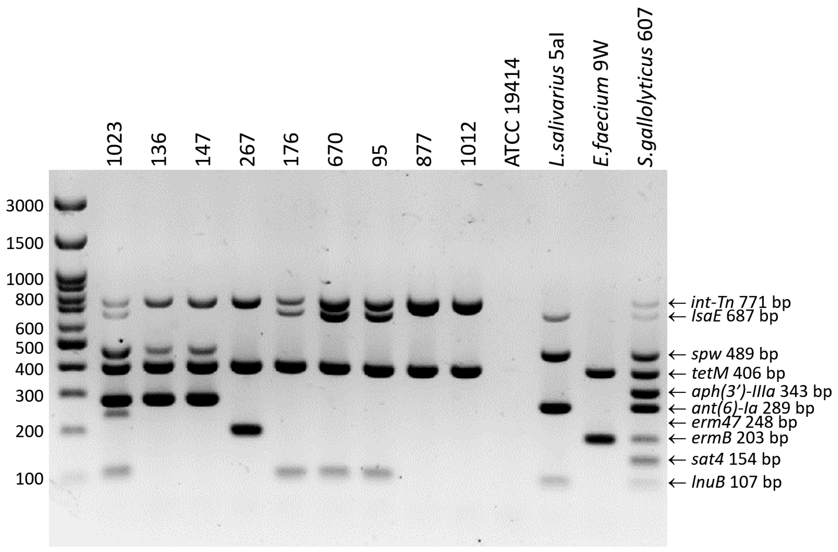
Figure 1. Electrophoretic separation of 10-plex PCR products in a 2% agarose gel. The strains E. rhusiopathiae 1023, 267, and 1012, Ligilactobacillus salivarius 5aI, Enterococcus faecium 9W, and Streptococcus gallolyticus 607 were used as positive controls. The negative control was the strain E. rhusiopathiae ATCC 19414.
Ijms 25 12192 g001
Figure 2. Clinker visualization showing homology between the Erysipelothrix phage phi1605 and Streptococcus phage Javan630 regions and genomes of E. rhusiopathiae (EMIAI_92, EMAI_141, 2604) and E. larvae LV19 strains. Arrows represent genes; the arrow’s colors represent the gene clusters identified by Clinker; homology between genes is represented by a gray gradient (%, the scale at the bottom of the figure).
Ijms 25 12192 g002
Figure 3. Dendrogram showing the similarity of the sequences of genes encoding the major capsid protein (A) and minor tail protein (B) detected in E. rhusiopathiae strain 489 to analogous sequences deposited in GenBank. Analysis was conducted by MEGA X software using the Maximum Likelihood method. The percentage of replicate trees in which the associated taxa were clustered together in the bootstrap test (500 replicates) is shown next to the branches. Scale bars show genetic distance.
Ijms 25 12192 g003
Figure 4. Minimum spanning tree based on the allelic profiles (pta, galK, purA, ldhA, recA, gpsA, and prsA) of 196 STs of E. rhusiopathiae strains (n = 557), including ST 4, 32, 113, 242, and 243 determined in isolates from waterfowl in Poland. Node size is proportional to the frequency of sequence occurrence.
Ijms 25 12192 g004
Table 1. Number and percentage of E. rhusiopathiae strains representing individual serotypes.
SerotypeE. rhusiopathiae
Strains Tested in This Work (n = 60)
E. rhusiopathiae Strains Whose WGS Were Derived from GenBank (n = 260)Notes
1a1 [1.7%]92 [35.4%]
1b17 [28.3%]57 [21.9%]
26 [10.0%]56 [21.5%]
1a/2022 [8.5%]
1a/1b01 [0.4%]
2/1501 [0.4%]
523 [38.3%]7 [2.7%]all strains from birds *
62 [3.3%]2 [0.8%]all strains from birds *
89 [15.0%]0all strains from birds *
903 [1.1%]
1101 [0.4%]
1501 [0.4%]
1601 [0.4%]
1702 [0.8%]
1901 [0.4%]
21011 [4.2%]
2301 [0.4%]
N2 [3.3%]1 [0.4%]
* Strains whose WGSs were analyzed originated from both poultry and wild birds; detailed information on the bird species is provided in Table S1.
Table 2. Distribution of MIC values obtained in antimicrobial susceptibility tests for E. rhusiopathiae isolates (n = 60) and resistance genes detected.
≤0.06≤0.125≤0.250.51248≥1632≥64128256≥512Number (%) of Resistant Isolates
PEN60 0
AMP37221 0
AMX60 0
AMC60 0
CEF59 1 0
TET 126 2tetM22tetM27tetM 51 (85%)
ENR 12 2919 48 (80%)
ERY234211 1erm471ermB 2 (3.3%)
TYL249511erm471 1ermB 3 (5.0%)
CLI4271 2lnuB5lnuB2lnuB 1 10 (16.7%)
LIN 15248111 10lnuB(9)
ermB (1)
10 (16.7%)
TIA 41022123 9lsaE 9 (15%)
FLO 3507 NA
STR 292322 4ant(6)-Ia
spw
NA
SPE 9281812ant(6)-Ia, spw2ant(6)-Ia,
spw
NA
GEN 159NA
NEO 60NA
TR/S 60NA
MIC values highlighted in grey indicate resistance, and those highlighted in blue indicate medium susceptibility. The number of strains in which the gene was detected is given in brackets next to the gene name. Where there is no numerical value after the gene name, the gene was present in all isolates with a given MIC value. Legend: PEN—penicillin; AMP—ampicillin; AMX—amoxicillin; AMC—amoxicillin + ca; CEF—ceftiofur; TET—tetracycline; ENR—enrofloxacin; ERY—erythromycin; TYL—tylosin; CLI—clindamycin; LIN—lincomycin; TIA—tiamulin; FLO—florfenicol; STR—streptomycin; SPE—spectinomycin; GEN—gentamicin; NEO—neomycin; TR/S—trimethoprim/sulfamethoxazole; NA—not applicable.
Table 3. Sequence analysis of the quinolone resistance-determining region (QRDR) in the gyrA and parC genes in enrofloxacin-susceptible and resistant E. rhusiopathiae strains.
IsolateSerotypeEnrofloxacin MIC [µg/mL] Mutation at Position 257 of the gyrA GeneGB or ENA Acc. No.Mutation at Position 242 of the parC GeneGB or ENA Acc. No.
ATCC 194142≤0.25SThr86 (ACA)LR134439.1Ser81 (AGT)LR134439.1
10122≤0.125SThr86 (ACA)OP921306Ser81 (AGT)ERR12736636
1023216RIle86 (ATA)OP921307Ile81 (ATT)ERR12736637
827KZ28RLys86 (AAA)PQ015304Ile81 (ATT)PQ015313
5845≤0.125SThr86 (ACA)ERR1273663Ser81 (AGT)ERR1273663
26758RLys86 (AAA)OQ625325 Ile81 (ATT)ERR12736634
13658RLys86 (AAA)PQ015305Ile81 (ATT)PQ015314
434W58RLys86 (AAA)PQ015306Ile81 (ATT)PQ015315
95816RIle86 (ATA)OP921308Ile81 (ATT)PQ015321
759W88RIle86 (ATA)PQ015307Ile81 (ATT)PQ015318
1092816RIle86 (ATA)PQ015308Ile81 (ATT)PQ015319
8496≤0.125SThr86 (ACA)PQ015309Ser81 (AGT)PQ015322
497616RIle86 (ATA)PQ015310Ile81 (ATT)PQ015320
6571b≤0.125SThr86 (ACA)OP921309Ser81 (AGT)PQ015323
1671b16RIle86 (ATA)PQ015311Ile81 (ATT)PQ015317
5261b16RIle86 (ATA)PQ015312Ile81 (ATT)PQ015316
Legend: S—susceptible, R—resistant; GB—GenBank; ENA—European Nucleotide Archive.
Table 4. MLST analysis results.
IsolateSerotypeHostYear of IsolationResistance GenesICE- and Phage-Specific GenesgpsArecApurAptaprsAgalKldhAST
958goose2020tetM, lnuB, lsaEint-Tn, rec 1, mcp 2, mtp 32422262113
759W8goose2019tetM, lnuB, lsaEint-Tn, rec, mcp, mtp2422262113
1768goose2020tetM, lnuB, lsaEint-Tn, rec, mcp, mtp2422262113
4898goose2020tetM, lnuB, lsaEint-Tn, rec, mcp, mtp2422262113
8S8pig2019tetM, lnuB, lsaEint-Tn, rec, mcp, mtp2422262113
10928goose2021tetMint-Tn2422262113
4511bduck2021tetMint-Tn24222524
1365goose2020tetM, ant(6)-Ia, spwint-Tn, mobL, rec24222524
1023 *2goose2021tetM, lnuB, lsaE, spw, ant(6)-Ia, erm47int-Tn, mobL, virB424222524
1012 *2goose2020tetMint-Tn2422452242
267 *5goose2021tetM, ermBint-Tn, prophage detected 422422252243
584 *5goose2021nonenone511111132
*—the ST of this strain was determined in our previous paper based on WGS analysis [14]. 1—gene coding for recombinase family protein (phage integrase). 2—gene coding for phage major capsid protein. 3—gene coding for phage minor tail protein. 4—analysis of prophage DNA sequence was presented in our previous paper [14]; it shows no homology to phage Javan630 or phi1605, but contains the ermB gene.
Table 5. MIC (µg/mL) breakpoints used to categorize E. rhusiopathiae strains as susceptible (S), intermediate (I), and resistant (R) in the broth microdilution method.
Antimicrobial AgentBreakpointsReference
SIR
Penicillin ≤0.12CLSI VET06 [40]
Ampicillin≤0.25CLSI VET06 [40]
AmoxicillinNo recommendations
Amoxicillin + caNo recommendations
Ceftiofur≤24≥8CLSI VET06 [40]
Erythromycin≤0.250.5≥1CLSI VET06 [40]
Tylosin≤0.250.5≥1Dec et al. [9]
Clindamycin ≤0.250.5≥1CLSI VET06 [40]
Lincomycin≤24 -8≥16Dec et al. [9]
Tiamulin≤16≥32Dec et al. [9]
Enrofloxacin≤0.51≥2CLSI VET06 [40]
Tetracycline≤48≥16Dec et al. [9]
FlorfenicolNo recommendations
GentamicinNo recommendations
NeomycinNo recommendations
StreptomycinNo recommendations
Spectinomycin No recommendations
Trimethoprim/
sulfamethoxazole
No recommendations
Table 6. Bacterial strains used as controls in 10-plex PCR.
Genotypic Resistance ProfileAcc. No.Ref.
E. rhusiopathiae 1023tetM, lsaE, lnuB, spw, ant(6)-Ia, erm47; int-TnENA: ERR12736637[14]
E. rhusiopathiae 267tetM, ermB; int-TnENA: ERR12736634[14]
E. rhusiopathiae 1012tetM; int-TnENA: ERR12736636[14]
E. rhusiopathiae ATCC 19414noneGB: NZ_LR134439.1unpublished
Streptococcus gallolyticus 607tetM, lsaE, lnuB, spw, ant(6)-Ia, sat4 aph(3′)-IIIa; int-Tn,Not applicableunpublished
Enterococcus faecium 9WtetM, ermBNot applicable[41]
Ligilactobacillus salivarius 5aIant(6)-Ia, spw, lsaE, lnuBGB: MK091478.1; MK091477.1[42]
GB—GenBank; ENA—European Nucleotide Archive.
Disclaimer/Publisher’s Note: The statements, opinions and data contained in all publications are solely those of the individual author(s) and contributor(s) and not of MDPI and/or the editor(s). MDPI and/or the editor(s) disclaim responsibility for any injury to people or property resulting from any ideas, methods, instructions or products referred to in the content.