Review

T-Type Voltage-Gated Calcium Channels: Potential Regulators of Smooth Muscle Contractility

1
Division of Clinical Pharmacology, Department of Pharmacology, Jichi Medical University, Shimotsuke 329-0498, Japan
2
School of Medicine, Faculty of Medicine, Gunma University, Maebashi 371-8511, Japan
3
Jichi Medical University, Shimotsuke 329-0498, Japan
4
Clinical Pharmacology Center, Jichi Medical University Hospital, Shimotsuke 329-0498, Japan
5
Division of Translational Research, Clinical Research Center, Jichi Medical University Hospital, Shimotsuke 329-0498, Japan
*
Author to whom correspondence should be addressed.
Int. J. Mol. Sci. 2024, 25(22), 12420; https://doi.org/10.3390/ijms252212420
Submission received: 27 October 2024 / Revised: 16 November 2024 / Accepted: 17 November 2024 / Published: 19 November 2024
(This article belongs to the Special Issue Calcium Handling in Cell Physiology and Pathophysiology)

Abstract

:
Emerging evidence has indicated a possible link between attenuation of contractility in aortic smooth muscle cells and pathogenesis of aortic dissection, as revealed through comprehensive, multi-omic analyses of familial thoracic aortic aneurysm and dissection models. While L-type voltage-gated calcium channels have been extensively investigated for their roles in smooth muscle contraction, more recent investigations have suggested that downregulation of T-type voltage-gated calcium channels, rather than their L-type counterparts, may be more closely associated with impaired contractility observed in vascular smooth muscle cells. This review provides a detailed examination of T-type voltage-gated calcium channels, highlighting their structure, electrophysiology, biophysics, expression patterns, functional roles, and potential mechanisms through which their downregulation may contribute to reduced contractile function. Furthermore, the application of multi-omic approaches in investigating calcium channels is discussed.

1. Introduction

Aortic dissection is characterized by a tear in the intima of the aortic wall, which allows blood to enter and separate the layers of the media [1]. This separation makes the aortic wall susceptible to rupture [1]. A false lumen may also form as a result of aortic dissection, which can cause cardiovascular and neurological complications [2]. Reduced contractility of smooth muscle cells due to phenotype switching correlates with the progression of aortic dissection [3]. Furthermore, malfunctions in the elastin–contractile unit, which induces smooth muscle contraction in response to mechanical stimuli, have been linked to the onset of aortic dissection [4,5]. Pathogenic variants in genes such as Efemp2, Eln, Emilin1, Flna, Mfap5, Acta2, Fbn1, Lox, Prkg1, and Mylk have proven responsible for those elastin–contractile unit malfunctions [4,5]. However, the precise mechanism by which impairment of smooth muscle contraction causes aortic dissection remains unclear.
Previously, we developed a familial thoracic aortic aneurysm and aortic dissection (FTAAD) model by deleting K1256 of myosin heavy chain 11 (Myh11), which led to the development of aortic dissection upon stimulation with angiotensin II [6]. In this model, aortas from Myh11 variant mice showed reduced contractility [6]. However, in our subsequent study, comprehensive multi-omics analysis revealed that genes involved in the elastin- unit were not affected by the K1256 deletion of Myh11, and several calcium channels, including Cacna1h, Trpm2, Hcn2, Hcn3, and Hcn4, were downregulated in the FTAAD model [7].
In smooth muscle cells, three types of voltage-gated calcium channels (VGCCs) are expressed: L-type (long-lasting current), T-type (transient current), and P/Q-type VGCCs [8,9]. Of these, L-type VGCCs are primarily responsible for Ca2+ entry into vascular smooth muscle cells, a crucial function in contraction [10]. L-type VGCCs work either individually or cooperatively [10]. They are also part of a signaling complex with other ion channels and receptors, such as Anoctamin-1 (a calcium-activated chloride channel) and IP3 receptors (IP3Rs) in pulmonary artery smooth muscle cells [11]. This complex is essential for generating calcium oscillations needed to sustain smooth muscle contraction [11]. In addition to L-type channels, T-type VGCCs, which are activated by low voltage, contribute to vasoconstriction [12]. Smooth muscle cells also express P/Q-type VGCCs, which contribute to global Ca2+ elevation [9].
While L-type VGCCs have traditionally been the primary focus of studies of smooth muscle contraction, emerging evidence suggests that T-type VGCCs, rather than L-type VGCCs, are more closely associated with impaired contractility observed in vascular smooth muscle cells. Therefore, this review will focus on the role of T-type VGCCs in smooth muscle function.

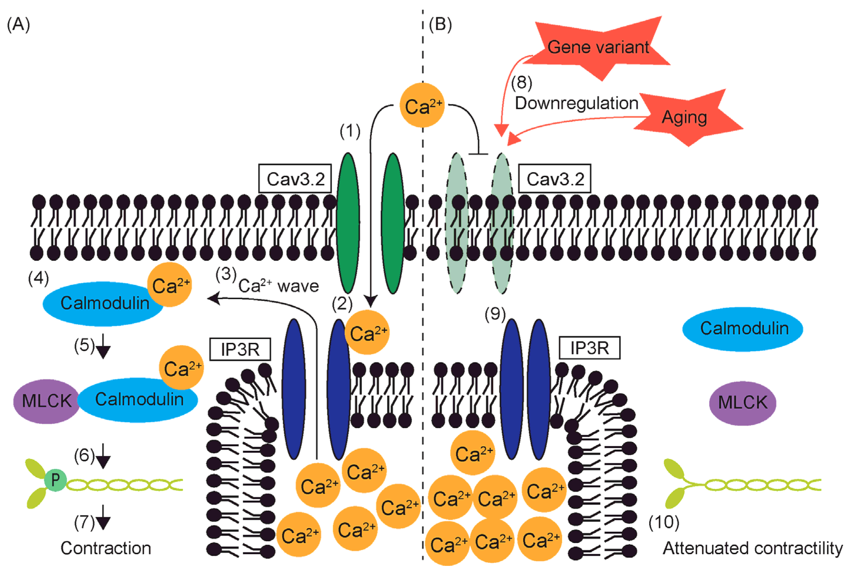
2. The Mechanism of Smooth Muscle Contraction Initiated by Calcium Influx

Ca2+ influx through L-type VGCCs initiates a cascade leading to smooth muscle cell contraction [13]. When membranes of smooth muscle cells depolarize by mechanosensory activation, L-type VGCCs open, allowing Ca2+ to enter the extracellular space [10]. This calcium influx is critical to initiating contraction by increasing intracellular Ca2+ levels [10]. After calcium enters through L-type VGCCs, it can trigger further release of calcium from the sarcoplasmic reticulum via inositol trisphosphate receptors (IP3R) or ryanodine receptors (RyR) [14]. This process amplifies the calcium signal, further enhancing contraction strength [14]. This coupling between L-type VGCCs and internal calcium release channels ensures sustained contraction [14]. Elevation of Ca2+ activates calmodulin via Ca2+ binding to calmodulin. Activated calmodulin triggers myosin light chain kinase, which phosphorylates myosin regulatory light chain [13]. Phosphorylation of myosin allows myosin to interact with actin [13]. Finally, the myosin cross-bridging cycle with actin results in contraction using the energy released from ATP by myosin ATPase activity [13]. While L-type channels are voltage-dependent, their function is modulated by surrounding signaling molecules. For instance, proteins like myristoylated alanine-rich C kinase substrate (MARCKS) interact with L-type VGCCs and regulate their activity [15]. When MARCKS is phosphorylated, it releases phosphatidylinositol 4,5-bisphosphate, which increases the open probability of these channels, promoting greater calcium entry and stronger contractions [15].

3. Subtypes of T-Type VGCCs

In contrast to L-type VGCCs, the highest activity of T-type VGCCS is observed at lower pressure in hyperpolarized vessels [16]. T-type VGCCs contribute to the development of myogenic tone to a lesser extent than L-type VGCCs do [16]. Nonetheless, T-type VGCCs can alter resting blood flow by 20–50% [16].
Three pore-forming subunits of T-type VGCCs have been identified: Cav3.1, Cav3.2, and Cav3.3, which are encoded by the genes Cacna1g, Cacna1h, and Cacna1i, respectively [17,18]. The three subunits can be differentiated by their activation kinetics. Of the three, Cav3.1 has the fastest activation and inactivation kinetics [19]. Cav3.2 and Cav3.3 share similar activation and inactivation profiles [19]. Both Cav3.1 and Cav3.2 deactivate with time constants of 6 milliseconds at a test potential of −80 mV, whereas Cav3.3 deactivates three times faster [19]. Cav3.1 also recovers most quickly from short-term inactivation, whereas Cav3.3 recovers most rapidly from long-term inactivation, with Cav3.2 recovering most slowly [19]. Additionally, Cav3.3 shows the greatest increase in calcium influx when positive pre-pulses are applied [19]. Sensitivity to Ni2+ provides another means of differentiating these subunits. The IC50 for Ni2+ on Cav3.1 is 13 µM, while those for Cav3.2 and Cav3.3 are significantly higher, at 250 µM and 216 µM, respectively (Table 1) [20].

4. Structural Properties of T-Type VGCCs

Unlike L-type VGCCs, individual T-type VGCCs consist of a single α1 subunit [21]. This subunit comprises four homologous domains labeled I, II, III, and IV, which are connected by large loops on the cytoplasmic side [22]. The loop connecting domains II and III is regulated by phosphorylation through kinases and G proteins. Each domain contains six α-helical transmembrane segments labeled S1 through S6, along with one re-entrant loop [22]. The S5 and S6 segments, along with the re-entrant loops, form the pore domain of the channel [22,23]. In the S4 segments, every third amino acid residue is positively charged, which enables the S4 segments to act as voltage sensors [22]. When a depolarizing pulse occurs, the electric field causes the voltage sensor to shift outward and rotate [24]. The S6 segments, which line the pore on the intracellular side of the membrane, form a bundle that regulates calcium influx [23,25]. When the membrane is polarized, this S6 bundle prevents calcium from entering the cell [23]. However, when the voltage sensor shifts, the S6 bundle opens, allowing Ca2+ to flow into the cytoplasm [23]. Additionally, the re-entrant loops, which line the outer side of the pore, carry a negative charge due to the presence of four glutamate or aspartate residues [23,26]. This negative charge is crucial for the channel’s selectivity for Ca2+ ions [23,25].

5. Electrophysiology and Biophysics of T-Type VGCCs

T-type VGCCs transition between three states, depending on the membrane potential. They assume a closed state when the cell membrane is hyperpolarized [22]. As the membrane becomes depolarized, T-type VGCCs open, and Ca2+ can pass through T-type VGCCs [22]. The threshold for T-type VGCCs’ is as low as −60 to −50 mV, which is close to the resting membrane potential of vascular smooth muscle cells [27,28]. This allows T-type VGCCs to be activated before other types of channels [22]. Ca2+ entry through T-type VGCCs further depolarizes the membrane, which then activates other channels [22].
Following their activation, T-type VGCCs are inactivated even with the continuation of the depolarizing pulse. Three types of inactivation have been identified: inactivation phase, fast inactivation, and slow inactivation [23]. The inactivation phase, which is the shortest of the three, occurs immediately after activation [23]. Replacement of the linker region of domains III and IV of Cav3.1 with that of an L-type VGCC drops the rate of inactivation, which suggests that the linker region contributes to the inactivation phase [29]. The C-terminal end, located in the cytoplasm, is also responsible for the occurrence of the inactivation phase, as deletion of its 161 amino acids decreases the rate of inactivation [29].
After a depolarizing pulse is extended from a few hundred milliseconds to 1 s or longer, T-type VGCCS enter fast or slow inactivation, respectively, recovery from which progresses at two very different time constants [23,30]. The time constant of fast inactivation is a few seconds [30], whereas that of slow inactivation is tens of seconds [30]. Fast or slow inactivation is sensitive to extracellular Ca2+ and can reduce Ca2+ influx by 50% [30].
Involvement of the loop linking domains I and II in inactivation has recently been discovered. The I and II linker has a helix–loop–helix motif called the gating brake on the I-domain side [31]. The gating brake contributes to the dependence of T-type VGCCs on low voltage [31]. The central region of the I and II linkers of Cav3.2 and Cav3.3 regulates surface expression, but that of Cav3.1 regulates current density [32,33]. The region proximal to domain II of Cav3.2 contributes to inactivation [32]. Furthermore, calmodulin binds to the gating brake and regulates activation and inactivation [31]. More recently, it was shown that high-frequency stimulation induces inactivation via Ca2+-sensitive phosphorylation that is not dependent on calmodulin and calcineurin [34].
Hyperpolarization of the plasma membrane causes T-type VGCCs to assume the closed state, and they again become available for activation [22].

6. Regulation of T-Type Voltage-Gated Calcium Channel Expression

6.1. T-Type VGCC-Expressing Cells

T-type voltage-gated calcium channels (VGCC) are expressed on the surface of various cells. Cav3.2 is expressed by white adipocytes, neurons, astrocytes, endocrine cells, mesangial cells, the heart, tracheal mesenchyme, sperm, oocytes, eggs, skeletal muscle, platelets, and smooth muscle cells [22,35,36,37,38,39,40,41,42,43]. Cav3.1 is expressed by white adipocytes, T lymphocytes, osteoblasts, endocrine cells, sperm, and neurons [22,38,44,45,46,47]. Cav3.3 is expressed in neurons [48,49,50].

6.2. Regulation by Transcription Factors

T-type VGCC expression is regulated by transcription factors, miRNAs, and epigenetic modifications. The promoter region of the Cav3.1 gene is GC-rich and lacks a TATA box [51]. As a result, transcription is initiated by specific binding of transcription factor 1 to this GC-rich region instead of the usual TATA-binding protein [52]. Additionally, the Cav3.1 promoter region is predicted to contain a nuclear factor kappa B (NF-κB) binding site [51].
A study using a retinoblastoma cell line showed that the MAPK pathway activates Cav3.1 expression, which can be inhibited by epidermal growth factor receptor (EGFR) or ERK inhibitors [53]. Another study found that SHOX2 activation also upregulates Cav3.1 expression [54]. In HL-1 cardiomyocyte cell lines, treatment with Yixin-Fumai granules, a traditional Chinese medicine, activated the SHOX2/BMP4/GATA4 axis, leading to downregulation of NKX2-5 and upregulation of Cav3.1 [54]. Similarly, the knockdown of NKX2-5 upregulates Cav3.1, suggesting that NKX2-5 negatively regulates its expression [55].
Both Cav3.1 and Cav3.2 are upregulated by VEGF stimulation in Purkinje cells [56]. In cardiomyocytes, aldosterone increases Cav3.1 expression by repressing the inhibitor of differentiation/DNA binding protein 2 (Id2) [57]. Overexpression of Id2, however, prevents aldosterone-induced Cav3.1 upregulation [57]. Aldosterone also increases EGFR expression, which leads to upregulation of Cav3.2 [58].
The Cav3.1 gene contains a regulatory DNA element, the deletion of which reduces Cav3.1 expression [59]. This element includes binding sites for several transcription factors, such as Nkx2-5, Tbx5, Hand2, Gata4, and Tbx3. In motor neuron cell lines (NSC-34), Cav3.2 was upregulated by cannabinoid treatment [60]. In trigeminal ganglion neurons, activation of adiponectin receptor 1 increased calcium influx through Cav3.2, although Cav3.2 is blocked by caveolin-3 [61].
In mice, rats, and humans, a neuron-restrictive silencer element (NRSE) has been identified in the first intron of Cav3.2 but not in Cav3.1 [62,63]. A gel electrophoresis mobility shift assay showed that the neuron-restrictive silencer factor (NRSF) binds to these NRSE-like sequences, suppressing Cav3.2 expression [62]. In mice expressing dominant-negative NRSF, Cav3.2 expression in cardiomyocytes was significantly higher than in wild-type mice [62], indicating that NRSF suppresses Cav3.2 expression, at least in cardiomyocytes.

6.3. Epigenetic Regulation

Downregulation by microRNA is a key mechanism for regulating Cav3.1 expression. In a cocaine-dependence model, calcium channels, including Cav3.1, were upregulated, but treatment with L-methionine induced expression of microRNA, which downregulated Cav3.1 [64]. In rat cerebral arteries, microRNA miR-137 also downregulated Cav3.1 [65].
Cav3.2 is similarly regulated by microRNAs. It was first identified as a target of miR-490-3p in the miRBase database, and the presence of a miR-490-3p mimic decreased Cav3.2 expression [66]. Upon aldosterone stimulation, miR-204 was upregulated, reducing NRSF levels, which led to the upregulation of both Cav3.1 and Cav3.2 in rat cardiomyocytes [67]. In a model of trigeminal ganglion injury in rats, Cav3.2 was identified as a direct target of miR-32-5p [68]. Bioinformatics tools such as TargetScan and miRDB predicted this interaction, which was confirmed with luciferase reporter assays [68]. These assays showed that miR-32-5p binding to the 3′UTR of Cav3.2 mRNA reduced luciferase activity, directly demonstrating that miR-32-5p downregulates Cav3.2 expression [68]. Knockdown of miR-32-5p increased Cav3.2 protein levels and enhanced T-type currents, reinforcing the role of miR-32-5p in regulating Cav3.2 channels [68].
There are also reports of T-type VGCC regulation via histone modification. In islet cells, treatment with HDAC1/3 inhibitors or Hdac1 knockdown led to the acetylation of H3K27 in the promoter region, which upregulated Cav3.1 expression [69]. Similarly, the intron of Cav3.1 includes a binding site for acetylated H3K27 [60]. Treatment with valproic acid increased histone H3 acetylation and mRNA expression of Cacna1g, Cacna1h, and Cacna1i in embryonic neural progenitor cells [70]. Additionally, histone H3 is bound to the promoter region of Cacna1g [70].
Histone methylation has also been implicated in regulating T-type VGCC expression. In response to pressure overload, Cav3.1 was upregulated in the heart; however, a deficiency in pax transactivation domain-interacting protein (PTIP) attenuated this upregulation [71]. PTIP methylates K4 of H3 (H3K4me3), and binding of H3K4me3 activates gene transcription [71]. This suggests that the reduced binding of H3K4me3 to the Cacna1g gene, due to PTIP deficiency, led to attenuation of Cacna1g expression under pressure overload conditions.
DNA methylation also contributes to the regulation of T-type VGCCs. Methylation of the 5′ CpG island upstream of the CACNA1G translation initiation site inhibits transcription in various cancers, including colorectal cancer, gastric cancer, and acute myelogenous leukemia [72]. Another study found that the CACNA1H gene is hypermethylated in human pancreatic islets following hyperglycemia. In cases of pheochromocytoma and abdominal paraganglioma, the methylation density of CACNA1H correlates with its expression levels [73].

6.4. Aging

Aging also affects the expression of T-type VGCCs. Aging downregulates Cav3.2, leading to a decrease in Ca2+ influx in vascular smooth muscle cells (VSMCs) and Purkinje cells [56,74]. Expression of Cav3.2 in VSMCs from 12–14-week-old to 48–56-week-old mice, as well as in Purkinje cells from 9-day-old to 30-day-old mice, was compared [56,74], demonstrating that aging upregulates Cav3.1 in Purkinje cells [56]. In young VSMCs, blocking Cav1.2 and Cav3.2 completely abolished Ca2+ sparks [74]. However, in aged VSMCs, Ca2+ sparks were only partially inhibited [74]. Blocking TRP channels with gadolinium ions inhibited the remaining Ca2+ sparks, suggesting that TRP channels are upregulated in aged cells to compensate for the reduction in Ca2+ sparks [74].

7. Functions of T-Type VGCCs Other than Contraction Induction

7.1. Proliferation

T-type VGCCs contribute to cell proliferation [75]. In smooth muscle cells from the pulmonary artery, knockdown of Cav3.1 using siRNA impaired cell proliferation [76]. Additionally, Cav3.1 expression increased following vascular injury in wild-type mice [77]. In contrast, vascular injury did not lead to neointimal formation in Cav3.1 knockout mice [77]. In these knockout mice, cyclin E was downregulated, and administration of a calmodulin agonist partially restored their proliferative activity [77].
Similarly, an in vitro study demonstrated that inhibition of Cav3.2, either by heme oxygenase-1 (HO-1) or the drug Mibefradil, impaired the proliferation of vascular smooth muscle cells [78]. Conversely, overexpression of Cav3.2 in HEK293 cells increased intracellular Ca2+ concentrations and promoted cell proliferation [78].

7.2. Apoptosis

T-type VGCCs participate in regulating apoptosis. Inhibition or knockdown of T-type VGCCs activates p53, leading to growth arrest and apoptosis in colon cancer cells [79]. Similarly, in cervical cancer cells, blocking T-type VGCCs with the compound KTt-45 demonstrated anti-apoptotic activity [80]. In medulloblastoma cells, T-type VGCC inhibition led to metabolic disruption, including reduced mitochondrial membrane potential and ATP levels, ultimately resulting in apoptosis [81]. Interestingly, overexpression of Cav3.1 in MCF-7 human breast cancer cells increased apoptotic activity, suggesting a context-dependent role for T-type VGCCs in the regulation of apoptosis [82]. In skeletal muscle cells, blocking T-type VGCCs upregulated endoplasmic reticulum stress-related genes such as 78-kDa glucose-regulated protein (GRP78), C/EBP homologous protein (CHOP), and apoptosis-related proteins like cleaved caspase 3 and cleaved caspase 9, triggering apoptosis [83].
In neural progenitor cells (NPCs), Cav3.1 helps maintain cell viability, and its inhibition induces apoptosis [84]. However, in neurons, calcium influx through T-type VGCCs contributes to cell death during cerebral ischemia/reperfusion injury, and blocking calcium influx has protective effects [85]. In Cav3.2 knockout mice, after cerebral ischemia/reperfusion injury, there was a reduction in infarct volume, brain water content, neurological dysfunction, oxidative stress, inflammation, and neuronal apoptosis [86]. These protective effects were reversed by overexpression of calcineurin [86]. Similarly, the knockdown of Cav3.2 by miR-490-3p reduced apoptosis after ischemic acute kidney injury by downregulating cleaved caspase-3, GRP78, and CHOP [66].

7.3. T Cell Differentiation and Cytokine Production

Wang et al. demonstrated that Cav3.1 influences cytokine production. In CD4+ naïve T cells from Cav3.1-deficient mice, differentiation into Th17 cells resulted in a 32% reduction in IL-17A+ cells, a 22% reduction in IL-17F+ cells, and a 38% reduction in IL-21+ cells, compared to Th17 cells differentiated from wild-type CD4+ naïve T cells [44,87,88]. In contrast, when Cav3.1-deficient CD4+ naïve T cells were differentiated into Th1 or Th2 cells, percentages of IFN-γ, TNF-α, and IL-4-producing cells were similar to those from wild-type T cells. Regulatory T-cell differentiation was also comparable between Cav3.1-deficient and wild-type mice [44]. The reduced differentiation of Th17 cells in Cav3.1-deficient mice was linked to lower expression of signal transducer and activator of transcription 3 (STAT3) and retinoic acid receptor-related orphan receptor γt (RORγt) [89], key transcription factors driving Th17 differentiation [44,89]. However, T-bet, which is important for Th1 differentiation, was unaffected [44]. During Th17 differentiation, Cav3.1 expression was upregulated, resulting in substantial Ca2+ influx, but Cav3.1 was not associated with general T cell development or maturation [44].
Moreover, Cav3.1-deficient mice exhibited decreased secretion of granulocyte–macrophage colony-stimulating factor (GM-CSF) by both Th1 and Th17 cells [44]. This reduction was attributed to decreased nuclear translocation of NFAT, a transcription factor that regulates GM-CSF production [90]. NFAT activation depends on Ca2+ influx, which activates calmodulin, leading to calcineurin-mediated NFAT dephosphorylation [44]. In Cav3.1-deficient cells, insufficient Ca2+ influx impaired NFAT translocation to the nucleus, resulting in reduced GM-CSF production [44].

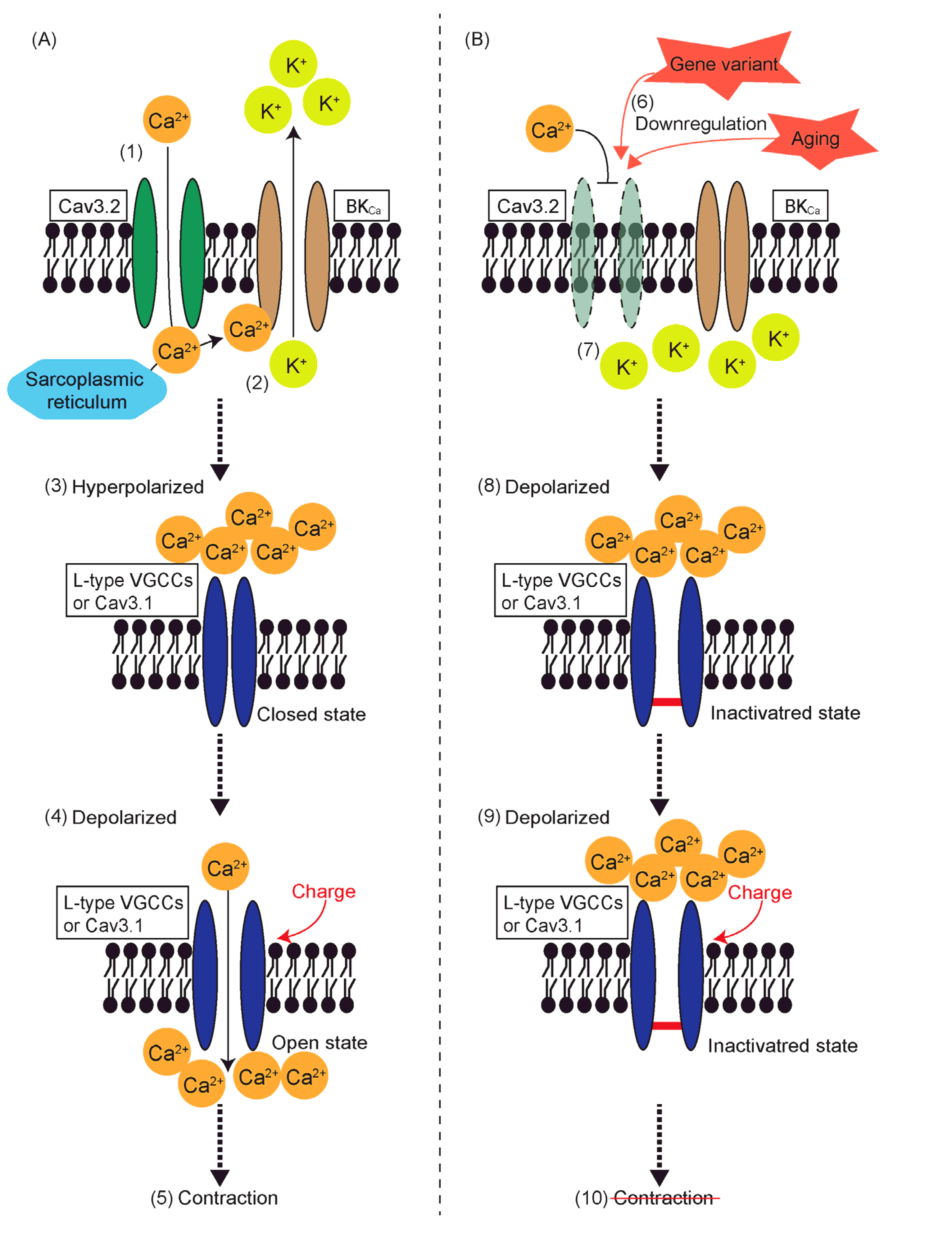
8. Reduced Contractility and T-Type VGCCs

The influx of Ca2+ through Cav3.1 induces contraction of the aortas [91]. Cav3.1 is located near inositol 1,4,5-trisphosphate receptors (IP3Rs), allowing Ca2+ that enters the cytoplasm via Cav3.1 to bind to IP3Rs [91]. This binding causes a conformational change in IP3Rs, which triggers the release of Ca2+ from the sarcoplasmic reticulum [92]. This release generates Ca2+ waves that activate calmodulin, leading to smooth muscle contraction [91].
In contrast, Ca2+ influx through Cav3.2 induces smooth muscle relaxation. Cav3.2 couples with ryanodine receptors to generate Ca2+ sparks, which act as negative feedback by activating large-conductance Ca2+-sensitive K+ channels (BKCa) [93,94]. Efflux of K+ through these activated BKCa channels repolarizes cell membranes, resulting in smooth muscle relaxation [93,94]. However, there are conflicting reports on this mechanism. In our previous study, deletion of K1256 of Myh11 impaired aortic contraction but not relaxation, and this coincided with the downregulation of Cav3.2, not Cav3.1 [6,7]. Similarly, in Notch3 knockout mice, reduced contractility of renal afferent arterioles coincided with downregulation of Cav3.2 [95]. These findings challenge the traditional view of Cav3.2 as solely an inducer of smooth muscle relaxation.
One possible explanation is that Cav3.2 induces contraction before triggering relaxation (Figure 1). In fact, while Ca2+ influx through L-type VGCCs induces contraction, recent studies suggest that L-type VGCCs also activate BKCa, leading to relaxation [96,97,98]. It is plausible that Cav3.2 serves a dual function, with Ca2+ influx inducing contraction through a mechanism similar to Cav3.1, in addition to promoting relaxation through K+ efflux via BKCa.
Another possibility is that proper interaction with Cav3.2 is necessary for L-type VGCCs or Cav3.1 to open and induce contraction in smooth muscle cells [95]. When the L-type VGCC blocker, diltiazem, is used alongside T-type VGCC blockers, pimozide or mibefradil, no additive effect on contraction is observed [100].
Failure to recover from the inactivation state of L-type VGCCs or Cav3.1 may also contribute to reduced contractility (Figure 2). As discussed earlier, Ca2+ influx activates BKCa channels, leading to hyperpolarization [93,101]. Since both L-type VGCCs and Cav3.1 are subject to voltage-dependent inactivation [23,102], extended depolarization caused by Cav3.2 downregulation may keep these channels inactivated. This could reduce the number of VGCCs available to open, thereby decreasing Ca2+ influx and attenuating contractility.
Alternatively, the downregulation of T-type VGCCs could affect the development of smooth muscle cells. In the trachea, for example, Cav3.2 is necessary for smooth muscle formation, which is partially controlled by RhoA-mediated cytoskeletal organization [103]. Additionally, T-type VGCCs are expressed in the heart during embryonic and fetal development, where they promote cell proliferation, migration, and resistance to apoptotic stress [79,83,104]. Therefore, downregulation of T-type VGCCs, as seen in Myh11 K1256 deletion or Notch3 knockout mice, may cause abnormalities in the phenotype, localization, or number of vascular smooth muscle cells, leading to the development of aortas or arteries with contractile dysfunction.

9. Multi-Omics Analysis of Smooth Muscle Cells

Previously, we analyzed transcriptomic and metabolomic data from the aortas of mice carrying a Myh11 K1256 deletion, where one lysine residue from a quadruple lysine repeat (1253–1256) was removed [6,7]. Using a comprehensive, data-driven, unbiased, multi-omics approach, we uncovered mechanisms that lead to reduced smooth muscle cell contraction. Our transcriptomic analysis revealed that Myh11 K1256-deletion mice did not show impaired expression of genes previously associated with familial thoracic aortic aneurysm and dissection (FTAAD) [7]. Additionally, the expression of genes encoding the elastin–contractile unit, which is also linked to FTAAD, was not affected [7].
With this multi-omics approach, we identified pathways that were impaired in Myh11 K1256-deletion mice, which involved the downregulation of genes encoding Cav3.2 and other calcium channels [7]. Our findings showed that the primary effect of the Myh11 K1256 deletion was the downregulation of membrane transporters [7]. Of the 22 affected molecular functions identified in the aortas of these mice, 19 were related to transmembrane transporters [7]. This suggests that the loss of one lysine in Myh11 has a significant impact on transmembrane transport, particularly with regard to calcium ion transport, which is critical for smooth muscle cell contraction. In our examination of individual gene expression, we found that several channels involved in calcium transport to the cytoplasm, including Cacna1h, Trpm2, Hcn2, Hcn3, and Hcn4, were downregulated [7].
Combining transcriptomics and metabolomics, we revealed Trpm2 dysfunction. By transcriptomics, we showed downregulation of Trpm2 [7]. By metabolomics, we found that poly ADP-ribose, which stimulates Trpm2 [105] synthesis, was attenuated in Myh11-K1256-deleted aortas [7]. This coincided with the downregulation of Parp6, Parp16, and Sarm1 in Myh11-K1256-deleted aortas [7], which convert NAD+ to ADP-ribose and nicotinamide [105]. Furthermore, cyclic ADP-ribose induces Ca2+ release from the sarcoplasmic reticulum to the cytoplasm. A decrease in ADP-ribose also contributes to reduced cytoplasmic Ca2+ [105]. Using a comprehensive, data-driven, unbiased, multi-omics approach, we proposed multiple mechanisms that led to reduced cytoplasmic Ca2+, revealing downregulation of multiple Ca2+ channels and metabolomic abnormalities that lead to Ca2+ insufficiency.
In addition to our approach, studies involving multi-omics with single-cell and spatial transcriptomics have been reported [106,107]. Kanemaru et al. explored the cellular composition of the human heart across eight distinct anatomical regions, including the left and right atria and ventricles, sinoatrial nodes, atrioventricular nodes, and other specialized cardiac tissues [106]. Integrating data from these regions allowed the creation of a spatially resolved map of cell types and interactions in cardiac niches and showed that only sinoatrial node P cells highly express CACNA1G [106]. Additionally, Qian et al. combined single-cell and spatial transcriptomics with spatial metabolomics to uncover smooth muscle phenotypic transformation and metabolic reprogramming in diabetic macroangiopathy [107]. Their method allowed the mapping of metabolites and gene expression across the entire anterior tibial artery [107]. By applying these advanced approaches, future studies are expected to reveal the localization of T-type VGCCs relative to other types of Ca2+ channels, their involvement in contraction, and the relationship between local downregulation of T-type VGCCS, metabolite abnormalities, and contractile dysfunction.

10. Conclusions

We have summarized the literature on the structure, electrophysiology, biophysics, transcriptional regulation, and function of T-type VGCCs. Although we have proposed three hypothetical models in which Cav3.2 downregulation attenuates contractility, it is likely that vascular contraction is multifactorial. While our understanding of the regulation and function of individual channels is growing, interactions between different channels remain poorly understood. A better understanding of these channel-to-channel interactions is expected to facilitate the development of therapeutic strategies targeting these interactions. Multi-omics are valuable for this purpose, as they allow us to analyze a wide range of factors simultaneously, beyond traditional research approaches. Additionally, developmental abnormalities may also contribute to contractile dysfunction. Although there is significant evidence supporting the involvement of T-type VGCCs in heart development, our understanding of their role in vascular development is limited. If attenuated vascular contractility is due to developmental abnormalities, interventions during the developmental phase would be necessary. Therefore, future studies that investigate the role of T-type VGCCs in vascular development are awaited.

Author Contributions

Conceptualization, S.T. and K.A.; writing—original draft preparation, S.T.; writing—review and editing, S.T., T.I. and K.A.; supervision, R.N. All authors have read and agreed to the published version of the manuscript.

Funding

This research received no external funding.

Institutional Review Board Statement

Not applicable.

Informed Consent Statement

Not applicable.

Data Availability Statement

Not applicable.

Conflicts of Interest

The authors declare no conflicts of interest.

References

  1. Tomida, S.; Aizawa, K.; Nishida, N.; Aoki, H.; Imai, Y.; Nagai, R.; Suzuki, T. Indomethacin reduces rates of aortic dissection and rupture of the abdominal aorta by inhibiting monocyte/macrophage accumulation in a murine model. Sci. Rep. 2019, 9, 10751. [Google Scholar] [CrossRef] [PubMed]
  2. Ahmed, S.; Malak, M.; Eshak, I.B. Aortic Dissection: A Review of the Pathophysiology, Management and Prospective Advances. Curr. Cardiol. Rev. 2021, 17, 12–26. [Google Scholar]
  3. Rombouts, K.B.; van Merrienboer, T.A.R.; Ket, J.C.F.; Bogunovic, N.; van der Velden, J.; Yeung, K.K. The role of vascular smooth muscle cells in the development of aortic aneurysms and dissections. Eur. J. Clin. Investig. 2022, 52, e13697. [Google Scholar] [CrossRef] [PubMed]
  4. Karimi, A.; Milewicz, D.M. Structure of the Elastin-Contractile Units in the Thoracic Aorta and How Genes That Cause Thoracic Aortic Aneurysms and Dissections Disrupt This Structure. Can. J. Cardiol. 2016, 32, 26–34. [Google Scholar] [CrossRef] [PubMed]
  5. Shen, Y.H.; LeMaire, S.A. Molecular pathogenesis of genetic and sporadic aortic aneurysms and dissections. Curr. Probl. Surg. 2017, 54, 95–155. [Google Scholar] [CrossRef]
  6. Negishi, K.; Aizawa, K.; Shindo, T.; Suzuki, T.; Sakurai, T.; Saito, Y.; Miyakawa, T.; Tanokura, M.; Kataoka, Y.; Maeda, M.; et al. An Myh11 single lysine deletion causes aortic dissection by reducing aortic structural integrity and contractility. Sci. Rep. 2022, 12, 8844. [Google Scholar] [CrossRef]
  7. Tomida, S.; Ishima, T.; Sawaki, D.; Imai, Y.; Nagai, R.; Aizawa, K. Multi-Omics of Familial Thoracic Aortic Aneurysm and Dissection: Calcium Transport Impairment Predisposes Aortas to Dissection. Int. J. Mol. Sci. 2023, 24, 15213. [Google Scholar] [CrossRef]
  8. Huang, J.; Fan, X.; Jin, X.; Lyu, C.; Guo, Q.; Liu, T.; Chen, J.; Davakan, A.; Lory, P.; Yan, N. Structural basis for human Cav3.2 inhibition by selective antagonists. Cell Res. 2024, 34, 440–450. [Google Scholar] [CrossRef]
  9. Andreasen, D.; Friis, U.G.; Uhrenholt, T.R.; Jensen, B.L.; Skøtt, O.; Hansen, P.B. Coexpression of Voltage-Dependent Calcium Channels Cav1.2, 2.1a, and 2.1b in Vascular Myocytes. Hypertension 2006, 47, 735–741. [Google Scholar] [CrossRef]
  10. Mironova, G.Y.; Haghbin, N.; Welsh, D.G. Functional tuning of Vascular L-type Ca2+ channels. Front. Physiol. 2022, 13, 1058744. [Google Scholar] [CrossRef]
  11. Akin, E.J.; Aoun, J.; Jimenez, C.; Mayne, K.; Baeck, J.; Young, M.D.; Sullivan, B.; Sanders, K.M.; Ward, S.M.; Bulley, S.; et al. ANO1, CaV1.2, and IP3R form a localized unit of EC-coupling in mouse pulmonary arterial smooth muscle. J. General. Physiol. 2023, 155, e202213217. [Google Scholar] [CrossRef] [PubMed]
  12. Gustafsson, F.; Andreasen, D.; Salomonsson, M.; Jensen, B.L.; Holstein-Rathlou, N.-H. Conducted vasoconstriction in rat mesenteric arterioles: Role for dihydropyridine-insensitive Ca2+ channels. Am. J. Physiol.-Heart Circ. Physiol. 2001, 280, H582–H590. [Google Scholar] [CrossRef] [PubMed]
  13. Webb, R.C. Smooth muscle contraction and relaxation. Adv. Physiol. Educ. 2003, 27, 201–206. [Google Scholar] [CrossRef] [PubMed]
  14. Rufenach, B.; Van Petegem, F. Structure and function of STAC proteins: Calcium channel modulators and critical components of muscle excitation–contraction coupling. J. Biol. Chem. 2021, 297, 100874. [Google Scholar] [CrossRef] [PubMed]
  15. Jahan, K.S.; Shi, J.; Greenberg, H.Z.E.; Khavandi, S.; Baudel, M.M.-A.; Barrese, V.; Greenwood, I.A.; Albert, A.P. MARCKS mediates vascular contractility through regulating interactions between voltage-gated Ca2+ channels and PIP2. Vasc. Pharmacol. 2020, 132, 106776. [Google Scholar] [CrossRef]
  16. El-Rahman, R.R.A.; Harraz, O.F.; Brett, S.E.; Anfinogenova, Y.; Mufti, R.E.; Goldman, D.; Welsh, D.G. Identification of L- and T-type Ca2+ channels in rat cerebral arteries: Role in myogenic tone development. Am. J. Physiol.-Heart Circ. Physiol. 2013, 304, H58–H71. [Google Scholar] [CrossRef]
  17. Lory, P.; Bidaud, I.; Chemin, J. T-type calcium channels in differentiation and proliferation. Cell Calcium 2006, 40, 135–146. [Google Scholar] [CrossRef]
  18. Yao, X.; Gao, S.; Yan, N. Structural biology of voltage-gated calcium channels. Channels 2024, 18, 2290807. [Google Scholar] [CrossRef]
  19. Klöckner, U.; Lee, J.-H.; Cribbs, L.L.; Daud, A.; Hescheler, J.; Pereverzev, A.; Perez-Reyes, E.; Schneider, T. Comparison of the Ca2+ currents induced by expression of three cloned α1 subunits, α1G, α1H and α1I, of low-voltage-activated T-type Ca2+ channels. Eur. J. Neurosci. 1999, 11, 4171–4178. [Google Scholar] [CrossRef]
  20. Lee, J.-H.; Gomora, J.C.; Cribbs, L.L.; Perez-Reyes, E. Nickel Block of Three Cloned T-Type Calcium Channels: Low Concentrations Selectively Block α1H. Biophys. J. 1999, 77, 3034–3042. [Google Scholar] [CrossRef]
  21. Perez-Reyes, E. Molecular Physiology of Low-Voltage-Activated T-type Calcium Channels. Physiol. Rev. 2003, 83, 117–161. [Google Scholar] [CrossRef] [PubMed]
  22. Iftinca, M.C.; Zamponi, G.W. Regulation of neuronal T-type calcium channels. Trends Pharmacol. Sci. 2009, 30, 32–40. [Google Scholar] [CrossRef] [PubMed]
  23. Guidelli, R.; Becucci, L. Deterministic model of Cav3.1 Ca2+ channel and a proposed sequence of its conformations. Bioelectrochemistry 2020, 136, 107618. [Google Scholar] [CrossRef] [PubMed]
  24. Catterall, W.A. Voltage-gated calcium channels. Cold Spring Harb. Perspect. Biol. 2011, 3, a003947. [Google Scholar] [CrossRef]
  25. He, L.; Yu, Z.; Geng, Z.; Huang, Z.; Zhang, C.; Dong, Y.; Gao, Y.; Wang, Y.; Chen, Q.; Sun, L.; et al. Structure, gating, and pharmacology of human CaV3.3 channel. Nat. Commun. 2022, 13, 2084. [Google Scholar] [CrossRef]
  26. Weiss, N.; Zamponi, G.W. T-type calcium channels: From molecule to therapeutic opportunities. Int. J. Biochem. Cell Biol. 2019, 108, 34–39. [Google Scholar] [CrossRef]
  27. Kuriyama, H.; Kitamura, K.; Itoh, T.; Inoue, R. Physiological Features of Visceral Smooth Muscle Cells, with Special Reference to Receptors and Ion Channels. Physiol. Rev. 1998, 78, 811–920. [Google Scholar] [CrossRef]
  28. Huguenard, J.R. Low-threshold calcium currents in central nervous system neurons. Annu. Rev. Physiol. 1996, 58, 329–348. [Google Scholar] [CrossRef]
  29. Staes, M.; Talavera, K.; Klugbauer, N.; Prenen, J.; Lacinová, L.; Droogmans, G.; Hofmann, F.; Nilius, B. The amino side of the C-terminus determines fast inactivation of the T-type calcium channel α1G. J. Physiol. 2001, 530, 35–45. [Google Scholar] [CrossRef]
  30. Hering, J.; Feltz, A.; Lambert, R.C. Slow inactivation of the CaV3.1 isotype of T-type calcium channels. J. Physiol. 2004, 555 Pt 2, 331–344. [Google Scholar] [CrossRef]
  31. Chemin, J.; Taiakina, V.; Monteil, A.; Piazza, M.; Guan, W.; Stephens, R.F.; Kitmitto, A.; Pang, Z.P.; Dolphin, A.C.; Perez-Reyes, E.; et al. Calmodulin regulates Cav3 T-type channels at their gating brake. J. Biol. Chem. 2017, 292, 20010–20031. [Google Scholar] [CrossRef] [PubMed]
  32. Vitko, I.; Bidaud, I.; Arias, J.M.; Mezghrani, A.; Lory, P.; Perez-Reyes, E. The I–II Loop Controls Plasma Membrane Expression and Gating of Cav3.2 T-Type Ca2+ Channels: A Paradigm for Childhood Absence Epilepsy Mutations. J. Neurosci. 2007, 27, 322–330. [Google Scholar] [CrossRef] [PubMed]
  33. Baumgart, J.P.; Vitko, I.; Bidaud, I.; Kondratskyi, A.; Lory, P.; Perez-Reyes, E. I–II Loop Structural Determinants in the Gating and Surface Expression of Low Voltage-Activated Calcium Channels. PLoS ONE 2008, 3, e2976. [Google Scholar] [CrossRef]
  34. Chemin, J.; Stamenic, T.T.; Cazade, M.; Llinares, J.; Blesneac, I.; Todorovic, S.M.; Lory, P. A novel phospho-modulatory mechanism contributes to the calcium-dependent regulation of T-type Ca2+ channels. Sci. Rep. 2019, 9, 15642. [Google Scholar] [CrossRef] [PubMed]
  35. Tamang, H.K.; Yang, R.B.; Song, Z.H.; Hsu, S.C.; Peng, C.C.; Tung, Y.C.; Tzeng, B.H.; Chen, C.C. CaV 3.2 T-type calcium channel regulates mouse platelet activation and arterial thrombosis. J. Thromb. Haemost. 2022, 20, 1887–1899. [Google Scholar] [CrossRef] [PubMed]
  36. Pandi, S.P.S.; Shattock, M.J.; Hendry, B.M.; Sharpe, C.C. Stimulated phosphorylation of ERK in mouse kidney mesangial cells is dependent upon expression of Cav3.1. BMC Nephrol. 2022, 23, 211. [Google Scholar] [CrossRef]
  37. Lin, S.-S.; Tzeng, B.-H.; Lee, K.-R.; Smith, R.J.H.; Campbell, K.P.; Chen, C.-C. Cav3.2 T-type calcium channel is required for the NFAT-dependent Sox9 expression in tracheal cartilage. Proc. Natl. Acad. Sci. USA 2014, 111, E1990–E1998. [Google Scholar] [CrossRef]
  38. Meng, Y.; Toledo-Rodriguez, M.; Fedorenko, O.; Smith, P.A. Sex and age affect depot expression of Ca2+ channels in rat white fat adipocytes. J. Mol. Endocrinol. 2024, 72, e230108. [Google Scholar] [CrossRef]
  39. Liu, Q.; Lu, Z.; Ren, H.; Fu, L.; Wang, Y.; Bu, H.; Ma, M.; Ma, L.; Huang, C.; Wang, J.; et al. Cav3.2 T-Type calcium channels downregulation attenuates bone cancer pain induced by inhibiting IGF-1/HIF-1α signaling pathway in the rat spinal cord. J. Bone Oncol. 2023, 42, 100495. [Google Scholar] [CrossRef]
  40. Mehregan, A.; Ardestani, G.; Akizawa, H.; Carvacho, I.; Fissore, R. Deletion of TRPV3 and CaV3.2 T-type channels in mice undermines fertility and Ca2+ homeostasis in oocytes and eggs. J. Cell Sci. 2021, 134, jcs257956. [Google Scholar] [CrossRef]
  41. Cai, H.; Chen, S.; Sun, Y.; Zheng, T.; Liu, Y.; Tao, J.; Zhang, Y. Interleukin-22 receptor 1-mediated stimulation of T-type Ca2+ channels enhances sensory neuronal excitability through the tyrosine-protein kinase Lyn-dependent PKA pathway. Cell Commun. Signal. 2024, 22, 307. [Google Scholar] [CrossRef] [PubMed]
  42. Leandrou, E.; Chalatsa, I.; Anagnostou, D.; Machalia, C.; Semitekolou, M.; Filippa, V.; Makridakis, M.; Vlahou, A.; Anastasiadou, E.; Vekrellis, K.; et al. α-Synuclein oligomers potentiate neuroinflammatory NF-κB activity and induce CaV3.2 calcium signaling in astrocytes. Transl. Neurodegener. 2024, 13, 11. [Google Scholar] [CrossRef] [PubMed]
  43. Ahn, J.W.; Kim, S.E.; Kim, D.Y.; Jeong, I.; Kim, S.; Chung, S.; Lee, S.E. Cav3.2 T-Type Calcium Channel Mediates Acute Itch and Contributes to Chronic Itch and Inflammation in Experimental Atopic Dermatitis. J. Investig. Dermatol. 2024, 144, 612–620.e6. [Google Scholar] [CrossRef] [PubMed]
  44. Wang, H.; Zhang, X.; Xue, L.; Xing, J.; Jouvin, M.H.; Putney, J.W.; Anderson, M.P.; Trebak, M.; Kinet, J.P. Low-Voltage-Activated CaV3.1 Calcium Channels Shape T Helper Cell Cytokine Profiles. Immunity 2016, 44, 782–794. [Google Scholar] [CrossRef]
  45. Shao, J.; Gao, D.-S.; Liu, Y.-H.; Chen, S.-P.; Liu, N.; Zhang, L.; Zhou, X.-Y.; Xiao, Q.; Wang, L.-P.; Hu, H.-L.; et al. Cav3.1-driven bursting firing in ventromedial hypothalamic neurons exerts dual control of anxiety-like behavior and energy expenditure. Mol. Psychiatry 2022, 27, 2901–2913. [Google Scholar] [CrossRef]
  46. Zöphel, D.; Hof, C.; Lis, A. Altered Ca2+ Homeostasis in Immune Cells during Aging: Role of Ion Channels. Int. J. Mol. Sci. 2020, 22, 110. [Google Scholar] [CrossRef]
  47. Mayadali, Ü.S.; Chertes, C.A.M.; Sinicina, I.; Shaikh, A.G.; Horn, A.K.E. Ion channel profiles of extraocular motoneurons and internuclear neurons in human abducens and trochlear nuclei. Front. Neuroanat. 2024, 18, 1411154. [Google Scholar] [CrossRef]
  48. Maksemous, N.; Blayney, C.D.; Sutherland, H.G.; Smith, R.A.; Lea, R.A.; Tran, K.N.; Ibrahim, O.; McArthur, J.R.; Haupt, L.M.; Cader, M.Z.; et al. Investigation of CACNA1I Cav3.3 Dysfunction in Hemiplegic Migraine. Front. Mol. Neurosci. 2022, 15, 892820. [Google Scholar] [CrossRef]
  49. Eickhoff, A.; Tjaden, J.; Stahlke, S.; Vorgerd, M.; Theis, V.; Matschke, V.; Theiss, C. Effects of progesterone on T-type-Ca2+-channel expression in Purkinje cells. Neural Regen. Res. 2022, 17, 2465–2471. [Google Scholar]
  50. Cmarko, L.; Weiss, N. Selective inhibition of neuronal Cav3.3 T-type calcium channels by TAT-based channel peptide. Mol. Brain 2020, 13, 95. [Google Scholar] [CrossRef]
  51. Bertolesi, G.E.; Jollimore, C.A.B.; Shi, C.; Elbaum, L.; Denovan-Wright, E.M.; Barnes, S.; Kelly, M.E.M. Regulation of α1G T-type calcium channel gene (CACNA1G) expression during neuronal differentiation. Eur. J. Neurosci. 2003, 17, 1802–1810. [Google Scholar] [CrossRef] [PubMed]
  52. González-Ramírez, R.; Martínez-Hernández, E.; Sandoval, A.; Felix, R. Transcription Factor Sp1 Regulates T-Type Ca2+ Channel CaV3.1 Gene Expression. J. Cell. Physiol. 2014, 229, 551–560. [Google Scholar] [CrossRef] [PubMed]
  53. Banderali, U.; Jain, M.; Thakur, S.; Jayanthan, A.; Belke, D.D.; Giles, W.R.; Narendran, A. The T-type Calcium Channel Cav3.1 in Y79 Retinoblastoma Cells is Regulated by the Epidermal Growth Factor Receptor via the MAPK Signaling Pathway. Curr. Eye Res. 2022, 47, 426–435. [Google Scholar] [CrossRef]
  54. Zhang, H.; Chen, C.; Liu, Y.; Ren, L.; Qi, J.; Yang, Y.; Chen, W.; Yao, Y.; Cai, X.; Liu, Z.; et al. NRF-2/HO-1 Pathway-Mediated SHOX2 Activation Is a Key Switch for Heart Rate Acceleration by Yixin-Fumai Granules. Oxidative Med. Cell. Longev. 2022, 2022, 8488269. [Google Scholar] [CrossRef] [PubMed]
  55. Wang, H.; Liu, Y.; Han, S.; Zi, Y.; Zhang, Y.; Kong, R.; Liu, Z.; Cai, Z.; Zhong, C.; Liu, W.; et al. Nkx2-5 Regulates the Proliferation and Migration of H9c2 Cells. Med. Sci. Monit. 2020, 26, e925388. [Google Scholar] [CrossRef] [PubMed]
  56. Tjaden, J.; Eickhoff, A.; Stahlke, S.; Gehmeyr, J.; Vorgerd, M.; Theis, V.; Matschke, V.; Theiss, C. Expression Pattern of T-Type Ca2+ Channels in Cerebellar Purkinje Cells after VEGF Treatment. Cells 2021, 10, 2277. [Google Scholar] [CrossRef]
  57. Ito, J.; Minemura, T.; Wälchli, S.; Niimi, T.; Fujihara, Y.; Kuroda, S.I.; Takimoto, K.; Maturana, A.D. Id2 Represses Aldosterone-Stimulated Cardiac T-Type Calcium Channels Expression. Int. J. Mol. Sci. 2021, 22, 3561. [Google Scholar] [CrossRef]
  58. Stroedecke, K.; Meinel, S.; Markwardt, F.; Kloeckner, U.; Straetz, N.; Quarch, K.; Schreier, B.; Kopf, M.; Gekle, M.; Grossmann, C. The mineralocorticoid receptor leads to increased expression of EGFR and T-type calcium channels that support HL-1 cell hypertrophy. Sci. Rep. 2021, 11, 13229. [Google Scholar] [CrossRef]
  59. Mohan, R.A.; Bosada, F.M.; van Weerd, J.H.; van Duijvenboden, K.; Wang, J.; Mommersteeg, M.T.M.; Hooijkaas, I.B.; Wakker, V.; de Gier-de Vries, C.; Coronel, R.; et al. T-box transcription factor 3 governs a transcriptional program for the function of the mouse atrioventricular conduction system. Proc. Natl. Acad. Sci. USA 2020, 117, 18617–18626. [Google Scholar] [CrossRef]
  60. Trainito, A.; Muscarà, C.; Gugliandolo, A.; Chiricosta, L.; Salamone, S.; Pollastro, F.; Mazzon, E.; D’Angiolini, S. Cannabinol (CBN) Influences the Ion Channels and Synaptic-Related Genes in NSC-34 Cell Line: A Transcriptomic Study. Cells 2024, 13, 1573. [Google Scholar] [CrossRef]
  61. Markandeya, Y.S.; Fahey, J.M.; Pluteanu, F.; Cribbs, L.L.; Balijepalli, R.C. Caveolin-3 Regulates Protein Kinase A Modulation of the CaV3.2 (α1H) T-type Ca2+ Channels. J. Biol. Chem. 2011, 286, 2433–2444. [Google Scholar] [CrossRef] [PubMed]
  62. Kuwahara, K.; Saito, Y.; Takano, M.; Arai, Y.; Yasuno, S.; Nakagawa, Y.; Takahashi, N.; Adachi, Y.; Takemura, G.; Horie, M.; et al. NRSF regulates the fetal cardiac gene program and maintains normal cardiac structure and function. EMBO J. 2003, 22, 6310–6321. [Google Scholar] [CrossRef] [PubMed]
  63. Somekawa, S.; Imagawa, K.; Naya, N.; Takemoto, Y.; Onoue, K.; Okayama, S.; Takeda, Y.; Kawata, H.; Horii, M.; Nakajima, T.; et al. Regulation of Aldosterone and Cortisol Production by the Transcriptional Repressor Neuron Restrictive Silencer Factor. Endocrinology 2009, 150, 3110–3117. [Google Scholar] [CrossRef] [PubMed]
  64. Wang, Y.; Yang, L.; Zhou, H.; Zhang, K.; Zhao, M. Identification of miRNA-mediated gene regulatory networks in L-methionine exposure counteracts cocaine-conditioned place preference in mice. Front. Genet. 2023, 13, 1076156. [Google Scholar] [CrossRef]
  65. Zhang, B.; Chen, L.; Bai, Y.-G.; Song, J.-B.; Cheng, J.-H.; Ma, H.-Z.; Ma, J.; Xie, M.-J. miR-137 and its target T-type CaV3.1 channel modulate dedifferentiation and proliferation of cerebrovascular smooth muscle cells in simulated microgravity rats by regulating calcineurin/NFAT pathway. Cell Prolif. 2020, 53, e12774. [Google Scholar] [CrossRef]
  66. Zheng, Q.; Li, X.; Xu, X.; Tang, X.; Hammad, B.; Xing, J.; Zhang, D. The mmu_circ_003062, hsa_circ_0075663/miR-490-3p/CACNA1H axis mediates apoptosis in renal tubular cells in association with endoplasmic reticulum stress following ischemic acute kidney injury. Int. Immunopharmacol. 2024, 132, 111956. [Google Scholar] [CrossRef]
  67. Koyama, R.; Mannic, T.; Ito, J.; Amar, L.; Zennaro, M.-C.; Rossier, M.F.; Maturana, A.D. MicroRNA-204 Is Necessary for Aldosterone-Stimulated T-Type Calcium Channel Expression in Cardiomyocytes. Int. J. Mol. Sci. 2018, 19, 2941. [Google Scholar] [CrossRef]
  68. Qi, R.; Cao, J.; Sun, Y.; Li, Y.; Huang, Z.; Jiang, D.; Jiang, X.-H.; Snutch, T.P.; Zhang, Y.; Tao, J. Histone methylation-mediated microRNA-32-5p down-regulation in sensory neurons regulates pain behaviors via targeting Cav3.2 channels. Proc. Natl. Acad. Sci. USA 2022, 119, e2117209119. [Google Scholar] [CrossRef]
  69. Zhang, Y.; Li, M.; Wang, Y.; Liu, X.; Zhou, L.; Zhang, C.; Shao, L. Histone deacetylase inhibition by MS-275 potentiates glucose-stimulated insulin secretion without affecting glucose oxidation. Life Sci. 2020, 257, 118073. [Google Scholar] [CrossRef]
  70. Kim, J.W.; Oh, H.A.; Kim, S.R.; Ko, M.J.; Seung, H.; Lee, S.H.; Shin, C.Y. Epigenetically Upregulated T-Type Calcium Channels Contribute to Abnormal Proliferation of Embryonic Neural Progenitor Cells Exposed to Valproic Acid. Biomol. Ther. 2020, 28, 389–396. [Google Scholar] [CrossRef]
  71. Stein, A.B.; Goonewardena, S.N.; Jones, T.A.; Prusick, P.J.; Bazzi, A.A.; Belyavskaya, J.M.; McCoskey, M.M.; Dandar, R.A. The PTIP-Associated Histone Methyltransferase Complex Prevents Stress-Induced Maladaptive Cardiac Remodeling. PLoS ONE 2015, 10, e0127839. [Google Scholar] [CrossRef] [PubMed]
  72. Toyota, M.; Ho, C.; Ohe-Toyota, M.; Baylin, S.B.; Issa, J.-P.J. Inactivation of CACNA1G, a T-Type Calcium Channel Gene, by Aberrant Methylation of Its 5′ CpG Island in Human Tumors1. Cancer Res. 1999, 59, 4535–4541. [Google Scholar] [PubMed]
  73. Svahn, F.; Solhusløkk Höse, K.; Stenman, A.; Liu, Y.; Calissendorff, J.; Tham, E.; Végvári, Á.; Zubarev, R.A.; Wang, N.; Korah, R.; et al. Genetic variants and down-regulation of CACNA1H in pheochromocytoma. Endocr. Relat. Cancer 2024, 31, e230061. [Google Scholar] [CrossRef] [PubMed]
  74. Fan, G.; Kaßmann, M.; Cui, Y.; Matthaeus, C.; Kunz, S.; Zhong, C.; Zhu, S.; Xie, Y.; Tsvetkov, D.; Daumke, O.; et al. Age attenuates the T-type Ca(V) 3.2-RyR axis in vascular smooth muscle. Aging Cell 2020, 19, e13134. [Google Scholar] [CrossRef]
  75. Yabuki, Y.; Matsuo, K.; Yu, M.; Xu, J.; Sakimura, K.; Shioda, N.; Fukunaga, K. Cav3.1 t-type calcium channel is critical for cell proliferation and survival in newly generated cells of the adult hippocampus. Acta Physiol. 2021, 232, e13613. [Google Scholar] [CrossRef]
  76. Rodman, D.M.; Reese, K.; Harral, J.; Fouty, B.; Wu, S.; West, J.; Hoedt-Miller, M.; Tada, Y.; Li, K.-X.; Cool, C.; et al. Low-Voltage-Activated (T-Type) Calcium Channels Control Proliferation of Human Pulmonary Artery Myocytes. Circ. Res. 2005, 96, 864–872. [Google Scholar] [CrossRef]
  77. Tzeng, B.-H.; Chen, Y.-H.; Huang, C.-H.; Lin, S.-S.; Lee, K.-R.; Chen, C.-C. The Cav3.1 T-type calcium channel is required for neointimal formation in response to vascular injury in mice. Cardiovasc. Res. 2012, 96, 533–542. [Google Scholar] [CrossRef]
  78. Duckles, H.; Boycott, H.E.; Al-Owais, M.M.; Elies, J.; Johnson, E.; Dallas, M.L.; Porter, K.E.; Giuntini, F.; Boyle, J.P.; Scragg, J.L.; et al. Heme oxygenase-1 regulates cell proliferation via carbon monoxide-mediated inhibition of T-type Ca2+ channels. Pflügers Arch.-Eur. J. Physiol. 2015, 467, 415–427. [Google Scholar] [CrossRef]
  79. Dziegielewska, B.; Brautigan, D.L.; Larner, J.M.; Dziegielewski, J. T-Type Ca2+ Channel Inhibition Induces p53-Dependent Cell Growth Arrest and Apoptosis through Activation of p38-MAPK in Colon Cancer Cells. Mol. Cancer Res. 2014, 12, 348–358. [Google Scholar] [CrossRef]
  80. Du, N.H.; Ngoc, T.T.B.; Cang, H.Q.; Luyen, N.T.T.; Thuoc, T.L.; Le Quan, T.; Thao, D.T.P. KTt-45, a T-type calcium channel blocker, acts as an anticancer agent by inducing apoptosis on HeLa cervical cancer cell line. Sci. Rep. 2023, 13, 22092. [Google Scholar] [CrossRef]
  81. Sedeeq, M.; Maklad, A.; Dutta, T.; Feng, Z.; Wilson, R.; Gueven, N.; Azimi, I. T-Type Calcium Channel Inhibitors Induce Apoptosis in Medulloblastoma Cells Associated with Altered Metabolic Activity. Mol. Neurobiol. 2022, 59, 2932–2945. [Google Scholar] [CrossRef] [PubMed]
  82. Ohkubo, T.; Yamazaki, J. T-type voltage-activated calcium channel Cav3.1, but not Cav3.2, is involved in the inhibition of proliferation and apoptosis in MCF-7 human breast cancer cells. Int. J. Oncol. 2012, 41, 267–275. [Google Scholar] [CrossRef] [PubMed]
  83. Chen, M.; Li, S.; Hao, M.; Chen, J.; Zhao, Z.; Hong, S.; Min, J.; Tang, J.; Hu, M.; Hong, L. T-type calcium channel blockade induces apoptosis in C2C12 myotubes and skeletal muscle via endoplasmic reticulum stress activation. FEBS Open Bio 2020, 10, 2122–2136. [Google Scholar] [CrossRef] [PubMed]
  84. Kim, J.W.; Oh, H.A.; Lee, S.H.; Kim, K.C.; Eun, P.H.; Ko, M.J.; Gonzales, E.L.T.; Seung, H.; Kim, S.; Bahn, G.H.; et al. T-Type Calcium Channels Are Required to Maintain Viability of Neural Progenitor Cells. Biomol. Ther. 2018, 26, 439–445. [Google Scholar] [CrossRef]
  85. Ferreira, A.F.F.; Britto, L.R.G. The transient receptor potential melastatin 2: A new therapeutical target for Parkinson’s disease? Neural Regen. Res. 2023, 18, 1652–1656. [Google Scholar] [CrossRef]
  86. Dai, F.; Hu, C.; Li, X.; Zhang, Z.; Wang, H.; Zhou, W.; Wang, J.; Geng, Q.; Dong, Y.; Tang, C. Cav3.2 channel regulates cerebral ischemia/reperfusion injury: A promising target for intervention. Neural Regen. Res. 2024, 19, 2480–2487. [Google Scholar] [CrossRef]
  87. Yang, F.; Zhou, L.; Shen, Y.; Wang, X.; Fan, X.; Yang, L. Multi-omics approaches for drug-response characterization in primary biliary cholangitis and autoimmune hepatitis variant syndrome. J. Transl. Med. 2024, 22, 214. [Google Scholar] [CrossRef]
  88. Erdogmus, S.; Concepcion, A.R.; Yamashita, M.; Sidhu, I.; Tao, A.Y.; Li, W.; Rocha, P.P.; Huang, B.; Garippa, R.; Lee, B.; et al. Cavβ1 regulates T cell expansion and apoptosis independently of voltage-gated Ca2+ channel function. Nat. Commun. 2022, 13, 2033. [Google Scholar] [CrossRef]
  89. Capone, A.; Volpe, E. Transcriptional Regulators of T Helper 17 Cell Differentiation in Health and Autoimmune Diseases. Front. Immunol. 2020, 11, 348. [Google Scholar] [CrossRef]
  90. Johnson, B.V.; Bert, A.G.; Ryan, G.R.; Condina, A.; Cockerill, P.N. Granulocyte-Macrophage Colony-Stimulating Factor Enhancer Activation Requires Cooperation between NFAT and AP-1 Elements and Is Associated with Extensive Nucleosome Reorganization. Mol. Cell. Biol. 2004, 24, 7914–7930. [Google Scholar] [CrossRef]
  91. El-Lakany, M.A.; Haghbin, N.; Arora, N.; Hashad, A.M.; Mironova, G.Y.; Sancho, M.; Gros, R.; Welsh, D.G. CaV3.1 channels facilitate calcium wave generation and myogenic tone development in mouse mesenteric arteries. Sci. Rep. 2023, 13, 20407. [Google Scholar] [CrossRef] [PubMed]
  92. Paknejad, N.; Sapuru, V.; Hite, R.K. Structural titration reveals Ca2+-dependent conformational landscape of the IP3 receptor. Nat. Commun. 2023, 14, 6897. [Google Scholar] [CrossRef] [PubMed]
  93. Hashad, A.M.; Harraz, O.F.; Brett, S.E.; Romero, M.; Kassmann, M.; Puglisi, J.L.; Wilson, S.M.; Gollasch, M.; Welsh, D.G. Caveolae Link Ca(V)3.2 Channels to BK(Ca)-Mediated Feedback in Vascular Smooth Muscle. Arterioscler. Thromb. Vasc. Biol. 2018, 38, 2371–2381. [Google Scholar] [CrossRef] [PubMed]
  94. Harraz, O.F.; Jensen, L.J. Aging, calcium channel signaling and vascular tone. Mech. Ageing Dev. 2020, 191, 111336. [Google Scholar] [CrossRef]
  95. Helle, F.; Hultström, M.; Kavvadas, P.; Iversen, B.; Chadjichristos, C.E.; Chatziantoniou, C. Deletion of Notch3 Impairs Contractility of Renal Resistance Vessels Due to Deficient Ca2+ Entry. Int. J. Mol. Sci. 2022, 23, 16068. [Google Scholar] [CrossRef]
  96. Plante, A.E.; Whitt, J.P.; Meredith, A.L. BK channel activation by L-type Ca2+ channels CaV1.2 and CaV1.3 during the subthreshold phase of an action potential. J. Neurophysiol. 2021, 126, 427–439. [Google Scholar] [CrossRef]
  97. Vivas, O.; Moreno, C.M.; Santana, L.F.; Hille, B. Proximal clustering between BK and CaV1.3 channels promotes functional coupling and BK channel activation at low voltage. eLife 2017, 6, e28029. [Google Scholar] [CrossRef]
  98. Numata, T.; Sato-Numata, K.; Yoshino, M. BK Channels Are Activated by Functional Coupling With L-Type Ca2+ Channels in Cricket Myocytes. Front. Insect Sci. 2021, 1, 662414. [Google Scholar] [CrossRef]
  99. Schmitz, E.A.; Takahashi, H.; Karakas, E. Structural basis for activation and gating of IP3 receptors. Nat. Commun. 2022, 13, 1408. [Google Scholar] [CrossRef]
  100. Feng, M.-G.; Li, M.; Navar, L.G. T-type calcium channels in the regulation of afferent and efferent arterioles in rats. Am. J. Physiol.-Ren. Physiol. 2004, 286, F331–F337. [Google Scholar] [CrossRef]
  101. Pérez, G.J.; Bonev, A.D.; Patlak, J.B.; Nelson, M.T. Functional Coupling of Ryanodine Receptors to KCa Channels in Smooth Muscle Cells from Rat Cerebral Arteries. J. Gen. Physiol. 1999, 113, 229–238. [Google Scholar] [CrossRef] [PubMed]
  102. Findlay, I. β-Adrenergic stimulation modulates Ca2+- and voltage-dependent inactivation of L-type Ca2+ channel currents in guinea-pig ventricular myocytes. J. Physiol. 2002, 541, 741–751. [Google Scholar] [CrossRef] [PubMed]
  103. Liu, Z.; Lu, C.; Ma, L.; Li, C.; Luo, H.; Liu, Y.; Liu, X.; Li, H.; Cui, Y.; Zeng, J.; et al. The T-Type Calcium Channel CACNA1H is Required for Smooth Muscle Cytoskeletal Organization During Tracheal Tubulogenesis. Adv. Sci. 2024, e2308622. [Google Scholar] [CrossRef] [PubMed]
  104. Ono, K.; Iijima, T. Cardiac T-type Ca2+ channels in the heart. J. Mol. Cell. Cardiol. 2010, 48, 65–70. [Google Scholar] [CrossRef]
  105. Yu, P.; Cai, X.; Liang, Y.; Wang, M.; Yang, W. Roles of NAD+ and Its Metabolites Regulated Calcium Channels in Cancer. Molecules 2020, 25, 4826. [Google Scholar] [CrossRef]
  106. Kanemaru, K.; Cranley, J.; Muraro, D.; Miranda, A.M.A.; Ho, S.Y.; Wilbrey-Clark, A.; Patrick Pett, J.; Polanski, K.; Richardson, L.; Litvinukova, M.; et al. Spatially resolved multiomics of human cardiac niches. Nature 2023, 619, 801–810. [Google Scholar] [CrossRef]
  107. Qian, Y.; Xiong, S.; Li, L.; Sun, Z.; Zhang, L.; Yuan, W.; Cai, H.; Feng, G.; Wang, X.; Yao, H.; et al. Spatial multiomics atlas reveals smooth muscle phenotypic transformation and metabolic reprogramming in diabetic macroangiopathy. Cardiovasc. Diabetol. 2024, 23, 358. [Google Scholar] [CrossRef]
Figure 1. Schematic representation of (A) a potential mechanism of smooth muscle cell contraction initiated by Ca2+ influx through Cav3.2 T-type voltage-gated calcium channels and (B) attenuation of contractility due to downregulation of Cav3.2. (1) Calcium ions enter the cytoplasm through Cav3.2. (2) Ca2+ binds to inositol triphosphate receptors (IP3Rs), acting as a co-activator [92,99]. (3) Upon activation, IP3R opens, leading to Ca2+ release from the sarcoplasmic reticulum, which induces Ca2+ waves [91]. (4) Ca2+ binds to and activates calmodulin [13]. (5) Activated calmodulin triggers myosin light chain kinase (MLCK) [13]. (6) Activated MLCK then phosphorylates myosin regulatory light chains [13]. (7) Myosin pulls on actin filaments, resulting in smooth muscle cell contraction [13]. (8) Aging or gene variants, such as deletion of lysine 1256 of myosin heavy chain 11 or knockout of neurogenic locus notch homolog protein 3, lead to downregulation of T-type VGCCs [7,56,74,95]. (9) Consequently, IP3Rs remain closed, preventing Ca2+ waves from being induced, which results in (10) attenuated contractility.
Ijms 25 12420 g001
Figure 2. Schematic representation of (A) a potential mechanism of recovery from the inactivated state initiated by Ca2+ influx through Cav3.2 T-type voltage-gated calcium channels (VGCCs) and (B) failure to recover from the inactivated state due to downregulation of Cav3.2. (1) Calcium ions enter the cytoplasm through Cav3.2. (2) Ca2+ entering through Cav3.2 or released from sarcoplasmic reticulum activates large-conductance Ca2+-sensitive K+ channels (BKCa), and K+ leaves the cell. (3) Consequentially, the plasma membrane becomes hyperpolarized, and L-type VGCCs or Cav3.1 recover from the inactivated state, assuming the closed state. (4) When charge depolarizes the plasma membrane, L-type VGCCs or Cav3.1 open, and Ca2+ enters the cytoplasm, (5) leading to contraction of the smooth muscle cell. (B) (6) Aging or gene variants, such as deletion of lysine 1256 of myosin heavy chain 11 or knockout of neurogenic locus notch homolog protein 3, lead to downregulation of T-type VGCCs. (7) Consequently, BkCa remains closed, preventing K+ from leaving the cell. (8) The plasma membrane remains depolarized; hence, L-type VGCCs or Cav3.1 are not able to recover from the inactivated state. (9) Even when a new depolarizing charge arises, Ca2+ is not able to enter the cytoplasm through inactivated L-type VGCCs or Cav3.1, (10) preventing the smooth muscle cell from contracting.
Ijms 25 12420 g002
Table 1. Properties of T-type VGCC subtypes. This summarizes genes encoding each subtype, plus activation kinetics and sensitivity to Ni2+ of each subtype.
PropertyCav3.1Cav3.2Cav3.3
Gene [17,18]CACNA1GCACNA1HCACNA1I
Activation [19]FastestSlowerSlower
Inactivation [19]FastestSlowerSlower
Deactivation [19]SlowerSlowerFastest
IC50 for Ni2+ [20]13 µM250 µM216 µM
Disclaimer/Publisher’s Note: The statements, opinions and data contained in all publications are solely those of the individual author(s) and contributor(s) and not of MDPI and/or the editor(s). MDPI and/or the editor(s) disclaim responsibility for any injury to people or property resulting from any ideas, methods, instructions or products referred to in the content.

