Article

Genome-Wide Analysis of CSL Family Genes Involved in Petiole Elongation, Floral Petalization, and Response to Salinity Stress in Nelumbo nucifera

Yunnan Province Engineering Research Center for Functional Flower Resources and Industrialization, College of Landscape Architecture and Horticulture Sciences, Southwest Forestry University, Kunming 650224, China
*
Authors to whom correspondence should be addressed.
These authors contributed equally to this work.
Int. J. Mol. Sci. 2024, 25(23), 12531; https://doi.org/10.3390/ijms252312531
Submission received: 30 October 2024 / Revised: 19 November 2024 / Accepted: 20 November 2024 / Published: 22 November 2024
(This article belongs to the Special Issue Crop Stress Biology and Molecular Breeding: 4th Edition)

Abstract

:
Lotus (Nelumbo nucifera), a perennial aquatic plant, endures various environmental stresses. Its diverse ornamental traits make it an ideal model for studying multigene family functional differentiation and abiotic stress responses. The cellulose synthase-like (CSL) gene family includes multiple subfamilies and holds potentially pivotal roles in plant growth, development, and stress responses. Thus, understanding this family is essential for uncovering the attributes of ancient dicotyledonous lotus species and offering new genetic resources for targeted genetic improvement. Herein, we conducted a genome-wide NnCSL gene identification study, integrating tissue-specific expression analysis, RNA-seq, and qRT-PCR validation. We identified candidate NnCSL genes linked to petiole elongation, floral petalization, salinity stress responses, and potential co-expressed TFs. 22 NnCSL genes were categorized into six subfamilies: NnCSLA, NnCSLB, NnCSLC, NnCSLD, NnCSLE, and NnCSLG. Promoter regions contain numerous cis-acting elements related to growth, development, stress responses, and hormone regulation. Nineteen NnCSL genes showed specific differential expression in LPA (large plant architecture) versus SPA (small plant architecture): petioles, petalized carpels (CP) and normal carpels (C), and petalized stamens (SP) and normal stamens (S). Notably, most NnCSLC, NnCSLA, and NnCSLB subfamily genes play diverse roles in various aspects of lotus growth and development, while NnCSLE and NnCSLG are specifically involved in carpel petalization and petiole elongation, respectively. Additionally, 11 candidate NnCSL genes responsive to salinity stress were identified, generally exhibiting antagonistic effects on growth and developmental processes. These findings provide an important theoretical foundation and novel insights for the functional study of NnCSL genes in growth, development, and stress resistance in lotus.

1. Introduction

The plant cell wall is a sophisticated and dynamic structure, predominantly classified into two distinct types: the primary cell wall and the secondary cell wall. The primary cell wall is crucial for cell elongation and division, whereas the secondary cell wall provides mechanical support necessary for growth. Both types are integral to plant development and the plant’s response to environmental stressors [1,2,3,4,5]. The cell wall is composed of three main polysaccharides: cellulose, hemicelluloses, and pectic polysaccharides [4]. Composed of linear chains of β-D-glucans interconnected by (1–4) glycosidic bonds, cellulose forms the structural backbone of the cell wall [6]. Hemicelluloses are a diverse group of polysaccharides, including xylans, xyloglucans, mannans, β-(1→3,1→4)-glucans (MLG), and glucomannans [7]. These polysaccharides fortify the cell wall by interacting synergistically with cellulose and, in certain instances, lignin. Studies have demonstrated that these function as the dominating backbone and linkers in the cell wall, with cellulose microfibrils providing tensile strength and hemicelluloses acting as cross-linking agents, creating a network structure that is vital for maintaining the mechanical strength and structural integrity of plant stems [8,9,10,11,12]. Pectin, primarily composed of rhamnogalacturonan I and homogalacturonan (HG), alongside minor constituents such as arabinan, xylogalacturonan, arabinogalactan I, and rhamnogalacturonan II, plays a crucial role in occupying the spaces between hemicelluloses and cellulose, thereby contributing to the stabilization of the cell wall matrix [13,14].
The cellulose synthase-like (CSL) gene family includes multiple subfamilies—CSLA, CSLB, CSLD, CSLC, CSLE, CSLF, CSLG, CSLH, CSLJ, and CSLM—each belonging to the glycosyltransferase-2 (GT2) superfamily and typically characterized by catalytic domains featuring the D, D, D, and QXXRW motifs. Notably, CSL genes exhibit high-sequence homology with the cellulose synthase (CESA) genes [3,15,16,17]. Among these subfamilies, CSLA, CSLC, CSLD, and CSLE are uniformly distributed across monocots and dicots, whereas CSLB, CSLG, and CSLM are specific to dicotyledons (eudicots). Conversely, CSLF, CSLH, and CSLJ are restricted to monocots (Poaceae) [16,18,19]. Studies have shown that the CSL family is primarily involved in the synthesis of the backbones of non-cellulosic polysaccharides, including glucuronoarabinoxylan, xyloglucan, galactoglucomannan, and MLG, and the β-galactan side chains of arabinogalactan proteins [20,21,22,23,24,25,26,27]. This pivotal role in polysaccharide synthesis significantly influences plant growth, development, and stress responses, thereby elucidating the functional diversity and evolutionary divergence within the CSL gene family.
The CSLD subfamily, closely related to the CESA family, has been extensively researched. CSLD proteins are pivotal in synthesizing xylan and homogalacturonan, and occasionally cellulose or mannan in tip-growing cells [17,28,29,30,31,32,33]. These genes can be categorized into three functional groups: (i) genes required for pollen germination and tube elongation, such as AtCSLD1, AtCSLD4, and PbrCSLD5 [34,35]; (ii) genes predominantly expressed in roots, with mutations resulting in root defects, such as AtCSLD2, AtCSLD3, OsCSLD1, ZmCSLD5, GhCSLD3, and LjCSLD1 [31,34,36,37,38,39,40]; (iii) genes affecting leaf morphology and plant architecture, including AtCSLD5, OsCSLD4, ZmCSLD1, and GhCSLD6 [28,41,42,43,44,45]. Some of these genes also affect cell division [43,46]. Moreover, AtCSLD5 and OsCSLD4 also play a vital role in osmotic stress tolerance [47,48]. Thus, the CSLD subfamily represents a crucial set of genes with multifaceted roles in various aspects of plant development and environmental adaptation.
The CSLA and CSLC subfamilies are closely related yet distinct from the CESA family [49]. Early biochemical investigations have demonstrated that the CSLA encodes β-1,4-mannan synthase, essential for mannan synthesis, including glucomannan [50,51]. In Populus trichocarpa, PtCSLA1 and PtCSLA3 were identified as mannan synthases, with PtCSLA1 also functioning as a glucomannan synthase critical for producing (1/4)-β-D-glucomannan, particularly in xylem tissue [16]. In Arabidopsis, studies using csla9 and triple mutants csla2csla3csla9 confirmed the role of AtCSLA glycosyltransferases in glucomannan synthesis [52]. In Dendrobium officinale, DofCSLA14 and DofCSLA15 were highly expressed in stems, suggesting their critical role in mannan synthesis [24]. Unconventionally, recent investigations have unveiled a striking positive correlation between the expression of ZjCESA1 and ZjCSLA1 and the activity of cellulose synthase. Temporary overexpression of either ZjCesA1 or ZjCSLA1 in jujube fruits led to an elevation in both cellulose synthase activity and cellulose content. This suggests that ZjCSLA plays a crucial role in cellulose biosynthesis during fruit development in Ziziphus jujuba [26]. The CSLC subfamily, on the other hand, encompasses enzymes that are integral to the formation of the 1,4-β-glucan backbone of xyloglucans and other polysaccharides in Arabidopsis [53]. Recent findings highlight that OsCSLC2 and its homologs are crucial for ethylene-mediated xyloglucan biosynthesis, particularly in the cell walls of root epidermal cells in rice [54]. A deficiency in xyloglucan leads to reduced turgor pressure and altered cell wall properties, negatively impacting early seedling establishment in Arabidopsis [55].
Research on monocotyledonous plants, such as rice, barley, sorghum, and Brachypodium distachyon, has revealed that the synthesis of MLG is mediated by proteins belonging to the CSLF and CSLH subfamilies [19,27,56,57,58,59]. Importantly, the overexpression of HvCSLF3 and HvCSLF10 genes has been found to produce a novel linear glucoxylan consisting of (1,4)-β-linked glucose and xylose residues [59]. The heterologous expression of HvCSLF3 in wild-type and root-hair-deficient Arabidopsis mutants (csld3 and csld5) demonstrated that this gene could compensate for the csld5 mutant phenotype, indicating that members of the CSLD and CSLF gene families have similar roles in regulating root growth [60]. This suggests that CSLF is involved in forming not only MLG but also (1,4)-β-glucosidic and (1,4)-β-xylosidic linkages. In contrast, the functions of the CSLB, CSLE, CSLG, and CSLM subfamilies remain poorly understood [22].
In summary, CSL family genes are integral to plant growth, development, and stress responses by mediating the synthesis of cellulosic polysaccharides, but current research is still limited. Lotus, a perennial aquatic herbaceous plant, stands out due to its diverse plant architecture, flower colors, and forms, making it an exemplary model for studying the functional differentiation within multigene families [61]. Moreover, as an ancient dicotyledon that retains certain monocot traits, lotus offers valuable insights for evolutionary and taxonomic investigations [62]. This perennial aquatic plant faces various environmental stresses, including salinity, alkalinity, low temperatures, and waterlogging [48,63,64]. Given this, this study aims to elucidate the dicotyledonous and monocotyledonous attributes of lotus through the conserved multigene family of CSL. Concurrently, it seeks to comprehensively elucidate the functions of various CSL subfamily genes in lotus, with particular emphasis on those subfamilies that have been less extensively studied, such as CSLB, CSLE, CSLG, and CSLM. Consequently, a comprehensive genome-wide identification of NnCSL family genes in lotus was performed, coupled with the investigation of candidate NnCSL genes associated with key ornamental traits, including petiole elongation and floral petalization. Additionally, the analysis of their responses to salinity stress along with potentially co-expressed TFs aims to establish a theoretical foundation for further studies. This not only enhances the understanding of lotus growth and stress tolerance but also offers novel insights into the functional investigation of CSL family genes, thereby providing new genetic resources for the targeted genetic improvement of lotus.

2. Results

2.1. Identification of CSL Family Members in N. nucifera

To identify all CSL genes in lotus (N. nucifera), we employed BLASTP and Hidden Markov Models (HMM). By integrating data from the Conserved Domain Database (CDD) and using batch conservative domain search tools, we identified gene sequences containing the Cellulose_synt (PF03552) and GT2 (PF00535) conserved domains, leading to the discovery of 22 NnCSL candidate genes (Table 1). These genes were named mainly based on their closer phylogenetic relationships with Arabidopsis and their chromosomal locations, resulting in the following designations: NnCSLA1/2, NnCSLB1/2, NnCSLC1/2/3/4/5, NnCSLD1/2/3/4/5, NnCSLE1/2/3/4/5, and NnCSLG1/2/3.
The basic information, including genomic length, amino acid residues, molecular weight (Mw), isoelectric point (PI), instability index, grand average of hydropathy (GRAVY), transmembrane domains (TMHs), and subcellular localization, was predicted (Table 1 and Table S1). Specifically, physicochemical analysis revealed that the open reading frame (ORF) lengths of these 22 NnCSL family genes ranged from 1425 bp (NnCSLE3) to 3456 bp (NnCSLD3). Mw varied between 61.230 kDa (NnCSLA2) and 129.122 kDa (NnCSLD3). The PI ranged from 5.86 (NnCSLB2) to 9.11 (NnCSLA2), with 27% of the proteins exhibiting a PI below 7, indicating weak acidity. Overall, 68% of NnCSL proteins exhibited relatively low stability. The GRAVY values ranged from −0.02 (NnCSLE1) to 0.208 (NnCSLA1), with 36% of proteins classified as hydrophilic and the remainder as hydrophobic. The number of TMHs varied from 2 (NnCSLB2) to 8 (NnCSLB1, NnCSLD2, NnCSLD4, NnCSLE1, NnCSLE5, NnCSLG2, and NnCSLG3). Subcellular localization analysis confirmed that all identified NnCSL proteins are localized to the plasma membrane.

2.2. Phylogenetic Analysis and Classification of the NnCSL Family

To elucidate the evolutionary relationships of the CSL gene family in N. nucifera, we constructed a phylogenetic tree using a total of 86 full-length protein sequences, including 30 Arabidopsis thaliana CSL genes (AtCSL), 34 Oryza sativa CSL genes (OsCSL), and 22 N. nucifera CSL genes (NnCSL) (Figure 1 and Tables S3–S5). The phylogenetic analysis revealed two principal branches of CSL genes: one branch comprised the CSLA, CSLC, CSLB, and CSLH subfamilies, with CSLA and CSLC showing higher homology; the other branch included the CSLD, CSLF, CSLE, and CSLG subfamilies, with CSLE and CSLG exhibiting higher homology. In detail, the CSLA subfamily included AtCSLA1/2/3/4/5/6/7/8/9/10/11/12/13/14/15, OsCSLA1/2/3/4/5/6/7/8/9, and NnCSLA1/2; the CSLC subfamily consisted of AtCSLC4/5/6/7/8/9/10/11/12, OsCSLC1/2/3/4/5/6/7/8/9/10, and NnCSLC1/2/3/4/5; the CSLD subfamily was represented by AtCSLD1/2/3/4/5/6, OsCSLD1/2/3/4/5, and NnCSLD1/2/3/4/5; and the CSLE subfamily included AtCSLE1, OsCSLE1/2/3/4/5/6, and NnCSLE1/2/3/4/5. These four subfamilies were common to all three species. The CSLB subfamily comprised AtCSLB1/2/3/4/5/6/7/8 and NnCSLB1/2, while the CSLG subfamily consisted of AtCSLG1/2/3 and NnCSLG1/2/3. These two subfamilies were specific to dicotyledonous plants. Conversely, CSLF and CSLH subfamilies were identified in monocotyledonous plants, including OsCSLF1/2/3/4/5/6/7/8 and OsCSLH1/2/3, respectively [18].

2.3. Structure Analysis of CSL Family Members in N. nucifera

The genomic annotation data of lotus indicated that 22 NnCSL family genes were primarily distributed across seven chromosomes (Chr): Chr1, Chr2, Chr3, Chr5, Chr6, Chr7, and Chr8 (Figure 2A). However, this distribution was uneven, with gene counts on these chromosomes being 8, 4, 3, 3, 1, 2, and 1, respectively. This uneven distribution could be due to non-uniform duplication events occurring across chromosomal segments. The distribution patterns of genes among various CSL subfamilies also showed significant variability. Specifically, the NnCSLB subfamily genes were exclusively located on Chr1, while other CSL subfamily genes were more dispersed. For instance, NnCSLA subfamily genes were found on Chr2 and Chr6; the NnCSLG subfamily genes were present on Chr1 and Chr3; the CSLC subfamily genes were dispersed across Chr1, Chr2, Chr3, Chr5, and Chr8; the CSLD subfamily genes were located on Chr1, Chr2, Chr3, and Chr7; the CSLE subfamily genes were distributed on Chr1, Chr5, and Chr7. These observations indicated that the distribution of CSL genes in lotus is influenced not only by the overall genomic structure but also by the functional evolution of each subfamily.
To explore the structural features of NnCSL proteins, motif identification was conducted using the MEME tool. It was observed that proteins within the same subfamily generally exhibited similar types and numbers of motifs, with comparable distribution patterns. In total, 10 distinct conserved motifs were identified among the NnCSL proteins, with each protein containing between 5 and 10 motifs (Figure 2B). Specifically, proteins from the NnCSLD, NnCSLB, NnCSLE, and NnCSLG subfamilies—except for NnCSLE3, NnCSLG2, and NnCSLB2—harbored 9 common motifs (motifs 1 through 9). In contrast, proteins from the CSLA and CSLC subfamilies shared only 5 motifs (motifs 2, 3, 4, 6, and 10), with motif 10 being unique to these subfamilies.
A domain analysis of the 22 NnCSLs identified a total of 5 conserved domains (Figure 2C). The genes in the NnCSLA and NnCSLC subfamilies (excluding NnCSLC1 and NnCSLC4) shared the same domains: Glycos_transf_2 (PF00535), Glyco_trans_2_3, and Glyco_transf_21. In contrast, genes from the NnCSLB, NnCSLD, NnCSLE, and NnCSLG subfamilies contained the cellulose synthase domain—Cellulose_synt (PF03552). This suggests that NnCSLs within the same subfamily have similar domain features. Additionally, the zinc finger domain was specifically present only in NnCSLD2, NnCSLD3, and NnCSLD4.
Furthermore, an analysis of the intron-exon structures of NnCSL genes revealed the following patterns (Figure 2D). Genes within the NnCSLA and NnCSLB subfamilies (excluding NnCSLB2) consistently had 9 exons. In contrast, genes from the NnCSLD subfamily displayed the greatest variability, with exon numbers ranging from 3 to 11. The NnCSLC subfamily genes had the fewest exons, ranging from 4 to 6. This analysis underscores the similarity in intron and exon numbers and distributions within the same subfamily, highlighting the conserved nature of NnCSL structures.
The secondary structure of proteins comprises α-helices, β-sheets, β-turns, extended chains, and random coils. Predictions of the secondary structure of 22 NnCSL proteins using the SOPMA tool (Figure 2E–J lower rows and Table S1) revealed that the NnCSL protein sequences primarily consist of α-helices, extended chains, and random coils, with comparable proportions of each. Notably, α-helices and random coils were dominant, while extended chains were less common, and β-sheets were absent.
Additionally, we predicted the tertiary structure of NnCSL family proteins using the Phyre2 threading method (de novo modeling). The results indicated significant variability in their three-dimensional structures (Figure 2E–J upper rows). Among them, proteins from the NnCSLA and NnCSLD subfamilies exhibited relatively high structural similarity within their respective groups. Specifically, NnCSLA1 and NnCSLA2 exhibited homologies of 62.61% and 75.5% with AtCSLA9, respectively. Within the NnCSLD subfamily, NnCSLD3 and NnCSLD4 demonstrated the closest structural similarity to AtCSLD4, with homologies of 67.85% and 75.8%, respectively. This suggests functional differentiation among NnCSL proteins. The validation of tertiary structure conformations using PDBsum Generate (Table S2) revealed that, except for NnCSLC3, all other NnCSL proteins had less than 1% of amino acids in the disallowed regions, indicating stable spatial structures. Moreover, the proportion of amino acids in the favored and allowed regions exceeded 88% for all NnCSL proteins, with most having over 90% in the most favored regions, suggesting reasonable conformations. All NnCSLs exhibited a generation factor value greater than −0.5, indicating normal spatial structures and potentially functional biological roles.

2.4. Cis-Acting Elements and Transcription Factor Binding Sites Analysis in the Promoter Region of NnCSL Family Genes

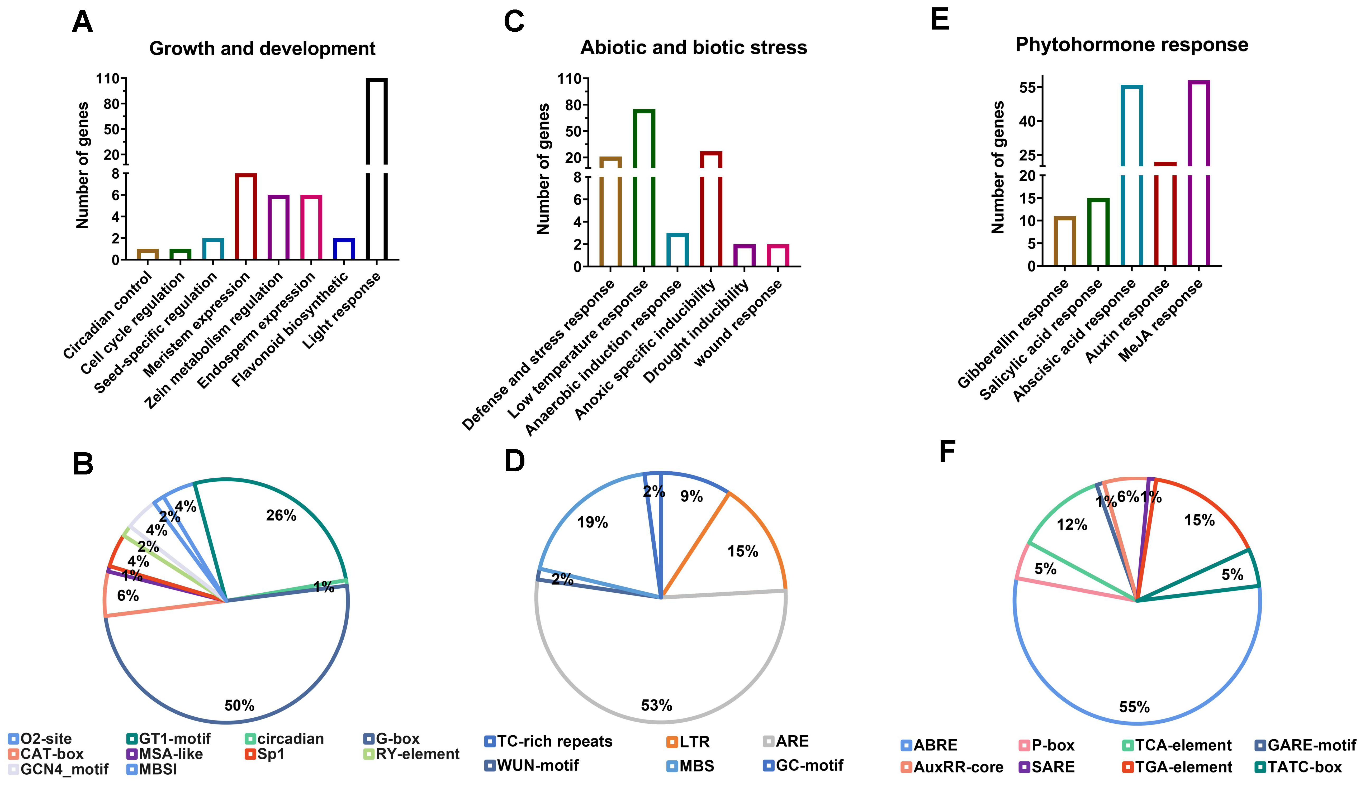
Promoters play a pivotal role in determining gene expression at the transcriptional level, with their regulation largely contingent upon cis-acting elements located upstream of the genes. Therefore, we extracted the 2000 bp upstream regions of NnCSL genes to identify their putative cis-elements (Figure 3 and Figure S1). We identified three types of cis-acting elements: growth and development-related elements, stress response elements, and hormone response elements. For growth and development-related elements (Figure 3A and Figure S1), we identified eight types of relevant elements, including light response, meristem expression, endosperm expression, and zein metabolism. The key motifs (Figure 3B) included G-box (50%), GT1 (26%), CAT-box (6%), O2-site (4%), Sp1 (4%), and GCN4 (4%). Regarding stress response elements (Figure 3C and Figure S1), we detected seven types of related elements, such as those responding to anaerobic conditions, drought, and low temperature. The key motifs (Figure 3D) included ARE (53%), MBS (19%), LTR (15%), and GC-motif (9%), with most being anaerobic response elements. We also identified five types of hormone response elements (Figure 3E and Figure S1), including MeJA, ABA, and Aux. The main motifs (Figure 3F) were ABRE (55%), TGA-element (15%), and TTCA-element (12%). Transcription factors (TFs) are essential proteins that regulate gene expression by binding to specific DNA sequences (promoter regions) and thus modulate various crucial developmental processes in plants. Accordingly, we further analyzed TF binding sites in the promoter regions of NnCSL genes (Figure S2). The analysis revealed a rich array of TF binding sites, with MYB, Dof, and C2H2 being the most abundant. MIKC_MADS, NAC, and WRKY also showed relatively high abundance. Notably, the NnCSLC4 gene had the highest number of TF binding sites, suggesting that it may have multiple functions in plant growth.

2.5. Gene Replication Events and Collinearity Analysis of CSL Family Genes

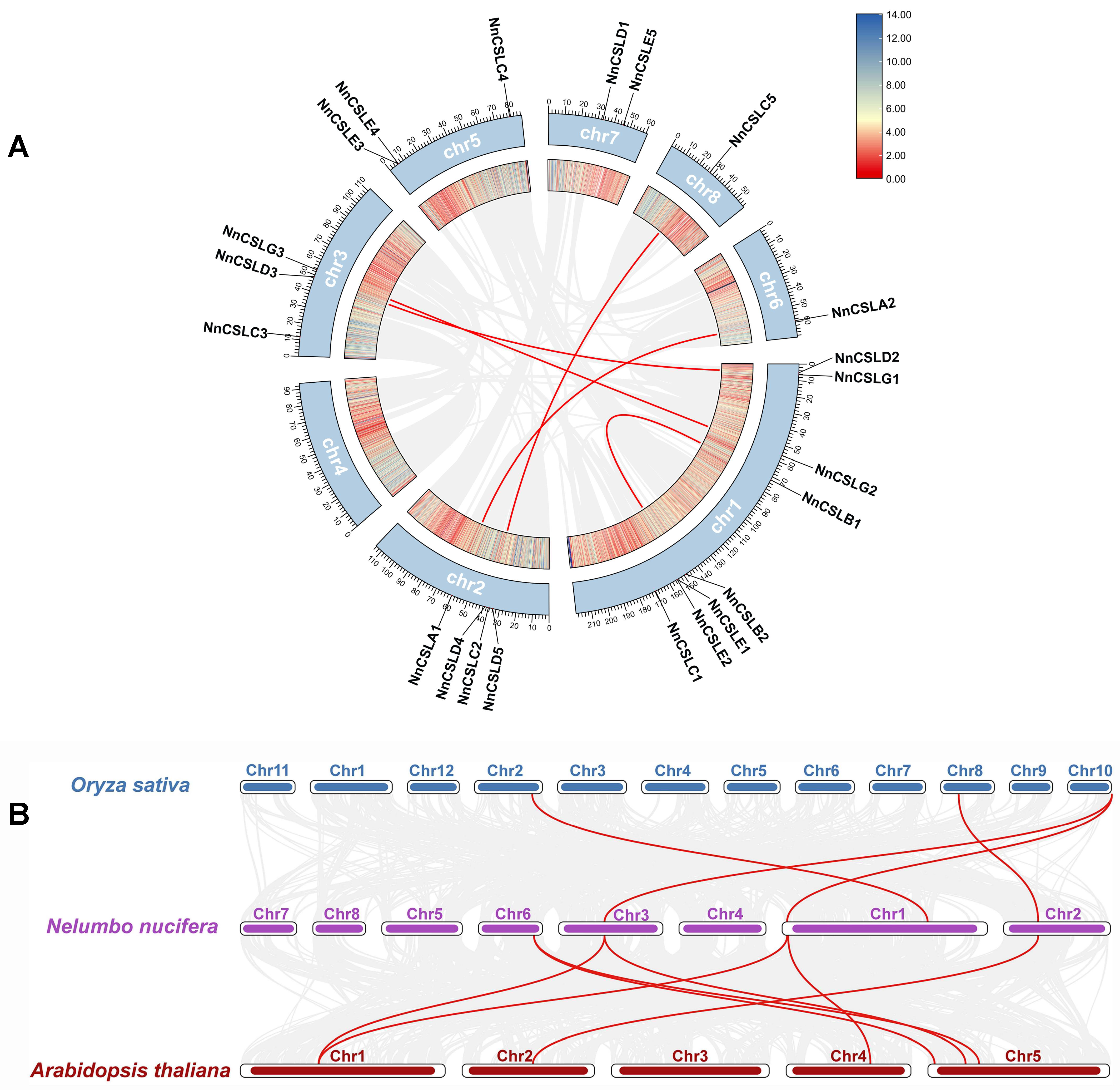
First, we conducted an intraspecific gene duplication analysis for the 22 NnCSL genes. We identified five pairs of segmental duplications on the chromosomes of N. nucifera. One pair occurred on Chr1, two pairs were found between Chr1 and Chr3, and the remaining two pairs were located between Chr2 and Chr6, and between Chr2 and Chr8, respectively. Notably, these five pairs of duplicated genes were predominantly within the same subfamilies, such as NnCSLB1 and NnCSLB2, NnCSLG2 and NnCSLG3, and NnCSLD2 and NnCSLD3 (Figure 4A). This observation suggests a certain level of conservation within the NnCSL subfamilies during the evolutionary process in the N. nucifera genome.
Subsequently, we performed comparative syntenic analyses between N. nucifera and two representative plant species (A. thaliana and O. sativa). The analysis identified seven pairs of syntenic homologous genes between N. nucifera and A. thaliana, and four pairs between N. nucifera and O. sativa. These NnCSL genes were concentrated on Chr1, Chr2, Chr3, and Chr6 in N. nucifera (Figure 4B). These findings suggest that the NnCSL genes may have similar biological functions to their homologous genes and highlight that N. nucifera exhibits more pronounced dicot characteristics, sharing a closer evolutionary relationship with A. thaliana.

2.6. Tissue Expression Patterns of NnCSL Genes in N. nucifera

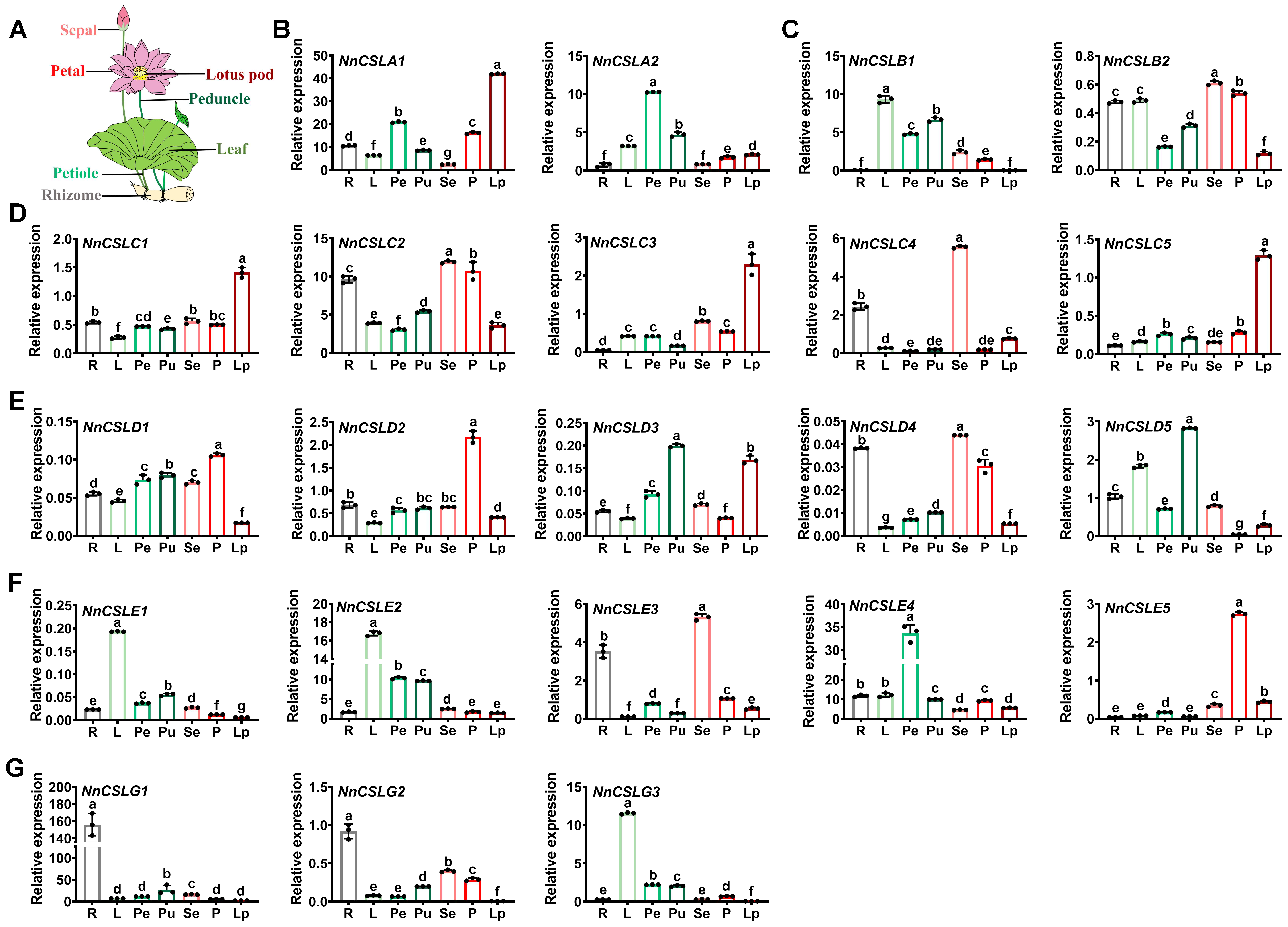
Tissue-specific expression profiles can reveal insights into gene function [65,66]. To explore the expression characteristics of NnCSL genes across various tissues, including rhizome (R), leaves (L), petioles (Pe), peduncles (Pu), sepals (Se), petals (P), and lotus pods (Lp), qRT-PCR analysis was performed. Rhizomes, leaves, and petioles are categorized as vegetative organs, whereas sepals, petals, and lotus pods are classified as reproductive organs. As illustrated in Figure 5, all 22 genes within the NnCSL family were expressed in both vegetative and reproductive organs. Specifically, genes in the NnCSLG subfamily were preferentially expressed in vegetative organs, whereas NnCSLC subfamily genes exhibited a dominant expression in reproductive organs, although they were also expressed at relatively high levels in vegetative organs. The expression patterns of NnCSLA1, NnCSLB2, NnCSLD1/2/4, and NnCSLE3/5 were similar to those of NnCSLC subfamily genes. In contrast, NnCSLA2, NnCSLB1, NnCSLD3/5, and NnCSLE1/2/4 showed the opposite trend of higher expression levels in vegetative organs.

2.7. Identification of NnCSL Genes as Candidates for Petiole Elongation in N. nucifera

Tissue expression patterns indicated that NnCSL genes were extensively involved in plant growth and development, with distinct roles in vegetative versus reproductive growth. We conducted RNA-seq on petioles from representative large and small lotus varieties (Figure 6A) to further explore the role of NnCSL genes in petiole elongation related to plant architecture. Among the 22 NnCSL genes, 18 differentially expressed genes (DEGs) were identified in the petioles of LPA (large plant architecture) versus SPA (small plant architecture) lotus varieties (Figure 6B). Notably, most DEGs, such as NnCSLC1/2/3/4/5, NnCSLA1/2, NnCSLB2, NnCSLD3/5, NnCSLG2/3, and NnCSLE1 were expressed at significantly higher levels in LPA compared to SPA. These results suggest that NnCSL genes play a crucial role in positively influencing petiole elongation.
Further validation of NnCSL genes was performed using qRT-PCR (Figure 6C–G). The results indicated that, except for NnCSLB1 and NnCSLD2, all other NnCSL genes were expressed at significantly higher levels in large lotus varieties compared to small ones. Among these, NnCSLC1/2/3/4/5, NnCSLA1/2, NnCSLB2, NnCSLD3, NnCSLD5, NnCSLG2/3, NnCSLB1, and NnCSLE1 were identified as DEGs in RNA-seq. Therefore, we conclude that, aside from NnCSLB1, which negatively impacts petiole elongation, the remaining 13 genes are candidates that positively influence petiole elongation in N. nucifera.
Co-expression analysis offers detailed insights into the transcriptional interactions between regulatory factors, which is essential for constructing a comprehensive transcriptional regulatory network [67]. This understanding is crucial for elucidating the biological functions and regulatory mechanisms of the NnCSL gene family at the protein level. Utilizing DEGs from RNA-seq, we constructed co-expression networks to investigate the relationships among 14 candidate NnCSL genes associated with petiole elongation and various TFs through a correlation-clustering analysis (Figure 6H). The analysis revealed that candidate genes positively mediating lotus petiole elongation, such as NnCSLA1/2, NnCSLC1/2/3/4/5, NnCSLD3/5, NnCSLE1, and NnCSLG2/3, were significantly positively correlated with TFs like NnNAC19/26/40, NnMYB47, NnWRKY7, and NnbHLH8/13/19/20. In contrast, NnCSLB1, which negatively affects petiole elongation, had negative correlations with these TFs. Additionally, NnCSLB1 was significantly positively correlated with NnMYB6/14, NnbHLH15, NnNAC17, and NnKNOX7/8, while NnCSLA1/2, NnCSLC1/2/3/4/5, NnCSLD3/5, NnCSLE1, and NnCSLG2/3 were negatively correlated with these TFs. These TFs, mainly from the MYB, NAC, and bHLH families, are known to be crucial in developing vegetative organs like leaves, lateral branches, and stems. Therefore, we hypothesize that NnCSL family genes regulate petiole elongation and plant architecture through a complex network involving these TFs.

2.8. Identification of NnCSL Genes as Candidates for Floral Petalization in N. nucifera

Previous studies have demonstrated that CSL family genes play a role in reproductive processes, including pollen germination and pollen tube growth. Additionally, our observations revealed that numerous NnCSL genes are specifically expressed in reproductive organs such as sepals, petals, and lotus seed pods (Figure 5B–G). To further elucidate the role of NnCSL family genes in the petalization of stamens and carpels in lotus, we conducted RNA-seq analysis on normal stamens (S), petalized stamens (SP), normal carpels (C), and petalized carpels (CP) of the lotus variety ‘Shen Nvzi’ (Figure 7A). The analysis identified 19 NnCSL genes with significant differential expression, excluding NnCSLE4 and NnCSLG1/2. Among these, 10 NnCSL genes were differentially expressed between SP and S: NnCSLC4, NnCSLD1/2/4/5, and NnCSLE2 were significantly downregulated in SP, whereas NnCSLA1, NnCSLC2, and NnCSLC5 were significantly upregulated in SP compared to S. In the comparison between CP and C, 13 differentially expressed NnCSL genes were identified: NnCSLA1/2, NnCSLB1/2, NnCSLC1/2/3/5, and NnCSLE1/2/3/5 showed significantly higher expression levels in CP, while NnCSLD3 was downregulated (Figure 7B). Notably, NnCSLA1 and NnCSLE2 were significantly upregulated in both SP and CP, with NnCSLE2 exhibiting higher expression levels in CP but lower expression levels in SP. These findings suggest that NnCSL family genes play a crucial role in the petalization of carpels and in maintaining normal stamen development.
qRT-PCR validation further confirmed that, with the exception of NnCSLC5 and NnCSLD1/3, 12 NnCSL genes were highly expressed in SP. These included NnCSLA1, NnCSLC1/2/3, NnCSLD2, NnCSLE2/3/4/5, and NnCSLG1/2/3, with expression levels ranging from 15% (NnCSLG2) to approximately 1760% (NnCSLC3) higher than in S. Among them, only NnCSLA1 and NnCSLC2 were identified as DEGs in RNA-seq. In S, 8 genes—NnCSLA2, NnCSLB1/2, NnCSLC4, NnCSLD1/4/5, and NnCSLE1—were highly expressed in S, but at levels 25% (NnCSLD1) to 100% (NnCSLD4) lower than in SP. Notably, NnCSLC4 and NnCSLD1/4/5 were also identified as DEGs in RNA-seq. Combining RNA-seq and qRT-PCR results, we identified two candidate genes (NnCSLA1 and NnCSLC2) that may positively mediate stamen petalization, and four candidate genes (NnCSLC4 and NnCSLD1/4/5) that may negatively influence this process in N. nucifera. Additionally, except for NnCSLD3/4 and NnCSLE4, all other genes exhibited significantly higher expression levels in CP. These genes—NnCSLA1/2, NnCSLB1/2, NnCSLC1/2/3/4/5, NnCSLD1/2/5, and NnCSLE1/2/3/5—exhibited expression levels in CP ranging from 24% (NnCSLB1 and NnCSLD1) to 3914% (NnCSLE2) higher than in C. The expression patterns of NnCSLA1, NnCSLB2, NnCSLC1/2/3/5, and NnCSLE2/3/5 were consistent with the RNA-seq data. NnCSLD3/4 were highly expressed in C, with expression levels more than 21% higher than in CP, and the expression pattern of NnCSLD3 matched RNA-seq data (Figure 7C–G). Based on these findings, NnCSLA1, NnCSLB2, NnCSLC1/2/3/5, and NnCSLE2/3/5 were proposed as candidate genes positively involved in carpel petalization, while NnCSLD3 is suggested as a candidate gene negatively involved in this process.
The analysis of co-expression networks involving 16 candidate NnCSL genes (NnCSLA1, NnCSLC2/4, NnCSLD1/4/5, NnCSLA1, NnCSLB2, NnCSLC1/2/3/5, NnCSLE2/3/5, and NnCSLD3) in relation to floral petalization and TFs revealed significant relationships among these genes (Figure 7H). Specifically, candidate genes involved in petalization, including NnCSLA2, NnCSLB1, NnCSLC1, and NnCSLE1/5, exhibited strong positive correlations with TFs such as NnbHLH137, NnERF027, NnNAC35, and NnAP2L4. Conversely, genes predominantly expressed in stamens, namely NnCSLC4 and NnCSLD5, displayed negative correlations with these TFs but were positively correlated with NnARF6-1, NnERF08, NnKNOX1, and NnNAC98. In carpels, the gene NnCSLD3, which is highly expressed, showed significant positive correlations with NnbZIP43-1, NnbHLH87, NnMADS6-1, and NnMADS6-3, while demonstrating predominantly negative correlations with other TFs. Other candidate genes did not show significant correlations with the selected TFs.

2.9. NnCSL Genes Responding to Salinity Stress Antagonize Growth and Development in N. nucifera

Salinity is a prominent abiotic stressor that induces osmotic stress and nutrient imbalances through excessive accumulation of Na+, K+, and Cl ions. This can lead to toxic effects during various stages of plant growth, including germination, seedling development, vegetative growth, flowering, and fruit set [68]. To examine whether NnCSL family genes exhibit similar responses, lotus seedlings were exposed to 300 mmol/L NaCl for 6 h. The results revealed that many NnCSL candidate genes, which are expected to positively impact petiole elongation and carpel petalization, actually responded negatively to salinity stress. This includes genes such as NnCSLC2/3/5, NnCSLG3, NnCSLD3/5, and NnCSLE1/5 (Figure 8). Specifically, within the NnCSLC subfamily, all genes except NnCSLC4 exhibited a significant decrease in expression after NaCl treatment, with NnCSLC2 decreasing by over 70% compared to the control. Similarly, the expression levels of NnCSLD5 and NnCSLE5 dropped by up to 93%. In contrast, genes in the NnCSLB subfamily exhibited a significant positive response to salinity stress. Notably, NnCSLB1, which is expressed relatively lower in lotus petioles of LPA, was significantly upregulated under salinity stress. Additionally, NnCSLD4, expressed at lower levels in petalized floral organs, also showed significant upregulation. These findings suggest that NnCSL genes mediate an antagonistic relationship between salinity stress and plant growth and development.

3. Discussion

As one of the multi-gene families in plants, the CSL gene family has been extensively characterized in various terrestrial species, including both dicotyledonous and monocotyledonous taxa, such as A. thaliana, cotton, P. trichocarpa, D. officinale, Z. jujuba, rice, barley, Avena sativa, and sorghum [16,19,24,26,27,53,54,57,58]. However, its presence and roles in the aquatic and ancient species, such as the lotus plant, remain under-examined. Here, we report the genome-wide identification, classification, and expression analysis of CSL family genes in lotus. Our findings revealed that 19 NnCSL genes exhibit specific differential expression patterns in LPA and SPA petioles, CP and C, and SP and S. Notably, 11 of these genes demonstrated antagonistic responses to salinity stress. Specifically, NnCSLC2, NnCSLA1, and NnCSLD3/5 play a significant and broad role in lotus responses to salinity stress, as well as in various growth and developmental processes.

3.1. Characteristics of the Lotus CSL Gene Family Indicative of Stronger Dicotyledonous Attributes

In this study, we identified 22 NnCSL genes in the lotus, a number that is fewer than those reported in the dicotyledon model Arabidopsis (30) and monocot model rice (34), given the relative genome sizes and phylogenetic positions in the species tree. Protein sequence analyses revealed that NnCSLs encompass the PF03552 and PF00535 conserved domains, consistent with the characteristics of the CSL family identified in various species, including A. thaliana [22], rice [18], Z. jujuba [26], D. officinal [24], pineapple [20], and strawberry [25]. Previous studies have indicated that CSLA, CSLC, CSLD, and CSLE are ubiquitous in both monocots and dicots, whereas CSLB, CSLG, and CSLM are dicot-specific, with CSLF, CSLH, and CSLJ being exclusive to monocots [16,18,19]. Here, we identified 22 NnCSL family genes, classified into the six subfamilies CSLA, CSLB, CSLC, CSLD, CSLE, and CSLG (Figure 1), consistent with the dicotyledon-like classification observed in Arabidopsis [15]. In contrast to rice [18], NnCSLs lack the subfamilies CSLF, CSLH, and CSLJ that are characteristic of monocots, suggesting a more pronounced dicotyledonous attribute in the ancient dicotyledonous lotus. Segmental gene duplication is the dominant factor for generating and maintaining gene families and is also considered the main source of gene structural changes and innovation [69]. Furthermore, comparative syntenic analyses among N. nucifera, A. thaliana, and O. sativa revealed seven syntenic gene pairs between N. nucifera and A. thaliana, and four pairs between N. nucifera and O. sativa (Figure 4B). This further indicates that NnCSLs exhibit stronger dicot characteristics, sharing closer evolutionary relationships with AtCSLs. Broadly, homologous genes from different species typically share similar functions [70]. Therefore, the functional study of a homologous gene can provide insights into the function of an unknown gene. Further research is essential to ascertain whether the functions of NnCSL genes are analogous to those of their AtCSL homologs.

3.2. Roles of NnCSLs in Plant Growth and Development

Studies have indicated that CSL family genes in dicotyledonous plants primarily contribute to the biosynthesis of various cell wall polysaccharides, including xyloglucan, homogalacturonan, mannan, and cellulose, thereby influencing the composition and structure of the cell wall. These genes are crucial for plant growth, development, and response to biotic and abiotic stresses [20,21,22,23,24,25,26,27,31,32]. However, the functional characterization of CSL genes in lotus remains underexplored. Tissue-specific expression profiles can elucidate gene function [65,66]. Previous research has demonstrated that genes like AtCSLD2 and AtCSLD3 in Arabidopsis [29], OsCSLD1 in rice [38], ZmCSLD1 in maize [39], and LjCSLD1 in Lotus japonicus [40] exhibit preferential expression in plant roots, crucial for root-hair development and nutrient absorption. Moreover, AtCSLD1 and AtCSLD4 in Arabidopsis are highly expressed in mature pollen, and their mutations impair cellulose deposition in pollen tube walls, affecting fertilization [71]. In tobacco, NaCSLD1 is primarily expressed during pollen maturation and tube growth, regulating flower development [1]. To investigate the role of NnCSL genes in plant growth and development, we performed tissue-specific expression analyses, revealing that 22 NnCSL genes were expressed in both vegetative and reproductive organs. Notably, genes in the NnCSLG subfamily showed preferential expression in vegetative organs, while most NnCSLC, NnCSLA, and NnCSLB subfamily genes were expressed at high levels in both reproductive and vegetative tissues. The NnCSLD and NnCSLE subfamily genes exhibited distinct expression patterns, with NnCSLD1/2/4 and NnCSLE3/5 predominantly expressed in reproductive organs, and NnCSLD3/5 and NnCSLE1/2/4 showing higher expression in vegetative organs (Figure 5).
In this study, we analyzed petioles from large and small lotus varieties as representative vegetative organs and included normal and petalized stamens and carpels as reproductive organs. RNA-seq and qRT-PCR analyses revealed that NnCSL gene expression patterns were consistent with their tissue-specific profiles (Figure 6 and Figure 7). Specifically, NnCSLG (NnCSLG2/G3) subfamily genes were predominantly expressed in larger lotus petioles, while NnCSLE (NnCSLE1/E2/E3/E5) subfamily genes were notably expressed in petalized carpels. Conversely, genes from the NnCSLC (NnCSLC1/C2/C3/C5), NnCSLA (NnCSLA1/A2), and NnCSLB (NnCSLB2) subfamilies exhibited high expression in both larger lotus petioles and petalized carpels. Notably, NnCSLA1 and NnCSLC2 were also significantly upregulated in petalized stamens. Additionally, consistent with their expression patterns in vegetative organs (Figure 5), NnCSLD3 and NnCSLD5 showed high expression in larger lotus petioles but were primarily expressed in normal stamens and carpels.
Extensive research has been conducted on the CSLA, CSLC, and CSLD subfamilies in dicotyledonous plants, while the functions of the CSLB, CSLE, and CSLG subfamilies remain relatively underexplored [22]. Notably, CSLA and CSLC genes share a high degree of homology and cluster with CSLB within the same major branch, whereas CSLD, which is closely related to CESA, forms a separate branch with greater sequence similarity to the CSLE and CSLG subfamilies (Figure 1). Our findings suggest that the NnCSLA, NnCSLC, and NnCSLB genes, which group within the same major branch, likely share similar functions related to cell wall polysaccharide synthesis, thereby playing a critical role in the entire plant growth and development period, encompassing both vegetative and reproductive growth. Existing studies indicate that CSL genes not only participate in cell wall construction during vegetative growth but also play a crucial role in the reproductive developmental stages of plants. It has been reported that the overexpression of AtCSLA2, AtCSLA7, and AtCSLA9 leads to an increase in glucomannan content in stems, which significantly impacts embryonic progression [52]. Research has also shown that CSLD genes are involved in the polarized growth of structures such as root hairs and pollen tubes, including OsCSLD1, AtCSLD3, and AtCSLD4 [38,71]. Additionally, evidence suggests that the PbrMADS52–PbrCSLD5 signaling pathway enhances cellulose content in the pear pollen tube cell wall, inhibiting pollen tube growth [35]. Collectively, our findings offer valuable insight into the potential specific roles of NnCSL genes in plant growth and development, particularly highlighting the largely unexplored functions of the CSLB, CSLE, and CSLG subfamilies. These subfamilies warrant further investigation to elucidate their specific mechanisms, contributing to a deeper understanding of the diverse roles these genes play in plant biology.

3.3. Roles of NnCSLs in Response to Salinity Stress Antagonize Growth and Development

Research has demonstrated that CSL family genes not only mediate plant growth and development but also respond to various stressors. In A. sativa, the expression of most AsCSL family genes were repressed under abiotic stress conditions [27]. Within the Orchidaceae family, the CSLA subfamily may play a crucial role in drought stress responses across different life forms, while the CSLD subfamily appears to be essential for epiphytic and saprophytic orchids to adapt to freezing stress [24]. In banana, the genes MaCSLA4/12, MaCSLD4, and MaCSLE2 are promising candidates associated with chilling tolerance [23]. In Fragaria vesca, the expression levels of numerous FveCSL genes were altered following treatment with nordihydroguaiaretic acid [25]. Moreover, CSLD proteins are important for how plants respond to environmental stresses. In Arabidopsis, the gene SOS6 encodes a protein called AtCSLD5, and its mutant allele sos6-1 is crucial for osmotic stress tolerance. Plants with the sos6-1 mutation are more sensitive to salt stress and osmotic pressure caused by mannitol or polyethylene glycol, as well as to drought conditions [47]. Similarly, knocking out the OsCSLD4 gene decreases salt and osmotic stress tolerance in rice, while overexpressing OsCSLD4 improves salt tolerance, thus highlighting its beneficial role in surviving salt stress [48].
Lotus, as a perennial aquatic herbaceous plant, confronts various environmental stresses, including salinity stress [48,63,64]. Consequently, the responsiveness of NnCSL family genes to salinity stress and their specific response patterns warrant in-depth investigation. In this study, through the prediction of cis-acting elements in the promoter regions of NnCSL genes in lotus, we identified 22 genes containing numerous abiotic stress response elements, suggesting that NnCSL genes play a pivotal role in stress responses. Further analysis of the expression changes of NnCSL genes under salinity stress revealed that eight genes—NnCSLC2/3/5, NnCSLG3, NnCSLD3/5, and NnCSLE1/5—expected to positively impact petiole elongation and carpel petalization, exhibited a negative response to salinity stress. Among them, NnCSLC2, NnCSLD5, and NnCSLE5 showed the most pronounced effects (Figure 8). Studies have shown that plants encounter abiotic stresses throughout their development, prompting the production of secondary metabolites to enhance resistance. However, this process demands considerable carbon and nutrients, diverting resources from growth and thereby decelerating development. Consequently, growth and resistance often exhibit antagonistic relationships [72]. This phenomenon could plausibly explain the antagonistic effects observed in our study, where NnCSLs response to salinity stress antagonizes growth and development. Nevertheless, the discrepancy between the antagonistic mechanism observed in NnCSL genes and the synergistic growth and resistance mediated by AtCSLD5 and OsCSLD4 [47,48] highlights species–specific diversity and complexity in stress response strategies and regulatory mechanisms. The underlying mechanisms merit further exploration. This complexity underscores the need for further exploration of the underlying mechanisms, which could provide valuable insights into the adaptive strategies employed by different species under environmental stress conditions.

3.4. Potential Complex Regulatory Networks of NnCSLs Involved in Plant Growth and Abiotic Stress Responses

Upstream TFs play a pivotal role in regulating gene expression levels, with the promoter region being essential for this process [73]. Therefore, analyzing cis-acting elements within the promoters of NnCSL genes provides an effective approach to predict their regulatory networks. To gain preliminary insights into the regulatory mechanisms of NnCSL candidate genes involved in lotus growth and abiotic stress responses, we initially examined TF binding sites within the promoter regions of NnCSL genes. The analysis uncovered a diverse array of TF binding sites, including MYB, NAC, and WRKY. Notably, the NnCSLC4 gene, which harbored the highest number of TF binding sites, showed specific high expression in larger lotus petioles (Figure 6E and Figure S2).
Furthermore, utilizing DEGs from RNA-seq analysis, we constructed co-expression networks based on identified NnCSL candidate genes associated with petiole elongation (e.g., NnCSLA1/2, NnCSLC1/2/3/4/5, NnCSLD3/5, NnCSLE1, and NnCSLG2/3) and those linked to floral petalization (e.g., NnCSLA1, NnCSLC2 and NnCSLC4, NnCSLD1/4/5; NnCSLA1, NnCSLB2, NnCSLC1/2/3/5, NnCSLE2/3/5, and NnCSLD3). Notably, TFs such as NnPIF2/3/9, NnKNOX2/4, NnMYB61/47/39/60/30/44/59/28, NnNAC40/28/26/19, bHLH8/13/20/19, and NnWRKY7 exhibited significant positive correlations with NnCSLC2/1/3/5, NnCLG2/3, and NnCSLD3, all of which were highly expressed in larger lotus petioles (Figure 6H). Conversely, most TFs exhibited negative correlations with NnCSL genes involved in petalization, with the exception of NnERF027, NnMYB06-1/C-6/S1, NnAP2L4, and NnNAC35, which positively correlated with NnCSLE1/5 and NnCLA2—genes that were predominantly expressed in petalized carpels (CP) or petalized stamens (SP). These results suggest that NnCSL plays a critical role in modulating both vegetative and reproductive growth through distinct regulatory networks mediated by specific TFs.
Numerous studies have elucidated the pivotal roles of various TFs, including NAC [74,75,76], MYB [77,78], KNOX [74,79], bHLH [80], PIF [81], WRKY [82], and ERF [83], in modulating plant growth, development, and responses to salinity stress. For instance, OsMYB110 has been identified as a direct target of PHOSPHATE STARVATION RESPONSE 2 (OsPHR2) and plays a crucial role in regulating OsPHR2-mediated suppression of rice height. The inactivation of MYB110 resulted in increased culm diameter and enhanced bending resistance, thereby improving lodging resistance despite the increase in plant height [78]. Furthermore, members of subgroup 19 (SG19) of the R2R3-MYB family have been demonstrated to play essential roles in the later stages of floral organ development and maturation. Extensive research, primarily conducted in Solanaceae and Arabidopsis, has documented the involvement of these TFs in flower opening [84,85], senescence [84], stamen development [86,87], ovule fertility [85], as well as pistil length and maturation [85,88]. In another context, the IbMYB308 gene from sweet potato (Ipomoea batatas) has been shown to enhance salt stress tolerance in transgenic tobacco [77]. Similarly, NAC1/NAC2 transcription factors from cowpea have been found to improve growth and tolerance to drought and heat in transgenic cowpea by activating photosynthetic and antioxidant mechanisms [76]. Collectively, these findings lead us to hypothesize that complex regulatory networks involving NnCSLs may exist, influencing plant growth and responses to abiotic stresses. This study, therefore, provides valuable insights that can further elucidate the biological functions and regulatory mechanisms of the NnCSL gene family, contributing to the broader understanding of plant growth and stress adaptation.

4. Materials and Methods

4.1. Plant Materials and Sample Collection

The lotus materials utilized in this study were sourced from the National Lotus Germplasm Repository at Southwest Forestry University, Kunming, China. Tissue samples, encompassing rhizomes, leaves, petioles, peduncles, sepals, petals, lotus pods, stamens, stamen petals, carpels, and carpel petals, were collected during the flowering season in the summer of 2024. Upon collection, samples were immediately flash frozen in liquid nitrogen and stored at −80 °C until further use.

4.2. Salinity Stress Treatment

For the salinity stress experiment, seedlings of the lotus variety ‘TKL 36’ were selected as the experimental material. Mature, plump seeds of uniform size were chosen, and the outer shell at the tail end was carefully trimmed. The seeds were then soaked in clean water, with the water changed 1–2 times daily. The soaking process was conducted under full light at 25–30 °C for approximately 15 days, until the first floating leaf was fully expanded. Healthy, uniformly growing seedlings were subsequently transferred to a 300 mM/L NaCl solution, with the water level maintained at about 5 cm, ensuring that the floating leaves remained fully above the water surface. After 6 h of treatment, petioles (3–5 cm below the base of the first floating leaf) were collected. Untreated lotus seedlings served as the control.

4.3. Identification and Property Analysis of NnCSLs Family Genes

(1) Data Acquisition: Download the Nelumbo genome, GFF, and protein files from the Nelumbo genome database (http://nelumbo.biocloud.net, accessed on 15 August 2024). Obtain the protein sequences of AtCSLs (Table S3) from the TAIR database (https://www.arabidopsis.org/, accessed on 15 August 2024). Acquire the protein sequences of OsCSLs (Table S4) from the Rice Genome Annotation Database (https://rice.plantbiology.msu.edu/index.shtml, accessed on 15 August 2024). (2) Initial screening with bLAST: Utilize AtCSLs and OsCSLs as seed sequences to perform a bidirectional BLAST search in TBtools, with a threshold set to 10−5, for the initial identification of NnCSL proteins. (3) HMM-based validation: Download Hidden Markov Model (HMM) configuration files for Cellulose_synt (PF03552) and Glycos_transf_2 (PF00535) from the Pfam database (http://pfam.xfam.org/, accessed on 18 August 2024). Validate the preliminarily identified NnCSLs protein candidates using these HMM models. Manually curate the sequences, removing those with incomplete domains. The remaining validated sequences represent the NnCSLs family members (Table S5). (4) Protein property analysis: Analyze the amino acid composition, isoelectric point, and molecular weight of the NnCSL proteins using the online tool Protparam (https://web.expasy.org/protparam/, accessed on 21 August 2024). (5) Structure and localization prediction: Predict the transmembrane structures of the NnCSL proteins using TMHMM-2.0 (http://www.cbs.dtu.dk/services/TMHMM/, accessed on 21 August 2024). Determine the subcellular localization of these proteins using WoLF PSORT (https://wolfpsort.hgc.jp/, accessed on 21 August 2024) and CELLO (http://cello.life.nctu.edu.tw/, accessed on 23 August 2024).

4.4. Phylogenetic Analysis

As previously described, CSL protein sequences for A. thaliana and O. sativa were sourced from the TAIR and TIGR databases, respectively, and subsequently combined with the CSL protein amino acid sequences from N. nucifera. These compiled sequences were imported into MEGA software (version 7.0) [89] for alignment. Alignment of all CSL proteins from the three species was performed using the MUSCLE algorithm. Phylogenetic analysis was then conducted using the neighbor-joining method, employing the optimal model with 1000 bootstrap replicates for reliability assessment. The resulting phylogenetic tree was visualized and edited using iTOL (https://itol.embl.de/, accessed on 18 November 2024). This streamlined method ensures clarity and consistency, enhancing the reproducibility of the phylogenetic analysis of CSL proteins across the three species.

4.5. Chromosome Localization, Gene Structure, Motif Distribution, and Conserved Domains of NnCSL Family Genes

The Gene Location Visualization function in TBtools (V2.056) was utilized to map the NnCSL family genes onto their respective chromosomes for visual representation [90] The intron–exon structures of 22 NnCSL genes were analyzed using TBtools, leveraging genomic GFF data. Next, conserved motifs within the CSL proteins were examined using the MEME Suite (https://meme-suite.org/meme/, accessed on 5 August 2024), allowing for the identification of up to 10 distinct motifs. Finally, the domain visualization of NnCSL proteins was conducted using the Batch SMART tool within TBtools.

4.6. Secondary and 3D Structure Analysis of NnCSL Family Genes

The secondary structure of the NnCSL protein was predicted using SOPMA (https://npsa-prabi.ibcp.fr/cgi-bin/npsa_automat.pl?page=/NPSA/npsa_server.html, accessed on 16 August 2024), which analyzes structural elements such as alpha helices, beta turns, random coils, and extended strands. To analyze the 3D structure of the NnCSL protein, the online tool SWISS-MODEL was employed (https://www.swissmodel.expasy.org/, accessed on 26 August 2024). Additionally, the stability of the 3D structure was assessed using PDBsum (https://www.ebi.ac.uk/msd-srv/prot_int/pistart.html, accessed on 26 August 2024).

4.7. Cis-Acting Elements Prediction and Transcription Factor Binding Sites Analysis of NnCSLs Promoter

The Fasta Extract function in TBtools was utilized to obtain a 2000 bp upstream nucleotide sequence fragment (promoter sequence) from the lotus genome file for the NnCSL genes. This fragment was then submitted to the PlantCARE website (http://bioinformatics.psb.ugent.be/webtools/plantcare/html/, accessed on 18 November 2024) and PLACE database (https://www.dna.affrc.go.jp/PLACE/?action=newplace, accessed on 20 November 2024) for the prediction and annotation of cis-acting elements in the NnCSL gene promoters, facilitating functional classification and statistical analysis. Ultimately, we utilized the binding site information predicted by the PlantCARE website to generate the graphical representations shown in Figures S1 and S2. Data visualization was conducted using TBtools and GraphPad Prism 5.0. Additionally, the 2000 bp upstream nucleotide sequence fragments of the NnCSL genes were submitted to the Plant Transcriptional Regulatory Map (https://plantregmap.gao-lab.org/index-chinese.php, accessed on 17 August 2024) for further prediction and analysis. The prediction results underwent classification and statistical analysis, with data visualization accomplished using the Graphics and Heat Map functions in TBtools.

4.8. Gene Duplication and Synteny Analyses

In TBtools, the One Step MCScanX tool [91] was utilized for synteny analysis within individual species and interspecies synteny analysis among N. nucifera, O. sativa, and A. thaliana. Subsequently, the “Advanced Circos” tool was employed to visualize the synteny map of the NnCSL family genes within each species, and the “Dual Synteny Plot for MCscanX” tool was used to visualize the interspecies synteny map.

4.9. RNA-Seq and Analysis

The Lotus petioles from LPA and SPA varieties, along with normal stamens (S), petalized stamens (SP), normal carpels (C), and petalized carpels (CP) of the lotus variety ‘Shen Nvzi’, were subjected to RNA extraction with three biological replicates. mRNA library construction and RNA-seq were performed using the DNBSEQ platform (BGI Co., Ltd., Shenzhen, China). To identify genes corresponding to the reads for each sample library, and the reads were aligned to the N. nucifera reference genome (GCF_000365185.1_Chinese_Lotus_1.1) using TopHat version 2.1.1. Gene expression levels were quantified as fragments per kilobase of exon per million fragments mapped (FPKM). Heatmaps for DEGs were generated using DEGseq version 1.60.0, applying criteria of q-value < 0.005 and |log2 (FPKM-LPA/FPKM-SPA)| > 1 or log2 (FPKM-SP/FPKM-S)| > 1 or log2 (FPKM-CP/FPKM-C)| > 1. For gene ontology (GO) term annotations, all N. nucifera genes were queried against the National Center for Biotechnology Information (NCBI) non-redundant (Nr) protein database using GOSeq version 1.58.0, with a corrected p-value threshold of <0.05. Finally, differentially expressed NnCSL genes were identified.

4.10. Nucleic Acid Isolation and qRT-PCR Analysis

Total RNA was isolated using the Eastep™ Super Total RNA Extraction Kit (Promega, Madison, WI, USA). First-strand cDNA synthesis was performed with 1 μg of total RNA in 20 μL reactions, using HiScript II Q RT SuperMix for qPCR (Vazyme, Nanjing, China) and oligo-dT (18)-MN primers following the manufacturer’s instructions. qRT-PCR was conducted with SYBR® Green Realtime PCR Master Mix-Plus (Takara, Tokyo, Japan) under the following conditions: polymerase activation for 30 s at 95 °C, followed by 40 cycles of 15 s at 95 °C, 15 s at 60 °C, and 25 s at 72 °C. The NnUBC (LOC104586755) gene served as an internal control, and gene expression was normalized to this reference gene. All primers used in these assays are listed in Table S6, and each assay was carried out with three biological replicates.

4.11. Heatmap of Enriched Correlations Between Candidate NnCSL Genes and TFs

TFs data were extracted from the transcriptome information described in Section 4.9. Correlation analysis between candidate NnCSL genes and TFs was performed using the enrichment tools available on the Metware Cloud platform (https://cloud.metware.cn, accessed on 2 September 2024). This analysis generated a correlation enrichment heatmap, which was subsequently visually enhanced using the HeatMap tool in TBtools. Pearson correlation coefficients were calculated to assess the relationships between structural genes and transcription factors, employing the filtering criteria of correlation coefficients >0.9 or <−0.9 and p < 0.05.

5. Conclusions

This study provides a comprehensive analysis of the 22 NnCSL genes identified within the whole genome of N. nucifera, which were categorized into six subfamilies: NnCSLA, NnCSLB, NnCSLC, NnCSLD, NnCSLE, and NnCSLG. Syntenic analyses revealed the genetic homologies between N. nucifera and other model plants such as A. thaliana and O. sativa, revealing not only the unique dicotyledonous characteristics of lotus but also its retention of certain monocotyledonous attributes. These findings underscore the profound implications for understanding gene conservation and variation across the plant kingdom. Beyond elucidating the structural and evolutionary relationships of these genes, the study also conducted an in-depth analysis of the promoter regions, highlighting the potential pivotal roles of NnCSL genes in plant growth and development, stress responses, and hormonal signaling pathways. Finally, we elucidated candidate NnCSL genes implicated in growth and development, specifically petiole elongation and floral petalization, as well as their responses to salinity stress in N. nucifera (Figure 9). These insights not only provide scientific evidence for the improvement of ornamental traits in lotus but also lay a foundation for the development of stress-tolerant breeding strategies. In summary, this research not only deepens our understanding of NnCSL genes in ancient dicotyledonous plants but also offers new perspectives and genetic resources for future investigations into plant gene functions and the development of novel breeding technologies.

Supplementary Materials

The following supporting information can be downloaded at: https://www.mdpi.com/article/10.3390/ijms252312531/s1.

Author Contributions

Investigation, Methodology, and Writing—Original Draft Preparation, J.Y. and J.W.; Validation, Methodology, and Software, D.Y., W.X., L.W. and S.W.; Visualization, H.Z.; Funding Acquisition and Supervision, L.C.; Conceptualization, Funding Acquisition, Project Administration, and Writing—Review and Editing, H.H. All authors have read and agreed to the published version of the manuscript.

Funding

This research was funded by the Yunnan Provincial Major Science and Technology Special Program (202202AE090028), Yunnan Agricultural Foundation Joint Project (202101BD070001-124), Yunnan Provincial High-level Talents Introduction of Young Talents Special Competitive Training Support Funds (YNQR-QNRC-2018-122), and the National Natural Science Foundation of China (32360070).

Institutional Review Board Statement

The authors declare that the collection of plant materials for this study complies with relevant institutional, national, and international guidelines and legislation.

Informed Consent Statement

Not applicable.

Data Availability Statement

The datasets supporting the results presented in this manuscript are included within the article (and its Supplementary Materials).

Acknowledgments

We thank Southwest Forestry University of College of Landscape Architecture and Horticulture Sciences for providing a platform for our experiments and all those who contributed to this article.

Conflicts of Interest

The authors declare no conflict of interest.

References

  1. Doblin, M.S.; Melis, L.D.; Newbigin, E.; Bacic, A.; Read, S.M. Pollen tubes of nicotiana alata express two genes from different B-Glucan synthase families. Plant Physiol. 2001, 125, 2040–2052. [Google Scholar] [CrossRef] [PubMed]
  2. Cosgrove, D.J. Growth of the plant cell wall. Nat. Rev. Mol. Cell Biol. 2005, 6, 850–861. [Google Scholar] [CrossRef] [PubMed]
  3. Burton, R.A.; Wilson, S.M.; Hrmova, M.; Harvey, A.J.; Shirley, N.J.; Medhurst, A.; Stone, B.A.; Newbigin, E.J.; Bacic, A.; Fincher, G.B. Cellulose synthase-like CslF genes mediate the synthesis of cell wall (1,3;1,4)-Beta-D-Glucans. Science 2006, 311, 1940–1942. [Google Scholar] [CrossRef]
  4. Hu, H.Z.; Zhang, R.; Feng, S.Q.; Wang, Y.M.; Wang, Y.T.; Fan, C.F.; Li, Y.; Liu, Z.Y.; Schneider, R.; Xia, T.; et al. Three AtCesA6-like members enhance biomass production by distinctively promoting cell growth in Arabidopsis. Plant Biotechnol. J. 2018, 16, 976–988. [Google Scholar] [CrossRef]
  5. Purushotham, P.; Ho, R.; Zimmer, J. Architecture of a catalytically active homotrimeric plant cellulose synthase complex. Science 2020, 369, 1089–1094. [Google Scholar] [CrossRef]
  6. McFarlane, H.E.; Döring, A.; Persson, S. The cell biology of cellulose synthesis. Annu. Rev. Plant Biol. 2014, 65, 69–94. [Google Scholar] [CrossRef]
  7. Scheller, H.V.; Ulvskov, P. Hemicelluloses. Annu. Rev. Plant Biol. 2010, 61, 263–289. [Google Scholar] [CrossRef]
  8. Somerville, C. Cellulose synthesis in higher plants. Annu. Rev. Cell Dev. Biol. 2006, 22, 53–78. [Google Scholar] [CrossRef] [PubMed]
  9. Wang, Y.T.; Fan, C.F.; Hu, H.Z.; Li, Y.; Sun, D.; Wang, Y.M.; Peng, L.C. Genetic modification of plant cell walls to enhance biomass yield and biofuel production in bioenergy crops. Biotechnol. Adv. 2016, 34, 997–1017. [Google Scholar] [CrossRef]
  10. Zhang, R.; Hu, Z.; Wang, Y.T.; Hu, H.Z.; Li, F.C.; Li, M.; Ragauskas, A.; Xia, T.; Han, H.Y.; Tang, J.F.; et al. Single-molecular insights into the breakpoint of cellulose nanofibers assembly during saccharification. Nat. Commun. 2023, 14, 1100. [Google Scholar] [CrossRef]
  11. Zhao, H.Q.; Wang, S.; Yang, R.J.; Yang, D.M.; Zhao, Y.J.; Kuang, J.H.; Chen, L.Q.; Zhang, R.; Hu, H.Z. Side chain of confined xylan affects cellulose integrity leading to bending stem with reduced mechanical strength in ornamental plants. Carbohydr. Polym. 2024, 329, 121787. [Google Scholar] [CrossRef]
  12. Hu, H.Z.; Zhang, R.; Zhao, Y.; Yang, J.; Zhao, H.Q.; Zhao, L.; Wang, L.; Cheng, Z.P.; Zhao, W.Y.; Wang, B.; et al. Cell wall remodeling confers plant architecture with distinct wall structure in Nelumbo nucifera. Plant J. 2024, 20, 1392–1409. [Google Scholar] [CrossRef]
  13. Somerville, C.; Bauer, S.; Brininstool, G.; Facette, M.; Hamann, T.; Milne, J.; Osborne, E.; Paredez, A.; Persson, S.; Raab, T.; et al. Toward a systems approach to understanding plant cell walls. Science 2004, 306, 2206–2211. [Google Scholar] [CrossRef] [PubMed]
  14. Mohnen, D. Pectin structure and biosynthesis. Curr. Opin. Plant Biol. 2008, 11, 266–277. [Google Scholar] [CrossRef] [PubMed]
  15. Richmond, T.A.; Somerville, C.R. The cellulose synthase superfamily. Plant Physiol. 2000, 124, 495–498. [Google Scholar] [CrossRef] [PubMed]
  16. Suzuki, S.; Li, L.G.; Sun, Y.H.; Chiang, V.L. The cellulose synthase gene superfamily and biochemical functions of xylem-specific cellulose synthase-like genes in populus trichocarpa. Plant Physiol. 2006, 142, 1233–1245. [Google Scholar] [CrossRef]
  17. Doblin, M.S.; Pettolino, F.; Bacic, A. Plant cell walls: The skeleton of the plant world. Funct. Plant Biol. 2010, 37, 357–381. [Google Scholar] [CrossRef]
  18. Wang, L.Q.; Guo, K.; Li, Y.; Tu, Y.Y.; Hu, H.Z.; Wang, B.R.; Cui, X.C.; Peng, L.C. Expression profiling and integrative analysis of the CESA/CSL superfamily in rice. BMC Plant Biol. 2010, 10, 282. [Google Scholar] [CrossRef]
  19. Little, A.; Schwerdt, J.G.; Shirley, N.J.; Khor, S.F.; Neumann, K.; O’Donovan, L.A.; Lahnstein, J.; Collins, H.M.; Henderson, M.; Fincher, G.B.; et al. Revised phylogeny of the Cellulose Synthase gene superfamily: Insights into cell wall evolution. Plant Physiol. 2018, 177, 1124–1141. [Google Scholar] [CrossRef]
  20. Cao, S.J.; Cheng, H.; Zhang, J.S.; Aslam, M.; Yan, M.; Hu, A.; Lin, L.L.; Ojolo, S.P.; Zhao, H.M.; Priyadarshani, S.V.G.N.; et al. Genome-wide identification, expression pattern analysis and evolution of the Ces/Csl gene superfamily in Pineapple (Ananas comosus). Plants 2019, 8, 275. [Google Scholar] [CrossRef]
  21. Li, G.H.; Liu, X.; Liang, Y.X.; Zhang, Y.; Cheng, X.; Cai, Y.P. Genome-wide characterization of the cellulose synthase gene superfamily in Pyrus bretschneideri and reveal its potential role in stone cell formation. Funct. Integr. Genom. 2020, 20, 723–738. [Google Scholar] [CrossRef] [PubMed]
  22. Daras, G.; Templalexis, D.; Avgeri, F.; Tsitsekian, D.; Karamanou, K.; Rigas, S. Updating insights into the catalytic domain properties of plant Cellulose Synthase (CesA) and Cellulose Synthase-like (Csl) proteins. Molecules 2021, 26, 4335. [Google Scholar] [CrossRef] [PubMed]
  23. Yuan, W.; Liu, J.; Takáč, T.; Chen, H.; Li, X.; Meng, J.; Tan, Y.; Ning, T.; He, Z.; Yi, G.; et al. Genome-wide identification of banana Csl gene family and their different responses to low temperature between chilling-sensitive and tolerant cultivars. Plants 2021, 10, 122. [Google Scholar] [CrossRef] [PubMed]
  24. Wang, Y.Z.; Zhao, K.K.; Chen, Y.; Wei, Q.Z.; Chen, X.Y.; Wan, H.J.; Sun, C.B. Species-specific gene expansion of the cellulose synthase gene superfamily in the orchidaceae family and functional divergence of mannan synthesis-related genes in Dendrobium officinale. Front. Plant Sci. 2022, 13, 777332. [Google Scholar] [CrossRef]
  25. Huang, H.X.; Zhao, S.; Chen, J.L.; Li, T.X.; Guo, G.G.; Xu, M.; Liao, S.F.; Wang, R.X.; Lan, J.Y.; Su, Y.X.; et al. Genome-wide identification and functional analysis of cellulose synthase gene superfamily in Fragaria vesca. Front. Plant Sci. 2022, 13, 1044029. [Google Scholar] [CrossRef]
  26. Niu, N.; Zhang, Y.; Li, S.J.; Meng, X.R.; Liu, M.J.; Wang, H.B.; Zhao, J. Genome-wide characterization of the cellulose synthase gene family in Ziziphus jujuba reveals its function in cellulose biosynthesis during fruit development. Int. J. Biol. Macromol. 2023, 239, 124360. [Google Scholar] [CrossRef]
  27. Zhang, S.S.; Hu, H.B.; Cui, S.M.; Yan, L.; Wu, B.; Wei, S.J. Genome-wide identification and functional analysis of the cellulose synthase-like gene superfamily in common oat (Avena sativa L.). Phytochemistry 2024, 218, 113940. [Google Scholar] [CrossRef]
  28. Bernal, A.J.; Jensen, J.K.; Harholt, J.; Sørensen, S.; Moller, I.; Blaukopf, C.; Johansen, B.; de Lotto, R.; Pauly, M.; Scheller, H.V.; et al. Disruption of ATCSLD5 results in reduced growth, reduced xylan and homogalacturonan synthase activity and altered xylan occurrence in Arabidopsis. Plant J. 2007, 52, 791–802. [Google Scholar] [CrossRef]
  29. Park, S.; Szumlanski, A.L.; Gu, F.; Guo, F.; Nielsen, E. A role for CSLD3 during cell-wall synthesis in apical plasma membranes of tip-growing root-hair cells. Nat. Cell Biol. 2011, 13, 973–980. [Google Scholar] [CrossRef]
  30. Verhertbruggen, Y.; Yin, L.; Oikawa, A.; Scheller, H.V. Mannan synthase activity in the CSLD family. Plant Signal. Behav. 2011, 6, 1620–1623. [Google Scholar] [CrossRef]
  31. Hu, H.Z.; Zhang, R.; Dong, S.C.; Li, Y.; Fan, C.F.; Wang, Y.T.; Xia, T.; Chen, P.; Wang, L.Q.; Feng, S.S.; et al. AtCSLD3 and GhCSLD3 mediate root growth and cell elongation downstream of the ethylene response pathway in Arabidopsis. J. Exp. Bot. 2018, 69, 1065–1080. [Google Scholar] [CrossRef] [PubMed]
  32. Hu, H.Z.; Zhang, R.; Tang, Y.W.; Peng, C.L.; Wu, L.M.; Feng, S.Q.; Chen, P.; Wang, Y.T.; Du, X.Z.; Peng, L.C. Cotton CSLD3 restores cell elongation and cell wall integrity mainly by enhancing primary cellulose production in the Arabidopsis cesa6 mutant. Plant Mol. Biol. 2019, 101, 389–401. [Google Scholar] [CrossRef]
  33. Yang, J.J.; Bak, G.; Burgin, T.; Barnes, W.J.; Mayes, H.B.; Peña, M.J.; Urbanowicz, B.R.; Nielsen, E. Biochemical and genetic analysis identify CSLD3 as a beta-1,4-glucan synthase that functions during plant cell wall synthesis. Plant Cell 2020, 32, 1749–1767. [Google Scholar] [CrossRef] [PubMed]
  34. Bernal, A.J.; Yoo, C.M.; Mutwil, M.; Jensen, J.K.; Hou, G.; Blaukopf, C.; Sørensen, I.; Blancaflor, E.B.; Scheller, H.V.; Willats, W.G.T. Functional analysis of the cellulose synthase-like genes CSLD1, CSLD2, and CSLD4 in tip-growing Arabidopsis cells. Plant Physiol. 2008, 148, 1238–1253. [Google Scholar] [CrossRef]
  35. Li, X.; Tang, C.; Li, X.Q.; Zhu, X.X.; Cai, Y.L.; Wang, P.; Zhang, S.L.; Wu, J.Y. Cellulose accumulation mediated by PbrCSLD5, a cellulose synthase-like protein, results in cessation of pollen tube growth in Pyrus bretschneideri. Physiol. Plant. 2022, 174, 13700. [Google Scholar] [CrossRef] [PubMed]
  36. Favery, B.; Ryan, E.; Foreman, J.; Linstead, P.; Boudonck, K.; Steer, M.; Shaw, P.; Dolan, L. LKOJAK encodes a cellulose synthase-like protein required for root hair cell morphogenesis in Arabidopsis. Genes Dev. 2001, 15, 79–89. [Google Scholar] [CrossRef]
  37. Wang, X.; Cnops, G.; Vanderhaeghen, R.; De Block, S.; Van Montagu, M.; Van Lijsebettens, M. AtCSLD3, a cellulose synthase-like gene important for root hair growth in Arabidopsis. Plant Physiol. 2001, 126, 575–586. [Google Scholar] [CrossRef]
  38. Kim, C.M.; Park, S.H.; Je, B.I.; Park, S.H.; Park, S.J.; Piao, H.L.; Eun, M.Y.; Dolan, L.; Han, C.D. OsCSLD1, a cellulose synthase-like D1 gene, is required for root hair morphogenesis in rice. Plant Physiol. 2007, 143, 1220–1230. [Google Scholar] [CrossRef] [PubMed]
  39. Penning, B.W.; Hunter, C.T.; Tayengwa, R.; Eveland, A.L.; Dugard, C.K.; Olek, A.T.; Vermerris, W.; Koch, K.E.; McCarty, D.R.; Davis, M.F.; et al. Genetic resources for maize cell wall biology. Plant Physiol. 2009, 151, 1703–1728. [Google Scholar] [CrossRef]
  40. Karas, B.J.; Ross, L.; Novero, M.; Amyot, L.; Shrestha, A.; Inada, S.; Nakano, M.; Sakai, T.; Bonetta, D.; Sato, S.; et al. Intragenic complementation at the Lotus japonicus CELLULOSE SYNTHASE-LIKE D1 locus rescues root hair defects. Plant Physiol. 2021, 186, 2037–2050. [Google Scholar] [CrossRef]
  41. Li, M.; Xiong, G.Y.; Li, R.; Cui, J.J.; Tang, D.; Zhang, B.C.; Pauly, M.; Cheng, Z.K.; Zhou, Y.H. Rice cellulose synthase-like D4 is essential for normal cell-wall biosynthesisand plant growth. Plant J. 2009, 60, 1055–1069. [Google Scholar] [CrossRef] [PubMed]
  42. Luan, W.J.; Liu, Y.Q.; Zhang, F.X.; Song, Y.L.; Wang, Z.Y.; Peng, Y.K.; Sun, Z.X. OsCD1 encodes a putative member of the cellulose synthase-like D sub-family and is essential for rice plant architecture and growth. Plant Biotechnol. J. 2011, 9, 513–524. [Google Scholar] [CrossRef] [PubMed]
  43. Yoshikawa, T.; Eiguchi, M.; Hibara, K.I.; Ito, J.I.; Nagato, Y. Rice SLENDER LEAF 1 gene encodes Cellulose Synthase-like D4 and is specifically expressed in M-Phase cells to regulate cell proliferation. J. Exp. Bot. 2013, 64, 2049–2061. [Google Scholar] [CrossRef]
  44. Li, F.C.; Xie, G.S.; Huang, J.F.; Zhang, R.; Yu, L.; Zhang, M.M.; Wang, Y.T.; LI, A.; Li, X.K.; Xia, T.; et al. OsCESA9 conserved-site mutation leads to largely enhanced plant lodging resistance and biomass enzymatic saccharification by reducing cellulose DP and crystallinity in rice. Plant Biotechnol. J. 2017, 15, 1093–1104. [Google Scholar] [CrossRef]
  45. Qiao, L.; Wu, Q.L.; Yuan, L.Z.; Huang, X.D.; Yang, Y.T.; Li, Q.Y.; Shahzad, N.; Li, H.F.; Li, W.Q. SMALL PLANT AND ORGAN 1 (SPO1) encoding a cellulose synthase-like protein D4 (OsCSLD4) is an important regulator for plant architecture and organ size in rice. Int. J. Mol. Sci. 2023, 24, 16974. [Google Scholar] [CrossRef]
  46. Gu, F.W.; Bringmann, M.; Combs, J.R.; Yang, J.Y.; Bergmann, D.C.; Nielsen, E. Arabidopsis CSLD5 functions in cell plate formation in a cell cycle-dependent manner. Plant Cell 2016, 28, 1722–1737. [Google Scholar] [CrossRef]
  47. Zhu, J.H.; Lee, B.H.; Dellinger, M.; Cui, X.P.; Zhang, C.Q.; Wu, S.; Nothnagel, E.A.; Zhu, J.K. A cellulose synthase-like protein is required for osmotic stress tolerance in Arabidopsis. Plant J. 2010, 63, 128–140. [Google Scholar] [CrossRef] [PubMed]
  48. Zhao, H.; Li, Z.X.; Wang, Y.Y.; Xiao, M.G.; Liu, H.; Quan, R.D.; Zhang, H.W.; Hang, R.F.; Zhu, L.; Zhang, Z. Cellulose synthase-like protein OsCSLD4 plays an important role in the response of rice to salt stress by mediating abscisic acid biosynthesis to regulate osmotic stress tolerance. Plant Biotechnol. J. 2022, 20, 468–484. [Google Scholar] [CrossRef] [PubMed]
  49. Liepman, A.H.; Cavalier, D.M. The CELLULOSE SYNTHASE-LIKE A and CELLULOSE SYNTHASE-LIKE C families: Recent advances and future perspectives. Front. Plant Sci. 2012, 3, 109. [Google Scholar] [CrossRef]
  50. Liepman, A.H.; Wilkerson, C.G.; Keegstra, K. Expression of cellulose synthase-like (Csl) genes in insect cells reveals that CslA family members encode mannan synthases. Proc. Natl. Acad. Sci. USA 2005, 102, 2221–2226. [Google Scholar] [CrossRef]
  51. Dhugga, K.S.; Barreiro, R.; Whitten, B.; Stecca, K.; Hazebroek, J.; Randhawa, G.S.; Dolan, M.; Kinney, A.J.; Tomes, D.; Nichols, S.; et al. Guar seed beta-mannan synthase is a member of the cellulose synthase super gene family. Science 2004, 303, 363–366. [Google Scholar] [CrossRef]
  52. Goubet, F.; Barton, C.J.; Mortimer, J.C.; Yu, X.L.; Zhang, Z.N.; Miles, G.P.; Richens, J.; Liepman, A.H.; Seffen, K.; Dupree, P. Cell wall glucomannan in Arabidopsis is synthesised by CSLA Glycosyltransferases, and influences the progression of embryogenesis. Plan. J. 2009, 60, 527–538. [Google Scholar] [CrossRef]
  53. Kim, S.J.; Chandrasekar, B.; Rea, A.C.; Danhof, L.; Durfee, S.Z.; Thrower, N.; Shepard, Z.S.; Pauly, M.; Brandizzi, F.; Keegstra, k. The synthesis of xyloglucan, an abundant plant cell wall polysaccharide, requires CSLC function. Proc. Natl. Acad. Sci. USA 2020, 117, 20316–20324. [Google Scholar] [CrossRef] [PubMed]
  54. Zhou, Y.; Gao, Y.H.; Zhang, B.C.; Yang, H.L.; Tian, Y.B.; Huang, Y.H.; Yin, C.C.; Tao, J.J.; Wei, W.; Zhang, W.K.; et al. CELLULOSE SYNTHASE-LIKE C proteins modulate cell wall establishment during ethylene-mediated root growth inhibition in rice. Plant Cell 2024, 36, 3751–3769. [Google Scholar] [CrossRef] [PubMed]
  55. Daher, F.B.; Serra, L.; Carter, R.; Jönsson, H.; Robinson, S.; Meyerowitz, E.M.; Gray, W.M. Xyloglucan deficiency leads to a reduction in turgor pressure and changes in cell wall properties, affecting early seedling establishment. Curr. Biol. 2024, 34, 2094–2106.e6. [Google Scholar] [CrossRef]
  56. Vega-Sánchez, M.E.; Verhertbruggen, Y.; Christensen, U.; Chen, X.W.; Sharma, V.; Varanasi, P.; Jobling, S.A.; Talbot, M.; White, R.G.; Joo, M.; et al. Loss of cellulose synthase-like F6 function affects mixed-linkage glucan deposition, cell wall mechanical properties, and defense responses in vegetative tissues of rice. Plant Physiol. 2012, 159, 56–69. [Google Scholar] [CrossRef] [PubMed]
  57. Kim, S.J.; Zemelis-Durfee, S.; Mckinley, B.; Sokoloski, R.; Aufdemberge, W.; Mullet, J.; Brandizzi, F. Cell- and development-specific degradation controls the levels of mixed-linkage glucan in sorghum leaves. Plant J. 2023, 116, 360–374. [Google Scholar] [CrossRef]
  58. Purushotham, P.; Ho, R.; Yu, L.; Fincher, G.B.; Bulone, V.; Zimmer, J. Mechanism of mixed-linkage glucan biosynthesis by barley cellulose synthase-like CslF6 (1,3;1,4)-β-glucan synthase. Sci. Adv. 2022, 8, eadd1596. [Google Scholar] [CrossRef]
  59. Little, A.; Lahnstein, J.; Jeffery, D.W.; Khor, S.F.; Schwerdt, J.G.; Shirley, N.J.; Hooi, M.; Xing, X.; Burton, R.A.; Bulone, V. A novel (1,4)-β-Linked glucoxylan is synthesized by members of the cellulose synthase-like F gene family in land plants. ACS Cent. Sci. 2019, 5, 73–84. [Google Scholar] [CrossRef]
  60. Lou, H.Y.; Tucker, M.R.; Shirley, N.J.; Lahnstein, J.; Yang, X.J.; Ma, C.; Schwerdt, J.; Fusi, R.; Burton, R.A.; Band, L.R.; et al. The cellulose synthase-like F3 (CslF3) gene mediates cell wall polysaccharide synthesis and affects root growth and differentiation in barley. Plant J. 2022, 110, 1681–1699. [Google Scholar] [CrossRef]
  61. Wang, Q.Z.; Zhang, X.Y. Lotus Flower Cultivars in China; China Forestry Publishing House: Beijing, China, 2005. [Google Scholar]
  62. Lin, Z.Y.; Zhang, C.; Cao, D.D.; Damaris, R.N.; Yang, P.F. The latest studies on lotus (Nelumbo nucifera)-an emerging horticultural model plant. Int. J. Mol. Sci. 2019, 20, 3680. [Google Scholar] [CrossRef] [PubMed]
  63. Li, X.; Zhang, D.; Xu, J.; Jiang, J.; Jiang, H.W. The protective effect of cold acclimation on the low temperature stress of the lotus (Nelumbo nucifera). Hortic. Sci. 2022, 49, 29–37. [Google Scholar] [CrossRef]
  64. Vartapetian, B.B.; Andreeva, I.N.; Generozova, I.P.; Polyakova, L.I.; Maslova, I.P.; Dolgikh, Y.I.; Stepanova, A.Y. Functional electron microscopy in studies of plant response and adaptation to anaerobic stress. Ann. Bot. 2003, 91, 155–172. [Google Scholar] [CrossRef] [PubMed]
  65. Guan, Y.; Zhang, Q.Y.; Li, M.H.; Zhai, J.W.; Wu, S.S.; Ahmad, S.; Lan, S.; Peng, D.H.; Liu, Z.J. Genome-wide identification and expression pattern analysis of TIFY family genes reveal Their potential roles in Phalaenopsis aphrodite flower opening. Int. J. Mol. Sci. 2024, 25, 5422. [Google Scholar] [CrossRef]
  66. Su, Y.T.; Fang, J.Y.; Zeeshan Ul Haq, M.; Yang, W.L.; Yu, J.; Yang, D.M.; Liu, Y.; Wu, Y.G. Genome-wide identification and expression analysis of the casparian strip membrane domain protein-like gene family in Peanut (Arachis hypogea L.) revealed its crucial role in growth and multiple stress tolerance. Plants 2024, 13, 2077. [Google Scholar] [CrossRef]
  67. Wu, H.P.; Zhang, R.L.; Diao, X.M. Genome-wide characterization and haplotypic variation analysis of the IDD gene family in Foxtail Millet (Setaria italica). Int. J. Mol. Sci. 2024, 25, 8804. [Google Scholar] [CrossRef]
  68. Chen, J.T.; Aroca, R.; Romano, D. Molecular aspects of plant salinity stress and tolerance. Int. J. Mol. Sci. 2021, 22, 4918. [Google Scholar] [CrossRef]
  69. Cannon, S.B.; Mitra, A.; Baumgarten, A.; Young, N.D.; May, G. The roles of segmental and tandem gene duplication in the evolution of large gene families in Arabidopsis thaliana. BMC Plant Biol. 2004, 4, 10. [Google Scholar] [CrossRef]
  70. Gabaldón, T.; Koonin, E.V. Functional and evolutionary implications of gene orthology. Nat. Rev. Genet. 2013, 14, 360–366. [Google Scholar] [CrossRef]
  71. Wang, W.; Wang, L.; Chen, C.; Xiong, G.; Tan, X.Y.; Yang, K.Z.; Wang, Z.C.; Zhou, Y.; Ye, D.; Chen, L.Q. Arabidopsis CSLD1 and CSLD4 are required for cellulose deposition and normal growth of pollen tubes. J. Exp. Bot. 2011, 62, 5161–5177. [Google Scholar] [CrossRef]
  72. Du, P.; Wang, Q.; Yuan, D.Y.; Chen, S.S.; Su, Y.N.; Li, L.; Chen, S.; He, X.J. WRKY transcription factors and OBERON histone-binding proteins form complexes to balance plant growth and stress tolerance. EMBO J. 2023, 42, e113639. [Google Scholar] [CrossRef] [PubMed]
  73. Weidemüller, P.; Kholmatov, M.; Petsalaki, E.; Zaugg, J.B. Transcription factors: Bridge between cell signaling and gene regulation. Proteomics 2021, 21, e2000034. [Google Scholar] [CrossRef] [PubMed]
  74. Zhao, Y.Q.; Song, X.Q.; Zhou, H.J.; Wei, K.L.; Jiang, C.; Wang, J.N.; Cao, Y.; Tang, F.; Zhao, S.T.; Lu, M.Z. KNAT2/6b, a class I KNOX gene, impedes xylem differentiation by regulating NAC domain transcription factors in poplar. New Phytol. 2020, 225, 1531–1544. [Google Scholar] [CrossRef]
  75. Han, K.J.; Zhao, Y.; Sun, Y.H.; Li, Y. NACs, generalist in plant life. Plant Biotechnol. J. 2023, 21, 2433–2457. [Google Scholar] [CrossRef] [PubMed]
  76. Srivastava, R.; Kobayashi, Y.; Koyama, H.; Sahoo, L. Cowpea NAC1/NAC2 transcription factors improve growth and tolerance to drought and heat in transgenic cowpea through combined activation of photosynthetic and antioxidant mechanisms. J. Integr. Plant Biol. 2023, 65, 25–44. [Google Scholar] [CrossRef]
  77. Wang, C.; Wang, L.J.; Lei, J.; Chai, S.S.; Jin, X.J.; Zou, Y.Y.; Sun, X.Q.; Mei, Y.Q.; Cheng, X.L.; Yang, X.S.; et al. IbMYB308, a sweet potato R2R3-MYB gene, improves salt stress tolerance in transgenic tobacco. Genes 2022, 13, 1476. [Google Scholar] [CrossRef]
  78. Wang, T.T.; Jin, Y.; Deng, L.X.; Li, F.; Wang, Z.Y.; Zhu, Y.Y.; Wu, Y.F.; Qu, H.Y.; Zhang, S.N.; Liu, Y.; et al. The transcription factor MYB110 regulates plant height, lodging resistance, and grain yield in rice. Plant Cell 2024, 36, 298–323. [Google Scholar] [CrossRef]
  79. Tsuda, K.; Maeno, A.; Otake, A.; Kato, K.; Tanaka, W.; Hibara, K.I.; Nonomura, K.I. YABBY and diverged KNOX1 genes shape nodes and internodes in the stem. Science 2024, 384, 1241–1247. [Google Scholar] [CrossRef]
  80. Nan, G.L.; Teng, C.; Fernandes, J.; O’Connor, L.; Meyers, B.C.; Walbot, V. A cascade of bHLH-regulated pathways programs maize anther development. Plant Cell 2022, 34, 1207–1225. [Google Scholar] [CrossRef]
  81. Gao, H.; Song, W.; Severing, E.; Vayssières, A.; Huettel, B.; Franzen, R.; Richter, R.; Chai, J.J.; Coupland, G. PIF4 enhances DNA binding of CDF2 to co-regulate target gene expression and promote Arabidopsis hypocotyl cell elongation. Nat. Plants 2022, 8, 1082–1093. [Google Scholar] [CrossRef]
  82. Wang, H.P.; Chen, W.Q.; Xu, Z.Y.; Chen, M.F.; Yu, D.Q. Functions of WRKYs in plant growth and development. Trends Plant Sci. 2023, 28, 630–645. [Google Scholar] [CrossRef] [PubMed]
  83. Garg, R.; Mahato, H.; Choudhury, U.; Thakur, R.S.; Debnath, P.; Ansari, N.G.; Sane, V.A.; Sane, A.P. The tomato EAR-motif repressor, SlERF36, accelerates growth transitions and reduces plant life cycle by regulating GA levels and responses. Plant Biotechnol. J. 2024, 22, 848–862. [Google Scholar] [CrossRef] [PubMed]
  84. Colquhoun, T.A.; Schwieterman, M.L.; Wedde, A.E.; Schimmel, B.C.; Marciniak, D.M.; Verdonk, J.C.; Kim, J.Y.; Oh, Y.; Gális, I.; Baldwin, I.T.; et al. EOBII controls flower opening by functioning as a general transcriptomic switch. Plant Physiol. 2011, 156, 974–984. [Google Scholar] [CrossRef]
  85. Schubert, R.; Dobritzsch, S.; Gruber, C.; Hause, G.; Athmer, B.; Schreiber, T.; Marillonnet, S.; Okabe, Y.; Ezura, H.; Acosta, I.F.; et al. Tomato MYB21 acts in ovules to mediate jasmonate-regulated fertility. Plant Cell 2019, 31, 1043–1062. [Google Scholar] [CrossRef]
  86. Song, S.S.; Qi, T.C.; Huang, H.; Ren, Q.C.; Wu, D.W.; Chang, C.Q.; Peng, W.; Liu, Y.L.; Peng, J.R.; Xie, D.X. The Jasmonate-ZIM domain proteins interact with the R2R3-MYB transcription factors MYB21 and MYB24 to affect jasmonate-regulated stamen development in Arabidopsis. Plant Cell 2011, 23, 1000–1013. [Google Scholar] [CrossRef] [PubMed]
  87. Battat, M.; Eitan, A.; Rogachev, I.; Hanhineva, K.; Fernie, A.; Tohge, T.; Beekwilder, J.; Aharoni, A.A. MYB triad controls primary and phenylpropanoid metabolites for pollen coat patterning. Plant Physiol. 2019, 180, 87–108. [Google Scholar] [CrossRef] [PubMed]
  88. Chopy, M.; Binaghi, M.; Cannarozzi, G.; Halitschke, R.; Boachon, B.; Heutink, R.; Bomzan, D.P.; Jäggi, L.; van Geest, G.; Verdonk, J.C.; et al. A single MYB transcription factor with multiple functions during flower development. New Phytol. 2023, 239, 2007–2025. [Google Scholar] [CrossRef]
  89. Kumar, S.; Stecher, G.; Tamura, K. MEGA7: Molecular evolutionary genetics analysis version 7.0 for bigger datasets. Mol. Biol. Evol. 2016, 33, 1870–1874. [Google Scholar] [CrossRef] [PubMed]
  90. Chen, C.J.; Wu, Y.; Li, J.W.; Wang, X.; Zeng, Z.H.; Xu, J.; Liu, Y.L.; Feng, J.T.; Chen, H.; He, Y.H.; et al. TBtools-II: A “one for all, all for one” bioinformatics platform for biological big-data mining. Mol. Plant 2023, 16, 1733–1742. [Google Scholar] [CrossRef]
  91. Wang, Y.P.; Tang, H.B.; Debarry, J.D.; Tan, X.; Li, J.P.; Wang, X.Y.; Lee, T.H.; Jin, H.Z.; Marler, B.; Guo, H.; et al. MCScanX: A toolkit for detection and evolutionary analysis of gene synteny and collinearity. Nucleic Acids Res. 2012, 40, e49. [Google Scholar] [CrossRef]
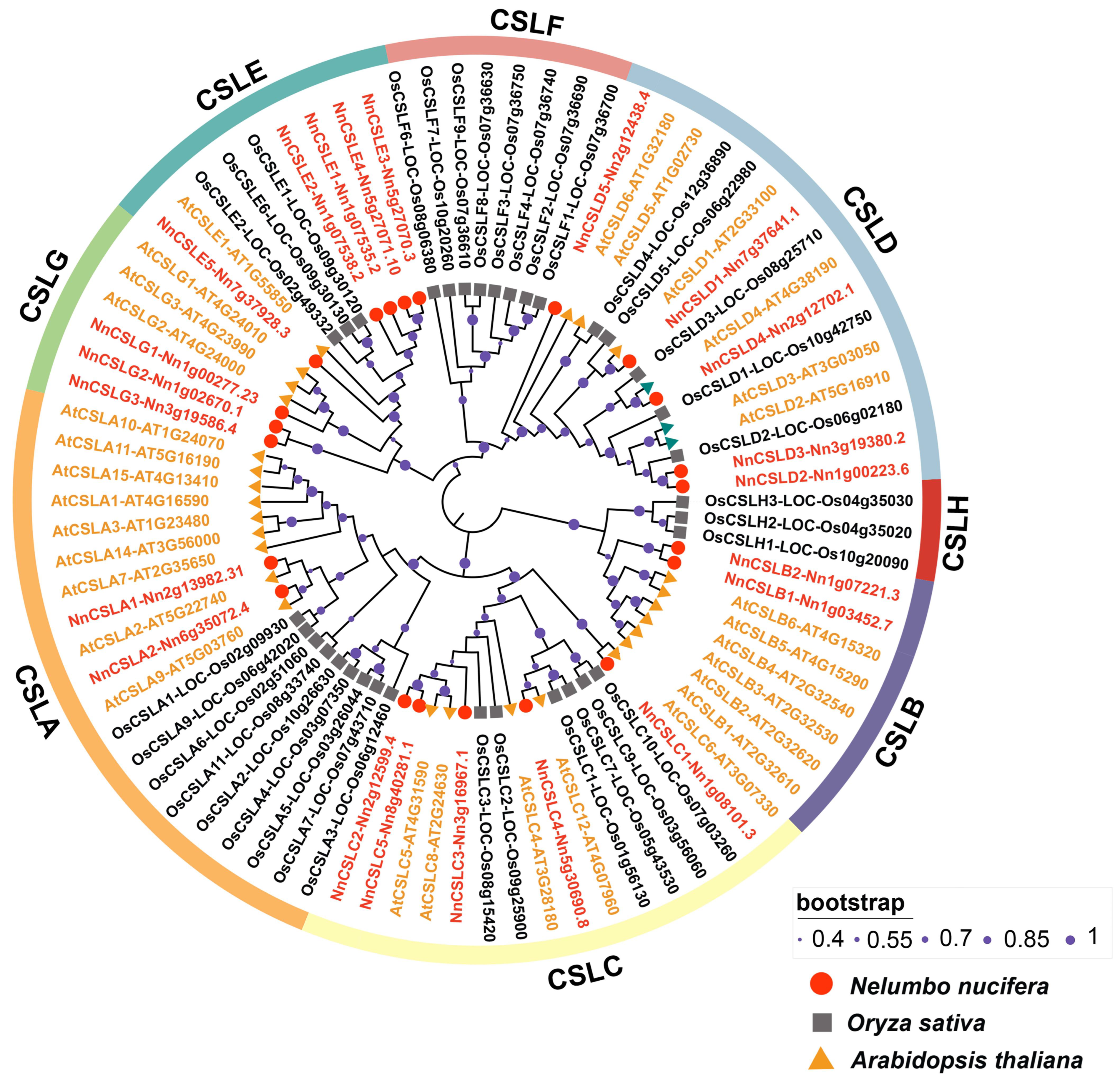
Figure 1. Phylogenetic tree of CSL proteins from N. nucifera (Nn), A. thaliana (At), and O. sativa (Os), using the neighbor-joining method. The eight CSL subfamilies (CSLA, CSLB, CSLC, CSLD, CSLE, CSLF, CSLG, CSLH) are indicated by different circle colors. Red circles denote NnCSL, orange triangles denote AtCSL, and black squares denote OsCSL. Bootstrap values are shown as colored points along the branches.
Ijms 25 12531 g001
Figure 2. Analysis of NnCSL family members: chromosome localization, conserved motifs, gene domains, gene and protein structure. (A) Chromosome localization of 22 NnCSL genes. The scale provided represents the chromosome size (Mbp). (B) Motif composition of NnCSL genes, with distinct motifs shown as colored boxes, as indicated in the scheme on the right. (C) Conserved domain, represented by different color boxes. (D) Exon–intron structure of NnCSL genes, with UTRs and CDS indicating untranslated regions and coding sequences, respectively. (EJ) Secondary and 3D structure predictions for 22 NnCSL proteins, AtCSLA9, and AtCSLD4. The top images display the 3D structures, while the bottom images show the secondary structures. In the secondary structure diagrams, blue, purple, and brown represent alpha helices, extended strands, and random coils, respectively. In the 3D structure visualizations, blue indicates a consistency greater than 70%, yellow represents a consistency between 60% and 70%, and red signifies a consistency of less than 50%.
Ijms 25 12531 g002
Figure 3. Analysis of cis-regulatory elements in the NnCSL promoter regions. (A,C,E) Counts of NnCSL genes associated with growth and development, abiotic and biotic stresses, and phytohormone responses. (B,D,F) Pie charts depicting the proportion of different cis-elements in each category: growth and development as shown in (A), abiotic and biotic stresses as shown in (C), and phytohormone responses as shown in (E). Different colors in the pie charts represent various cis-regulatory elements and their proportions in NnCSL genes.
Ijms 25 12531 g003
Figure 4. Gene duplication, and synteny of NnCSL genes. (A) Interchromosomal relationships with red lines indicating segmental gene duplications. (B) Synteny analysis of CSL genes among N. nucifera, O. sativa, and A. thaliana, with red lines highlighting syntenic regions.
Ijms 25 12531 g004
Figure 5. Tissue-specific expression of 22 NnCSL genes in N. nucifera. R, L, Pe, Pu, Se, P, and Lp represent rhizome, leaves, petioles, peduncles, sepals, petals, and lotus pod, respectively. (A) Schematic of the lotus plant showing the tissues analyzed. (BG) qRT-PCR analysis of 22 NnCSL gene expressions in different tissues. Bars represent means ± SD (n = 3 biological replicates, with individual data points shown). Different letters indicate significant differences (p < 0.05) based on one-way ANOVA followed by Tukey’s test. NnUBC served as the internal control with its expression value normalized to 100.
Ijms 25 12531 g005
Figure 6. Expression patterns of NnCSL genes in petioles of LPA (large plant architecture) and SPA (small plant architecture) lotus varieties. (A) Phenotypic comparison of LPA and SPA lotus varieties. (B) RNA-seq analysis showing differentially expressed NnCSL genes in petioles of LPA and SPA varieties, with expression levels reported as normalized log2 FPKM values. (CG) qRT-PCR analysis of NnCSL gene expression in petioles of LPA and SPA varieties, with NnUBC as the internal control (normalized to 100). Data represent means ± SD of three biological replicates. * and ** indicate significant differences between LPA and SPA varieties (t-test, p < 0.05 or p < 0.01, n = 3). Percentage changes relative to SPA are shown; ns denotes not significant. (H) Heatmap of enriched correlations between petiole elongation-related candidate NnCSL genes and TFs. Green for positive correlation and brown for negative correlation, where darker colors represent stronger correlations.
Ijms 25 12531 g006
Figure 7. Expression patterns of NnCSL genes in floral petalization-related tissues. (A) Phenotypic observation of floral structures in duplicate-petalled N. nucifera. S, SP, C, and CP denote stamens, stamen petals, carpels, carpel petals, respectively. (B) RNA-seq analysis showing differentially expressed NnCSL genes related to floral petalization, with expression levels reported as normalized log2 FPKM values. (CG) qRT-PCR analysis of NnCSL gene expression in floral tissues, with NnUBC as the internal control (normalized to 100). Data represent means ± SD of three biological replicates. * and ** indicate significant differences between SP and S, or CP and C tissues (t-test, p < 0.05 or p < 0.01, n = 3). Percentage changes relative to S or C are shown; ns denotes not significant. (H) Heatmap of enriched correlations between floral petalization-related candidate NnCSL genes and TFs. Blue for positive correlation and pink for negative correlation, where darker colors represent stronger correlations.
Ijms 25 12531 g007
Figure 8. Expression patterns of the NnCSL family genes under 300 mM/L NaCl treatment for 6 hours (h). (A) Morphological comparison of lotus seedlings treated with 300 mM/L NaCl for 6 h. (BG) qRT-PCR analysis of NnCSL gene expression in NaCl-treated seedlings versus control (CK), with NnUBC as the internal control (normalized to 100). Data represent means ± SD of three biological replicates. * and ** indicate significant differences between NaCl treatment and CK (t-test, p < 0.05 or p < 0.01, n = 3). Percentage changes relative to CK are shown; ns denotes not significant.
Ijms 25 12531 g008
Figure 9. Hypothetical model of candidate NnCSL genes related to growth and development (petiole elongation, stamen petalization, and carpel petalization) and response to salinity stress. The model also highlights which NnCSL genes exhibit contrasting expression patterns in response to salinity stress compared to their roles in growth and development.
Ijms 25 12531 g009
Table 1. The information of the CSL family genes in N. nucifera.
Gene NameGene IDLOCORF (bp)AA(aa)Mw (KDa)PIInstability IndexAliphatic IndexGRAVYTMHsSubcellular Localization
NnCSLA1Nn2g13982.31104612509163854661.9068.8443.55101.390.2085Plasma membrane
NnCSLA2Nn6g35072.4104587567160253361.239.1136.1399.770.1555Plasma membrane
NnCSLB1Nn1g03452.7104588296230476785.9236.4745.696.620.0678Plasma membrane
NnCSLB2Nn1g07221.3104594052210069978.9885.8645.2497.870.0282Plasma membrane
NnCSLC1Nn1g08101.3104589898207969279.6868.6343.58101.010.0225Plasma membrane
NnCSLC2Nn2g12599.4104612365208569479.7618.7138.34102.850.0966Plasma membrane
NnCSLC3Nn3g16967.1104595557198966275.8469.1337.11106.190.1945Plasma membrane
NnCSLC4Nn5g30690.8104609129200166676.1628.2939.495.440.0656Plasma membrane
NnCSLC5Nn8g40281.1104605331210069980.6298.8136.45100.30.0726Plasma membrane
NnCSLD1Nn7g37641.110459837832491082120.3156.8743.6380.6−0.2366Plasma membrane
NnCSLD2Nn1g00223.610460480334441147129.0676.8445.1180.94−0.2118Plasma membrane
NnCSLD3Nn3g19380.210461145134561151129.1226.6442.2682.17−0.1896Plasma membrane
NnCSLD4Nn2g12702.110459913533781125126.1996.642.7379.68−0.2158Plasma membrane
NnCSLD5Nn2g12438.41046032422769938104.6428.2437.6483.73−0.168Plasma membrane
NnCSLE1Nn1g07535.2104594270202867577.4077.4739.8385.93−0.028Plasma membrane
NnCSLE2Nn1g07538.2104594272230776887.1768.4345.7792.860.0726Plasma membrane
NnCSLE3Nn5g27070.3104594007142547453.7948.9243.0596.880.074Plasma membrane
NnCSLE4Nn5g27071.10109114471219973283.928.5940.4491.24−0.0446Plasma membrane
NnCSLE5Nn7g37928.3104609477236478789.3318.6740.4186.61−0.0688Plasma membrane
NnCSLG1Nn1g00277.23104604823225975285.0348.745.8387.650.0757Plasma membrane
NnCSLG2Nn1g02670.1104610102203467776.5888.7351.21102.540.1898Plasma membrane
NnCSLG3Nn3g19586.4104600820213070980.8287.5340.8295.560.1368Plasma membrane
Disclaimer/Publisher’s Note: The statements, opinions and data contained in all publications are solely those of the individual author(s) and contributor(s) and not of MDPI and/or the editor(s). MDPI and/or the editor(s) disclaim responsibility for any injury to people or property resulting from any ideas, methods, instructions or products referred to in the content.