Article

Hypoxia-Inducible Factor-2α Promotes Liver Fibrosis by Inducing Hepatocellular Death

by
Raja Gopal Reddy Mooli
1,*,
Dhanunjay Mukhi
1,
Mikayla Watt
1,
Veerababu Nagati
1,
Sara M. Reed
1,
Nikita K. Gandhi
1,
Michael Oertel
2,3,4 and
Sadeesh K. Ramakrishnan
1,2,5,*
1
Division of Endocrinology and Metabolism, Department of Medicine, University of Pittsburgh, Pittsburgh, PA 15261, USA
2
Pittsburgh Liver Research Center, University of Pittsburgh, Pittsburgh, PA 15261, USA
3
Division of Experimental Pathology, Department of Pathology, University of Pittsburgh, Pittsburgh, PA 15261, USA
4
McGowan Institute for Regenerative Medicine, University of Pittsburgh, Pittsburgh, PA 15261, USA
5
Hillman Cancer Center, University of Pittsburgh, Pittsburgh, PA 15261, USA
*
Authors to whom correspondence should be addressed.
Int. J. Mol. Sci. 2024, 25(23), 13114; https://doi.org/10.3390/ijms252313114
Submission received: 3 November 2024 / Revised: 2 December 2024 / Accepted: 3 December 2024 / Published: 6 December 2024

Abstract

:
The activation of hypoxia-inducible factors (HIF)-1α and 2α in the liver is closely linked to the progression of fatty liver diseases. Prior studies indicated that disrupting hepatocyte HIF-2α attenuates diet-induced hepatic steatosis, subsequently decreasing fibrosis. However, the direct role of hepatocyte HIF-2α in liver fibrosis has not been addressed. Hepatic HIF-2α expression was examined in mouse model of carbon tetrachloride (CCl4)-induced liver fibrosis. Conditional hepatocyte Hif-2α knockout mice were employed to investigate the role of hepatocyte HIF-2α in fibrosis. Markers of apoptosis, proliferation, inflammation, and fibrosis were assessed through biochemical, molecular, and histological analyses. We found an induction of HIF-2α in CCL4-injected liver injury and fibrosis mouse models. Hepatocyte-specific deletion of HIF-2α attenuated stellate cell activation and fibrosis, with no significant difference in inflammation. Disrupting hepatocyte HIF-2α led to reduced injury-mediated hepatocellular apoptosis. Surviving hepatocytes exhibited hypertrophy, which was strongly associated with the activation of c-JUN signaling. Our study demonstrates a direct role of hepatocyte HIF-2α in liver fibrosis by promoting hepatocyte apoptosis. The reduction in apoptosis and induction of hepatocyte hypertrophy following HIF-2α disruption is closely linked to enhanced c-JUN signaling, a survival mechanism in response to liver injury. These findings highlight HIF-2α as a potential therapeutic target for liver fibrosis.

1. Introduction

Liver fibrosis can progress to cirrhosis and is responsible for up to 40% of deaths in developed countries [1,2]. It is characterized by the excessive accumulation of the extracellular matrix (ECM), primarily crosslinked collagens of type I and type III, which disrupts liver structure and function [2,3]. Fibrosis is initiated by the activation of hepatic stellate cells (HSCs) and portal fibroblasts, leading to the proliferation of ECM-expressing myofibroblasts [3,4]. HSCs are also activated by mitogens, such as platelet-derived growth factor and transforming growth factor-β (TGF-β) [3,5]. Additionally, inflammatory immune cells, predominantly macrophages, are the major source of TGF-β, playing a critical role in HSC activation and fibrosis [6,7,8]. Moreover, hepatocytes are the primary cells affected during liver injury, leading to changes in their gene and secretion profiles, which can promote fibrosis [9,10]. For example, the expression of hepatocyte-enriched transcription factors and genes was repressed in injured liver, and they release intracellular molecules known as damage-associated molecular patterns (DAMPs) that trigger fibrosis [11,12]. Excessive wound healing due to uncontrolled hepatocyte death can result in fibrogenesis [13]. On the other hand, persistent liver injury triggers the compensatory proliferation of surviving hepatocytes, potentially leading to carcinogenesis [14]. The factors that influence hepatocyte survival mechanisms during injury and fibrosis remain unclear, highlighting the need to understand these mechanisms to identify novel targets for liver fibrosis.
Hypoxia signaling, mediated by the transcription factors hypoxia-inducible factor (HIF)-1α and HIF-2α [15], plays a crucial role in liver injury [16,17]. HIFs are heterodimeric proteins composed of an oxygen-sensitive subunit (HIF-α) and an oxygen-insensitive subunit (HIF-β). Under normoxia, prolyl hydroxylase domain (PHD) enzymes hydroxylate HIF-α at specific proline residues using oxygen as a substrate. Hydroxylated HIF is then ubiquitinated by the Von Hippel Lindau (VHL) complex, an E3 ubiquitin ligase, and is then subsequently degraded in the proteasome [18,19]. HIF-α is also regulated by oxygen-independent mechanisms, such as tricarboxylic acid (TCA) metabolites, inflammatory cytokines, and reactive oxygen species (ROS) [20,21]. Elevated levels of HIF-1α and HIF-2α are associated with liver dysfunction in both alcoholic and metabolic dysfunction-associated steatotic liver diseases [22]. Mouse models of bile duct ligation and carbon tetrachloride (CCl4) combined with moderate alcohol feeding show that HIF-1α promotes liver fibrosis [22,23]. However, patients with hepatitis and cholestasis, express similar levels of HIF-1α and HIF-2α [24]. Genetic models with HIF-2α overexpression in hepatocytes show a significant dysregulation of lipid homeostasis leading to spontaneous steatosis and hypercholesterolemia, culminating in liver fibrosis [25,26]. Consistently, deletion of HIF-2α in hepatocytes attenuates diet-induced hepatic steatosis and fibrosis [27]. However, it is difficult to discern whether the attenuation of liver fibrosis is secondary to lower steatosis or a direct effect of hepatocyte HIF-2α [27]. Lentiviral-mediated knockdown of HIF-2α in the liver attenuated CCl4-induced liver fibrosis [24]. However, lentiviral vectors non-specifically target all cell types, including hepatocytes. Thus, the causal role of hepatocyte HIF-2α in liver fibrosis remains elusive.
In this study, we investigated the role of hepatocyte HIF-2α in liver fibrosis using a CCl4-induced liver injury mouse model. Temporal deletion of HIF-2α in hepatocytes reduced the expression of fibrotic markers independent of inflammation. Hepatocyte-specific deletion of HIF-2α attenuated hepatocyte death and resulted in hepatocyte hypertrophy and was strongly associated with elevated levels of c-Jun, a pro-survival signaling mechanism crucial for hepatocyte survival and liver regeneration.

2. Results

2.1. HIF-2α Expression Is Upregulated in Acute Liver Injury and Fibrosis

Acute liver injury induces hypoxia signaling mediated by HIF-1α and HIF-2α [28,29]. Our analysis showed that liver injury induced the expression of HIF-2α as early as 4 h post-injury (Figure 1A,B). Using immunostaining, we show that HIF-2α stabilization occurred in cells with prominent nuclei, which are likely hepatocytes (Figure 1C). These data indicate that HIF-2α is upregulated in liver injury, raising the possibility that it may play a role in liver fibrosis.

2.2. Hepatocyte-Specific HIF-2α Inhibition Protects Against Liver Fibrosis

To test our hypothesis that HIF-2α induction in hepatocytes promotes fibrosis independent of steatosis, we used mice with temporal disruption of HIF-2α, specifically in hepatocytes (Hif-2αF/F;Alb-Cre-ERT2) [30]. One week after tamoxifen treatment, mice were administered CCl4 twice a week for 7 weeks (Figure 2A). As expected, HIF-2a expression was strongly induced in CCl4-treated Hif-2αF/F mice but not in Hif-2αF/F;Alb-Cre-ERT2 mice (Figure 2B). Histological analysis using Sirius red and Masson’s trichrome staining showed a significant reduction in liver fibrosis in Hif-2αF/F;Alb-Cre-ERT2 mice compared to Hif-2αF/F mice (Figure 2C). Similarly, Hif-2αF/F;Alb-Cre-ERT2 mice showed reduced lipid accumulation and fibrosis in a diet-induced fibrosis model (Supplementary Figure S1).
HSCs transdifferentiate into myofibroblast-like cells, which rapidly proliferate and secrete numerous pro-fibrotic molecules around the area of injury [31,32]. Among these molecules, alpha-smooth muscle actin (α-SMA) serves as a marker of HSC activation [25]. Immunohistochemical analysis showed increased expression of α-SMA in the livers of Hif-2αF/F mice, whereas its expression was significantly downregulated in Hif-2αF/F;Alb-Cre-ERT2 mice (Figure 2D). Additionally, collagen type III A (COL3A) expression was elevated in the livers of Hif-2αF/F mice but not in Hif-2αF/F;Alb-Cre-ERT2 mice (Figure 2D). Similarly, Western blot analysis showed increased protein levels for α-SMA and collagen type 1A (COL1A) in the livers of Hif-2αF/F mice but not in Hif-2αF/F;Alb-Cre-ERT2 mice (Figure 2E). Furthermore, mRNA levels of α-Sma were significantly reduced in CCl4-treated Hif-2αF/F;Alb-Cre-ERT2 mice (Figure 2F). However, no differences were observed in the mRNA levels of Col1a1, Col4a1, and plasminogen activator inhibitor-1 (Pai-1) between Hif-2αF/F and Hif-2αF/F;Alb-Cre-ERT2 mice (Figure 2F). To further investigate whether HIF-2α in HSCs modulates fibrosis, we treated the HSC cell line LX2 with TGFβ in the presence or absence of hypoxia. An increase in HIF-2α in hypoxia-exposed LX2 cells did not modulate TGFβ-mediated induction of COL1A1 at the protein level (Supplementary Figure S2). Thus, our data reveal that hepatocyte HIF-2α regulates hepatic stellate cell activation and fibrosis.

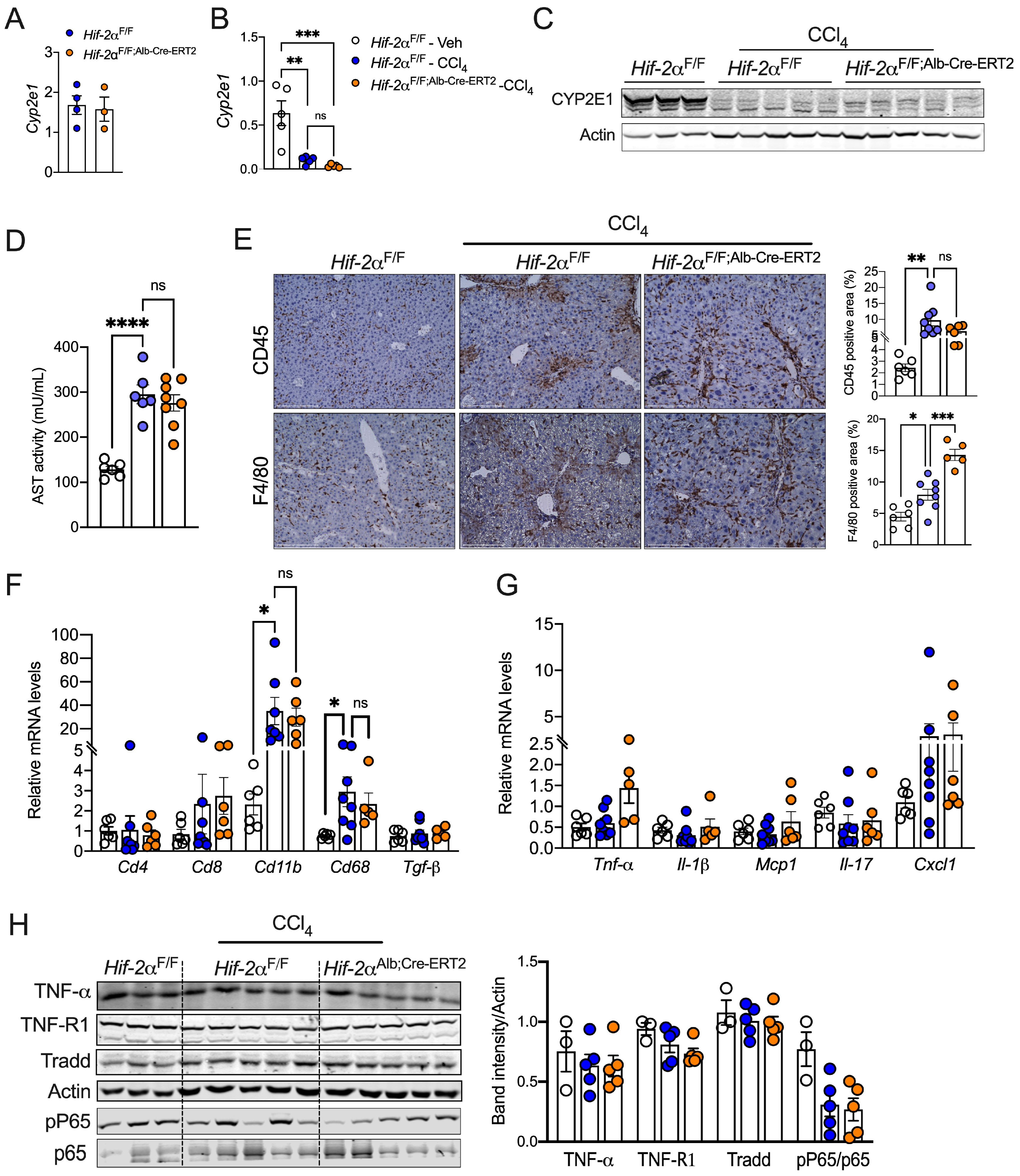
2.3. HIF-2α Induces Fibrosis Independent of Inflammation

Next, we sought to determine how the disruption of hepatocyte HIF-2α attenuates liver fibrosis. The enzyme cytochrome P450 2E1 (CYP2E1) metabolizes CCl4, and Cyp2e1 KO mice are resistant to CCl4-induced hepatotoxicity [33]. Given that hypoxia represses cytochrome P450 activity and expression [34], we assessed whether HIF-2α regulates liver fibrosis by modulating xenobiotic metabolism. We observed no difference in Cyp2e expression between Hif-2αF/F and Hif-2αF/F;Alb-Cre-ERT2 mice assessed under steady-state conditions (Figure 3A). Moreover, CCl4 treatment reduced the mRNA and protein levels of CYP2E1 in both Hif-2αF/F and Hif-2αF/F;Alb-Cre-ERT2 mice (Figure 3B,C), suggesting that HIF-2α may not alter CCl4 metabolism. Additionally, we observed no difference in serum aspartate transaminase (AST) levels, indicating that the decreased fibrosis is not due to improved liver damage (Figure 3D).
Inflammatory cells are critical drivers of liver fibrosis [35]. We assessed whether reduced fibrosis in Hif-2αF/F;Alb-Cre-ERT2 mice is driven by attenuated inflammatory response. Immunostaining revealed no difference in the number of CD45+ cells between Hif-2αF/F and Hif-2αF/F;Alb-Cre-ERT2 mice (Figure 3E). Paradoxically, F4/80+ cells were higher in Hif-2αF/F;Alb-Cre-ERT2 mice, indicating an increase in macrophages (Figure 3E). qPCR analysis showed an increase in the mRNA levels of Tnf-α in Hif-2αF/F;Alb-Cre-ERT2 mice; however, the expression of other cytokines and macrophage markers was not different between the two groups (Figure 3F,G). Activation of inflammatory pathways, such as NF-kB and TNF-R1, induces a number of pro-fibrotic factors [36,37]. However, we did not observe any changes between the genotypes (Figure 3H). Together, the data suggest that attenuated fibrosis observed in the Hif-2αF/F;Alb-Cre-ERT2 mice is not due to a decrease in hepatic inflammation.

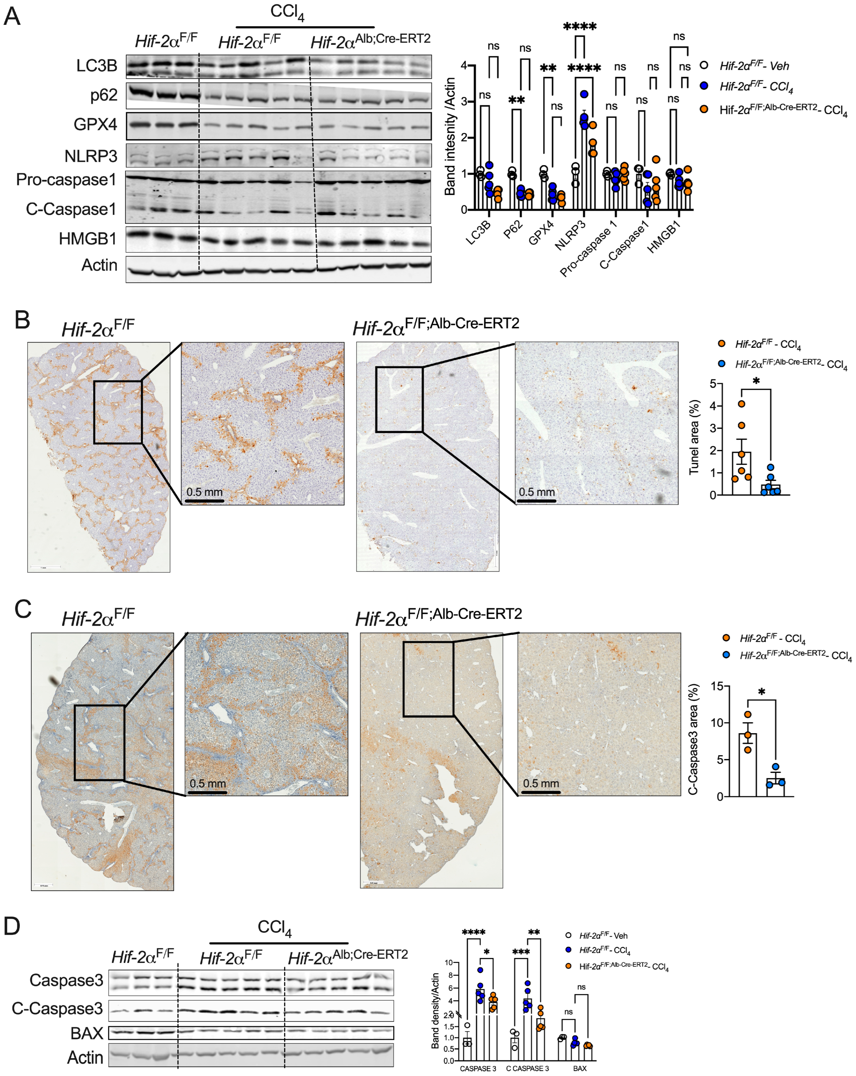
2.4. HIF-2α Deletion Prevents Hepatocyte Apoptosis in Fibrotic Livers

Apoptosis of injured hepatocytes significantly contributes to liver fibrosis [38]. Moreover, necroptosis, pyroptosis, ferroptosis, and autophagy are dysregulated in injury-mediated liver fibrosis and NASH [39,40,41,42]. In our study, hepatocyte-specific deletion of HIF-2α did not affect markers of pyroptosis (NLRP3, cleaved caspase-1), autophagy (p62 and LC3 II/I), and ferroptosis (GPX4) (Figure 4A). To investigate whether HIF-2α influences hepatocyte cell death, we performed terminal deoxynucleotidyl transferase dUTP nick-end labeling (TUNEL) staining. Our results showed increased TUNEL-positive cells in CCl4-treated fibrotic livers from Hif-2αF/F mice, indicating apoptotic cell death. However, the number of apoptotic cells was significantly reduced in Hif-2αF/F;Alb-Cre-ERT2 mice (Figure 4B). Immunohistochemistry for cleaved caspase 3 revealed a similar increase in apoptosis in Hif-2αF/F mice but not in Hif-2αF/F;Alb-Cre-ERT2 mice (Figure 4C). Additionally, Western blot analysis showed decreased protein levels of total caspase 3 and cleaved caspase 3 in Hif-2αF/F;Alb-Cre-ERT2 mice compared to Hif-2αF/F mice, but no change in BAX expression was observed (Figure 4D). These results suggest that hepatocyte HIF-2a promotes hepatocyte apoptosis.

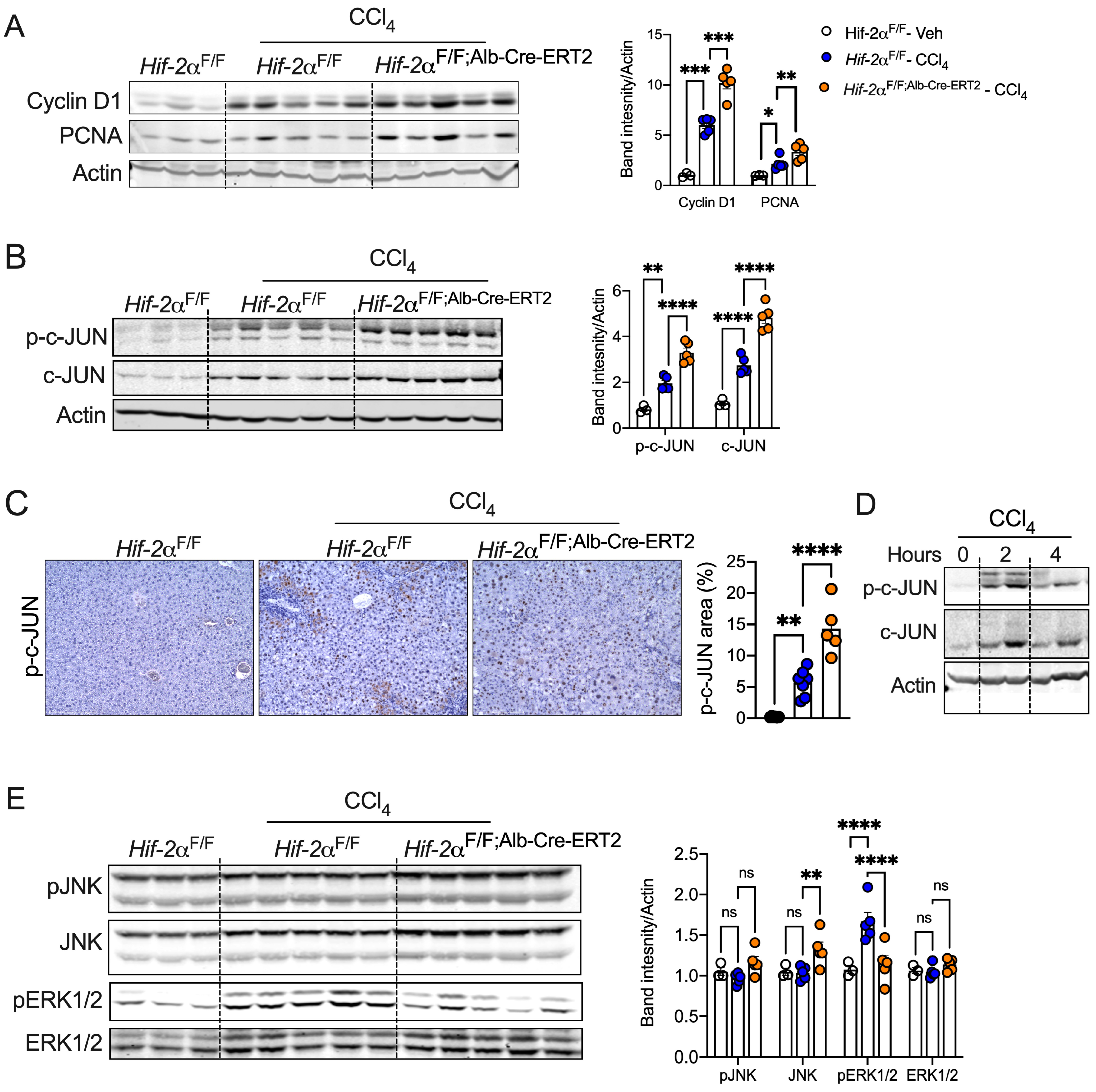
2.5. Disruption of HIF-2α Enhances Hepatocyte Survival and c-JUN Activation

Hepatocyte survival and proliferation are transcriptionally regulated, protecting against injury-mediated fibrosis and inflammation [43,44]. We observed a significant increase in the protein levels of cyclin D1 and PCNA in CCl4-treated Hif-2αF/F;Alb-Cre-ERT2 mice compared to Hif-2αF/F mice (Figure 5A). Cyclin D1 and PCNA expression are regulated by the transcription factor c-Jun, which is crucial for the survival of injured hepatocytes [45,46,47,48]. Western blot analysis revealed increased expression of c-JUN and phosphorylated c-JUN protein levels in chronic CCl4-treated Hif-2αF/F;Alb-Cre-ERT2 mice compared to Hif-2αF/F mice (Figure 5B). c-JUN elicits discordant effects on liver injury depending on the cell type. For instance, activation of c-JUN in hepatic stellate cells or myofibroblasts induces fibrosis, whereas, in hepatocytes, c-JUN promotes survival and regeneration [48,49,50,51,52]. Immunostaining revealed intense phospho-c-JUN staining in the hepatocytes of CCl4-treated Hif-2αF/F;Alb-Cre-ERT2 mice (Figure 5C). In addition to chronic CCl4 treatment models, acute treatment also elevated c-JUN phosphorylation in Hif-2αF/F;Alb-Cre-ERT2 mice compared to Hif-2αF/F mice (Figure 5D). c-JUN expression and activation are modulated by upstream kinases, such as c-JUN N-terminal kinase (JNK) and extracellular regulated kinase (ERK1/2) [45]. We observed a modest decrease in ERK1/2 phosphorylation and a trend towards an increase in JNK levels in Hif-2αF/F;Alb-Cre-ERT2 mice (Figure 5E). Together, these data demonstrate that the hepatocyte HIF-2α regulates c-JUN phosphorylation in liver injury, and the induction of hepatocyte c-JUN in mice with disruption of hepatocyte HIF-2α could be associated with protection against CCl4-induced liver fibrosis.

2.6. HIF-2α Inhibition Promotes Hepatocyte Expansion and Preserves Cell Identity

We next examined whether hepatocyte HIF-2α regulates cell proliferation in liver injury and found no difference in Ki-67-positive cells in CCl4-treated Hif-2αF/F;Alb-Cre-ERT2 mice (Figure 6A). Chronic CCl4 treatment increased the nuclear and hepatocyte size in Hif-2αF/F;Alb-Cre-ERT2 mice, indicating hepatocyte hypertrophy (Figure 6B). Hepatocyte-enriched transcription factors, such as hepatocyte nuclear factor (HNF4α), play a critical role in hepatocellular identity and protect against injury-mediated dysfunction and fibrosis [53]. We found a significant increase in HNF4α in Hif-2αF/F;Alb-Cre-ERT2 mice (Figure 6C). Moreover, the expression of lipid metabolism genes, such as PPARα, CPT1α, and ACSL1, whose repression is linked to injury-mediated hepatocyte death [54], recuperates in CCl4-treated Hif-2αF/F;Alb-Cre-ERT2 mice (Figure 6C,D). Collectively, our data suggest that disruption of HIF-2α in hepatocytes induces adaptive hypertrophy and preserves hepatocyte identity in the liver injury model.

3. Discussion

Hypoxia, prevalent in liver injury, induces HIF-1α and HIF-2α, which are known to contribute to the expression of pro-fibrotic genes [32,55,56,57,58]. The role of HIF-1α in liver fibrosis is well-documented [22,56,59]. This study elucidates the cell-autonomous function of hepatocyte HIF-2α in liver fibrosis using a CCl4-induced liver injury model. We show that both acute and chronic liver injuries induce HIF-2α in hepatocytes. Deletion of HIF-2α specifically in hepatocytes attenuates liver fibrosis, partly due to a reduction in hepatocyte death. Furthermore, disruption of HIF-2α promotes hepatocyte hypertrophy, an adaptive survival mechanism against cellular injury. Finally, our data indicate that the disruption of hepatocyte HIF-2α enhances injury-induced activation of c-JUN, a key transcription factor involved in hepatocyte survival, and preserves the markers of hepatocellular identity. Together, these findings demonstrate that hepatocyte HIF-2α acts as a critical driver of liver fibrosis by promoting hepatocyte death, associated with the attenuation of injury-mediated induction of c-JUN (Figure 6E).
Several interventions targeting hypoxic signaling have shown promise in preserving liver function during inflammation and fibrosis. For instance, overexpression of VHL, an E3 ubiquitin ligase that targets HIF-α for proteasomal degradation, prevents liver injury [24]. Similarly, loss of prolyl hydroxylase domain (PHD) enzymes attenuates liver fibrosis in a bile duct injury model [60]. Several studies have shown that HIF-2α is activated in various liver diseases, including steatosis, NASH, and hepatocellular carcinoma models [27,61,62], which are interconnected with each other [28]. Additionally, HIF-2α inhibition protects against fibrosis in NASH models, potentially through improved lipotoxicity, ROS, and inflammation [25,63]. For example, liver injury induces reactive oxygen species and induces NFκB signaling, which triggers and mediates NLRP3 inflammasomes’ activation to produce pyroptotic death of hepatocytes via caspase-1 [64,65]. Inhibition of HIF-2α also modulates immune cells, such as macrophages, which are key players in fibrosis [66]. Given the complex role of HIF-2α in hepatic steatosis and inflammation, its causal role in fibrosis remained unclear. Our data suggest that HIF-2α promotes liver fibrosis through hepatocyte-intrinsic mechanisms independent of steatosis and inflammation.
Hepatocyte apoptosis is a common and critical component in liver injury, including in patients with NASH, alcoholic hepatitis, and chemically-induced fibrosis [67,68]. Apoptosis of damaged hepatocytes drives fibrosis by activating liver myofibroblasts, making the regulation of hepatocyte apoptosis crucial for liver homeostasis [68,69]. Attenuating hepatocyte apoptosis and increasing their survival can ameliorate fibrosis through various mechanisms. Recent studies have shown that hepatocyte apoptotic bodies can activate hepatic stellate cells [67]. For example, mice lacking apoptotic caspases are protected against liver injury and fibrosis [41,70]. Interestingly, inhibition of apoptosis using caspase inhibitors has been shown to reduce liver injury and fibrosis [71,72]. Thus, all these studies provide evidence that protecting hepatocytes from apoptosis is sufficient to attenuate liver injury and fibrosis. Moreover, we found that HIF-2α regulates cleaved caspase and hepatocyte apoptosis in liver injury, which is consistent with previous reports identifying HIF-2α as an inducer of apoptosis [62,73,74]. Therefore, we anticipate that HIF-2α promotes fibrosis by regulating hepatocyte survival mechanisms in liver injury.
Activation of hepatocyte survival transcription factors in response to liver injury has been shown to ameliorate liver fibrosis by regulating various signaling pathways. In particular, c-JUN is an important regulator of hepatocyte survival by regulating hepatocyte proliferation in various injury-mediated liver pathologies [48,75]. For example, c-JUN promotes hepatocyte survival in hepatitis and chemically induced stress models [76]. A study by Schulien et.al has shown that hepatocyte specific deletion of c-JUN mice is more prone to liver fibrosis [47]. In addition, it was shown that cyclin D1 that plays a critical role in hepatocyte survival, which is the downstream target of c-JUN [45]. Moreover, it has been also shown that while HIF-1α induces c-JUN expression, HIF-2α inhibits its expression [77]. Our study shows abundant levels of c-JUN and its phosphorylated form in CCl4-induced liver fibrosis, which were further enhanced by the deletion of HIF-2α in hepatocytes. Additionally, our data showed an increase in the expression of cyclin D1 in response to injury under deletion of HIF-2α in hepatocytes. Furthermore, the induction of c-JUN in CCl4-treated Hif-2αF/F;Alb-Cre-ERT2 mice is associated with hepatocyte hypertrophy, a survival mechanism that inhibits apoptotic cell death. Thus, we propose that HIF-2α deletion attenuates liver fibrosis partly by inhibiting c-JUN-mediated hepatocyte survival and hypertrophy.
Hepatocyte nuclear factors (HNFs), including HNF1, HNF3, HNF4, and other hepatocyte-enriched transcriptional networks, such as FOXA2, C/EBPα, PPARα, and CPT1α, control hepatocyte differentiation and function [10,12,43,78]. For example, constitutive expression of HNF4α attenuates fibrosis by protecting hepatocytes against apoptosis and inhibiting epithelial-mesenchymal transition [79]. Moreover, resetting the hepatocyte-specific transcriptional network, particularly HNF4α, preserves hepatocyte function and inhibits liver fibrosis [12]. Thus, reestablishing dysregulated hepatocyte-specific transcription factors protects injured hepatocytes from chronic liver injury. Inhibition of HIF-2α increases the expression of hepatic genes, such as HNF4α, ACSL1, PPARα, and CPT1α [61,80]. We observed that disrupting hepatocyte HIF-2α increases the expression of HNF4α, ACSL1, and CPT1α in CCl4-treated mice, suggesting that hepatocyte HIF-2α inhibits hepatocyte-enriched genes and transcriptional machinery. Therefore, targeting the HIF-2α-HNF4α axis may provide potential therapeutic target for liver fibrosis.
In conclusion, we propose that HIF-2α in hepatocytes promotes injury-mediated liver fibrosis in a cell-autonomous manner. HIF-2α drives fibrosis by enhancing apoptosis that is strongly associated with c-JUN signaling, a survival mechanism. Additionally, markers of functional hepatocytes are enhanced in hepatocytes with disrupted HIF-2α. These findings provide a strong rationale for further exploring the therapeutic potential of HIF-2α inhibitors for treating liver fibrosis.

Limitations of the Study

Apoptotic bodies derived from hepatocytes activate the stellate cells and induce fibrosis [41,67]. Consistently, apoptosis inhibitors reduce liver injury and fibrosis [71,72] indicating that hepatocyte apoptosis could promote the progression of liver disease. We show that HIF-2a regulates hepatocyte survival mechanisms and fibrosis. Our future studies will understand the mechanistic underpinnings of HIF-2α-mediated hepatocyte survival in regulating liver fibrosis

4. Materials and Methods

4.1. Animals

Conditional Hif-2α knockout mice (Hif-2αF/F;Alb-Cre-ERT2) were generated by crossing Hif-2αF/F mice with mice expressing tamoxifen-inducible Cre recombinase driven by the albumin promoter (Alb-Cre-ERT2). Littermates (Hif-2αF/F) that did not express Cre recombinase were used as controls. Animals were maintained under a 12 h day and night cycle with free access to food and water throughout the experiment. For chronic treatment, 6–8 week-old male Hif-2αF/F;Alb-Cre-ERT2 and Hif-2αF/F mice were injected intraperitoneally with tamoxifen (Cayman Chemicals, Ann Arbor, MI, USA) AT (200 mg/kg bodyweight) in corn oil for three consecutive days. Twenty-four hours before carbon tetrachloride (CCl4) injection, animals were moved into a biosafety cabinet for acclimatization. For the fibrosis models, mice were injected intraperitoneally with CCl4 in corn oil (0.6 mL/kg body weight) twice a week for 7 weeks. Animals were sacrificed three days after the last CCl4 injection. For acute treatment, 8-week-old C57BL6 mice was administered intraperitoneally with a single dosage of CCl4 (1 mL/kg body weight) and the mice were sacrificed at different time points. Control mice received corn oil intraperitoneally throughout the experiments. For diet-induced fibrosis model, 6–8 week-old male Hif-2αF/F;Alb-Cre-ERT2 and Hif-2αF/F mice were injected intraperitoneally with tamoxifen, and 1 week later, the mice were provided with a CDAA-HFD with 60% kcal% fat with 0.1% methionine and no added choline (AO6071302, Research Diets Inc., New Brunswick, NJ, USA) for the final 8 weeks of the study. Animals handling procedures followed and approved by the Institutional Animal Care and Use Committee (IACUC) at the University of Pittsburgh (IACUC Approval # 24044761).

4.2. Cell Culture

The human hepatic stellate cell line (LX2) obtained from the Department of Pathology at the University of Pittsburgh was cultured in DMEM supplemented with 10% fetal bovine serum with 1% penicillin–streptomycin at 37 C in 5% CO2. After 75–80% confluence in 12-well plates, the cells were treated with TGF-β (MedChem Express, Monmouth Junction, NJ, USA), after exposure to 1% hypoxia. The detailed information about the treatment duration and dosage were described in the figure legends.

4.3. RNA Isolation, cDNA Synthesis, and qPCR Analysis

Total RNA from the liver was extracted using Trizol reagent (Invitrogen, Carlsbad, CA, USA) following manufacturer’s instructions [81]. Here, 1 μg of total RNA was reverse transcribed (cDNA) using M-MLV reverse transcriptase (Invitrogen) for 5 min at 25 °C (annealing), 120 min at 42 °C (cDNA synthesis), and 5 min at 95 °C (enzyme denaturation). Gene expression at mRNA levels was analyzed by comparative CT (2−CT) method with Radiant SYBR green PCR mix (#QY9001, Alkali Scientific Inc., Fort Lauderdale, FL, USA) on a QuantStudio-3 instrument (#A28137, Applied Biosystems, Carlsbad, CA, USA). The relative expression of each gene was represented after normalizing with β-actin. Primers sequences were listed in Supplementary Table S1.

4.4. Western Blotting

Liver lysates were prepared in radio immunoprecipitation assay lysis buffer (RIPA, 50 mM Tris-HCl, 150 mM NaCl, 2 mM EDTA, 1% NP-40, and 0.1% SDS, pH 7.5) supplemented with protease and phosphatase inhibitors. About 25 μg of protein was subjected to polyacrylamide gel electrophoresis at 100 V and transferred onto a nitrocellulose membrane. Membranes were blocked with 3% skim milk in Tris-buffered saline (pH 7.5) and probed with primary antibodies overnight at 4 °C. They were then incubated with IR-conjugated secondary antibodies (Cell Signaling Technology, Inc., Danvers, MA, USA) for one hour at room temperature. Images were visualized using an Odyssey CLx Imaging System (LI-COR, Inc., Lincoln, NE, USA) and quantified using ImageJ Software Version 1.5g (NIH, Bethesda, MD, USA). Antibodies and their source are listed in Supplementary Table S2.

4.5. H&E, Sirius Red, and Masson’s Trichrome Staining

Liver tissues were fixed overnight in 10% phosphate-buffered saline, embedded in paraffin, and sectioned (5 microns). The liver sections were stained with hematoxylin and eosin (H&E) as per the standard protocol. For Sirius Red staining, slides were incubated with Picrosirius Red stain (#24901-250, Polyscience Inc., Warrington, PA, USA) for one hour until equilibrium was achieved, then washed under tap water and rinsed in PBS several times. For Masson’s Trichrome staining, slides were incubated in Bouin’s fixative for one hour at room temperature, and we followed the manufacturer’s protocol (#ab150686, Abcam, Waltham, MA, USA).

Immunohistochemistry Staining

FFPE sections were deparaffinized and antigen-retrieved using citrate buffer (10 mM Na3C6H5O7 and 0.05% Tween 20, pH 6.0). Sections were blocked with 5% goat serum for 30 min and incubated with primary antibodies (αSMA, COL3A, CD45, F4/80, p-c-JUN, and Ki-67) overnight at 4 °C. After washing with PBS-Tween 20 (PBST), sections were incubated with secondary antibodies conjugated with horseradish peroxidase (HRP) (#SK-4100, Vector labs, Newark, CA, USA) for 30 min at room temperature. Sections were developed using 3,3′-diaminobenzidnie (DAB) and counterstained with hematoxylin. Antibodies and their source are listed in Supplementary Table S2. For determining apoptosis, terminal deoxynucleotidyl transferase-mediated dUTP nick-end labelling (TUNEL) was performed on FFPE sections as described.

4.6. Immunofluorescence Staining

OCT-embedded frozen livers sections (5 microns) were fixed with 4% ice-cold paraformaldehyde for 10 min and permeabilized with 0.1% Triton X-100 in PBS. Sections were blocked with 5% goat serum in PBS for 30 min at room temperature. Slides were washed with PBST and incubated overnight at 4 °C with anti-HIF-2α and anti-actin antibodies (1:100 dilution). Sections were then incubated with Alexa flour-conjugated secondary antibodies (#A11034, Invitrogen, Carlsbad, CA, USA) for one hour at room temperature, washed with PBST, and mounted with anti-fading mounting medium containing DAPI (#P36935; Invitrogen, Carlsbad, CA, USA). Antibodies and their source are listed in Supplementary Table S2.

4.7. Microscopy, Imaging, and Staining Quantification

Stained tissue sections were mounted using Permount toluene solution (immunohistochemistry) or DAPI (immunofluorescence). All the images of stained sections were captured using an EVOS-Auto2 fluorescence microscope (Invitrogen, Carlsbad, CA, USA) or an AxioObserver Z1 microscope (Carl Zeiss, Carl-Zeiss-Str, Oberkochen, Germany). Hepatocyte and nuclear size were quantified using ImageJ software with approximately 10 hepatocytes measured per animal, and the average size per mouse was presented in a bar graph. All the IHC staining quantifications were determined using ImageJ software and considered 4–6 mice per group with 3–4 images per mice.

4.8. Statistical Analysis

Statistical significance was calculated using GraphPad prism 9.0 software (Version 9.0.0). Results were expressed as mean ± SEM. Significant differences among the groups were calculated using t-tests (two groups) and one-way ANOVA (three groups). A p-value <0.05 was considered significant. The staining intensity for immunohistochemistry and band intensity for Western blotting were quantified using ImageJ software.

Supplementary Materials

The following supporting information can be downloaded at: https://www.mdpi.com/article/10.3390/ijms252313114/s1.

Author Contributions

Conceptualization, R.G.R.M., D.M. and S.K.R.; methodology, R.G.R.M. and D.M.; software, R.G.R.M. and D.M.; validation, R.G.R.M. and D.M.; formal analysis, V.N., S.M.R., N.K.G., M.W. and M.O.; investigation, R.G.R.M., D.M. and S.K.R.; resources, S.K.R.; writing—original draft preparation, R.G.R.M., D.M. and S.K.R.; writing—review and editing, M.O. and S.K.R.; supervision, S.K.R.; project administration, S.K.R.; funding acquisition, M.O. and S.K.R. All authors have read and agreed to the published version of the manuscript.

Funding

This work is supported by NIH grants DK134581, DK133406 to S.K.R. and DK130949 to M.O.

Institutional Review Board Statement

The animal study protocol was approved by the Animal Care and Use Committee at the University of Pittsburgh.

Informed Consent Statement

Not applicable.

Data Availability Statement

The data presented in this study are available from the corresponding author upon reasonable request.

Conflicts of Interest

The authors declare no conflicts of interest.

References

  1. Zhang, D.Y.; Friedman, S.L. Fibrosis-dependent mechanisms of hepatocarcinogenesis. Hepatology 2012, 56, 769–775. [Google Scholar] [CrossRef] [PubMed]
  2. Kisseleva, T.; Brenner, D. Molecular and cellular mechanisms of liver fibrosis and its regression. Nat. Rev. Gastroenterol. Hepatol. 2021, 18, 151–166. [Google Scholar] [CrossRef] [PubMed]
  3. Bataller, R.; Brenner, D.A. Liver fibrosis. J. Clin. Investig. 2005, 115, 209–218. [Google Scholar] [CrossRef] [PubMed]
  4. Luedde, T.; Kaplowitz, N.; Schwabe, R.F. Cell death and cell death responses in liver disease: Mechanisms and clinical relevance. Gastroenterology 2014, 147, 765–783 e764. [Google Scholar] [CrossRef]
  5. Dewidar, B.; Meyer, C.; Dooley, S.; Meindl-Beinker, A.N. TGF-beta in Hepatic Stellate Cell Activation and Liver Fibrogenesis-Updated 2019. Cells 2019, 8, 1419. [Google Scholar] [CrossRef]
  6. Tsuchida, T.; Friedman, S.L. Mechanisms of hepatic stellate cell activation. Nat. Rev. Gastroenterol. Hepatol. 2017, 14, 397–411. [Google Scholar] [CrossRef] [PubMed]
  7. Kikuchi, A.; Pradhan-Sundd, T.; Singh, S.; Nagarajan, S.; Loizos, N.; Monga, S.P. Platelet-Derived Growth Factor Receptor alpha Contributes to Human Hepatic Stellate Cell Proliferation and Migration. Am. J. Pathol. 2017, 187, 2273–2287. [Google Scholar] [CrossRef]
  8. Wells, R.G.; Kruglov, E.; Dranoff, J.A. Autocrine release of TGF-beta by portal fibroblasts regulates cell growth. FEBS Lett. 2004, 559, 107–110. [Google Scholar] [CrossRef]
  9. Bonzo, J.A.; Ferry, C.H.; Matsubara, T.; Kim, J.H.; Gonzalez, F.J. Suppression of hepatocyte proliferation by hepatocyte nuclear factor 4alpha in adult mice. J. Biol. Chem. 2012, 287, 7345–7356. [Google Scholar] [CrossRef]
  10. Wang, W.; Yao, L.J.; Shen, W.; Ding, K.; Shi, P.M.; Chen, F.; He, J.; Ding, J.; Zhang, X.; Xie, W.F. FOXA2 alleviates CCl(4)-induced liver fibrosis by protecting hepatocytes in mice. Sci. Rep. 2017, 7, 15532. [Google Scholar] [CrossRef]
  11. An, P.; Wei, L.L.; Zhao, S.; Sverdlov, D.Y.; Vaid, K.A.; Miyamoto, M.; Kuramitsu, K.; Lai, M.; Popov, Y.V. Hepatocyte mitochondria-derived danger signals directly activate hepatic stellate cells and drive progression of liver fibrosis. Nat. Commun. 2020, 11, 2362. [Google Scholar] [CrossRef] [PubMed]
  12. Nishikawa, T.; Bell, A.; Brooks, J.M.; Setoyama, K.; Melis, M.; Han, B.; Fukumitsu, K.; Handa, K.; Tian, J.; Kaestner, K.H.; et al. Resetting the transcription factor network reverses terminal chronic hepatic failure. J. Clin. Investig. 2015, 125, 1533–1544. [Google Scholar] [CrossRef] [PubMed]
  13. Pellicoro, A.; Ramachandran, P.; Iredale, J.P.; Fallowfield, J.A. Liver fibrosis and repair: Immune regulation of wound healing in a solid organ. Nat. Rev. Immunol. 2014, 14, 181–194. [Google Scholar] [CrossRef] [PubMed]
  14. Bhushan, B.; Apte, U. Liver Regeneration after Acetaminophen Hepatotoxicity: Mechanisms and Therapeutic Opportunities. Am. J. Pathol. 2019, 189, 719–729. [Google Scholar] [CrossRef] [PubMed]
  15. Ratcliffe, P.J.; O’Rourke, J.F.; Maxwell, P.H.; Pugh, C.W. Oxygen sensing, hypoxia-inducible factor-1 and the regulation of mammalian gene expression. J. Exp. Biol. 1998, 201 Pt 3, 1153–1162. [Google Scholar] [CrossRef]
  16. Chen, J.; Chen, J.; Fu, H.; Li, Y.; Wang, L.; Luo, S.; Lu, H. Hypoxia exacerbates nonalcoholic fatty liver disease via the HIF-2alpha/PPARalpha pathway. Am. J. Physiol. Endocrinol. Metab. 2019, 317, E710–E722. [Google Scholar] [CrossRef]
  17. Xie, C.; Yagai, T.; Luo, Y.; Liang, X.; Chen, T.; Wang, Q.; Sun, D.; Zhao, J.; Ramakrishnan, S.K.; Sun, L.; et al. Activation of intestinal hypoxia-inducible factor 2alpha during obesity contributes to hepatic steatosis. Nat. Med. 2017, 23, 1298–1308. [Google Scholar] [CrossRef]
  18. Marxsen, J.H.; Stengel, P.; Doege, K.; Heikkinen, P.; Jokilehto, T.; Wagner, T.; Jelkmann, W.; Jaakkola, P.; Metzen, E. Hypoxia-inducible factor-1 (HIF-1) promotes its degradation by induction of HIF-alpha-prolyl-4-hydroxylases. Biochem. J. 2004, 381, 761–767. [Google Scholar] [CrossRef]
  19. Maxwell, P.H.; Wiesener, M.S.; Chang, G.W.; Clifford, S.C.; Vaux, E.C.; Cockman, M.E.; Wykoff, C.C.; Pugh, C.W.; Maher, E.R.; Ratcliffe, P.J. The tumour suppressor protein VHL targets hypoxia-inducible factors for oxygen-dependent proteolysis. Nature 1999, 399, 271–275. [Google Scholar] [CrossRef]
  20. Bailey, P.S.J.; Nathan, J.A. Metabolic Regulation of Hypoxia-Inducible Transcription Factors: The Role of Small Molecule Metabolites and Iron. Biomedicines 2018, 6, 60. [Google Scholar] [CrossRef]
  21. Metzen, E.; Zhou, J.; Jelkmann, W.; Fandrey, J.; Brune, B. Nitric oxide impairs normoxic degradation of HIF-1alpha by inhibition of prolyl hydroxylases. Mol. Biol. Cell 2003, 14, 3470–3481. [Google Scholar] [CrossRef] [PubMed]
  22. Roychowdhury, S.; Chiang, D.J.; McMullen, M.R.; Nagy, L.E. Moderate, chronic ethanol feeding exacerbates carbon-tetrachloride-induced hepatic fibrosis via hepatocyte-specific hypoxia inducible factor 1alpha. Pharmacol. Res. Perspect. 2014, 2, e00061. [Google Scholar] [CrossRef] [PubMed]
  23. Moon, J.O.; Welch, T.P.; Gonzalez, F.J.; Copple, B.L. Reduced liver fibrosis in hypoxia-inducible factor-1alpha-deficient mice. Am. J. Physiol. Gastrointest. Liver Physiol. 2009, 296, G582–G592. [Google Scholar] [CrossRef] [PubMed]
  24. Wang, J.; Lu, Z.; Xu, Z.; Tian, P.; Miao, H.; Pan, S.; Song, R.; Sun, X.; Zhao, B.; Wang, D.; et al. Reduction of hepatic fibrosis by overexpression of von Hippel-Lindau protein in experimental models of chronic liver disease. Sci. Rep. 2017, 7, 41038. [Google Scholar] [CrossRef]
  25. Qu, A.; Taylor, M.; Xue, X.; Matsubara, T.; Metzger, D.; Chambon, P.; Gonzalez, F.J.; Shah, Y.M. Hypoxia-inducible transcription factor 2alpha promotes steatohepatitis through augmenting lipid accumulation, inflammation, and fibrosis. Hepatology 2011, 54, 472–483. [Google Scholar] [CrossRef]
  26. Ramakrishnan, S.K.; Taylor, M.; Qu, A.; Ahn, S.H.; Suresh, M.V.; Raghavendran, K.; Gonzalez, F.J.; Shah, Y.M. Loss of von Hippel-Lindau protein (VHL) increases systemic cholesterol levels through targeting hypoxia-inducible factor 2alpha and regulation of bile acid homeostasis. Mol. Cell Biol. 2014, 34, 1208–1220. [Google Scholar] [CrossRef]
  27. Morello, E.; Sutti, S.; Foglia, B.; Novo, E.; Cannito, S.; Bocca, C.; Rajsky, M.; Bruzzi, S.; Abate, M.L.; Rosso, C.; et al. Hypoxia-inducible factor 2alpha drives nonalcoholic fatty liver progression by triggering hepatocyte release of histidine-rich glycoprotein. Hepatology 2018, 67, 2196–2214. [Google Scholar] [CrossRef]
  28. Anstee, Q.M.; Reeves, H.L.; Kotsiliti, E.; Govaere, O.; Heikenwalder, M. From NASH to HCC: Current concepts and future challenges. Nat. Rev. Gastroenterol. Hepatol. 2019, 16, 411–428. [Google Scholar] [CrossRef]
  29. Chaudhuri, S.; McCullough, S.S.; Hennings, L.; Letzig, L.; Simpson, P.M.; Hinson, J.A.; James, L.P. Acetaminophen hepatotoxicity and HIF-1alpha induction in acetaminophen toxicity in mice occurs without hypoxia. Toxicol. Appl. Pharmacol. 2011, 252, 211–220. [Google Scholar] [CrossRef]
  30. Ramakrishnan, S.K.; Zhang, H.; Takahashi, S.; Centofanti, B.; Periyasamy, S.; Weisz, K.; Chen, Z.; Uhler, M.D.; Rui, L.; Gonzalez, F.J.; et al. HIF2alpha Is an Essential Molecular Brake for Postprandial Hepatic Glucagon Response Independent of Insulin Signaling. Cell Metab. 2016, 23, 505–516. [Google Scholar] [CrossRef]
  31. Gressner, A.M. Transdifferentiation of hepatic stellate cells (Ito cells) to myofibroblasts: A key event in hepatic fibrogenesis. Kidney Int. Suppl. 1996, 54, S39–S45. [Google Scholar] [PubMed]
  32. Copple, B.L.; Bai, S.; Burgoon, L.D.; Moon, J.O. Hypoxia-inducible factor-1alpha regulates the expression of genes in hypoxic hepatic stellate cells important for collagen deposition and angiogenesis. Liver Int. 2011, 31, 230–244. [Google Scholar] [CrossRef] [PubMed]
  33. Wong, F.W.; Chan, W.Y.; Lee, S.S. Resistance to carbon tetrachloride-induced hepatotoxicity in mice which lack CYP2E1 expression. Toxicol. Appl. Pharmacol. 1998, 153, 109–118. [Google Scholar] [CrossRef]
  34. Fradette, C.; Du Souich, P. Effect of hypoxia on cytochrome P450 activity and expression. Curr. Drug Metab. 2004, 5, 257–271. [Google Scholar] [CrossRef] [PubMed]
  35. Wynn, T.A.; Ramalingam, T.R. Mechanisms of fibrosis: Therapeutic translation for fibrotic disease. Nat. Med. 2012, 18, 1028–1040. [Google Scholar] [CrossRef]
  36. Wandrer, F.; Liebig, S.; Marhenke, S.; Vogel, A.; John, K.; Manns, M.P.; Teufel, A.; Itzel, T.; Longerich, T.; Maier, O.; et al. TNF-Receptor-1 inhibition reduces liver steatosis, hepatocellular injury and fibrosis in NAFLD mice. Cell Death Dis. 2020, 11, 212. [Google Scholar] [CrossRef]
  37. Luedde, T.; Schwabe, R.F. NF-kappaB in the liver—Linking injury, fibrosis and hepatocellular carcinoma. Nat. Rev. Gastroenterol. Hepatol. 2011, 8, 108–118. [Google Scholar] [CrossRef]
  38. Gao, R.; Tang, H.; Mao, J. Programmed Cell Death in Liver Fibrosis. J. Inflamm. Res. 2023, 16, 3897–3910. [Google Scholar] [CrossRef]
  39. Guo, R.; Jia, X.; Ding, Z.; Wang, G.; Jiang, M.; Li, B.; Chen, S.; Xia, B.; Zhang, Q.; Liu, J.; et al. Loss of MLKL ameliorates liver fibrosis by inhibiting hepatocyte necroptosis and hepatic stellate cell activation. Theranostics 2022, 12, 5220–5236. [Google Scholar] [CrossRef]
  40. Gaul, S.; Leszczynska, A.; Alegre, F.; Kaufmann, B.; Johnson, C.D.; Adams, L.A.; Wree, A.; Damm, G.; Seehofer, D.; Calvente, C.J.; et al. Hepatocyte pyroptosis and release of inflammasome particles induce stellate cell activation and liver fibrosis. J. Hepatol. 2021, 74, 156–167. [Google Scholar] [CrossRef]
  41. Lei, Z.; Yu, J.; Wu, Y.; Shen, J.; Lin, S.; Xue, W.; Mao, C.; Tang, R.; Sun, H.; Qi, X.; et al. CD1d protects against hepatocyte apoptosis in non-alcoholic steatohepatitis. J. Hepatol. 2024, 80, 194–208. [Google Scholar] [CrossRef] [PubMed]
  42. Chen, J.; Li, X.; Ge, C.; Min, J.; Wang, F. The multifaceted role of ferroptosis in liver disease. Cell Death Differ. 2022, 29, 467–480. [Google Scholar] [CrossRef]
  43. Yan, T.; Yan, N.; Xia, Y.; Sawaswong, V.; Zhu, X.; Dias, H.B.; Aibara, D.; Takahashi, S.; Hamada, K.; Saito, Y.; et al. Hepatocyte-specific CCAAT/enhancer binding protein alpha restricts liver fibrosis progression. J. Clin. Investig. 2024, 134. [Google Scholar] [CrossRef]
  44. Guzman-Lepe, J.; Cervantes-Alvarez, E.; Collin de l’Hortet, A.; Wang, Y.; Mars, W.M.; Oda, Y.; Bekki, Y.; Shimokawa, M.; Wang, H.; Yoshizumi, T.; et al. Liver-enriched transcription factor expression relates to chronic hepatic failure in humans. Hepatol. Commun. 2018, 2, 582–594. [Google Scholar] [CrossRef]
  45. Wisdom, R.; Johnson, R.S.; Moore, C. c-Jun regulates cell cycle progression and apoptosis by distinct mechanisms. EMBO J. 1999, 18, 188–197. [Google Scholar] [CrossRef] [PubMed]
  46. Schwabe, R.F.; Bradham, C.A.; Uehara, T.; Hatano, E.; Bennett, B.L.; Schoonhoven, R.; Brenner, D.A. c-Jun-N-terminal kinase drives cyclin D1 expression and proliferation during liver regeneration. Hepatology 2003, 37, 824–832. [Google Scholar] [CrossRef]
  47. Schulien, I.; Hockenjos, B.; Schmitt-Graeff, A.; Perdekamp, M.G.; Follo, M.; Thimme, R.; Hasselblatt, P. The transcription factor c-Jun/AP-1 promotes liver fibrosis during non-alcoholic steatohepatitis by regulating Osteopontin expression. Cell Death Differ. 2019, 26, 1688–1699. [Google Scholar] [CrossRef]
  48. Hasselblatt, P.; Rath, M.; Komnenovic, V.; Zatloukal, K.; Wagner, E.F. Hepatocyte survival in acute hepatitis is due to c-Jun/AP-1-dependent expression of inducible nitric oxide synthase. Proc. Natl. Acad. Sci. USA 2007, 104, 17105–17110. [Google Scholar] [CrossRef]
  49. Kluwe, J.; Pradere, J.P.; Gwak, G.Y.; Mencin, A.; De Minicis, S.; Osterreicher, C.H.; Colmenero, J.; Bataller, R.; Schwabe, R.F. Modulation of hepatic fibrosis by c-Jun-N-terminal kinase inhibition. Gastroenterology 2010, 138, 347–359. [Google Scholar] [CrossRef]
  50. Cosse, J.P.; Ronvaux, M.; Ninane, N.; Raes, M.J.; Michiels, C. Hypoxia-induced decrease in p53 protein level and increase in c-jun DNA binding activity results in cancer cell resistance to etoposide. Neoplasia 2009, 11, 976–986. [Google Scholar] [CrossRef]
  51. Eferl, R.; Ricci, R.; Kenner, L.; Zenz, R.; David, J.P.; Rath, M.; Wagner, E.F. Liver tumor development. c-Jun antagonizes the proapoptotic activity of p53. Cell 2003, 112, 181–192. [Google Scholar] [CrossRef] [PubMed]
  52. Hettinger, K.; Vikhanskaya, F.; Poh, M.K.; Lee, M.K.; de Belle, I.; Zhang, J.T.; Reddy, S.A.; Sabapathy, K. c-Jun promotes cellular survival by suppression of PTEN. Cell Death Differ. 2007, 14, 218–229. [Google Scholar] [CrossRef] [PubMed]
  53. Yeh, M.M.; Bosch, D.E.; Daoud, S.S. Role of hepatocyte nuclear factor 4-alpha in gastrointestinal and liver diseases. World, J. Gastroenterol. 2019, 25, 4074–4091. [Google Scholar] [CrossRef] [PubMed]
  54. Dong, H.; Zhong, W.; Zhang, W.; Hao, L.; Guo, W.; Yue, R.; Sun, X.; Sun, Z.; Bataller, R.; Zhou, Z. Loss of long-chain acyl-CoA synthetase 1 promotes hepatocyte death in alcohol-induced steatohepatitis. Metab. Clin. Exp. 2023, 138, 155334. [Google Scholar] [CrossRef]
  55. Copple, B.L.; Bustamante, J.J.; Welch, T.P.; Kim, N.D.; Moon, J.O. Hypoxia-inducible factor-dependent production of profibrotic mediators by hypoxic hepatocytes. Liver Int. 2009, 29, 1010–1021. [Google Scholar] [CrossRef]
  56. Ju, C.; Colgan, S.P.; Eltzschig, H.K. Hypoxia-inducible factors as molecular targets for liver diseases. J. Mol. Med. 2016, 94, 613–627. [Google Scholar] [CrossRef] [PubMed]
  57. Aron-Wisnewsky, J.; Minville, C.; Tordjman, J.; Levy, P.; Bouillot, J.L.; Basdevant, A.; Bedossa, P.; Clement, K.; Pepin, J.L. Chronic intermittent hypoxia is a major trigger for non-alcoholic fatty liver disease in morbid obese. J. Hepatol. 2012, 56, 225–233. [Google Scholar] [CrossRef]
  58. Thurman, R.G.; Ji, S.; Matsumura, T.; Lemasters, J.J. Is Hypoxia Involved in the Mechanism of Alcohol-Induced Liver Injury? ☆ Toxicol. Sci. 1984, 4, 125–133. [Google Scholar] [CrossRef]
  59. Han, J.; He, Y.; Zhao, H.; Xu, X. Hypoxia inducible factor-1 promotes liver fibrosis in nonalcoholic fatty liver disease by activating PTEN/p65 signaling pathway. J. Cell. Biochem. 2019, 120, 14735–14744. [Google Scholar] [CrossRef]
  60. Strowitzki, M.J.; Kirchberg, J.; Tuffs, C.; Schiedeck, M.; Ritter, A.S.; Biller, M.; Harnoss, J.M.; Lasitschka, F.; Schmidt, T.; Radhakrishnan, P.; et al. Loss of Prolyl-Hydroxylase 1 Protects against Biliary Fibrosis via Attenuated Activation of Hepatic Stellate Cells. Am. J. Pathol. 2018, 188, 2826–2838. [Google Scholar] [CrossRef]
  61. Mooli, R.G.R.; Rodriguez, J.; Takahashi, S.; Solanki, S.; Gonzalez, F.J.; Ramakrishnan, S.K.; Shah, Y.M. Hypoxia via ERK Signaling Inhibits Hepatic PPARalpha to Promote Fatty Liver. Cell Mol. Gastroenterol. Hepatol. 2021, 12, 585–597. [Google Scholar] [CrossRef] [PubMed]
  62. Foglia, B.; Sutti, S.; Cannito, S.; Rosso, C.; Maggiora, M.; Autelli, R.; Novo, E.; Bocca, C.; Villano, G.; Ramavath, N.N.; et al. Hepatocyte-Specific Deletion of HIF2alpha Prevents NASH-Related Liver Carcinogenesis by Decreasing Cancer Cell Proliferation. Cell Mol. Gastroenterol. Hepatol. 2022, 13, 459–482. [Google Scholar] [CrossRef] [PubMed]
  63. Claveria-Cabello, A.; Avila, M.A. HIF2alpha Activation in NASH: A New Force Pushing Toward HCC. Cell Mol. Gastroenterol. Hepatol. 2022, 13, 678–680. [Google Scholar] [CrossRef]
  64. Elmorsy, E.A.; Saber, S.; Hamad, R.S.; Abdel-Reheim, M.A.; Nadwa, E.H.; Alibrahim, A.O.E.; Alkhamiss, A.S.; AlSalloom, A.A.; Mohamed, E.A.; Nour-El-Din, M.; et al. Modulating the HSP90 control over NFkappaB/NLRP3/Caspase-1 axis is a new therapeutic target in the management of liver fibrosis: Insights into the role of TAS-116 (Pimitespib). Life Sci. 2024, 354, 122966. [Google Scholar] [CrossRef]
  65. Shi, J.; Zhao, Y.; Wang, K.; Shi, X.; Wang, Y.; Huang, H.; Zhuang, Y.; Cai, T.; Wang, F.; Shao, F. Cleavage of GSDMD by inflammatory caspases determines pyroptotic cell death. Nature 2015, 526, 660–665. [Google Scholar] [CrossRef] [PubMed]
  66. Imtiyaz, H.Z.; Williams, E.P.; Hickey, M.M.; Patel, S.A.; Durham, A.C.; Yuan, L.J.; Hammond, R.; Gimotty, P.A.; Keith, B.; Simon, M.C. Hypoxia-inducible factor 2alpha regulates macrophage function in mouse models of acute and tumor inflammation. J. Clin. Investig. 2010, 120, 2699–2714. [Google Scholar] [CrossRef]
  67. Lu, J.L.; Yu, C.X.; Song, L.J. Programmed cell death in hepatic fibrosis: Current and perspectives. Cell Death Discov. 2023, 9, 449. [Google Scholar] [CrossRef]
  68. Ribeiro, P.S.; Cortez-Pinto, H.; Sola, S.; Castro, R.E.; Ramalho, R.M.; Baptista, A.; Moura, M.C.; Camilo, M.E.; Rodrigues, C.M. Hepatocyte apoptosis, expression of death receptors, and activation of NF-κB in the liver of nonalcoholic and alcoholic steatohepatitis patients. Am. J. Gastroenterol. 2004, 99, 1708–1717. [Google Scholar] [CrossRef]
  69. Canbay, A.; Friedman, S.; Gores, G.J. Apoptosis: The nexus of liver injury and fibrosis. Hepatology 2004, 39, 273–278. [Google Scholar] [CrossRef]
  70. Thapaliya, S.; Wree, A.; Povero, D.; Inzaugarat, M.E.; Berk, M.; Dixon, L.; Papouchado, B.G.; Feldstein, A.E. Caspase 3 inactivation protects against hepatic cell death and ameliorates fibrogenesis in a diet-induced NASH model. Dig. Dis. Sci. 2014, 59, 1197–1206. [Google Scholar] [CrossRef]
  71. Witek, R.P.; Stone, W.C.; Karaca, F.G.; Syn, W.K.; Pereira, T.A.; Agboola, K.M.; Omenetti, A.; Jung, Y.; Teaberry, V.; Choi, S.S.; et al. Pan-caspase inhibitor VX-166 reduces fibrosis in an animal model of nonalcoholic steatohepatitis. Hepatology 2009, 50, 1421–1430. [Google Scholar] [CrossRef] [PubMed]
  72. Barreyro, F.J.; Holod, S.; Finocchietto, P.V.; Camino, A.M.; Aquino, J.B.; Avagnina, A.; Carreras, M.C.; Poderoso, J.J.; Gores, G.J. The pan-caspase inhibitor Emricasan (IDN-6556) decreases liver injury and fibrosis in a murine model of non-alcoholic steatohepatitis. Liver Int. 2015, 35, 953–966. [Google Scholar] [CrossRef] [PubMed]
  73. Yang, S.L.; Liu, L.P.; Niu, L.; Sun, Y.F.; Yang, X.R.; Fan, J.; Ren, J.W.; Chen, G.G.; Lai, P.B. Downregulation and pro-apoptotic effect of hypoxia-inducible factor 2 alpha in hepatocellular carcinoma. Oncotarget 2016, 7, 34571–34581. [Google Scholar] [CrossRef]
  74. Garlapati, C.; Joshi, S.; Turaga, R.C.; Mishra, M.; Reid, M.D.; Kapoor, S.; Artinian, L.; Rehder, V.; Aneja, R. Monoethanolamine-induced glucose deprivation promotes apoptosis through metabolic rewiring in prostate cancer. Theranostics 2021, 11, 9089–9106. [Google Scholar] [CrossRef]
  75. Dorn, C.; Engelmann, J.C.; Saugspier, M.; Koch, A.; Hartmann, A.; Muller, M.; Spang, R.; Bosserhoff, A.; Hellerbrand, C. Increased expression of c-Jun in nonalcoholic fatty liver disease. Lab. Investig. 2014, 94, 394–408. [Google Scholar] [CrossRef]
  76. Fuest, M.; Willim, K.; MacNelly, S.; Fellner, N.; Resch, G.P.; Blum, H.E.; Hasselblatt, P. The transcription factor c-Jun protects against sustained hepatic endoplasmic reticulum stress thereby promoting hepatocyte survival. Hepatology 2012, 55, 408–418. [Google Scholar] [CrossRef]
  77. Zarrabi, A.J.; Kao, D.; Nguyen, D.T.; Loscalzo, J.; Handy, D.E. Hypoxia-induced suppression of c-Myc by HIF-2alpha in human pulmonary endothelial cells attenuates TFAM expression. Cell. Signal. 2017, 38, 230–237. [Google Scholar] [CrossRef] [PubMed]
  78. Yang, T.; Poenisch, M.; Khanal, R.; Hu, Q.; Dai, Z.; Li, R.; Song, G.; Yuan, Q.; Yao, Q.; Shen, X.; et al. Therapeutic HNF4A mRNA attenuates liver fibrosis in a preclinical model. J. Hepatol. 2021, 75, 1420–1433. [Google Scholar] [CrossRef]
  79. Yue, H.Y.; Yin, C.; Hou, J.L.; Zeng, X.; Chen, Y.X.; Zhong, W.; Hu, P.F.; Deng, X.; Tan, Y.X.; Zhang, J.P.; et al. Hepatocyte nuclear factor 4alpha attenuates hepatic fibrosis in rats. Gut 2010, 59, 236–246. [Google Scholar] [CrossRef]
  80. Rankin, E.B.; Rha, J.; Selak, M.A.; Unger, T.L.; Keith, B.; Liu, Q.; Haase, V.H. Hypoxia-inducible factor 2 regulates hepatic lipid metabolism. Mol. Cell. Biol. 2009, 29, 4527–4538. [Google Scholar] [CrossRef]
  81. Garlapati, C.; Joshi, S.; Bhattarai, S.; Krishnamurthy, J.; Turaga, R.C.; Nguyen, T.; Li, X.; Aneja, R. PLK1 and AURKB phosphorylate survivin differentially to affect proliferation in racially distinct triple-negative breast cancer. Cell Death Dis. 2023, 14, 12. [Google Scholar] [CrossRef] [PubMed]
Figure 1. HIF-2α is activated in liver injury and fibrosis models. (A) Western blot and band intensity quantification graph showing HIF-2α in liver lysates at 0 h, 2 h, and 4 h after CCl4 injection in C57BL6 mice. (B) Western blot analysis of HIF-2α expression in liver lysates from the C57BL6 mice. (C) Immunofluorescence analysis of HIF-2α expression in livers from C57BL6 mice treated with CCl4. Images were acquired using a Nikon Eclipse fluorescence microscope at 20X magnification. Scale bars indicate 100 μm length. n = 5–6 mice per group. White arrows indicating HIF-2α expression. All the data are presented as mean ± SEM. p < 0.05 (*), as analyzed by a two-tailed Student’s t-test.
Ijms 25 13114 g001
Figure 2. Hepatocyte-specific disruption of HIF-2α attenuates stellate cell activation and fibrosis. (A) Schematic showing corn oil or CCl4 (0.6 mL/kg body weight) injection to Hif-2αF/F and Hif-2αF/F;Alb-Cre-ERT2 mice. (B) Western blot analysis of HIF-2α in the liver lysates from corn oil (Hif-2αF/F) or CCl4-injected Hif-2αF/F mice and knockout Hif-2αF/F;Alb-Cre-ERT2 mice. (C) Sirius red and trichrome staining and intensity quantification in liver of Hif-2αF/F and Hif-2αF/F;Alb-Cre-ERT2 mice injected with CCl4. (D) IHC analysis of α-SMA and COL3A and staining quantification in CCl4-injected Hif-2αF/F and Hif-2αF/F;Alb-Cre-ERT2 mice. Images were acquired at 20X magnification, and scale bars indicate 200 μm length. (E) Western blotting analysis of α-SMA and COl1A1. n = 6 mice per group. (F) mRNA expression levels for the indicated genes in CCl4-injected Hif-2αF/F and Hif-2αF/F;Alb-Cre-ERT2 mice. Experiments performed in two cohorts with both males and females. ns = non-significant. All the data are presented as mean ± SEM. p < 0.05 (*); or p <0.005 (**); or p < 0.001 (***) or p < 0.0001 (****), as analyzed by one-way ANOVA (Tukey multiple comparisons test).
Ijms 25 13114 g002
Figure 3. Hepatocyte HIF-2α does not affect injury-mediated liver inflammation. Quantitative PCR analysis of Cyp2e1 expression in Hif-2αF/F and Hif-2αF/F;Alb-Cre-ERT2 mice fed on (A) standard chow diet and (B) CCl4-treated mice. (C) Western blotting analysis of CYP2E1 expression in livers of CCl4-treated mice. (D) Serum AST levels in CCl4-injected Hif-2αF/F and Hif-2αF/F;Alb-Cre-ERT2 mice. (E) Immunostaining for CD45 and F4/80 proteins in the livers from CCl4-injected Hif-2αF/F and Hif-2αF/F;Alb-Cre-ERT2 mice. Images were acquired at 20X magnification and scale bars indicate 200 μm length. n = 6 mice per group. (F,G) Quantitative PCR analysis of cytokines and chemokines. (H) Western blot showing proteins involved in inflammatory signaling in CCl4-injected Hif-2αF/F and Hif-2αF/F;Alb-Cre-ERT2 mice. ns = non-significant. All the data are presented as mean ± SEM. p < 0.05 (*); or p < 0.005 (**); or p < 0.001 (***); or p < 0.0001 (****), as analyzed by one-way ANOVA (Tukey multiple comparisons test).
Ijms 25 13114 g003
Figure 4. Hepatocyte-specific disruption of HIF-2α attenuates cell death (A) Western blot showing markers for autophagy, ferroptosis, and pyroptosis of livers from CCl4-injected Hif-2αF/F and Hif-2αF/F;Alb-Cre-ERT2 mice. (B,C) TUNEL staining and immunostaining for cleaved caspase 3 (C-Caspase 3) in liver samples from CCl4-treated Hif-2αF/F and Hif-2αF/F;Alb-Cre-ERT2 mice. Images were acquired using a PreciPoint microscope at 20X magnification. (D) Western blot showing proteins for apoptosis markers. ns = non-significant. All the data are presented as mean ± SEM. p < 0.05 (*); or p < 0.005 (**); or p < 0.001 (***) or p < 0.0001 (****), analyzed by one-way ANOVA (Tukey multiple comparisons test) and a two-tailed Student’s t-test.
Ijms 25 13114 g004
Figure 5. Hepatocyte HIF-2α deletion activates cell survival mechanisms in response to injury. (A) Western blotting analysis for PCNA and cyclin D1 of livers from CCl4-injected Hif-2αF/F and Hif-2αF/F;Alb-Cre-ERT2 mice. (B) Western blot showing c-JUN and p-c-JUN in the livers of chronic CCl4 treatment of Hif-2αF/F and Hif-2αF/F;Alb-Cre-ERT2 mice. (C) Immunohistochemical analysis of p-c-JUN in Hif-2αF/F and Hif-2αF/F;Alb-Cre-ERT2 mice. Images were acquired at 20X magnification and scale bars indicate 200 μm length. n = 6 mice per group. (D) Western blot showing c-JUN and p-c-JUN in liver lysates at 2 h and 4 h after CCl4 injection in C57BL6 mice. (E) Western blot showing c-JUN regulatory signaling in the livers of Hif-2αF/F and Hif-2αF/F;Alb-Cre-ERT2 mice. ns = non-significant. All the data are presented as mean ± SEM. p < 0.05 (*); or p < 0.005 (**) or p < 0.001 (***); or p < 0.0001 (****), as analyzed by one-way ANOVA (Tukey multiple comparisons test).
Ijms 25 13114 g005
Figure 6. HIF-2α inhibition preserved hepatocyte cell identity and expansion. (A) Ki-67 immunostaining in liver from mice administered with CCl4. Images were acquired at 20X magnification and scale bars indicate 200 μm length. (B) H&E and immunofluorescence for actin/DAPI staining images of livers from CCl4-injected Hif-2αF/F and Hif-2αF/F;Alb-Cre-ERT2 mice were acquired at 60X, and the scale bar indicates 75 μm. Nuclei were encircled by a white line. (C) Western blotting analysis of Hif-2αF/F and Hif-2αF/F;Alb-Cre-ERT2 mice injected with CCl4. (D) Western blot for ACSL1. (E) Graphical abstract showing the mechanisms by which HIF-2α attenuates fibrosis. ns = non-significant. All the data are presented as mean ± SEM. p < 0.05 (*); or p < 0.005 (**); or p < 0.001 (***); or p < 0.0001 (****), as analyzed by one-way ANOVA (Tukey multiple comparisons test) and a two-tailed Student’s t-test.
Ijms 25 13114 g006
Disclaimer/Publisher’s Note: The statements, opinions and data contained in all publications are solely those of the individual author(s) and contributor(s) and not of MDPI and/or the editor(s). MDPI and/or the editor(s) disclaim responsibility for any injury to people or property resulting from any ideas, methods, instructions or products referred to in the content.

