Article

Semisynthesis and Antitumour Evaluation of Natural Derivatives from ent-Kaurene ent-15α-Angeloyloxykaur-l6-en-3β-ol Isolated from Distichoselinum tenuifolium

by
Yass K. Yasser
1,
Daniel Gil
2,3,
Houda Zentar
1,
María Jesús Durán-Peña
3,
Belen Prados-Lopez
1,
Jorge Juárez-Moreno
1,
José Manuel Botubol-Ares
3,
Ali Haidour
2,
Juan Sainz
1,4,5,6,
Antonio Fernández
2,
Ramón Alvarez-Manzaneda
7,
Rachid Chahboun
2,* and
Fernando J. Reyes-Zurita
1,*
1
Department of Biochemistry and Molecular Biology I, Faculty of Sciences, University of Granada, 18071 Granada, Spain
2
Department of Organic Chemistry, Faculty of Sciences, University of Granada, 18071 Granada, Spain
3
Department of Organic Chemistry, Faculty of Sciences, Campus Universitario Río San Pedro, University of Cádiz, 11510 Puerto Real, Spain
4
Genomic Oncology Area, GENYO, Centre for Genomics and Oncological Research: Pfizer/University of Granada/Andalusian Regional Government, PTS, 18016 Granada, Spain
5
CIBER Epidemiología y Salud Pública (CIBERESP), 28029 Madrid, Spain
6
Instituto de Investigación Biosanitaria IBs.Granada, 18012 Granada, Spain
7
Área de Química Orgánica, Departamento de Química y Física, Universidad de Almería, 04120 Almería, Spain
*
Authors to whom correspondence should be addressed.
Int. J. Mol. Sci. 2024, 25(23), 13222; https://doi.org/10.3390/ijms252313222
Submission received: 14 November 2024 / Revised: 30 November 2024 / Accepted: 5 December 2024 / Published: 9 December 2024
(This article belongs to the Special Issue Small Molecule Drug Design and Research: 3rd Edition)

Abstract

:
Two natural ent-kaurene diterpenoids, ent-15α-angeloyloxykaur-16-en-3β-ol (7) and ent-15α-angeloyloxykaur-16-en-3β,9-diol (8), were extracted from the aerial parts of Distichoselinum tenuifolium, and six new derivatives were synthesised from compound (7). The antitumour properties of these natural and derivative ent-kaurenes (2, 7, 913) were evaluated in three cancer cell lines: HT29 (colon cancer), HepG2 (hepatocellular carcinoma), and B16-F10 (murine melanoma). Among them, the synthesised ent-kaurene (13) containing an exomethylene–cyclopentanone moiety showed the strongest antiproliferative effects in all cell lines tested, with significantly lower IC50 values around 2.5 μM. Compounds 13 and 12, together with their precursor (7), were selected for further comparative cytometric and microscopic analyses. Cell cycle studies revealed that derivatives 12 and 13 exhibited promising cytostatic activity by inducing selective G2/M phase arrest, particularly effective in HT29 and HepG2 cells. Conversely, precursor (7) showed no significant effect on B16-F10 cell cycle distribution. The Annexin V-FITC/PI double staining assay confirmed the robust apoptotic effects of compounds (7), 12 and 13, with compound 13 inducing up to 99% total apoptosis and exhibiting significant apoptotic activity in all cell lines tested. These apoptotic effects were closely linked to mitochondrial dysfunction, as evidenced by a marked loss of mitochondrial membrane potential and reduced Rh123 fluorescence in treated cells, thereby activating the intrinsic apoptotic pathway. These findings highlight the critical role of mitochondrial disruption in the cytotoxic mechanisms of these ent-kaurenes and underscore their potential as promising anticancer agents.

Graphical Abstract

1. Introduction

Diterpenes are an abundant natural product in plant species and constitute a source of important structural variety. As secondary metabolites, they are not considered essential for the life of the species that contain them; however, they play an important role in their survival. The ent-kauranoids are a relatively small group of diterpenoids characterised by a tetracyclic structure and are generally found in higher plants. These diterpenes exhibit interesting pharmacological activities, including anticancer, antimicrobial, and anti-inflammatory properties [1,2,3]. Since their initial discovery in 1961, approximately 1300 ent-kaurene derivatives have been identified across various plant families such as Apiaceae, Asteraceae, Annonaceae, Lamiaceae, Euphorbiaceae, Jungermanniaceae, and Chrysobalanaceae. Among these, the genus Isodon stands out for its abundant production of structurally distinct and bioactive ent-kaurenes, including oridonin, a potent tetracyclic diterpenoid that has progressed to phase I clinical trials due to its promising anticancer activity against diverse malignancies such as acute myeloid leukaemia and pancreatic, colorectal, and liver cancers [4]. Among the four subgroups of ent-kaurane diterpenoids, non-oxygenated ent-kaurenes, oxygenated ent-kaurenes, and 8,9-seco-ent-kauranoids can be highlighted [2,5].
Aglycone steviol (1) is an example of non-oxygenated ent-kaurenes isolated from the species Stevia rebaudiana, which is produced via enzymatic hydrolysis of the steviol glycoside stevioside. Steviol glycoside extracts are natural low-calorie sweeteners since they are up to 300 times sweeter than sucrose; they are widely used sweeteners in many countries, including the United States. ent-Kaur-16-ene-3b,15b-diol (2) was first isolated from the Japanese species Jungermunnia vulcanicola [6], and later was extracted from Distichoselinum tenuifolium [7] and isolated from the roots of Gelonium multiflorum [8]. Recently, diol 2 was isolated from Erythroxylum bezerrae, and its anti-inflammatory activity was evaluated. It was found to exhibit significant nitric oxide (NO)-inhibitory activity with an IC50 NO value of 3.76 mM [9]. Moreover, Neotripterifordin (3), which was extracted from the Chinese medicinal plant Tripterygium wilfordii [10,11], inhibited HIV replication in H9 human lymphocyte cells with an EC50 of 25 nM. Oridonin (4), which was isolated from the ethanolic extract of Isodon japonica, shows strong specific antibacterial activity against Gram-positive Bacillus subtilis bacteria with a potent and selective antitumor activity [12,13,14]. This high biological activity is mainly attributed to the conjugate bond system, which facilitates Michael-type nucleophilic addition reactions involving, for example, cysteine residues. 8,9-seco-ent-kauranoids are tricyclic diterponoids characterised by an unusual cyclohexyl-fused bicyclo [7.2.1]dodecane framework. This group of ent-kaurenes exhibits activities such as cytotoxicity against HeLa, KB, and FM 3A/B cell lines, antimycobacterial or antimalarial activities, antibacterial activity against Staphylococcus aureus, and apoptosis in SW480 cells [15,16,17,18]. Kongeniod A (5), which was extracted from the aerial parts of Croton kongensis [15], and shikodomedin (6), which was isolated from Rabdosia shikokiana var. intermedia [16], are two examples of the 8,9-seco-kauranoids group (Figure 1).
Within the Apiaceae family, Distichoselinum tenuifolium, a monotypic species endemic to Spain, has emerged as an intriguing source of structurally unique ent-kaurene diterpenoids [7,19]. Recent studies have identified these compounds as potent cytotoxic agents against cancer cell lines, with an activity influenced by specific structural features. These structural elements were linked to their mechanism of action, which involves both cytostatic and cytotoxic effects, as exemplified by compounds like pharicin A and kaurenoic acid [20,21].
Grande’s research group conducted a study of the hexane extract of the roots of Distichoselinium tenuifolium, a botanic species traditionally used for the treatment of dermatitis and skin infections [22]. From this plant, six compounds with an ent-kauran diterpene skeleton were isolated, with ent-15a-Angeloyloxykaur-l6-en-3b-ol (7) and ent-15a-angeloyloxykaur- 16-ene- 3b, 9-diol (8) beign the most important products [7,19,23,24] (Figure 2).
In this work, we focus on the anticancer potential of ent-kaurene diterpenoids derived from D. tenuifolium. Specifically, we explore their mechanisms of action, their efficacy across various cancer types, and how structural modifications can enhance their therapeutic potential. The main ent-kaurenes in the Distichoselinium tenuifolium were isolated and two natural compounds and some derivatives were prepared from 7. The antiproliferative activities of the isolated natural products and the different derivatives that were prepared were evaluated in colorectal (HT29), liver (HepG2), and melanoma (B16-F10) cancer cell lines. A comprehensive analysis combining cytotoxicity assays, cell cycle studies, apoptosis evaluations, and mitochondrial membrane potential (MMP) assessments provided detailed mechanistic insights into their mode of action.

2. Results

2.1. Chemistry

Distichoselinum tenuifolium was collected from the northern part of Granada (South Spain). The hexane-soluble fraction of the methanolic extract was fractionated and purified using silica gel column chromatography to isolate ent-kaurenes 7 and 8, in which compound 7 represents more than 70% of the extract. The NMR data of 7 (Tables S1 and S2, see the ESI) pointed to a kaurane skeleton with an angeloyloxy group and a hydroxyl group attached at C-15 and C-3, respectively, determined using 2D NMR data. Their 1H and 13C NMR data were in accordance with those previously described for ent-15α-angeloyloxykaur-16-en-3β-ol (7) [7,19]. However, limited NMR data were provided in that report, and the relative configuration of the angeloyloxy and hydroxyl groups were tentatively assigned on the basis of the calculated 13C NMR chemical shifts. The one-dimensionalNOE correlations between H-3/H-5/H-1β/H3-18 indicated that they all are β-oriented, while the correlations between H3-19/H-14α, H-15/H2-17/H-14α/H-7α and H-13α/H2-17/H2-14 supported their α-orientation (Figure S1, included in the Supplementary Materials). Compound 8 was isolated from the most polar fractions and showed spectroscopic data that were closely structurally related to 7, with an additional hydroxyl group from a hydroxylated quaternary carbon at δC 79.2. The position of this group was assigned to C-9 because of the correlations between the protons δH 2.12 and 1.54-1.42 (H2-11), 1.26 (H-7), 1.39-1.29 (H-14), 3.36 (9-OH) and 1.15 (H3-20), and carbon C-9 (δC 79.2) in the g-HMBC spectrum. Furthermore, the spectroscopic data of 8 were in agreement with those described in the literature for ent-15β-angeloyloxykaur-16-en-3β,9α-diol (8) [7]. The NMR data were recorded in CDCl3 to collate them with the literature. However, due to the stability issues in CDCl3, NMR in acetone-d6 was used to acquire the full NMR characterisation of 8. The relative configuration of 8 was confirmed based on the correlations observed in the NOESY spectrum. The correlations of H-3/H-5/H3-18/H-7β indicated that these protons are β-oriented, whereas the correlations of H3-20/H3-19/H-14a/H-12α/H-11α and H-15/H-7α/H-14b/H-17a were used to place these protons on the opposite face of the molecule (Figure S2, included in the Supplementary Materials).
Next, some derivatives were prepared from ent-kaurene 7. First, compound 6 was acetylated with Ac2O/pyridine to create compound 9. It should be noted that this reaction was described previously [19]. Next, the treatment of 7 with Jones reagent in acetone provided ketone 10, and the oxidation reaction of 7 to 10 was also achieved by Grande’s group using the reagent PCC in CHCl3 [19]. The selective oxidation of 10 with ozone was conducted to obtain diketone 11. Compound 7 was directly oxidised with ozone to afford hydroxyketone 12, which was obtained with a moderate yield 39% (Scheme 1).
Furthermore, attempts to remove the angelate group from 7 using KOH/MeOH at room temperature or reflux was unsuccessful. However, diol 2 was obtained by reducing 7 with LiAlH4 in THF after 90 min at room temperature. The spectroscopic data for 2 are in agreement with those described in the literature [6,7,8]. Moreover, the selective oxidation of diol 2 with MnO2 at reflux in CHCl3 led to the production of hydroxyenone 13 with a 93% yield. The spectroscopic data for compound 13 were identical to those reported in the literature [6] (Scheme 2). In summary, the preparation of compounds 2 and 13 was performed for the first time from the compound ent-15a-angeloyloxikaur-16-en-3b-ol (7) very abundant in the species Distichoselinum tenuifolium. The structure of compound 7, which was identified in several studies, allows us to establish the absolute stereochemistry of the two synthesised natural compounds.

2.2. Anticancer Activity

2.2.1. Cell Viability

The evaluation of cell viability is fundamental in determining the cytotoxic potential of new compounds against cancer cells. In this study, both synthesised and naturally derived ent-kaurenes (compounds 2, 7, 8, 9, 11, 12, 13) were evaluated for their in vitro antiproliferative activity against three tumour cell lines: human colon adenocarcinoma (HT29, ECACC No. 9172201; ATCC No. HTB-38), human hepatocarcinoma (HepG2, ECACC No. 85011430), and murine melanoma (B16-F10, ATCC No. CRL-6475). The MTT (3-(4,5-dimethylthiazol-2-yl)-2,5-diphenyltetrazolium bromide) colorimetric assay was used, a standard method for assessing cell metabolic activity, which directly correlates with cell viability. This assay measures the reduction in MTT dye to create formazan via the mitochondria in viable cells.
To assess the potency of these ent-kaurenes, each product was incubated in different cell lines at concentrations ranging from 0 to 100 µg/mL. IC20, IC50, and IC80 values (the concentrations required to inhibit cell growth by 20%, 50% and 80%, respectively) were calculated for each compound, as shown in Table 1.
These results show that all tested compounds exhibited dose-dependent cytotoxicity in the three cancer cell lines, as shown in Figure 3. Among the tested ent-kaurenes, compound 13 showed the strongest antiproliferative effects, with significantly lower IC50 values in HT29, HepG2, and B16-F10 cells: 2.71 ± 0.23 μM, 2.12 ± 0.23 μM, and 2.65 ± 0.13 μM, respectively. These low IC50 values suggest that compound 13 may be a particularly promising candidate for further study due to its potent cytotoxic activity at relatively low concentrations, as illustrated by the sigmoidal dose–response curves in Figure 4.
In addition, compounds 2, 7 and 12 showed selectivity against HT29, B16-F10, and HepG2 cell lines, with IC50 values of 10.08 ± 2.69 μM for compound 2 in HT29 cells, 32.43 ± 3.28 μM for compound 7 in B16-F10 cells, and 24.43 ± 16.35 μM for compound 12 in HepG2 cells. Notably, compound 13 exhibited higher toxicity against HepG2 cells, with an IC50 value almost 35 times lower than that of its precursor 7, highlighting a potential specificity for hepatocarcinoma cells. Similarly, compound 13 was 12 and 38 times more cytotoxic than its precursor 7 against B16-F10 and HT29 cells, respectively. This significant difference in cytotoxicity suggests that compound 13 may exert its effects through different mechanisms of action or may exhibit enhanced uptake or metabolic activation in certain cell types.
Other kaurenes, including compounds 8, 9, and 11, showed moderate cytotoxic effects in all cell lines, with IC50 values ranging from 42.03 μM to 153.23 μM. Although less potent than compound 13, the variation in the IC50 values between cell lines suggests a potential for differential activity that could be explored in further structure–activity relationship (SAR) studies of these ent-kaurenes.
On the basis of these results, and in view of their promising cytotoxic activity, natural kaurene 7 and its kaurene derivatives, compounds 12 and 13, were selected for further cytometric and microscopic analyses. These analyses aimed to characterise the changes in cell cycle progression, the apoptotic mechanisms, and the alterations in cell morphology and mitochondrial membrane potential, which will help elucidate the cellular mechanisms underlying their cytotoxic effects.

2.2.2. Cell Morphology

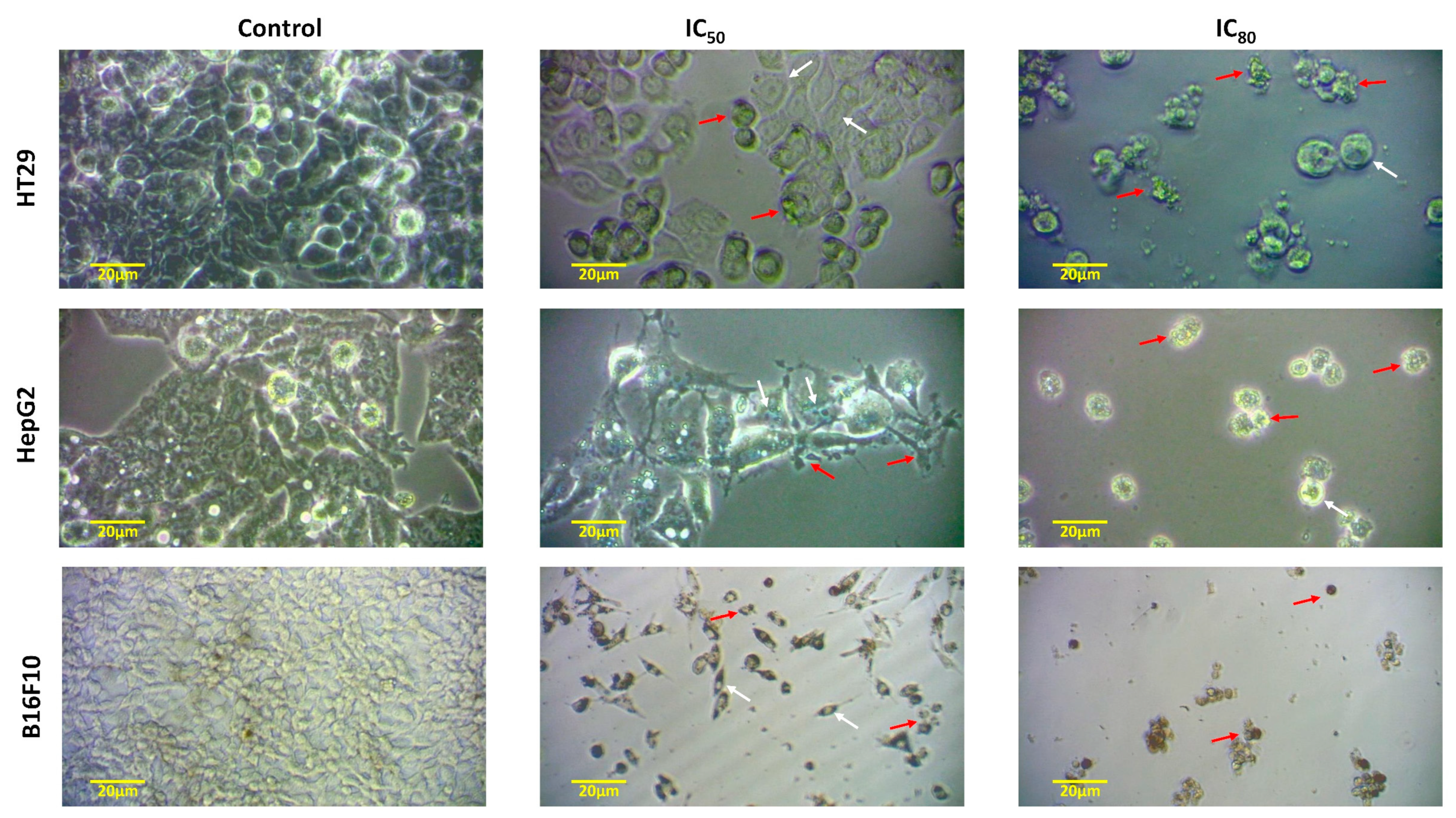
The cytotoxic effects of ent-kaurene 13 on HT29, HepG2, and B16-F10 cells and their morphological changes were confirmed and evaluated via inverted microscopy. The results of these observations reveal a progressive sequence of morphological changes that correlate with dose-dependent cytotoxic effects across the three cell lines. At IC50 concentrations, compound 13, together with other active derivatives, such as compounds 2, 7, and 12, showed a significant inhibition of cell proliferation. The results of the compound 13 were consistent after 72 h of treatment in the three treated lines, showing distinct apoptotic characteristics.
Figure 5 provides a clear comparison, showing that untreated control cells retained a large, flattened morphology with strong adhesion to the culture surface and a generally uniform, rounded shape with visible membrane integrity. Following treatment with IC50 concentrations of compound 13, cells of all lines (HT29, HepG2, and B16-F10) exhibited marked changes in morphology. Cells appeared smaller and more rounded, with a scattered distribution suggesting reduced intercellular adhesion. In addition, cell shrinkage was evident, indicating an early apoptotic response characterised by cytoplasmic condensation and loss of typical membrane morphology.
As the concentration increased to IC80 levels, more pronounced morphological features of cell death were observed. A marked increase in apoptotic cells was observed, and membrane blebbing and rupture of the cell membrane were identified, leading to the formation of apoptotic bodies and cellular debris. These effects, observed at high concentrations, suggest that compound 13 induces extensive cell death beyond the initial cytostatic effects, progressing to apoptosis, cell degradation, and disintegration.
The contrasting morphologies (control versus treated cells) shown in Figure 5 emphasise this differential response, as untreated cells showed adherence, a uniform shape and intact cell membranes, in contrast to the rounded, fragmented, and scattered morphology observed in treated cells. This clear apoptotic response, coupled with mitochondrial membrane alterations, highlights the potential of compound 13 and related ent-kaurene derivatives as promising anticancer agents that are capable of inducing cell death in colorectal, liver, and melanoma cancer cell lines.

2.2.3. Induction of Apoptosis

Apoptosis, or programmed cell death, is characterised by a cascade of biochemical events leading to morphological and structural changes, including chromatin condensation, nuclear fragmentation, and cell membrane blebbing. These features culminate in the formation of apoptotic bodies [25]. The induction of apoptosis is a central target in cancer therapy as it allows for the focused elimination of cancer cells via programmed cell death mechanisms. While cell cycle arrest is crucial to halt proliferation, apoptotic pathways provide an additional layer of cytotoxicity that enhances the anticancer effects of therapeutic agents. The majority of currently used anticancer drugs trigger and induce apoptosis as a key target in cancer cells [25]. Therefore, a comprehensive evaluation of the apoptotic potential of compounds 7, 12, and 13 was performed on the three cancer cell lines to elucidate their mechanisms of action beyond cell cycle inhibition.
In this context, we further investigated the potential mechanisms underlying the cytotoxic and cytostatic effects of compounds 7, 12, and 13 on the three cancer cells using Annexin V-FITC/PI double staining together with a flow-activated cell sorter (FACS). This method allowed for differentiation between early apoptosis, late apoptosis, and necrosis. This approach allowed for the detection of phosphatidylserine (PS) externalisation. In brief, the loss of cytoplasmic membrane asymmetry that occurs during apoptosis causes the translocation of phosphatidylserine (PS) from the inner membrane leaflet to the outer membrane, exposing PS to the external environment, where it is recognised and rapidly phagocytosed by macrophage cells [26]. Annexin V binds specifically to exposed PS, allowing for the identification of early apoptotic cells (Annexin V+/PI−), late apoptotic cells (Annexin V+/PI+), and necrotic cells (Annexin V−/PI+), as shown in Figure 6, Figure 7 and Figure 8. This staining was performed 72 h after treatment at IC50 and IC80 concentrations, providing insight into concentration-dependent apoptotic responses.
The apoptotic effects of compound 7 on B16-F10 cells are as follows: Exposure to compound 7 resulted in a dose-dependent increase in apoptotic cell populations on the B16-F10 melanoma cell line. Control samples showed 91% normal cells, which decreased to 85% and 62% at IC50 and IC80 concentrations, respectively. This decrease in normal cells was accompanied by an increase in total apoptosis rates from 6% in control cells to 13% and 26% at IC50 and IC80, respectively. Notably, early apoptotic cells constituted the majority of the apoptotic population at both concentrations, accounting for 11% and 16% of the total cells, respectively. No significant necrotic activity was observed, suggesting that compound 7 primarily induces apoptosis rather than necrosis in melanoma cells, thereby reducing non-specific cell death (Figure 6).
The apoptotic effects of compound 12 on HepG2 cells were as follows: Compound 12 showed significant apoptotic effects on the HepG2 hepatocarcinoma cell line, with total apoptosis rates reaching 67% at both the early and late stages, even at lower concentrations. Early and late apoptosis accounted for 43% and 24% of the total apoptotic population, respectively, while necrotic cell populations did not exceed 4% in treated samples.
The proportion of normal cells decreased significantly, from 87% in untreated controls to 29% with compound 12 at both IC50 and IC80 concentrations, indicating a strong induction of programmed cell death at both concentration levels. These results underscore the potent apoptotic induction of compound 12 in hepatocarcinoma cells, suggesting its potential for targeted cytotoxicity against liver tumours (Figure 7).
The apoptotic effects of compound 13 in the three cell lines were as follows: Among the compounds tested, compound 13 exhibited the most pronounced apoptotic effects in all three cell lines (HT29, HepG2, and B16-F10), demonstrating both early and late apoptotic induction in a dose-dependent manner (Figure 8). In HT29 (colon adenocarcinoma) cells, compound 13 produced a significant apoptotic response, achieving a total apoptosis rate of 32% (21% early and 11% late) at the IC50 concentration. At the IC80 concentration, the apoptotic effects were even more pronounced, with 87% total apoptosis (50% early and 37% late apoptosis), confirming the potential of compound 13 for high-impact cytotoxicity in colon cancer cells. In HepG2 cells, compound 13 induced remarkably high levels of apoptosis, achieving total apoptosis rates of 95% at IC50 (41% early; 54% late) and reaching 99% at IC80 (36% early and 63% late apoptosis). This exceptional response suggests a particularly potent effect on hepatocarcinoma cells, probably involving a rapid apoptotic response, in agreement with the results found in the cytotoxicity analysis of this compound.
Finally, in the B16-F10 melanoma cell line, compound 13 produced moderate apoptotic rates at the IC50 concentration (28% total apoptosis; 9% early, 19% late), which increased dramatically to 96% at the IC80 concentration (92% early, 4% late apoptosis), indicating a broad apoptotic response at higher concentrations.
The observed apoptotic responses suggest that compounds 12 and 13 are particularly effective in inducing apoptosis, with concentration-dependent shifts from cytostatic to cytotoxic effects. While compound 13 exhibits broad apoptotic potential across cell lines, compound 12 appears to have a more selective effect on HepG2 cells, suggesting specific interactions with cellular components or regulatory pathways unique to liver cells. In addition, the low necrotic activity across treatments highlights a preference for apoptotic pathways that may minimise the inflammatory responses commonly associated with necrosis. These findings emphasise the therapeutic potential of ent-kaurene derivatives, especially compound 13, as selective apoptotic agents with applicability across diverse cancer cell types. Further investigations into their molecular mechanisms could provide valuable insights into the apoptotic pathways engaged by these compounds. As a result, these ent-kaurene derivatives could serve as promising candidates for targeted cancer therapies with apoptosis-focused mechanisms.

2.2.4. Analysis of Cell Cycle

Flow cytometry was used to measure DNA ploidy and examine changes in cell cycle profiles, aiming to confirm the cytostatic effect associated with the inhibitory response of the selected ent-kaurenes on cell growth in the three cancer cell lines tested. By analysing the DNA histograms of treated cells stained with propidium iodide (PI), we were able to precisely quantify cell populations across G0/G1, S, and G2/M phases, revealing distinct arrest patterns associated with each compound. The cell cycle effects of ent-kaurene derivatives 7, 12, and 13 were investigated in B16-F10, HepG2, and HT29 cells after 72 h of treatment at IC50 and IC80 concentrations. These derivatives, previously identified due to their remarkable antiproliferative activities, were selected based on their efficacy in previous cytotoxicity assays. Cell cycle phase distributions and DNA content analyses were performed to determine whether cell cycle arrest mechanisms contributed to the observed growth inhibition.
The effects of compound 7 on B16-F10 cell cycle distribution were as follows: As shown in Figure 9, compound 7 had a minimal effect on cell cycle phase distribution in B16-F10 cells, with results that were not significant compared to control conditions. Cell populations in the G0/G1, S, and G2/M phases remained similar to those of untreated controls at both IC50 and IC80 concentrations, suggesting that the cytotoxic effects of compound 7 may occur independently of cell cycle disruption. This observation suggests that there are alternative cytotoxic pathways for compound 7, possibly through direct mitochondrial disruption or alternative apoptotic pathways not primarily involving cell cycle arrest.
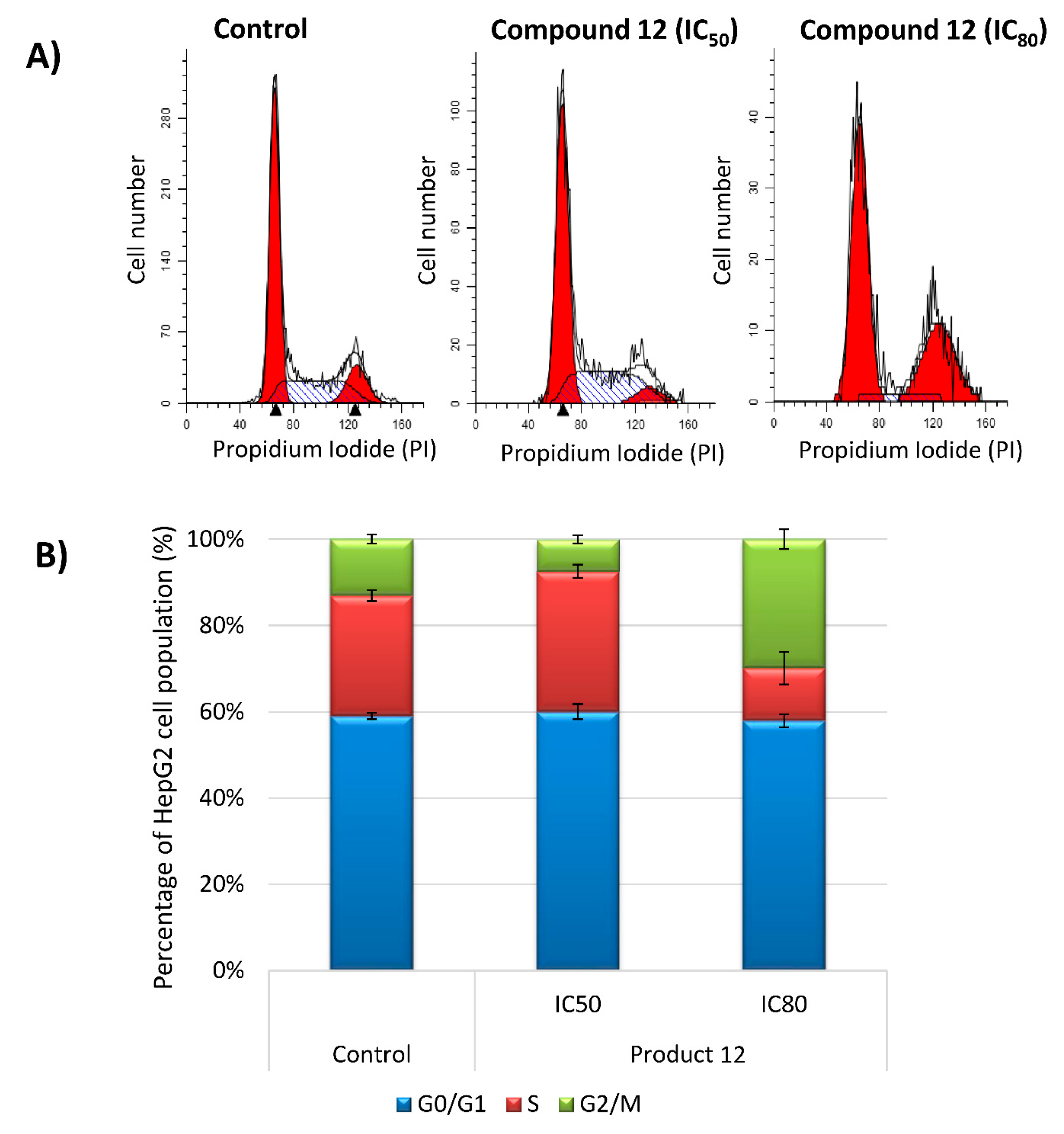
The effects of compound 12 on HepG2 cell cycle arrest and distribution are presented as follows: Treatment with compound 12 resulted in a significant increase in the G2/M phase cell population at IC80 concentration, with 17% more cells in G2/M compared to untreated controls, while the S phase population decreased significantly, by approximately 16% (Figure 10). This shift suggests that compound 12 induces a G2/M phase block at higher concentrations, potentially halting DNA synthesis and inhibiting mitosis. However, at IC50 concentrations, compound 12 showed limited changes in cell cycle progression, with no significant differences in phase distribution, indicating a concentration-dependent effect on G2/M arrest in HepG2 cells. This result suggests the involvement of the G2/M checkpoint at higher doses, where compound 12 may interfere with the key regulatory proteins required for mitotic entry.
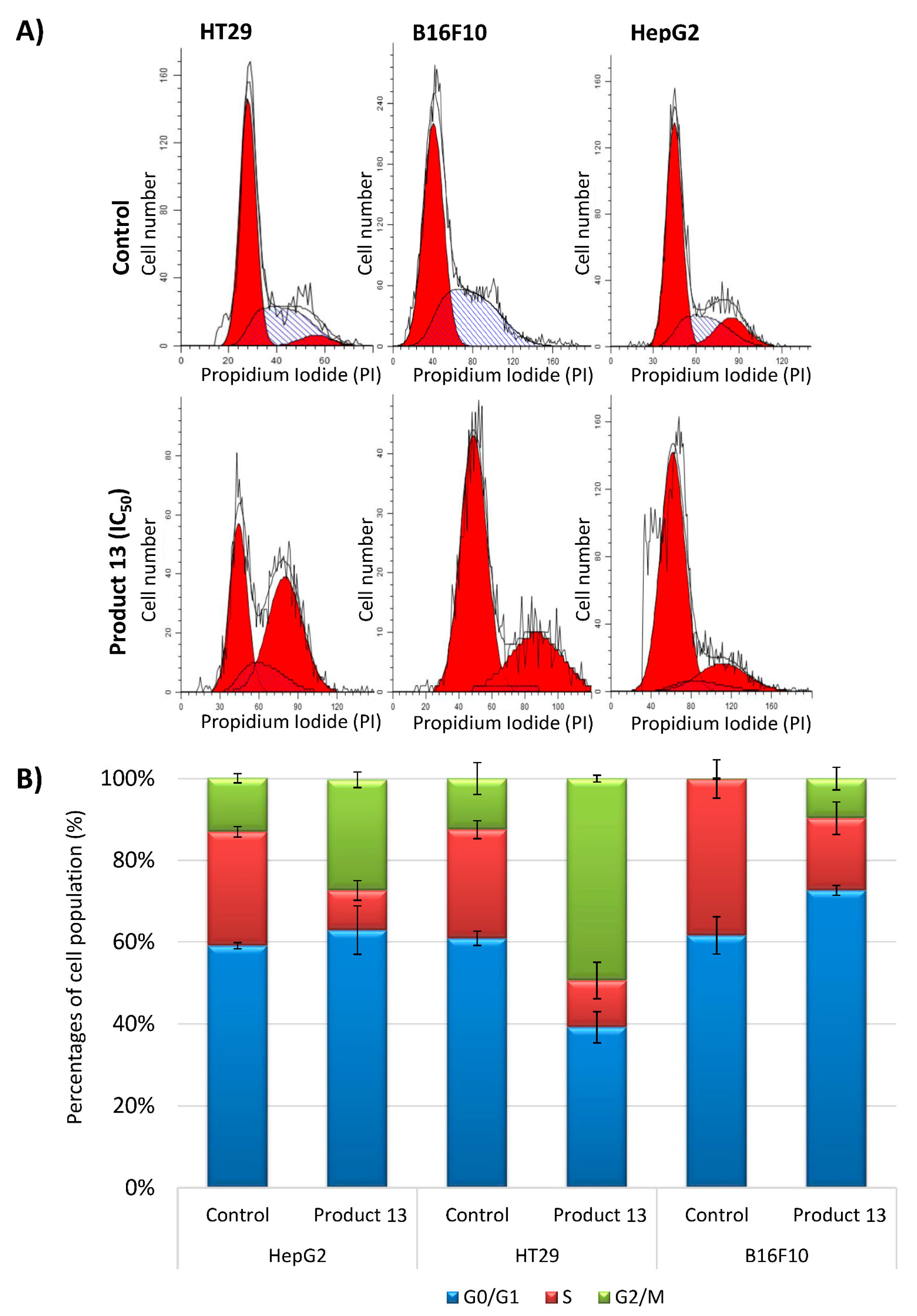
The effects of compound 13 on HT29, HepG2, and B16-F10 cell cycle arrest were as follows: Compound 13 showed the strongest effect on cell cycle arrest among the ent-kaurenes tested (Figure 11). In HT29 cells, treatment with compound 13 resulted in a significant accumulation in the G2/M phase: approximately 49.4% of the cells were detected in the G2/M phase, an increase of 36.9% compared to control untreated cells. This redistribution was accompanied by a significant reduction in S phase cells of 15% and a 21% reduction in G0/G1 phase cells, indicating an effective blockade at the G2/M checkpoint. In HepG2 cells, compound 13 similarly induced G2/M arrest, increasing the G2/M cell population by 14% while reducing S phase cells by 18%. Interestingly, G0/G1 changes were negligible, suggesting a specific effect on G2/M. This suggests that compound 13 may inhibit critical proteins, such as cyclin-dependent kinases (CDKs).
In B16-F10 cells, compound 13 showed a moderate effect, with G2/M and G0/G1 cell cycle arrest both increasing by approximately 11% each. Correspondingly, S-phase cells decreased by approximtely 21% compared to controls, suggesting a partial G2/M arrest coupled with reduced DNA synthesis. The ability of compound 13 to induce G2/M arrest in all cell lines tested underscores its potential as a universal cytostatic agent, and its consistent effect highlights its selective action on cell cycle regulatory mechanisms. Notably, at the IC80 concentration, compound 13 showed no further enhancement of G2/M arrest in any of the cell lines, suggesting that higher doses may favour the activation of apoptotic pathways over cell cycle arrest. This is consistent with the observations of increased apoptosis and markers such as chromatin condensation, membrane blebbing, and apoptotic body formation in parallel microscopy assays, indicating a shift from cytostatic to cytotoxic effects at elevated concentrations.
These cell cycle analyses show that ent-kaurene derivatives 12 and 13 exhibit promising cytostatic activity through selective G2/M-phase arrest; this is particularly effective in HT29 and HepG2 cells. The concentration-dependent effects suggest a dual mechanism, whereby the compounds first inhibit cell proliferation and subsequently induce apoptosis at higher doses. Further studies focusing on protein-level interactions, particularly at the G2/M checkpoint, may provide insight into the molecular basis of these responses and provide a basis for the potential therapeutic use of ent-kaurenes in targeted cancer treatments.

2.2.5. Analysis of Mitochondrial Membrane Potential

The mitochondrial membrane potential (Δψm) is a critical parameter in assessing mitochondrial function and serves as an early indicator of apoptosis in cells. The disruption of Δψm indicates mitochondrial destabilisation, which is often associated with the intrinsic apoptotic pathway. Therefore, studying changes in mitochondrial membrane potential (MMP) in tumour cells provides valuable insights into the mechanism and pathway of apoptosis induced by anticancer agents. Apoptotic effects can manifest due to the activation of intrinsic (mitochondrial) or extrinsic apoptotic pathways. If apoptosis is accompanied by loss of MMP, this suggests mitochondrial involvement and activation of the intrinsic pathway. Conversely, when apoptosis is induced without changes in MMP, this typically indicates activation by the extrinsic pathway, often mediated by cell-surface-death receptors.
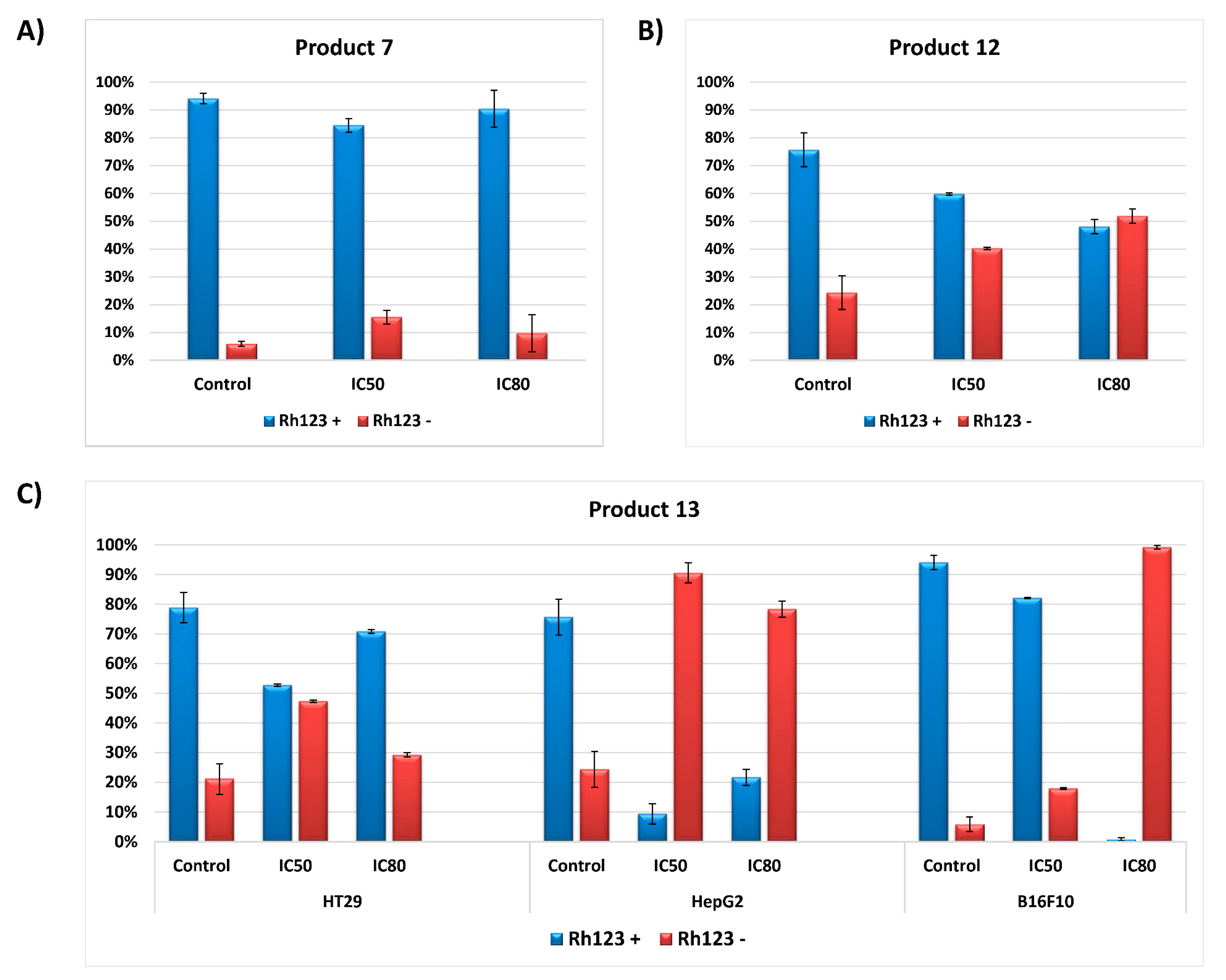
In this study, MMP changes were analysed in HT29, HepG2, and B16-F10 cell lines after treatment with compounds 7, 12, and 13. Flow cytometry with Rhodamine 123 (Rh123) and propidium iodide (PI) double-staining was used to assess MMP integrity. Rhodamine 123 is a lipophilic cationic dye that selectively accumulates in mitochondria, and the logarithmic ratio of the probe concentration between the inner and outer mitochondria membrane is proportional to the mitochondrial membrane potential. A decrease in Rh123 fluorescence indicates a loss of mitochondrial potential, signalling mitochondrial dysfunction. After 72 h exposure to compounds 7, 12, and 13 at IC50 and IC80 concentrations, the percentage of Rh123-positive and Rh123-negative cells was determined, providing a comparative measure of MMP integrity relative to untreated controls (Figure 12).
The effects of compound 7 on B16-F10 cells were as follows: The MMP analysis for compound 7 on B16-F10 melanoma cells showed no significant reduction in Rh123 fluorescence. Both IC50 and IC80 concentrations produced high Rh123-positive staining, consistent with untreated cells. This lack of MMP disruption implies that compound 7 does not induce mitochondrial destabilisation in B16-F10 cells, suggesting that apoptosis is likely to be mediated by an extrinsic apoptotic pathway, independent of mitochondrial influence. This extrinsic pathway activation could involve cell-surface-death receptors such as Fas ligand or tumour necrosis factor receptor (TNF-R), bypassing the mitochondrial apoptotic cascade.
The effects of compound 12 on HepG2 cells were as follows: In HepG2 cells, compound 12 induced a significant decrease in Rh123-positive cell populations. The increase in Rh123-negative cells at both IC50 and IC80 concentrations suggests significant MMP loss, suggesting that compound 12 induces apoptosis via the intrinsic or mitochondrial pathway in hepatocarcinoma cells. The dose-dependent increase in mitochondrial dysfunction may reflect early mitochondrial membrane permeability changes, cytochrome c release, and the activation of downstream caspases, all hallmarks of intrinsic apoptosis. This mitochondria-targeted mechanism is consistent with the broader apoptotic efficacy of compound 12 in HepG2 cells.
The effects of compound 13 in all cell lines were as follows: Compound 13 demonstrated the most potent effect on mitochondrial membrane potential among all compounds tested in all three cell lines. In HT29 colon adenocarcinoma cells, treatment with compound 13 resulted in a significant reduction in Rh123-positive cells at both IC50 and IC80 concentrations, indicating a pronounced loss of MMP and suggesting intrinsic apoptosis. These results correlate with the high apoptosis rates observed in HT29 cells, suggesting that compound 13 exerts a robust pro-apoptotic effect through mitochondrial destabilisation. In HepG2 cells, compound 13 also induced a dose-dependent loss of MMP, as evidenced by the significant increase in the Rh123-negative population compared to untreated controls. This decrease in MMP at both concentrations highlights the potent mitochondrial action of compound 13, which may involve mitochondrial membrane permeabilisation and the release of pro-apoptotic factors. In contrast, in B16-F10 melanoma cells, compound 13 induced a marked increase in the Rh123-negative population at the IC80 concentration, indicating MMP destabilisation and the activation of intrinsic apoptosis, although this effect was not observed at the IC50 concentration, which could mean that, in this cell line, there may be an initial activation of the extrinsic apoptosis-inducing extrinsic pathway and a secondary activation of the intrinsic pathway. Future studies are required to clarify this point. These consistent effects across cell lines support the role of compound 13 as a broad-spectrum mitochondrial disruptor.
Comparative analysis of the MMP effects between compounds shows that compounds 5 and 7 induce apoptosis primarily through the mitochondrial pathways in HepG2 and HT29 cells, respectively, with compound 13 exerting this effect in all cell lines tested. In contrast, the lack of effect of compound 7 on MMP in B16-F10 cells suggests an alternative apoptotic pathway, probably extrinsic in nature. These findings suggest compound-specific mechanisms of action, with compounds 12 and 13 acting as potent inducers of mitochondrial destabilisation, whereas compound 7 bypasses mitochondrial involvement. The consistent induction of MMP degradation via compounds 12 and 13 highlights their potential as selective mitochondrial targeting agents for cancer therapy. Future investigations could focus on elucidating the specific molecular targets within the mitochondrial apoptotic pathway, including the role of Bcl-2 family proteins, caspase activation and cytochrome c release. The differential effects observed between the compounds suggest the utility of ent-kaurene derivatives in tailored cancer therapies that exploit apoptotic pathways to selectively induce apoptosis in cancer cells while minimising non-specific toxicity.

3. Discussion

The significant anticancer potential of ent-kaurene diterpenoids, particularly compounds 7, 12, and 13, has been demonstrated in the assayed three cancer cell lines: colorectal (HT29), liver (HepG2), and melanoma (B16-F10) cancer cell lines. Also, we analysed the anticancer activity and mechanisms of action of compounds isolated and structurally modified from D. tenuifolium, supporting their potential as promising therapeutic agents for a range of cancers.
Cell viability assays revealed a range of cytotoxic potentials for the investigated compounds, with IC50 values ranging from 2.12 μM to 153.83 μM. Among these, compound 13 emerged as the most potent cytotoxic agent, demonstrating a dose-dependent anti-proliferative effect in all cancer cell lines tested (HT29, HepG2, and B16-F10). In HT29 cells, compound 13 achieved an impressive IC50 of 2.71 ± 0.23 µM, significantly outperforming compounds 2 and 12, which exhibited IC50 values of 10.08 ± 2.69 µM and 45.37 ± 5.34 µM, respectively. Notably, compound 12 exhibited selective cytotoxicity towards HepG2 cells, suggesting its potential application in the treatment of hepatocellular carcinoma. This observation is consistent with previous findings on other diterpenoids, such as kaurenoic acid, which demonstrated cell line-specific activity influenced by structural modifications [21]. Structurally, this compound is characterised by the presence of an exo-methylene cyclopentanone, which is consistent with previous studies highlighting the critical role of this moiety in the cytotoxic activity of ent-kaurene diterpenoids [27].
Oridonin, one of the most potent tetracyclic ent-kaurenes, was isolated from the genus Isodon and one of its derivatives has progressed to phase I clinical trials [28]. Oridonin shows activity against several types of cancer, including acute myeloid leukaemia, pancreatic, oesophageal, nasopharyngeal, colorectal, and liver cancer, with an IC50 of 25.7 µM in HepG2 cells [29]. Another example is kaurenic acid, isolated from Espeletia semiglobulata, which showed significant antimelanoma activity, with an IC50 of 0.79 µM in B16-F10 cells. In addition, ponicidin, extracted from Isodon adenolomus, was identified as a cytotoxic agent against hepatocellular carcinoma, lung cancer, monocytic leukaemia, and colorectal cancer. Treatment with ponicidin was reported to significantly suppress the growth of HT29 cells (~4-fold reduction), highlighting its potential as an anticancer agent [30,31,32].
A cell cycle analysis showed that compounds 12 and 13 induced a marked G2/M cell cycle arrest in HT29 and HepG2 cell lines in a dose-dependent manner, highlighting their cytostatic potential. G2/M arrest is a well-documented mechanism for stopping cancer cell proliferation through preventing mitotic entry and allowing for DNA damage repair or apoptotic signalling. In HT29 cells, compound 13 caused a significant 49.4% increase in the G2/M population, accompanied by a reduction in the S and G0/G1 phases. Similarly, compound 12 induced a 17% increase in the G2/M population in HepG2 cells at its IC80 concentration, further emphasising its role in arresting mitotic progression. These results suggest that ent-kaurenoids interfere with mitotic entry by modulating cyclin-dependent kinases (CDKs) and checkpoint regulators [33]. Interestingly, the G2/M arrest observed with these compounds is similar to the effects of oridonin, a structurally related diterpenoid known to inhibit microtubule dynamics and spindle assembly checkpoints [34]. This common mechanism of action among diterpenoids underscores their potential as inhibitors of mitotic progression and highlights their therapeutic relevance in cancer treatment [20].
Our findings are consistent with previous research on pharicin A, a novel natural ent-kaurenoid isolated from Isodon pharicus, which has been identified as a cytostatic and cytotoxic agent. Pharicin A induces G2/M phase arrest in both paclitaxel-sensitive and resistant tumour cells, an effect attributed to its ability to bind BubR1, disrupt its subcellular localisation, and inhibit its kinase activity [20]. Similarly, other studies have shown that ent-kaurenoids can induce cell cycle arrest and apoptosis while exhibiting moderate to low toxicity in normal cell lines. [35] Many cytotoxic compounds exert inhibitory effects on cancer cell growth by arresting the cell cycle at specific checkpoints, inducing apoptosis, or a combination of both mechanisms [26,36,37].
One of the most compelling findings of this study was the strong apoptotic induction observed with compounds 7, 12, and 13, particularly compound 13, which exhibited broad apoptosis rates across multiple cell lines, with total apoptosis percentages ranging from 20% to 99%. In HepG2 cells, compound 13 achieved total apoptosis rates of over 95%, with a dose-dependent progression from the early to late apoptotic stages. Apoptosis studies using Annexin V-FITC/PI staining and flow cytometry confirmed these results and demonstrated that the apoptotic effects of compounds 12 and 13 were strongly associated with mitochondrial dysfunction, as evidenced by the loss of Δψm in treated cells. The reduction in Rh123 fluorescence in HT29 and HepG2 cells further supports the activation of the intrinsic apoptotic pathway, highlighting the mechanistic involvement of mitochondrial disruption in their cytotoxic effects.
A comparative analysis of mitochondrial membrane potential (MMP) effects revealed that compounds 12 and 13 induced apoptosis primarily through mitochondrial pathways in HepG2 and HT29 cells, respectively, with compound 13 exerting this effect in all cell lines tested. Mitochondrial destabilisation, as evidenced by a significant decrease in Rh123 fluorescence, was particularly pronounced in HT29 and HepG2 cells. This effect is likely to correlate with the release of pro-apoptotic factors such as cytochrome c into the cytosol, leading to caspase activation.
In contrast, compound 7 induced apoptosis in B16-F10 cells without significant MMP disruption, suggesting an alternative, extrinsic apoptotic pathway, possibly mediated by death receptors such as Fas or TNFR, which operate independently of mitochondrial involvement. These results highlight the compound specific mechanisms of action, with compounds 12 and 13 acting as potent inducers of mitochondrial destabilisation, whereas compound 7 appears to bypass the mitochondrial pathways. Notably, compound 13, which exhibited the strongest apoptotic effect, contains an α,β-unsaturated ketone group, a structural feature that may explain its efficacy in all tumour cell lines tested. Further molecular studies will be essential to confirm these mechanistic findings and to fully elucidate the apoptotic pathways involved.
Consistent with these findings, previous studies on the apoptotic effects of ent-kaurenoids suggest that these biomolecules primarily activate intrinsic apoptotic mechanisms, particularly in compounds containing an α,β-unsaturated ketone group. Oridonin and related compounds have been shown to induce apoptosis through a mitochondrial-related pathway, in which cytochrome c plays a central role. The α,β-unsaturated ketone moiety was identified as an active functional group that is critical for their cytotoxicity, mitochondrial localisation, and uptake [38]. Furthermore, there is evidence supporting mitochondrial redox changes as a potential mediator of the apoptotic activities of oridonin in HepG2 cells [39].
From a chemical perspective, the widely accepted mechanism of action of these diterpenoids involves the Michael addition of soft nucleophiles, such as thiols and protein sulfhydryl (SH) groups, to the α,β-unsaturated ketone moiety. This reaction deactivates SH-dependent enzymes and coenzymes, leading to the accumulation of reactive oxygen species (ROS). The deactivation of SH enzymes and coenzymes disrupts intracellular redox homeostasis, while elevated ROS levels induce apoptosis through oxidative damage to tumour cells [40]. The importance of the α,β-unsaturated ketone group was highlighted in the potent induction of mitochondrial apoptosis and promotion of ROS generation in cancer cells [29]. For example, pteisolic acid G, a novel ent-kaurene diterpenoid, was reported to inhibit cell viability and induce apoptosis in human colorectal cancer cells through an intrinsic apoptotic mechanism. Polyisolic acid G downregulated anti-apoptotic proteins such as Bcl-2 and Bcl-XL and factors such as NF-κB p65 and p-p65. Conversely, it upregulated pro-apoptotic proteins, including Puma, Bax, and Bim, and the tumour suppressor p53.
These differential effects highlight the versatility of ent-kaurene derivatives in targeting multiple apoptotic pathways. The ability of ent-kaurene derivatives to target both intrinsic and extrinsic pathways increases their therapeutic versatility and may overcome the resistance mechanisms in apoptosis-deficient cancers. A similar dual-pathway activation was observed in other diterpenoids, such as triptolide, which simultaneously activates mitochondrial dysfunction and death receptor signalling [41]. This highlights the potential of ent-kaurene derivatives as multi-target agents capable of addressing the heterogeneity of cancer. Future investigations could focus on elucidating the specific molecular targets within the mitochondrial apoptotic pathway, including the role of Bcl-2 family proteins, caspase activation, and cytochrome c release. The differential effects observed between compounds 7, 12, and 13 suggest the utility of ent-kaurene derivatives in tailored cancer therapies that exploit apoptotic pathways to selectively induce apoptosis in cancer cells while minimising non-specific toxicity.

4. Materials and Methods

4.1. Chemistry

4.1.1. General Experimental Procedures

Thin-layer chromatography (TLC) was performed using F254 precoated plates (0.25 mm) and visualised using UV fluorescence quenching and phosphomolybdic acid solution in ethanol staining. Flash chromatography was performed on silica gel (230–400 mesh). Chromatography separations were carried out via conventional column on silica gel 60 (230–400 Mesh), using hexanes-AcOEt (AcOEt-hexane) mixtures of increasing polarity. 1H and 13C NMR spectra were recorded on a Bruker Nanobay Avance NEO III HD High Definition NMR Spectrometer, Billerica, MA, USA, at 500 MHz and at 125 MHz, respectively. CDCl3 was treated with K2CO3. Chemical shifts (δ H) are quoted in parts per million (ppm), noting the appropriate residual solvent peak and tetramethylsilane. Data for 1H NMR spectra are reported with the abbreviations s, br s, d, br d, t, dt, dq, and m denoting singlet, broad singlet, doublet, broad doublet, triplet, doublet triplet, doublet quartet, and multiplet, respectively. J = coupling constant in Hertz (Hz). Data for 13C NMR spectra are reported in terms of chemical shift relative to Me4Si (δ 0.0) and the signals were assigned utilising DEPT experiments and on the basis of heteronuclear correlations. Infrared spectra (IR) were recorded as thin films or as solids on a Perkin Elmer model, USA. One FTIR spectrophotometer with samples between sodium chloride plates or as potassium bromide pellets and are reported in terms of their frequency of absorption (cm−1). Only selected absorbances (νmax) are reported.

4.1.2. Extraction and Isolation

The plant was collected in the north of Granada, Spain. The air-dried aerial parts of D. tenuifolium (158.25 g) was powdered and extracted with 500 mL of MeOH at room temperature. The MeOH extract was evaporated under reduced pressure to yield a crude extract of 43.1g. The extract was subjected to column chromatography on silica gel, eluting with an increasingly polar gradient of ethyl acetate (AcOEt) to yield 10.1 g of 7 (10% AcOEt/hexane) and 250 mg of 8 (15% AcOEt/hexane).

4.1.3. Synthesis and Structural Characterisation

Structural Characterisation of Compounds (7) and (8)

ent-15α-angeloyloxykaur-16-en-3β-ol (7): white solid; 1H NMR data (CDCl3, 500 MHz) (see Table S1, included in the Supplementary Materials); 13C NMR data (CDCl3, 125 MHz) (see Table S2, included in the Supplementary Materials). HRMS (ESI) m/z calcd for C25H38O3Na (M+Na+) 409.2719 was 409.2727. The spectroscopic data of 7 were obtained using those described in the literature [23].
ent-15α-angeloyloxykaur-16-en-3β,9α-diol (8): white solid; 1H NMR data (acetone-d6, 500 MHz) (see Table S1, included in the Supplementary Materials); 13C NMR data (acetone-d6, 125 MHz) (see Table S2, included in the Supplementary Materials). HRMS (ESI) m/z calcd for C25H38O4Na (M + Na+) 425.2668 was 425.2680. Spectroscopic data of 8 were obtained according to the description in the literature [7].

Synthesis of Acetate 9

Ac2O (0.5 mL) was added to a solution of alcohol 7 (123 mg, 0.318 mmol) in pyridine (1 mL) and the mixture was stirred for 2 h. Then, water (5 mL) was added, and the mixture was stirred for 5 min. Next, it was diluted with AcOEt (15 mL), and the phases were shaken and separated. The organic phases were washed with 2 N HCl (3 × 5 mL), saturated NaHCO3 (3 × 5 mL), and brine (5 mL), and dried over anhydrous Na2SO4. Removal of the solvent under vacuum yielded a crude product (138 mg). Flash chromatography (20% AcOEt/hexane) yielded 9 (130 mg, 96%) as a colourless syrup. 1H NMR (CDCl3, 400 MHz); δ 0.78 (dd, J = 11.5, 1.6 Hz, 1H); 0.86 (s, 6H); 1.02 (m, 1H); 1.09 (s, 3H); 1.20 (m, 1H); 1.92 (dt, J= 13.3, 3.2, 3.2 Hz, 1H); 1.95 (br s, 3H); 2.06 (q, J = 7.2 Hz, 3H); 2.06 (s, 3H); 1.22–1.81 (m, 12H); 2.71 (br s, 1H); 4.48 (dd, J = 11.0, 5.0 Hz, 1H); 4.92 (br s, 1H); 4.95 (br s, 1H); 5.27 (t, J= 2.5 Hz, 1H); 6.14 (br q, J = 7.2 Hz, 1H). 13C NMR (125 MHz, CDCl3): 15.9 (CH3); 16.6 (CH3); 17.7 (CH3); 17.8 (CH2); 19.4 (CH2); 20.8 (CH3); 21.3 (CH3); 23.6 (CH2); 28.3 (CH3); 33.4 (CH2); 36.5 (CH2); 37.7 (C); 38.6 (CH2); 38.6 (CH2); 38.6 (C); 40.7 (CH); 46.0 (C); 48.1 (CH); 54.9 (CH); 80.9 (CH); 81.1 (CH); 106.3 (CH2); 127.9 (C); 138.4 (CH); 153.7 (C); 168.1 (C); 171.0 (C). HRMS (ESI) m/z calcd for C27H40O4Na (M + Na+) 451.2824 was 451.2823.

Synthesis of Ketone 10

Jones reagent (0.2 mL) was added dropwise at 0 °C to a solution of compound 7 (187 mg, 0.48 mmol) in acetone (4 mL) and the reaction mixture was stirred for 10 min, at which point no starting material for TLC was found. The solvent was then removed under vacuum, and the crude product was diluted with AcOEt–water (20:10 mL). The phases were shaken and separated, and the organic layer was washed with water (3 × 10 mL) and brine and dried over anhydrous Na2SO4. Removal of the solvent under vacuum provided a crude product that was purified using flash chromatography to yield 10 (173 mg, 93%) as a colourless syrup.1H NMR (CDCl3 400 MHz); δ 1.04 (s, 3H); 1.08 (s, 3H); 1.13 (s, 3H); 1.94 (br s, 3H); 2.05 (q, J = 7.2 Hz, 3H); 2.06 (s, 3H); 1.18–2.19 (m, 12H); 2.45–2.53 (m, 2H); 2.74 (br s, 1H); 4.94 (br s, 1H); 4.97 (br s, 1H); 5.31 (t, J= 2.5 Hz, 1H); 6.13 (dq, J = 7.2, 1.4 Hz, 1H). 13C NMR (125 MHz, CDCl3): 15.8 (CH3); 17.8 (CH3); 18.2 (CH2); 20.8 (CH3); 21.0 (CH3); 21.1 (CH2); 27.2 (CH3); 33.2 (CH2); 34.1 (CH2); 36.2 (CH2); 37.8 (C); 38.2 (CH2); 39.5 (CH2); 40.6 (CH); 45.9 (C); 47.1 (C); 54.1 (CH); 81.0 (CH); 106.5 (CH2); 127.8 (C); 138.5 (CH); 153.5 (C); 168.0 (C); 217.8 (C). HRMS (ESI) m/z calcd for C25H36O3Na (M + Na+) 407.2562 was 407.2572.

Synthesis of Diketone 11

An O3/O2 mixture was slowly bubbled through a stirred solution of 10 (113 mg, 0.294 mmol) in dichloromethane (10 mL) at −78 °C for 20 min. At this point, no remaining TLC starting material was found. Then, the excess ozone was removed by bubbling the solution with argon, PPh3 (105 mg, 0.4 mmol) was added at −78 °C, the reaction mixture was stirred for 2 h at room temperature, and the solvent was evaporated under vacuum to obtain a crude product. Purification via chromatography on silica gel (20% AcOEt/hexane) yielded pure diketone 11 (59 mg, 52%) as a colourless syrup. [ α ] D 25 = -28.3 (c 0.38, CHCl3). IR (film) ʋmax: 2929, 1759, 1703, 1448, 1373, 1136, 1111, 1025, 978 cm−1. 1H NMR (CDCl3, 400 MHz); δ 1.07 (s, 3H); 1.10 (s, 3H); 1.19 (s, 3H); 1.27 (br s, 3H); 1.49 (q, J = 7.2 Hz, 3H); 1.90 (m, 1H); 2.09 (m, 1H); 2.40 (m, 1H); 2.47–2.55 (m, 2H); 2.58 (br s, 1H); 4.91 (s, 1H); 5.40 (q, J = 7.2 Hz; 1H). 13C NMR (125 MHz, CDCl3): 15.5 (CH3); 18.1 (CH3); 18.8 (CH3); 18.9 (CH2); 20.0 (CH2); 21.1 (CH3); 27.2 (CH3); 29.8 (CH2); 32.6 (CH2); 33.9 (CH2); 37.8 (CH2); 38.3 (C); 39.2 (CH2); 44.0 (CH); 45.0 (C); 46.1 (CH); 47.1 (C); 54.1 (CH); 78.8 (CH); 82.6 (CH); 102.5 (CH); 104.6 (C); 168.2 (C); 213.9 (C); 217.1 (C). HRMS (ESI) m/z calcd for C24H34O4Na (M + Na+) 409.2355 was 409.2363.

Synthesis of Hydroxyketone 12

An O3/O2 mixture was slowly bubbled through a stirred solution of 7 (87 mg, 0.225 mmol) in dichloromethane (20 mL) at −78 °C for 30 min. At this point, no remaining TLC starting material was observed. The excess ozone was then removed via bubbling the solution with argon, and PPh3 (393 mg, 1.5 mmol) was added at −78 °C. Next, the reaction mixture was stirred for 4 h at room temperature. Hydroxyketone 12 (34 mg, 39%) was obtained following the same method as that used for 11 after chromatography, using (30% AcOEt/hexane) as the eluent. [ α ] D 25 −46.0 (c 0.05, CHCl3).IR (film) ʋmax: 3492, 2932, 1759, 1453, 1276, 1139, 981, 680 cm−1. 1H NMR (CDCl3, 400 MHz); δ 0.81 (s, 3H); 0.99 (s, 3H); 1.11 (s, 3H); 1.25 (br s, 3H); 1.49 (q, J = 7.2 Hz, 3H); 1.36–2.02 (m, 15H); 2.53 (br s, 1H); 3.21 (m, 1H); 4.89 (br s, 1H); 5.40 (br q, J = 7.2 Hz, 1H). 13C NMR (125 MHz, CDCl3): 15.5 (CH3); 15.6 (CH3); 18.0 (CH3); 18.3 (CH2); 18.6 (CH2); 18.9 (CH3); 27.1 (CH2); 28.4 (CH3); 30.0 (CH2); 32.9 (CH2); 38.7 (CH2); 38.8 (CH2); 38.8 (CH2); 38.9 (C); 44.1 (CH); 45.1 (C); 47.1 (CH); 54.7 (CH); 78.8 (CH); 82.8 (CH); 102.5 (CH2); 104.6 (C); 168.3 (CH); 2014.3 (C). HRMS (ESI) m/z calcd for C24H36O4Na (M + Na+) 411.2511 was 411.2512.

Synthesis of Diol 2

LiAlH4 (190 mg, 5.05 mmol) was carefully added to a solution of 7 (484 mg, 1.25 mmol) in anhydrous THF (10 mL), previously kept cold at 0 °C, and the reaction mixture was stirred at room temperature for 90 min. At this point, no remaining TLC starting material was observed. The mixture was poured onto ice and washed with AcOEt (3 × 20 mL). The mixture was transferred to a separating funnel and the phases were shaken and separated. The aqueous phase was extracted using AcOEt (10 mL) and the organic phases were combined. Then, it was washed with water and brine and dried over anhydrous Na2SO4. The removal of the solvent under vacuum yielded a crude product that was chromatographed on silica gel (30% AcOEt/hexane) to yield pure diol 2 (346 mg, 91%) as a white amorphous solid. 1H NMR (CDCl3, 400 MHz) δ 0.79 (s, 3H); 1.00 (s, 3H); 1.05 (s, 3H); 1.06 (ddd, J= 13.1, 13.1, 4.5 Hz, 1H); 1.28–1.95 (m, 15H); 1.99 (d, J = 15.0 Hz, 1H); 2.67 (br s, 1H); 3.23 (dd, J = 11.2, 5.1 Hz, 1H); 3.77 (s, 1H); 4.98 (s, 1H), 1H); 5.11 (s, 1H); 13C NMR (CDCl3, 125 MHz) δ 15.5 (CH3); 17.6 (CH3); 18.2 (CH2); 19.7 (CH2); 27.3 (CH2); 28.3 (CH3); 33.2 (CH2); 36.4 (CH2); 38.6 (CH2); 38.7 (CH2); 38.8 (C); 38.8 (C); 40.1 (CH); 45.6 (C); 46.3 (CH); 54.5 (CH); 79.0 (CH); 82.3 (CH); 104.9 (CH2); 158.3 (C). HRMS (ESI) m/z calcd for C20H32O2Na (M + Na+) 327.2300 was 327.2308.

Synthesis of Hydroxyenone 13

MnO2 (1 g, 11.49 mmol) was added to a solution of diol 2 (267 mg, 0.88 mmol) in CHCl3 (10 mL), and the mixture was stirred at reflux for 1 h. The filtrate was then filtered through silica gel (5 g), washed with AcOEt (10 mL), and concentrated to yield hydroxyenone 13 (246 mg, 93%) as a white solid. 1H NMR (CDCl3 400 MHz); δ 0.78 (s, 3H); 1.00 (s, 3H); 1.08 (s, 3H); 0.82–1.97 (m, 17H); 2.39 (d, J= 11.9 Hz, 1H, 3H); 3.04 (br s, 1H); 3.20 (dd, J= 11.3, 5.0 Hz, 1H); 5.25 (s, 1H); 5.94 (s, 1H). 13C NMR (125 MHz, CDCl3): 15.4 (CH3); 17.5 (CH3); 18.2 (CH2); 18.4 (CH2); 27.0 (CH2); 28.2 (CH3); 32.3 (CH2); 33.5 (CH2); 36.6 (CH2); 38.0 (CH); 38.1 (CH2); 38.8 (C); 39.7 (C); 52.2 (C); 52.5 (CH); 54.2 (CH); 78.7 (CH); 114.6 (CH2); 149.4(C); 210.9 (C). HRMS (ESI) m/z calcd for C20H30O2Na (M + Na+) 325.2143 was 325.2140.

4.2. Biological Experimental Procedures

4.2.1. Materials Used

Dulbecco’s modified Eagle’s medium (DMEM), RPMI 1640 medium with L-glutamine, foetal bovine serum (FBS), penicillin/streptomycin (Biowest, Nuaillé, France), dimethylsulfoxide (DMSO, Merck Life Science S.L., Madrid, Spain), and 3-(4,5-dimethylthiazol-2-yl)-2,5-diphenyltetrazolium bromide (MTT) were purchased from Thermo Fisher Scientific Inc. (Ward Hill, MA, USA). Cell culture flasks and multiwell plates were purchased from VWR International Ltd. (Radnor, PA, USA). All reagents were of analytical grade.

4.2.2. Test Compounds

The test compounds (2, 79, 1113) were initially dissolved in DMSO (25% in PBS). Stock solutions were stored at −20 °C until use and diluted in cell culture medium to achieve the appropriate working concentrations for each experimental assay. For the antiproliferative assay performed on all cell lines, the concentrations of the compounds required to achieve a 20%, 50%, and 80% inhibition of cell growth (IC20, IC50, and IC80, respectively) were determined. This approach was designed to evaluate the full range of cytotoxicity and to assess graded versus acute cellular responses to these compounds. Untreated control cells were included in all experiments for comparative analysis. All assays were performed in triplicate to ensure reproducibility and data are expressed as mean ± standard deviation.

4.2.3. Cell Culture and Viability Assay

The human colorectal adenocarcinoma cell line HT29 (ECACC no. 9172201; ATCC no. HTB-38), the human hepatocarcinoma cell line HepG2 (ECACC no. 85011430), and the mouse melanoma cell line B16-F10 (ATCC no. CRL-6475) were cultured in Dulbecco’s modified Eagle’s medium (DMEM), supplemented with 2 mM L-glutamine, 10% heat-inactivated foetal bovine serum (FBS), and antibiotics (10,000 units/mL penicillin and 10 mg/mL streptomycin). Cells were maintained at 37 °C in a humidified atmosphere of 5% CO2. Media were changed every 48 h and confluent cultures were subcultured with 0.25% trypsin–EDTA solution. For all experiments, cells were grown as monolayers to 80-90% confluence in sterile cell culture flasks. All cell lines were obtained from the cell bank of the University of Granada, Spain, and authenticated to confirm their identity and mycoplasma-free status.
Cytotoxicity was assessed using the MTT (3-(4,5-dimethylthiazol-2-yl)-2,5-diphenyltetrazolium bromide) assay. Fresh cell suspensions were prepared and seeded into 96-well plates at a density of 6.0 × 10³ cells/well for HT29, 5.0 × 10³ cells/well for B16-F10, and 1.5 × 10⁴ cells/well for HepG2, with a final volume of 100 µL per well. Cell attachment was achieved via incubation for 24 h at 37 °C in a humidified atmosphere containing 5% CO2. Test compounds were prepared in fresh growth medium. After 24 h, 100 µL of medium containing the test compounds at different final concentrations (0–100 µg/mL) were added to the corresponding wells, while control wells received only medium. Cells were incubated with the compounds for 72 h under the same conditions. After incubation, the medium was removed and 100 µL of MTT solution (0.5 mg/mL in a 1:1 mixture of PBS and medium) was added to each well. The plates were further incubated for 1.5 h at 37 °C and the resulting formazan crystals were solubilized by adding 100 µL DMSO per well.
Cell viability relative to untreated controls was quantified by measuring absorbance at 570 nm using an ELISA plate reader (Tecan Sunrise MR20-301, TECAN, Grödig, Austria). Each experiment was performed in triplicate and data were expressed as mean ± standard deviation. Compounds with low IC50 values (compounds 1, 5, and 7) were selected for further flow cytometric assays, including apoptosis analysis, cell cycle distribution, and mitochondrial membrane potential determination.

4.2.4. Cell Cycle Analysis

The method used to quantify the amount of DNA in the different phases of the cell cycle (G0/G1, S, and G2/M) was flow cytometry after propidium iodide (PI) staining. For this assay, cells were seeded and allowed to adhere for 24 h prior to treatment. After 24 h, cells were treated with IC50 and IC80 concentrations of compounds (1, 5, and 7) for 72 h. The cells were then washed twice with PBS, trypsinised and resuspended in 1 × TBS (10 mM Tris and 150 mM NaCl). A Vindelov buffer (100 mM Tris, 100 mM NaCl, 10 mg/mL RNase and 1 mg/mL PI, at pH 8) was then added. Cells were stored on ice and samples were stained with 20 µL of 1 mg/mL PI solution immediately before measurement. Approximately 10 × 10³ cells per sample were analysed in each experiment. Experiments were performed in triplicate, with two replicates per assay. The number of cells at each stage of the cell cycle was estimated using fluorescence-associated cell sorting (FACS) at 488 nm in an Epics XL flow cytometer (Coulter Corporation, Hialeah, FL, USA).

4.2.5. Apoptosis Analysis

To confirm the pro-apoptotic effect of compounds (1, 5 and 7), annexin V-FITC and propidium iodide (PI) double staining was detected via flow cytometry. An early indicator of apoptosis is the translocation of the membrane phospholipid phosphatidylserine from the cytoplasmic interface to the outer surface of the plasma membrane. This event can be specifically detected using annexin V, which binds to phosphatidylserine exposed on the outer leaflet of the plasma membrane. The phospholipid phosphatidylserine that accumulates on the outer plasmatic membrane can be detected by Annexin V/PI, where PI is a fluorescent dye that binds to the nuclei of dead cells. Apoptosis was assessed via flow cytometry using a FACScan (fluorescence-activated cell sorter) flow cytometer (Coulter Corporation, Hialeah, FL, USA).
For this assay, approximately 5 × 10⁴ B16-F10, HT29 and HepG2 cells were plated in 24-well plates with 1.5 mL of medium and incubated for 24 h. Cells were then treated with the selected compounds in triplicate for 72 h at their respective IC50 and IC80 concentrations. Cells were collected and resuspended in binding buffer (10 mM HEPES/NaOH, pH 7.4, 140 mM NaCl, 2.5 mM CaCl2). Annexin V-FITC conjugate (1 µg/mL) was then added and incubated for 15 min at room temperature in the dark. Immediately before flow cytometric analysis, cells were stained with 5 µL of 1 mg/mL PI solution. Approximately 10 × 10³ cells were analysed in each experiment, and the experiment was replicated in triplicate.

4.2.6. Flow Cytometric Analysis of the Mitochondrial Membrane Potential

The electrochemical gradient across the mitochondrial membrane was studied via analytical flow cytometry using the fluorescent probe dihydrorhodamine 123 (DHR 123). DHR is oxidised upon contact with the mitochondria of living cells to form a highly fluorescent product called rhodamine 123 (Rh123). The emitted fluorescence can be monitored via fluorescence spectroscopy using excitation and emission wavelengths of 500 and 536 nm, respectively. For the assay, approximately 5 × 10⁴ cancer cells were plated in 24-well plates, incubated for 24 h, and treated with the selected compounds for 48 h at their respective IC50 and IC80 concentrations.
After treatment, the culture medium was refreshed through the addition of fresh medium containing DHR to a final concentration of 5 µM. Cells were incubated for 1 h at 37 °C in an atmosphere of 5% CO2 and 95% humidity, then washed and resuspended in PBS with 5 µg/mL PI. The fluorescence intensity of Rh123 was then measured using a FACScan (fluorescence-activated cell sorter) flow cytometer. A total of 10 × 103 cells were analysed per sample and experiments were performed in triplicate with two replicates per assay.

4.2.7. Statistical ANALYSIS

Experimental cytotoxicity data were fitted to a sigmoidal function (y = ymax/(x/a)-b) via non-linear regression. IC20, IC50, and IC80 values (concentrations causing 80%, 50%, and 20% inhibition of cell viability, respectively) were obtained via interpolation. These analyses were performed using SigmaPlot® version 12.5 software. All data presented are representative of at least two independent experiments conducted in triplicate. All quantitative data were summarised as mean ± standard deviation (SD).

5. Conclusions

In conclusion, in this study we successfully developed the semi-synthesis of six novel ent-kaurene derivatives, expanding the structural diversity of this important class of compounds. Among these derivatives, compounds 12 and 13 were subjected to a detailed evaluation of their anticancer potential in direct comparison with their precursor, compound 7.
Our results showed a significant increase in anticancer activity for derivatives 12 and 13, demonstrating their improved cytotoxic efficacy compared to compound 7. This highlights the value of structural modifications in optimising bioactivity and positions these derivatives as promising candidates for further development as anticancer agents. The comparative analysis of the tested compounds revealed that compound 13 exhibited a broad spectrum of activity across cell lines, making it suitable for cancers with deregulated intrinsic apoptotic pathways. Additionally, the selective induction of apoptosis and the absence of significant necrosis underscore the potential of compounds 12 and 13 as anticancer agents with selective and potent apoptotic mechanisms. In contrast to the necrotic pathways, apoptosis minimises inflammation and preserves the tumour microenvironment, making it a preferred mechanism in cancer therapy. Further studies are essential to optimise the efficacy of these derivatives and extend their clinical applicability, offering hope for novel treatments in oncology.
Overall, this work provides critical insights into the structure–activity relationships of the ent-kaurene derivatives, paving the way for future explorations in the field of anticancer drug discovery. Future studies are required to determine the full molecular mechanism activated by these compounds and to extend their clinical applicability, offering hope for novel treatments in oncology.

Supplementary Materials

The following supporting information can be downloaded at: https://www.mdpi.com/article/10.3390/ijms252313222/s1.

Author Contributions

Conceptualization: R.C., M.J.D.-P., A.F. and F.J.R.-Z.; methodology: R.C., J.M.B.-A., Y.K.Y., B.P.-L., J.J.-M. and F.J.R.-Z.; validation: R.C., R.A.-M., M.J.D.-P., A.H., Y.K.Y., H.Z., J.S. and F.J.R.-Z.; formal analysis: A.F., A.H., R.C., Y.K.Y., H.Z., J.S. and F.J.R.-Z.; investigation: D.G., R.C., A.F., Y.K.Y., B.P.-L., J.J.-M. and F.J.R.-Z.; resources: R.C., J.S. and F.J.R.-Z.; data curation: R.A.-M., Y.K.Y., B.P.-L., J.J.-M. and F.J.R.-Z.; writing—original draft preparation: R.C., J.M.B.-A., H.Z. and F.J.R.-Z.; writing—review and editing: R.C., M.J.D.-P., H.Z., J.S. and F.J.R.-Z.; visualisation: R.C., J.M.B.-A., H.Z. and F.J.R.-Z.; supervision: R.C., J.M.B.-A. and F.J.R.-Z.; project administration, R.C. and F.J.R.-Z.; funding acquisition, R.C. and F.J.R.-Z. All authors have read and agreed to the published version of the manuscript.

Funding

This research was funded by grants from FEDER (EDRF)/Junta de Andalucía-Consejería de Universidad, Investigación e Innovación, grant numbers C-EXP-053-UGR23 and B-FQM-650-UGR-20 and assistance provided to the groups FQM-348 and BIO-157.

Institutional Review Board Statement

Not applicable.

Informed Consent Statement

Not applicable.

Data Availability Statement

The original contributions presented in the study are included in the article/Supplementary Materials, further inquiries can be directed to the corresponding authors.

Acknowledgments

Y.K.Y and F.J.R-Z would like to thank the Ministry of Higher Education and Scientific Research of the Republic of Iraq and the University of Kerbala for providing the scholarship that enabled Y.K.Y. to participate in this work. We give special thanks to the BIO 157 and FQM 348 groups and the departments of Biochemistry and Molecular Biology I and Organic Chemistry, as well as the CIC of the University of Granada for their support.

Conflicts of Interest

The authors declare no conflicts of interest.

References

  1. Fujita, E.; Nagao, Y.; Kaneko, K.; Nakazawa, S.; Kuroda, H. Antitumor and Antibacterial Activity of Isodon Diterpenoids. Chem. Pharm. Bull. 1976, 24, 2118–2127. [Google Scholar] [CrossRef] [PubMed]
  2. Riehl, P.S.; DePorre, Y.C.; Armaly, A.M.; Groso, E.J.; Schindler, C.S. New avenues for the synthesis of ent-kaurene diterpenoids. Tetrahedron 2015, 71, 6629–6650. [Google Scholar] [CrossRef]
  3. Ding, C.; Ding, Y.; Chen, H.; Zhou, J. Chemistry and Bioactivity of ent-Kaurene Diterpenoids. Stud. Nat. Prod. Chem. 2017, 54, 141–197. [Google Scholar]
  4. Li, J.L.; Chen, Q.Q.; Jin, Q.P.; Gao, J.; Zhao, P.J.; Lu, S.; Zeng, Y. IeCPS2 is potentially involved in the biosynthesis pharmacologically active Isodon diterpenoids rather than gibberellin. Phytochemistry 2012, 76, 32–39. [Google Scholar] [CrossRef] [PubMed]
  5. Sun, H.D.; Huang, S.X.; Han, Q.B. Diterpenoids from Isodon species and their biological activities. Nat. Prod. Rep. 2006, 23, 673–698. [Google Scholar] [CrossRef]
  6. Nagashima, F.T.M.; Asakawa, Y. Terpenoids from some Japonese Liverworts. Phytochemistry 1990, 29, 673–698. [Google Scholar] [CrossRef]
  7. Grande, M.; Macias, M.J.; Mancheno, B.; Segura, M.; Zarzo, A. New Kaurane Diterpenoids from the Aerial Parts of Distichoselinum-Tenuifolium. J. Nat. Prod. 1991, 54, 866–869. [Google Scholar] [CrossRef]
  8. Das, B.; Chakravarty, A.K.; Masuda, K.; Suzuki, H.; Ageta, H. A Diterpenoid from Roots of Gelonium-Multiflorum. Phytochemistry 1994, 37, 1363–1366. [Google Scholar] [CrossRef]
  9. Brito, L.S.; Batista, A.; Santos, F.A.; de Lima, R.P.; Ayala, A.P.; Canuto, K.M.; Silveira, E.R.; Pessoa, O.D.L. Anti-inflammatory kaurane diterpenoids of Erythroxylum bezerrae. Fitoterapia 2023, 165, 105424. [Google Scholar] [CrossRef]
  10. Chen, K.; Shi, Q.A.; Fujioka, T.; Nakano, T.; Hu, C.Q.; Jin, J.Q.; Kilkuskie, R.E.; Lee, K.H. Anti-Aids Agents.19. Neotripterifordin, a Novel Anti-Hiv Principle from Tripterygium-Wilfordii-Isolation and Structural Elucidation. Bioorgan. Med. Chem. 1995, 3, 1345–1348. [Google Scholar] [CrossRef]
  11. Corey, E.J.; Liu, K. Enantioselective total synthesis of the potent anti-HIV agent neotripterifordin. Reassignment of stereochemistry at C(16). J. Am. Chem. Soc. 1997, 119, 9929–9930. [Google Scholar] [CrossRef]
  12. Meade-Tollin, L.C.; Wijeratne, E.M.K.; Cooper, D.; Guild, M.; Jon, E.; Fritz, A.; Zhou, G.X.; Whitesell, L.; Liang, J.Y.; Gunatilaka, A.A.L. Ponicidin and oridonin are responsible for the antiangiogenic activity of Rabdosia rubescens, a constituent of the herbal supplement PCSPES. J. Nat. Prod. 2004, 67, 2–4. [Google Scholar] [CrossRef] [PubMed]
  13. Liu, J.J.; Huang, R.W.; Lin, D.J.; Peng, J.; Wu, X.Y.; Pan, X.L.; Li, M.Q.; Lin, Q. Anti-proliferative effects of oridonin on SPC-A-1 cells and its mechanism of action. J. Int. Med. Res. 2004, 32, 617–625. [Google Scholar] [CrossRef] [PubMed]
  14. Liu, J.J.; Wu, X.Y.; Peng, J.; Pan, X.L.; Lu, H.L. Antiproliferation effects of oridonin on HL-60 cells. Ann. Hematol. 2004, 83, 691–695. [Google Scholar] [CrossRef]
  15. Shi, S.Q.; Fan, Y.Y.; Xu, C.H.; Ding, J.; Wang, G.W.; Yue, J.M. Cytotoxic 8,9-seco-ent-kaurane diterpenoids from Croton kongensis. J. Asian Nat. Prod. Res. 2018, 20, 920–927. [Google Scholar] [CrossRef]
  16. Fujita, T.T.Y.; Shingu, T.; Kido, M.; Taira, Z. Structures of shikodomedin (X-ray analysis) and shikokiamedin: New cytotoxic 8,9-seco-ent-kaurenoids from Rabdosia shikokiana var. Intermedia. J. Chem. Soc. Chem. Commun. 1982, 162. [Google Scholar] [CrossRef]
  17. Wang, M.J.; Wang, M.; Zhan, X.Q.; Liu, L.; Wu, Q.; An, F.L.; Lu, Y.B.; Guo, L.L.; Zhang, Z.X.; Fei, D.Q. Antimicrobial diterpenoids from the leaves and twigs of Croton kongensis Gagnepain. Fitoterapia 2023, 164, 105350. [Google Scholar] [CrossRef]
  18. Li, Y.Q.; Hou, B.L.; Wang, M.J.; Wang, R.Y.; Chen, X.H.; Liu, X.; Fei, D.Q.; Zhang, Z.X.; Li, E.W. Diterpenoids and C(13) Nor-Isoprenoid Identified From the Leaves and Twigs of Croton yanhuii Activating Apoptosis and Pyroptosis. Front. Chem. 2022, 10, 861278. [Google Scholar] [CrossRef]
  19. Grande, M.; Segura, M.; Mancheno, B. New Kaurane Diterpenoids from the Roots of Elaeoselinum-Tenuifolium. J. Nat. Prod. 1986, 49, 259–264. [Google Scholar] [CrossRef]
  20. Xu, H.Z.; Huang, Y.; Wu, Y.L.; Zhao, Y.; Xiao, W.L.; Lin, Q.S.; Sun, H.D.; Dai, W.; Chen, G.Q. Pharicin A, a novel natural ent-kaurene diterpenoid, induces mitotic arrest and mitotic catastrophe of cancer cells by interfering with BubR1 function. Cell Cycle 2010, 9, 2897–2907. [Google Scholar] [CrossRef]
  21. Da Costa, R.M.; Bastos, J.K.; Costa, M.C.A.; Ferreira, M.M.C.; Mizuno, C.S.; Caramori, G.F.; Nagurniak, G.R.; Simao, M.R.; Dos Santos, R.A.; Veneziani, R.C.S.; et al. In vitro cytotoxicity and structure-activity relationship approaches of ent-kaurenoic acid derivatives against human breast carcinoma cell line. Phytochemistry 2018, 156, 214–223. [Google Scholar] [CrossRef] [PubMed]
  22. Goes, L.D.M.C.H.O.; Duarte, J.L.; Salgueiro, L.R.; Cavaleiro, C.; Perazzo, F.F.; Carvalho, J.C.T. Effect of Distichoselinum tenuifolium (Lag.) Garcia Martin & Silvestre essential oil on analgesic and behavioral assays. Afr. J. Pharm. Pharmacol. 2015, 9, 460–467. [Google Scholar]
  23. Grande, M.; Moran, J.R.; Macias, M.J.; Mancheno, B. C-13 Nuclear-Magnetic-Resonance Spectra of Some Tetracyclic Diterpenoids Isolated from Elaeoselinum Species. Phytochem. Anal. 1993, 4, 19–24. [Google Scholar] [CrossRef]
  24. Villalobos, N.; Martin, L.; Macias, M.J.; Mancheno, B.; Grande, M. Gibberellin-Like Activity of Some Tetracyclic Diterpenoids from Elaeoselinum Species and Their Derivatives. Phytochemistry 1994, 37, 635–639. [Google Scholar] [CrossRef]
  25. Jannus, F.; Medina-O’Donnell, M.; Rivas, F.; Díaz-Ruiz, L.; Rufino-Palomares, E.E.; Lupiáñez, J.A.; Parra, A.; Reyes-Zurita, F.J. A Diamine-PEGylated Oleanolic Acid Derivative Induced Efficient Apoptosis through a Death Receptor and Mitochondrial Apoptotic Pathway in HepG2 Human Hepatoma Cells. Biomolecules 2020, 10, 1375. [Google Scholar] [CrossRef]
  26. Zentar, H.; Jannus, F.; Medina-O’Donnell, M.; El Mansouri, A.E.; Fernández, A.; Justicia, J.; Alvarez-Manzaneda, E.; Reyes-Zurita, F.J.; Chahboun, R. Synthesis of Tricyclic Pterolobirin H Analogue: Evaluation of Anticancer and Anti-Inflammatory Activities and Molecular Docking Investigations. Molecules 2023, 28, 6208. [Google Scholar] [CrossRef]
  27. Hossain, M.; Das, U.; Dimmock, J.R. Recent advances in α,β-unsaturated carbonyl compounds as mitochondrial toxins. Eur. J. Med. Chem. 2019, 183, 111687. [Google Scholar] [CrossRef]
  28. Ding, Y.; Ding, C.Y.; Ye, N.; Liu, Z.Q.; Wold, E.A.; Chen, H.Y.; Wild, C.; Shen, Q.; Zhou, J. Discovery and development of natural product oridonin-inspired anticancer agents. Eur. J. Med. Chem. 2016, 122, 102–117. [Google Scholar] [CrossRef]
  29. Sarwar, M.S.; Xia, Y.X.; Liang, Z.M.; Tsang, S.W.; Zhang, H.J. Mechanistic Pathways and Molecular Targets of Plant-Derived Anticancer ent-Kaurane Diterpenes. Biomolecules 2020, 10, 144. [Google Scholar] [CrossRef]
  30. Du, J.; Chen, C.Y.; Sun, Y.Q.; Zheng, L.; Wang, W.C. Ponicidin suppresses HT29 cell growth via the induction of G1 cell cycle arrest and apoptosis. Mol. Med. Rep. 2015, 12, 5816–5820. [Google Scholar] [CrossRef]
  31. Liu, J.J.; Zhang, Y.; Guang, W.B.; Yang, H.Z.; Lin, D.J.; Xiao, R.Z. Ponicidin Inhibits Monocytic Leukemia Cell Growth by Induction of Apoptosis. Int. J. Mol. Sci. 2008, 9, 2265–2277. [Google Scholar] [CrossRef] [PubMed]
  32. Zhang, J.X.; Han, Q.B.; Zhao, A.H.; Sun, H.D. Diterpenoids from Isodon japonica. Fitoterapia 2003, 74, 435–438. [Google Scholar] [CrossRef] [PubMed]
  33. Lim, S.H.; Kaldis, P. Cdks, cyclins and CKIs: Roles beyond cell cycle regulation. Development 2013, 140, 3079–3093. [Google Scholar] [CrossRef] [PubMed]
  34. Zhao, J.; Zhang, M.; He, P.C.; Zhao, J.J.; Chen, Y.; Qi, J.; Wang, Y. Proteomic analysis of oridonin-induced apoptosis in multiple myeloma cells. Mol. Med. Rep. 2017, 15, 1807–1815. [Google Scholar] [CrossRef]
  35. Kibet, S.; Kimani, N.M.; Mwanza, S.S.; Mudalungu, C.M.; Santos, C.B.R.; Tanga, C.M. Unveiling the Potential of ent-Kaurane Diterpenoids: Multifaceted Natural Products for Drug Discovery. Pharmaceuticals 2024, 17, 510. [Google Scholar] [CrossRef]
  36. Zentar, H.; Jannus, F.; Medina-O’Donnell, M.; Lupiáñez, J.A.; Justicia, J.; Alvarez-Manzaneda, R.; Reyes-Zurita, F.J.; Alvarez-Manzaneda, E.; Chahboun, R. Synthesis and Biological Evaluation of Cassane Diterpene (5α)-Vuacapane-8(14), 9(11)-Diene and of Some Related Compounds. Molecules 2022, 27, 5705. [Google Scholar] [CrossRef]
  37. Zentar, H.; Jannus, F.; Gutierrez, P.; Medina-O’Donnell, M.; Lupiáñez, J.A.; Reyes-Zurita, F.J.; Alvarez-Manzaneda, E.; Chahboun, R. Semisynthesis and Evaluation of Anti-Inflammatory Activity of the Cassane-Type Diterpenoid Taepeenin F and of Some Synthetic Intermediates. J. Nat. Prod. 2022, 85, 2372–2384. [Google Scholar] [CrossRef]
  38. Li, D.; Han, T.; Liao, J.; Hu, X.; Xu, S.; Tian, K.; Gu, X.; Cheng, K.; Li, Z.; Hua, H.; et al. Oridonin, a Promising ent-Kaurane Diterpenoid Lead Compound. Int. J. Mol. Sci. 2016, 17, 1395. [Google Scholar] [CrossRef]
  39. Liu, X.; Kang, J.; Wang, H.; Huang, T. Mitochondrial ROS contribute to oridonin-induced HepG2 apoptosis through PARP activation. Oncol. Lett. 2018, 15, 2881–2888. [Google Scholar] [CrossRef]
  40. Santagata, S.; Xu, Y.M.; Wijeratne, E.M.; Kontnik, R.; Rooney, C.; Perley, C.C.; Kwon, H.; Clardy, J.; Kesari, S.; Whitesell, L.; et al. Using the heat-shock response to discover anticancer compounds that target protein homeostasis. ACS Chem. Biol. 2012, 7, 340–349. [Google Scholar] [CrossRef]
  41. You, L.T.; Dong, X.X.; Ni, B.R.; Fu, J.; Yang, C.J.; Yin, X.B.; Leng, X.; Ni, J. Triptolide Induces Apoptosis Through Fas Death and Mitochondrial Pathways in HepaRG Cell Line. Front. Pharmacol. 2018, 9, 813. [Google Scholar] [CrossRef] [PubMed]
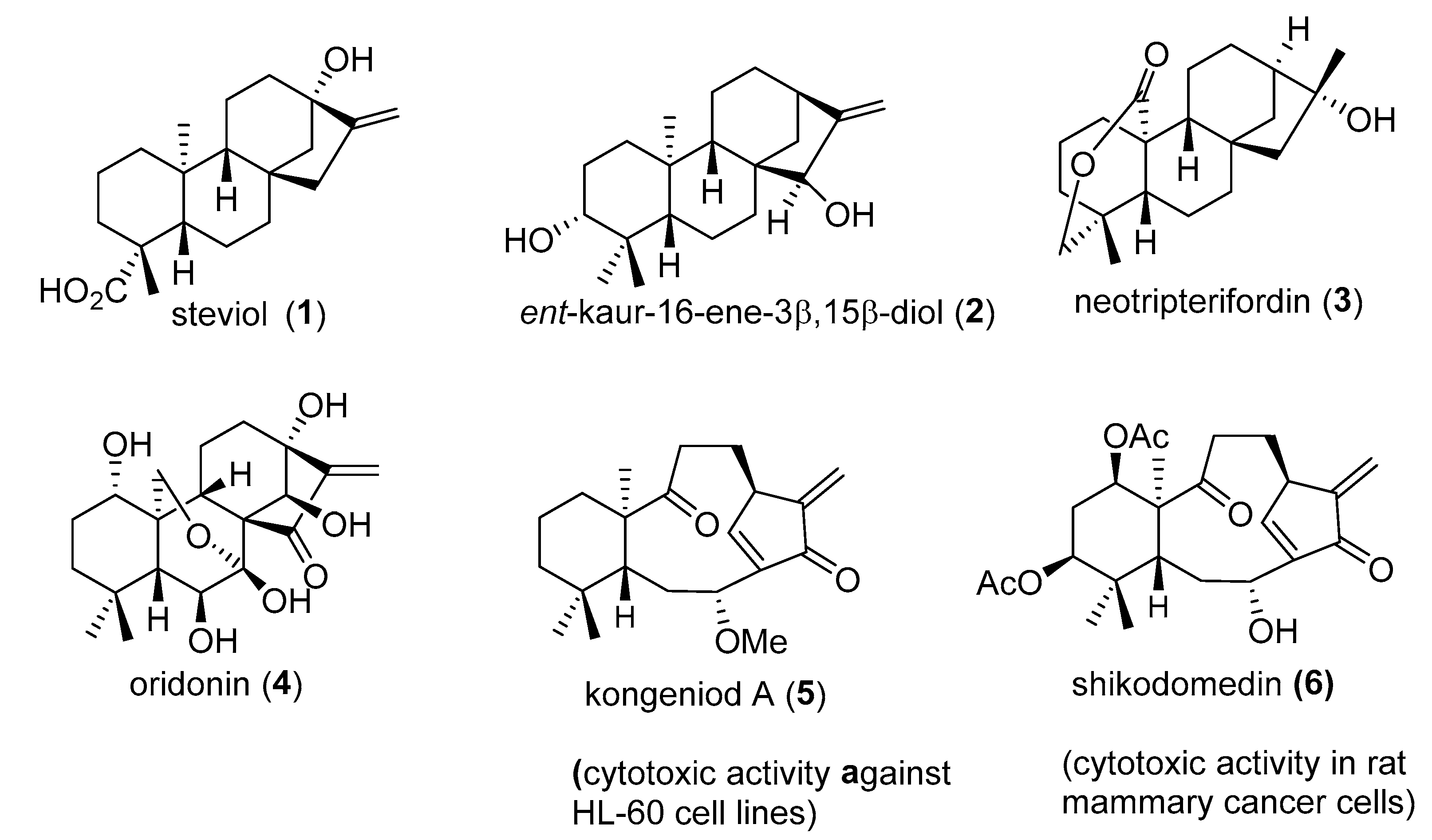
Figure 1. Some examples of ent-kaurane types.
Ijms 25 13222 g001
Figure 2. Representative ent-kaurane from Distichoselinium tenuifolium.
Ijms 25 13222 g002
Scheme 1. Preparation of some derivatives of 7.
Ijms 25 13222 sch001
Scheme 2. Semisynthesis of natural products ent-kaurene-3b,15a-diol (2) and ent-3b-hydroxykaurene-15-one (13) from 7.
Ijms 25 13222 sch002
Figure 3. Sigmoidal cell viability curves (or cytotoxicity) for kaurene derivatives in the three cell lines studied.
Ijms 25 13222 g003
Figure 4. Dose–response curves of ent-kaurene 13 in the three cancer cell lines. Relative viability percentages with respect to the untreated control were plotted against the logarithm of the concentration of each tested compound. Each data point represents the mean ± standard deviation from three replicates per concentration.
Ijms 25 13222 g004
Figure 5. Light microscopy images from the contrast phase of control cells (untreated) and cells treated with IC50 and IC80 concentrations of ent-kaurene 13 (white arrows indicate live cells; red arrows indicate apoptotic or dead cells).
Ijms 25 13222 g005
Figure 6. Effects of compound 7 on apoptosis in B16-F10 cells after 72 h treatment, at IC50 and IC80 concentrations. (A) Annexin V/propidium iodide (IP) cytometry plots: Q1, necrotic cells (Annexin V− PI+); Q2, late apoptotic cells (Annexin V+ PI+); Q3, normal cells (Annexin V− PI−); Q4, early apoptotic cells (Annexin V+ PI−); normal cells, blue quadrant; apoptotic cells, purple quadrant. (B) Flow cytometric analysis of Annexin V-FITC staining and PI accumulation: normal cells (blue bars), early apoptotic cells (brown bars), late apoptotic cells (green bars), and total apoptotic cells (purple bars). Values represent mean ± SD of three experiments performed in duplicate.
Ijms 25 13222 g006
Figure 7. Effects of compound 12 on apoptosis in HepG2 cells after 72 h treatment at IC50 and IC80 concentrations. (A) Annexin V/propidium iodide (IP) cytometry plots. Q1, necrotic cells (Annexin V− PI+); Q2, late apoptotic cells (Annexin V+ PI+); Q3, normal cells (Annexin V− PI−); Q4, early apoptotic cells (Annexin V+ PI−); normal cells, blue quadrant; apoptotic cells, purple quadrant. (B): Flow cytometric analysis of Annexin V-FITC staining and PI accumulation: normal cells (blue bars), early apoptotic cells (brown bars), late apoptotic cells (green bars), and total apoptotic cells (purple bars). Values represent mean ± SD of three experiments performed in duplicate.
Ijms 25 13222 g007
Figure 8. Effects of compound 13 on apoptosis in HT29 cells (A,B), HepG2 cells (C,D), and B16-F10 (E,F) after 72 h treatment at IC50 and IC80 concentrations. (A,C,E) Annexin V/propidium iodide (IP) cytometry plots; normal cells, blue quadrant; apoptotic cells, purple quadrant. (B,D,F): Flow cytometric analysis of annexin V-FITC staining and PI accumulation. Values represent mean ± SD of three experiments performed in duplicate.
Ijms 25 13222 g008
Figure 9. Flow cytometric analysis of the cell cycle of B16F10 melanoma cells after 72h of treatment with compound 7 at IC50 and IC80. (A) Representative histograms obtained via FACS analysis. Red: cells in G0/G1 and G2/M phases; blue diagonal lines: cells in S phase. (B) Changes in the percentage of cells in each phase of the cell cycle with respect to the control untreated cells. Values represent the mean ± standard deviation from at least two experiments in triplicate.
Ijms 25 13222 g009
Figure 10. Flow cytometric analysis of the cell cycle of HepG2 hepatocellular carcinoma cells with compound 12 at IC50 and IC80 concentrations after 72 h of treatment. (A) Representative histograms obtained by FACS analysis. Red: cells in G0/G1 and G2/M phases; blue diagonal lines: cells in S phase. (B) Changes in the percentage of cells in each phase of the cell cycle with respect to the untreated control cells. Values represent the mean ± standard deviation of at least two experiments in triplicate.
Ijms 25 13222 g010
Figure 11. Flow cytometric analysis of the cell cycle of HT29, B16-F10, and HepG2 cancer cells with compound 13 at IC50 and IC80 concentrations after 72 h of treatment. (A) Representative histograms obtained by FACS analysis. Red: cells in G0/G1 and G2/M phases; blue diagonal lines: cells in S phase. (B) Changes in the percentage of cells in each phase of the cell cycle with respect to untreated control cells. Values represent the mean ± standard deviation of at least two experiments in triplicate.
Ijms 25 13222 g011
Figure 12. Effects of products 7, 12, and 13 on the mitochondrial membrane potential. Compound 7 on B16-F10 (A), compound 12 on HepG2 (B), and compound 13 on HT29, HepG2, and B16-F10 cancer cell lines (C). The experiment was performed at IC50 and IC80 concentrations after 72 h treatment. The percentage of Rh123-positive or -negative cells relative to untreated control cells (control) is shown. Values are expressed as mean ± SD of two independent experiments performed in triplicate.
Ijms 25 13222 g012
Table 1. Effect of ent-kaurene diterpenoids on the viability of tumour cell lines HT29, HepG2, and B16-F10 after 72 h of treatment. IC20, IC50, and IC80 values are presented as mean ± standard deviation for three replicates. The ratio between the IC50 of compound 7 (precursor) and that of the other compounds (#) is also provided.
Cell LineCompoundIC20 (µM)IC50 (µM)IC80 (µM)IC50 7/IC50 #
HT2921.90 ± 1.1210.08 ± 2.6950.55 ± 4.377.46
771.82 ± 0.2775.27 ± 0.9079.47 ± 2.531.00
883.57 ± 4.89111.72 ± 3.39148.32 ± 2.670.67
992.42 ± 2.27153.83 ± 0.71202.16 ± 1.700.49
1149.71 ± 3.2974.96 ± 5.91114.91 ± 11.431.00
1227.28 ± 8.3645.37 ± 5.3485.58 ± 27.541.66
130.73 ± 0.082.71 ± 0.238.43 ± 2.6127.76
HepG2239.15 ± 15.3770.22 ± 9.92125.00 ± 4.661.04
771.38 ± 0.6672.84 ± 0.2974.30 ± 0.151.00
866.06 ± 12.57107.69 ± 6.08166.59 ± 7.720.68
948.02 ± 7.57101.49 ± 7.65171.30 ± 6.000.72
1131.76 ± 4.1047.26 ± 5.7872.90 ± 10.871.54
128.79 ± 9.3024.43 ± 16.3581.75 ± 13.642.98
130.79 ± 0.302.12 ± 0.235.36 ± 0.8634.42
B16-F10252.32 ± 3.7479.58 ± 3.58125.56 ± 5.550.41
727.40 ± 4.6232.43 ± 3.2837.93 ± 2.941.00
8128.36 ± 2.81145.32 ± 2.76168.42 ± 3.600.22
983.33 ± 4.50107.13 ± 3.95138.94 ± 4.680.30
1125.04 ± 2.2842.04 ± 1.5566.97 ± 1.050.77
1264.26 ± 6.6594.54 ± 8.33143.50 ± 9.360.34
130.79 ± 0.132.65 ± 0.1354.29 ± 4.0012.26
Disclaimer/Publisher’s Note: The statements, opinions and data contained in all publications are solely those of the individual author(s) and contributor(s) and not of MDPI and/or the editor(s). MDPI and/or the editor(s) disclaim responsibility for any injury to people or property resulting from any ideas, methods, instructions or products referred to in the content.