Communication

Association of Neuroblastoma (NB) SH-SY5Y Cells with Antibodies of Parasitic Origin (Anti-Acanthamoeba and Anti-Toxocara canis)

by
Víctor Alberto Maravelez Acosta
1,
Maria de Lourdes Caballero Garcia
1,
Genaro Patiño López
2,
María del Pilar Crisóstomo Vázquez
1,
Luz Ofelia Franco Sandoval
1 and
Leticia Eligio García
1,*
1
Laboratorio de Investigación en Parasitología, Hospital Infantil de México Federico Gómez (HIMFG), Dr. Márquez 162. Col Doctores, Cuauhtémoc, México City 06720, Mexico
2
Unidad de Investigación en Inmunología y Proteomica, Hospital Infantil de México Federico Gómez (HIMFG), Dr. Márquez 162. Col Doctores, Cuauhtémoc, México City 06720, Mexico
*
Author to whom correspondence should be addressed.
Int. J. Mol. Sci. 2024, 25(24), 13577; https://doi.org/10.3390/ijms252413577
Submission received: 28 September 2024 / Revised: 27 October 2024 / Accepted: 28 October 2024 / Published: 19 December 2024

Abstract

:
It is little known that Acanthamoeba trophozoites and Toxocara canis eggs can reduce tumors in vitro and animal models. Although this has been known for many years, the mechanism that induces the antitumor effect in these parasites is still not known. We employed Western blot (WB) and immunofluorescence (IFC) by confocal microscopy to explore the potential protein binding between neuroblastoma (NB) SH-SY5Y cells and anti-Acanthamoeba and anti-Toxocara canis antibodies. Using WB, we detected two fragments of 70 kDa and 60 kDa recognized by the anti-Acanthamoeba antibodies, and two fragments of 115 kDa and 70 kDa recognized by the anti-Toxocara canis antibodies. In both cases, the IFC results were positive in the cell membrane of the SH-SY5Y cells. Our findings suggest a potential overlap of similar molecules between these parasites and tumor cells, which may contribute to tumor elimination. Investigating the relationship between anti-Acanthamoeba and anti-Toxocara canis antibodies in neoplastic cells could provide evidence for the future use of these anti-parasitic antibodies in targeting NB or other cancers.

1. Introduction

Neuroblastoma (NB), the most common extracranial solid tumor in children under the age of 5, was described as early as the 19th century, and its complexity has continued to intrigue researchers, as well as medical and surgical specialists. At one end of the phenotypic spectrum, neuroblastoma is self-limiting with minimal to no intervention required, while on the opposite end exists the challenge of refractory disease despite aggressive management and toxic systemic treatments [1]. Immunotherapy has faced challenges in pediatric patients, and although the etiology of neuroblastoma is multifactorial, a significant portion of the lack of response is thought to be attributed to the tumor microenvironment [2]. A prominent immunotherapeutic strategy in NB involves using antibodies against the tumor-associated disialoganglioside GD2. This strategy is used due to the fact that the Fc portion of the anti-GD2 antibodies, which binds to neuroblastoma tumor antigens, is recognized by natural killer (NK) cells, through antibody-dependent cell-mediated cytotoxicity (ADCC) [3]. It is possible to combine immunotherapy with standard chemotherapy or other immunomodulatory agents to enhance the therapeutic effect. Additionally, research is being conducted to prolong the efficacy and potency of therapies aimed at improving the expansion and activation of NK cells [4]. Despite recent advances in the management and therapeutics of cancer, the treatment of the disease is limited by its excessive cost and severe side effects. In this scenario, there is an unmet need to identify novel treatment alternatives for this dreaded disease [5].
Several parasites have shown the ability to slow certain types of cancer growth [6]. Table 1 presents the parasites that have been most extensively studied for their antitumor effects.
A little-known fact is that Acanthamoeba and Toxocara canis have been associated with possible anticancer activity.
Acanthamoeba is a unicellular protozoan and has an evolutionary history that spans at least a billion years [49]; its pathological significance was acknowledged in the 1960s and 1970s, and it was recognized as the etiological agent of amoebic granulomatous encephalitis and keratitis [50]. Acanthamoeba is widely distributed in nature, existing in the form of free-living organisms or parasites, and is frequently associated with biofilms in various environments. Since 1986, Acanthamoeba has emerged as a global public health concern due to the use of contact lenses, although they have historically caused disease in immunosuppressed individuals [51]. Acanthamoeba can interact with various microorganisms, including bacteria, fungi and viruses [52]. As a result, Acanthamoeba can act as a predator, a vehicle for transmission, or an incubator in natural environments [53]. Acanthamoeba feeds on bacteria by absorbing them through phagocytosis and subsequently lysing them in phagolysosomes. Additionally, Acanthamoeba can function as a ‘Trojan horse’, serving as a vehicle for microbial transmission in the environment. In other words, Acanthamoeba can incubate microorganisms that are pathogenic to humans. These microorganisms utilize the parasite’s defense mechanisms to evade the immune system and treatment while reproducing freely within it. Consequently, Acanthamoeba serves as a ‘genetic melting pot’, promoting gene exchange and the adaptation of microorganisms, which in turn enhances their pathogenicity [52]. Acanthamoeba castellanii trophozoites demonstrated a strong chemotactic response toward human melanoma (OCM-1) and murine melanoma (D5.1G4) cells. This response was observed by injecting either live parasites or cell-free parasite lysates into a melanoma animal model, which led to a reduction in tumor mass between 53% and 83% [54]. Murine NB cells (NB41A3) were exposed to a parasite extract of Acanthamoeba castellanii. The results indicated that over 70% of NB41A3 DNA was fragmented, and several morphological features of apoptosis were observed, including cell shrinkage, vesicle formation on the cell membrane, the formation of apoptotic bodies, and nuclear condensation. The results suggest that at least one species of pathogenic free-living amoeba can lyse tumor cells by a process that culminates in apoptosis [55]. The cytotoxic effect of five isolates of Acanthamoeba (trophozoítes) on human cervix cancer HeLa cells culture was investigated. The HeLa cells showed a sequence of morphological features of apoptosis and 50% of the tumor cells underwent cytolysis. These five strains of Acanthamoeba exhibit cytotoxic effects of varying degrees on HeLa cells [56]. Signs of apoptosis were observed when NB cells were exposed to an isolate of Acanthamoeba known as AS. Notable features included cell contraction, nuclear condensation, and DNA fragmentation. Apoptosis was determined to be mediated by the involvement of caspase enzymes and the pro-apoptotic and anti-apoptotic mitochondrial proteins Bax and Bcl-2 [57]. The effect of cell-free supernatants that was obtained from four axenic cultures of Acanthamoeba (trophozoítes) was investigated on three cancer cell lines (human prostate cancer, rat prostate cancer and breast cancer). All analyzed samples demonstrated an inhibitory effect on prostate cancer cells; however, this effect was not observed in breast cancer cells [58].
Toxocara canis infection (toxocariasis) is a cosmopolitan, zoonotic and neglected disease [59,60]. Toxocariasis is an infection caused in canines, felines, humans and other vertebrates by species of the genus Toxocara, such as Toxocara canis and Toxocara cati [61]. The definitive hosts are domestic or wild canids (such as dogs), and due to perinatal transmission, it also affects puppies under three months of age [62,63]. The primary mode of infection acquisition is through the ingestion of embryonated eggs, affecting both definitive hosts (dogs and cats) and paratenic hosts, including humans [61]. Toxocara canis has been shown to have both antitumor and protumor growth properties [9,10,11,12,13,14,15,16,17,18,19,20,21,22,23,24,25,26,27,28,29,30,31,32,33,34,35,36,37,38,39,40,41,42,43,44,45,46,47,48,49,50,51,52,53,54,55,56,57,58,59,60,61,62,63,64,65,66]. Toxocara canis egg antigens induced the inhibition of tumor growth in the fibrosarcoma mouse model; the mean tumor area in Toxocara canis-injected mice over four different days was 24.5 mm2 compared to the control group (alum treated) which was 155 mm2 [9]. A peptide sequence was synthesized from an excretory–secretory antigen of Toxocara canis (TOXCA Troponin T protein) and the possible anticancer properties and their effect on gastrointestinal and liver cancer cell proliferation-related genes in laboratory conditions were evaluated. The peptide, when administered at high concentrations, promotes cancer cell mortality and alters gene expression related to apoptosis, metastasis and angiogenesis [24]. Due to their ability to alter the host’s immune response and their widespread presence in the environment, helminths have been found to promote tumor growth. This has been demonstrated in studies involving Schistosoma haematobium, Clonorchis sinensis and Opisthorchis viverrine [65,66]. Toxocara canis exhibits immunomodulatory properties that enable it to infect humans and other hosts [67]. By modulating the immune response, Toxocara canis can influence tumor growth in mice, leading to the development of larger tumors; in other words, Toxocara canis modulates the immune microenvironment of the tumor [68]. The intratumoral injection of Toxocara canis excretory/secretory antigens (EST) promotes lung metastasis through the modulation of the tumor immune microenvironment in mice. ESTs were injected intratumorally into mice with lung cancer, resulting in no significant changes in tumor size or weight. However, there was a marked increase in microvasculature and the development of both micro- and macro-metastases in the lungs, along with elevated levels of VEGF [69]. Toxocara canis increases the potential of breast cancer by reducing the expression of P53, a tumor suppressor protein, and increases the Ki-67 protein; this means that cancer cells divide rapidly [70]. Evidence suggests that Toxocara canis has an antitumor effect in its egg form and a protumor effect during infection in its larval form.
Acanthamoeba trophozoite and Toxocara canis egg antigens induce apoptosis in neoplastic cells, but which parasite antigens interfere with tumor growth has not been investigated. For this reason, we focused on first identifying whether there is a recognition of anti-Acanthamoeba and anti-Toxocara canis antibodies on the membranes of SH-5S5Y and then detected possible proteins that have unexpected cross-linking between anti-Acanthamoeba/anti-Toxocara canis antibodies in the antigens of SH-5S5Y cells.

2. Results

2.1. Recognition of Anti-Acanthamoeba and Anti-Toxocara canis Antibodies on the Total Proteins of SH-5S5Y

To identify SH-5S5Y proteins recognized by anti-Acanthamoeba and anti-Toxocara canis antibodies, we performed WB analysis using total protein extracts of SH-5S5Y against a panel of anti-Acanthamoeba and anti-Toxocara canis antibodies, both at a concentration of 8.8 μg/μL and at a dilution of 1:100. Prominent fragments were consistently detected (Figure 1); we found two fragments of 70 Kdal and 60 Kdal recognized by the anti-Acanthamoeba antibodies and two fragments of 115 Kdal and 70 Kdal recognized by the anti-Toxocara canis antibodies. These findings suggest that anti-Acanthamoeba and anti-Toxocara canis antibodies may effectively target the specific antigens that are present within SH-5S5Y.

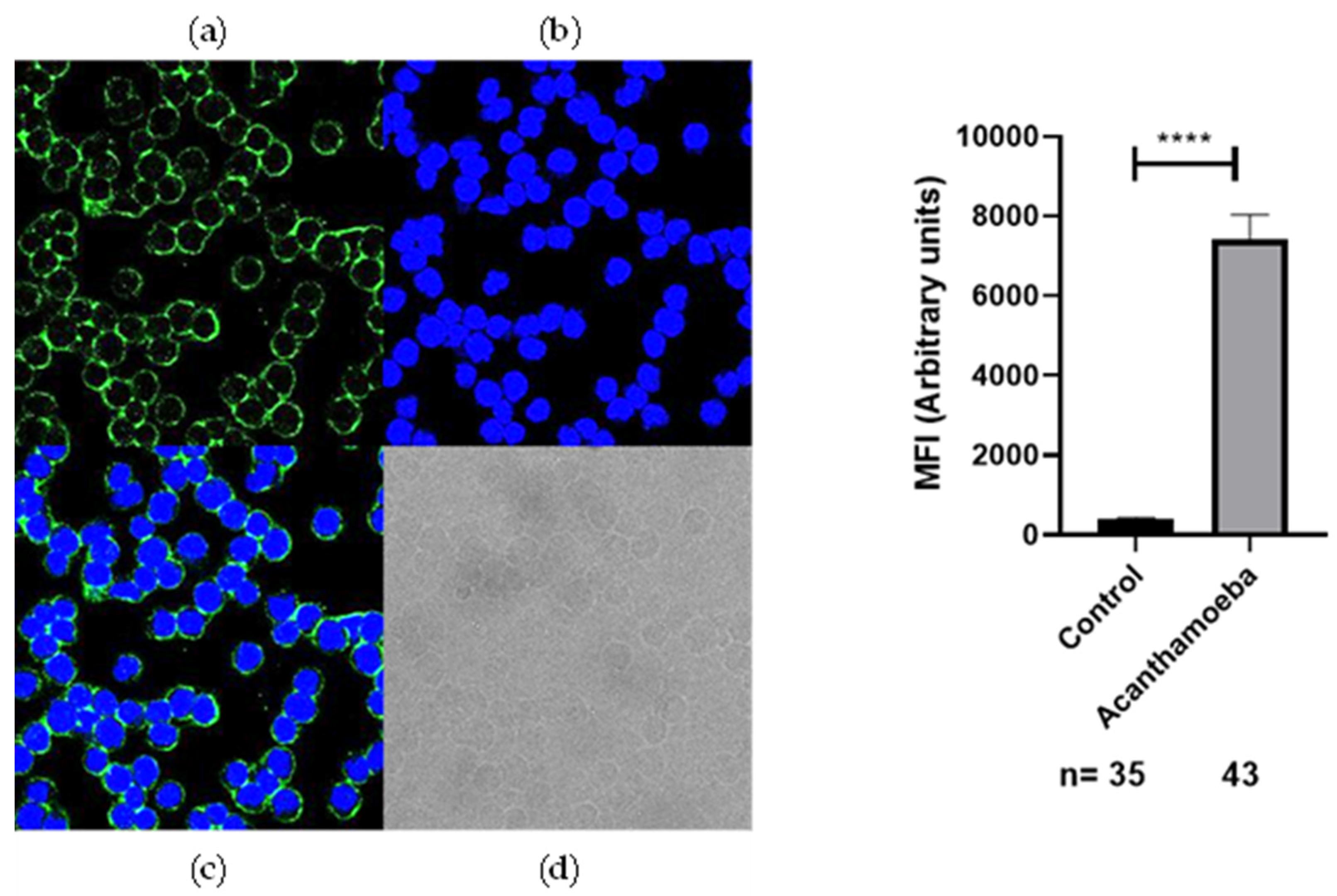
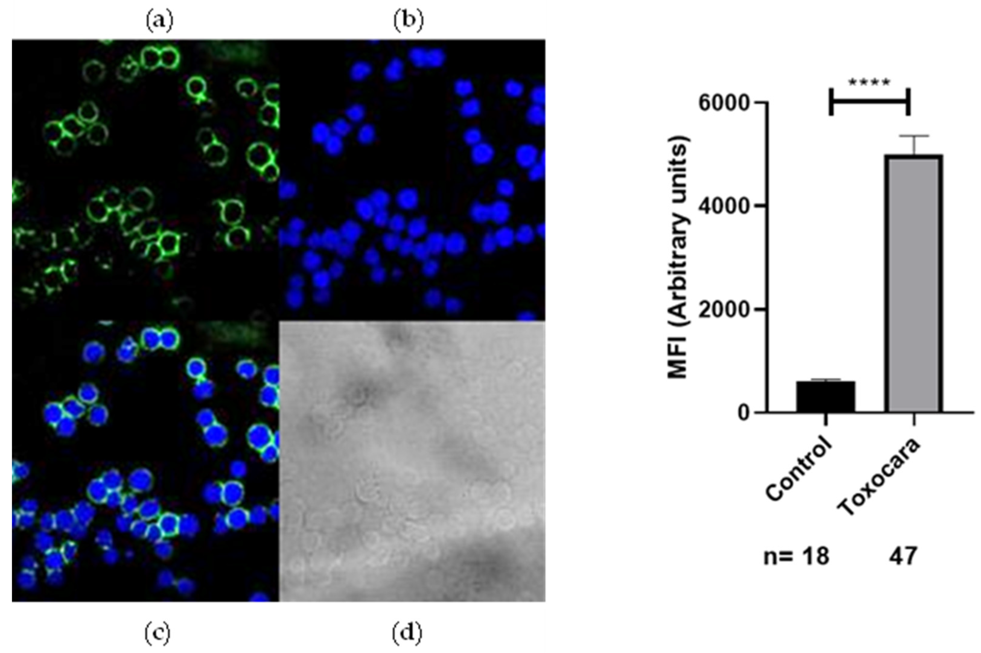
2.2. Recognition of Anti-Acanthamoeba and Anti-Toxocara canis Antibodies on the Membrane of SH-5S5Y

To investigate the recognition of anti-Acanthamoeba and anti-Toxocara canis-specific antibodies by the membrane protein SH-5S5Y, IFC analysis was performed. We evaluated non-permeabilized SH-5S5Y. Anti-Acanthamoeba and anti-Toxocara canis antibodies (8.8 μg/µL) were diluted (1:300) and incubated with SH-5S5Y-expressing cells. The binding of these antibodies was detected using Alexa Fluor 488-conjugated anti-rabbit IgG antibodies (1:300). The results indicated that SH-5S5Y-expressing cells displayed positive staining for anti-Acanthamoeba (Figure 2) and anti-Toxocara canis (Figure 3) in the membrane of the cells. These findings suggest that a substantial proportion of SH-5S5Y molecules on the cell surface are recognized by anti-Acanthamoeba/anti-Toxocara canis-specific antibodies. The fluorescence intensity quantification for anti-Acanthamoeba and anti-Toxocara canis antibodies was analyzed alongside their respective control (rabbit preimmune IgG). The statistical analysis shows a p-value of <0.0001 (****) for both antibodies, indicating an extremely statistically significant result. Figure 2 and Figure 3 display the graphs of fluorescence intensity quantification for each antibody alongside its respective control.

3. Discussion

In studies conducted with Acanthamoeba, it has been demonstrated that the antigens of the parasite, when injected into a murine melanoma model, suppress tumor cells [54]. Similarly, in Toxocara canis, antigens have been shown to suppress tumor cells in a murine fibrosarcoma model [9]. The antitumoral effect of Acanthamoeba and Toxocara canis has ceased to be a subject of study, and the mechanisms involved in tumor suppression have not yet been defined. The role of anti-Acanthamoeba and anti-Toxocara canis antibodies in this antitumoral effect has not been studied. This is the first study to analyze the potential of antibodies induced by Acantamoeba trophozoites and Toxocara canis eggs antigens on cancer cells. In this study, neuroblastoma cells from the SH-5S5Y cell line were used.
To better understand the possible usefulness of using anti Acantamoeba and anti Toxocara canis antibodies to recognize the antigens of SH-5S5Y, we performed WB, where we used the total proteins of SH-5S5Y vs. the anti-Acantamoeba and anti-Toxocara canis antibodies. The results demonstrate that there is a cross-linking between anti-Acantamoeba and anti-Toxocara canis antibodies with SH-5S5Y antigens; see Figure 1. We identified two bands of approximately 70 Kdal and 60 Kdal that cross-link with the anti-Acantamoeba antibodies, and two bands of approximately 115 Kdal and 70 Kdal that cross-link with the anti-Toxocara canis antibodies. Each band contains a mixture of proteins, where one or more may contribute to the antitumor effect. Another strategy involved analyzing whether the anti-Acanthamoeba and anti-Toxocara canis antibodies could specifically recognize antigens on the SH-5S5Y membrane; therefore, we conducted IFC by confocal microscopy. We also found that the membrane antigens of SH-5S5Y specifically recognize the anti-Acantamoeba antibodies Figure 2 and the anti-Toxocara canis antibodies Figure 3. The results of the IFC were consistent for both antibodies, showing A clear recognition of anti-Acantamoeba and anti-Toxocara canis antibodies binding to the antigens present in the membrane of SH-5S5Y. The statistical analysis of fluorescence intensity quantification for the anti-Acanthamoeba and anti-Toxocara canis antibodies shows a p-value of <0.0001 (****) for both antibodies, indicating an extremely statistically significant result. Figure 2 and Figure 3 present the fluorescence intensity quantification graphs for each antibody alongside its respective control. The recognition of SH-5S5Y membrane antigens by anti-Acantamoeba and anti-Toxocara antibodies suggests that these antibodies may be involved in cellular cytotoxicity mechanisms against cancer cells, potentially leading to a reduction in tumor mass, as demonstrated in studies utilizing murine models of melanoma and fibrosarcoma [54,58].
The antitumor effect of Acantamoeba on NB cells has been previously studied, revealing that (i) antigens from lysate trophozoites induce apoptosis in cultured NB cells [55] and (ii) in co-culture experiments involving trophozoites and NB cells, AS trophozoites (T4) attacked the NB cell monolayers immediately upon addition, penetrated the monolayers, and induced apoptosis in NB cells via the Bax pathway [57]. Additionally, we have demonstrated that anti-Acanthamoeba antibodies exhibit cross-linking with both total antigens and the antigens present in the SH-5S5Y membrane, highlighting an intriguing direction for further study. In a previous study, Acanthamoeba was examined in vivo and it was revealed that the injection of trophozoites lysates into progressively growing subcutaneous melanomas resulted in 83% and 53% reductions in the tumor masses [54]; in other words, anti-Acantamoeba antibodies were generated and contributed to tumor reduction through the cross-recognition of antigens present in cancer cells.
There is evidence that some helminths are inductors of different types of tumors [71]. Toxocara canis chronic infection in BALB/c mice results in a type 2 response with an incipient regulatory response [71], and chronic infection with Toxocara canis in BALB/c mice with breast cancer accelerates tumor growth and is associated with alterations in the tumor microenvironment. The higher proportions of immune cells found in the tumor microenvironment comprised F4/80+ macrophages and CD19+ B cells, which could contribute to tumor enlargement and reduce the proportions of CD8+ lymphocytes. Furthermore, it was found that the IL-4 and VEGF levels were elevated, and a microenvironment characterized by higher levels of IL-10 was observed. This study demonstrated that Toxocara canis infection promotes a protumor effect through the modulation of the tumor immune microenvironment [69]. Additionally, the intratumoral injection of excretion/secretion antigens from Toxocara canis (EST) in lung cancer models induces a protumoral effect, without significant changes in tumor size or weight; it was noted that lung tumors exhibited increased micro- and macro-metastasis, as well as enhanced microvasculature. In contrast, EST did not alter the proportions of immune cells in the tumor, spleen, or peripheral lymph nodes. Therefore, the effect of EST was to promote lung metastasis by modulating the tumor’s immune microenvironment [64]. Therefore, Toxocara canis infection in a murine model of breast cancer, as well as the intratumoral injection of EST in a lung cancer model, alters the tumor microenvironment, thereby promoting a protumoral effect. Helminths have been recognized as inducers or promoters of cancer due to their ability to regulate the host’s immune response [69,70]. In contrast, we utilize a total extract of Toxocara canis antigens derived from larvae eggs to generate antibodies. ESTs were not included in our experiments, as we did not collect them; however, we do not rule out the possibility that small amounts could be present within the parasite before secretion. Nonetheless, larval Toxocara canis eggs have never been shown to provide the conditions necessary to stimulate the production of EST. We found that anti-Toxocara canis antibodies recognized SH-5S5Y antigens Figure 1 and Figure 3, suggesting that these antibodies may be associated with the antitumor effect of Toxocara canis due to the similarity between the proteins of the parasite and those of SH-5S5Y. Toxocara canis egg antigens inhibited tumor growth in a fibrosarcoma mouse model, likely because the immune responses elicited by parasite antigens may exert non-specific effects on tumor cells [9]. By comparing our results with those of previous studies, we can deduce that the antitumor effect of Toxocara canis may be attributed to the ability of anti-Toxocara canis antibodies to recognize antigens on the SH-5S5Y membrane and potentially participate in therapeutic mechanisms such as antibody-dependent cellular cytotoxicity (ADCC) or complement-dependent cytotoxicity (CDC) against cancer cells. In the case of other parasites with antitumor effects, such as Trypanosoma cruzi, total antigens, antibodies and induced infections in mice have demonstrated antitumor activity [35,72,73,74]. We did not observe this effect with Toxocara canis. Although it has been established that helminths are associated with cancer induction [66], we find a duality: (i) the in vivo infections in mice and the EST of the parasite modify the tumor microenvironment and promote tumor growth [69]; on the other hand, (ii) antigens of the parasite that are absent from ESTs could induce an antitumor effect in vivo tests [9]. Our results indicate that the antitumor effect of Toxocara canis could be attributed to the cross-linking between anti-Toxocara canis antibodies and cancer cell antigens. This interaction could initiate mechanisms involving antibodies, such as ADCC and CDC. Our results also suggest the possibility that helminth lysates may exert an antitumor effect, contrasting with the protumor effects associated with the helminth infections of EST. However, this remains a hypothesis that requires thorough investigation. The involvement of Trypanosoma cruzi in the antitumor effect can elicit both humoral and cellular responses [36]. This suggests that a similar response could occur with Acantamoeba and Toxocara canis antigens.
The cross-linking between anti-Acantamoeba and anti-Toxocara canis antibodies and SH-5S5Y cell antigens could be responsible for the tumor cell suppressor effect, since there are similar antigens with SH-5S5Y. In future research, it will be essential to determine the cytotoxic potential (ADCC and CDC) of anti-Acantamoeba and anti-Toxocara canis antibodies on NB cells and other cancer types. Additionally, identifying the specific proteins involved in tumor suppression is essential, as this knowledge could help advance the development of future cancer therapies based on parasitic-origin molecules. The antitumor effects of Acanthamoeba and Toxocara canis require further investigation. This work brings us closer to establishing that antibodies induced by Acanthamoeba and Toxocara canis antigens play a role in the antitumor effect.

4. Materials and Methods

4.1. Cell Line of SH-SY5Y and Obtaining the Antigen

The NB cell lines SH-SY5Y were obtained from ATCC (Manassas, VA, USA). The cell line was cultured in DMEM media (Gibco, Gran Island, NY, USA) supplemented with 10% fetal calf serum (Gibco, NY, USA) and 1X Antibiotic-Antimycotic (Gibco, Gran Island, NY, USA). These were counted in a Neubauer chamber and assessed for viability with trypan blue.

4.2. Acanthamoeba Trophozoites Culture and Obtaining the Antigen

Acanthamoeba trophozoites were obtained from viable cysts stored in Petri dishes with non-nutritive agar at 4 °C. For excystment, a 1 × 0.5 cm gel sample was taken and sown in Petri dishes with non-nutritive agar (NaCL 0.06 g, magnesium sulfate 0.002, anhydrous dibasic sodium phosphate 0.007 g, bacteriological agar 7.5 g and monobasic potassium phosphate 0.068 g) together with 100 uL of culture (5% meat peptone) saturated with inactivated E. coli for 1 h at 56 °C and incubated at 37 °C for 96 h for excystment. The trophozoites were expanded in PBSMG medium (peptone biotryptase 8.3 g, dextrose 1.3 g, anhydrous dibasic sodium phosphate 0.75 g, monobasic potassium phosphate 0.45 g and 10% FBS) for 7 days at 28 °C. To obtain the antigen, the Acanthamoeba trophozoites were harvested and washed. The trophozoites were counted in a Neubauer chamber and the viability was assessed with trypan blue. To extract the antigen subsequently, the cell pellet was resuspended in cold protein lysis buffer (1% Nonidet P-40, 150 mM NaCl, 10 mM Tris-HCl pH 7.6, 10 mM PMSF, 2 mM EDTA, Protinin A 1 µg/mL, Pestatin A 1 µg/mL y Leupeptin A 1 µg/mL) and lysed by freezing for 30 min. The total antigen concentration obtained was determined on a UV-Visible spectrophotometer at 280 nm Epoch (Biotec Instruments, Winooski, VT, USA) [72].

4.3. Toxocara canis Eggs Culture and Obtaining the Antigen

To obtain Toxocara canis eggs, the small intestine of a puppy sacrificed by the Canine Control Center of CDMX, Mexico, was recovered. The ends of the intestine were tied together for transport. In the laboratory, the intestine was cut into fragments of approximately 3 cm; subsequently, each fragment was cut longitudinally. When Toxocara canis was identified, it was placed in culture flasks with 1X PBS. To obtain embryonic eggs, 2 females and 2 males were placed in a culture box with warm 1X PBS and incubated at 37 °C for 24 h. The next day, all of the PBS were collected and centrifuged at 2500 rpm for 5 min and the larvae eggs were recovered. EST were not considered in this study. The eggs were counted in a Neubauer chamber. Subsequently, to extract the antigen, the cell pellet was resuspended in cold protein lysis buffer (1% Nonidet P-40, 150 mM NaCl, 10 mM Tris-HCl pH 7.6, 10 mM PMSF, 2 mM EDTA, Protinin A 1 µg/mL, Pestatin A 1 µg/mL y Leupeptin A 1 µg/mL) and mechanically lysed in a mortar under freezing conditions for 30 min using liquid nitrogen. The total antigen concentration obtained was determined on a UV-Visible spectrophotometer at 280 nm Epoch (Biotec Instruments, Winooski, VT, USA) [72].

4.4. Generation of Anti-Acanthamoeba and Anti-Toxocara canis In Vivo with New Zeeland Rabbits

Six-week-old rabbits were obtained from of the animal facility of the Children’s Hospital de Mexico Federico Gomez (Mexico City, Mexico). The animal study protocol was approved by the Ethics Committee of the Children’s Hospital of Mexico Federico Gomez (HIM-2014-035) and follows the guidelines for animal experiments in Mexico. Antibody induction was conducted separately using proteins derived from Acanthamoeba trophozoites and Toxocara canis eggs as immunogens. At time 0, the rabbits were injected with 2 mg of antigen dissolved in 150 µL physiological solution saline via an intradermal route and were emulsified with 150 µL Freud’s complete adjuvant (Sigma, St. Louis, MO, USA); on day 15, 2 mg of antigen dissolved in 150 µL physiological solution saline was injected via an intradermal route and emulsified with 150 µL Freud’s complete adjuvant (Sigma, St. Louis, MO, USA). On day 30, the rabbits were injected intravenously with 0.25 mg antigen dissolved in 150 µL physiological solution saline, on day 31, they were injected intravenously with 0.50 mg antigen dissolved in 150 µL physiological solution saline and on day 32, they were injected intravenously with0.50 mg antigen dissolved in 150 µL physiological solution saline. The total collection of the serum was on day 39 [72].

4.5. Purification of Anti-Acanthamoeba and Anti-Toxocara canis Antibodies by Affinity Chromatography with Protein A/G

IgG purification followed the manufacturer’s protocol from Thermo Scientific, using affinity chromatography with protein A/G using Spin Columns (Thermo Scientific, Rockford, IL, USA). The concentrations of purified antibodies were quantified by measuring the absorbance of each fraction at 280 nm in an Epoch (Biotec Instruments, Winooski, VT, USA) UV/Vis spectrophotometer [72].

4.6. Identification of SH-SY5Y Proteins Recognized by Anti-Acanthamoeba and Anti-Toxocara canis Antibodies by Western Blotting

To identify the proteins of the SH-SY5Y that are recognized by anti-Acanthamoeba and anti-Toxocara canis antibodies, Western blotting was performed. The separating gel was prepared with 12% acrylamide–bisacrylamide (Sigma, Amsterdam, The Netherlands) and the concentrator gel with 4% acrylamide–bisacrylamide. The samples were prepared 1:5 with sample buffer under reducing conditions. The voltage was set to 200 V (constant) for 50 min. The electrophoresis gel was placed on a 0.2 µM nitrocellulose membrane (Amersham Biosciences, Region, UK) using the Trans-Blot-Transfer (Bio-Rad, Hercules, CA, USA) chamber cassette; the voltage was conditioned at 100 V for 2 h. To verify the proper transfer of proteins to the nitrocellulose membrane, the membrane was stained with 0.2% Ponceau red and washed with water until the dye was completely removed. The dilution ratio of the anti-Acanthamoeba and anti-Toxocara canis antibodies was 1:100 overnight, both starting at a concentration of 8.8 μg/μL. The rabbit anti-IgG antibody coupled to horseradish peroxidase (Sigma, Jerusalem, Israel) was at a 1:5000 dilution for 2 h. The blot was developed with 4-CN (Sigma, USA) and H2O2 (ProdQuimMonterrey, Monterrey, Mexico) [72].

4.7. Determination of Anti-Acantamoeba and Anti-Toxocara canis Antibodies Recognition with Neuroblastom Cells by Immunofluorescence in Confocal Microscopy

For evaluating the recognition of anti-Acantamoeba and anti-Toxocara canis antibodies by IFC analyses, the line cell SH-SY5Y was used. The used dilution of the anti-Acantamoeba and anti-Toxocara canis antibodies was 1:300 for 1 h, both starting at a concentration of 8.8 μg/μL. For reactivity, specific anti-rabbit IgG antibodies conjugated to Alexa-Fluor 488 (Invitrogen, Lane County, OR, USA) were used and diluted 1:300 for 1 h. Core staining was carried out with 20 µL of mounting medium with 4′,6-diamidino-2-phenylindole diluent (DAPI) (Vector Laboratories, Newark, CA, USA). Images were captured using a TCS-5P8X (Leica, Microsystems, Wetzlar, Germany) confocal microscope and analyzed with Fiji software version 1.54k (https://imagej.net/software/fiji/downloads (accessed on 22 October 2024)) and included the analysis of fluorescence intensity quantification.

Author Contributions

Conceptualization, V.A.M.A.; methodology, V.A.M.A., M.d.L.C.G., G.P.L., L.E.G., L.O.F.S. and M.d.P.C.V.; software, V.A.M.A. and G.P.L.; validation, V.A.M.A. and G.P.L.; formal analysis, V.A.M.A. and G.P.L.; investigation, V.A.M.A. and G.P.L.; resources, V.A.M.A.; data curation, V.A.M.A., G.P.L. and L.E.G.; writing—original draft preparation, V.A.M.A.; writing—review and editing, V.A.M.A. and G.P.L.; visualization, V.A.M.A., M.d.L.C.G., G.P.L., L.E.G., L.O.F.S. and M.d.P.C.V.; supervision, L.E.G.; project administration V.A.M.A.; funding acquisition, V.A.M.A. All authors have read and agreed to the published version of the manuscript.

Funding

This research was funded by Federal Funds, grant number HIM/2014/035 of Hospital Infantil de México Federico Gómez, Secretaría de Salud de México.

Institutional Review Board Statement

The animal study protocol was approved by the Institutional Review Board (or Ethics Committee) of HOSPITAL INFANTIL DE MEXICO FEDERICO GOMEZ (protocol code HIM(2014/035 approved 20 February 2014).

Informed Consent Statement

Not applicable.

Data Availability Statement

Data are contained within the article.

Acknowledgments

The authors would like to thank the veterinarians VMD Raul Castro Luna and VMD Leonel Martínez Cristóbal from the Animal Facility, as well as the Technician Andres Flores Luna from the Hospital Infantil de México Federico Gómez, for their valuable support.

Conflicts of Interest

The authors declare no conflict of interest. The funders had no role in the study design, data collection, analysis or interpretation, in the writing of the manuscript, or in the decision to publish the results.

References

  1. Williams, K.M.; Shah, N.R.; Chukkapalli, S.; King, S.; Grant, C.N.; Brown, E.G.; Avanzini, S.; Lal, D.R.; Sarnacki, S.; Newman, E.A. Modern surgical strategies in pediatric neuroblastoma: Evolving approaches and treatment principles. Pediatr. Blood Cancer, 2024; e31317Advance online publication. [Google Scholar] [CrossRef] [PubMed]
  2. Rados, M.; Landegger, A.; Schmutzler, L.; Rabidou, K.; Taschner-Mandl, S.; Fetahu, I.S. Natural killer cells in neuroblastoma: Immunological insights and therapeutic perspectives. Cancer Metastasis Rev. 2024; Advance online publication. [Google Scholar] [CrossRef] [PubMed]
  3. Esser, R.; Müller, T.; Stefes, D.; Kloess, S.; Seidel, D.; Gillies, S.D.; Aperlo-Iffland, C.; Huston, J.S.; Uherek, C.; Schönfeld, K.; et al. NK cells engineered to express a GD2-specific antigen receptor display built-in ADCC-like activity against tumour cells of neuroectodermal origin. J. Cell. Mol. Med. 2012, 16, 569–581. [Google Scholar] [CrossRef] [PubMed]
  4. Raiser, P.; Schleiermacher, G.; Gambart, M.; Dumont, B.; Defachelles, A.S.; Thebaud, E.; Tandonnet, J.; Pasqualini, C.; Proust, S.; Entz-Werle, N.; et al. Chemo-immunotherapy with dinutuximab beta in patients with relapsed/progressive high-risk neuroblastoma: Does chemotherapy backbone matter? Eur. J. Cancer 2024, 202, 114001. [Google Scholar] [CrossRef] [PubMed]
  5. Walter, N.S.; Bhattacharyya, S. Mining parasites for their potential as novel therapeutic agents against cancer. Med. Oncol. 2024, 41, 211. [Google Scholar] [CrossRef]
  6. Eissa, M.M.; Salem, A.E.; El Skhawy, N. Parasites revive hope for cancer therapy. Eur. J. Med. Res. 2024, 29, 489. [Google Scholar] [CrossRef]
  7. Xin, W.; Li, S.; Li, Z.; Zheng-Quan, L.; Jiang, L.; Lin-Xi, Z. Impact of Toxoplasma gondii on the proliferation and apoptosis of tumor cell lines. Zhongguo Ji Sheng Chong Xue Yu Ji Sheng Chong Bing Za Zhi (CJPPD) 2012, 30, 17. [Google Scholar]
  8. Pyo, K.-H.; Jung, B.-K.; Chai, J.-Y.; Shin, E.-H. Suppressed CD31 expression in sarcoma-180 tumors after injection with Toxoplasma gondii lysate antigen in BALB/c mice. Korean J. Parasitol. 2010, 48, 171–174. [Google Scholar] [CrossRef]
  9. Darani, H.Y.; Shirzad, H.; Mansoori, F.; Zabardast, N.; Mahmoodzadeh, M. Effects of Toxoplasma gondii and Toxocara canis antigens on WEHI-164 fibrosarcoma growth in a mouse model. Korean J. Parasitol. 2009, 47, 175–177. [Google Scholar] [CrossRef]
  10. Hunter, C.A.; Yu, D.; Gee, M.; Ngo, C.V.; Sevignani, C.; Goldschmidt, M.; Golovkina, T.V.; Evans, S.; Lee, W.F.; Thomas-Tikhonenko, A. Cutting edge: Systemic inhibition of angiogenesis underlies resistance to tumors during acute toxoplasmosis. J. Immunol. 2001, 166, 5878–5881. [Google Scholar] [CrossRef]
  11. Boghozian, R.; Saei, A.; Mirzaei, R.; Jamali, A.; Vaziri, B.; Razavi, A.; Hadjati, J. Identification of Toxoplasma gondii protein fractions induce immune response against melanoma in mice. Apmis 2015, 123, 800–809. [Google Scholar] [CrossRef] [PubMed]
  12. Fox, B.A.; Sanders, K.L.; Rommereim, L.M.; Guevara, R.B.; Bzik, D.J. Secretion of rhoptry and dense granule effector proteins by nonreplicating Toxoplasma gondii uracil auxotrophs controls the development of anti-tumor immunity. PLOS Genet. 2016, 12, e1006189. [Google Scholar] [CrossRef] [PubMed]
  13. Choo, J.-D.; Lee, J.-S.; Kang, J.-S.; Lee, H.-S.; Yeom, J.-Y.; Lee, Y.-H. Inhibitory effects of Toxoplasma antigen on proliferation and invasion of human glioma cells. J. Korean Neurosurg. Soc. 2005, 37, 129–136. [Google Scholar]
  14. Nguyen, Y.; Zhao, X.; Ewald, S.; Harris, T.; Zong, H. Harness the immune-modulatory activities of Toxoplasma gondii to improve lymphocyte infiltration into brain tumors. Cancer Immunol. Res. 2022, 10, 40. [Google Scholar] [CrossRef]
  15. Pyo, K.-H.; Jung, B.-K.; Xin, C.-F.; Lee, Y.-W.; Chai, J.-Y.; Shin, E.-H. Prominent IL-12 production and tumor reduction in athymic nude mice after Toxoplasma gondii lysate antigen treatment. Korean J. Parasitol. 2014, 52, 605–612. [Google Scholar] [CrossRef]
  16. Luo, Q.; Sun, L.; Tian, Q.-Q.; Ren, H.; Liu, H.; Yang, C.-J.; Li, X. Effect of culture supernatant of Toxoplasma gondii on the proliferation and apoptosis of BGC-823 cells. Chin. J. Parasitol. Parasit. Dis. 2014, 32, 123–127. [Google Scholar]
  17. Wang, G.; Gao, M. Influence of Toxoplasma gondii on in vitro proliferation and apoptosis of hepatoma carcinoma H7402 cell. Asian Pac. J. Trop. Med. 2016, 9, 63–66. [Google Scholar] [CrossRef]
  18. Zhang, X.; Cai, N.; Sun, L.; Luo, Q.; An, F. Apoptosis of human leukemia K562 cell in vitro induced by Toxoplasma gondii. Chin. J. Parasitol. Parasit. Dis. 2007, 25, 185–188. [Google Scholar]
  19. Wang, X.; Fu, B.; Yang, S.; Wu, X.; Cui, G.; Zhao, Y.; Yu, Y.; Liu, X.; Deng, H.; Chen, Q.; et al. Trichinella spiralis—A potential anti-tumor agent. Vet. Parasitol. 2009, 159, 249–252. [Google Scholar] [CrossRef]
  20. Eissa, M.M.; Ismail, C.A.; El-Azzouni, M.Z.; Ghazy, A.A.; Hadi, M.A. Immuno-therapeutic potential of Schistosoma mansoni and Trichinella spiralis antigens in a murine model of colon cancer. Investig. New Drugs 2019, 37, 47–56. [Google Scholar] [CrossRef]
  21. Kang, Y.-J.; Jo, J.-O.; Cho, M.-K.; Yu, H.-S.; Leem, S.-H.; Song, K.S.; Ock, M.S.; Cha, H.-J. Trichinella spiralis infection reduces tumor growth and metastasis of B16-F10 melanoma cells. Vet. Parasitol. 2013, 196, 106–113. [Google Scholar] [CrossRef] [PubMed]
  22. Tsocheva-Gaytandzhieva, N.; Toshkova, R.; Gardeva, E.; Yossifova, L.; Petkova, S.; Nanev, V. Antiproliferative activity against tumour cells of biologically active substances isolated from livers of healthy and Trichinella spiralis infected rats. Proc. Bulg. Acad. Sci. 2016, 69, 1443–1448. [Google Scholar]
  23. Li, X.; Zhang, G.; Zhang, X. Effect of Trichinella on growth of human colorectal carcinoma HCT-8 cells in BALB/c mice. Chin. J. Biol. 2008, 4, 285–287. [Google Scholar]
  24. Luo, J.; Yu, L.; Xie, G.; Li, D.; Su, M.; Zhao, X.; Du, L. Study on the mitochondrial apoptosis pathways of small cell lung cancer H446 cells induced by Trichinella spiralis muscle larvae ESPs. Parasitology 2017, 144, 793–800. [Google Scholar] [CrossRef]
  25. Sanders, K.L.; Fox, B.A.; Bzik, D.J. Attenuated Toxoplasma gondii stimulates immunity to pancreatic cancer by manipulation of myeloid cell populations. Cancer Immunol. Res. 2015, 3, 891–901. [Google Scholar] [CrossRef]
  26. Qiao, J.-C.; Zhang, H.; Jiao, Y.-M.; Yang, Y.-T.; Dong, J.-J.; Wang, Z.-Z.; Li, H.-Y.; Meng, L.-W.; Yang, X.-D.; Tao, Z.-Y.; et al. Anti-tumor effect of Plasmodium yoelii infection on melanoma in mice. Chin. J. Schistosomiasis Control 2017, 29, 315. [Google Scholar] [CrossRef]
  27. Tao, Z.; Ding, W.; Cheng, Z.; Feng, Y.; Kang, Z.; Qiu, R.; Zhao, S.; Hu, W.; Zhou, F.; Wu, D.; et al. Preclinical study of Plasmodium immunotherapy combined with radiotherapy for solid tumors. Cells 2022, 11, 3600. [Google Scholar] [CrossRef]
  28. Yao, X.; Cao, Y.; Lu, L.; Xu, Y.; Chen, H.; Liu, C.; Chen, D.; Wang, K.; Xu, J.; Fang, R.; et al. Plasmodium infection suppresses colon cancer growth by inhibiting proliferation and promoting apoptosis associated with disrupting mitochondrial biogenesis and mitophagy in mice. Parasites Vectors 2022, 15, 192. [Google Scholar] [CrossRef]
  29. Adah, D.; Yang, Y.; Liu, Q.; Gadidasu, K.; Tao, Z.; Yu, S.; Dai, L.; Li, X.; Zhao, S.; Qin, L.; et al. Plasmodium infection inhibits the expansion and activation of MDSCs and Tregs in the tumor microenvironment in a murine Lewis lung cancer model. Cell Commun. Signal. 2019, 17, 32. [Google Scholar] [CrossRef]
  30. Pan, J.; Ma, M.; Qin, L.; Kang, Z.; Adah, D.; Tao, Z.; Li, X.; Dai, L.; Zhao, S.; Chen, X.; et al. Plasmodium infection inhibits triple negative 4T1 breast cancer potentially through induction of CD8+ T cell-mediated anti-tumor responses in mice. Biomed. Pharmacother. 2021, 138, 111406. [Google Scholar] [CrossRef]
  31. Wang, B.; Li, Q.; Wang, J.; Zhao, S.; Nashun, B.; Qin, L.; Chen, X. Plasmodium infection inhibits tumor angiogenesis through effects on tumor-associated macrophages in a murine implanted hepatoma model. Cell Commun. Signal. 2020, 18, 157. [Google Scholar] [CrossRef] [PubMed]
  32. Tong, Z.-Z.; Fang, Z.-M.; Zhang, Q.; Zhan, Y.; Zhang, Y.; Jiang, W.-F.; Hou, X.; Li, Y.-L.; Wang, T. Plasmodium yoelii infection inhibits murine leukaemia WEHI-3 cell proliferation in vivo by promoting immune responses. Infect. Dis. Poverty 2018, 7, 48. [Google Scholar] [CrossRef] [PubMed]
  33. Sheklakova, L.; Kallinikova, V.; Karpenko, L. Genetic heterogeneity of Trypanosoma cruzi and its direct anti-cancer effect in cultured human tumor cells. Bull. Exp. Biol. Med. 2003, 135, 89–92. [Google Scholar] [CrossRef] [PubMed]
  34. Aguilar-Guzmán, L.; Lobos-González, L.; Rosas, C.; Vallejos, G.; Falcón, C.; Sosoniuk, E.; Coddou, F.; Leyton, L.; Lemus, D.; Quest, A.F.G.; et al. Human survivin and Trypanosoma cruzi calreticulin act in synergy against a murine melanoma in vivo. PLoS ONE 2014, 9, e95457. [Google Scholar] [CrossRef]
  35. Eligio García, L.; Crisóstomo Vázquez, M.D.P.; Maravelez Acosta, V.A.; Soria Guerrero, M.; Cortés Campos, A.; Jiménez Cardoso, E. Trypanosoma cruzi Antigenic Proteins Shared with Acute Lymphoblastic Leukemia and Neuroblastoma. Pharmaceuticals 2022, 15, 1421. [Google Scholar] [CrossRef] [PubMed] [PubMed Central]
  36. Ubillos, L.; Freire, T.; Berriel, E.; Chiribao, M.L.; Chiale, C.; Festari, M.F.; Medeiros, A.; Mazal, D.; Rondán, M.; Bollati-Fogolín, M.; et al. Trypanosoma cruzi extracts elicit protective immune response against chemical-ly induced colon and mammary cancers. Int. J. Cancer 2016, 138, 1719–1731. [Google Scholar] [CrossRef]
  37. Freire, T.; Landeira, M.; Giacomini, C.; Festari, M.F.; Pittini, Á.; Cardozo, V.; Brosque, A.; Monin, L.; da Costa, V.; Faral-Tello, P.; et al. Trypanosoma cruzi-derived molecules induce anti-tumour protection by favouring both innate and adaptive immune responses. Int. J. Mol. Sci. 2022, 23, 15032. [Google Scholar] [CrossRef]
  38. Barati, N.; Tanzadehpanah, H.; Asl, S.S.; Khazaei, S.; Motavallihaghi, S. Anticancer Activity of Antigen B from hydatid cyst fluid of Echinococcus granulosus on melanoma cancer cell line. Chemother. Open Access 2022, 11, 1–7. [Google Scholar]
  39. Ranasinghe, S.L.; Boyle, G.M.; Fischer, K.; Potriquet, J.; Mulvenna, J.P.; McManus, D.P. Kunitz type protease inhibitor EgKI-1 from the canine tapeworm Echinococcus granulosus as a promising therapeutic against breast cancer. PLoS ONE 2018, 13, e0200433. [Google Scholar] [CrossRef]
  40. Motavallihaghi, S.; Tanzadehpanah, H.; Soleimani Asl, S.; Shojaeian, A.; Yousefimashouf, M.; Barati, N. In vitro anti-cancer activity of hydatid cyst fluid on colon cancer cell line (C26). Egypt. J. Med. Hum. Genet. 2023, 24, 15. [Google Scholar]
  41. Doğan, S.; Çakir, M.; Kartal, A.; Öztaş, H.; Oltulu, P. Can Echinococcus granulosus infestation prevent pancreatic cancer? An in vivo experimental study. Asian Pac. J. Cancer Prev. 2023, 24, 1307–1312. [Google Scholar] [CrossRef] [PubMed]
  42. Karadayi, S.; Arslan, S.; Sumer, Z.; Turan, M.; Sumer, H.; Karadayi, K. Does hydatid disease have protective effects against lung cancer? Mol. Biol. Rep. 2013, 40, 4701–4704. [Google Scholar] [CrossRef] [PubMed]
  43. Yousofi Darani, H.; Soozangar, N.; Khorami, S.; Taji, F.; Yousofi, M.; Shirzad, H. Hydatid cyst protoscolices induce cell death in WEHI-164 fibrosarcoma cells and inhibit the proliferation of baby hamster kidney fibroblasts in vitro. J. Parasitol. Res. 2012, 2012, 1–4. [Google Scholar] [CrossRef] [PubMed]
  44. Asouli, A.; Sadr, S.; Mohebalian, H.; Borji, H. Anti-tumor effect of protoscolex hydatid cyst somatic antigen on inhibition cell growth of K562. Acta Parasitol. 2023, 68, 385–392. [Google Scholar] [CrossRef] [PubMed]
  45. Zhu, Z.; Zhao, L.; Brittingham, A.; Bai, Q.; Wakefield, M.R.; Fang, Y. Trichomonas vaginalis inhibits HeLa cell growth through modulation of critical molecules for cell proliferation and apoptosis. Anticancer Res. 2018, 38, 5079–5086. [Google Scholar] [CrossRef]
  46. Zhu, Z.; Davidson, K.T.; Brittingham, A.; Wakefield, M.R.; Bai, Q.; Xiao, H.; Fang, Y. Trichomonas vaginalis: A possible foe to prostate cancer. Med. Oncol. 2016, 33, 115. [Google Scholar] [CrossRef]
  47. Salvador-Membreve, D.M.C.; Jacinto, S.D.; Rivera, W.L. Trichomonas vaginalis induces cytopathic effect on human lung alveolar basal carcinoma epithelial cell line A549. Exp. Parasitol. 2014, 147, 33–40. [Google Scholar] [CrossRef]
  48. Pereira, F.E.L.; Raso, P.; Coelho, P.M.Z. Evolution of sarcoma 180 (ascitic tumor) in mice infected with Schistosoma mansoni. Rev. Soc. Bras. Med. Trop. 1986, 19, 39–42. [Google Scholar] [CrossRef]
  49. Siddiqui, R.; Khan, N.A. Acanthamoeba is an evolutionary ancestor of macrophages: A myth or reality? Exp. Parasitol. 2012, 130, 95–97. [Google Scholar] [CrossRef] [PubMed]
  50. Castellani, A. An amoeba found in cultures of a yeast preliminary note-third note-an amoeba growing in cultures of a yeast second note-fourth note. J. Trop. Med. Hyg. 1930, 33, 160. [Google Scholar]
  51. Pinto, L.F.; Andriolo, B.N.G.; Hofling-Lima, A.L.; Freitas, D. The role of Acanthamoeba spp. in biofilm communities: A systematic review. Parasitol. Res. 2021, 120, 2717–2729. [Google Scholar] [CrossRef] [PubMed]
  52. Mungroo, M.R.; Siddiqui, R.; Khan, N.A. War of the microbial world: Acanthamoeba spp. interactions with microorganisms. Folia Microbiol. 2021, 66, 689–699. [Google Scholar] [CrossRef] [PubMed] [PubMed Central]
  53. Khan, N.A.; Siddiqui, R. Depredadores vs. alienígenas: Interacciones de bacterias con Acanthamoeba. Parasitología 2014, 141, 869–874. [Google Scholar] [CrossRef]
  54. Pidherney, M.S.; Alizadeh, H.; Stewart, G.L.; McCulley, J.P.; Niederkorn, J.Y. In vitro and in vivo tumoricidal properties of a pathogenic/free-living amoeba. Cancer Lett. 1993, 72, 91–98. [Google Scholar] [CrossRef] [PubMed]
  55. Alizadeh, H.; Pidherney, M.S.; McCulley, J.P.; Niederkorn, J.Y. Apoptosis as a mechanism of cytolysis of tumor cells by a pathogenic free-living amoeba. Infect. Immun. 1994, 62, 1298–1303. [Google Scholar] [CrossRef] [PubMed] [PubMed Central]
  56. Qian, M.; Zhang, X.H.; Mei, B.; Zhang, P.; Yan, Z.; Yu, M. [Cytotoxic effect of Acanthamoeba trophozoite on HeLa cells]. Zhongguo Ji Sheng Chong Xue Yu Ji Sheng Chong Bing Za Zhi 2001, 19, 37–40. (In Chinese) [Google Scholar] [PubMed]
  57. Chusattayanond, A.D.; Boonsilp, S.; Kasisit, J.; Boonmee, A.; Warit, S. Thai Acanthamoeba isolate (T4) induced apoptotic death in neuroblastoma cells via the Bax-mediated pathway. Parasitol. Int. 2010, 59, 512–516. [Google Scholar] [CrossRef] [PubMed]
  58. Zeybek, Z.; Kebabcı, Ç.; Bugan Gül, İ.; Akbaş, F. Çevresel Acanthamoeba Suşlarının Hücresiz Sıvılarının Metastatik Hücre Hatlarının Canlılığı Üzerindeki İnhibisyon Etkisi [Inhibition Effect of Cell-Free Supernatants of Environmental Acanthamoeba Strains on the Viability of Metastatic Cell-Lines]. Mikrobiyoloji Bul. 2023, 57, 283–292. (In Turkish) [Google Scholar] [CrossRef] [PubMed]
  59. Archelli, S.; Leonora, K. Toxocara y toxocariosis. Acta Bioquím. Clín. Latinoam En Linea 2008, 42, 379–384. [Google Scholar]
  60. Magnaval, J.F.; Glickman, L.T.; Dorchies, P.; Morassin, B. Highlights of human toxocariasis. Korean J. Parasitol. 2001, 39, 1. [Google Scholar] [CrossRef]
  61. Bonilla-Aldana, D.K.; Morales-Garcia, L.V.; Ulloque Badaracco, J.R.; Mosquera-Rojas, M.D.; Alarcón-Braga, E.A.; Hernandez-Bustamante, E.A.; Al-Kassab-Córdova, A.; Benites-Zapata, V.A.; Rodriguez-Morales, A.J.; Delgado, O. Prevalence of Toxocara eggs in Latin American parks: A systematic review and meta-analysis. Infez. Med. 2023, 31, 329–349. [Google Scholar] [CrossRef] [PubMed] [PubMed Central]
  62. Strube, C.; Heuer, L.; Janecek, E. Toxocara spp. infections in paratenic hosts. Vet. Parasitol. 2013, 193, 375–389. [Google Scholar] [CrossRef] [PubMed]
  63. Rassier, G.L.; Borsuk, S.; Pappen, F.; Scaini, C.J.; Gallina, T.; Villela, M.M.; Farias, N.A.d.R.; Benavides, M.V.; Berne, M.E.A. Toxocara spp. seroprevalence in sheep from southern Brazil. Parasitol. Res. 2013, 112, 3181–3186. [Google Scholar] [CrossRef] [PubMed]
  64. Bahadory, S.; Sadraei, J.; Zibaei, M.; Pirestani, M.; Dalimi, A. In vitro anti-gastrointestinal cancer activity of Toxocara canis-derived peptide: Analyzing the expression level of factors related to cell proliferation and tumor growth. Front. Pharmacol. 2022, 13, 878724. [Google Scholar] [CrossRef] [PubMed] [PubMed Central]
  65. Botelho, M.C.; Soares, R.; Vale, N.; Ribeiro, R.; Camilo, V.; Almeida, R.; Medeiros, R.; Gomes, P.; Machado, J.C.; da Costa, J.M.C. Schistosoma haematobium: Identificación de nuevas moléculas estrogénicas con actividad antagonista del estradiol y capacidad para inactivar el receptor de estrógeno en células de mamíferos. Exp. Parasitol. 2010, 126, 526–535. [Google Scholar] [CrossRef]
  66. Machicado, C.; Marcos, L.A. Carcinogenesis associated with parasites other than Schistosoma, Opisthorchis and Clonorchis: A systematic review. Int. J. Cancer 2016, 138, 2915–2921. [Google Scholar] [CrossRef]
  67. Maizels, R.M. Toxocara canis: Molecular basis of immune recognition and evasion. Vet. Parasitol. 2013, 193, 365–374. [Google Scholar] [CrossRef]
  68. Ruiz-Manzano, R.A.; Palacios-Arreola, M.I.; Hernández-Cervantes, R.; Del Río-Araiza, V.H.; Nava-Castro, K.E.; Ostoa-Saloma, P.; Muñoz-Cruz, S.; Morales-Montor, J. Potential Novel Risk Factor for Breast Cancer: Toxocara canis Infection Increases Tumor Size Due to Modulation of the Tumor Immune Microenvironment. Front. Oncol. 2020, 10, 736. [Google Scholar] [CrossRef] [PubMed] [PubMed Central]
  69. Aragón-Franco, R.; Ruiz-Manzano, R.A.; Nava-Castro, K.E.; Del Rìo Araiza, V.H.; Garay-Canales, C.A.; Pérez-Torres, A.; Chacón-Salinas, R.; Girón-Pérez, M.I.; Morales-Montor, J. Convergence between helminths and breast cancer: Intratumoral injection of the excretory/secretory antigens of the human parasite Toxocara canis (EST) increase lung macro and micro metastasis. Front. Immunol. 2024, 15, 1332933. [Google Scholar] [CrossRef] [PubMed] [PubMed Central]
  70. Kazemi, F.; Moradi-Sardareh, H.; Arjmand, R.; Tavalla, M.; Amari, A.; Cheraghian, B. Toxocara Canis Increases the Potential of Breast Cancer by Reducing the Expression of the P53 Protein. Curr. Mol. Med. 2024, 24, 335–343. [Google Scholar] [CrossRef] [PubMed]
  71. Ruiz-Manzano, R.A.; Hernández-Cervantes, R.; Del Río-Araiza, V.H.; Palacios-Arreola, M.I.; Nava-Castro, K.E.; Morales-Montor, J. Immune response to chronic Toxocara canis infection in a mice model. Parasite Immunol. 2019, 41, e12672. [Google Scholar] [CrossRef] [PubMed]
  72. Maravelez Acosta, V.A.; Crisóstomo Vázquez, M.D.P.; Eligio García, L.; Franco Sandoval, L.O.; Castro Pérez, D.; Patiño López, G.; Medina Contreras, O.; Jiménez Cardoso, E. Tumor-Suppressive Cross-Linking of Anti-T. cruzi Antibodies in Acute Lymphoblastic Leukemia. Int. J. Mol. Sci. 2024, 25, 8307. [Google Scholar] [CrossRef] [PubMed] [PubMed Central]
  73. Zenina, A.V.; Kravtsov, E.G.; Tsetsegsaikhan, B.; Yashina, N.V.; Dalin, M.V.; Karpenko, L.P.; Sheklakova, L.A.; Kallinikova, V.D. The study of immunological component in antitumor effect of Trypanosoma cruzi. Bull. Exp. Biol. Med. 2008, 145, 352–354. [Google Scholar] [CrossRef] [PubMed]
  74. Alejandra Morillo, D.; Elena González, B.; Sulbarán Larrarte, A.J.; Ibarra, A.; Álvarez, C.; Colmenares, M.V.; Bonfante-Cabarcas, R.A. La infección por Trypanosoma cruzi disminuye el desarrollo del melanoma maligno e incrementa la supervivencia en ratones C57BL/6 [Trypanosoma cruzi infection decreases malignant melanoma development and increases survival in C57BL/6 mice]. Investig. Clin. 2014, 55, 227–237. [Google Scholar]
Figure 1. Protein profile and recognition of SH-5S5Y with anti-Acanthamoeba and anti-Toxocara canis antibodies by WB. Shown: (1) molecular weight and protein extract of SH-5S5Y (1000 μg). Preparation was separated by SDS_PAGE 12% and electro-transferred to nitrocellulose membranes. Reactivity was carried out with (2) anti-Acanthamoeba and (3) anti-Toxocara canis. Anti-rabbit IgG coupled to peroxidase 1:10,000 was used for immunostaining and rebelled with 4-chloro-naphthol.
Ijms 25 13577 g001
Figure 2. IFC by confocal microscopy. (Left), recognition of membrane antigens of SH-5S5Y with specific anti-Acanthamoeba antibodies. SH-SY5Y cells were incubated with anti-Acanthamoeba antibodies (8.8 μg/µL, diluted 1:300), Alexa 488-conjugated secondary antibodies (1:300) and DAPI for nuclear staining. The images shown include the following: (a) Alexa-488 staining, (b) DAPI nuclear staining, (c) Merge, Alexa-488 and DAPI, (d) black and white representation. All images were captured using a 63X objective lens with 2X digital magnification. (Right), fluorescence intensity quantification for anti-Acanthamoeba antibodies alongside its respective control (rabbit preimmune IgG). p < 0.0001 (****).
Ijms 25 13577 g002
Figure 3. IFC by confocal microscopy. (Left), recognition of membrane antigens of SH-5S5Y with specific anti-Toxocara canis antibodies. SH-SY5Y cells were incubated with anti-Toxocara canis antibodies (8.8 μg/µL, diluted 1:300), Alexa 488-conjugated secondary antibodies (1:300) and DAPI for nuclear staining. The images shown include the following: (a) Alexa-488 staining, (b) DAPI nuclear staining, (c) Merge, Alexa-488 and DAPI, (d) black and white representation. All images were captured using a 63X objective lens with 2X digital magnification. (Right), fluorescence intensity quantification for anti-Toxocara canis antibodies alongside its respective control (rabbit preimmune IgG). p < 0.0001 (****).
Ijms 25 13577 g003
Table 1. The parasites that have been most studied for their antitumor effects.
Parasite Antitumor Effect
Toxoplasma gondiiBreast cancer, prostate cancer DU-145 cells and lung cancer cells A549 [7], murine sarcoma 180 cells [8], fibrosarcoma WEHI-164 cells [9], human melanoma B16-F10 cells [10], mouse melanoma [11], ovarian cancer A2780 and resistant A2780-CP cells [12], human glioma U373MG and U87MG cells [13], medulloblastoma Shh-subtype cells [14], colorectal carcinoma CT26 cells [15], human gastric cancer BGC-823 cells [16], hepatocellular carcinoma H7402 cells [17] and human chronic myeloid leukemia K562 cells [18].
Trichinella spiralisMurin sarcoma cells 180, hepatoma H22 and H7402 cells murine forestomach carcinoma MFC cells and human chronic myeloid leukemia K562 cells [19], human osteosarcoma MG-63 cells [20], melanoma B16-F10 cells [21], human cervical carcinoma HeLa and T24 cells, human transitional cell bladder carcinoma, IV grade [22], HCT-8 human colorectal carcinoma [23], lung cancer H446 SCLC cells [24] and breast cancer [25].
Plasmodium yoeliiHuman melanoma B16-F10 cells [26], mouse glioma GL261 cells and lung cancer LLC cells [27], colon cancer cells and murin melanoma B16-F10 cells [28]. Murine Lewis lung cancer cells [29], murine triple-negative breast cancer [30], hepatoma cells [31] and murine WEHI-3 leukemia cells [32].
Trypanosoma cruziBreast cancer cell [33], murine melanoma B16-F10 cells [34], human neuroblastoma SH.5S5Y cells and human leukemia SUPB15 cells [35], colon and mammary rat cancer cells and human colon cancer cells [36] and murine lung cancer [37].
Echinococcus granulosusHuman melanoma cancer A375 cells [38], breast cancer MDA-MB-231, MCF-7 and T47D cells [39], mouse colon cancer C26 cells [40], pancreas cancer induced in a rat [41], lung cancer HCL-H209/Anl cells [42], murine fibrosarcoma WEHI-164 cells [43] and cronic myeloid leukemia K562 cells [44].
Trichomonas vaginalisHuman cervical carcinoma HeLa cells [45], prostate cancer PC-3 and DU145 cells [46] and human lung alveolar basal carcinoma epithelial A549 cells [47].
Schistosoma mansoniMurine sarcoma 180 cells [48], murine fibrosarcoma WEHI-164 cells [43] and DMH-induced colon carcinogenesis [20].
Disclaimer/Publisher’s Note: The statements, opinions and data contained in all publications are solely those of the individual author(s) and contributor(s) and not of MDPI and/or the editor(s). MDPI and/or the editor(s) disclaim responsibility for any injury to people or property resulting from any ideas, methods, instructions or products referred to in the content.

