Article

Functional Analysis of β-Carotene Oxygenase 2 (BCO2) Gene in Yesso Scallop (Patinopecten yessoensis)

1
MOE Key Laboratory of Molecular Genetics and Breeding, College of Marine Life Sciences, Ocean University of China, Qingdao 266003, China
2
Laboratory for Marine Fisheries Science and Food Production Processes, Qingdao National Laboratory for Marine Science and Technology, Qingdao 266237, China
3
Laboratory of Tropical Marine Germplasm Resources and Breeding Engineering, Sanya Oceanographic Institution, Ocean University of China, Sanya 572000, China
*
Authors to whom correspondence should be addressed.
These authors contributed equally to this work.
Int. J. Mol. Sci. 2024, 25(7), 3947; https://doi.org/10.3390/ijms25073947
Submission received: 22 February 2024 / Revised: 23 March 2024 / Accepted: 28 March 2024 / Published: 2 April 2024
(This article belongs to the Special Issue Aquatic Biotechnology and Its Application in Genetic Breeding)

Abstract

:
Carotenoids are essential nutrients for humans and animals, and carotenoid coloration represents an important meat quality parameter for many farmed animals. Increasingly, studies have demonstrated that vertebrate carotenoid cleavage oxygenases (CCOs) are essential enzymes in carotenoid metabolism and are therefore potential candidate genes for improving carotenoid deposition. However, our understanding of carotenoid bioavailability and CCOs functions in invertebrates, particularly marine species, is currently quite limited. We previously identified that a CCO homolog, PyBCO-like 1, was the causal gene for carotenoid coloration in the ‘Haida golden scallop’, a variety of Yesso scallop (Patinopecten yessoensis) characterized by carotenoid enrichment. Here, we found that another CCO-encoding gene named PyBCO2 (β-carotene oxygenase 2) was widely expressed in P. yessoensis organs/tissues, with the highest expression in striated muscle. Inhibiting BCO2 expression in P. yessoensis through RNA interference led to increased carotenoid (pectenolone and pectenoxanthin) deposition in the striated muscle, and the color of the striated muscle changed from white to light orange. Our results indicate that PyBCO2 might be a candidate gene used for improving carotenoid content in normal Yesso scallops, and also in ‘Haida golden scallops’.

1. Introduction

Carotenoids are the most widespread color pigments found in nature and perform a host of functions in living organisms, such as being a major source of provitamin A and preventing oxidative damage [1,2,3]. As essential nutrients for humans and animals, carotenoids are primarily produced by plants, bacteria, and fungi, and are obtained by animals through their diet [4]. Therefore, a good carotenoid source can be used as a supplement to a balanced diet [5,6]. Muscle carotenoid accumulation has become an important economic trait in meat animals such as livestock, poultry, and fish, and increasing carotenoid content can improve the nutritional value and quality of meat [7,8,9]. Identifying the genes that regulate carotenoid deposition could enhance our understanding of the genetic basis of this trait, and thus the development of carotenoid-enriched animals [10].
Carotenoids are selectively absorbed, transported, and metabolized in animals, and can be enzymatically converted into a variety of products [11,12]. Carotenoid cleavage oxygenases (CCOs) mediate the degradation of carotenoids to colorless substances, which is a key step in carotenoid utilization [13,14]. Genetic studies in animals suggest that CCOs can control carotenoid homeostasis to affect carotenoid coloration in the body surface or tissues, thereby being potential candidate genes for improving carotenoid deposition [15]. In mammals, the loss of function of the CCO gene, named BCO2 (β-carotene oxygenase 2), increased the content levels of carotenoids, resulting in the yellow color of milk and serum [16] as well as in fat tissues in cows [17], and a nonsense mutation (c.196C>T) of BCO2 gene was also strongly associated with yellow fat in sheep [18]. In chickens, the BCO2 gene affects the deposition of carotenoids in the skin and is used for the selective breeding of chickens with yellow skin to meet consumer preferences [19]. In the aquatic animal salmon, which is a famous seafood for its carotenoid-enriched muscle, BCO2-like controls muscle carotenoid pigmentation [20].
Carotenoid enrichment is also an important economic trait for some marine invertebrates [21,22]. In our previous study, a new variety named ‘Haida golden scallop’, which is characterized by an orange adductor muscle with carotenoid deposition, was developed for the Yesso scallop (Patinopecten yessoensis), representing a high-quality scallop in the market [23,24]. A CCO homolog, PyBCO-like 1, was found to be the causal gene of carotenoid enrichment in ‘Haida golden scallops’ [25]. Through transcriptome analysis, we found that another CCO-encoding gene, PyBCO2, was also highly expressed in striated muscle, suggesting that BCO2 may be another candidate gene for improving the carotenoid content of normal Yesso scallops and also ‘Haida golden scallops’. In this study, the PyBCO2 gene was characterized, and its possible function in carotenoid metabolism was analyzed. We inhibited PyBCO2 expression through RNA interference (RNAi), which led to the color of striated muscle changing from white to light orange, and an increase in carotenoid (pectenolone and pectenoxanthin) deposition in striated muscle. Our results indicate the role of BCO2 in the carotenoid metabolism of aquatic invertebrates and suggest that it could be a candidate gene for gene editing to improve muscle carotenoid content in scallops.

2. Results and Discussion

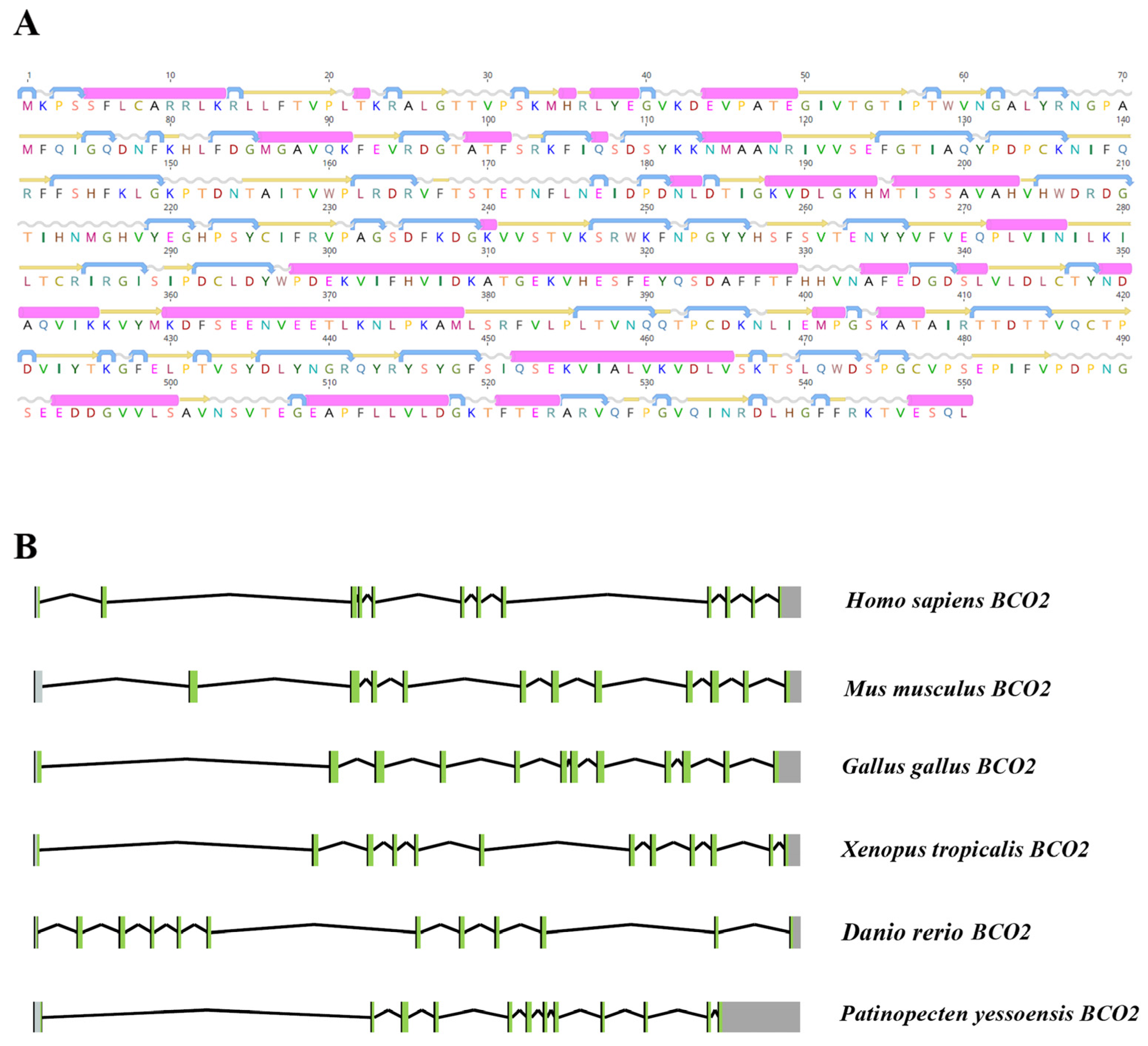
2.1. Characterization of PyBCO2 Gene Sequence

Through a transcriptome analysis of the Yesso scallop muscle, a gene named PyBCO2 was found, which was homologous with the causal PyBCO-like 1 gene for carotenoid coloration in ‘Haida golden scallop’ and showed high similarity with BCO2s in other animals. The full length of the PyBCO2 cDNA sequence was determined to be 5583 bp, containing an open reading frame (ORF) of 1653 bp, a 5′ untranslated region (UTR) of 252 bp, and a 3’UTR of 3678 bp (Figure S1A). The ORF encoded a protein of 550 amino acids with a predicted molecular weight (MW) of about 62.2 kDa and an isoelectric point (pI) value of 6.41.
Based on the bioinformatics analysis, PyBCO2 protein contained the typical RPE65 domain of the CCO superfamily from positions 38 to 544 (Figure S1B). A secondary structure analysis revealed that PyBCO2 protein was composed of 26 alpha helices, 40 beta strands, 45 coils, and 53 turns (Figure 1A). The subcellular localization of PyBCO2 was analyzed by Wolf PSORT and YLoc software, both of which predicted that PyBCO2 is located in the mitochondria. A mitochondrial import sequence (30-aa leader sequence) was found in its N-terminal extension part. This result was consistent with the subcellular localization of vertebrate BCO2, which was reported to be a mitochondrial carotenoid oxygenase with broad substrate specificity [12,26].
Furthermore, a genomic structure analysis of PyBCO2 was conducted. The results showed that the PyBCO2 gene contained 12 exons and 11 introns, which have the same exon/intron numbers as other vertebrate species, including Homo sapiens, Mus musculus, Gallus gallus, Xenopus tropicalis, and Danio rerio (Figure 1B). These results indicate that the functions of PyBCO2 and vertebrate BCO2 might be conserved.

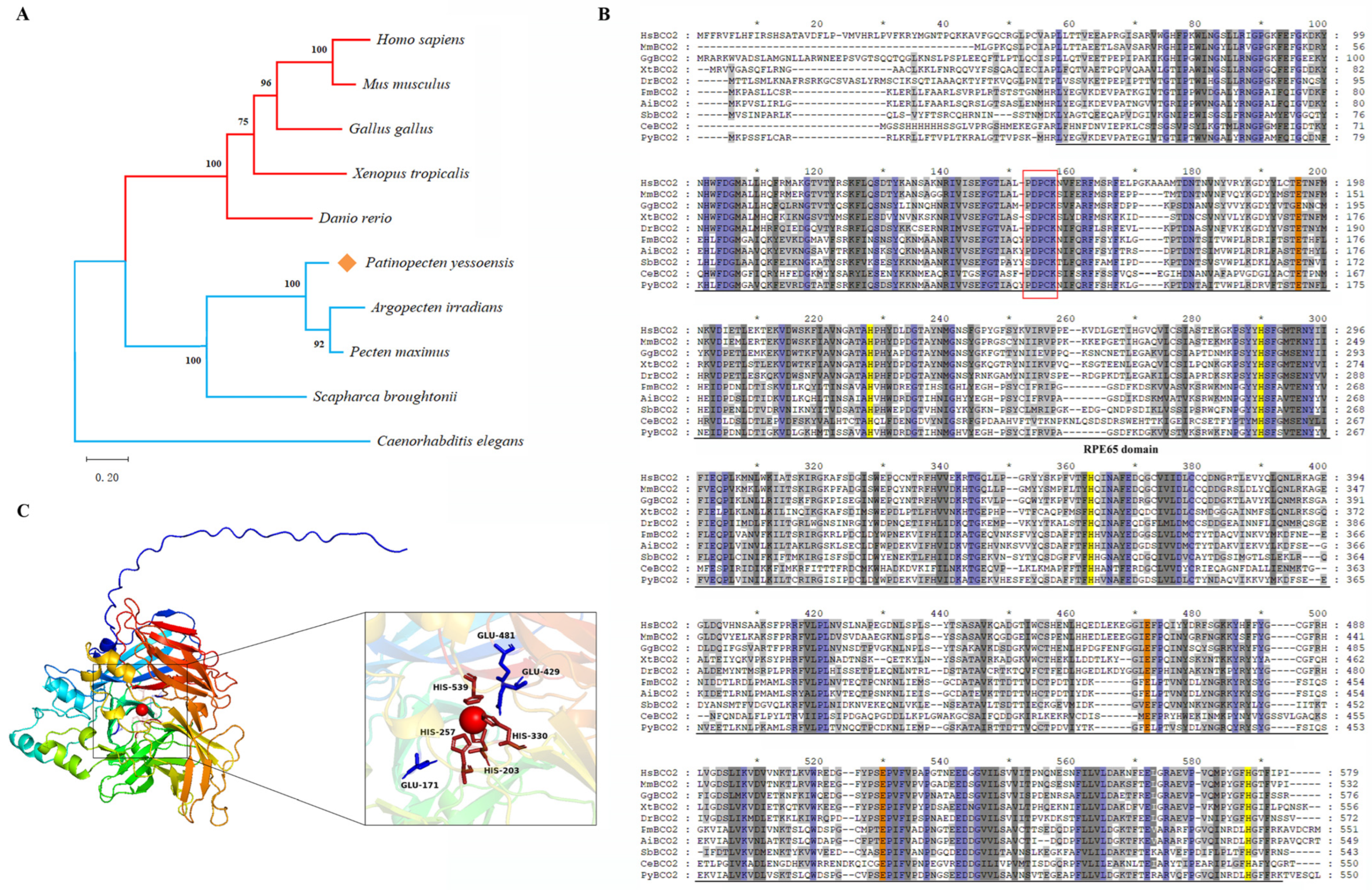
2.2. Homology and Phylogenetic Analysis

The PyBCO2 protein sequence was aligned with the homologous protein sequences from various species of other vertebrates and invertebrates. PyBCO2 displayed higher identities with the BCO2 homologs of Argopecten irradians (73.64%) and Pecten maximus (78.58%), but relatively lower identities with other vertebrate species, including H. sapiens, M. musculus, G. gallus, X. tropicalis, and D. rerio (38.48~40.65%), and the lowest identity with Caenorhabditis elegans (31.62%) (Table S1), which was further supported by phylogenetic analysis (Figure 2A).
Similar to vertebrate BCO2s, PyBCO2 exhibited all of the characteristic features of carotenoid cleavage oxygenases, including four tightly conserved His residues and three Glu residues, as well as the conserved PDPCK motif (Figure 2B) [27]. As a non-heme iron oxygenase, BCO2 cleaves the carotenoid substrate by activating molecular oxygen with a Fe2+ ion as a cofactor, which is coordinated by four conserved His residues, with three Glu residues forming the second coordination sphere [14]. Sequence alignment analysis confirmed the conservation of these catalytic sites in PyBCO2, which was further supported by tertiary structure prediction (Figure 2C), showing the structural basis of PyBCO2 for carotenoid cleavage. The PDPCK motif in metazoan BCO2 proteins was reported to be a signature motif providing specificity for palmitoylation of the cysteine residue within this motif, and, thus, may mediate interactions with the internal membranes to capture their carotenoid substrates [28]. The conservation of catalytic sites and the PDPCK motif between PyBCO2 and its vertebrate homologs suggests a similar function for BCO2 in scallops and higher organisms.

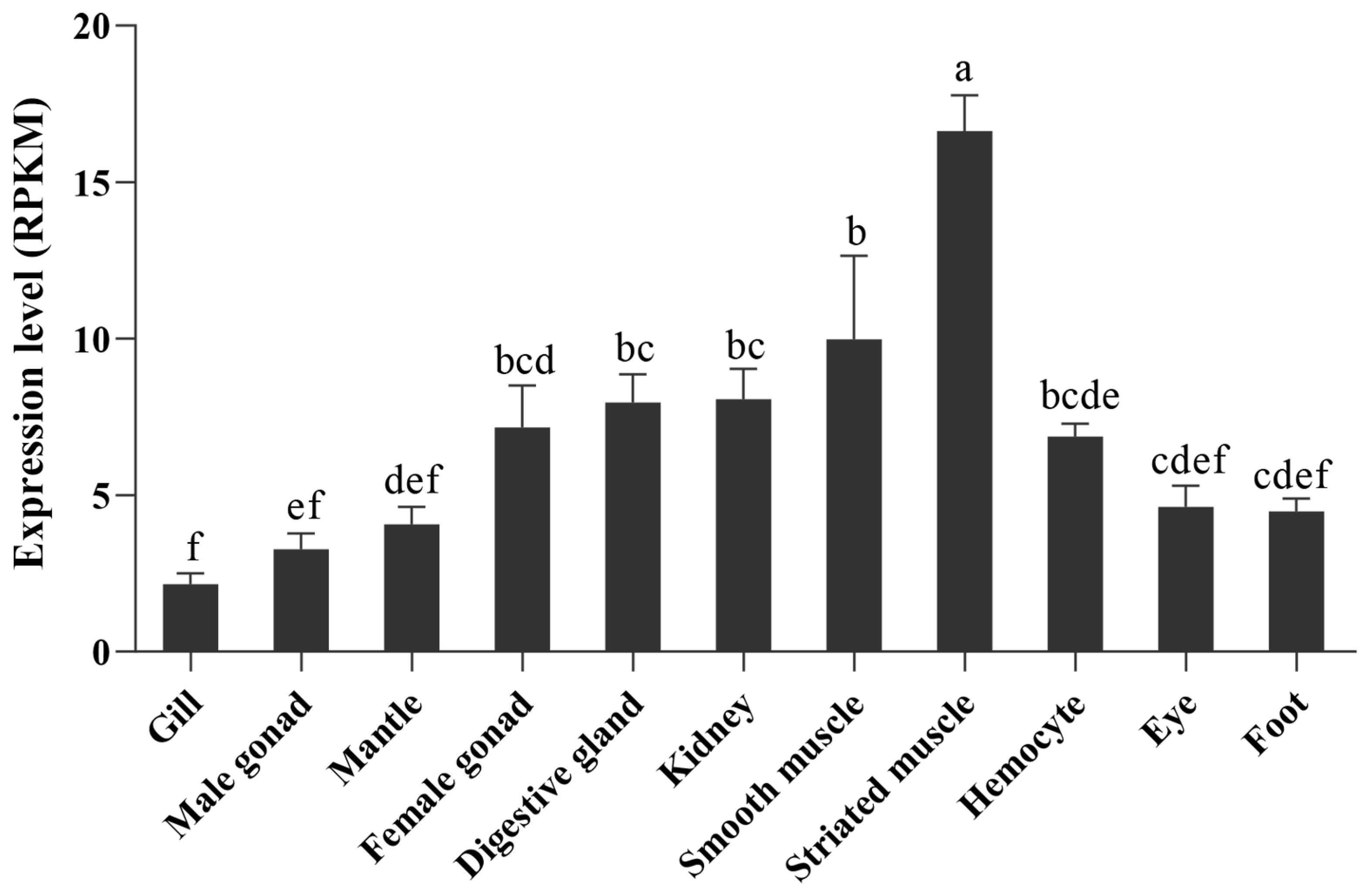
2.3. Expression Pattern of PyBCO2 in Adult Organs/Tissues

The PyBCO2 gene was ubiquitously expressed in all examined scallop organs/tissues, with the highest expression levels detected in the striated muscle, followed by the smooth muscle (Figure 3). The average RPKM (reads per kilobase per million mapped reads) of PyBCO2 gene expression in the female gonads, kidneys, digestive glands, and hemocytes was between 5 and 10, while that in the gills, male gonads, mantle, eyes, and feet was less than 5 (Figure 3). The widespread expression of BCO2 was also reported in vertebrate species, such as H. sapiens, G. gallus, and Oncorhynchus mykiss [19,29,30]. Studies have shown that the accumulation of carotenoids can induce oxidative stress in mitochondria, and BCO2 functions as a key regulator to prevent the adverse effects caused by excess carotenoids [31]. The main food for scallops in the sea is microalgae, such as chrysophyceae, diatoms, and chlorella, which all can produce carotenoids [32,33,34]. The wide expression of PyBCO2 in scallop organs/tissues implies that it might be involved in the degradation of excessive carotenoids accumulated from the ingested microalgae, or in providing the metabolites of carotenoids, in the whole body. The high expression of PyBCO2 in the striated muscle, the main tissue of scallops that is edible by humans, indicates the potential of this gene to be used in carotenoid enrichment in scallops.

2.4. Functional Analysis of PyBCO2

In order to verify whether PyBCO2 has the function of degrading carotenoids, RNAi experiments were carried out by injecting PyBCO2 dsRNA into the striated muscle of Yesso scallops. As shown in Figure 4A, the expression of PyBCO2 in the striated muscle was significantly more reduced in the PyBCO2 inhibited group than that in the PBS control group (p = 2.04 × 10⁻⁴), indicating that the dsRNA successfully entered into the cells in the striated muscle and PyBCO2 expression was successfully suppressed.
The phenotypes of the Yesso scallops in the PBS control group and the PyBCO2 inhibited group are shown in Figure 4B. The color of the striated muscles in the PBS control group was white, whereas that of the PyBCO2 inhibited group appeared slightly orange. To determine which types of carotenoids are responsible for the orange striated muscle after PyBCO2 inhibition, an HPLC analysis was performed. The result showed that after inhibiting PyBCO2 expression, two obvious carotenoid peaks were detected in the striated muscle with the retention time at 5.527 min and 5.583 min, respectively, but no obvious carotenoid peaks were observed in the PBS control group (Figure 4C), suggesting that the orange color of striated muscle in the PyBCO2 inhibited group was mainly caused by the enrichment of these two carotenoids. Further analysis based on a previously published method indicates that these two carotenoid peaks were pectenolone and pectenoxanthin, respectively [23,24]. Compared with the PBS control group without detecting any carotenoids, the peak areas of pectenolone and pectenoxanthin in the PyBCO2 inhibited group were (1.40 × 10⁴) ± (5.40 × 10³) µv·s and (6.05 × 10³) ± (2.85 × 10³) µv·s, respectively, indicating that PyBCO2 has the function of degrading pectenolone and pectenoxanthin. Additionally, no obvious phenotypic changes in the growth and development of scallops were observed compared with the control group. However, the potential impact of carotenoid accumulation on growth and development cannot be ignored. Our previous studies have found that the ‘Haida golden scallop’ with carotenoid-enriched orange muscle showed advantages in growth and stress resistance, which may be affected by carotenoid accumulation [35]. The corresponding phenotypes were not observed in this study, which may be because the time of RNAi is not enough to see these phenotypic changes in scallops.
Pectenolone and pectenoxanthin are two main carotenoids in Yesso scallops, especially in female gonads, and they are also the main carotenoids responsible for the muscle coloration of the ‘Haida golden scallop’ [23,36,37]. Pectenolone shows excellent antioxidative activity by inhibiting lipid peroxidation and contributing to protection against oxidative stress [38,39]. Pectenoxanthin (alloxanthin/cynthiaxanthin) also has beneficial effects on human health, such as anti-inflammatory activities and anti-carcinogenesis [40,41]. Increasing the content of pectenolone and pectenoxanthin in the striated muscle could largely improve the nutritional value of scallops. Our findings prove that it is a feasible approach to increasing the content of pectenolone and pectenoxanthin in scallops by decreasing the expression of PyBCO2.
As a carotene-cleaving enzyme, BCO2 could catalyze the oxidative cleavage of colored carotenoids acquired from the diet into colorless apocarotenoids, which is an essential step in carotenoid degradation [12]. Numerous studies have demonstrated that missense mutations in BCO2 or BCO2 knockout resulted in increased carotenoid accumulation. For example, a cis-acting mutation that lowered BCO2 expression caused domestic chickens to have yellow skin due to carotenoid accumulation [42,43]. The genetic disruption of BCO2 function in mice resulted in increased plasma and blood accumulation of the dietary carotenoid [44]. A nonsynonymous mutation in the BCO2 gene also induced higher carotenoid accumulation in canary tissues [45]. These studies emphasize the important role of BCO2 in regulating carotenoid metabolism and accumulation. In this study, through inhibiting PyBCO2 expression, the deposition of the carotenoids pectenolone and pectenoxanthin was observed in scallop muscle, indicating the function of PyBCO2 in the bioavailability of marine carotenoids. Our result not only proves that PyBCO2 could participate in the degradation of pectenolone and pectenoxanthin, but also provides a reliable target gene for improving carotenoid content in scallops and even other aquatic animals.

3. Materials and Methods

3.1. Biological Materials

The healthy one-year-old Yesso scallops used in this experiment were collected from natural populations by Zoneco Group Co., Ltd. (Liaoning, China), and then, transported to the laboratory. After collection, the Yesso scallops were acclimated in filtered and aerated seawater at 9–12 °C for one week and were fed Isochrysis galbana twice daily before the experiments.

3.2. RNA Isolation and cDNA Synthesis

Total RNA was extracted from the striated muscle of Yesso scallops using guanidinium isothiocyanate, as described by Hu et al. [46], and then, was digested with DNase I (TaKaRa, Shiga, Japan). RNA concentration and purity were measured by a Nanovue Plus spectrophotometer (GE Healthcare, Piscataway, NJ, USA), and the RNA integrity was checked by running electrophoresis on 1% agarose gel. RNA samples with clear bands corresponding to 18S and 28S rRNA on the gel and OD260/OD280 ratios between 1.8 and 2.0 were used for cDNA synthesis. First-strand cDNA was synthesized from 2 µg of total RNA using oligo(dT)18 and MMLV reverse transcriptase (TaKaRa, Shiga, Japan) in a total volume of 20 µL. The reaction was performed at 42 °C for 90 min and terminated by heating at 70 °C for 10 min. The cDNA products were diluted to 20 ng/mL and stored at −20 °C.

3.3. Cloning of PyBCO2 Gene and Sequence Analysis

The DNA and mRNA gene sequences of PyBCO2 were obtained from the whole genome [47] and transcriptome sequence databases [25] of the Yesso scallop. The coding sequence of PyBCO2 was predicted by ORF Finder (https://www.ncbi.nlm.nih.gov/orffinder/, accessed on 1 January 2018) and was confirmed by a polymerase chain reaction (PCR) with a gene-specific primer (PyBCO2-Fw/Rv, Table 1); the primer pairs were designed using the software Primer Premier v.5.0 [48]. Conserved domains of PyBCO2 were analyzed using the Simple Modular Architecture Research Tool (SMART) (http://smart.embl-heidelberg.de/, accessed on 1 January 2018), and the physicochemical characteristics, including MW and pI, were predicted using ProtParam (http://br.expasy.org/, accessed on 1 January 2018). Secondary and tertiary structures were predicted using Geneious Prime (http://www.geneious.com/, accessed on 5 March 2018) and Alphafold2 (http://github.com/deepmind/alphafold, accessed on 5 March 2018), respectively. The subcellular localization of PyBCO2 was predicted using WOLF PSORT (https://www.genscript.com/wolf-psort.html, accessed on 20 May 2020), YLoc (https://abi-services.cs.uni-tuebingen.de/yloc/webloc.cgi, accessed on 20 May 2018), iPSORT Prediction (https://ipsort.hgc.jp/predict.cgi, accessed on 20 May 2018), and Predotar (https://urgi.versailles.inra.fr/predotar/, accessed on 20 May 2018).
The BCO2s of various species, including H. sapiens, M. musculus, G. gallus, X. tropicalis, and D. rerio, were selected for comparing their gene structure with PyBCO2. The positional information of exons and introns was characterized by comparing the cDNA sequence to the genomic sequence. The gene structure was drawn using the R package “ggbio” [49].
Multiple sequence alignment and phylogenetic analysis were performed using PyBCO2 and its homologous proteins from various species of other vertebrates and invertebrates, including H. sapiens (Q9BYV7.5), M. musculus (NP_573480.1), G. gallus (XP_040546304.1), X. tropicalis (AAH75500.1), D. rerio (NP_001035402.1), P. maximus (XP_033763808.1), A. irradians, S. broughtonii, and C. elegans (NP_494694.2). The BCO2 protein sequences were aligned using the ClustalX program [50], and alignment editing was performed using the GeneDoc multiple sequence alignment editor (http://www.nrbsc.org/gfx/genedoc/index.html, accessed on 20 May 2018). The amino acid sequence identity between PyBCO2 and its homologous protein of other species was calculated using BLAST (http://blast.ncbi.nlm.nih.gov/Blast.cgi, accessed on 20 May 2018). A phylogenetic tree was constructed using the maximum-likelihood (ML) method with the LG model using MEGA X software [51]. Bootstrapping with 1000 replications was performed to evaluate the phylogenetic tree.

3.4. Expression Analysis of PyBCO2 in Different Adult Organs/Tissues

PyBCO2 gene expression in adult organs/tissues, including the mantle, gill, male gonad, female gonad, kidney, digestive gland, smooth muscle, striated muscle, foot, eye, and hemocyte, was analyzed using transcriptomic data from Yesso scallops [47]. The expression of PyBCO2 was measured as RPKM values.

3.5. Inhibiting PyBCO2 Expression

To verify the function of PyBCO2 in carotenoid degradation, a dsRNA-mediated RNAi experiment was performed. The sequence of the PyBCO2 gene was submitted to siDirect 2.0 (http://sidirect2.rnai.jp/, accessed on 12 September 2018) for prediction and screening of the RNAi target sequence. siDirect 2.0 is an online server that utilizes a fast and sensitive homology search algorithm to minimize any off-target effects and ensure functional dsRNA design. In addition, the RNAi target sequence that was uniquely aligned with the Yesso scallop genome was selected for further dsRNA synthesis. PyBCO2 dsRNA was produced using the MEGAscript® RNAi Kit (Thermo, Waltham, MA, USA) following the manufacturer’s protocol. The acclimated Yesso scallops were randomly divided into two groups (PBS control group and PyBCO2 inhibited group). For each individual, 100 μL of specific dsRNA (1 μg/μL, for PyBCO2 inhibited group) or l× PBS (for PBS control group) was injected into the striated muscle. Each scallop was injected once a week for four weeks. The striated muscles of the two scallop groups (PBS control group and PyBCO2 inhibited group) were sampled for gene expression analysis and carotenoid extraction.

3.6. Quantitative Real-Time PCR (qRT-PCR)

The expression pattern of PyBCO2 after inhibiting PyBCO2 expression was analyzed by qRT-PCR. The cDNAs of striated muscles of Yesso scallops from the PyBCO2 inhibited group and the PBS control group were used as templates. Gene-specific primers (PyBCO2-qPCR-Fw/Rv, Table 1) were designed using Primer Premier v5.0 [48].
qRT-PCR was performed on a Light-Cycler Roche 480 Real-time PCR System (Roche Applied Science, Mannheim, Germany). The qRT-PCR reactions were carried out in a total volume of 20 μL containing 2× Light Cycler 480 SYBR Green I Master (Roche Applied Science, Mannheim, Germany), 0.4 μL of each primer (10 μM), 2 μL of diluted cDNA template (10 ng/μL), and 7.2 μL of nuclease-free water. The running program was as follows: 94 °C for 10 min, followed by 40 cycles of 94 °C for 15 s and 62 °C for 1 min. Melting curve analysis was performed to verify that the primer set amplified a single product. Ubiquitin (UBQ) was used as an internal control to normalize gene expression. The relative expression level of PyBCO2 was calculated using the 2−ΔΔCt method [52].

3.7. High-Performance Liquid Chromatography (HPLC) Analysis

The carotenoids of striated muscles in scallops after PyBCO2 inhibition were analyzed using reversed-phase HPLC, as previously described [23,24], with minor modifications. The solvents and chemicals used for carotenoid extraction were on the scale of analytical reagents. To avoid the degradation and isomerization of carotenoids, amber glassware was used and all the extraction procedures were carried out in an aphotic environment. Briefly, 1 g of striated muscle was lyophilized and extracted three times with 4 mL of methanol containing 0.1% butylated hydroxytoluene (BHT). After drying the methanol with nitrogen, the residue was dissolved in 2 mL of a mobile phase solution (acetonitrile/methanol/dichloromethane, 50:46:4, v/v/v) and filtered through a 0.22 μm nylon-6 membrane (13 mm, Solarbio, Beijing, China). HPLC analysis of carotenoids was performed using a Chromaster (HITACHI, Tokyo, Japan) equipped with an analytical-scale LaChrom C18 column (4.6 × 250 mm, 5 μm inner diameter; HITACHI, Tokyo, Japan). The mobile phase for chromatographic separation was a ternary, isocratic solvent system of acetonitrile/methanol/dichloromethane at a ratio of 50:46:4. The other detail parameters were as follows: a 10 μL injection volume; a 25 °C column temperature; a 1.0 mL per minute flow rate; a 450 nm UV-Vis detector; and a 10 min separation time. Peak area (μv·s) was used as a relative quantification for inter-group comparison.

3.8. Statistical Analysis

All statistical analyses were performed using SPSS 19.0 software [53]. Significant differences in PyBCO2 expression among adult organs/tissues were analyzed by one-way analysis of variance. The expression difference of PyBCO2 in the striated muscles between the PBS control group and PyBCO2 inhibited group were determined using a dependent-samples t-test. p values less than 0.05 were considered statistically significant.

4. Conclusions

A gene encoding carotenoid cleavage oxygenase, PyBCO2, was identified in Yesso scallops. We provided evidence that PyBCO2 is widely expressed in scallop organs/tissues, with the highest expression in the striated muscle, the main edible tissue of scallops. Functional studies showed that the inhibition of PyBCO2 expression led to the deposition of the scallop carotenoids pectenolone and pectenoxanthin in the striated muscle. Our results demonstrated that PyBCO2 plays a key role in carotenoid metabolism in Yesso scallops, and it could be used in the breeding of carotenoid-enriched scallops, including further improving carotenoid content in ‘Haida golden scallop’.

Supplementary Materials

The following supporting information can be downloaded at: https://www.mdpi.com/article/10.3390/ijms25073947/s1.

Author Contributions

Conceptualization, X.H. and H.W.; methodology, S.L. and S.W.; formal analysis, S.L. and S.W.; investigation, T.L. and Y.Z.; resources, L.Z.; data curation, H.W.; writing—original draft preparation, S.L.; writing—review and editing, X.H. and H.W.; supervision, Z.B.; funding acquisition, X.H. All authors have read and agreed to the published version of the manuscript.

Funding

This research was funded by the Key Research and Development Project of Shandong Province (2021ZLGX03), the National Natural Science Foundation of China (32302968), and the Shandong Provincial Natural Science Foundation (ZR2022QC032).

Institutional Review Board Statement

Not applicable.

Informed Consent Statement

Not applicable.

Data Availability Statement

The data are contained within the article.

Conflicts of Interest

The authors declare no competing or financial interests.

References

  1. Eggersdorfer, M.; Wyss, A. Carotenoids in human nutrition and health. Arch. Biochem. Biophys. 2018, 652, 18–26. [Google Scholar] [CrossRef] [PubMed]
  2. Cooper, D.A. Carotenoids in health and disease: Recent scientific evaluations, research recommendations and the consumer. J. Nutr. 2004, 134, 221S–224S. [Google Scholar] [CrossRef] [PubMed]
  3. Maoka, T. Carotenoids as natural functional pigments. J. Nat. Med. 2020, 74, 1–16. [Google Scholar] [CrossRef] [PubMed]
  4. Rao, A.V.; Rao, L.G. Carotenoids and human health. Pharmacol. Res. 2007, 55, 207–216. [Google Scholar] [CrossRef] [PubMed]
  5. Xavier, A.A.O.; Perez-Galvez, A. Carotenoids as a source of antioxidants in the diet. Sub-Cell. Biochem. 2016, 79, 359–375. [Google Scholar]
  6. da Costa, D.P.; Campos Miranda-Filho, K. The use of carotenoid pigments as food additives for aquatic organisms and their functional roles. Rev. Aquac. 2020, 12, 1567–1578. [Google Scholar] [CrossRef]
  7. Langi, P.; Kiokias, S.; Varzakas, T.; Proestos, C. Carotenoids: From plants to food and feed industries. Methods Mol. Biol. 2018, 1852, 57–71. [Google Scholar] [PubMed]
  8. Jensen, S.K.; Jensen, C.; Jakobsen, K.; Engberg, R.M.; Andersen, J.O.; Lauridsen, C.; Sorensen, P.; Skibsted, L.H.; Bertelsen, G. Supplementation of broiler diets with retinol acetate, β-carotene or canthaxanthin: Effect on vitamin status and oxidative status of broilers in vivo and on meat stability. Acta Agric. Scand. Sect. A-Anim. Sci. 1998, 48, 28–37. [Google Scholar] [CrossRef]
  9. Muramoto, T.; Nakanishi, N.; Shibata, M.; Aikawa, K. Effect of dietary β-carotene supplementation on beef color stability during display of two muscles from Japanese black steers. Meat Sci. 2003, 63, 39–42. [Google Scholar] [CrossRef]
  10. Hubbard, J.K.; Uy, J.A.C.; Hauber, M.E.; Hoekstra, H.E.; Safran, R.J. Vertebrate pigmentation: From underlying genes to adaptive function. Trends Genet. 2010, 26, 231–239. [Google Scholar] [CrossRef]
  11. Kiefer, C.; Hessel, S.; Lampert, J.M.; Vogt, K.; Lederer, M.O.; Breithaupt, D.E.; von Lintig, J. Identification and characterization of a mammalian enzyme catalyzing the asymmetric oxidative cleavage of provitamin A. J. Biol. Chem. 2001, 276, 14110–14116. [Google Scholar] [CrossRef]
  12. Amengual, J.; Lobo, G.P.; Golczak, M.; Li, H.N.M.; Klimova, T.; Hoppel, C.L.; Wyss, A.; Palczewski, K.; von Lintig, J. A mitochondrial enzyme degrades carotenoids and protects against oxidative stress. Faseb J. 2011, 25, 948–959. [Google Scholar] [CrossRef]
  13. Amengual, J.; Widjaja-Adhi, M.A.K.; Rodriguez-Santiago, S.; Hessel, S.; Golczak, M.; Palczewski, K.; von Lintig, J. Two carotenoid oxygenases contribute to mammalian provitamin a metabolism. J. Biol. Chem. 2013, 288, 34081–34096. [Google Scholar] [CrossRef] [PubMed]
  14. Sui, X.; Kiser, P.D.; von Lintig, J.; Palczewski, K. Structural basis of carotenoid cleavage: From bacteria to mammals. Arch. Biochem. Biophys. 2013, 539, 203–213. [Google Scholar] [CrossRef]
  15. Sun, Y.; Liu, M.; Yan, C.; Yang, H.; Wu, Z.; Liu, Y.; Su, N.; Hou, J.; Zhang, J.; Yang, F.; et al. CRISPR/Cas9-mediated deletion of β, β-carotene 9′, 10′-oxygenase gene (EcBCO2) from Exopalaemon carinicauda. Int. J. Biol. Macromol. 2020, 151, 168–177. [Google Scholar] [CrossRef]
  16. Berry, S.D.; Davis, S.R.; Beattie, E.M.; Thomas, N.L.; Burrett, A.K.; Ward, H.E.; Stanfield, A.M.; Biswas, M.; Ankersmit-Udy, A.E.; Oxley, P.E.; et al. Mutation in bovine β-carotene oxygenase 2 affects milk color. Genetics 2009, 182, 923–926. [Google Scholar] [CrossRef] [PubMed]
  17. Tian, R.; Pitchford, W.S.; Morris, C.A.; Cullen, N.G.; Bottema, C.D.K. Genetic variation in the β, β-carotene-9′,10-dioxygenase gene and association with fat colour in bovine adipose tissue and milk. Anim. Genet. 2010, 41, 253–259. [Google Scholar] [CrossRef] [PubMed]
  18. Vage, D.I.; Boman, I.A. A nonsense mutation in the beta-carotene oxygenase 2 (BCO2) gene is tightly associated with accumulation of carotenoids in adipose tissue in sheep (Ovis aries). BMC Genet. 2010, 11, 10. [Google Scholar] [CrossRef]
  19. Fallahshahroudi, A.; Sorato, E.; Altimiras, J.; Jensen, P. The domestic BCO2 allele buffers low-carotenoid diets in chickens: Possible citness increase through species hybridization. Genetics 2019, 212, 1445–1452. [Google Scholar] [CrossRef]
  20. Lehnert, S.J.; Christensen, K.A.; Vandersteen, W.E.; Sakhrani, D.; Pitcher, T.E.; Heath, J.W.; Koop, B.F.; Heath, D.D.; Devlin, R.H. Carotenoid pigmentation in salmon: Variation in expression at BCO2-l locus controls a key fitness trait affecting red coloration. Proc. R. Soc. B-Biol. Sci. 2019, 286, 20191588. [Google Scholar] [CrossRef]
  21. Wade, N.M.; Gabaudan, J.; Glencross, B.D. A review of carotenoid utilisation and function in crustacean aquaculture. Rev. Aquac. 2017, 9, 141–156. [Google Scholar] [CrossRef]
  22. Tan, K.; Zhang, H.; Zheng, H. Carotenoid content and composition: A special focus on commercially important fish and shellfish. Crit. Rev. Food Sci. Nutr. 2024, 64, 544–561. [Google Scholar] [CrossRef] [PubMed]
  23. Li, N.; Hu, J.; Wang, S.; Cheng, J.; Hu, X.; Lu, Z.; Lin, Z.; Zhu, W.; Bao, Z. Isolation and identification of the main carotenoid pigment from the rare orange muscle of the Yesso scallop. Food Chem. 2010, 118, 616–619. [Google Scholar] [CrossRef]
  24. Li, N. What Accounting for Orange Color of Yesso Scallop (Patinopecten yessoensis) Muscle and Its Application in Breeding. Ph.D. Thesis, Ocean University of China, Qingdao, China, 2009. [Google Scholar]
  25. Li, X.; Wang, S.; Xun, X.; Zhang, M.; Wang, S.; Li, H.; Zhao, L.; Fu, Q.; Wang, H.; Li, T.; et al. A carotenoid oxygenase is responsible for muscle coloration in scallop. Biochim. Biophys. Acta-Mol. Cell Biol. Lipids 2019, 1864, 966–975. [Google Scholar] [CrossRef]
  26. Palczewski, G.; Amengual, J.; Hoppel, C.L.; von Lintig, J. Evidence for compartmentalization of mammalian carotenoid metabolism. Faseb J. 2014, 28, 4457–4469. [Google Scholar] [CrossRef] [PubMed]
  27. Uppal, S.; Rogozin, I.B.; Redmond, T.M.; Poliakov, E. Palmitoylation of metazoan carotenoid oxygenases. Molecules 2020, 25, 1942. [Google Scholar] [CrossRef]
  28. Poliakov, E.; Uppal, S.; Rogozin, I.B.; Gentleman, S.; Redmond, T.M. Evolutionary aspects and enzymology of metazoan carotenoid cleavage oxygenases. Biochim. Biophys. Acta-Mol. Cell Biol. Lipids 2020, 1865, 158665. [Google Scholar] [CrossRef]
  29. Wu, S.; Zhao, L.; Huang, J.; Li, Y.; Liu, Z.; Zhang, D. miR-330 targeting BCO2 is involved in carotenoid metabolism to regulate skin pigmentation in rainbow trout (Oncorhynchus mykiss). BMC Genom. 2023, 24, 124. [Google Scholar] [CrossRef] [PubMed]
  30. Lindqvist, A.; He, Y.-G.; Andersson, S. Cell type-specific expression of β-carotene 9′, 10′-monooxygenase in human tissues. J. Histochem. Cytochem. 2005, 53, 1403–1412. [Google Scholar] [CrossRef]
  31. Wu, L.; Guo, X.; Hartson, S.D.; Davis, M.A.; He, H.; Medeiros, D.M.; Wang, W.; Clarke, S.L.; Lucas, E.A.; Smith, B.J.; et al. Lack of β, β-carotene-9′, 10′-oxygenase 2 leads to hepatic mitochondrial dysfunction and cellular oxidative stress in mice. Mol. Nutr. Food Res. 2017, 61, 1600576. [Google Scholar] [CrossRef]
  32. Gong, M.; Bassi, A. Carotenoids from microalgae: A review of recent developments. Biotechnol. Adv. 2016, 34, 1396–1412. [Google Scholar] [CrossRef] [PubMed]
  33. Cheng, P.; Zhou, C.; Chu, R.; Chang, T.; Xu, J.; Ruan, R.; Chen, P.; Yan, X. Effect of microalgae diet and culture system on the rearing of bivalve mollusks: Nutritional properties and potential cost improvements. Algal Res. 2020, 51, 102076. [Google Scholar] [CrossRef]
  34. Shahidi, F.; Metusalach; Brown, J.A. Carotenoid pigments in seafoods and aquaculture. Crit. Rev. Food Sci. Nutr. 1998, 38, 1–67. [Google Scholar] [CrossRef] [PubMed]
  35. Zhang, L.; Li, X.; Li, N.; Wang, S.; Li, X.; Sun, G.; Hu, X.; Bao, Z. Corralation analysis of carotenoid concentration and the growth traits of ‘Haida golden scallop’. Period. Ocean Univ. China/Zhongguo Haiyang Daxue Xuebao 2018, 48, 41–47. [Google Scholar]
  36. Li, X.; Li, N.; Zhao, L.; Shi, J.; Wang, S.; Ning, X.; Li, Y.; Hu, X. Tissue distribution and seasonal accumulation of carotenoids in Yesso scallop (Mizuhopecten yessoensis) with orange adductor muscle. Food Chem. 2022, 367, 130701. [Google Scholar] [CrossRef]
  37. Matsuno, T.; Hiraoka, K.; Maoka, T. Carotenoids in the gonad of scallops [Pactinopecten yessoensis]. Bull. Jap. Soc. Sci. Fish. 1981, 47, 385–390. [Google Scholar] [CrossRef]
  38. Narita, M.; Maoka, T.; Ebitani, K.; Nishino, H. Characteristics of chemical constituents and red pigment of orange adductor muscle of scallop Mizuhopecten yessoensis in the Okhotsk Sea and anti-oxidative activity of the pigment. Nippon. Suisan Gakkaishi 2013, 79, 48–54. [Google Scholar] [CrossRef]
  39. Maoka, T.; Kuwahara, T.; Narita, M. Carotenoids of sea angels Clione limacina and Paedoclione doliiformis from the perspective of the food chain. Mar. Drugs 2014, 12, 1460–1470. [Google Scholar] [CrossRef] [PubMed]
  40. Konishi, I.; Hosokawa, M.; Sashima, T.; Maoka, T.; Miyashita, K. Suppressive effects of alloxanthin and diatoxanthin from Halocynthia roretzi on LPS-induced expression of pro-inflammatory genes in RAW264.7 cells. J. Oleo. Sci. 2008, 57, 181–189. [Google Scholar] [CrossRef]
  41. Tsushima, M.; Maoka, T.; Katsuyama, M.; Kozuka, M.; Matsuno, T.; Tokuda, H.; Nishino, H.; Iwashima, A. Inhibitory effect of natural carotenoids on Epstein-Barr virus activation activity of a tumor promoter in Raji cells. A screening study for anti-tumor promoters. Biol. Pharm. Bull. 1995, 18, 227–233. [Google Scholar] [CrossRef]
  42. Eriksson, J.; Larson, G.; Gunnarsson, U.; Bed’hom, B.; Tixier-Boichard, M.; Strömstedt, L.; Wright, D.; Jungerius, A.; Vereijken, A.; Randi, E.; et al. Identification of the Yellow Skin gene reveals a hybrid origin of the domestic chicken. PLOS Genet. 2008, 4, e1000010. [Google Scholar] [CrossRef] [PubMed]
  43. Pointer, M.A.; Mundy, N.I. Chicken skin sheds light on carotenoid genetics. Heredity 2008, 101, 393–394. [Google Scholar] [CrossRef] [PubMed]
  44. Li, B.; Vachali, P.P.; Gorusupudi, A.; Shen, Z.; Sharifzadeh, H.; Besch, B.M.; Nelson, K.; Horvath, M.M.; Frederick, J.M.; Baehr, W.; et al. Inactivity of human beta,beta-carotene-9′, 10′-dioxygenase (BCO2) underlies retinal accumulation of the human macular carotenoid pigment. Proc. Natl. Acad. Sci. USA 2014, 111, 10173–10178. [Google Scholar] [CrossRef] [PubMed]
  45. Gazda, M.A.; Toomey, M.B.; Araujo, P.M.; Lopes, R.J.; Afonso, S.; Myers, C.A.; Serres, K.; Kiser, P.D.; Hill, G.E.; Corbo, J.C.; et al. Genetic basis of de novo appearance of carotenoid ornamentation in bare parts of canaries. Mol. Biol. Evol. 2020, 37, 1317–1328. [Google Scholar] [CrossRef] [PubMed]
  46. Hu, X.; Bao, Z.; Hu, J.; Shao, M.; Zhang, L.; Bi, K.; Zhan, A.; Huang, X. Cloning and characterization of tryptophan 2,3-dioxygenase gene of Zhikong scallop Chlamys farreri (Jones and Preston 1904). Aquac. Res. 2006, 37, 1187–1194. [Google Scholar] [CrossRef]
  47. Wang, S.; Zhang, J.; Jiao, W.; Li, J.; Xun, X.; Sun, Y.; Guo, X.; Huan, P.; Dong, B.; Zhang, L.; et al. Scallop genome provides insights into evolution of bilaterian karyotype and development. Nat. Ecol. Evol. 2017, 1, 0120. [Google Scholar] [CrossRef] [PubMed]
  48. Ahern, K. Primer premier. Science 1999, 286, 433. [Google Scholar] [CrossRef]
  49. Yin, T.; Cook, D.; Lawrence, M. ggbio: An R package for extending the grammar of graphics for genomic data. Genome Biol. 2012, 13, R77. [Google Scholar] [CrossRef] [PubMed]
  50. Jeanmougin, F.; Thompson, J.D.; Gouy, M.; Higgins, D.G.; Gibson, T.J. Multiple sequence alignment with Clustal x. Trends Biochem. Sci. 1998, 23, 403–405. [Google Scholar] [CrossRef]
  51. Kumar, S.; Stecher, G.; Li, M.; Knyaz, C.; Tamura, K. MEGA X: Molecular evolutionary genetics analysis across computing platforms. Mol. Biol. Evol. 2018, 35, 1547–1549. [Google Scholar] [CrossRef]
  52. Livak, K.J.; Schmittgen, T.D. Analysis of relative gene expression data using real-time quantitative PCR and the 2−ΔΔCT method. Methods 2001, 25, 402–408. [Google Scholar] [CrossRef] [PubMed]
  53. Popovic, B.V. Handbook of Univariate and Multivariate Data Analysis with IBM SPSS, 2nd ed.; Chapman and Hall/CRC: Boca Raton, FL, USA, 2013; Volume 42, p. 2291. [Google Scholar]
Figure 1. (A) The secondary structure of PyBCO2 protein. The pink cylinder represents the alpha helix; the straight yellow arrow represents the beta strand; the wavy line represents the coil; the curved arrow represents the turn. (B) The gene structure of BCO2s in P. yessoensis, H. sapiens, M. musculus, G. gallus, X. tropicalis, and D. rerio. The ORF and UTR regions are shown as green boxes and grey boxes, respectively; introns are shown as fold lines.
Ijms 25 03947 g001
Figure 2. Phylogenetic analysis, sequence alignment, and protein structure of BCO2 proteins. (A) Phylogenetic tree of BCO2 protein in P. yessoensis and other species. The numbers show the bootstrap percentages (1000 replicates) obtained using the maximum-likelihood (ML) method. The vertebrate and invertebrate BCO2s are denoted by red and blue branch lines, respectively. P. yessoensis BCO2 is marked with an orange diamond. (B) Multiple alignments of amino acid sequences of BCO2 from P. yessoensis and other species. The shade residues in blue, dark gray, and light gray represent completely (=100%), strongly (≥75%), and weakly (≥50%) conserved residues. The conservative His residues and Glu residues are highlighted with yellow and orange backgrounds, respectively. The PDPCK motif is enclosed in a red rectangular frame. The RPE65 domain is denoted by a black line. The species abbreviations are as follows: Hs: H. sapiens; Mm: M. musculus; Gg: G. gallus; Xt: X. tropicalis; Dr: D. rerio; Pm: P. maximus; Ai: A. irradians; Sb: S. broughtonii; Ce: C. elegans; Py: P. yessoensis. (C) The structure and catalytic active centers of PyBCO2 protein. The active sites, where four His residues coordinate with the Fe2+ and three Glu residues form the second coordination sphere, are highlighted with an enlarged black border in the right box. The Fe2+ ion is shown as a red sphere. The His residues and Glu residues are highlighted in red and blue, respectively.
Ijms 25 03947 g002
Figure 3. Expression of PyBCO2 gene in adult organs/tissues of Yesso scallop. Different lowercase letters mean significant differences between organs/tissues (p < 0.05).
Ijms 25 03947 g003
Figure 4. Functional verification of PyBCO2. (A) Expression of PyBCO2 in the striated muscles of Yesso scallops in the PBS control group and the PyBCO2 inhibited group. p < 0.05 indicates differences that are statistically significant. (B) The phenotype of the striated muscles of Yesso scallops in the PBS control group and the PyBCO2 inhibited group. (C) HPLC separation of carotenoids at 450 nm in the striated muscles of Yesso scallops in the PBS control group and the PyBCO2 inhibited group. The peaks of pectenolone and pectenoxanthin are marked by arrows with the retention time at 5.527 min and 5.583 min, respectively.
Ijms 25 03947 g004
Table 1. Primers used in this study.
Primer NameSequenceSequence Information
PyBCO2-FwATGAAGCCATCATCATTTCTSequence validation
PyBCO2-RvCTATAACTGACTCTCTACAGTSequence validation
RNAi-FwGATCACTAATACGACTCACTATAGGGGCGACGGAACCATCCACAARNAi
RNAi-RvGATCACTAATACGACTCACTATAGGGCCTCACTGAAATCCTTCATATACACCTRNAi
PyBCO2-qPCR-FwGATGCCAGGCTCTAAAGCAACqRT-PCR
PyBCO2-qPCR-RvCTGAACCCGTACGAGTAACGATACTqRT-PCR
UBQ-FwTCGCTGTAGTCTCCAGGATTGCqRT-PCR
UBQ-RvTCGCCACATACCCTCCCACqRT-PCR
Disclaimer/Publisher’s Note: The statements, opinions and data contained in all publications are solely those of the individual author(s) and contributor(s) and not of MDPI and/or the editor(s). MDPI and/or the editor(s) disclaim responsibility for any injury to people or property resulting from any ideas, methods, instructions or products referred to in the content.

