Article

Redosing with Intralymphatic GAD-Alum in the Treatment of Type 1 Diabetes: The DIAGNODE-B Pilot Trial

1
Division of Pediatrics, Department of Biomedical and Clinical Sciences, Faculty of Medicine and Health Sciences, Linköping University, 581 83 Linköping, Sweden
2
Department of Clinical Diagnostics, School of Health and Welfare, Jönköping University, 551 11 Jönköping, Sweden
3
Division of Medical Diagnostics, Department of Laboratory Medicine, Ryhov County Hospital, 551 85 Jönköping, Sweden
4
Department of Pediatrics, Ryhov County Hospital, 551 85 Jönköping, Sweden
5
Diamyd Medical AB, 111 56 Stockholm, Sweden
6
Crown Princess Victoria Children’s Hospital, Linköping University, 581 85 Linköping, Sweden
*
Author to whom correspondence should be addressed.
Int. J. Mol. Sci. 2025, 26(1), 374; https://doi.org/10.3390/ijms26010374
Submission received: 25 November 2024 / Revised: 20 December 2024 / Accepted: 23 December 2024 / Published: 4 January 2025
(This article belongs to the Special Issue Molecular Diagnosis and Treatments of Diabetes Mellitus)

Abstract

:
Immunotherapies aimed at preserving residual beta cell function in type 1 diabetes have been successful, although the effect has been limited, or raised safety concerns. Transient effects often observed may necessitate redosing to prolong the effect, although this is not always feasible or safe. Treatment with intralymphatic GAD-alum has been shown to be tolerable and safe in persons with type 1 diabetes and has shown significant efficacy to preserve C-peptide with associated clinical benefit in individuals with the human leukocyte antigen DR3DQ2 haplotype. To further explore the feasibility and advantages of redosing with intralymphatic GAD-alum, six participants who had previously received active treatment with intralymphatic GAD-alum and carried HLA DR3-DQ2 received one additional intralymphatic dose of 4 μg GAD-alum in the pilot trial DIAGNODE-B. The participants also received 2000 U/day vitamin D (Calciferol) supplementation for two months, starting one month prior to the GAD-alum injection. During the 12-month follow-up, residual beta cell function was estimated with Mixed-Meal Tolerance Tests, and clinical and immune responses were observed. C-peptide decreased minimally, and most patients showed stable HbA1c and IDAA1c. The mean % TIR increased while the mean daily insulin dose decreased at month 12 compared to the baseline. Redosing with GAD-alum seems to be safe and tolerable, and may prolong the disease modification elicited by the original GAD-alum treatment.

1. Introduction

The incidence of type 1 diabetes (T1D) is increasing worldwide for reasons that remain unknown. Despite recent promising advances with immune and cell therapies, preventing stage 3 is difficult, and there is currently no cure. Even though treatment of T1D has improved, with better insulin analogues, glucose sensors, and “smarter” pumps, the disease management burden is heavy. Most patients do not reach treatment targets [1,2] and often suffer from hypoglycemia and anxiety, long-term disease complications resulting in decreased life expectancy [3,4,5,6]. Interventions to preserve residual beta cell function aimed at the immune system [7] have been tried since the 1970s [7,8,9,10,11,12,13,14,15,16,17], but their effect has been limited and transient, with some interventions raising safety concerns [13,18,19,20,21,22,23,24,25,26,27,28,29,30,31,32,33,34,35,36,37,38,39,40,41,42]. Transient effects may require redosing, but administration of an extra dose may not always be feasible or safe [43].
The rapid progress in the last few years has created hope [44], with new efficacious treatments including the first Phase III trial with teplizumab showing significant preservation of beta cell function [37,45]. However, although teplizumab is clinically available in the USA for high-risk individuals in stage 2 [45], this treatment has not yet been approved for treatment of newly diagnosed T1D. In order for immune interventions to affect the quality of life of individuals living with T1D, such therapies must be not only efficacious but also safe, and not add to the already heavy burden of disease and disease management.
Antigen-specific immunotherapy with glutamic acid decarboxylase (GAD)65 formulated with aluminum hydroxide (GAD-alum) aims to modulate the autoimmune response to the beta cell antigen GAD65 thereby interrupting the disease progression. The treatment has been shown to be easy and safe to administer, but the clinical results were inconclusive [46,47,48], even though meta-analysis of the data supported a high probability that the treatment was efficacious [49]. To improve the effect of GAD-alum, the drug was injected into an inguinal lymph node in the open-label clinical trial DIAGNODE-1, testing three injections of 4 μg of GAD-alum one month apart. DIAGNODE-1 was the first human trial with an autoantigen given intralymphatically, and the results were positive [50,51,52]. DIAGNODE-1 was followed by a randomized double-blind placebo-controlled Phase IIb trial, DIAGNODE-2. In DIAGNODE-2, significant efficacy in preserving C-peptide was limited to patients with the human leukocyte antigen (HLA) DR3DQ2 haplotype, in line with previous findings in clinical trials [53,54]. The DR and DQ HLA genes code for class II Major Histocompatibility Complex proteins that present antigen peptides to CD4 T-cells, and DR3DQ2 haplotype alleles, are associated with increased risk of developing T1D. To investigate if treatment effects could be extended, a fourth dose was given 2.5 years after the initial treatment to three adult patients with DR3DQ2 in a DIAGNODE-1 extension study, which seemed to prolong the preservation of beta cell function [55].
In addition to the intralymphatic GAD-alum injections, patients have received oral vitamin D, as adequate vitamin D concentrations may play a role in achieving an appropriate effect on the immune system [56]. Vitamin D is thought to improve dendritic cell function and may also induce TH2 deviation [56,57] and a shift of T cells from an effector to a regulatory phenotype [58]. It has also been suggested that vitamin D may protect β cells and improve insulin sensitivity [59], and epidemiological studies have suggested a correlation with type 1 diabetes incidence [60]. However, several studies have contradicted that vitamin D by itself has an effect on preservation of beta cell function [61,62,63,64], in accordance with our own previous results [54]. We therefore in this pilot study focused our investigations on the consequences of redosing with intralymphatic GAD-alum.
It has been shown that administration of GAD-alum both subcutaneously and into the lymph nodes has a specific immunomodulatory effect, indicated by in vitro cytokine secretion and proliferation upon GAD65-stimulation [51,52,65,66,67,68] and induction of anti-GAD65 antibodies (GADA) [69]. Furthermore, low doses of GAD-alum directly into the lymph node have had a more potent impact on immune responses, shown by induction of higher GADA titers and GAD-induced cytokine secretion than observed after subcutaneous administration [70].
The aim of this study was to further explore the feasibility and advantages of redosing with intralymphatic GAD-alum participants who previously received active treatment with GAD-alum in DIAGNODE-1 or DIAGNODE-2, and carried HLA DR3-DQ2, received one additional intralymphatic injection and were followed for 12 months in the DIAGNODE-B first-in-human pilot trial

2. Results

2.1. Safety and Feasibility of Repeated Dosing with GAD-Alum

The design of the DIAGNODE-B clinical trial is illustrated in Figure 1. Briefly, two patients (A1 and 2) received the first injection of GAD-alum (4 µg) into the lymph nodes, followed by two booster injections one month apart in the DIAGNODE-1 open label trial. They received a further dose of GAD-alum (4 µg) after 29 months as an extension of DIAGNODE-1, and a fifth GAD-alum injection (4 µg) 44 months later during the current trial. The other four patients (B1–B4) participated in the placebo-controlled DIAGNODE-2 trial, where they received the first injection of GAD-alum (4 µg) into the lymph nodes, followed by two injections one month apart. They received a fourth dose of GAD-alum (4 µg) during the current trial, patients B1 and B2 after 50 months and patients B3 and B4 after 39 months. All patients took vitamin D (Calciferol) for 120 days during the DIAGNODE-1 and DIAGNODE-2 studies, and a second course of vitamin D (60 days) at the time of the four and fifth injections.
The baseline characteristics of the patients are shown in Table 1. All six enrolled patients completed the trial, and none of the safety assessments raised any concerns. There were no differences in the safety and tolerability between the two patients from the DIAGNODE-1 cohort (A1, A2) who received a fifth injection and the four patients from the DIAGNODE-2 trial (B1, B2, B3, B4) after the fourth dose. Five patients reported 15 treatment-emergent adverse events (TEAEs), nine of them classified as mild and six as moderate events. No serious adverse events were reported. Three patients reported one TEAE each (all transient injection site reactions) assessed by the investigator as related to the study drug. No treatment-emergent episodes of severe hypoglycemia or diabetic ketoacidosis were reported.

2.2. Clinical Response

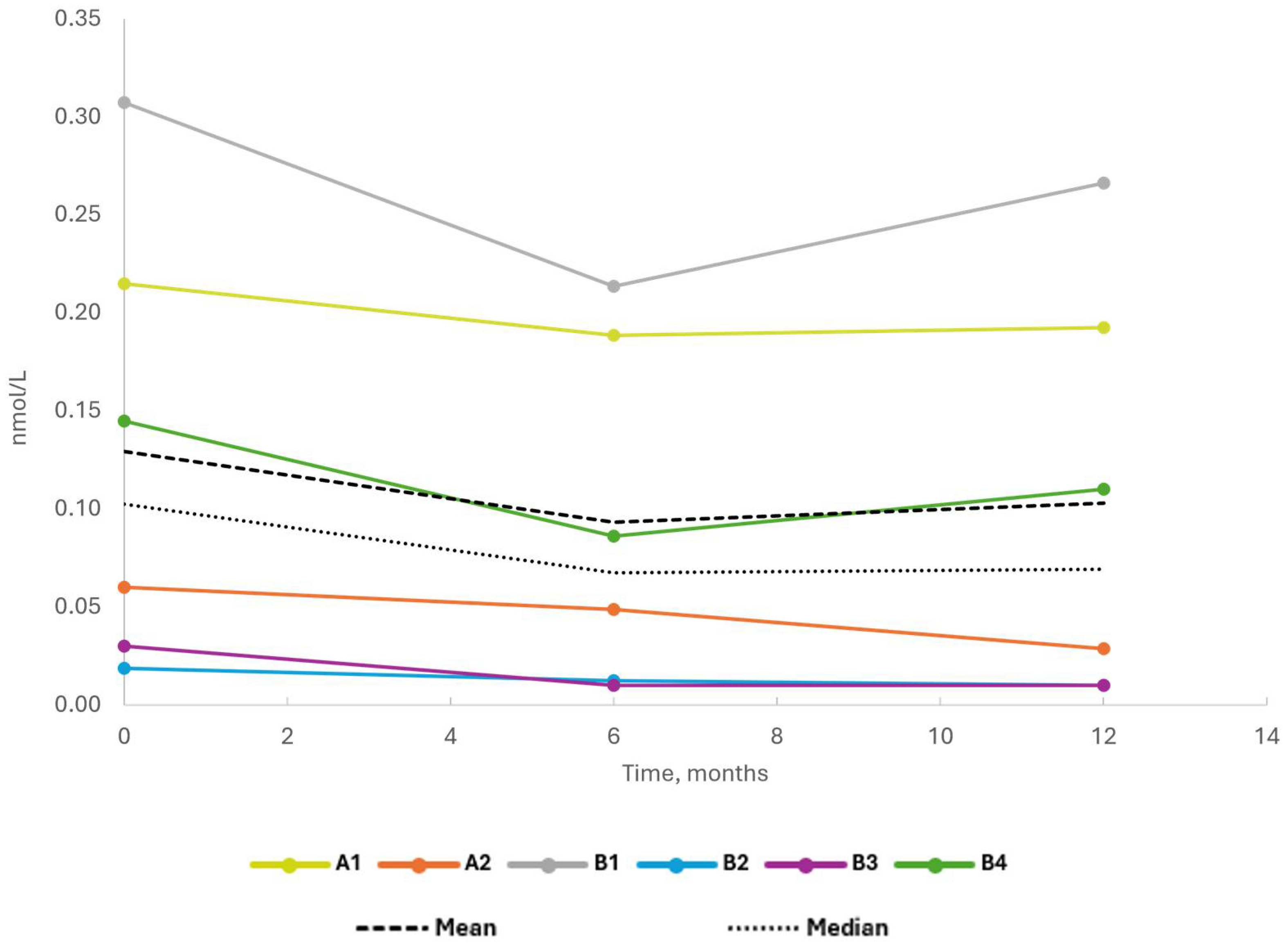
Only small decreases in endogenous insulin secretion were observed during the trial (Figure 2; Supplementary Table S1). The mean C-peptide AUC (area under the curve) after the Mixed-Meal Tolerance test (MMTT, 0–120 min) decreased from 0.083 nmol/L/min at baseline to 0.053 nmol/L/min and 0.050 nmol/L/min at months 6 and 12, respectively. This corresponded to a mean change ratio of 0.635 from baseline to month 6 and 0.607 to month 12. The absolute decrease in C-peptide AUC response over the course of the trial was low in all patients.
The mean HbA1c serum levels were similar both at six (53.0 mmol/mol) and twelve (55.3 mmol/mol) months as compared to baseline (53.0 mmol/mol) (Supplementary Tables S2 and S3). The mean daily exogenous insulin consumption decreased from 0.701 IU/kg per day at baseline to 0.529 IU/kg per day at month 6 and 0.553 IU/kg per day at month 12 (Supplementary Table S4). This corresponded to mean changes from baseline at month 6 of −0.172 IU/kg per day and 0.147 IU/kg per day at month 12.
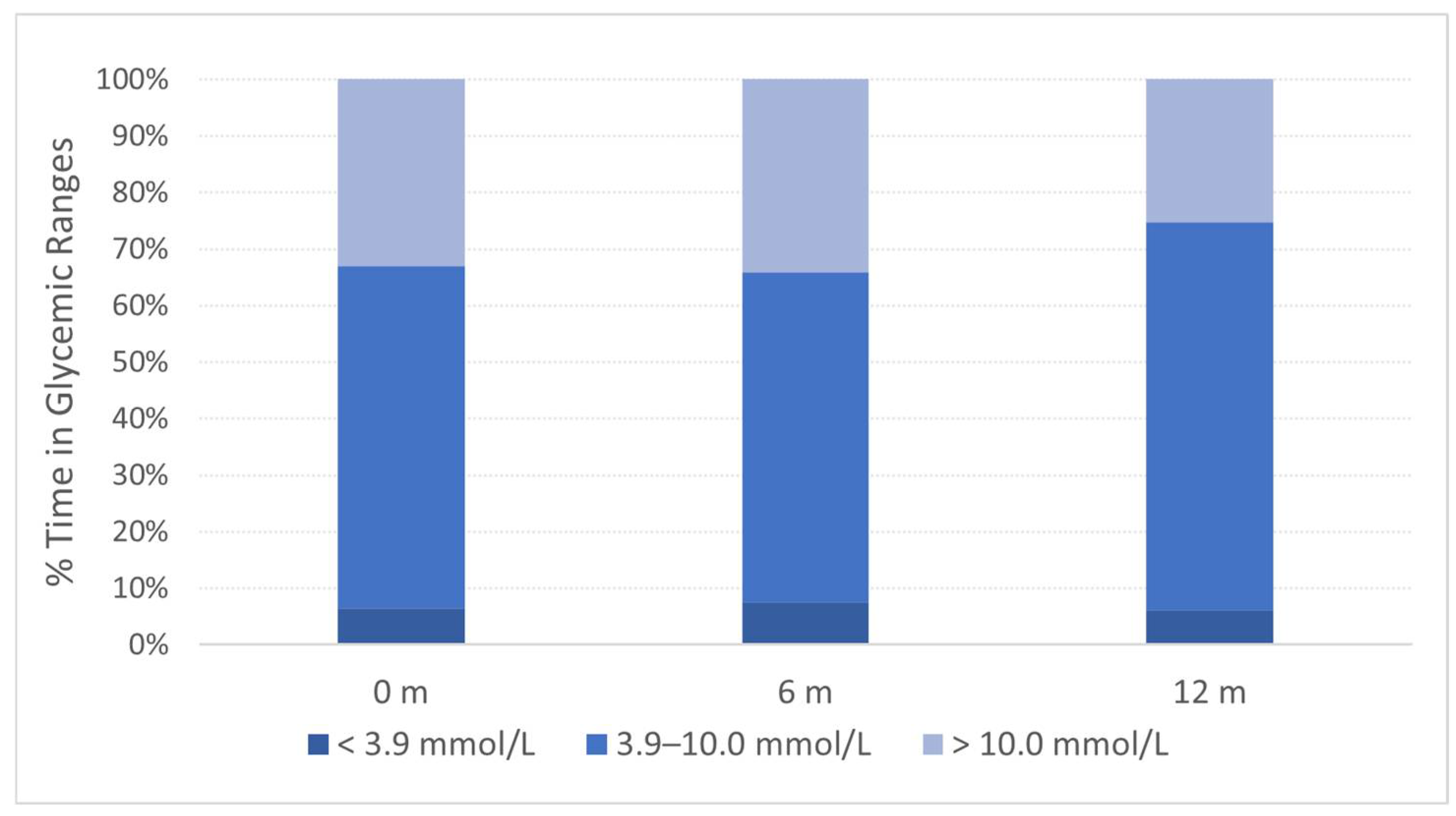
The mean percentage of time spent in the glycemic target range (TIR) of 3.9 to 10 mmol/L (70 to 180 mg/dL) was similar at baseline (60.7%) and month 6 (58.3%) but increased at month 12 (68.6%) (Figure 3). This corresponded to a mean change of 7.9% from baseline to month 12 (12.9 h). The increase in % TIR was reflected in a decreased percentage of time spent in the hyperglycemic range of >10 mmol/L (>180 mg/dL), with a mean change from baseline to month 12 of −7.7% (26.9 h) (Supplementary Tables S5 and S6).

2.3. Individual Longitudinal Follow-Up from Original Treatment

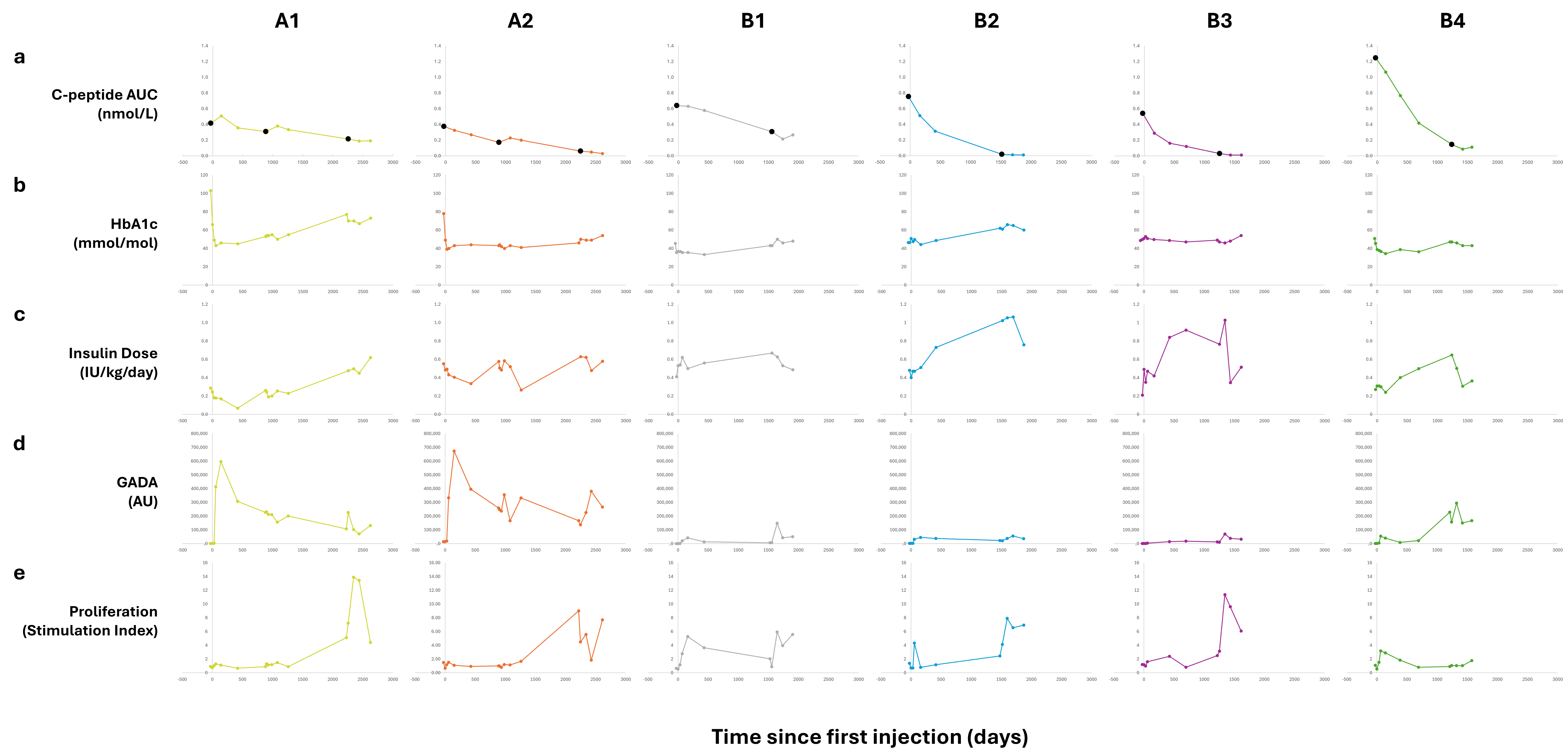
Individual longitudinal data from the DIAGNODE-B trial were combined with results from the preceding trials (DIAGNODE-1 and DIAGNODE-2) for the six patients (Figure 4). The disease progression was clearly seen in the decline in stimulated C-peptide over time after diagnosis (Figure 4a). Beta cell function declined in a linear fashion, with patients A1, A2 and B1 declining more slowly at a similar rate, while patients B2, B3 and B4 declined more rapidly at a similar rate, leveling out as C-peptide levels approached 0.2 nmol/L. Patients A1 and A2 most clearly showed a perturbation of disease development after initial treatment and/or redosing. All patients had a slightly lower rate of decline after dosing, though this was clearly confounded by the expectation that the decline would naturally decrease over time.
As expected, initial HbA1c levels were higher in patients initially randomized closer to diagnosis (Figure 4b, second row). HbA1c declined initially for all patients and most rapidly for those randomized closer to diagnosis. HbA1c increased over time from all patients to a varying degree, from minimum values in the first year after randomization.
The rise in exogenous insulin dose requirements over time mirrors the decrease in C-peptide decline for all patients, including the perturbations observed in connection with dosing (Figure 4c, third row). The latter may also be affected by inclusion in new clinical trial activities.

2.4. Immunological Responses

Generally, the results reflect varied immunological responses at different timepoints. However, the statistical analysis did not show significant differences in the immunological responses.
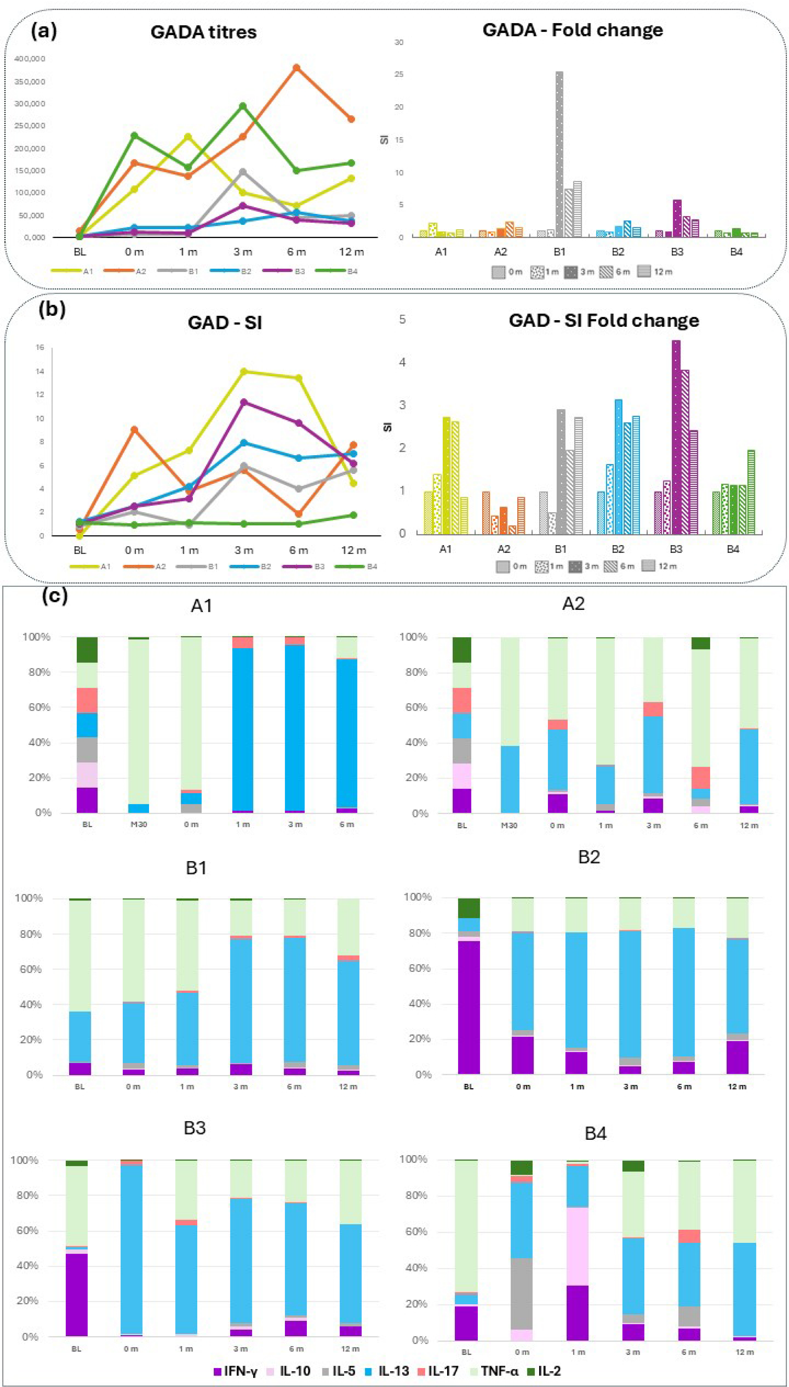
The serum GADA titers varied substantially between the patients at the trial baseline (0 month) (Figure 5a), and the mean GADA titers increased from 92,892 U/mL at baseline to 145,920 U/mL at month 3, 122,630 U/mL at month 6 and 113,408 U/mL at month 12. The mean GAD65-induced proliferation of PBMCs increased from 3.5 at baseline to 6.1 at month 6 and 5.4 at month 12 (Figure 5b; Supplementary Tables S7 and S8). Both GADA levels and proliferation SI were low at the time of the first injection (BL) (Figure 5a,b). A transient rise in both GADA and the proliferation of varying amplitudes was observed after each dosing in all individual patients, with occasional exceptions (Figure 4).
Analysis of GAD-induced cytokine secretion upon in vitro stimulation of PBMCs showed an elevation for several cytokines in most of the patients (Supplementary Figure S1). Analysis of the relative cytokine contribution for each individual when including cytokines that were also analyzed in previous studies (IL-2, -5, -10, -13, -17, IFN-γ and TNF-α) illustrates that redosing with GAD-alum predominantly induced a relative increase in IL-13 (0 m to 12 m) (Figure 5c). While the relative cytokine contribution before the original treatment (BL) differed between patients, IL-13 predominated at the time of the redosing (0 m), i.e., 39–49 months after the latest dosing. Patients A1 and A2 had previously been redosed 30 months after the initial treatment, at which time (30 months) the cytokine profile was similar to that at 0 months (baseline in DIAGNODE-B trial). There was a noticeable contribution of TNF-α to the relative cytokine profile, especially before initial treatment and redosing, which is most likely explained by the high levels characteristic of this cytokine (Supplementary Figure S1).

3. Discussion

The results of the trial suggest that redosing with intralymphatic GAD-alum was tolerable and seemed to be safe. Redosing, even several years after the first course of the therapy, showed the same pattern of immunological response seen at initial treatment in previous trials.
As the DIAGNODE-B trial included only six patients with no control arm, an important limitation of the study, few conclusions can and should be drawn regarding efficacy. Additionally, three out of six individuals had minimal beta cell function at redosing, as indicated by stimulated C-peptide levels, limiting the possibility of seeing effects on disease progression. That said, C-peptide decreased minimally during the 12-month trial period for all patients, in contrast to the expected natural course (7). Regarding clinical outcomes, most patients showed stable and well-controlled blood sugar levels as measured by HbA1c and IDAA1c. The mean % TIR (3.9 to 10 mmol/L) increased, while mean time in hyperglycemic range decreased, and mean daily exogenous insulin consumption decreased at month 12 when compared to baseline. Treatment with GAD-alum affects the immune system and may have most effect in patients with adequate vitamin D concentrations [72], although vitamin D on its own is insufficient for preserving beta cell function.
The lengthy continuous follow-up of individual stimulated C-peptide levels suggests that the six patients represent two different disease trajectories, with patients A1, A2 and B1 showing a slower progression, either as a result of disease heterogeneity or a difference in ability to respond to the treatment. When comparing trajectories against demographics and baseline values, the pattern was not straightforward. It has been previously shown that patients positive for DR3-DQ2 phenotype and negative for DR4-DQ8 possibly respond even better to GAD-alum treatment [53]. Patients A1 and A2 belong to this HLA category, while B1 does not, and nor do the other three patients. One common feature for patients A1, A2 and B1 was that they were initially treated closer to diagnosis than B2 and B3, suggesting that treatment closer to diagnosis might be advantageous. However, this does not apply to subject B4, who was also treated within 90 days of diagnosis. Regarding trajectory in relation to age, while individuals A1 and A2 were above 20 years old at diagnosis, commonly associated with slower disease progression, B1 was only 13 years old. Additionally, despite showing a faster decline, B4 was also above 20 years of age at diagnosis.
Though there was no control group included in the DIAGNODE-B trial, the longitudinal C-peptide values suggest clinical effect based on changes to the disease trajectories. Ideally, an efficacious treatment would be indicated by a break in the trajectory after redosing, or otherwise by a bump or shift in the trajectory indicating a transient delay. No conclusions can be drawn from the initial three-dose treatment as there was no earlier C-peptide data. Although no strong breaks in the trajectory for any subject were observed, there are indications of bumps at both redosing instances for A1 and A2, and possibly for B1 and B4. The perturbation in the trajectory appears largest in the first redosing of A1 and A2, perhaps because the time since the previous treatment administration was shorter.
Due to the small number of patients, furthermore with two different treatment regimens, statistical analysis of any immune markers included in this trial in relation to stimulated C-peptide was precluded. Graphical illustration of the relative cytokine profile showed more pronounced GAD65-stimulated IL-13 secretion after GAD-alum redosing in all patients. In the DIAGNODE-1 trial, it was observed that the elevation of cytokine secretion induced by GAD65 treatment waned after 30 months, when almost all analyzed cytokines were undetectable with the exception of IL-13, which was elevated compared to both baseline and 15 months [55]. In the DIAGNODE-2 trial, it was shown that GAD65-induced secretion of IL-13, IL-10 and IL-5 and proliferation was increased significantly more in the DR3-DQ2 responder group than in non DR3-DQ2 patients [54,73] These factors were also significantly increased in those patients who showed the lowest rate of disease progression after active treatment regardless of HLA [73]. Interleukin-13 has potent anti-inflammatory activities both in vitro and in vivo [74,75,76] and together with IL-5 has a central role in immune regulation and differentiation, governing the onset of inflammatory responses and providing signals that help turn off chronic inflammation and protect tissues from ongoing damage [76,77]. Thus, it is possible that the effect of GAD-alum might be related to the generation of a predominant anti-autoimmune response to GAD65 that counteracts proinflammatory factors and generates an environment where autoreactive Th1 effector cells are suppressed, thereby restoring immunological balance.
In conclusion, this DIAGNODE-B pilot trial provides evidence that redosing with GAD-alum seems to be safe and tolerable. Additionally, though there is considerable individual variation, redosing appears to induce an immunological response comparable to original treatment consistent with GAD65-specific anti-inflammatory modulation. There was some indication that redosing may prolong the disease modification elicited by the original GAD-alum treatment, but further placebo-controlled studies are needed to confirm this hypothesis and to establish the best redosing interval. If for no other reason, earlier redosing may be preferred to further slow the progression as soon as possible. It cannot be excluded that different autoantigens should be combined depending on endotype of T1D [78,79,80,81,82], and other treatment modalities may need to be added [83,84,85]. As all treatments to preserve residual beta cell function so far have only shown a transient effect, the possibility to redose is crucial.

4. Materials and Methods

4.1. Trial Design and Participants

DIAGNODE-B (Eudra CT number 2021-005441-32; NCT NCT05351879) was an open-label Phase I/II pilot trial in GADA-positive T1D patients carrying the HLA DR3-DQ2 haplotype who had previously been treated with four intralymphatic injections of GAD-alum within the DIAGNODE-1 trial or three intralymphatic injections of GAD-alum within the DIAGNODE-2 trial.
The DIAGNODE-B trial comprised five visits to the clinic:
Visit 1: Month 0. Screening and informed consent
Visit 2: Month 1, baseline. Mixed-Meal Tolerance Test (MMTT) and intralymphatic administration of GAD-alum
Visit 3: Month 3. Safety follow-up
Visit 4: Month 6. Safety follow-up and MMTT
Visit 5: Month 12. Safety follow-up and MMTT
All patients were evaluated at day 1 (0 months, baseline) and at 1, 3, 6 and 12 months with clinical examination and blood samples. Mixed-Meal Tolerance Tests (MMTT) [71] were performed at day 1 (baseline) and at 6 and 12 months to measure C-peptide (mean area under the curve [AUC]).
Eight patients from two clinics in Sweden were initially invited to participate. One was unable to participate because of pregnancy and one other for practical reasons. Two individuals who had earlier participated in the DIAGNODE-1 extension trial [55] received a fifth injection of 4 μg of GAD-alum (Diamyd Medical, Stockholm, Sweden) into an inguinal lymph node administered using an ultrasound technique, and three individuals who had participated in DIAGNODE-2 [54] received a fourth injection of GAD-alum into an inguinal lymph node. All participants also received 2000 U/day vitamin D (Calciferol) supplementation (Divisun, Meda AB, Solna, Sweden) for 2 months, starting 1 month prior to the GAD-alum injection. Informed consent was obtained from all patients prior to any trial procedures. Safety, beta cell function, diabetes status and immunological responses were followed for 12 months post-baseline.

4.2. Investigational Product and Mode of Administration

The GAD-alum (Diamyd®) drug product was composed of the rhGAD65 protein formulated in a sterile, non-pyrogenic phosphate-buffered saline containing an aluminum hydroxide adjuvant, Alhydrogel® (Croda Pharma, Fredrikssund, Denmark). GAD-alum (Diamyd®; 4 μg, Protein Sciences Corporation, Meriden, CT, USA) was administered as an ultrasound-guided 0.1 mL intralymphatic injection into an inguinal lymph node.
The trial was approved by the relevant regulatory authorities and research ethics board. All participants and their parents/caregivers gave their consent after receiving oral and written information.

4.3. Laboratory Tests

Laboratory analyses were performed at Linköping University, Sweden. Blood and serum samples from all the participants were collected at start of trial (0 months), before the GAD-alum redosing (1 months) and after 3, 6 and 12 months. Samples were drawn during the morning hours, and peripheral blood mononuclear cells (PBMCs) were isolated within 24 h using Leucosep (Greiner Bio One, Kremsmünster, Austria) according to the manufacturer’s instructions.
Analysis of serum C-peptide was performed using a solid phase-two side enzyme immunoassay (Mercodia, Uppsala, Sweden). Results for each assay were validated with the inclusion of a human diabetes antigen control (low/high) (Mercodia). The assay is calibrated against the international reference reagent for C-peptide IRR c-peptide 84/510. Inter- and intra-assay variations were 6.6% and 3.5%, respectively.

4.4. Serum Antibodies

Serum GAD autoantibodies (GADA) were estimated in duplicate samples by means of a radio-binding assay, using 35S-labeled recombinant human GAD65 (rhGAD65) as previously described [69]. Sepharose protein A was used to separate free from antibody-bound labelled GAD65. Results were expressed as units/mL.

4.5. Lymphocyte Proliferation Assay

Proliferative responses were analyzed in PBMCs cultured in triplicate in medium alone (AIM-V medium with β-mercaptoethanol), in the presence of 5 μg/mL rhGAD65 (Diamyd Medical, Stockholm, Sweden) or with CD3/CD28 beads (Gibco, Life Technologies AS, Oslo, Norway). After 3 days, cells were incubated with 0.2 µCi of [3H] thymidine/well for 18 h and then harvested. The [3H] thymidine incorporation was counted in a beta-scintillation counter (Perkin Elmer, Shelton, CT, USA) as counts per minute (cpm). The proliferation was expressed as a stimulation index (SI), calculated as the mean cpm of cells cultured in triplicate in the presence of stimulus divided by the mean cpm of cells with medium alone.

4.6. Cytokine Secretion Assay

PBMCs were cultured for 7 days in the presence of 5 μg/mL rhGAD65 or in medium alone at 37 °C in 5% CO2, as previously described [68]. The cytokines IL-1β, IL-2, IL-4, IL-5, IL-6, IL-7, IL-8, IL-10, IL-12, IL-13, IL-17, interferon (IFN)-γ, tumor necrosis factor (TNF)-α, and chemokines: monocyte chemoattractant protein (MCP)-1, granulocyte (G) colony stimulating factor (CSF), macrophage inflammatory proteins (MIP)-1β, and granulocyte–monocyte (GM)-CSF were detected by Luminex in supernatants collected after 7 days culture. GAD65-induced cytokine secretion is given as pg/mL after subtraction of spontaneous secretion.
The cytokines were measured in cell culture supernatants using a Bio-Plex Pro Cytokine Panel (Bio-Rad, Hercules, CA, USA) according to the manufacturer’s instructions. Data were collected using the Luminex 200 ™ (Luminex xMAP™ Corporation, Austin, TX, USA). Antigen-induced cytokine secretion level was calculated by subtracting the spontaneous secretion (i.e., secretion from PBMCs cultured in medium alone) from the one following stimulation with GAD65, and the levels were expressed as pg/mL.

4.7. Statistical Analysis

Since the sample size was limited, non-parametric tests were used to compare immunological observations at the different timepoints. Friedman’s test was used to compare differences in the immunological responses at the different timepoints. Differences were considered statistically significant for p < 0.05.

Supplementary Materials

The following supporting information can be downloaded at: https://www.mdpi.com/article/10.3390/ijms26010374/s1.

Author Contributions

K.Å. Resources;Writing—review; editing, Project administration; J.L. had the idea, designed the trial, wrote the protocol, recruited the patients and performed the clinical studies. A.T. performed the laboratory experiments. R.C., A.T., A.L. and P.F.T. analyzed the data. J.L. wrote the first draft of manuscript, and all other authors critically read the manuscript, and all approved the final version. R.C. and J.L. are the guarantors of this work and, as such, have full access to all the data in the trial and takes responsibility for the integrity of the data and the accuracy of the data analysis. All authors have read and agreed to the published version of the manuscript.

Funding

This work was supported by Barndiabetesfonden (Swedish Child Diabetes Foundation), Diabetesfonden (the Swedish Diabetes Foundation) and an unrestricted grant from Diamyd Medical.

Institutional Review Board Statement

The trial was approved by the Swedish Ethical Review Authority 2021 (Dnr 2021/05681) and by the Medical Product Agency, Uppsala, Sweden. Trial monitoring to ensure adherence to the clinical study protocol, Good Clinical Practice and local guidelines was performed by Forum Östergötland (Linköping).

Informed Consent Statement

All patients gave their oral and written informed consent to participate in the trial.

Data Availability Statement

Data are available from corresponding author on reasonable request after ethical approval.

Acknowledgments

We are very grateful to Joachim Davidsson and Anders Knutsson for skillful performance of the needle guided lymph node injections, to research nurses Sofia Sjöberg, Mona Ahlberg and Malin Huss for taking care of the patients and to Ingela Johansson and Gosia Smolinska for skillful laboratory work. The trial has been generously supported by Barndiabetesfonden (Swedish Child Diabetes Foundation), Diabetesfonden (Swedish Diabetes Foundation), and an unrestricted grant from Diamyd Medical.

Conflicts of Interest

P.F.T. and A.L. are employees of Diamyd Medical. No other conflicts of interest relevant to this article were reported.

Abbreviations

GADAGlutamic acid decarboxylase autoantibodies
GAD-alumGlutamic acid decarboxylase formulated in aluminum hydroxide
LNlymph node
PBMCPeripheral blood mononuclear cell
SCsubcutaneous
ThT helper

References

  1. Cardona-Hernandez, R.; Schwandt, A.; Alkandari, H.; Bratke, H.; Chobot, A.; Coles, N.; Corathers, S.; Goksen, D.; Goss, P.; Imane, Z.; et al. Glycemic Outcome Associated with Insulin Pump and Glucose Sensor Use in Children and Adolescents with Type 1 Diabetes. Data from the International Pediatric Registry SWEET. Diabetes Care 2021, 44, 1176–1184. [Google Scholar] [CrossRef] [PubMed]
  2. Holman, N.; Woch, E.; Dayan, C.; Warner, J.; Robinson, H.; Young, B.; Elliott, J. National Trends in Hyperglycemia and Diabetic Ketoacidosis in Children, Adolescents, and Young Adults with Type 1 Diabetes: A Challenge Due to Age or Stage of Development, or Is New Thinking About Service Provision Needed? Diabetes Care 2023, 46, 1404–1408. [Google Scholar] [CrossRef] [PubMed]
  3. Bojestig, M.; Arnqvist, H.J.; Hermansson, G.; Karlberg, B.E.; Ludvigsson, J. Declining Incidence of Nephropathy in Insulin-Dependent Diabetes Mellitus. N. Engl. J. Med. 1994, 330, 15–18. [Google Scholar] [CrossRef]
  4. Lind, M.; Svensson, A.-M.; Kosiborod, M.; Gudbjörnsdottir, S.; Pivodic, A.; Wedel, H.; Dahlqvist, S.; Clements, M.; Rosengren, A. Glycemic Control and Excess Mortality in Type 1 Diabetes. N. Engl. J. Med. 2014, 371, 1972–1982. [Google Scholar] [CrossRef]
  5. Rawshani, A.; Sattar, N.; Franzén, S.; Rawshani, A.; Hattersley, A.T.; Svensson, A.-M.; Eliasson, B.; Gudbjörnsdottir, S. Excess mortality and cardiovascular disease in young adults with type 1 diabetes in relation to age at onset: A nationwide, register-based cohort study. Lancet 2018, 392, 477–486. [Google Scholar] [CrossRef]
  6. Gubitosi-Klug, R.A.; Braffett, B.H.; Hitt, S.; Arends, V.; Uschner, D.; Jones, K.; Diminick, L.; Karger, A.B.; Paterson, A.D.; Roshandel, D.; et al. Residual β cell function in long-term type 1 diabetes associates with reduced incidence of hypoglycemia. J. Clin. Investig. 2021, 131, e143011. [Google Scholar] [CrossRef]
  7. Taylor, P.N.; Collins, K.S.; Lam, A.; Karpen, S.R.; Greeno, B.; Walker, F.; Lozano, A.; Atabakhsh, E.; Ahmed, S.T.; Marinac, M.; et al. C-peptide and metabolic outcomes in trials of disease modifying therapy in new-onset type 1 diabetes: An individual participant meta-analysis. Lancet Diabetes Endocrinol. 2023, 11, 915–925. [Google Scholar] [CrossRef]
  8. Ludvigsson, J.; Heding, L.; Lieden, G.; Marner, B.; Lernmark, A. Plasmapheresis in the initial treatment of insulin-dependent diabetes mellitus in children. BMJ 1983, 286, 176–178. [Google Scholar] [CrossRef] [PubMed]
  9. Pinckney, A.; Rigby, M.R.; Keyes-Elstein, L.; Soppe, C.L.; Nepom, G.T.; Ehlers, M.R. Correlation Among Hypoglycemia, Glycemic Variability, and C-Peptide Preservation After Alefacept Therapy in Patients with Type 1 Diabetes Mellitus: Analysis of Data from the Immune Tolerance Network T1DAL Trial. Clin. Ther. 2016, 38, 1327–1339. [Google Scholar] [CrossRef] [PubMed]
  10. Bogdanou, D.; Penna-Martinez, M.; Filmann, N.; Chung, T.; Moran-Auth, Y.; Wehrle, J.; Cappel, C.; Huenecke, S.; Herrmann, E.; Koehl, U.; et al. T-lymphocyte and glycemic status after vitamin D treatment in type 1 diabetes: A randomized controlled trial with sequential crossover. Diabetes/Metabolism Res. Rev. 2017, 33, e2865. [Google Scholar] [CrossRef]
  11. Sharma, S. Does Vitamin D Supplementation Improve Glycaemic Control in Children with Type 1 Diabetes Mellitus?—A Randomized Controlled Trial. J. Clin. Diagn. Res. 2017, 11, SC15–SC17. [Google Scholar] [CrossRef] [PubMed]
  12. White, P.C.; Adhikari, S.; Grishman, E.K.; Sumpter, K.M. A phase I study of anti-inflammatory therapy with rilonacept in adolescents and adults with type 1 diabetes mellitus. Pediatr. Diabetes 2018, 19, 788–793. [Google Scholar] [CrossRef] [PubMed]
  13. Greenbaum, C.J.; Serti, E.; Lambert, K.; Weiner, L.J.; Kanaparthi, S.; Lord, S.; Gitelman, S.E.; Wilson, D.M.; Gaglia, J.L.; Griffin, K.J.; et al. IL-6 receptor blockade does not slow β cell loss in new-onset type 1 diabetes. J. Clin. Investig. 2021, 6, 150074. [Google Scholar] [CrossRef] [PubMed]
  14. Dharadhar, S.; Sharma, A.; Dey, D. Efficacy and safety of oral insulin versus placebo for patients with diabetes mellitus: A systematic review and meta-analysis. Indian J. Pharmacol. 2022, 54, 244–252. [Google Scholar] [CrossRef] [PubMed]
  15. Basak, E.A.; de Joode, K.; Uyl, T.J.; van der Wal, R.; Schreurs, M.W.; Berg, S.A.v.D.; Hoop, E.O.-D.; van der Leest, C.H.; Chaker, L.; Feelders, R.A.; et al. The course of C-peptide levels in patients developing diabetes during anti-PD-1 therapy. Biomed. Pharmacother. 2022, 156, 113839. [Google Scholar] [CrossRef]
  16. Bastos, T.S.B.; Braga, T.T.; Davanso, M.R. Vitamin D and Omega-3 Polyunsaturated Fatty Acids in Type 1 Diabetes Modulation. Endocrine, Metab. Immune Disord.—Drug Targets 2022, 22, 815–833. [Google Scholar] [CrossRef]
  17. Forlenza, G.P.; McVean, J.; Beck, R.W.; Bauza, C.; Bailey, R.; Buckingham, B.; DiMeglio, L.A.; Sherr, J.L.; Clements, M.; Neyman, A.; et al. Effect of Verapamil on Pancreatic Beta Cell Function in Newly Diagnosed Pediatric Type 1 Diabetes. JAMA 2023, 329, 990–999. [Google Scholar] [CrossRef]
  18. Coutant, R.; Landais, P.; Rosilio, M.; Johnsen, C.; Lahlou, N.; Chatelain, P.; Carel, J.C.; Ludvigsson, J.; Boitard, C.; Bougnères, P.F. Low dose linomide in Type I juvenile diabetes of recent onset: A randomised placebo-controlled double blind trial. Diabetologia 1998, 41, 1040–1046. [Google Scholar] [CrossRef]
  19. Stiller, C.R.; Laupacis, A.; Dupre, J.; Jenner, M.R.; Keown, P.A.; Rodger, W.; Wolfe, B.M. Abuse of Cimetidine in Outpatient Practice. N. Engl. J. Med. 1983, 308, 1226. [Google Scholar] [CrossRef]
  20. Herold, K.C.; Hagopian, W.; Auger, J.A.; Poumian-Ruiz, E.; Taylor, L.; Donaldson, D.; Gitelman, S.E.; Harlan, D.M.; Xu, D.; Zivin, R.A.; et al. Anti-CD3 Monoclonal Antibody in New-Onset Type 1 Diabetes Mellitus. N. Engl. J. Med. 2002, 346, 1692–1698. [Google Scholar] [CrossRef]
  21. Keymeulen, B.; Vandemeulebroucke, E.; Ziegler, A.G.; Mathieu, C.; Kaufman, L.; Hale, G.; Gorus, F.; Goldman, M.; Walter, M.; Candon, S.; et al. Insulin Needs after CD3-Antibody Therapy in New-Onset Type 1 Diabetes. N. Engl. J. Med. 2005, 352, 2598–2608. [Google Scholar] [CrossRef] [PubMed]
  22. Mastrandrea, L.; Yu, J.; Behrens, T.; Buchlis, J.; Albini, C.; Fourtner, S.; Quattrin, T. Etanercept Treatment in Children with New-Onset Type 1 Diabetes. Diabetes Care 2009, 32, 1244–1249. [Google Scholar] [CrossRef] [PubMed]
  23. Pescovitz, M.D.; Greenbaum, C.J.; Krause-Steinrauf, H.; Becker, D.J.; Gitelman, S.E.; Goland, R.; Gottlieb, P.A.; Marks, J.B.; McGee, P.F.; Moran, A.M.; et al. Rituximab, B-Lymphocyte Depletion, and Preservation of Beta-Cell Function. N. Engl. J. Med. 2009, 361, 2143–2152. [Google Scholar] [CrossRef] [PubMed]
  24. Sherry, N.; Hagopian, W.; Ludvigsson, J.; Jain, S.M.; Wahlen, J.; Ferry, R.J.; Bode, B.; Aronoff, S.; Holland, C.; Carlin, D.; et al. Teplizumab for treatment of type 1 diabetes (Protégé study): 1-year results from a randomised, placebo-controlled trial. Lancet 2011, 378, 487–497. [Google Scholar] [CrossRef]
  25. Hagopian, W.; Ferry, R.J.; Sherry, N.; Carlin, D.; Bonvini, E.; Johnson, S.; Stein, K.E.; Koenig, S.; Daifotis, A.G.; Herold, K.C.; et al. Teplizumab Preserves C-Peptide in Recent-Onset Type 1 Diabetes: Two-year results from the randomized, placebo-controlled protege trial. Diabetes 2013, 62, 3901–3908. [Google Scholar] [CrossRef]
  26. Aronson, R.; Gottlieb, P.A.; Christiansen, J.S.; Donner, T.W.; Bosi, E.; Bode, B.W.; Pozzilli, P.; the DEFEND Investigator Group. Low-Dose Otelixizumab Anti-CD3 Monoclonal Antibody DEFEND-1 Study: Results of the Randomized Phase III Study in Recent-Onset Human Type 1 Diabetes. Diabetes Care 2014, 37, 2746–2754. [Google Scholar] [CrossRef]
  27. Orban, T.; Bundy, B.; Becker, D.J.; DiMeglio, L.A.; Gitelman, S.E.; Goland, R.; Gottlieb, P.A.; Greenbaum, C.J.; Marks, J.B.; Monzavi, R.; et al. Costimulation Modulation with Abatacept in Patients with Recent-Onset Type 1 Diabetes: Follow-up 1 Year After Cessation of Treatment. Diabetes Care 2014, 37, 1069–1075. [Google Scholar] [CrossRef]
  28. Haller, M.J.; Gitelman, S.E.; Gottlieb, P.A.; Michels, A.W.; Rosenthal, S.M.; Shuster, J.J.; Zou, B.; Brusko, T.M.; Hulme, M.A.; Wasserfall, C.H.; et al. Anti-thymocyte globulin/G-CSF treatment preserves β cell function in patients with established type 1 diabetes. J. Clin. Investig. 2015, 125, 448–455. [Google Scholar] [CrossRef]
  29. Rigby, M.R.; Harris, K.M.; Pinckney, A.; DiMeglio, L.A.; Rendell, M.S.; Felner, E.I.; Dostou, J.M.; Gitelman, S.E.; Griffin, K.J.; Tsalikian, E.; et al. Alefacept provides sustained clinical and immunological effects in new-onset type 1 diabetes patients. J. Clin. Investig. 2015, 125, 3285–3296. [Google Scholar] [CrossRef]
  30. Rosenzwajg, M.; Salet, R.; Lorenzon, R.; Tchitchek, N.; Roux, A.; Bernard, C.; Carel, J.-C.; Storey, C.; Polak, M.; Beltrand, J.; et al. Low-dose IL-2 in children with recently diagnosed type 1 diabetes: A Phase I/II randomised, double-blind, placebo-controlled, dose-finding study. Diabetologia 2020, 63, 1808–1821. [Google Scholar] [CrossRef]
  31. Rapini, N.; Schiaffini, R.; Fierabracci, A. Immunotherapy Strategies for the Prevention and Treatment of Distinct Stages of Type 1 Diabetes: An Overview. Int. J. Mol. Sci. 2020, 21, 2103. [Google Scholar] [CrossRef] [PubMed]
  32. von Herrath, M.; Bain, S.C.; Bode, B.; Clausen, J.O.; Coppieters, K.; Gaysina, L.; Gumprecht, J.; Hansen, T.K.; Mathieu, C.; Morales, C.; et al. Anti-interleukin-21 antibody and liraglutide for the preservation of β-cell function in adults with recent-onset type 1 diabetes: A randomised, double-blind, placebo-controlled, phase 2 trial. Lancet Diabetes Endocrinol. 2021, 9, 212–224. [Google Scholar] [CrossRef] [PubMed]
  33. Groele, L.; Szajewska, H.; Szalecki, M.; Świderska, J.; Wysocka-Mincewicz, M.; Ochocińska, A.; Stelmaszczyk-Emmel, A.; Demkow, U.; Szypowska, A. Lack of effect of Lactobacillus rhamnosus GG and Bifidobacterium lactis Bb12 on beta-cell function in children with newly diagnosed type 1 diabetes: A randomised controlled trial. BMJ Open Diabetes Res. Care 2021, 9, e001523. [Google Scholar] [CrossRef] [PubMed]
  34. Martin, A.; Mick, G.J.; Choat, H.M.; Lunsford, A.A.; Tse, H.M.; McGwin, G.G.; McCormick, K.L. A randomized trial of oral gamma aminobutyric acid (GABA) or the combination of GABA with glutamic acid decarboxylase (GAD) on pancreatic islet endocrine function in children with newly diagnosed type 1 diabetes. Nat. Commun. 2022, 13, 7928. [Google Scholar] [CrossRef]
  35. Reddy, R.; Dayal, D.; Sachdeva, N.; Attri, S.V.; Gupta, V.K. Combination therapy with lansoprazole and cholecalciferol is associated with a slower decline in residual beta-cell function and lower insulin requirements in children with recent onset type 1 diabetes: Results of a pilot study. Einstein-Sao Paulo 2022, 20, eAO0149. [Google Scholar] [CrossRef]
  36. Waibel, M.; Thomas, H.E.; Wentworth, J.M.; Couper, J.J.; MacIsaac, R.J.; Cameron, F.J.; So, M.; Krishnamurthy, B.; Doyle, M.C.; Kay, T.W. Investigating the efficacy of baricitinib in new onset type 1 diabetes mellitus (BANDIT)—Study protocol for a phase 2, randomized, placebo controlled trial. Trials 2022, 23, 433. [Google Scholar] [CrossRef]
  37. Ramos, E.L.; Dayan, C.M.; Chatenoud, L.; Sumnik, Z.; Simmons, K.M.; Szypowska, A.; Gitelman, S.E.; Knecht, L.A.; Niemoeller, E.; Tian, W.; et al. Teplizumab and β-Cell Function in Newly Diagnosed Type 1 Diabetes. N. Engl. J. Med. 2023, 389, 2151–2161. [Google Scholar] [CrossRef]
  38. Waibel, M.; Wentworth, J.M.; So, M.; Couper, J.J.; Cameron, F.J.; MacIsaac, R.J.; Atlas, G.; Gorelik, A.; Litwak, S.; Sanz-Villanueva, L.; et al. Baricitinib and β-Cell Function in Patients with New-Onset Type 1 Diabetes. N. Engl. J. Med. 2023, 389, 2140–2150. [Google Scholar] [CrossRef]
  39. Tatovic, D.; Marwaha, A.; Taylor, P.; Hanna, S.J.; Carter, K.; Cheung, W.Y.; Luzio, S.; Dunseath, G.; Hutchings, H.A.; Holland, G.; et al. Ustekinumab for type 1 diabetes in adolescents: A multicenter, double-blind, randomized phase 2 trial. Nat. Med. 2024, 30, 2657–2666. [Google Scholar] [CrossRef]
  40. Roy, S.S.; Keshri, U.S.P.; Alam, S.; Wasnik, A. Effect of Immunotherapy on C-peptide Levels in Patients with Type I Diabetes Mellitus: A Systematic Review of Randomized Controlled Trials. Cureus 2024, 16, e58981. [Google Scholar] [CrossRef]
  41. Lokesh, M.N.; Kumar, R.; Jacob, N.; Sachdeva, N.; Rawat, A.; Yadav, J.; Dayal, D. Supplementation of High-Strength Oral Probiotics Improves Immune Regulation and Preserves Beta Cells among Children with New-Onset Type 1 Diabetes Mellitus: A Randomised, Double-Blind Placebo Control Trial. Indian J. Pediatr. 2024; ahead of print. [Google Scholar] [CrossRef] [PubMed]
  42. Quattrin, T.; Haller, M.J.; Steck, A.K.; Felner, E.I.; Li, Y.; Xia, Y.; Leu, J.H.; Zoka, R.; Hedrick, J.A.; Rigby, M.R.; et al. Golimumab and Beta-Cell Function in Youth with New-Onset Type 1 Diabetes. N. Engl. J. Med. 2020, 383, 2007–2017. [Google Scholar] [CrossRef] [PubMed]
  43. Gitelman, S.E.; Gottlieb, P.A.; Felner, E.I.; Willi, S.M.; Fisher, L.K.; Moran, A.; Gottschalk, M.; Moore, W.V.; Pinckney, A.; Keyes-Elstein, L.; et al. Antithymocyte globulin therapy for patients with recent-onset type 1 diabetes: 2 year results of a randomised trial. Diabetologia 2016, 59, 1153–1161. [Google Scholar] [CrossRef]
  44. Ludvigsson, J. Immune Interventions at Onset of Type 1 Diabetes—Finally, a Bit of Hope. N. Engl. J. Med. 2023, 389, 2199–2201. [Google Scholar] [CrossRef]
  45. Herold, K.C.; Bundy, B.N.; Long, S.A.; Bluestone, J.A.; DiMeglio, L.A.; Dufort, M.; Gitelman, S.E.; Gottlieb, P.A.; Krischer, J.P.; Linsley, P.S.; et al. An Anti-CD3 Antibody, Teplizumab, in Relatives at Risk for Type 1 Diabetes. N. Engl. J. Med. 2019, 381, 603–613. [Google Scholar] [CrossRef]
  46. Ludvigsson, J.; Faresjö, M.; Hjorth, M.; Axelsson, S.; Chéramy, M.; Pihl, M.; Vaarala, O.; Forsander, G.; Ivarsson, S.; Johansson, C.; et al. GAD Treatment and Insulin Secretion in Recent-Onset Type 1 Diabetes. N. Engl. J. Med. 2008, 359, 1909–1920. [Google Scholar] [CrossRef]
  47. Wherrett, D.K.; Bundy, B.; Becker, D.J.; DiMeglio, L.A.; Gitelman, S.E.; Goland, R.; Gottlieb, P.A.; Greenbaum, C.J.; Herold, K.C.; Marks, J.B.; et al. Antigen-based therapy with glutamic acid decarboxylase (GAD) vaccine in patients with recent-onset type 1 diabetes: A randomised double-blind trial. Lancet 2011, 378, 319–327. [Google Scholar] [CrossRef]
  48. Ludvigsson, J.; Krisky, D.; Casas, R.; Battelino, T.; Castaño, L.; Greening, J.; Kordonouri, O.; Otonkoski, T.; Pozzilli, P.; Robert, J.-J.; et al. GAD65 Antigen Therapy in Recently Diagnosed Type 1 Diabetes Mellitus. N. Engl. J. Med. 2012, 366, 433–442. [Google Scholar] [CrossRef]
  49. Beam, C.A.; MacCallum, C.; Herold, K.C.; Wherrett, D.K.; Palmer, J.; Ludvigsson, J. GAD vaccine reduces insulin loss in recently diagnosed type 1 diabetes: Findings from a Bayesian meta-analysis. Diabetologia 2017, 60, 43–49. [Google Scholar] [CrossRef]
  50. Ludvigsson, J.; Wahlberg, J.; Casas, R. Intralymphatic Injection of Autoantigen in Type 1 Diabetes. N. Engl. J. Med. 2017, 376, 697–699. [Google Scholar] [CrossRef]
  51. Tavira, B.; Barcenilla, H.; Wahlberg, J.; Achenbach, P.; Ludvigsson, J.; Casas, R. Intralymphatic Glutamic Acid Decarboxylase-Alum Administration Induced Th2-Like-Specific Immunomodulation in Responder Patients: A Pilot Clinical Trial in Type 1 Diabetes. J. Diabetes Res. 2018, 2018, 9391845. [Google Scholar] [CrossRef] [PubMed]
  52. Casas, R.; Dietrich, F.; Barcenilla, H.; Tavira, B.; Wahlberg, J.; Achenbach, P.; Ludvigsson, J. Glutamic Acid Decarboxylase Injection Into Lymph Nodes: Beta Cell Function and Immune Responses in Recent Onset Type 1 Diabetes Patients. Front. Immunol. 2020, 11, 564921. [Google Scholar] [CrossRef] [PubMed]
  53. Hannelius, U.; Beam, C.A.; Ludvigsson, J. Efficacy of GAD-alum immunotherapy associated with HLA-DR3-DQ2 in recently diagnosed type 1 diabetes. Diabetologia 2020, 63, 2177–2181. [Google Scholar] [CrossRef]
  54. Ludvigsson, J.; Sumnik, Z.; Pelikanova, T.; Chavez, L.N.; Lundberg, E.; Rica, I.; Martínez-Brocca, M.A.; de Adana, M.R.; Wahlberg, J.; Katsarou, A.; et al. Intralymphatic Glutamic Acid Decarboxylase With Vitamin D Supplementation in Recent-Onset Type 1 Diabetes: A Double-Blind, Randomized, Placebo-Controlled Phase IIb Trial. Diabetes Care 2021, 44, 1604–1612. [Google Scholar] [CrossRef]
  55. Casas, R.; Dietrich, F.; Puente-Marin, S.; Barcenilla, H.; Tavira, B.; Wahlberg, J.; Achenbach, P.; Ludvigsson, J. Intra-lymphatic administration of GAD-alum in type 1 diabetes: Long-term follow-up and effect of a late booster dose (the DIAGNODE Extension trial). Acta Diabetol. 2022, 59, 687–696. [Google Scholar] [CrossRef]
  56. Piemonti, L.; Monti, P.; Sironi, M.; Fraticelli, P.; Leone, B.E.; Dal Cin, E.; Allavena, P.; Di Carlo, V. Vitamin D3 Affects Differentiation, Maturation, and Function of Human Monocyte-Derived Dendritic Cells. J. Immunol. 2000, 164, 4443–4451. [Google Scholar] [CrossRef]
  57. Boonstra, A.; Barrat, F.J.; Crain, C.; Heath, V.L.; Savelkoul, H.F.J.; O’garra, A. 1α,25-Dihydroxyvitamin D3 Has a Direct Effect on Naive CD4+ T Cells to Enhance the Development of Th2 Cells. J. Immunol. 2001, 167, 4974–4980. [Google Scholar] [CrossRef]
  58. Mathieu, C.; Gysemans, C.; Giulietti, A.; Bouillon, R. Vitamin D and diabetes. Diabetologia 2005, 48, 1247–1257. [Google Scholar] [CrossRef]
  59. Infante, M.; Ricordi, C.; Sanchez, J.; Clare-Salzler, M.J.; Padilla, N.; Fuenmayor, V.; Chavez, C.; Alvarez, A.; Baidal, D.; Alejandro, R.; et al. Influence of Vitamin D on Islet Autoimmunity and Beta-Cell Function in Type 1 Diabetes. Nutrients 2019, 11, 2185. [Google Scholar] [CrossRef]
  60. Zipitis, C.S.; Akobeng, A.K. Vitamin D supplementation in early childhood and risk of type 1 diabetes: A systematic review and meta-analysis. Arch. Dis. Child. 2008, 93, 512–517. [Google Scholar] [CrossRef]
  61. Bizzarri, C.; Pitocco, D.; Napoli, N.; Di Stasio, E.; Maggi, D.; Manfrini, S.; Suraci, C.; Cavallo, M.G.; Cappa, M.; Ghirlanda, G.; et al. No Protective Effect of Calcitriol on β-Cell Function in Recent-Onset Type 1 Diabetes. Diabetes Care 2010, 33, 1962–1963. [Google Scholar] [CrossRef] [PubMed]
  62. Pitocco, D.; Crinò, A.; Di Stasio, E.; Manfrini, S.; Guglielmi, C.; Spera, S.; Anguissola, G.B.; Visalli, N.; Suraci, C.; Matteoli, M.C.; et al. The effects of calcitriol and nicotinamide on residual pancreatic β-cell function in patients with recent-onset Type 1 diabetes (IMDIAB XI). Diabet. Med. 2006, 23, 920–923. [Google Scholar] [CrossRef] [PubMed]
  63. Gabbay, M.A.L.; Sato, M.N.; Finazzo, C.; Duarte, A.J.S.; Dib, S.A. Effect of Cholecalciferol as Adjunctive Therapy With Insulin on Protective Immunologic Profile and Decline of Residual β-Cell Function in New-Onset Type 1 Diabetes Mellitus. Arch. Pediatr. Adolesc. Med. 2012, 166, 601–607. [Google Scholar] [CrossRef]
  64. Holick, M.F.; Binkley, N.C.; Bischoff-Ferrari, H.A.; Gordon, C.M.; Hanley, D.A.; Heaney, R.P.; Murad, M.H.; Weaver, C.M. Evaluation, Treatment, and Prevention of Vitamin D Deficiency: An Endocrine Society Clinical Practice Guideline. Med. J. Clin. Endocrinol. Metab. 2011, 96, 1911–1930. [Google Scholar] [CrossRef]
  65. Axelsson, S.; Hjorth, M.; Åkerman, L.; Ludvigsson, J.; Casas, R. Early induction of GAD65-reactive Th2 response in type 1 diabetic children treated with alum-formulated GAD65. Diabetes/Metabolism Res. Rev. 2010, 26, 559–568. [Google Scholar] [CrossRef]
  66. Hjorth, M.; Axelsson, S.; Rydén, A.; Faresjö, M.; Ludvigsson, J.; Casas, R. GAD-alum treatment induces GAD65-specific CD4+CD25highFOXP3+ cells in type 1 diabetic patients. Clin. Immunol. 2011, 138, 117–126. [Google Scholar] [CrossRef]
  67. Axelsson, S.; Chéramy, M.; Hjorth, M.; Pihl, M.; Åkerman, L.; Martinuzzi, E.; Mallone, R.; Ludvigsson, J.; Casas, R. Long-Lasting Immune Responses 4 Years after GAD-Alum Treatment in Children with Type 1 Diabetes. PLoS ONE 2011, 6, e29008. [Google Scholar] [CrossRef]
  68. Axelsson, S.; Chéramy, M.; Åkerman, L.; Pihl, M.; Ludvigsson, J.; Casas, R. Cellular and Humoral Immune Responses in Type 1 Diabetic Patients Participating in a Phase III GAD-alum Intervention Trial. Diabetes Care 2013, 36, 3418–3424. [Google Scholar] [CrossRef]
  69. Chéramy, M.; Skoglund, C.; Johansson, I.; Ludvigsson, J.; Hampe, C.S.; Casas, R. GAD-alum treatment in patients with type 1 diabetes and the subsequent effect on GADA IgG subclass distribution, GAD65 enzyme activity and humoral response. Clin. Immunol. 2010, 137, 31–40. [Google Scholar] [CrossRef]
  70. Dietrich, F.; Barcenilla, H.; Tavira, B.; Wahlberg, J.; Achenbach, P.; Ludvigsson, J.; Casas, R. Immune response differs between intralymphatic or subcutaneous administration of GAD-alum in individuals with recent onset type 1 diabetes. Diabetes/Metabolism Res. Rev. 2022, 38, e3500. [Google Scholar] [CrossRef]
  71. Greenbaum, C.J.; Mandrup-Poulsen, T.; McGee, P.F.; Battelino, T.; Haastert, B.; Ludvigsson, J.; Pozzilli, P.; Lachin, J.M.; Kolb, H. Mixed-Meal Tolerance Test Versus Glucagon Stimulation Test for the Assessment of β-Cell Function in Therapeutic Trials in Type 1 Diabetes. Diabetes Care 2008, 31, 1966–1971. [Google Scholar] [CrossRef] [PubMed]
  72. Grant, W.B.; Boucher, B.J.; Bhattoa, H.P.; Lahore, H. Why vitamin D clinical trials should be based on 25-hydroxyvitamin D concentrations. J. Steroid Biochem. Mol. Biol. 2018, 177, 266–269. [Google Scholar] [CrossRef] [PubMed]
  73. Puente-Marin, S.; Dietrich, F.; Achenbach, P.; Barcenilla, H.; Ludvigsson, J.; Casas, R. Intralymphatic glutamic acid decarboxylase administration in type 1 diabetes patients induced a distinctive early immune response in patients with DR3DQ2 haplotype. Front. Immunol. 2023, 14, 1112570. [Google Scholar] [CrossRef] [PubMed]
  74. Wynn, T.A. IL-13 Effector Functions. Annu. Rev. Immunol. 2003, 21, 425–456. [Google Scholar] [CrossRef]
  75. Russell, M.A.; Cooper, A.C.; Dhayal, S.; Morgan, N.G. Differential effects of interleukin-13 and interleukin-6 on Jak/STAT signaling and cell viability in pancreatic β-cells. Islets 2013, 5, 95–105. [Google Scholar] [CrossRef]
  76. A Russell, M.; Morgan, N.G. The impact of anti-inflammatory cytokines on the pancreatic β-cell. Islets 2014, 6, e950547. [Google Scholar] [CrossRef]
  77. Dougan, M.; Dranoff, G.; Dougan, S.K. GM-CSF, IL-3, and IL-5 Family of Cytokines: Regulators of Inflammation. Immunity 2019, 50, 796–811. [Google Scholar] [CrossRef]
  78. Ludvigsson, J. Autoantigen Treatment in Type 1 Diabetes: Unsolved Questions on How to Select Autoantigen and Administration Route. Int. J. Mol. Sci. 2020, 21, 1598. [Google Scholar] [CrossRef]
  79. Dabiri, H.; Habibi-Anbouhi, M.; Ziaei, V.; Moghadasi, Z.; Sadeghizadeh, M.; Hajizadeh-Saffar, E. Candidate Biomarkers for Targeting in Type 1 Diabetes; A Bioinformatic Analysis of Pancreatic Cell Surface Antigens. Cell J. 2024, 26, 51–61. [Google Scholar] [CrossRef]
  80. Van Rampelbergh, J.; Achenbach, P.; Leslie, R.D.; Kindermans, M.; Parmentier, F.; Carlier, V.; Bovy, N.; Vanderelst, L.; Van Mechelen, M.; Vandepapelière, P.; et al. First-in-human, double-blind, randomized phase 1b study of peptide immunotherapy IMCY-0098 in new-onset type 1 diabetes: An exploratory analysis of immune biomarkers. BMC Med. 2024, 22, 259. [Google Scholar] [CrossRef]
  81. Groegler, J.; Callebaut, A.; James, E.A.; Delong, T. The insulin secretory granule is a hotspot for autoantigen formation in type 1 diabetes. Diabetologia 2024, 67, 1507–1516. [Google Scholar] [CrossRef] [PubMed]
  82. Peakman, M.; Santamaria, P. Autoantigen-Specific Immunotherapies for the Prevention and Treatment of Type 1 Diabetes. Cold Spring Harb. Perspect. Med. 2024; ahead of print. [Google Scholar] [CrossRef] [PubMed]
  83. Begum, M.; Choubey, M.; Tirumalasetty, M.B.; Arbee, S.; Mohib, M.M.; Wahiduzzaman; Mamun, M.A.; Uddin, M.B.; Mohiuddin, M.S. Adiponectin: A Promising Target for the Treatment of Diabetes and Its Complications. Life 2023, 13, 2213. [Google Scholar] [CrossRef] [PubMed]
  84. Herold, K.C.; Delong, T.; Perdigoto, A.L.; Biru, N.; Brusko, T.M.; Walker, L.S.K. The immunology of type 1 diabetes. Nat. Rev. Immunol. 2024, 24, 435–451. [Google Scholar] [CrossRef]
  85. Ludvigsson, J. Time to Leave Rigid Traditions in Type 1 Diabetes Research. Immunotherapy 2017, 9, 619–621. [Google Scholar] [CrossRef]
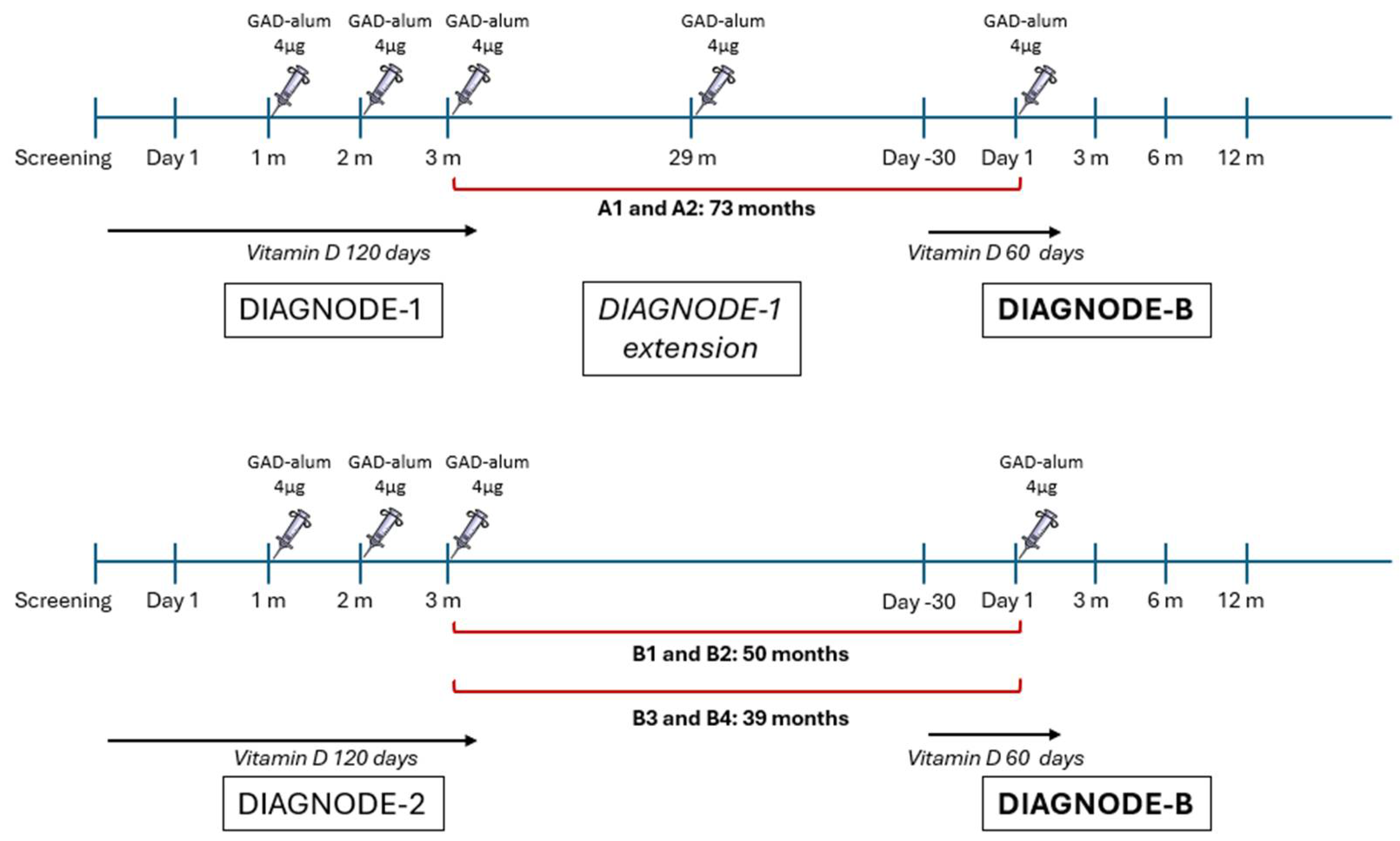
Figure 1. Overview of the GAD-alum treatment in DIAGNODE studies. Patients A1 and A2 (n = 2) received a course of three injections of GAD-alum (4 µg) into the lymph nodes (LN) one month apart in the DIAGNODE-1 trial. These patients received a further dose of GAD-alum (4 µg) after 29 months as an extension of DIAGNODE-1, and a fifth GAD-alum injection (4 µg) 44 months later during DIAGNODE-B. Patients who participated in the placebo-controlled DIAGNODE-2 (n = 4, B1–B4) trial received a course of three injections of GAD-alum (4 µg) into the lymph nodes (LN) one month apart. A fourth dose of GAD-alum (4 µg) was administered during the current trial, in the case of patients B1 and B2 after 50 months and patients B3 and B4 after 39 months. All patients were also taking vitamin D (Calciferol) from day 1, for 120 days, both in DIAGNODE-1 and DIAGNODE-2. A second course of vitamin D (60 days) was taken by the patients at the time of the four and fifth injections.
Ijms 26 00374 g001
Figure 2. C-peptide AUC. Individual, mean and median C-peptide AUC (area under the curve) after Mixed-Meal Tolerance Test (MMTT) [71] at day 1 (baseline) and at 6 and 12 months. C-peptide is expressed as nmol/L.
Ijms 26 00374 g002
Figure 3. Time in glycemic ranges. Mean percentage of time spent in hypoglycemic (<3.9 mmol/L), glycemic (3.9–10.0 mmol/L) and hyperglycemic (>10.0 mmol/L) range from baseline, 0 months (m), to 12 months (m) follow-up in patients (A1, A2, B1, B2, B3, B4) in the DIAGNODE-B trial.
Ijms 26 00374 g003
Figure 4. Individual longitudinal follow-up. Individual longitudinal values for: (a) C-peptide AUC (first row), (b) HbA1c (second row), (c) exogenous insulin dose (third row), (d) GADA (fourth row) and (e) GAD-stimulated proliferation of PBMCs (fifth row) from first GAD-alum treatment to 12 months follow-up in DIAGNODE-B for the patients from the DIAGNODE-1 cohort (patients: A1, A2) and the four patients from the DIAGNODE-2 cohort (patients: B1, B2, B3, B4). Black dots in individual C-peptide plots (first row) mark the instances of GAD-alum injections. The figure combines data from DIAGNODE-1, DIAGNODE-2 and DIAGNODE-B.
Ijms 26 00374 g004
Figure 5. Immunological responses upon in vitro peripheral mononuclear blood cell (PMBC) stimulation in patients (A1, A2, B1, B2, B3, B4) who received an additional injection of GAD-alum in the DIAGNODE-B trial. (a) GADA titers (U/mL) expressed as arbitrary units (AUs). Data from DIAGNODE-B. (b) PBMC proliferative responses to GAD65. Cells were cultured for 3 days with GAD65 (5 µg/mL) or medium; thereafter, cells were pulsed with [3H] thymidine and harvested. Proliferation is expressed as stimulation index (SI) and calculated from the mean of triplicates in the presence of stimulus divided by the mean of triplicates with medium alone. SI scale started from 1 after dividing by the unstimulated value index (SI unstimulated  =  1). (a) GADA and (b) proliferation fold change were calculated in relation to baseline (BL) values. Data from DIAGNODE-B. (c) Cytokine relative contribution (%). Interleukin (IL)-2, -5, -10, -13, -17, IFN-γ and TNF-α were detected by Luminex in PBMCs supernatants after 7-day culture in the presence of GAD65 (5 µg/mL) or medium alone. Secretion levels were calculated after subtraction of spontaneous secretion from each individual and expressed as pg/mL D-PBMC proliferative responses to GAD65. The results are based on samples collected before the original treatment (BL), before the first redosing 30 months after the initial treatment (30M, only A1 and A2), at start of DIAGNODE-B (0 m), before the GAD-alum redosing (1 m) and after 3, 6 and 12 months.
Ijms 26 00374 g005
Table 1. Demographics and baseline characteristics. Subject level demographic data and baseline characteristics both at the time of first treatment with GAD65 and at baseline for DIAGNODE-B.
StaticPrevious TrialDIAGNODE-B
SubjectGenderRelative with T1DHLAPrevious Trial (Number of Previous GAD-Alum Injections)Age (Year)BMI (kg/m2)Time Since Diagnosis (Days)Baseline C-Peptide (nmol/L)Baseline HbA1c (mmol/mol)Age (Year)BMI (kg/m2)Time Since Diagnosis (Months)Time Since the Last GAD-Alum
Injection (Months)
Baseline C-Peptide (nmol/L)Baseline HbA1c (mmol/mol)
A1MaleYesDR3-DQ2/DR4-DQ7DIAGNODE-1 (4)2123.43260.415662827.9376440.1370
A2MaleYesDR3-DQ2/DR10-DQ5.1DIAGNODE-1 (4)2319.05410.375492922.7076430.0250
B1FemaleNoDR3-DQ2/DR4-DQ8DIAGNODE-2 (3)1318.30760.639361823.6354490.0843
B2MaleNoDR3-DQ2/DR4-DQ8DIAGNODE-2 (3)1720.001450.755462222.0555480.0161
B3MaleYesDR3-DQ2/DR4-DQ8DIAGNODE-2 (3)1318.801440.540501723.6146390.0347
B4FemaleNoDR3-DQ2/DR4-DQ8DIAGNODE-2 (3)2326.60731.246452629.3444390.0647
Disclaimer/Publisher’s Note: The statements, opinions and data contained in all publications are solely those of the individual author(s) and contributor(s) and not of MDPI and/or the editor(s). MDPI and/or the editor(s) disclaim responsibility for any injury to people or property resulting from any ideas, methods, instructions or products referred to in the content.