Review

Exploring the Chemical Features and Biomedical Relevance of Cell-Penetrating Peptides

by
Liliana Marisol Moreno-Vargas
*,† and
Diego Prada-Gracia
*,†
Research Unit on Computational Biology and Drug Design, Children’s Hospital of Mexico Federico Gómez, Mexico City 06720, Mexico
*
Authors to whom correspondence should be addressed.
These authors contributed equally to this work.
Int. J. Mol. Sci. 2025, 26(1), 59; https://doi.org/10.3390/ijms26010059
Submission received: 23 October 2024 / Revised: 27 November 2024 / Accepted: 28 November 2024 / Published: 25 December 2024
(This article belongs to the Section Biochemistry)

Abstract

:
Cell-penetrating peptides (CPPs) are a diverse group of peptides, typically composed of 4 to 40 amino acids, known for their unique ability to transport a wide range of substances—such as small molecules, plasmid DNA, small interfering RNA, proteins, viruses, and nanoparticles—across cellular membranes while preserving the integrity of the cargo. CPPs exhibit passive and non-selective behavior, often requiring functionalization or chemical modification to enhance their specificity and efficacy. The precise mechanisms governing the cellular uptake of CPPs remain ambiguous; however, electrostatic interactions between positively charged amino acids and negatively charged glycosaminoglycans on the membrane, particularly heparan sulfate proteoglycans, are considered the initial crucial step for CPP uptake. Clinical trials have highlighted the potential of CPPs in diagnosing and treating various diseases, including cancer, central nervous system disorders, eye disorders, and diabetes. This review provides a comprehensive overview of CPP classifications, potential applications, transduction mechanisms, and the most relevant algorithms to improve the accuracy and reliability of predictions in CPP development.

1. Introduction

Cell-penetrating peptides (CPPs) are small peptides, either synthetic or natural, consisting of 4 to 40 amino acids. They possess a net positive charge at a physiological pH and, unlike most peptides, exhibit the remarkable ability to cross cell membranes while preserving the functional integrity of the cargo they transport [1]. Since the pioneering discovery of CPPs over thirty years ago [2,3], these peptides have found diverse applications. CPPs can serve as vectors for delivering various payloads, including small peptides, full-length proteins, nucleic acids (such as RNA and DNA), liposomes, nanoparticles, and viral particles, as well as radioisotopes and other fluorescent probes [4,5]. Importantly, CPPs are not limited to acting solely as carriers for functional peptides into the cell interior; they can also incorporate functional motifs themselves [6,7].
CPPs exhibit a diverse range of cationic, anionic, and neutral sequences, displaying varying degrees of hydrophobicity and polarity. These characteristics are influenced by the amino acid composition and three-dimensional (3D) structure of the CPPs. While CPPs, in general, lack sequence identity, certain groups of CPPs share high-sequence homology and common structural features. The specific modes and extents of cellular uptake are determined not only by the structural diversity of CPPs but also by the nature of the cargo they carry, whether it is covalently or non-covalently attached. This cargo attachment profoundly affects the transport of CPPs across the cell membrane.
The cell membrane serves as a barrier to the intracellular delivery of potential diagnostic or therapeutic cargoes. The cellular uptake of CPPs involves various pathways, including macropinocytosis, caveolae and/or lipid raft-mediated endocytosis, as well as those mediated by membrane proteins packaged in clathrin-coated vesicles [8,9,10]. Cells continuously internalize and recycle one to five times the equivalent of their cell surface area hourly [11,12]. This continuous internalization process likely facilitates the entry of peptides with a strong affinity for the membrane through at least some endocytic pathways. Alternatively, CPPs with specific physicochemical properties may be capable of directly translocating across the cell membrane, similar to small molecules. However, the cell membrane is not a homogeneous double layer; certain regions exhibit higher density, while others are more fluid, depending on their lipid composition and density [13,14]. In turn, the lipid composition, density, and dynamics vary depending on the cell type, specific membrane region, and various signaling pathways [15,16,17]. This heterogeneity gives rise to different levels and modes of CPP uptake [18].
The uptake mode of several cationic CPPs varies depending on the CPP concentration. When the concentration exceeds a certain limit, rapid cytosolic uptake occurs, indicating direct translocation. At lower concentrations, the main uptake mechanism is primarily endocytic. The concentration limit differs for each CPP but is typically in the micromolar (μM) range. Studies conducted on CHO-K1 cells have shown that Penetratin translocation only occurs below 2 μM [19], suggesting that the cell type and membrane composition influence the balance between different entry pathways. Many cationic CPPs interact electrostatically with glycosaminoglycans (GAGs) on the cell surface as the initial step for cellular entry [20]. This interaction induces the clustering of GAGs on the cell surface, activating intracellular signals that facilitate cell entry through various internalization pathways, including direct translocation and endocytosis. For example, the activation of acid sphingomyelinase, followed by a change in the lipid composition of the cell membrane, facilitates the direct translocation of cationic CPPs [21].
Similar to most peptides, many CPPs exhibit poor absorption, distribution, metabolism, and excretion (ADME) properties, including rapid renal clearance, short half-life, low permeability, and occasionally low solubility [22]. Enhancing the pharmacological properties of CPPs involves strategies to increase permeability, reduce proteolysis and renal clearance, and prolong their half-life. Proteolysis can be reduced by stabilizing the 3D structure with unnatural amino acids, while renal clearance can be addressed by lowering free peptide concentrations through depot formation or carrier protein association [23].
The development of peptide drugs presents challenges but also holds promising prospects for targeted drug delivery due to their high specificity, selectivity, small sizes, ease of modification, and high biocompatibility. Incorporating CPPs into drug design provides an opportunity to address biological targets that would otherwise be difficult to target using small molecules. Over a quarter of a century since their discovery, refining the design of CPPs involves improving loading strategies, enhancing our understanding of how these peptides traverse cell membranes, and gaining deeper insights into the mechanisms that govern specificity in recognizing their molecular targets. This review highlights some of the most notable examples of CPPs, demonstrating their significant potential as delivery vehicles for in vivo applications. Special emphasis is placed on evaluating their structural and physicochemical characteristics.

Overview of CPPs Classification

The initial and simplest classification of CPPs was based on their origin, categorizing CPPs into protein-derived and synthetic peptides. The sequences of the first CPPs were found in proteins from various origins and functions, and were accordingly named membrane translocation sequences (MTSs), protein transduction domains (PTDs), and Trojan peptides based on their respective roles. The identification and characterization of early protein-derived CPPs, such as the Tat peptide from the transactivator protein of HIV-1 transcription and the Penetratin from the homeotic transcription factor of Antennapedia, established the foundation for classifying peptides responsible for protein translocation.
The second classification of CPPs reflects advancements in peptide design, introducing chimeric peptides. Chimeric peptides are combinations of two or more different natural protein sequences or a combination of natural and/or synthetic peptide sequences. Various combinations of cationic, amphipathic, and hydrophobic sequences have led to the development of numerous delivery constructs. For instance, a potent secondary amphipathic peptide was designed for siRNA delivery into mammalian cells [24]. In another study, a series of steric blocking peptide nucleic acid oligonucleotides conjugated to an 18-mer PNA705 model oligonucleotide demonstrated enhanced cell-penetrating activity [25]. Additionally, an effective and versatile vehicle for the intracellular delivery of pDNA, SCO, and siRNA into various adherent and suspension cells was developed without cytotoxic side effects [26].
While these classifications provide insight into the relevance of cellular penetration, they do not correlate the properties of CPPs with their mechanisms of interaction with cell membranes or their modes of action. Therefore, it is essential to group CPPs based on their physicochemical features, as these features ultimately determine the effectiveness of their interactions and the potency of their effects.
To advance the understanding of CPPs, it is essential to classify them based on their physicochemical properties and origin, particularly in light of current developments in CPP design and application. This classification framework sheds light on the molecular mechanisms that govern CPP interactions with cellular membranes, including specific translocation pathways, binding affinities, and cellular uptake efficiencies, which are fundamental to optimizing their use as delivery vectors. CPPs are broadly categorized by their charge, hydrophobicity, and structural sequence motifs, characteristics that significantly impact their modes of cellular entry, endosomal escape, and cargo-release efficacy. Additionally, the origin of CPPs, whether derived from protein sequences, synthetic constructs, or biological sources such as viral proteins and animal toxins, further elucidates their functional adaptability and specificity for various therapeutic applications. Figure 1 provides a structured representation of CPP classifications, identifying key peptides within each category. This detailed categorization supports a more granular analysis of CPP properties, underscoring the diverse design strategies available to customize CPPs for targeted biomedical interventions, as will be elaborated in the following sections.

2. The CPPs Classification by Their Physicochemical Properties

CPPs can be classified into three categories based on their physicochemical properties, including charge, hydrophobicity, and the distribution of these descriptors along the peptide sequence. Although CPPs exhibit a wide range of sequences, three main classes can generally be identified: cationic, amphipathic, and hydrophobic. Data from publications and patents, excluding mutants of the same peptides, indicate that the majority of CPPs carry a net-positive charge. Amphipathic CPPs, which encompass both cationic and anionic peptides, constitute the largest class, while hydrophobic peptides represent only about 15% of the total [23,32]. This classification correlates with their interactions with cell membranes and other organelles [10]. Under physiological conditions, a significant percentage of peptides in all three CPP classes are positively charged.
Within the group of cationic CPPs (cCPPs), peptides are classified based on specific characteristics: they contain a continuous stretch of basic amino acids and lack the formation of an amphipathic helix in their 3D structure. Amphipathic CPPs (aCPPs) are chimeric peptides, with the most common structural motif being an amphipathic α -helix. Amphipathic α -helical CPPs possess a highly hydrophobic patch on one side, while the other side can be cationic, anionic, or polar. Hydrophobic CPPs (hCPPs) consist solely of non-polar residues or have very few charged amino acids, typically cationic, which are often derived from signal peptide sequences. In recent years, the discovery and characterization of novel natural peptide sequences with cell-penetrating activity have expanded the length limits and modified the physicochemical properties of constituent residues, enabling the design of neutral and even anionic CPPs.

2.1. Cationic CPPs (cCPPs)

Cationic cell-penetrating peptides (cCPPs) are a subclass of CPPs defined by a high density of basic amino acids, predominantly arginine (Arg) and lysine (Lys), which confer a net positive charge under physiological conditions. Arginine, due to its guanidinium side chain, exhibits unique hydrogen-bonding capabilities, forming stable bidentate interactions with anions such as phosphates and sulfates on the cell surface. These interactions enhance the cellular uptake efficiency by facilitating electrostatic binding to negatively charged glycosaminoglycans, including heparan sulfate. In contrast, lysine, with its primary amine side chain, shows lower affinity for these anions, leading to reduced internalization compared to arginine-rich sequences. For instance, polyarginines (e.g., R8–R10) demonstrate significantly higher translocation efficiency than polylysines of equivalent chain length, highlighting the superior electrostatic and hydrogen-bonding properties of the guanidinium group. Unlike amphipathic CPPs, cCPPs generally lack the formation of an amphipathic helix in their three-dimensional structure, typically adopting disordered or random coil conformations. This structural feature, combined with their cationic nature, enables cellular entry through mechanisms such as direct translocation and endocytosis. Prototypical examples of cCPPs include the HIV-1 Tat peptide (49–57) and polyarginines, which are widely used for cargo delivery in various biological contexts [33,34,35].

2.1.1. Tat

A cCPP contains a continuous stretch of basic amino acids, and its 3D structure does not form an amphipathic helix. The first discovered cCPP was derived from the HIV-1 protein Tat, a potent trans-activator essential for virus replication, which is considered a prototypic cCPP [36]. The HIV-1 Tat-protein-derived Tat peptide, denoted as Tat (residues 49 RKKRRQRRR 57, functional region III [3]), is fully polymorphic and unable to adopt any specific secondary structure, regardless of its environment. Tat remains disordered in the presence of phospholipids [37,38], exhibiting a random coil conformation in solution [39]. In aqueous solutions, polyarginine peptides typically form unstructured or polyproline type II helical conformations. This phenomenon is likely due to the strong repulsive interactions between the guanidinium groups of the arginine residues [40,41,42,43]. The abundant arginine residues in Tat, along with the guanidinium moieties in this peptide, facilitate strong binding to carboxylic, sulfate, and phosphate groups on the surface of cell membranes. These unique binding properties induce endocytosis and/or membrane transduction, enabling the transport of Tat and its cargo across the blood–brain barrier and cellular membranes [44]. Tat is known to aggregate at phospholipid membranes and occasionally fuse vesicles [45,46,47]. Previous studies have shown that the deletion of one arginine residue from either the amine terminus (Tat50–57) or the carboxyl terminus (Tat49–56) resulted in a significant (80%) loss in intracellular fluorescence compared to the parent sequence (Tat49–57) [48]. Tat and its derivatives have been evaluated as potential carriers of molecules for intracellular release in the treatment of various diseases [49,50,51,52,53,54], as well as possible bactericides against Gram-positive and Gram-negative bacteria. Additionally, there are studies reporting the antifungal activity of Tat [55,56,57]. Recently, significant progress has been made in utilizing Tat complexes, which have greatly contributed to the advancement of therapeutic approaches based on this CPP. Numerous Tat-conjugated drugs were under clinical development for applications as diverse as heart diseases (NCT00785954, phase II, completed 2011), intraocular inflammation (NCT02235272, phase III, completed 2016; NCT02508337, completed 2017, phase III), and acute inner ear and hearing loss (NCT02561091, completed 2017, phase III) [58,59,60] (see Table 1).

2.1.2. PTD4

Several modified PTD peptides exhibit significantly enhanced cell penetration capabilities compared to the original TAT PTD, offering potential for the development of novel therapeutic strategies [61]. One such modified peptide, known as PTD4 (a less basic Ala-scan analog of the TAT peptide, YARAAARQARA-NH2), has garnered significant attention due to its improved transduction efficiency. PTD4 contains fewer arginine residues than the original TAT(49–57) sequence, yet it demonstrates superior cell membrane translocation efficiency [62]. This enhanced efficiency is attributed to an optimized balance between positive charge and hydrophobicity, which facilitates more effective interactions with negatively charged cell membranes. Consequently, PTD4 promotes entry into the cell interior via mechanisms involving outer membrane receptors or transmembrane channels, facilitating efficient intracellular delivery of cargo [63].
Preclinical investigations have extensively explored the potential of PTD4 for drug delivery applications. Initial studies showed that PTD4 could efficiently transport therapeutic peptides and proteins across cellular membranes, enhancing their intracellular availability. In vitro experiments using cell lines demonstrated that PTD4-conjugated cargoes exhibited significantly higher uptake compared to unmodified TAT PTD [61].
Further in vitro studies using mouse B16-F1 and human A875 and SK-MEL-5 melanoma cells, along with in vivo studies in a B16-F1 melanoma mouse model, demonstrated that the PTD4-mediated delivery of chemotherapeutics like dacarbazine enhances tumor regression while reducing systemic toxicity. Combined PTD4–apoptin/dacarbazine treatment, with a 50% reduction in dacarbazine dosage, achieved similar antitumor efficacy without the hematologic side effects typically associated with full-dose dacarbazine. These findings underscore the potential of PTD4 to enhance chemotherapeutic efficacy, enabling dose reduction, minimizing adverse effects, and ultimately improving patient outcomes [64]. In another study, apoptin was fused with PTD4 to facilitate its delivery across cell membranes. The PTD4–apoptin complex induced apoptosis in cervical carcinoma cells by increasing active caspase-3 levels. Interestingly, apoptin-induced Mfn-2 accumulation was unaffected by Bcl-2, while Bcl-2 inhibited apoptin-mediated AKT activation. In vivo, cervical carcinoma xenografts treated with PTD4–apoptin for seven days exhibited significant tumor reduction compared to PTD4-GFP controls. TUNEL analysis confirmed apoptosis induction by PTD4–apoptin, highlighting its potential as an anticancer agent in cervical carcinoma [65].
On another front, Wang et al. designed anticancer agents targeting CyclinD1/CDK4 and CyclinD3/CDK4 complexes, which are key regulators of cell cycle progression. They synthesized chimeric peptides derived from key motifs in these complexes and conjugated them to PTD4. The resulting peptides—PTD4-D1, PTD4-D3, and PTD4-K4—exhibited significant antiproliferative effects by competing with CyclinD/CDK4 complexes, leading to G1/S phase arrest and apoptosis. Tumor challenge experiments further demonstrated potent antitumor activity with minimal side effects, positioning these PTD4-conjugated peptides as promising lead compounds for cancer therapy [66].
PTD4 has also been applied to inhibit the expression of connective tissue growth factor (CTGF), thereby reducing fibrosis and minimizing scar formation [67]. A notable example is the AZX100-PTD4 conjugate, which comprises a phosphorylated peptide analog of HSP20 (WLRRAS(phospho)APLPGLK) covalently conjugated to PTD4 [68]. Preclinical studies have shown that AZX100 effectively reduces stress fiber formation, induces morphological changes in human dermal keloid fibroblasts, and minimizes scar tissue formation in vivo. As a result of these promising outcomes, AZX100-PTD4 has advanced to clinical testing, specifically targeting keloid scar reduction. To date, it has successfully completed three Phase II clinical trials, underscoring the therapeutic potential of PTD4 in treating human diseases characterized by excessive fibrosis and scarring (NCT00811577; NCT00825916; NCT00892723) (see Table 1).
The continued evaluation of PTD4 in clinical settings remains crucial for fully understanding its therapeutic potential. Future studies should focus on optimizing dosing regimens, improving delivery specificity, and minimizing potential immunogenic responses. Moreover, exploring the use of PTD4 for delivering a broader range of therapeutic agents, including nucleic acids, proteins, and small molecules, will expand its clinical applicability. The efficacy of PTD4 in clinical applications, particularly for conditions requiring intracellular delivery of therapeutic agents, is supported by multiple studies [63,68,69,70,71,72,73].

2.1.3. Penetratin

Another widely studied cCPP is Penetratin (43 RQIKIWFQNRRMKWKK 58), which corresponds to the 16 residues of the third α -helix of the Antennapedia homeodomain of Drosophila. This region is responsible for the translocation of the entire homeodomain across cell membranes and is structurally conserved in a wide range of homeodomains, indicating high evolutionary conservation [74,75,76,77]. A minimal active translocation region has been identified through cell penetration assays using human cell cultures. These assays have revealed that the C-terminal segment 52 RRMKWKK 58 of the original sequence is necessary and sufficient for efficient membrane translocation, retaining 60% of the full-length Penetratin uptake efficiency [78]. Penetratin exhibits structural variations depending on its environment. Studies on its secondary structure when interacting with charged vesicles have demonstrated that it can adopt a helical conformation [79] or a dominant β -structure [80], depending on the specific conditions. Experiments using Penetratin variants in membrane-mimicking environments have indicated that the translocation process does not involve chiral recognition by a receptor, and induced helical secondary structure does not appear to be necessary [75,76]. The presence of positively charged basic amino acids contributes to its solubility in water, leading to a predominantly random coil conformation, although a significant contribution from β -structure is also observed [81]. While charged residues play a crucial role in the uptake of cationic CPPs, other residues are also important for membrane binding and translocation. The significance of aromatic residues in the cellular uptake of Penetratin has been demonstrated, with tryptophan 48 and 56, as well as phenylalanine 49, identified as crucial for efficient translocation. Substituting Trp48 with Phe has been shown to greatly impair cellular uptake. The authors suggest that the electrostatic interactions established by Penetratin on the cell surface stabilize it, and they propose that Trp48 and the positively charged amino acids promote the creation of inverted micelles, which capture the peptide and facilitate its transport to the cytoplasm [74,78,82,83]. These findings suggest a delicate balance between hydrophobic and positively charged residues that can impact the interaction with membranes and the translocation process [84]. The ability of Penetratin to translocate across cell membranes has established it as a widely utilized CPP for the development of drug delivery systems. It enables the direct delivery of diverse cargo types, including peptides, nanoparticles, proteins, and nucleic acids, into the cell [85,86,87,88,89,90]. The evidence supporting Penetratin’s cell-penetrating capability includes studies demonstrating its entry into various cell types such as murine fibrosarcoma cells (WC/1), chicken fibroblast cells (CEC-32), chicken monocytic cells (HD-11), human fibroblast cells (SV 80), and human monocytic cells (MonoMac-6) [91].

2.1.4. Polyarginines

Oligoarginines or polyarginines have been reported to penetrate various types of cells efficiently without causing significant cytotoxicity, under non-invasive conditions and at lower concentrations (in the range of tens of micromolar). They have been used to facilitate the uptake of a variety of small molecules and biomolecules [92,93,94,95,96,97,98]. It is known that the hepta-L-arginine (R7) sequence represents the lowest threshold for cellular uptake, and increasing the number of arginines is positively correlated with the uptake level. For delivery purposes, the optimal range is between R8 and R10 [33,48,95,99]. Several studies suggest that the R8 peptide possesses favorable characteristics in terms of achieving a balance between membrane uptake and cytosolic release [35,100,101,102]. It is important to emphasize that cellular uptake is not solely determined by charge, chirality, or backbone length. It has been determined that the guanidine headgroup of arginine serves as the critical structural component responsible for the observed biological activity. This importance was confirmed by the inability of citrulline heptamers to enter cells [33,48,100]. The guanidinium headgroup (arginine) is capable of hydrogen donation, while the oxygen of urea (citrulline) acts as a hydrogen bond acceptor. This interaction is particularly evident in the strong ability of guanidine to form a stable bidentate hydrogen bond with anions like phosphate or sulfate [35]. The exact mechanism of the internalization of polyarginine peptides is currently the subject of ongoing research. Numerous investigations have identified multiple uptake mechanisms that seem to be influenced by factors such as peptide concentration, the transported cargo, and the length of the peptide chain. Among these mechanisms are clathrin-mediated endocytosis (CME) [103,104] and macropinocytosis [94,105,106].

ATX-101

ATX-101 (MDRWLVKWKKKRKIRRRRRRRRRRR) is a cationic CPP composed of three parts: an AlkB homologue 2 proliferating cell nuclear antigen (PCNA)-interacting motif (APIM; RWLVK), an SV40 nuclear localization signal (KKKRK), and the CPP undeca-arginine (R11) [107]. Through the APIM motif, PCNA interacts with numerous cellular proteins that are crucial for cellular stress responses, DNA damage repair, intracellular signaling, apoptosis, metabolism, and antitumor immunity. Consequently, ATX-101 enhances the efficacy of various anticancer drugs by disrupting PCNA/APIM interactions. This includes DNA-damaging agents, microtubule-targeting drugs, molecular targeting agents (such as p38 MAPK and EGFR inhibitors), and γ -irradiation [108,109]. Müller et al. demonstrated that ATX-101 induces caspase-dependent apoptosis in multiple myeloma cell lines and primary cancer cells, independent of the cell cycle phase. Additionally, ATX-101 increased the sensitivity of multiple myeloma cells to melphalan, a DNA-damaging agent commonly used in the treatment of multiple myeloma [107]. Preclinical studies demonstrated that ATX-101 rapidly penetrated cells and targeted PCNA/APIM-containing protein complexes, which are crucial for cell survival and DNA repair. A completed Phase I clinical trial of ATX-101 (NCT01462786) revealed a favorable safety profile, with no significant adverse effects reported. ATX-101 is currently being evaluated in several clinical trials (see Table 1). For instance, in a Phase I/II study involving patients with platinum-sensitive fallopian tube and primary peritoneal cancer, ATX-101 was combined with platinum-based chemotherapy (NCT04814875). Additionally, a Phase II study (NCT05116683) has been initiated to assess the efficacy of ATX-101 as a monotherapy in sarcoma, with the aim of evaluating its antitumor activity and further characterizing its safety and pharmacokinetics.
cCPPs have garnered significant interest for their potential in the intracellular delivery of various biomolecules, including nucleic acids, proteins, and small drugs. However, the efficiency of cCPPs in facilitating the release of these biomolecules into the cytoplasm or nucleus is often hindered by endocytic sequestration. Once internalized, many cCPPs are trapped within endosomes and subsequently directed towards lysosomal degradation, which limits their functional delivery [110,111].

(R-X-R)4, X = 6-Aminohexanoic Acid

To overcome this challenge, innovative CPPs have been engineered, such as spaced oligoarginine conjugates linked with 6-aminohexanoic acid ((R-X-R)4, X = 6-aminohexanoic acid). These novel CPPs have shown the ability to internalize vectors via endocytic pathways that involve proteoglycans, which are essential components of the cell surface and play a crucial role in endocytosis [112]. Unlike traditional cCPPs, the spaced oligoarginine conjugates can escape endosomal entrapment and avoid lysosomal degradation, thereby facilitating the delivery of their cargo to the nuclear compartment. This capability is particularly advantageous for applications requiring direct nuclear delivery, such as gene editing and nucleic acid-based therapies [113,114,115]. In HeLa pLuc705 cells, conjugates of branched and linear (R-X-R) peptides—with X being a 6-aminohexanoic acid—are primarily internalized via an energy-dependent, clathrin-mediated pathway, with additional contributions from caveolae-mediated endocytosis. However, advanced cellular techniques are required to delineate the predominant endocytic mechanisms specific to these peptide–cargo conjugates [110,111].
A phase II clinical study registered under NCT00451256 aimed to evaluate whether immersing the excised saphenous vein in a novel antisense anti-c-myc solution (AVI-5126) would prevent graft failure after one year, compared to immersion in physiological saline (placebo) before grafting (see Table 1). The study was discontinued because the authors determined that the predefined efficacy outcomes described in the protocol would not be achieved. Consequently, the safety and efficacy evaluation of AVI-5126 for use in vein grafts prior to cardiac bypass graft (CABG) surgery was not completed. Further studies will be necessary to determine if this pharmaceutical formulation could have potential use in the future.
In cancer surgery, the complete resection of tumor lesions is crucial for optimal outcomes. However, distinguishing between tumor and normal tissues is challenging, often leading to residual tumor tissue and subsequent recurrence [116]. Intraoperative imaging with fluorescent molecular probes aids surgeons in visualizing tumor lesions and their boundaries, enhancing precision and reducing the likelihood of residual disease [117]. CPPs are crucial in imaging applications, especially when conjugated with agents like fluorophores and radioisotopes. These conjugates improve the imaging of cells and tissues, greatly enhancing diagnostic precision and advancing research capabilities. Specifically, CPP conjugates with fluorescent probes have been successfully designed to precisely delineate tumor boundaries, facilitating surgical removal.

2.1.5. AVB-620

AVB-620 is a protease-cleavable peptide conjugated with the fluorophores Cy5 and Cy7 at the cationic and anionic terminals, respectively. To improve water solubility, an α -aminoxyl- ω -methoxy PEG (mPEG, Mw ≈ 2000) moiety was conjugated. Its unique hairpin structure enables efficient fluorescence resonance energy transfer (FRET) between the fluorophores. The peptide linker is designed to be cleaved by MMP2 and MMP9, enzymes highly expressed in human breast cancer cells. MMPs hydrolyze AVB-620, causing it to be retained in the tissue and triggering a ratiometric fluorescence color change. This change can be visualized using camera systems that simultaneously capture fluorescence and white light images [117,118,119]. Consequently, surgeons can visualize and remove the tumor during surgery. In the phase I clinical trial (NCT02391194), AVB-620 was proven to be safe and effective for detecting tumor-positive tissue during surgery [120]. A phase II, single-arm, open-label study in women with primary breast cancer undergoing surgery evaluated how the timing of AVB-620 administration affects fluorescence and imaging accuracy, as well as its ability to differentiate between malignant and non-malignant tissues (NCT03113825). The intraoperative imaging of surgical specimens infused with AVB-620 enabled real-time tumor detection. The infusion of AVB-620 is safe and has the potential to enhance the intraoperative detection of malignant tissue during breast cancer surgeries.

2.1.6. Polylysines

It is known that other cationic poly amino acids, such as polylysines (PL), enhance the uptake of small molecules, peptides, proteins, or even larger particles, like viruses, into cells. PL peptides have been used for the delivery of methotrexate [121,122,123], 5-Fluorouracil [124], oligonucleotides [125,126,127], albumin [128] or horseradish peroxidase [128,129], and adenovirus [130,131,132,133]. Polylysine peptides have been demonstrated to bind to the membrane surface via electrostatic interactions, subsequently inserting into the membrane interface region, facilitating their translocation [134]. Contrarily, polyarginines exhibit higher cellular uptake efficiency compared to lysine polymers with similar chain lengths. The distinction in membrane translocation between polyarginine and polylysine was observed in early studies on CPPs, where investigations comparing oligoarginine and polylysine revealed the distinctive properties of the guanidinium group in arginine concerning proteoglycan binding, encompassing both the affinity and clustering of binding [33,134,135].

TransMTS®

TransMTS® proprietary peptides are simple, straight-chain peptides characterized by two distinct domains. The main core of the peptide consists of consecutive lysine residues, which carry a positive charge under physiological conditions. This positively charged core forms non-covalent (electrostatic) bonds with negatively charged macromolecules intended for transport. The second domain is a PTD, crucial for delivering the macromolecule to the target site. Each peptide is flanked by two identical PTDs at its termini, enhancing its ability to penetrate cell membranes and effectively transport the macromolecule into target cells. The positively charged lysine core not only facilitates binding to negatively charged macromolecules, such as nucleic acids or proteins, but also enhances the overall stability of the complex in biological environments. The electrostatic interactions ensure that the macromolecule is securely bound to the peptide, preventing premature release and degradation. The PTD domains, often derived from well-known transduction sequences like the TAT peptide from HIV-1, are responsible for the efficient cellular uptake of the peptide–macromolecule complex [136]. These domains exploit the cell’s natural endocytic pathways, allowing the complex to traverse cellular membranes and reach intracellular targets. By flanking the peptide with two PTD domains, the design maximizes transduction efficiency, offering a robust delivery mechanism [137].
Overall, the unique structure of TransMTS® peptides, with their bifunctional domains, represents a sophisticated approach to intracellular delivery. This design not only improves the targeting and uptake of therapeutic macromolecules but also enhances their therapeutic potential by ensuring precise delivery to specific intracellular locations (see Table 1). A TransMTS®–botulinum toxin A (the active ingredient in Botox) conjugate is currently in Phase III clinical trials for cervical dystonia (involuntary and painful neck contractions) and plantar fasciitis (heel pain) (NCT03608397) [138]. Botulinum toxin A conjugates have been developed for various therapeutic and cosmetic applications, including the treatment of moderate to severe lateral canthal lines (NCT02580370) and primary axillary hyperhidrosis (NCT02565732) [139,140,141].

2.1.7. Z12

Numerous studies have explored the utility of the Epstein–Barr virus transactivator protein ZEBRA as a vector for the delivery of diverse cargo proteins. Investigations focusing on truncated versions of ZEBRA have identified a minimal domain (MD), comprising amino acids 170-220, essential for its internalization capabilities [142]. Within this domain, a specific peptide segment termed Z12 has been demonstrated to efficiently transport reporter proteins across various normal and tumor cell lines. Notably, the internalized cargo proteins retained their functionality, and no cellular toxicity associated with MD was observed. The mechanism of MD translocation across the cell membrane involves binding to heparan sulfate proteoglycans (HSPGs) on the cell surface, occurring primarily through direct translocation across the lipid bilayer rather than via endocytosis [143].
The peptide Z12 (KRYKNRVASRKCRAKFKQLLQHYREVAAAKSSENDRLRLLLK) has been employed in the development of cellular-based vaccines by conjugating its sequence with multi-epitopic antigens. Treatments with Z12-formulated vaccines have extended survival rates in three tumor models, with the most significant increase observed in an aggressive brain cancer model. Analysis of tumor sites showed favorable immune modulation, indicating that these vaccines not only promote an integrated immune response but enhance the persistence and homing of CD8+ effector T cells to the tumor site. Furthermore, the presence of Th1 polarized CD4+ T cells was noted, contributing to potent antitumor immunity in various models, including gliomas [144]. These findings underscore the versatility and effectiveness of Z12 as a carrier for multi-epitope antigens, showcasing its potential in advancing cancer immunotherapies. The ability of Z12 to promote persistent and targeted immune responses highlights its value as a tool for developing innovative therapeutic strategies against cancer [145,146]. Furthermore, this promising potential has led to the initiation of a Phase I clinical trial (NCT04046445) to evaluate the safety, tolerability, and antitumor activity of ATP128, VSV-GP128, and BI 754091 in patients with stage IV colorectal cancer. This trial represents a critical step in translating the preclinical successes of Z12 into clinical applications in humans and animals [147].
Several studies have investigated CPPs derived from the ZEBRA protein, beyond the Z12 fragment, in the development of therapeutic cancer vaccines. These investigations have evaluated various truncated forms of ZEBRA for their structural properties, in vitro transduction efficiency, and their ability to induce CD4 and CD8 T cell responses in vivo. The findings underscore the critical role of selecting appropriate CPP sequences and adjuvants to optimize antitumor immunity. Optimal CPP-adjuvant combinations significantly enhance immune responses and control tumor growth in aggressive tumor models, demonstrating the promising potential of ZEBRA-derived CPP-based vaccines in cancer therapy [145,148,149].

2.2. Amphipathic CPPs (aCPPs)

Multiple investigations have indicated that among the identified CPPs to date, amphipathic peptides are the most abundant, accounting for approximately 40% of them. Amphiphilic CPPs (aCPPs) consist of both polar and non-polar amino acid regions, with the non-polar regions being rich in hydrophobic amino acids like Ala, Val, Leu, and Ile. The amphipathic α -helix is the most common structural motif of many amphipathic CPPs. Amphipathic α -helical CPPs exhibit a highly hydrophobic region on one side, while the other side can be cationic, anionic, or polar. Previous studies have demonstrated that variations in conditions can lead to different secondary structures in the same sequence, thereby altering its capacity to interact with the hydrophobic/hydrophilic interface [39]. For example, the α -helical MAP (model amphipathic peptide) spontaneously inserts into the lipid monolayer, exhibiting strong interactions with negatively charged phospholipids. Conversely, the structural analysis of peptide/lipid interactions of MPG (N-methylpurine DNA Glycosylase) revealed that its β -sheet structures are more responsive to external forces compared to its α -helical structures [150].
Based on their sequence, length, and lipid association, amphipathic CPPs can be classified into primary amphipathic, secondary amphipathic, β -sheet amphipathic, and proline-rich categories [151,152].

2.2.1. Primary Amphipathic CPPs (paCPPs)

Primary amphipathic CPPs (paCPPs) can be defined as the sequential arrangement of a hydrophobic residue domain with a hydrophilic residue domain. Numerous paCPPs are chimeric peptides, with several being derived from partially covalently linking a hydrophobic domain to a nuclear localization sequence (NLS). paCPPs generally exhibit a longer length compared to cationic CPPs, typically consisting of 20 or more amino acids, which allows them to traverse the hydrophobic core of membranes. Many paCPPs demonstrate a strong affinity for both neutral and anionic membranes, potentially leading to lipid reorganization and disruptions in membrane structure. While the majority of paCPPs are chimeric or synthetic in nature, certain sequences are derived from proteins. Examples include the VE-cadherin-derived CPP, pVEC [153], the N-terminal sequence (1–30) of the bovine prion protein, BPrPp(1–30), the N-terminal sequence (1–28) of the mouse prion protein, MPrPp(1–28) [154,155], and the N-terminal sequence (1–22) of the tumor suppressor p14ARF, ARF(1–22) [7].

pVEC

The uptake of pVEC (LLIILRRRIRKQAHAHSK) occurs through a non-endocytic translocation mechanism without causing changes in plasma membrane permeability or cell morphology. Once internalized, it predominantly localizes to nuclear structures, making it a potent carrier for peptide nucleic acids (PNAs) and proteins [153]. Studies investigating the pVEC sequence to determine the significance of each residue and the impact of single substitutions on translocation ability have demonstrated the crucial role of the N-terminal hydrophobic region of pVEC in efficient cellular translocation [156]. pVEC has also been found to enhance the translocation of homing peptides that selectively target molecular markers on tumor cells. One such homing peptide is the cyclic peptide PEGA (CPGPEGAGC), which has previously shown accumulation in breast tumor tissue in mice. In vitro studies have shown that PEGA peptide conjugated with the CPP pVEC can be taken up by various breast cancer cells. Additionally, the conjugation of the anticancer drug chlorambucil to pVEC-PEGA has been demonstrated to significantly increase drug efficacy by more than fourfold, leading to a reduction in the clonogenic survival of MCF-7 cells, thus highlighting its potential as a carrier for anticancer drugs [157,158,159].

bPrPp(1–30)

A notable paCPP is the bPrPp(1–30) peptide (MVKSKIGSWILVLFVAMWSDVGLCKKRPKP). Its structure and membrane interaction have been examined using spectroscopy techniques. Circular dichroism (CD) spectroscopy demonstrated that the peptide adopts predominantly an α -helical conformation in zwitterionic bicelles and DHPC micelles, while exhibiting a lesser degree of α -helical structure in partially charged bicelles. The structure of bPrPp(1–30) was determined in DHPC micelles, revealing an α -helix spanning residues Ser8 to Ile21. The peptide induced some degree of ordering within the bilayer, consistent with positive hydrophobic mismatch, and its stable helical conformation allowed insertion at a transmembrane position within the bilayer [160]. The internalization of bPrPp predominantly occurs through macropinocytosis, a fluid-phase endocytic process initiated by binding to cell-surface proteoglycans [155].

MPrPp(1–28)

Another N-terminal sequence, MPrPp(1–28) (MANLGYWLLALFVTMWTDVGLCKKRPKP), exhibits a strong propensity for aggregation and β -structure formation, particularly in its interaction with negatively charged phospholipid membranes. It has been observed that the conformational characteristics adopted by the MPrPp(1–28) peptide vary significantly depending on the environment. In the presence of neutral POPC or negatively charged POPG vesicles, the CD spectra of PrP(1–28) indicate a predominant α -helical or β -structure, respectively [154]. The PrPp(1–28) segment may play a crucial role in the cellular transport of prion proteins and their infectivity. This is attributed to the potent β -structure induction within the (1–28) region facilitated by the interaction with a negatively charged membrane surface. Notably, MPrPp(1–28) is capable of transporting large hydrophilic cargoes across the cell membrane, likely through an α -to- β transition catalyzed by a negatively charged lipid surface.

ARF(1–22)

Johansson et al. conducted a study to assess the impact of the ARF(1–22) peptide (MVRRFLVTLRIRRACGPPRVRV) on cell proliferation, apoptosis induction, stability, and cellular uptake mechanisms. ARF (1–22) demonstrated a dose-dependent decrease in cell proliferation and induced apoptosis. The evaluation of peptide stability revealed that ARF (1–22) remained stable within cells for a minimum of three hours, with consistent concentrations of both intact and degraded peptide over time. This suggests that uptake and degradation occur at similar rates during the initial hours. The primary uptake mechanism for ARF (1–22) was identified as vesicular uptake, indicating endocytosis as the main pathway. This uptake mechanism enables the peptide to transport bioactive charges to the nucleoli of cells. For instance, when ARF (1–22) was conjugated with splice-correcting PNA, correct splicing was restored [7].

MPG and Pep-1

Two other relevant paCPPs are N-methylpurine DNA Glycosylase (MPG, GALFLGFLGAAGSTMGAWSQPKKKRKV) and Pep-1 (KETWWETWWTEWSQPKKKRKV). Both peptides consist of three domains: firstly, a variable N-terminal hydrophobic motif; secondly, a hydrophilic lysine-rich domain derived from the NLS (nuclear localization sequence) of SV40 (simian virus 40) large T-antigen (KKKRKV); and thirdly, a linker domain (WSQP) containing a proline residue. This linker domain improves the flexibility and integrity of both the hydrophobic and hydrophilic domains [161,162,163,164,165]. However, the peptides mainly differ in their hydrophobic domain. The MPG hydrophobic domain (GALFLGFLGAAGSTMGA) is derived from the fusion sequence of the HIV glycoprotein 41 and is necessary for efficient targeting to the cell membrane and cellular uptake. On the other hand, the Pep-1 hydrophobic motif (KETWWETWWTEW) originates from a tryptophan-rich cluster, which is also crucial for efficient targeting to the cell membrane and for forming hydrophobic interactions with proteins [165]. Additionally, a cysteamine group is present at the C-terminal, and an acetyl group caps the N-terminus. Under oxidizing conditions, dimers may form due to the disulfide linkage of cysteamine groups [166].
MPG was originally designed for the rapid and efficient delivery of plasmid DNA into the nucleus [161]. Variations and optimizations were subsequently made for siRNA delivery [167,168,169,170]. Subsequently, through a single mutation substituting the second Lys residue in NLS with Ser (changing KKKRKV to KSKRKV, denoted as Δ NLS), the nuclear translocation function was eliminated, allowing for the swift release of siRNA in the cytoplasm. This modification enabled the effective down-regulation of target mRNA [163,171]. Research findings have demonstrated that MPG Δ NLS exhibits a robust interaction with the phospholipid cell membrane by means of its hydrophobic gp41 sequence. This sequence has the ability to transiently adopt a β -sheet structure and integrate into the plasma membrane. Consequently, the insertion of the β -sheet brings about a temporary alteration in the organization of the cell membrane, generating a transient channel. This channel facilitates the entry of the siRNA/MPG Δ NLS complex into the cytoplasm [172]. The cationic end plays a major role in siRNA condensation, while the hydrophobic region stabilizes the CPP/siRNA complex via intermolecular hydrophobic interactions. MPG has been demonstrated to interact with the extracellular matrix via negatively charged HSPGs. The initiation of cellular uptake is a highly dynamic mechanism in which the binding of MPG to the extracellular matrix rapidly triggers a remodeling of the actin network, involving the activation of the GTPase Rac1 [173]. In another study, MPG has been utilized to enhance antigen cross-presentation and elicit an antitumor immune response. When MPG binds to antigens encapsulated in nanovaccines, it influences the spatial and temporal intracellular localization of antigens, promoting antigen cross-presentation and stimulating antigen-specific immune responses, particularly cytotoxic T lymphocyte (CTL) responses. This innovative approach has demonstrated promising results in enhancing the efficacy of nanovaccines for cancer immunotherapy [174]. Experimental evidence suggests that most CPPs are taken up via endocytosis, although MPG and Pep-1 uptake are known to be energy independent [162,163].
Pep-1 undergoes a conformational rearrangement upon interaction with lipids, and its insertion into the membrane is accompanied by segregation of lipids, membrane disorganization, and transient pore formation, allowing for transient ionic currents [175,176]. Evidence indicates that the highly charged hydrophilic domain is responsible for the first contact with the membrane due to electrostatic interactions between the polar head group of phospholipids and the positive charges of Pep-1, and that the dehydration and insertion of the hydrophobic domains promote membrane destabilization [166,177,178,179]. It has been observed that vesicles mimicking normal and cancer cell membranes exhibit differential responses to the CPP Pep-1. Pep-1 exhibits selectivity towards model membranes based on their composition, binding sites, and peptide concentration. The interaction of Pep-1 with cancer cells is relatively stronger compared to normal cells in terms of the C=O group, whereas the interaction of Pep-1 with normal cells is relatively stronger compared to cancer cells in terms of the phosphate group [180]. Hydrophobic interactions have been shown to be the primary driving force behind peptide interactions with normal cell membranes, whereas electrostatic interactions play a dominant role in peptide interactions with cancer cell membranes [181]. Cell selectivity is primarily determined by an elevation in the concentration of acidic components on the cancer cell wall rather than a slight increase in the level of phosphatidylserine (PS) on the outer surface of the cancer cell membranes [182]. Numerous studies have shown that the presence of the membrane induces various secondary structures in Pep-1. However, it remains unclear which structure is crucial for transmembrane administration [183,184]. Pep-1 has the capability to transport a wide range of peptides and proteins into various cell lines, regardless of their nature and size, while preserving their biological activity. These cell lines include neuronal cells [185], pancreatic cells [186,187], neural retinal cells [188], macrophages [189], and hepatocytes [190]. The mechanism by which Pep-1 delivers active macromolecules has been determined to be independent of the endosomal pathway. Furthermore, the dissociation of the Pep-1 particle–macromolecule complex occurs immediately upon crossing the cell membrane [191]. Pep-1 offers several advantages, including high nanomolar affinity for most proteins and peptides, stability in physiological buffers, and the absence of toxicity. These attributes make it a promising candidate for the development of therapeutic applications utilizing covalent protein transduction domains.

PEP-010

PEP-010 is a CPP specifically designed to disrupt key intracellular interactions involved in apoptosis, particularly those between caspase-9 and protein phosphatase 2A (PP2A). While the exact sequence of PEP-010 is proprietary and not disclosed in clinical study summaries for intellectual property and confidentiality reasons, it is known to be a bifunctional peptide comprising 30 amino acids. It includes the DPT sequence, which facilitates cell penetration, and the Pep-1 sequence, which actively interferes with PP2A [192]. Upon administration, the cell-penetrating segment of PEP-010 facilitates its entry into the cytosol, where the interfering segment disrupts the interaction between caspase-9 and PP2A. This disruption leads to the release and activation of caspase-9, thereby restoring apoptosis in tumor cells [193,194]. Activated caspase-9 induces caspase-dependent apoptosis. PP2A, a serine/threonine phosphatase, plays a crucial role in regulating cell growth and DNA damage repair.
As a bifunctional peptide, PEP-010 can penetrate cells and interfere with essential protein interactions, thereby promoting apoptosis in cancer cells. This mechanism of action has been validated through both in vitro and in vivo studies, showing efficacy in patient-derived xenograft (PDX) models of triple-negative breast cancer (TNBC) and hormone receptor-positive, HER2-negative breast adenocarcinoma [195]. NCT04733027 is a clinical trial designed to test PEP-010 in humans for the first time. This Phase I trial aims to determine the safety, tolerability, and optimal dosage of PEP-010 when administered alone and in combination with other chemotherapy agents, specifically, paclitaxel and gemcitabine. The study seeks to collect preliminary data on the pharmacodynamics, pharmacokinetics, and potential therapeutic benefits of PEP-010 in treating cancer.

2.2.2. Secondary Amphipathic CPPs (saCPPs)

The α -helical secondary structure of secondary amphipathic CPPs (saCPPs) is crucial for their interaction with biological membranes. However, their internalization efficiency depends on various properties, including charge, guanidinium content, and primarily amphipathicity, which interact with one another. saCPPs are generally shorter than paCPPs and form stable associations exclusively with negatively charged membranes. Although saCPPs can adopt a helical amphipathic structure, they do not deeply insert into the membrane, resulting in fewer changes to biological membranes compared to paCPPs. Typically, secondary amphipathic saCPPs do not possess a well-defined structure when in solution. They acquire their amphipathic properties through a change in secondary structure, which occurs when they interact with negatively charged polyanions, membranes, or glycosaminoglycans.

MAP

Numerous studies have demonstrated that amphiphilic saCPPs, like the model amphipathic peptide (MAP) (KLALKLALKALKAALKLA), an artificial CPP incorporating hydrophobic and hydrophilic residues on opposite sides of its helical structure, exhibit robust interactions with negatively charged phospholipids and readily insert into the lipid monolayer, thanks to its well-defined amphipathic α -helix. Studies on the cellular uptake kinetics of various CPPs have revealed that MAP exhibits faster uptake and has the ability to induce membrane leakage by altering membrane integrity, even at concentrations as low as 1 μ M. Additionally, MAP has been found to exert a strong toxic effect on various cell lines [196,197]. For example, a recent study investigated whether coupling the drug tacrine with the MAP peptide could enhance the antiproliferative properties of the drug. Both MAP and its tacrine conjugate were found to exhibit high toxicity against two cancer cell lines, namely, breast (MCF-7) and neuroblastoma (SH-SY5Y) cells. Since the unconjugated MAP peptide exhibited the same cytotoxic effect, it can be inferred that the peptide functions more as a cell-killing agent rather than a CPP. This highlights the potential utility of MAP as an antimicrobial peptide (AMP) capable of rapidly destroying cell membranes, even at low concentrations (1 μ M) [198]. Regarding MAP analogs with inverse charge, like MAP17 (QLALQLALQALQAALQLA), the evidence suggests that their membrane translocation ability stems from their amphiphilic nature [199].

M918

The saCPP class encompasses other peptides, such as M918 (MVTVLFRRLRIRRACGPPRVRV), a highly hydrophobic and positively charged peptide derived from the tumor suppressor protein p14ARF. This peptide, comprising amino acids 1–22 with positions 3–8 inverted, demonstrates efficient translocation into various cells without toxicity. M918 can be utilized either as a covalent conjugate or in a non-covalent complex with the cargo. Its internalization is independent of GAGs on the cell surface and primarily relies on macropinocytosis for cellular uptake. Consequently, M918 is an effective carrier for large cargoes, such as proteins and peptide nucleic acids (PNAs) [200,201].

GALA and KALA

The saCPP class includes anionic peptides, one of which is GALA (WEAALAEALAEALAEHLAEALAEALEALAA). GALA is a synthetic peptide composed of 30 amino acids, featuring a repetitive sequence of glutamic acid–alanine–leucine–alanine (EALA). The glutamic acids (E) create a pH-dependent negatively charged side chain. Additionally, GALA contains histidine and tryptophan residues that act as spectroscopic probes. The EALA motif allows the peptide to have a hydrophobic surface, enabling interaction with the bilayer when it adopts an α -helical conformation. GALA is soluble in water at neutral pH, but at an acidic pH, it undergoes a conformational change from a random coil structure to an amphipathic α -helix. When GALA binds to bilayer membranes, it assembles into a transmembrane peptide pore composed of approximately 10 helical monomers aligned perpendicular to the membrane plane [202]. Studies suggest that GALA incorporates into the vesicular bilayer and aggregates to form a transbilayer pore consisting of around 10 peptides (±2). It has been observed that the lipid composition of the bilayer model influences the peptide’s orientation and insertion mechanism [203]. Due to its net negative charges, GALA is commonly used in combination with other DNA or oligodeoxynucleotides (ODN) condensation reagents to create particles for effective gene transfection. By partially replacing glutamic acid with lysine in the GALA sequence, the KALA peptide is derived.
KALA (WEAKLAKALAKALAKHLAKALAKALKACEA) is a cationic, endosomolytic, and fusogenic peptide capable of binding to DNA, destabilizing membranes, and mediating DNA transfection. At physiological pH (7.4), KALA adopts an α -helical conformation and exhibits a pH-dependent ability to form pores, distinct from those formed by GALA. As a cationic peptide, KALA disrupts membranes, condenses DNA, and can deliver ODN and plasmid DNA into the cell nucleus without requiring additional condensation reagents. A conformational change from an α -helix to a random coil occurs as the pH shifts from neutral to acidic [202]. Studies on gene expression have shown that KALA significantly enhances gene expression in substrate-mediated transfections [204,205]. The optimization of this peptide has led to the development of an amphiphilic cationic system that serves as an efficient non-viral gene delivery vector, exhibiting improved DNA condensation ability and reduced cytotoxicity [206].

p28

p28 is a naturally occurring bacterial peptide that has recently attracted considerable attention as both an efficient CPP and a promising anticancer agent. p28 (Leu50-Asp77) is an amphipathic, α -helical peptide composed of 28 amino acids, derived from azurin—a 128-amino acid (14 kDa) copper-binding member of the cupredoxin family of redox proteins, secreted as a periplasmic protein by Pseudomonas aeruginosa [207]. As a post-translational, multi-target anticancer agent, p28 demonstrates the ability to penetrate a wide range of solid tumor cells [208]. Mechanistically, p28 exerts its effects through two primary pathways. After cellular internalization, p28 binds to both wild-type and mutant p53 proteins, inhibiting their ubiquitination and subsequent proteasomal degradation mediated by the constitutive photomorphogenesis 1 (Cop1) protein. This inhibition results in elevated levels of p53, inducing G2/M phase cell-cycle arrest and apoptosis, thereby contributing to tumor cell reduction and death [209]. Preclinical studies and early-phase clinical trials have demonstrated the safety and potential efficacy of p28 in treating various cancers (see Table 1), including solid tumors (NCT00914914), glioblastoma and central nervous system tumors (NCT06102525; NCT01975116), and hepatocellular carcinoma (NCT05359861). The dual role of p28 as a CPP and anticancer agent positions it as a unique and promising candidate in therapeutic development [210,211,212].

2.2.3. Amphipathic β -Sheet Peptides

An amphipathic β -sheet peptide is characterized by the presence of both hydrophobic and hydrophilic regions, which are exposed to the solvent. When folded, this peptide exhibits amphipathicity. The folding process is influenced by various weak non-covalent interactions, including electrostatic interactions, hydrogen bonding, the hydrophobic effect, and van der Waals forces. It is their ability to form β -sheets that enhances their efficiency in penetrating cells [213,214]. Amphipathic peptides have a strong tendency to spontaneously form one-dimensional fibril structures that resemble amyloids, which subsequently self-assemble into higher-order fibrils. Despite the absence of sequence homology among constituent peptides, amyloid assemblies derived from different peptides exhibit many shared structural characteristics. Amyloid fibrils adopt a quaternary structure known as the “cross- β ” structure. Each individual peptide assumes an extended β -strand conformation, with the amino acid side chains oriented perpendicular to the amide backbone. The β -strands self-associate to form β -sheets, which are stabilized by hydrogen bonding between neighboring amide backbone groups, as well as hydrophobic, aromatic, and Coulombic interactions between adjacent side chain groups. Cross- β architectures emerge when multiple β -sheets laminate together to form fibrils [213,214]. These self-assembled peptide biomaterials form a unique scaffold in the shape of a fibrillar network. This scaffold has diverse applications, including regenerative medicine and tissue engineering, particularly through the use of hydrogels made from various β -sheet peptides [215,216].

EAK16 and RAD16

Significant progress in the field of amphipathic self-assembling peptides has unveiled a wide range of sequences, one of which is EAK16 (AEAEAKAKAEAEAKAK). This specific sequence demonstrates ionic self-complementarity and consists of an equal number of cationic Lys (K) and anionic Glu (E) residues, alternating with hydrophobic Ala (A) residues. Through the optimization of the EAK16 peptide, several other peptides with immense potential as vaccine adjuvants or hydrogels for cell culture support have been developed. By substituting Lys with Arg (R) and Glu with Asp (D) in EAK16, a distinct self-assembling peptide named RAD16 or RADA16 is formed [217,218]. Similar to EAK16, RAD16 exhibits the ability to spontaneously generate stable hydrogels in physiological buffers. These remarkable properties, including favorable cell attachment, high stability, and biocompatibility, have led to the successful commercialization of RAD16 as PuraMatrixTM peptide hydrogels (AcN-(RADA)4-CONH2) [219].

MAX1, MAX8 and Q11

In the field of biomedical applications, significant progress has been made by replicating the molecular design features of EAK16 to create self-assembling β -sheet peptides. Two notable examples of these peptides are MAX1 and Q11, which possess the remarkable ability to spontaneously form β -sheet structures. This unique property makes them highly appealing for various biomedical applications, including drug delivery and tissue engineering. MAX1 is composed of alternating hydrophobic Val residues and hydrophilic, cationic Lys residues within the β -strands. These β -strands are interconnected by the turn sequence VdPPT, where V represents Val, DP represents D-proline, P represents proline, and T represents threonine [220]. MAX1 exhibits the remarkable ability to form viscoelastic hydrogels, which vary in stiffness based on temperature and salt concentration [221]. MAX8, one of its derivatives, displays rapid gelation kinetics, enabling the homogeneous encapsulation of mesenchymal stem cells and hepatocytes. Additionally, MAX8 ensures high cell survival rates after immobilization in the gel matrix [222]. In contrast, Q11 (QQKFQFQFEQQ) assembles into a highly intricate gel-like network of β -sheet peptides, serving as an optimal modular biomaterial platform. Studies have revealed that incorporating cell-adhesive ligands through the co-assembly of Q11 and N-terminally modified Q11 with RGD or IKVAV significantly enhances cell attachment, proliferation, and growth [215]. On the other hand, Q11 acts as an adjuvant when fused to a peptide antigen. By attaching epitopes to the N-terminus of Q11, the epitopes can be displayed on the surface of the assembled nanofibers without affecting the peptide assembly. The unmodified Q11 peptide does not generate an immune response. However, when Q11 is conjugated with different epitopes, it induces detectable immune responses that can persist for weeks. These Q11 conjugated fibers elicit strong antibody responses, highlighting the potential of Q11-based vaccines for providing long-term protection [223,224,225].

E1Y9

E1Y9 peptide (EYEYKYEYKY) is an emerging biomaterial with great potential for cell culture applications. It has demonstrated the ability to form hydrogels that promote cell growth and proliferation. Numerous studies have showcased the biocompatibility of E1Y9 hydrogels with various cell types, including 3T3-L1 cells and PC12 cells [216,226]. Furthermore, these hydrogels have exhibited low toxicity levels comparable to commercially available microtiter plate surfaces. These findings position E1Y9 hydrogels as a compelling and versatile candidate for a wide range of cell culture applications [227].
Amphipathic β -sheet peptides offer a promising alternative to α -helical amphipathic peptides as vectors for delivering drugs, proteins, and oligonucleotides into mammalian cells. Further investigations are currently being conducted to better understand the underlying mechanism and explore the potential of utilizing β -sheet peptides as transport vectors for biomolecules.

2.2.4. Proline-Rich Amphipathic Peptides

Proline-rich peptides are a chemically and structurally diverse family of CPPs characterized by the presence of pyrrolidine rings from proline residues. These peptides tend to assume helical conformations responsible for their internalization and enter cells via caveolae- or lipid raft-mediated endocytosis. The cellular uptake of proline-rich peptides occurs through their binding to anionic phosphate groups followed by interaction with the lipophilic region of the cell membrane. Proline-rich peptides are particularly effective, demonstrating efficient cellular uptake and low cytotoxicity [228]. Examples of these peptides include SAP (sweet arrow peptide) and its derivatives [229,230,231], bactenecin-7 (Bac-7) and its derivatives [232], and polyprolines [233].

SAP

SAP is an intracellular delivery peptide ((VRLPPP)3) derived from the natural sequence of the N-terminal domain of γ -zein VHL(PPP)8 [234]. SAP adopts a polyproline II helical structure (PPII) in an aqueous buffer, is highly soluble in aqueous media, and exhibits no cytotoxicity at very high concentrations (1000 μ M) [229]. Substitution of the arginine residue for a glutamate residue results in the negative version of SAP, SAP(E) ((VELPPP)3), the first anionic CPP, which retains its propensity to adopt a PII helical secondary structure and is not cytotoxic at high concentrations (1000 μ M). Since SAP(E) is negatively charged at physiological pH, the initial electrostatic interaction with the cell membrane is not necessary for its internalization. It is hypothesized that the mechanism of entry involves an initial aggregation on the cell surface, leading to an internalization mechanism that is independent of clathrin and probably caveolin mediated [231,235]. The chemical modification of SAP(E), by adding the Ac-CGGW sequence to the N-terminus of the primary structure, allows attachment to biologically active substances. For example, this linker contains an activated thiol and aminooxy functionality capable of generating a stable oxime bond, required for drug modification at the terminal cysteine, with the C-13 carbonyl group of doxorubicin. Cellular uptake and cytotoxicity studies in MCF-7 and HT-29 cancer cells demonstrated that this CPP–drug conjugate can efficiently transport doxorubicin through the cell membrane. The conjugate can be efficiently cleaved by glutathione within a short period, delivering the toxic cargo into the nucleus [236]. This delivery system solves the serious problem of passage through membranes, as doxorubicin’s anticancer activity targets DNA intercalation and topoisomerase inhibition [237].

Bac-7

Bac-7 (RRI RPRPPRLPRPRPRP LPFPRPGPRPIPRP LPFPRPGPRPIPRP LPFPRPGPRPIPRP), an antimicrobial proline-rich peptide, with four 14-residue repeats from the bactenecin family, is divided into five distinct regions: a charged cap (RRI), a degenerated repeat (RPRPPRLPRPRPRP), and three copies of a 14-residue repeat ((LPFPRPGPRPIPRP)3) [238]. Shortened Bac-7 fragments, regardless of their charge or hydrophobic content, penetrate cells and lack membranolytic activity. The cell-permeant antimicrobial activity of Bac-7 is localized in the N-terminal residues 1-24 (Bac1-24). This sequence is amphipathic and displays segregated charge and hydrophobic areas in three distinctive regions [232]. This proline-rich peptide was able to deliver a non-covalently bound protein to cells, indicating that Bac-7 and its derivatives are highly efficient CPPs for the intracellular delivery of proteins or peptides. For example, in a study to develop a thermally responsive polypeptide inhibitor of c-Myc, the cellular uptake, intracellular distribution, and potency of the Pen, Tat, and Bac1-24 cell-penetrating peptides fused to ELP (Elastin-Like Polypeptide)-H1 (a peptide which blocks c-Myc/Max dimerization) were evaluated. The conjugate Bac1-24-ELP-H1 localized to the nucleus of a subset of the cells and was the most potent MCF-7 cell proliferation inhibitor, compared to the CPPs Penetratin and Tat, resulting in a more potent c-Myc inhibitory polypeptide [239]. Massodi et al. developed a polypeptide carrier for a cell cycle inhibitor peptide, using a similar strategy. The coding sequence of ELP was modified by adding the CPP Bac-71-24 at the N-terminus and a 23-amino acid peptide derived from p21 at the C-terminus (Bac1-24-ELP1-p21). Bac1-24-ELP1-p21 displayed both cytoplasmic and nuclear distribution in SKOV-3 cells, inducing caspase activation, PARP cleavage, and cell cycle arrest in the S-phase and G2/M-phase [240]. These studies suggest that such macromolecular biopolymers with antiproliferative activity have great potential in cancer therapy for the targeted treatment of solid tumors.

Poly-L-Proline Type II Helix (PPII) Based

A specific class of CPPs is based on polyproline secondary structures, the poly-L-proline type II helix (PPII). PPII has a left-handed helical structure with distinct trans isomers of peptide bonds with dihedral angles of [−75°, +150°]. The rise per residue of the PPII helix is 3.1 Å with three residues per turn, resulting in a helical structure rising 9.3 Å per turn compared to the 6.0 Å pitch of a 310 helix. The primary reason for such an open and elongated geometry of PPII is the absence of H-donor atoms due to the cyclic side chain of proline residues. The PPII conformation is highly acceptable of H-donor atoms from its environment or third-party moieties, enhancing its solvation energy [241,242,243]. This architecture allows for the precise orientation of hydrophilic and hydrophobic moieties along different faces of the helix, making the structure soluble in aqueous media. To ensure that the designed CPPs adopt a PPII helical structure, the key is to maintain a proline content of at least 50%. These structural characteristics have been used in the design of CPPs with a rigid secondary structure, such as proline-based dendrimers [233], proline-derived γ -peptides like cis- γ -amino-L-proline [244,245], and cationic amphiphilic polyproline helices (CAPHs) [246,247,248,249]. Proline-rich peptides are highly water soluble, an invaluable property for life science applications.

2.3. Hydrophobic CPPs

The design of a new class of peptides, the hydrophobic ones, aims not only to mimic but also to improve the translocation properties of known peptides. To achieve this goal, the design considers various parameters such as electrostatics, secondary structures, and hydrophobic properties. Hydrophobic peptides are characterized by having only non-polar residues, a low net charge (less than 20% of the sequence), and a hydrophobic motif or chemical group that is critical for uptake independent of the rest of the sequence. These peptides are subdivided into two types: linear hydrophobic peptides based on natural amino acids and chemically modified peptides, including stapled peptides, prenylated peptides, and pepducins.

2.3.1. Linear Hydrophobic Peptides Based on Natural Amino Acids

Pentapeptides (CPP5)

Linear hydrophobic peptides are formed by combining sequences of up to 70% to 90% aliphatic residues with aromatic, anionic, or cationic residues (10% to 30%). These peptides typically consist of pentapeptides (CPP5) and serve as the minimal cell-penetrating sequence within longer CPPs. An example of such CPP5s is derived from the Bax-binding domain of Ku70, known as Bax-inhibiting peptides (BIPs). These antiapoptotic peptides were designed based on the Bax-binding domain of Ku70 found in various species, including humans, rats, and mice. The CPP5s derived from this domain include VPMLK (V5 antiapoptotic pentapeptide), PMLKE, VPTLK, VPALR, and VPALK. Among these, VPTLK and KLPVM demonstrated protein-transduction activity [250]. Evaluation of the protein transduction activity of these two pentapeptides in cell culture revealed that, when VPTLK and KLPVM were added to the N-terminus of the Cre protein, the complexes were able to activate the expression of the Cre-inducible GFP gene, suggesting that the Cre complexes were able to reach the chromosomal DNA in the nucleus [251].
The cytoprotective activities of various other BIPs have been previously reported in both cell culture and animal models. For instance, the V5 antiapoptotic pentapeptide (VPMLK) was shown to prolong the survival of mice with acute liver failure when they were transplanted with monkey hepatocytes previously cultured with V5. V5 aids in increasing the survival of isolated hepatocytes and protects the cells by enhancing the expression of CAS. Thus, the action of V5 on monkey hepatocytes can be considered to be a stimulation of the kinase cascade and its complex, resulting in the stabilization of hepatocytes against stress signals [252]. Hepatocyte transplantation (HTX) is a potential treatment for liver failure and inborn errors of liver metabolism. The results of recent studies suggest that V5 could be used to administer other molecules that facilitate cell growth and proliferation or decrease the cellular immune response in target cells in other diseases.
For example, pancreatic islet transplantation holds great promise as a treatment for type 1 diabetes but often requires transplantation of islets from two to four donors. Following transplantation, islets undergo apoptosis and necrosis from transient hypoxia, lack of nutrient support, and hyperglycemia-induced toxicity. V5 inhibits a wide range of caspases, improving pancreatic islet recovery. This molecule binds Bax and prevents mitochondrial cytochrome c translocation, resulting in the global inhibition of caspases through the activation of NF- κ B–dependent and BH1–4 genes [253]. The use of V5 in treating isolated islets increases their viability, improves their function, and prevents apoptosis. When islets are transplanted together with fibroblast growth factor-2 (FGF-2) and V5, it becomes possible to use a smaller mass of islets from a single donor pancreas for transplantation [254]. The approach described above effectively restores normal blood sugar levels and preserves insulin content and islet function after transplantation. Based on their findings, the authors suggest that exploring a timed release of V5, possibly achieved through gelatinization, could be a viable strategy to enhance long-term prevention of apoptosis and further improve outcomes in human islet transplantation.

Pep-7, SG3 and FGF

Several studies using phage and plasmid display techniques have identified a significant number of unusual, non-amphipathic, and minimally charged peptides that could potentially be classified as hydrophobic CPPs. Some examples include Pep-7 (SDLWEMMMVSLACQY) [255], SG3 (RLSGMNEVLSFRWL) [256], and FGF (PIEVCMYREP) [257]. These peptides have approximately 60%, 57%, and 60% apolar residues, respectively, and exhibit a net charge ranging from +2 (Pep-7) to +1 (SG3). Further investigations are necessary to elucidate the functions of these peptides and determine whether their hydrophobicity or other factors influence their absorption properties.

2.3.2. Stapled Peptides

Hydrocarbon stapling, pioneered by Schafmeister et al. in 2000, is a chemical strategy developed to create a novel class of hydrophobic CPPs, known as stapled peptides. Initially, this method used α , α -disubstituted non-natural amino acids with olefin-bearing tethers to generate an all-hydrocarbon “staple” through ruthenium-catalyzed ring-closing metathesis (RCM). This hydrocarbon stapling approach stabilizes the α -helical structure of peptides, enhancing their membrane permeability and proteolytic stability, thereby improving their potential as therapeutic agents [258].
In all cases, the peptide stapling technique involves cross-linking the side chain of an amino acid residue to either the peptide terminus or another side chain within the native peptide. This ensures that the selected or substituted amino acids are positioned on the same helical face, allowing covalent cross-linking [259,260,261,262]. In α -helices, residues align along the same helical face every 3.6 residues. Specific positions for stapling include i/i + 3, i/i + 4 (one-turn staple), i/i + 7 (two-turn staple), and i/i + 11 (three-turn staple). Covalent connections at these positions stabilize the helical conformation, enhancing structural integrity and proteolytic stability. Various chemical reactions have been employed to achieve these cross-links, broadening the applicability of peptide stapling in drug development [259,263,264,265]. Peptide stapling produces peptides with superior binding affinity, selectivity, and enhanced metabolic stability compared to their unmodified counterparts. This technique not only increases the bioavailability of peptide drugs but also extends their half-life, providing a robust solution for accelerating the development and clinical application of peptide-based therapeutics [266,267,268,269].
Stapled peptides offer increased proteolytic stability and enhanced cell permeability, addressing key limitations of conventional peptides and making them highly effective as therapeutic agents. By maintaining a stable α -helical structure in biological environments, they are particularly useful for modulating intracellular pathways. These advantages have demonstrated significant potential in targeting protein–protein interactions, which are often challenging for traditional small molecule drugs. Stapling increases peptide helicity by rigidifying the peptide structure and fortifying the natural α -helical structure that would otherwise unfold outside the context of the host protein. This is important because the α -helix, a major structural motif of proteins, often mediates intracellular protein–protein interactions that govern many biological pathways and enhances the cell penetration capacity of various CPPs.

SAHBs

This chemical strategy has been used to generate BH3 peptides with improved pharmacological properties. The stapled peptides, called stabilized α -helix of BCL-2 domains (SAHBs), proved to be helical, protease-resistant, and cell-permeable molecules that bind with increased affinity to multidomain BCL-2 member pockets. A SAHB of the BH3 domain from the BID protein, SAHBA (EDIIRNIARHLA(S5)VGD(S5)NLDRSIW), specifically activated the apoptotic pathway to kill leukemia cells. SAHBA effectively inhibited the growth of human leukemia xenografts in vivo. Histological examination of SAHBA-treated mice showed no toxicity of the compound to normal tissue [270]. The hydrocarbon stapling technique applied to BH3 death domain peptides has been instrumental in addressing the shortcomings of CPPs, such as the loss of their bioactive structure in solution, rapid proteolytic degradation in vivo, and limited cell permeability [271].
Researchers have utilized the natural activity of the Bcl-2-interacting mediator of cell death (BIM), which interacts with BCL-2 and possesses one of the most potent BH3 death domains in the BCL-2 protein family, to attempt to restore BH3-dependent cell death in resistant hematological cancers. A notable example of this approach comes from the work of LaBelle and colleagues. Their studies demonstrate that BIM-SAHBA, a stapled BIM BH3 helix that contains an i, i + 4 all-hydrocarbon cross-link spanning positions 154 and 158 (designated “A”), effectively reactivates cell death in vitro and in vivo in a BH3 sequence-dependent manner. Furthermore, BIM-SAHBA blocks the antiapoptotic sequestration of BAX/BAK BH3 helices, leading to the release of mitochondrial cytochrome c in a BAX/BAK-dependent manner. The treatment with BIM-SAHBA also activates caspase-3/7 and induces cell death in resistant hematologic cancer cells [272]. An interesting example involves the complex of MCL-1 and the peptide SAHBD (EDIIRNIAR(R5)LAQVGD(S8)NLDRSIW). This complex effectively targets native MCL-1, disrupting its capacity to suppress the death pathway through protein interactions. Consequently, it sensitizes caspase-dependent cancer cell apoptosis when death receptor stimulation occurs. The evidence obtained has enabled the authors to develop a model for the creation of novel therapies aimed at reactivating apoptosis in diseases driven by pathological cell survival mediated by MCL-1 and chemoresistance [273].

NYAD-1

Another case of interest is the modified peptide NYAD-1, derived from a 12-mer α -helical peptide (CAI). In cell culture, the original peptide (CAI) failed to inhibit HIV-1 due to its inability to penetrate cells. However, NYAD-1, with enhanced α -helicity through molecular stapling chemical modification, is capable of cell penetration without the need for a carrier protein. It disrupts the formation of both immature- and mature-like HIV-1 particles and effectively inhibits HIV-1 infection in cell cultures. NYAD-1 demonstrates high-affinity binding to the C-terminal domain of the capsid and holds potential as a new class of drugs for AIDS treatment [274].

ALRN-6924

ALRN-6924 (Sulanemadlin: AcLTF(R8)EYWAQL(S5)AAAAA(dA)-NH2, with a staple between R8 and S5) is a stapled peptide designed to mimic the N-terminal domain of the p53 tumor suppressor protein. This peptide exhibits high-affinity binding to MDM2 and MDMX (MDM4), the natural inhibitors of p53, thus activating p53 signaling in cells with a wild-type TP53 genotype (TP53-WT). Ingelshed et al. have demonstrated that MDM2/MDMX inhibition by sulanemadlin reduces cell growth in a p53-dependent manner. They also showed that p53 activation by sulanemadlin increases the expression of immunogenicity markers, enhances lymphocyte infiltration when combined with anti-PD-1 immunotherapy, and synergistically improves overall survival [275]. Through iterative structure–activity relationship (SAR) optimization, ALRN-6924 has been engineered to possess favorable characteristics in terms of cell permeability, solubility, pharmacokinetics, and safety. The intracellular proteolysis of ALRN-6924 results in a long-acting metabolite that maintains a strong binding affinity to MDM2 and MDMX, with slow dissociation kinetics. At high doses (10 mg/Kg), ALRN-6924 exhibits mechanism-based anticancer activity in TP53-WT tumor models. Conversely, at lower doses, it transiently arrests the cell cycle in healthy tissues, thereby providing chemoprotection without affecting TP53-mutant cancer cells [276]. ALRN-6924 activates the transcription factor p53, resulting in a unique pharmacodynamic response. Saleh, M. N., et al., have shown that after ALRN-6924 administration, serum levels of MIC-1, a protein regulated by p53, increased rapidly and remained elevated for more than 48 h at the RP2D, indicating sustained p53 activation. This finding suggests that effective therapies can achieve prolonged effects despite the drug’s plasma half-life of 5.4 h [277]. Recent reports indicate that ALRN-6924 enhances the antitumor efficacy of chemotherapy in TP53 wild-type hormone receptor-positive breast cancer models. Pairawan et al. demonstrated that ALRN-6924 is active in WT-TP53 cancer cell lines but not in mutant TP53. In ER+ breast cancer cell lines, it showed synergistic effects in vitro and improved in vivo antitumor activity with paclitaxel and eribulin. Apoptotic assays revealed a significantly higher in vivo apoptotic rate with the combination of ALRN-6924 and paclitaxel compared to either agent alone, warranting further evaluation in hormone receptor-positive breast cancer patients [278]. In preclinical studies, ALRN-6924 significantly improved survival outcomes in an animal model of human acute myeloid leukemia (AML). Mice transplanted with human leukemia cells exhibited a threefold increase in median survival, from 50 days to approximately 150 days, highlighting the therapeutic potential of ALRN-6924 (NCT02909972) [279]. These and other findings have led to the conduct of multiple clinical trials to investigate the potential of ALRN-6924, both as a monotherapy and in combination, for the treatment of various diseases (NCT02264613, NCT04022876, NCT03654716, NCT05622058, NCT02264613) (see Table 1).
Some of these CPPs have been shown to enter cells via an endosomal mechanism, rather than a membrane rupture mechanism [280]. Studies on the application of molecular stapling to multiple cell targets have revealed several key findings. First, the stapled peptides are localized in the mitochondria and multivesicular bodies of intact cells. Second, the stapled peptides exhibit reduced binding affinity and poor cell permeability, which may lead to a loss of activity [281]. Molecular stapling, in some cases, has been found to increase cytotoxicity. Several studies indicate that certain constrained peptides can induce cell lysis by rupturing the cell membrane, which is undesirable and likely leads to non-specific toxicity [282,283]. The molecular stapling technique has been applied in various contexts. For a more comprehensive review, it is advisable to consult reports related to the generation of stapled peptides for high-affinity protein binding [284], intracellular targeting [270,273], binding site discovery [285,286,287], potential therapeutic applications in cancer [288,289,290], diabetes [291], vaccine development [292], and viral infection [293]. In the context of the COVID-19 pandemic, some research groups have explored the possibility that stapled peptides can inhibit the binding of SARS-CoV-2 to ACE2 receptors [294,295]. However, one of these peptides was reported to have no antiviral activity against SARS-CoV-2. Additionally, further studies are needed to improve the ability of stapled peptides to block viral entry [296,297].
In summary, hydrocarbon stapling is a potent technique in peptide drug development, providing a method to improve the pharmacokinetic properties and therapeutic potential of peptides. The ongoing evolution and optimization of this technology promise novel treatments for a broad spectrum of diseases.

2.3.3. Prenylated Peptides

The prenylation of peptides involves adding a lipid chain, consisting of three isoprene units (farnesyl) or four isoprene units (geranylgeranyl), to a free thiol group found in specific Cys residues near the C-terminus of a protein. This addition of either a farnesyl (C15) or geranylgeranyl (C20) isoprenoid moiety has been shown to confer inherent cell-penetrating capacity to peptides. These peptides can efficiently cross the cell membrane through an ATP-independent, non-endocytic pathway, leading to their accumulation in the cytosol [298,299]. Prenylated peptides have potential applications in cell-penetrating therapies, especially when the cargo needs to be readily released in the cytosol. It is worth noting that the specific sequence of the peptide does not significantly affect uptake, as long as the geranylgeranyl group is present [300,301]. Various studies have demonstrated that prenylated peptides are internalized via a non-endocytic pathway, irrespective of their sequence. Additionally, both the length and identity of the sequence can influence peptide uptake, with even short prenylated sequences containing just two amino acids exhibiting significant cell-penetrating properties [299].
Natural prenylated indoles have been proposed as potential anticancer agents. The design of a novel pentapeptide sequence, including a prenyl residue in one of the tryptophan residues (N-tert-prenylated tryptophan), was based on the Substance P Antagonist G (Arg–d-Trp–NMePhe–d-Trp–Leu–Met-NH2), a known anticancer agent for small cell lung cancer (SCLC). The resulting prenylated peptide showed favorable cytotoxicity against H69 and DMS79 SCLC cell lines when compared with the unmodified pentapeptide or the original SPG sequence. In vivo studies demonstrated that the pentapeptide exhibited antitumor activity at relatively low doses (1.5 mg/kg) against the growth of the DMS79 xenograft and remained stable in plasma for at least 3 h [27].

2.3.4. Pepducins

Pepducins are cell-penetrating, membrane-tethered lipopeptides designed to target the intracellular region of various transmembrane proteins, such as GPCRs and MMPs. Depending on the peptide sequence, they can act as agonists, antagonists, or modulators of the protein’s activity. Pepducins consist of a lipid moiety, such as myristate, palmitate, or lithocholic acid, which enables them to easily penetrate cell membranes. This lipid is attached to a peptide corresponding to an amino acid segment from one of the cytoplasmic loops (intracellular i1–i3 domains) or the C-terminal tail (intracellular i4 domain) of the target GPCR. In the presence of their cognate receptor, pepducins activate receptor G-protein signaling and exhibit high selectivity for the receptor type. Pepducins are commonly modified at the N-terminus, and both the lipid moiety and amino acid sequence can be optimized to achieve the desired pharmacological and pharmaceutical properties. While pepducins are believed to function as allosteric modulators of GPCR signaling by binding to the intracellular surface of target receptors, the precise pharmacological basis of their activity remains unknown [28,302,303,304].
In the orthosteric activation mechanism of a GPCR, an agonist binds to the orthosteric binding pocket located within the receptor’s transmembrane bundle near the extracellular surface. This binding triggers a conformational change in the receptor, leading to the externalization of part of TM6 and rotation of TM7. As a result, an ionic lock between TM3 and TM6 is broken. The E(D)RY sequence in helix 3 facilitates proton uptake, stabilizing the active receptor conformation. In this state, the receptor’s intracellular portion exposes a predominantly hydrophobic cavity, allowing the C-terminal helix of the G- α protein to engage and exchange GDP for GTP [305,306,307]. Allosteric inhibition, which involves the modulation of signaling through a site other than the orthosteric ligand binding site, offers new drug-targeting opportunities. Pepducins can access the intracellular face of GPCRs chemically and physically, enabling the identification of both orthosteric and allosteric modulators for these significant drug discovery targets [308].
Due to the above, cell-penetrating lipidated peptides represent a class of compounds that can be used as novel tools to probe complex mechanisms of diverse GPCR activity [309] and other membrane proteins, including muscarinic acetylcholine receptor, chemokine receptors (CXCR1, CXCR2, and CXCR4) [310,311], protease-activated receptors (PAR1, PAR2, and PAR4) [312], the melanocortin-4 receptor, the Smoothened receptor, formyl peptide receptor-2 (FPR2), the relaxin receptor (LGR7), sphingosine 1-phosphate receptor-3 (S1P3), G-proteins (G α q/11/o/13), and the GPIIb integrin. Pepducins are being investigated as a potential treatment strategy for various human diseases, including cardiovascular diseases [302,313,314,315,316,317], cancer [312,318,319,320,321], inflammation, sepsis [322,323,324], asthma [304], and bone marrow transplant [325].

P1pal-7

P1pal-7, also known as PZ-128, is a 7-mer palmitoylated pepducin (palmitate-KKSRALF, PAR1 pepducin) that functions as a reversible inhibitor of the protease-activated receptor 1 (PAR1) on platelets and other vascular cells. This inhibition is achieved by targeting the intracellular surface of the receptor, thereby preventing signal transduction associated with platelet activation and other cellular responses. Structurally, PZ-128 mimics the predicted off-state conformation of the juxtamembrane region of the third intracellular loop of PAR1, effectively blocking its activation. It targets the cytoplasmic surface of PAR1 and interrupts signaling to internally located G proteins [302,303,323,326]. The efficacy of PZ-128 as an antiplatelet agent has been demonstrated in vivo, where it significantly reduces ischemic events by inhibiting platelet aggregation and thrombosis [317]. In clinical studies, PZ-128 has progressed successfully through phase 2 trials in cardiac patients, demonstrating safety and efficacy in reducing myonecrosis and arterial thrombosis (NCT01806077, NCT02561000) (see Table 1) [327]. The advancement of PZ-128 underscores its potential as a therapeutic agent in cardiovascular diseases, providing a novel approach to antiplatelet therapy by specifically targeting the intracellular mechanisms of PAR1. Beyond cardiovascular applications, PZ-128 shows promise in cancer and systemic inflammation. In cancer studies, nude mice were inoculated in their mammary fat pads with the invasive breast cancer cell line MCF7-PAR1/N55 and treated with either a vehicle or the PAR1 pepducin, P1pal-7. By the end of the treatment period, P1pal-7 significantly inhibited the growth of MCF7-PAR1/N55 xenografts by 62% (p < 0.01). Tumors excised from the mammary pads revealed extensive replacement of normal mammary tissue upon hematoxylin and eosin staining. Moreover, P1pal-7 treatment resulted in a significant reduction in blood vessel density at the center of the tumors by 75% (p < 0.002) [312].
Pepducins represent a promising class of cell-penetrating peptides with significant therapeutic potential. Their unique mechanism of action, targeting the intracellular surface of GPCRs, offers a novel approach to inhibiting receptor-mediated signaling. Extensive preclinical testing has shown that pepducins possess favorable pharmacodynamic and pharmacokinetic properties, along with suitable biodistribution and low-toxicity profiles. Moreover, their simplicity in synthesis and purification in large quantities makes them viable candidates for drug development [328]. These findings underscore the potential of pepducins as versatile therapeutic agents in the treatment of various diseases, including cancer, cardiovascular conditions, and systemic inflammation [329,330,331].

2.4. Cyclic CPPs (cyCPPs)

Cyclic cell-penetrating peptides (cyCPPs) have garnered substantial interest within the scientific community for their exceptional ability to traverse cellular membranes, representing a pivotal advancement in the field of biomedical research [332,333,334,335]. The synthesis of cCPPs leverages advanced peptide chemistry strategies in conjunction with solid-phase synthesis techniques. This process systematically forms peptide bonds between amino acids, progressively lengthening the peptide chain. The defining feature of cCPPs is their distinctive cyclic structure, which is achieved by the strategic insertion of a linker or bridge between the N- and C-termini, resulting in a closed-loop formation [336,337]. The establishment of a covalent bond between the peptide’s amino and carboxyl ends enhances its stability, provides significant resistance to enzymatic degradation, and markedly improves its cellular uptake capabilities [338,339]. To further enhance these properties, an array of chemical modifications are employed. Methods including cyclization through disulfide bonds or other cross-linking approaches are utilized to improve the stability and effectiveness of cCPPs [340]. Specifically, peptides can be made cyclic by creating disulfide or lactam bridges or through the modification and cyclization of the peptide main chain for increased performance [341]. Recent advancements have led to the development of methodologies that facilitate the rapid construction of extensive cyclic peptide libraries [342,343,344,345], broadening the spectrum of potential inhibitors for targets that were previously considered challenging to address with drug-based interventions [343].
Numerous studies have demonstrated that several cyclic CPPs directly interact with phospholipids on the plasma membrane, facilitating their cellular entry through endocytosis while exhibiting minimal cytotoxic effects [332]. Endosomal escape studies suggest an alternative cargo release mechanism, where CPPs directly bind to the luminal side of the endosomal membrane, inducing curvature and forming CPP-enriched small vesicles. This process involves significant membrane distortion, marked by sharp negative Gaussian curvatures, representing the highest energy transition state. CPPs selectively bind to budding necks, reducing the energy barrier and accelerating budding. Endosomal acidification enhances this process by increasing the affinity of arginine-rich CPPs for the membrane. Upon detachment from the endosomal membrane, small vesicles likely become less stable due to the dissipation of pH gradients across their membrane, resulting in luminal content release into the cytosol [332]. pH gradients across artificial membranes can persist for minutes to hours, indicating efficient proton movement across lipid bilayers [346]. The late endosome membrane, rich in negatively charged lipids like bis(monooleoylglycero) phosphate (BMP) and phosphatidylinositol (PI), further facilitates cationic CPP binding, promoting vesicle budding and concentrating CPPs within budding vesicles.
Compared to linear CPPs, cyCPPs exhibit notable advantages, including enhanced structural stability, superior cellular penetration capabilities, and reduced susceptibility to intracellular proteolysis [332,336,347]. These attributes position cyCPPs as promising candidates for the efficient delivery of drugs, therapeutic molecules, therapeutic approaches known as Peptide Receptor Radionuclide Therapy (PRRT), and imaging probes across a myriad of biomedical applications. Firstly, cyCPPs excel in drug delivery, efficiently ferrying therapeutic agents ranging from small-molecule drugs to peptides and nucleic acids such as siRNA, DNA, and RNA. This capability is invaluable for addressing diseases requiring precise intracellular targeting [348,349,350,351,352,353,354,355,356,357]. Moreover, cCPPs play a pivotal role in imaging applications, where they can be conjugated with imaging agents like fluorophores and radioisotopes. This conjugation enhances cellular and tissue imaging, significantly advancing diagnostic capabilities and research endeavors [118,358,359,360,361,362]. Additionally, in gene therapy, cyCPPs are instrumental in facilitating the delivery of gene-editing tools and therapeutic genes, enabling precise manipulation of cellular functions for therapeutic purposes [363]. Lastly, cyCPPs serve as indispensable research tools in both cell biology and molecular biology, facilitating the study of intricate cellular processes and protein functions, driving advancements in scientific understanding and therapeutic innovation [364,365].
In drug design, cyCPPs have emerged as potent inhibitors of protein–protein interactions (PPIs). These interactions, characterized by intricate interfaces spanning substantial surface areas—typically between 1000 and 5000 Å2—pose a challenge to conventional small molecule approaches. The distinctive attributes of cyCPPs, including their resistance to chemical or enzymatic degradation and their heightened receptor selectivity [338], render them particularly promising. The utilization of these innovative CPPs has significantly broadened the spectrum of potential inhibitors for targets previously deemed “undruggable”. In this context, cyclic peptides are gaining traction as a promising foundation for drug development, offering novel avenues to enhance drug delivery and efficacy. These peptides, along with cyclic depsipeptides and bicyclic peptides found in nature, exhibit a wide array of chemical structures. Known for their diverse biological effects—ranging from fighting cancer, bacteria, viruses, and fungi to reducing inflammation and clotting—cyclic peptides and their derivatives underscore their importance in the search for new pharmacological agents, driving ongoing research into their synthesis, characterization, and clinical applications [366].

2.4.1. BT1718

BT1718 exemplifies a Bicycle Toxin Conjugate (BTC), a novel class of therapeutics that leverages bicyclic peptide technology for targeted drug delivery. This conjugate comprises a bicyclic peptide that binds with high affinity and selectivity to the hemopexin domain of membrane type 1 matrix metalloproteinase (MT1-MMP) [29]. MT1-MMP is critically involved in cell invasion and metastasis and is often overexpressed in solid tumors associated with poor patient prognosis [367,368,369,370]. The peptide component of BT1718 is connected via a molecular spacer and a cleavable disulfide linker to DM1 (N2′-deacetyl-N2′-[3-mercapto-1-oxopropyl]-maytansine), a potent cytotoxic tubulin inhibitor.
BT1718 targets cells expressing MT1-MMP, exploiting its overexpression on tumor cells to deliver the cytotoxic payload specifically to the tumor site. The conjugate’s design ensures that once the linker is cleaved in the tumor microenvironment, the active DM1 is released, binding to microtubules and inhibiting tumor cell division, leading to cell death and tumor size reduction. Unlike traditional MMP inhibitors, BT1718 does not inhibit MT1-MMP activity but uses it as a delivery mechanism for its cytotoxic cargo [29].
BT1718 has demonstrated promising efficacy against treatment-resistant cancer samples. Additionally, it has shown reduced toxicity compared to other potent cancer treatments, highlighting its potential as a safer and more effective therapeutic option for aggressive cancers. By specifically targeting MT1-MMP, BT1718 effectively induces tumor cell death and inhibits tumor growth while minimizing toxicity. The promising preclinical results and favorable safety profile of BT1718 position it as a highly innovative and effective therapeutic candidate for treating aggressive and treatment-resistant cancers (NCT03486730) (see Table 1).

2.4.2. 177Lu-DOTA0-Tyr3-Octreotate

177Lu-DOTA0-Tyr3-octreotate is a radioconjugate comprising the somatostatin analog Tyr3-octreotate (TATE) conjugated to the chelating agent DOTA and radiolabeled with the β -emitting isotope lutetium-177 (177Lu). This radioconjugate possesses both imaging and antineoplastic capabilities, demonstrating high-affinity binding to type 2 somatostatin receptors (SSTR2s) expressed on neuroendocrine tumor (NET) cells. Upon receptor binding and subsequent internalization, it delivers a cytotoxic dose of β radiation directly to SSTR2-positive cells, thereby selectively targeting and eradicating these tumor cells. 177Lu-DOTA0-Tyr3-octreotate represents a significant advancement in PRRT. It demonstrates superior tumor uptake and extended residence times while maintaining reduced whole-body retention, thereby mitigating the risk of bone marrow toxicity and preserving renal function post-PRRT [371,372]. Furthermore, the use of renal protective agents significantly reduces adverse effects, which are generally mild, with therapy responses extending beyond two years. Its radioligand properties facilitate the real-time monitoring of treatment efficacy and enable precise dose adjustments [373,374,375,376]. Numerous clinical trials have been conducted to assess its efficacy, both as a monotherapy and in combination with other agents, for the treatment of various conditions (see Table 1), including pulmonary neuroendocrine tumors (NCT03325816) [377], bronchial and gastroenteropancreatic neuroendocrine tumors [378], and progressive neuroendocrine tumors [379], among others. Approved by the FDA as Lutathera® in January 2018 and by the EMA in September 2017, it is the first radiopharmaceutical agent indicated for the treatment of unresectable or metastatic, progressive, well-differentiated (G1 and G2) SSTR-positive gastroenteropancreatic neuroendocrine tumors (GEP-NETs) in adults [380,381].

3. The CPPs Classification by Their Origin

3.1. Protein-Derived CPPs

Natural proteins contain valuable structural motifs that serve as CPPs. These CPPs have the ability to enter cells independently, but they may not confer inherent cell-penetrating capabilities to the entire protein. Conversely, certain natural proteins may contain CPP sequences that do not have cell-penetrating abilities when isolated. The role of the CPP sequence within the full-length protein is often unknown, which adds complexity to understanding cell-penetrating mechanisms.
In this section, CPPs are classified based on the originating group of proteins or peptides. In certain instances, the cell-penetrating ability of CPPs is directly linked to the function of the parent protein or peptide. However, this correlation is not universally observable. Despite numerous reports documenting biologically active CPPs, the exact pharmacological mechanisms underlying their activity often remain unclear, even after thorough characterization of the source proteins. This ambiguity frequently arises from insufficient biochemical and mechanistic data.

3.1.1. Homeoprotein-Derived Cell-Penetrating Peptides

Homeoproteins are a family of transcription factors that contain a conserved DNA-binding motif called a homeodomain, which plays a crucial role in regulating the subcellular localization of these proteins. The homeodomain is a 60-amino acid DNA-binding domain that exhibits high conservation across homeoproteins and species, adopting a three-helical fold with a characteristic helix-turn-helix topology [382]. The presence of the homeodomain defines the homeoprotein family, with its molecular signature being the third α helix that exactly corresponds to the Penetratin motif. This third helix is responsible for recognizing the DNA binding site, serving as the structural fragment that binds to the main groove of DNA [383]. More than 300 members of homeoproteins have been identified in humans, and many of them demonstrate efficient translocation through biological membranes. Some extensively studied homeoproteins include Antennapedia [384,385], HoxA-5 [386], Engrailed [387], pIsl peptide [388], and PDX-1 [389], among others.
To analyze the effects of sequence divergence in the homeodomain, several authors compared the cellular uptake efficiencies and interaction properties of four peptides corresponding to the third helix sequence of Antennapedia, Engrailed-2, HoxA-13, and Knotted-1 in a membrane-mimicking environment. Although all these peptides were found to translocate into cells, significant differences in their uptake efficiencies were observed. As expected, these peptides adopt helical conformations and are positioned parallel to the surface of the micelle. However, subtle differences in immersion depth were noticed among them. Interestingly, the peptide with the highest uptake efficiency was the least deeply inserted within the micelle, suggesting that electrostatic surface interactions may play a major role in membrane translocation [84]. These peptides, among others, have been widely used to internalize charges into the cytoplasm and nucleus of various cell types in vivo and in vitro. For example, the pIsl peptide, which corresponds to the third helix of the homeodomain from the rat insulin-1 gene enhancer protein, efficiently internalizes into human Bowes melanoma cells. The researchers demonstrated the ability of pIsl to carry large molecules into the cells by coupling biotinylated pIsl peptide to fluorescently labeled avidin, a biotin-binding protein with a molecular weight of approximately 63 kDa. The observations showed that pIsl, with a sequence analogous to that of penetratin, translocates into the cells in a nonendocytotic manner. Based on structural studies of pIsl, it has been suggested that utilizing the native Cys residue in the pIsl sequence may enable various coupling reactions. This characteristic makes pIsl a valuable candidate for the cellular delivery of diverse hydrophilic cargo molecules [388].
Homeodomain-derived CPPs have been used to block the function of specific transcription factors involved in the progression of different types of cancer. For example, the neural-specific transcription factor, Engrailed 1 (EN1), is exclusively overexpressed in extremely aggressive cancers. Experimental results have shown that the knockdown of EN1 triggered potent and selective cell death. In contrast, the ectopic overexpression of EN1 in normal cells activated cell survival pathways and conferred resistance to chemotherapeutic agents. This knowledge has been used to propose the inhibition of EN1 function as a potential strategy for containing cancer cell proliferation. To block EN1 function, synthetic interfering peptides (iPeps) have been designed, comprising the EN1-specific sequences that mediate essential protein–protein interactions required for EN1 function, and an N-terminal cell-penetrating peptide/nuclear localization sequence. These peptide conjugates (EN1-iPeps) elicited a strong apoptotic response in EN1-overexpressing tumor cells, with no toxicity to EN1-non-expressing or normal cells, effectively inhibiting its role as an activator of intrinsic inflammatory pathways associated with survival in basal breast cancer [390]. The engineering of homeoprotein-derived CPPs as a novel and selective therapeutic strategy is seen as a promising alternative to combat lethal forms of various types of cancer.

3.1.2. Heparin Binding Proteins

Proteins that have an affinity for heparin or heparan sulfate (HS) serve as excellent sources of CPPs due to their ability to interact with cell surface receptors and effectively cross cell membranes. These proteins possess specific domains or motifs that enable them to bind to the highly abundant heparin or HS molecules present on the cell surface [391]. By leveraging these interactions, CPPs derived from such proteins can efficiently transport various cargoes, including drugs or therapeutic molecules, into the cell interior. This unique characteristic makes heparin/heparan-binding proteins highly valuable for the development of innovative strategies in biomedical and pharmaceutical applications, particularly for intracellular delivery.
The initial step for numerous CPPs in cell uptake involves binding to GAGs. Multiple studies have provided evidence of the interaction between negatively charged GAGs on the surface of biological cells and CPPs, resulting in the formation of clusters upon binding. This clustering phenomenon amplifies the endocytic uptake of CPPs. Proteins with GAG-interacting domains have been shown to serve as potential sources of CPPs. Based on this principle, a group of cationic CPPs, called Vectocell® or Diatos Peptide Vectors (DPVs), was identified from human heparin-binding proteins and/or anti-DNA antibodies. These proteins include superoxide dismutase (DPV3 and DPV3/10), platelet-derived growth factor (DPV6), epidermal-like growth factors (DPV7-DPV7b), intestinal mucin (DPV10/6), apolipoprotein B (DPV1047), and cationic antimicrobial protein of 37 kDa (CAP 37) [392]. DPVs possess the ability to effectively facilitate the internalization of molecules ranging from a few Daltons to as large as 200 kDa. The internalization of DPVs (excluding DPV1047) relies on their interaction with GAGs present on the cell surface. The uptake mechanism of DPVs-GAG complexes involves active caveolar endocytosis, a pathway associated with signal transduction and the intracellular transport of lipid raft-associated molecules. Importantly, the utilization of different peptides enables the delivery of molecules to either the cell nucleus or cytoplasm.
A study was conducted to enhance the therapeutic index of doxorubicin by chemically conjugating it with short peptide sequences (15 to 23 amino acids) from the Vectocell family. The efficacy of these conjugates using different linkers was assessed. The results of the investigation showed that the therapeutic index of doxorubicin in vivo could be improved by linking it to DPV1047 using an ester linker at the C14 position of doxorubicin, in both colon and breast tumor models. The conjugated form of doxorubicin was more effective in treating tumors while reducing its toxicity to normal cells. Furthermore, the doxorubicin-peptide conjugate demonstrated significant in vivo antitumor activity in a model of doxorubicin-resistant cancer, indicating its potential to overcome the multidrug resistance (MDR) phenotype. This is an important finding, as MDR is a common challenge in cancer treatment where cancer cells become resistant to multiple drugs [393].
Subsequent investigations revealed that structurally diverse CPPs share the binding and clustering properties of GAGs. These investigations utilized techniques such as isothermal titration calorimetry and dynamic light scattering to measure GAG binding and clustering for different monovalent and multivalent CPPs. The results revealed microscopic dissociation constants in the range of 0.34 to 1.34 μ M, which are biologically significant. The interactions between CPPs and GAGs led to the formation of aggregates with a hydrodynamic radius spanning from 78 nm to 586 nm, indicating multiple GAG chains interconnected by CPPs [20]. Remarkably, these binding and clustering characteristics were observed consistently across all examined CPPs. This valuable insight could guide the design of CPPs tailored for specific cellular uptake and internalization processes. Furthermore, the investigations have shed light on the potential sources of CPPs, which may be derived from proteins that naturally undergo internalization in cells. Moreover, the findings have enabled the identification of proteins exhibiting specificity toward particular cell types.
At the cellular level, it is well known that HS, a negatively charged molecule on the cell surface, plays a crucial role in the initial attachment of various basic CPPs through electrostatic interactions, leading to diverse cellular effects. The binding of HS molecules to specific sequences of basic amino acids, also known as Cardin–Weintraub or heparan sulfate/heparin-binding motifs, occurs in a structurally appropriate conformation. These motifs are further reinforced with other polar residues to enhance the stability of the complex [394,395]. In a study by Chen C. J. et al., a series of HS-binding CPPs derived from natural proteins were investigated, including CPPecp (NYRWRCKNQN). CPPecp was identified from a critical HS binding region in hRNase3, a unique member of the RNase family with known in vitro antitumor activity. Notably, CPPecp demonstrated multiple functions, such as strong binding activity to tumor cell surfaces with higher HS expression, significant inhibitory effects on cancer cell migration, and the ability to suppress angiogenesis both in vitro and in vivo [396].

3.1.3. Viral Proteins

Numerous CPPs have been derived from motifs found in viral proteins that possess cell-penetrating abilities. One of the earliest CPPs discovered was the TAT peptide from the human immunodeficiency virus (HIV). The TAT peptide, rich in arginine and lysine residues and adopting an α -helical structure, has been extensively used in biomedical research and vaccine design [397,398].
Members of the Circoviridae family have also yielded CPPs like CVP1 and CVP1-N2, derived from the VP1 protein of the chicken anemia virus (CAV). These CPPs, containing multiple arginine residues and an α -helical structure, effectively transport plasmids and proteins into cells. The effectiveness of the CVP1 peptide as a macromolecule transporter was demonstrated through the successful delivery of β -galactosidase and nucleotides (plasmid and poly(I:C)) to cells. The data indicate that the cell-penetrating activity of CVP1 was significantly higher than that of TAT. Despite being used as a tool to deliver biomolecules for treating various diseases, including cancer, it has been documented that the TAT peptide sequence contains a motif recognized and cleaved by furin, compromising its stability and cell-penetrating ability when delivering exogenous payloads. Consequently, CVP1, lacking such a motif, could be considered a viable alternative to TAT for enhanced drug delivery or the delivery of active molecules, such as apoptin and functional siRNA, into cells for therapeutic applications in vivo [399,400,401]. On the other hand, CVP1-N2 exhibited even higher uptake efficiency compared to TAT. It effectively delivered the red fluorescent protein (RFP) and apoptin gene into cells, leading to apoptosis induction. CVP1-N2 may be suitable as a nucleic acid drug delivery tool in gene therapy applications [402,403].
In the same context, the potential of two new CPPs named pepR (Arg-rich CPP, LKRWGTIKKSKAINVLRGFRKEIGRMLNILNRRRR—residues 67-100 of DENV-2 C protein) and pepM (hydrophobic Pro-rich CPP, KLFMALVAFLRFLTIPPTAGILKRWGTI—residues 45–72 of DENV-2 C protein) has been examined. These CPPs were designed based on two domains of the dengue virus (DENV) capsid protein. The primary objective of the study was to investigate their ability to deliver nucleic acids into cells as non-covalently bound cargo. Translocation studies were conducted in various cell lineages, including HepG2, BHK, and HEK cells, as well as in astrocytes and peripheral blood mononuclear cells. The studies revealed distinct internalization routes for pepR and pepM: pepM demonstrated a direct translocation across lipid membranes, while pepR utilized an endocytic pathway. Both peptides preferentially bound to anionic lipid membranes, adopting an α -helical conformation. However, fluorescence quenching studies suggested that pepM is deeply inserted into the lipid bilayer, in contrast to pepR. Furthermore, both peptides successfully facilitated plasmid transfection, resulting in fully functional GFP protein expression. This indicates that while a portion of the cargo may be located in the membranes, a significant fraction is efficiently delivered [404].
Yamamoto et al. identified novel CPPs derived from the hemagglutinin cleavage site (pHACS) peptides of highly pathogenic influenza viruses. The study aimed to compare the CPP potential of peptides originating from the pHACS of four subtypes of influenza A virus (H1, H3, H5, and H7) and an influenza B virus (H1-pHACS, H3-pHACS, H5-pHACS, H7-pHACS, and B-pHACS). The data revealed that only the H5-pHACS and H7-pHACS peptides exhibited strong binding affinity to both mouse dendritic cells and human epithelial cells. These peptides were efficiently internalized into the cells, and the process required glycosaminoglycans, particularly HS and neuropilins, to bind effectively. The conducted studies further demonstrated that when H5-pHACS and H7-pHACS peptides were conjugated to antigens, they robustly induced the production of antigen-specific antibodies. This finding highlights the potential of these CPPs as effective vehicles for delivering antigens [405].
Recently, a study was conducted to characterize two new CPPs derived from the capsid protein (Cap) of the beak and feather disease virus (BFDV), which belongs to the Circoviridae family. The peptides were designated as CPP1 (26 RRYRRRRRYFRKRR 39) and CPP2 (11 RRRRYARPYYRRR 23). The internalization of both peptides was dose- and time dependent, but their absorption efficiency varied depending on the cell type. Their cellular internalization was found to involve multiple pathways, including endocytosis and direct translocation. Both CPPs were able to deliver green fluorescent protein (GFP) and the pc-mCherry, pc-Rep, and pc-Cap plasmids, whose proteins encoded by these plasmids were subsequently expressed in recipient cells successfully. Additionally, CPP1 and CPP2 were able to effectively trigger apoptosis by delivering the apoptin gene, confirming their potential as delivery vehicles [406].
After the identification of the first case of COVID-19 in December 2019, the World Health Organization declared the coronavirus disease of 2019 a pandemic. COVID-19 was caused by a new coronavirus, initially designated as 2019-nCoV and subsequently named severe acute respiratory syndrome coronavirus 2 (SARS-CoV-2). The collected data indicated that we were dealing with a highly infectious virus with a high ability to penetrate cells. Since the virus’s genomic sequence became publicly available, multiple groups have recognized it as a significant source of new CPPs. CPPs could be used to carry pharmacological cargoes or could act as bioactive peptides on their own. In this context, Hemmati et al., using a comprehensive computational approach, identified potential regions of cell penetration in the SARS-CoV-2 proteome. They discovered approximately 310 CPPs, including cationic, amphipathic, and hydrophobic sequences. The critical characteristics of these peptides were verified for their development as drug carriers or biotherapeutic agents, such as antimicrobial and anticancer compounds. The researchers conducted theoretical evaluations of their absorption efficiency, physicochemical properties, solubility, half-life, toxicity, immunogenicity, potential for red blood cell lysis, susceptibility to proteases, inherent and membrane-induced secondary structure, amphipathicity, and lipid-binding potential [407]. In vitro studies are essential to validate the activity of the identified CPPs and accurately assess their antibacterial, antifungal, antiviral, or anticancer properties. Before considering these peptides as potential drug delivery vectors, it is crucial to evaluate their absorption, distribution, metabolism, excretion, and toxicity (ADMET), and initiate studies to determine their stability and effectiveness.
CPPs derived from viral proteins are increasingly being studied to improve the cell permeability of cargo molecules. Among the viral sources of CPPs, Xentry (LCLRPVG) and X-pep (MAARLCCQ) are derived from the X-protein of the hepatitis B virus [408], and VG-21 (VTPHHVLVDEYTGEWVDSQFK) is derived from the vesicular stomatitis virus glycoprotein [409]. These viral CPPs serve as examples of facilitators for the cellular uptake of cargo. Moreover, several viral-derived CPPs have undergone experimental validation, including NLS-A (MTYPRRRFRRRRHRPRS) from porcine circovirus 2 (PCV2) [410], FHV coat-(35-49) (RRRRNRTRRNRRRVR) from the flock house virus (FHV) [100,411], peptides corresponding to the C-terminal domain of Erns from the classical swine fever virus (CSFV) [412], and chimeric Pep1 (KETWWETWWTEWSQPKKKRKV) derived from the simian virus (SV40) [162]. Many of the mechanisms of action for these CPPs have been analyzed using theoretical methods. Additionally, most of these CPPs have been validated for cell permeability using at least one cell line.

3.2. CPPs Derived from Animal Venoms and Toxins

Animal venoms and toxins contain pharmacologically active peptide components used by venomous and poisonous organisms for defense and predation. So far, native peptides with cell-penetrating properties have been identified in a limited range of species, including insects (bees and wasps), arachnids (spiders and scorpions), fishes, amphibians, and snakes (elapids and pit vipers). These CPPs exhibit various biological activities, such as toxigenic, cytotoxic, and antimicrobial effects [413]. Numerous studies have explored the potential applications of these peptides in treating pathological processes involving specific interactions with cell membranes or receptor binding. These peptides can translocate through cell membranes and target subcellular compartments, making them suitable for biomedical applications and as potential biopharmaceuticals.

3.2.1. Maurocalcine (MCa)

Maurocalcine (MCa) is the first demonstrated example of an animal toxin peptide with efficient cell-penetration properties. MCa (GDCLPHLKLCKENKDCCSKKCKRRGTNIEKRCR) is a cationic peptide derived from the venom of the Tunisian scorpion Scorpio maurus palmatus. In solution, MCa folds to adopt the canonical ICK/knottin motif, in a three-strand arrangement ( β β β ) constrained by three disulfide bonds. MCa activates the ryanodine receptor (RyR), leading to the rapid release of calcium ions from the sarcoplasmic reticulum in skeletal muscle cells [364]. To achieve this, MCa enters cells, accumulating in both the cytoplasm and nucleus. Its cellular uptake is facilitated by interactions with negatively charged lipids on the cell membrane. MCa has been found to transport various molecules into cells, including the chemotherapy drug doxorubicin [414,415]. To optimize its properties for drug delivery, modified versions of MCa were created, including smaller derivatives without disulfide bonds [416]. Despite these modifications, the version of MCa with native disulfide bonds remained stable in the bloodstream when injected into mice [417].

3.2.2. Imperatoxin A

MCa shares roughly 82% of its sequence with Imperatoxin A (IpTxA, GDCLPHLKRCKADNDCCGKKCKRRGTNAEKRCR), a 3.7 kDa scorpion toxin from Pandinus imperator venom [418]. IpTxA activates Ca2+-release channels/ryanodine receptors (RyRs) and stands out from typical scorpion toxins and insect defensins by lacking a common consensus motif and deviating from the conventional α / β scaffold. It possesses a cluster of positively charged, basic residues concentrated on one side of the molecule, which may interact with cell membrane phospholipids. In a study focused on understanding how charged amino acid residues in IpTxA affect the gating of RyR1, the authors conducted experiments using synthetic wild-type IpTxA and mutants with alanine substitutions [419]. These mutant toxins were tested on RyR1 incorporated into planar lipid bilayers. Notably, mutants with modified basic amino acids (such as K19A, K20A, K22A, R23A, and R24A) exhibited a significant reduction in substrate production within RyR1. This finding corroborates earlier research suggesting that the essential basic domain of the toxin is responsible for its binding to the channel [419,420]. Furthermore, the domain containing these basic residues, which plays a role in substrate production, shares structural similarities with both MCa and Peptide A [421]. This observation implies a common function of highly concentrated positive charges in influencing the gating of the RyR1 channel. Alternatively, IpTxA could serve as a valuable tool for pinpointing crucial regulatory domains that impact channel gating and for analyzing how skeletal-type RyRs contribute to the intracellular Ca2+ waveforms generated by various RyR isoforms upon stimulation [422]. IpTxA has the potential to transport large, membrane-impermeable cargo across the cell’s outer membrane, which has promising implications for innovative drug delivery methods [423].

3.2.3. Melittin

Melittin (GIGAVLKVLTTGLPALISWIKRKRQQ), a cationic CPP, is the principal component of European honeybee (Apis mellifera) venom. It adopts an amphipathic α -helix structure characteristic of membrane-interacting and lytic peptides. At low concentrations (lower than 70 μ M), melittin inserts into neutral lipid bilayers and phospholipid membranes, while at higher concentrations, it forms transmembrane pores (70 μ M). Due to its membrane-disrupting and cytolytic properties, melittin exhibits broad-spectrum anti-infective and anticancer activities [424,425,426,427]. Various melittin derivatives and analogs have been developed to enhance druggability and specific cell membrane penetrability. For example, a chimeric peptide combining melittin with a pro-apoptotic peptide has shown promising results in inducing apoptosis in tumor-associated macrophages, thereby reducing tumor growth [428,429]. Additionally, modified melittin-derived peptides containing Arg and His residues have demonstrated the inhibition of cell proliferation and metastasis, as well as the efficient delivery of siRNAs into the cytoplasm [430]. Truncated versions of melittin have been shown to enhance endosomal escape and transfection efficiency in eukaryotic cells. Moreover, the N-terminal fragment of melittin has been highly effective in facilitating the cellular uptake of nanocrystals and large proteins. These findings suggest the potential of converting cytolytic venom peptides like melittin into safe and effective therapeutics for various biomedical applications.

3.2.4. Anoplin

Anoplin (GLLKRIKTLL) is an amphipathic, α -helical antimicrobial peptide isolated from the venom of the Japanese solitary wasp Anoplius samariensis. Anoplin carries a positive charge and interacts directly with negatively charged biological membranes, leading to the formation of an α -helix that disrupts the lipid bilayer. Due to its simple structure and diverse biological and pharmacological functions, numerous research teams have investigated modifications to enhance its effectiveness and availability [431]. For instance, to enhance anoplin’s bactericidal properties by stabilizing its helical structure, Wojciechowska et al. designed and synthesized peptide analogs incorporating hydrocarbon staples. These analogs exhibited improved antimicrobial activity compared to the unmodified peptide. The peptides’ effectiveness varied against Gram-negative and Gram-positive bacteria depending on the staple’s location. The results demonstrated that anoplin’s charge, amphipathicity, and hydrophobic residue positioning influence its ability to disrupt cell walls, affecting its antibacterial activity. Furthermore, the introduced molecular staple increased proteolytic resistance by stabilizing the helical secondary structure [432]. Similarly, Stergiou et al. focused on developing and testing new antibiotic compounds derived from anoplin. By strategically modifying specific amino acids (Leu3 and Arg5 in the interphase, and Thr8 in the polar phase) and attaching fatty acids (substitutions at the N-terminus with octanoic and decanoic acid), the researchers created several anoplin variations. Some of these compounds demonstrated strong activity against both Gram-negative and Gram-positive bacteria, with the most promising compound showing a minimum inhibitory concentration (MIC) value of 0.5 μ g/mL [433]. These findings suggest that targeted modifications in the anoplin sequence, along with fatty acid attachments, could yield effective new antimicrobial peptides [434,435,436,437,438].

3.2.5. Mastoparan

Mastoparan (INLKALAALAKKIL) is a cationic peptide extracted from the venom of the wasp Vespula lewisii, comprising up to 50% of the venom’s content. Mastoparan and related peptides have served as foundational structures for designing various CPPs, including the mitoparan series and transportans [439,440]. Among these, Transportan (GWTLNSAGYLLGKINLKALAALAKKIL), a 27-residue chimeric peptide combining the N-terminal sequence of the neuropeptide galanin with the 14-residue sequence of mastoparan, has been pivotal in developing a novel series of CPPs known as PepFects and NickFects. Notable examples include PF6 [441], PF14 [442], NF51 [26], and NF55 [443]. A key distinction between traditional CPPs and PepFect or NickFect vectors is that the latter form nanoparticles when loaded with oligonucleotide cargos [444]. Peptides derived from mastoparan exhibit significant potential for various therapeutic applications and promise in biomedical research. Mitoparan (INLKKLAKLAbiKKIL), a mastoparan derivative with structural modifications to reduce cytotoxicity and enhance pharmacodynamic properties, demonstrates greater potency than mastoparan. It functions as both a secretagogue and a cytotoxic agent, efficiently translocating across mammalian cell membranes and targeting mitochondria to trigger apoptosis-mediated cell death [445].

3.2.6. Lycosin-I and Lycosin-II

Lycosin-I (RKGWFKAMKSIAKFIAKEKLKEHL) is a linear cationic peptide derived from the venom of the Lycosa singorensis spider. It activates the mitochondrial death pathway, inducing apoptosis in tumor cells, and upregulates p27 to inhibit cell proliferation [446]. When interacting with lipid membranes, it adopts an amphiphilic α -helix structure [447]. Numerous studies have demonstrated lycosin-I’s antibacterial properties and its ability to inhibit tumor growth [435,448]. Notably, lycosin-I induces apoptosis and impedes the migration of prostate cancer cells [449]. Furthermore, it can enter the cytoplasm of tumor cells, initiating signaling pathways that lead to reduced cell proliferation and cell death [446]. However, its potential as a new anticancer medication is limited due to challenges in cell penetration and the effective targeting of solid tumors. To enhance its inhibitory effects on cancer cells, a new peptide variant has been developed. This variant involves replacing lycosin-I’s original lysine with arginine (R-lycosin-I: RGWFRAMRSIARFIARERLREHL), aiming to improve its binding affinity to cell membranes and overall bioavailability. This peptide demonstrated greater anticancer activity and better penetrability against solid tumor cells compared to lycosin-I. Notable distinctions were observed in their physicochemical properties, including secondary structure, hydrodynamic size, and zeta potential [450].
Another peptide similar to lycosin, lycosin-II (VWLSALKFIGKHLAKHQLSKL), displays potent bacteriostatic effects on drug-resistant bacterial strains isolated from hospital patients. Lycosin-II effectively suppressed the growth of all tested bacterial strains, with MIC values ranging from 3.1 to 25 μ M, depending on the specific bacteria. Among these strains, Staphylococcus saprophyticus, S. epidermidis, Viridans streptococci, S. aureus, and A. baumannii showed the highest susceptibility to lycosin-II, while Klebsiella pneumoniae and Streptococcus pyogenes demonstrated lower sensitivity to the peptide. Notably, significant inhibitory responses were observed only at the highest concentration (50 μ M) of lycosin-II. These findings suggest that lycosin-II holds potential as a leading candidate in the development of innovative antibiotics for effectively treating drug-resistant bacterial infections [451].

3.2.7. Pardaxins

Pardaxins (P1 to P5) represent a group of potent ichthyotoxic peptides released into seawater by Moses sole fishes of the genus Pardachirus (P. marmoratus and P. pavoninus). These peptides share a common structure consisting of a single-chain acidic peptide composed of 33 amino acids, rich in aspartic acid, serine, glycine, and alanine, and devoid of arginine, tyrosine, and tryptophan. Their three-dimensional structure includes an N-terminal hydrophobic α -helix connected to a C-terminal amphiphilic α -helix through a dipeptide (SerPro) [452,453,454]. Pardaxins (GFFALIPKIISSPLFKTLLSAVGSALSSSGGQE) typically insert into biological membranes, creating pores that result in cytolysis [455]. Numerous studies have demonstrated that these effects are achieved through the hydrophobic and pore-forming characteristics of pardaxins, which can cause the lysis of both healthy and tumor cells. For instance, in vitro experiments have shown that pardaxin exhibits antitumor activity against human fibrosarcoma (HT-1080) cells and epithelial carcinoma (HeLa) cells. Importantly, at a concentration of 15 μ g/mL, pardaxin did not induce the lysis of human red blood cells. Furthermore, pardaxin dose-dependently inhibited the proliferation of HT1080 cells and induced programmed cell death in HeLa cells [456]. Uen et al. demonstrated that pardaxin can induce the differentiation of leukemic cells into macrophage-like cells with immunostimulatory functions, such as phagocytosis and superoxide anion production. In leukemic THP-1 and U937 cells, pardaxin significantly reduced cell viability and arrested the cell cycle at the G0/G1 phase [457]. These data suggest that pardaxin could be a potential candidate for leukemia treatment, although further studies are required to establish it as a promising therapeutic agent.
Another group demonstrated the anticancer activity of pardaxin in two distinct types of ovarian cancer cells: PA-1 (teratocarcinoma) and SKOV3 (adenocarcinoma). Pardaxin triggers a cytotoxic mechanism by inducing the overproduction of reactive oxygen species (ROS) in mitochondria. This leads to mitochondrial membrane depolarization, resulting in an imbalance in mitochondrial membrane potential and the subsequent activation of pro-caspases 9 and 3. The induction of mitochondria-mediated apoptosis by pardaxin is further supported by the upregulation of t-Bid and Bax. The authors concluded that pardaxin induces excessive mitophagy and mitochondria-mediated apoptosis in human ovarian cancer through the generation of ROS [458].
Additionally, pardaxin has been shown to exhibit anticancer activity both in vitro and in vivo, inhibiting the proliferation and growth of oral squamous cell carcinoma (OSCC). Cell viability assays and colony formation tests on OSCC cell lines (SCC-4) demonstrated that pardaxin reduces cell viability in a dose-dependent manner. Trials for cleaved caspase-3 in SCC-4 cells revealed a significant increase in activated caspase-3 expression after a 24-h treatment with pardaxin. Furthermore, cell cycle analysis indicated that pardaxin treatment led to cell cycle arrest in the G2/M phase, thereby inhibiting cell proliferation in SCC-4 cells. In the 7,12-dimethylbenz[a]anthracene (DMBA)-induced hamster buccal pouch model, pardaxin treatment significantly reduced prostaglandin E2 (PGE2) levels and alleviated carcinogenesis. These results suggest that pardaxin holds potential as a drug for adjuvant chemotherapy in human OSCC and oral cancer [459].
As discussed earlier, CPPs derived from animal venoms have a broad spectrum of applications in biomedicine and biotechnology. These applications encompass diagnostics for detecting diseased cell populations or tissues, as well as therapies that induce selective cell death by delivering drugs or organic compounds that would not typically enter cells. Consequently, discoveries related to venom-derived peptides with cell-penetrating abilities will remain valuable contributions to both basic and applied research in the rational design of CPPs derived from venomous animal components.

4. Types of CPP Attachment to Cargo: Covalent and Non-Covalent Strategies with Recent Advances

CPPs are versatile vectors capable of delivering a wide array of therapeutic cargos, including proteins, nucleic acids, and small molecules, across cellular membranes. The method by which CPPs attach to their cargo significantly impacts the efficiency, stability, and functionality of the delivery process. Broadly, CPP–cargo complexes are established through two primary strategies: covalent conjugation and non-covalent complexation. Each approach offers unique benefits and is selected based on the required stability, release mechanism, and nature of the cargo. Below, we explore these strategies in detail, with insights from recent advancements in the field.

4.1. Covalent Conjugation: Stability and Targeted Release

Covalent conjugation involves forming stable chemical bonds between the CPP and cargo, ensuring proximity and integrity throughout delivery. This method is frequently used in applications that demand controlled release and stability during systemic circulation. Covalent attachments can be categorized as either non-cleavable or cleavable, each with distinct functional implications.

4.1.1. Non-Cleavable Conjugation

In non-cleavable systems, the bond between CPP and cargo remains intact even after cellular entry, providing a sustained interaction that enhances intracellular retention. This approach is commonly achieved through recombinant DNA technology, where CPPs and cargos—often proteins—are expressed as a fusion protein, or by chemical ligation [460,461]. However, non-cleavable attachment may restrict the activity of the cargo due to the persistent linkage with the CPP, a limitation particularly relevant for bioactive proteins [461].

4.1.2. Cleavable Conjugation

Cleavable conjugates are designed to release the cargo in response to specific intracellular conditions, such as changes in pH or redox potential, which allows targeted action within the cell. Common cleavable bonds include disulfide linkages that are reduced by intracellular glutathione and pH-sensitive linkers that respond to the acidic environments in endosomes and lysosomes [460,461]. For instance, disulfide-linked CPP conjugates allow efficient cargo release within the cytosol, thus preserving cargo bioactivity upon cellular entry [462]. Maleimide linkers, meanwhile, are sensitive to pH changes and can release the cargo selectively in acidic tumor microenvironments, enhancing the specificity of therapeutic action [463].
Recent advancements emphasize the development of bifunctional linkers for CPP–drug conjugates, which have become prominent due to their ability to provide tumor-specific release. These linkers, such as succinyl and acid-sensitive maleimide linkers, facilitate the controlled release of the drug specifically within the tumor, minimizing off-target effects and enhancing therapeutic efficacy. CPP–drug conjugates utilizing bifunctional linkers have shown promise in improving the selectivity and potency of anticancer therapies, establishing a controlled delivery mechanism that responds to environmental stimuli within cancer cells [463].

4.2. Non-Covalent Complexation: Flexibility and Adaptability

Non-covalent complexation forms CPP–cargo complexes through weaker interactions, such as electrostatic or hydrophobic forces, without the permanency of a chemical bond. This approach provides flexibility in cargo attachment and can simplify the production process, which is particularly advantageous for sensitive biomolecules like nucleic acids.

4.2.1. Electrostatic Interactions

CPPs with a high content of positively charged residues, such as arginine and lysine, can form electrostatic complexes with negatively charged cargos like DNA or siRNA. This interaction facilitates membrane translocation while preserving the cargo’s structural integrity, making it suitable for nucleic acid delivery [462].

4.2.2. Hydrophobic Interactions and Adaptor Complexes

Hydrophobic cargos benefit from the amphipathic properties of certain CPPs, enabling close contact during delivery through hydrophobic bonding. Additionally, adaptor strategies, such as avidin–biotin coupling, enhance complex stability while allowing cargo release inside the cell. Despite these benefits, non-covalent complexes can be prone to dissociation, particularly within the bloodstream, necessitating careful design to maintain complex integrity during transit to the target site [461].

4.3. Innovations in CPP–Drug Conjugation Techniques

Emerging approaches emphasize the use of CPPs conjugated with metal complexes such as ruthenium, which improves both the water solubility and nuclear localization of therapeutic agents. Such CPP–metal conjugates have shown high efficacy in vitro, particularly in cancer cell lines, where they reduce toxicity to non-cancerous cells [463]. Metal–CPP conjugates represent an evolving area of research, offering a unique mechanism for enhancing selectivity and potency in anticancer applications.
Figure 2 illustrates these attachment modalities, contrasting the robust stability of covalent conjugation—exemplified by fusion proteins and chemically ligated compounds—with the adaptable yet transient nature of non-covalent complexes, which are particularly advantageous for delivering sensitive biomolecules, such as nucleic acids.
In summary, advancements in CPP–drug conjugation techniques have expanded the scope for specific and controlled cargo release, tailoring conjugation strategies to meet the environmental conditions unique to tumor cells. These innovations leverage the stability of covalent bonds and the adaptability of non-covalent complexes, positioning CPPs as promising vehicles in targeted drug delivery. Through continued optimization of hybrid approaches and responsive linkers, CPPs hold significant potential for developing safer and more effective therapeutics for a range of diseases, particularly in oncology.

5. Cell Translocation Mechanisms of CPPs

While many CPPs share certain properties, particularly their cationic nature, it is widely acknowledged that the uptake mechanisms vary across different CPP families. Most CPPs employ two or more internalization pathways, contingent upon the specific experimental conditions [10]. Numerous studies have proposed that CPPs can enter cells actively through an energy-dependent mechanism or passively via an energy-independent mechanism. One of the primary routes for CPP internalization is endocytosis [464]. In this scenario, CPPs are engulfed by the cell in vesicles or vacuoles pinched off from the plasma membrane, involving distinct steps: interaction with cell surface proteoglycans and/or proteins, followed by endocytic uptake and subsequent endosomal escape [465]. The type of cellular uptake of the CPP depends on several factors, including the physicochemical properties of the peptide, its charge, length, structure, and the applied concentration [466].
Comprehending the influence of various factors is crucial to elucidate the uptake mechanisms of CPPs. In particular, the correlation between the secondary structure of a peptide and its cell penetration capability presents a challenging and intricate aspect. The secondary structure of peptides is profoundly affected by their ambient environment, which plays a pivotal role in determining their efficacy in penetrating cells. This means that peptides can adopt diverse structural forms depending on their location—whether in an aqueous environment, adjacent to or within the cell membrane, or interacting with proteins. Additionally, the importance of a secondary structure in facilitating cellular uptake varies depending on both the type of peptide (cationic, amphipathic, or hydrophobic) and the specific cellular uptake mechanism involved.
For instance, peptides with α -helical and β -strand structures may exhibit heightened sensitivity to mutations that disrupt their three-dimensional conformation. A prominent example of this sensitivity is observed in the amphipathic CPP lactoferrin, which loses both its helical structure and cell penetration efficacy when the disulfide bonds stabilizing its structure are disrupted [467]. Similarly, research on the β -sheet peptide VT5 demonstrated that mutations impairing its β -sheet structure significantly diminished its cellular uptake [468]. Moreover, it is crucial to consider the role of peptide interactions with specific cellular components, such as cell surface receptors, in influencing their ability to penetrate cells. These interactions can play a key role in either facilitating or hindering peptide translocation across cell membranes. Thus, an in-depth analysis of how these interactions affect peptide secondary structure can yield valuable insights. Such insights are essential for advancing our understanding and manipulation of peptide-based cellular penetration in both therapeutic contexts and scientific research.
The following subsections explore, mechanism by mechanism, the various uptake pathways employed by CPPs. To complement this discussion, Figure 3 provides a schematic representation of these mechanisms, along with a list of known CPPs that use them for membrane penetration.

5.1. Direct Translocation

The possibility of CPPs translocating directly through the cell membrane via an energy-independent mechanism, as an alternative to endocytosis, was first suggested when CPP internalization was observed at low temperatures [76]. Direct translocation is considered a single-step process that does not require energy and involves mechanisms such as the formation of inverted micelles, pores, and the ’carpet’ model [469]. Essentially, direct translocation necessitates the interaction of positively charged CPPs with negatively charged components of the cellular membrane, such as the phospholipid bilayer, ultimately facilitating CPP entry. Moreover, direct translocation requires either a permanent or temporary destabilization of the membrane to enable internalization. It is widely acknowledged that direct translocation is more likely to occur at high CPP concentrations (higher than 10 μ M) and is most probable for primary amphipathic CPPs, such as transportan analogues and MPG [10,94].

5.1.1. Inverted Micelle Formation

In this type of cellular uptake, the formation of inverted micelles occurs as follows: The first step in the internalization process is the formation of an electrostatic interaction between the peptide and the cell membrane, which affects the supramolecular organization of lipids. This process can lead to changes in membrane curvature. These curvatures or invaginations of the membrane can lead to the formation of inverted micelles that trap the peptide. The hydrophilic environment within the inverted micelle allows for peptide accumulation and is favorable for the transport of hydrophilic compounds conjugated to the peptide. Subsequently, the micelle is destabilized, and the peptide–cargo complex is released into the cytoplasm [10,76,470].

5.1.2. Direct Translocation via Pore Formation

In direct translocation via pore formation, there are two primary models: the ‘barrel-stave’ model and the ‘toroidal’ model. The ‘barrel-stave’ model is characteristic of amphipathic α -helical peptides. These peptides organize into bundles upon interaction with the cellular membrane, forming channels at their centers. The pore is created by the inward-facing hydrophilic surfaces and the interaction between outward-facing hydrophobic residues and the lipid membrane. On the other hand, the ‘toroidal’ model is applicable to peptides that can form α -helices upon interacting with cellular membranes. According to this model, the interaction between the positive side chains of the peptide and the phosphate groups leads to the accumulation of the peptide on the outer leaflet of the membrane. Subsequently, these peptides induce bending of the lipid monolayer towards the interior, creating a hydrophilic gap within the membrane, housing both phospholipid heads and peptides [471,472,473].

5.1.3. Carpet-like Model

In this model, positively charged segments of the peptide align parallel to the membrane surface, binding to the acidic phospholipid headgroups. The peptides self-associate in a ’carpet’-like manner. It is hypothesized that the hydrophobic sites embed into the lipid region of the membrane, while the hydrophilic parts orient towards the hydrophilic region, leading to structural reorganization and internalization of the CPP. Given the necessity of hydrophobic interaction for this model, it appears unlikely to be used for the internalization of strongly cationic peptides. Electrostatic interaction is crucial for the binding between CPP and the membrane. Achieving a high local concentration at the membrane’s surface is also a critical factor for inducing membrane penetration in this model. An alternative model to the ‘carpet’ model is the ‘membrane-thinning’ effect. In this model, a ‘carpet’ formation is initially established, followed by perturbation caused by the interaction between the negatively charged lipids in the outer leaflet of the membrane and the cationic groups of the CPP. This interaction leads to a lateral rearrangement of the negative charges and subsequent thinning of the membrane. The CPPs aggregate on the membrane surface, reducing local surface tension and allowing for CPP intercalation within the membrane. Following peptide internalization, the membrane reseals [10,474,475].

5.1.4. Direct Translocation Mechanisms Used by Arginine-Rich Peptides

Cationic CPPs have been demonstrated to translocate across membranes at low temperatures and in the presence of metabolic or endocytic inhibitors. Experiments using living cells showed that the majority of CPPs are associated with the outer leaflet of the cell membrane. This evidence led to the conclusion that an energy-dependent process is the major route for the internalization of CPPs. Nonetheless, novel studies on living cells show that the uptake of arginine-rich peptides could be a combination of both direct translocation and endocytosis. It has been shown that at low concentrations (below 5 μ M), arginine-rich CPPs are mainly endocytosed, whereas rapid cytoplasmic entry occurs at higher concentrations (above 5 μ M) [102]. The latter is associated with the accumulation of the peptide at certain membrane areas called nucleation zones [476].
The initial mechanism elucidating the direct penetration of arginine-rich peptides underscores the significance of guanidine groups. It has been demonstrated that oligoarginines can partition into lipid phases from the aqueous phase, especially in the presence of phosphatidylglycerol. The guanidine group in arginine has been shown to form bidentate hydrogen bonds and electrostatic interactions with sulfate, phosphate, and carboxylate moieties—all of which are present on cell surface components. This suggests that the formation of these hydrophobic counterion complexes enhances the accumulation of CPPs on the cell surface and facilitates their internalization. However, during membrane translocation, the peptide backbone must traverse the lipid core. It is hypothesized that hydrophobic interactions between the less hydrophilic peptide backbone and the lipid core are involved in this process [470].
Studies investigating the internalization of polyarginines (R12 peptide) in HeLa cells have revealed a distinct uptake behavior. This behavior is characterized by the formation of structures resembling particles during the interaction and uptake of polyarginines. Both membrane components and R12 contribute to the formation of these “particle-like” structures. The formation and uptake of peptides occur at a low temperature (4 °C) within the first 10–20 min of incubation. The authors propose that these particle-like structures, primarily composed of membrane peptides, lead to membrane inversion and the subsequent absorption of polyarginines. The formation of membrane particles may vary significantly based on the number of arginine residues in the peptide. Specifically, the R12 peptide exhibits a higher affinity for the plasma membrane compared to R8 and R4 peptides, resulting in a more pronounced direct influx [477].
Another mechanism used by arginine-rich peptides to directly traverse cell membranes involves pore formation. Molecular dynamics simulations were conducted to observe the translocation of the cationic peptide TAT, emphasizing the significance of the peptide–phosphate interaction during pore formation. The proposed model suggests that when a specific concentration threshold is met within one membrane leaflet, TAT peptides migrate towards phosphate groups in the opposing leaflet. These peptides collaborate to aid translocation. As the TAT concentration increases, neighboring phospholipids’ phosphate groups are attracted to the peptide due to their opposite charge, ultimately dividing the membrane into regions rich in TAT and phosphate groups. This separation creates uncharged regions, leading to membrane thinning. TAT forms complexes with phospholipids by interacting with negatively charged phosphate groups through arginine and lysine side chains, allowing penetration into the membrane. Concurrently, water molecules infiltrate and solvate the charged groups. Over time, this interaction leads to a transient water pore. TAT then smoothly moves towards the pore walls, transporting attached phospholipids, and successfully crosses the membrane [478].

5.2. Endocytosis

It is now widely accepted that CPPs, especially when bound to cargo, are taken up by cells through an energy-dependent process at low concentrations. Endocytosis is an active process, whereby macromolecules are transported into the cell within vesicles or vacuoles pinched off from the plasma membrane. This process involves two distinct steps: endocytic uptake followed by endosomal escape. Endocytosis is a complex process that can be divided into two main types: phagocytosis, which involves the uptake of large particles and occurs in specific cells like macrophages, monocytes, and neutrophils, and pinocytosis, which involves the uptake of fluids and solutes and is a fundamental process in all cells. Pinocytosis includes four distinct mechanisms: macropinocytosis, clathrin-mediated endocytosis (CME), caveolae-mediated endocytosis (CvME), and clathrin- and caveolae-independent endocytosis. Endocytic mechanisms vary based on the cell types and differentiation states, with the choice of pathway influenced by these factors. Additionally, for internalizing carriers like CPPs, their physicochemical properties and surface reactivity are crucial considerations.

5.2.1. Macropinocytosis

Several studies strongly indicate that macropinocytosis is the primary pathway for CPPs to enter cells. Macropinocytosis is a rapid form of endocytosis that does not depend on specific receptors but is dependent on lipid rafts [479]. It occurs when cells are stimulated by growth factors or other signals, leading to membrane ruffling. In this process, actin-driven membrane protrusions extend, resulting in an increased uptake of fluid-phase material [480]. Unlike other endocytic mechanisms where ligand-coated particles are enveloped, macropinocytosis involves the collapse and fusion of these protrusions with the plasma membrane, forming large endocytic vesicles known as macropinosomes [479]. Macropinocytosis is a well-organized process involving significant signaling events that lead to cytoskeleton remodeling. Key regulators of macropinocytosis include kinases (e.g., Src, PI3) and GTPases (e.g., the Rho family, Ras family, and Rab proteins) that drive the formation of actin-driven membrane protrusions [481].

5.2.2. Clathrin-Mediated Endocytosis (CME)

The molecular mechanisms governing clathrin-mediated endocytosis (CME) have been extensively studied, making it the most well-understood type of endocytosis to date. CME is a receptor-dependent process that requires clathrin and dynamin [479]. CME is a fundamental cellular process found in all mammalian cells, playing a crucial role in continuously facilitating the uptake of vital nutrients and intercellular communication during tissue and organ development. In CME, a ligand binds strongly to a specific receptor on the cell surface, initiating the assembly of clathrin proteins into a lattice-like structure on the inner side of the cell membrane. This binding causes the membrane to fold inward, forming a coated pit. Initially, these pits are shallow and progress into dome-like structures that remain connected to the plasma membrane by a funnel-shaped rim. Further invagination leads to the formation of a spherical bud, with the rim transforming into an hourglass-like neck made of membrane. Ultimately, this neck undergoes fission, a critical step facilitated by dynamin, a type of GTPase. After fission, the resulting clathrin-coated vesicles (CCVs) are released and promptly shed their clathrin coats. These uncoated vesicles then travel to early endosomes, which subsequently mature into late endosomes characterized by their low pH environment. The late endosomes transport their cargo to lysosomes, the final destination in this uptake process. CME also regulates the levels of surface signaling receptors and rapidly clears and downregulates activated signaling receptors. Additionally, it plays a significant role in maintaining cell and serum homeostasis by governing the internalization of membrane pumps that control the transport of small molecules and ions across the plasma membrane [482]. CME involves five stages, each intricately coordinated by molecular interactions: (i) the initiation of endocytic events, (ii) cargo loading, (iii) membrane bending, (iv) vesicle scission, and (v) the disassembly of the coat. Each of these stages is highly orchestrated by a series of molecular interactions [470].

5.2.3. Caveolae-Mediated Endocytosis (CvME)

Caveolae are flask-shaped membrane invaginations primarily found on the plasma membrane. These structures owe their shape and structural organization to caveolin proteins, particularly members of the caveolin gene family: caveolin-1 (Cav-1), caveolin-2 (Cav-2), and caveolin-3 (Cav-3). Cav-1 and Cav-2 are widely expressed across various cell types, such as fibroblasts, adipocytes, endothelial cells, and pneumocytes. In contrast, Cav-3 is expressed independently and is primarily confined to skeletal muscle and cardiac myocytes [483]. Among these caveolin proteins, Cav-1 stands out as a key player in shaping caveolae. This small integral membrane protein is characterized by hydrophobic amino acids that insert into the inner leaflet of the membrane bilayer in a hairpin-like fashion. The cytosolic region of Cav-1 serves as a scaffolding domain and is associated with binding to membrane domains rich in cholesterol and sphingolipids. When embedded in the inner leaflet of the plasma membrane, Cav-1 self-associates to create a striated caveolin coat on the surface of these membrane invaginations [484,485]. Notably, Cav-1 exhibits limited mobility at the plasma membrane, contributing to the stabilization of these invaginations’ association with the membrane. This phenomenon delays dynamin-dependent budding and detachment, thereby regulating constitutive endocytosis.
In most cells, caveolae are slowly internalized, a process that takes more than 20 min. The binding of different ligands to caveolin/caveolae, the cross-linking of caveolar components, and the accumulation of receptors within caveolae promote downstream signaling events, ultimately leading to caveolar internalization. Caveolae-mediated endocytosis is highly regulated and, like phagocytosis and macropinocytosis, driven by the cargo molecules themselves [486]. However, the exact molecular mechanisms connecting cargo molecules, caveolae-localized receptors, and the triggered endocytosis process are still not fully understood. Caveolae serve a critical role in delineating regions of the plasma membrane enriched in cholesterol and sphingolipids. These microdomains are instrumental in concentrating a diverse array of signaling molecules and membrane transporters.
Caveolae formation involves proteins other than caveolins. A distinct group of proteins, known as cavins, plays a pivotal role in this process. Unlike caveolins, cavins are peripheral membrane proteins that interact with the molecular components of the caveolar domain facing the cytosol. One key player in this process is PTRF (polymerase I and transcript release factor), also known as cavin-1, which is recruited to the plasma membrane and serves as a coat protein for caveolae. Its binding to the domain containing oligomerized caveolins, cholesterol, and phosphatidylserine stabilizes the membrane curvature, resulting in the characteristic flask-shaped structure of caveolae [484].

5.2.4. Clathrin- and Caveolae-Independent Endocytosis

Lipids and lipid–protein interactions play a crucial role in the functional compartmentalization of the plasma membrane into microdomains or lipid domains. These domains, formed through the interaction of sterols and sphingolipids, give rise to lipid rafts. Lipid rafts diffuse freely over the cell surface; consequently, the division of certain macromolecules into lipid rafts facilitates their internalization through an endocytic pathway independent of both clathrin and caveolae.
Although the mechanisms governing clathrin- and caveolae-independent endocytosis are not fully understood, it is known that this coat-free pathway can be dynamin dependent or -independent. Research has demonstrated that fluid-phase internalization persists even in the presence of a dominant-negative dynamin mutant, which effectively inhibits traditional endocytic mechanisms. This suggests that alternative, dynamin-independent pathways are capable of facilitating the cellular uptake under these conditions [470,479]. The interleukin-2 receptor (IL-2R) is a well-characterized example of a receptor internalized via the dynamin-dependent pathway. Studies have demonstrated that IL-2R subunits localize to lipid raft domains, where dynamin mediates vesicle scission from the plasma membrane. Additionally, actin cytoskeletal elements contribute to the subsequent internalization process, facilitating the receptor’s entry into the cell [479].
Recent evidence suggests that, in the absence of a specific coat protein, lipid accumulation alone can drive membrane deformation, leading to vesicle budding. Clathrin- and caveolae-independent endocytosis may involve not only dynamin but also actin or alternative dynamin-independent mechanisms, such as the ARF-mediated pathway, which has been implicated in SV40 virus entry. Studies on SV40 indicate that this pathway is cholesterol dependent and involves coat-free vesicles characterized by neutral pH and rapid uptake kinetics [487].
As highlighted by Ruseska, flotillin-mediated endocytosis represents another dynamin- and coat-independent pathway as demonstrated by the internalization of GPI-anchored CD59 in HeLa cells. Flotillins 1 and 2 induce membrane invagination in a dose-dependent manner, with phosphorylation by tyrosine kinases likely activating this process. Some evidence also suggests that flotillins may contribute to dynamin-dependent pathways by acting as adaptor proteins for specific cargo [470].
The internalization of CPPs is influenced by their intrinsic physicochemical properties and the characteristics of the membranes they traverse. This internalization process varies across different CPP families, highlighting the diversity of the molecular mechanisms involved. Significant research efforts, including molecular dynamics (MD) simulations, have aimed to clarify the peptide/lipid membrane interactions that promote CPP internalization [62,488,489,490]. However, a comprehensive understanding of the underlying molecular mechanisms, particularly regarding membrane binding and penetration dynamics of CPPs, is yet to be achieved. Take, for instance, the cationic peptide BP100 (KKLFKKILKYL), notable for its strong antimicrobial effects and low hemolytic activity. MD simulations suggest that lysine side chains may play a role in stabilizing the BP100 monomer’s insertion into the membrane through a mechanism known as ‘snorkeling’. Nonetheless, the precise mechanisms by which BP100 and similar antimicrobial peptides disrupt membranes are not fully understood. Experimental studies have demonstrated that BP100 aligns horizontally on the membrane surface at high temperatures in saturated phosphatidylcholine lipids. Near the lipid phase transition temperature, BP100 tends to orient itself vertically, spanning the membrane. This orientation remains stable in thinner bilayers at lower peptide concentrations, while thicker membranes require higher concentrations for similar stability. Intriguingly, even at higher temperatures, BP100 maintains its inserted orientation in the presence of lysolipids [491,492].
Despite these advancements, the complete molecular picture of CPP internalization, including aspects of membrane binding and penetration, continues to be elusive. Integrating a variety of complementary research methodologies is essential for identifying the critical factors that influence CPP cellular entry, thereby facilitating the rational design of efficient molecular transporters.

6. Computationally-Aided Design and Prediction of New CPPs

Since the first reports in the literature [2,3], the number of experimentally characterized CPPs has increased rapidly. This growing dataset has allowed researchers to investigate the physicochemical and structural properties that distinguish CPPs from other peptides, with the goal of predicting a peptide’s ability to cross cellular membranes [493]. As a result, not only are these efforts crucial for designing novel CPPs but they have also provided a broader perspective on the common ingredients that enable these peptides to penetrate cellular membranes.
The pioneering attempts to answer these questions came from Hallbrink et al. in 2005 [494] and Hansen et al. in 2008 [495]. Both teams worked with the z-descriptors proposed by Sandberg et al. [496]. These z-descriptors, or z-values, are dimensionless values derived from partial least squares and principal component analysis of physicochemical properties from 87 amino acids. Essentially, z-values capture the key physicochemical differences between amino acids in a reduced-dimensional space. Hallbrink and colleagues examined the average z-values for each peptide in a set of 53 CPPs reported in the literature at the time. By defining specific ranges where these CPPs fell, they reported a prediction success rate of 90% for the CPPs they studied, as well as for 25 non-CPPs. Although this approach might seem simple, they successfully confirmed the cell-penetrating ability of peptides predicted as positive from randomly selected protein segments.
Later, Hansen et al. [495] reported a similar approach using 66 CPPs found in the literature. And shortly after, in 2010, Dobchev et al. [497] introduced the first machine learning predictor for CPPs: an Artificial Neural Network (ANN) trained and tested on a dataset of 52 CPPs and 10 non-CPPs, and validated on a separate set of 25 CPPs and 5 non-CPPs, all with experimental evidence. Despite working with a relatively small CPPs dataset, they achieved an accuracy of 83% in their predictions.
The race that followed in the search for better CPP predictors was vertiginous [493]. A variety of machine learning algorithms have proven efficient in predicting CPPs. In 2011, Sanders et al. developed a predictor based on Support Vector Machines (SVMs) [498], an approach later adopted by Fu et al. [499] and Tang et al. [500,501]. Holton et al. published the CPPpred predictor, using N-to-1 neural networks [502]. The Random Forest (RF) approach was explored by several teams, including Chen et al. [503], Wei et al. with SkipCPP-Pred [504], and Qiang et al. with CPPred-FL [505]. Pandey et al. introduced KELM-CPPred, a predictor based on Extreme Learning Machines [506], while Arif et al. developed TargetCPP using the Gradient Boost Decision Tree (GBDT) algorithm [507]. Liu et al. explored multiview TSK fuzzy systems via HSIC [508], and Rodrigues et al. implemented XGBoost with CSM-Peptides [509]. Most recently, Maroni et al. tested the LightGBM algorithm in their LightCPPgen predictor [510].
Other researchers explored improvements in prediction efficiency by combining different machine learning algorithms. MLCPP, developed by Manavalan et al. [511], employed Random Forests (RFs), Support Vector Machines (SVMs), Extremely Randomized Trees (ERTs), and k-Nearest Neighbors (k-NNs), while MLCPP 2.0 [512] tested a new architecture using SVM, RF, AdaBoost (AB), Light Gradient Boosting (LGB), Gradient Boosting (GB), XGBoost, and ERT. Similarly, CellPPDMod by Kumar et al. [513] utilized RFs, SVMs, Sequential Minimal Optimization (SMO), J48, and Naive Bayes, and Fu et al.’s StackCPPred [514] integrated eXtreme Gradient Boosting (XGBoost), Light Gradient Boosting Machine (LightGBM), KNN, RFs, and SVMs as a meta-classifier.
BChemRF-CPPred, developed by Lima de Oliveira et al., employed an ANN, Gaussian Process Classifier (GPC), and SVM [515], while PreTP-EL by Guo et al. combined SVMs, RFs, and a genetic algorithm [516]. More recent predictors combining ML algorithms are TP-MV and TPpred-ATMV by Yan et al. [517,518], DeepCPPred by Arif et al. [519], and TriplEP-CPP by Serebrennikova et al. [520], among other noteworthy approaches [493].
These earlier predictors differ not only in their choice of machine learning algorithms but also in the input features they utilize. The performance of different algorithms as classifiers depends significantly on the input data. In the case of CPP prediction, machine learning models work by identifying patterns and features specific to CPPs. To achieve accurate predictions, these models must be trained to effectively correlate the selected features with a peptide’s classification as a CPP or a not-CPP.
Initially, the features were based on the z-descriptors proposed by Sandberg et al. [496], which are linear combinations of physicochemical properties, such as molecular weight, side chain van der Waals volume, logP (octanol/water partition coefficient), total accessible molecular surface area, and others (26 features in total). As the field started to advance, Dobchev’s predictor became the first to incorporate features derived from peptide conformations of minimized structures [497]. Building on this, Sanders et al. utilized 61 physicochemical descriptors, including peptide length, charge, weight, and amino acid composition, as well as the percentage of polar and hydrophobic amino acids, secondary structure, steric bulk, and net hydrogen bond donors [498].
CellPPD represented another milestone by integrating dipeptide composition alongside features from the AAindex database [521]. Subsequent predictors greatly expanded the number of features. For example, Chen’s predictor incorporated 400 features, including PseAAC (pseudo-amino acid composition) for the first time [503], while C2Pred also utilized 400 features, focusing on dipeptide frequency to account for sequence information [501]. CPPpred-RF extended this even further to a total of 636 features [522], while CellPPDMod utilized a remarkable 15,537 descriptors [521].
More recent predictors have added even more specialized feature sets. These include composition–transition–distribution (CPPred-FL [505], MLCPP 2.0 [512]), unique motif-based features (KELM-CPPpred [506]), and residue pairwise energy content matrices (StackCPPred [514]). Other predictors used k-mers and top-n-grams (PreTP-EL [516], TP-MV [517], PreTP-Stacks [523]), BLOSUM62 indices (CSM-peptides [509]), and adaptive k-skip-2-g features (SkipCPP-Pred [504]), adding another layer of complexity to the evolving CPP predictors landscape [493].
With the rise in more complex datasets, researchers began exploring deep learning techniques to overcome limitations in feature engineering and capture more intricate patterns [493]. In 2021, Cai et al. introduced ITP-Pred, which utilizes a CNN-BiLSTM model to test this approach [524]. A year later, Zhang et al. developed SiameseCPP, applying natural language processing (NLP) techniques through a Siamese Neural Network combined with fully connected (FCN) layers [525].
Other examples of deep learning-based predictors include PrMFTP by Yan et al. [526], DeepTPpred by Cui et al. [527], AiCPP by Park et al. [528], and PractiCPP by Shi et al. [529]. It is important to note that deep learning approaches do not rely on predefined features; instead, they learn feature representations automatically, and in some cases, they may utilize pretrained models.
These predictors are useful not only for identifying novel CPPs but also for extracting the most relevant features that explain the cell-penetrating ability of these peptides. Many of the characteristics highlighted by the predictors discussed in this section are related to the amino acid composition, charge distribution, and physicochemical properties associated with the hydrophobicity and lipophilicity of the peptides [493]. For example, early predictors revealed physicochemical attributes such as lipophilicity, steric bulk, and polarity [494,495], as well as topographic electronic indices, differences in the charged partial surface area, hydrogen bond donors and acceptors, and the water-octanol partition coefficient [497,498].
Regarding the importance of specific amino acids, Sanders et al. emphasized the role of positively charged residues such as lysine, arginine, and histidine, as well as aromatic amino acids. Similarly, the authors of CellPPD highlighted the significance of residues like Arg and Lys in any position; and specifically Trp, Leu, Ala, and Ile at the N-terminus; and Leu, Ser, and Pro at the C-terminus [521]. Complementing these findings, SkipCPP-Pred identified frequent dipeptides in CPP sequences, including RR, KR, KK, LR, and MM [504]. While traditional machine learning methods rely on predefined features, deep learning models take a different approach. These models typically analyze common patterns in peptides predicted as CPPs, without predefined feature selection. For example, both DeepTPpred [527] and PractiCPP [529] found that amino acids such as Arg, Leu, Trp, and Lys are highly likely to be present in these peptides.
Other physicochemical descriptors following this trend have been uncovered by more recent predictors. For instance, models developed by Chen [503], ITP-Pred [524], CSM-peptides [509], and Multi_CycGT [530] revealed descriptors such as effective partition energy (hydrophobicity), T/T/D charge, conjoint k-spaced triads with n-gaps, cyclohexane-to-water transfer energy, amino acid composition in membrane-spanning regions of proteins, and MlogP (a partition coefficient indicating lipophilicity).
It is worth mentioning that some predictors were designed not only to identify CPPs, but also to assess whether their uptake efficiency is high or low. This is the case for several models, including CellPPD [521], CPPpred-RF [522], MLCPP [511], MLCPP 2.0 [512], and DeepCPPred [519]. These predictors aim to provide not just a binary classification but a deeper insight into how efficiently CPPs penetrate cells, which is crucial for their potential therapeutic applications.
On the other hand, some models were developed with very specific use cases in mind. For example, Wolfe et al. created a predictor tailored to identifying CPPs capable of delivering charge-neutral antisense oligonucleotides, such as phosphorodiamidate morpholino oligonucleotides [531]. Meanwhile, Cao et al. developed Multi_CycGT, a specialized tool designed to predict cyclic CPPs [530]. These focused approaches highlight the growing diversity of CPP prediction tools, each adapted to particular challenges within the field.
Finally, researchers have also attempted to predict CPPs alongside other therapeutic peptides. Diener et al. developed in 2016 a model to predict AMPs and DNA-binding peptides in addition to CPPs [532]. Similarly, Wei et al. introduced the PEPred-Suite, which can predict a variety of peptide types, including antiangiogenic peptides, antibacterial peptides, anticancer peptides, anti-inflammatory peptides, antiviral peptides, CPPs, quorum sensing peptides, and surface-binding peptides [533].
Other valuable predictors have been developed to handle a wide range of therapeutic peptides, including anticoronavirus peptides, antidiabetic peptides, anti-endo-toxin peptides, antifungal peptides, anti-HIV peptides, antihypertensive peptides, anti-MRSA peptides, antiparasitic peptides, antitubercular peptides, blood–brain barrier peptides, biofilm-inhibitory peptides, dipeptidyl peptidase IV peptides, and tumor homing peptides, in addition to those mentioned earlier, including CPPs. Some key models include TP-MV, TPpred-ATMV [518], CSM-Peptides [509], PrMFTP [526], PreTP-Stack [523], and DeepTPpred [527], among others [493].
To conclude this overview of CPP predictors, it is important to acknowledge that the development of machine learning and deep learning-based models has not been without its critiques and areas for improvement. CPPs, after all, are far from a homogeneous group. They exhibit a wide range of chemical features, utilize different cellular mechanisms, and are assessed using diverse experimental methods to measure their uptake efficiency. This complexity often stands in contrast to the predominant approach of training predictors as binary classifiers, either CPP or non-CPP.
One significant area of untapped potential is predicting more nuanced properties, such as different uptake mechanisms or the ability to deliver specific cargos. Although some models have moved beyond simple binary classification, continued development in this direction could greatly enhance our understanding and broader application of CPPs. Furthermore, the lack of unified standards for comparing and evaluating these predictors remains a notable challenge. Establishing a curated, standardized database of CPPs and non-CPPs, together with a consistent set of evaluation tests and metrics, would lay the groundwork for more reliable and comparable models [493].
Despite these challenges, numerous studies in the recent literature demonstrated the successful use of these predictors. For example, CPP predictors have been employed to assess the cell-penetrating potential of the following: antimicrobial peptides isolated from Capsicum annuum [534], the LL-37 human antimicrobial peptide as a therapeutic against SARS-CoV-2 [535], and peptides with the antimicrobial properties found in goat and sheep milk, as well as feta cheese [536].
Additionally, recent work in optimizing and designing novel CPPs demonstrated the potential of these predictors. Some notable examples found in the recent literature include the design of peptides for sustained ocular drug delivery [537], the development of analog peptides derived from Melittin and CXCL14-C17 [538], and the discovery of novel anticancer peptides with cell-penetrating properties targeting lung cancer cells [539].
Ultimately, the remarkable progress made over the past two decades in CPP predictor development highlights the unique and indispensable role these peptides play as therapeutic molecules and drug delivery systems. These advances underscore the versatility of CPPs, and as research continues to push the boundaries of predictive models, CPPs will become increasingly central to next-generation therapeutic strategies.

7. Discussion

CPPs share the ability to penetrate membranes, which allows them to be considered members of a single family of therapeutic peptides. However, this family is extremely heterogeneous, with widely diverse physicochemical properties. Based on these properties, they can be categorized into groups such as cationic, amphipathic, hydrophobic, cyclic, or chemically modified peptides. Yet, this variation in physicochemical characteristics is just one aspect of the complexity of this family. In addition to their intrinsic properties, CPPs utilize multiple pathways for cellular uptake, such as macropinocytosis, caveolae/lipid raft-mediated endocytosis, and direct translocation across the cell membrane. And these pathways can also be influenced by external factors, including peptide concentration, cell type, temperature, or pH.
In this rich and intriguing scenario, the role of specific features and interactions that explain and characterize the various uptake mechanisms and their efficiencies remain largely unexplored. For instance, how the conformational landscape of different CPPs, whether in bulk conditions or during membrane translocation—alone or assisted—could offer valuable insights into optimizing and designing new peptides, is still an open area for future research. Additionally, the mechanisms and interactions with membrane proteins that assist some of these peptides in crossing the membrane also require further investigation.
Despite—or perhaps because of—their diversity, CPPs serve as versatile vectors capable of traversing cell membranes while preserving cargo integrity, making them essential and full of potential tools in drug delivery systems. Their specificity, selectivity, and biocompatibility present promising prospects for targeted drug delivery, allowing them to address challenging biological targets with high precision. However, the use of these peptides is not without its drawbacks. There remains significant room for improvement. CPPs face hurdles such as rapid renal clearance and low permeability, which can be mitigated by incorporating non-natural amino acids and associating them with carrier proteins to enhance stability and delivery efficacy. Strategies to improve the pharmacological properties of CPPs—such as increasing permeability, reducing proteolysis, and prolonging half-life—are crucial for enhancing their stability and efficacy in vivo. These approaches aim to develop CPPs with greater resistance to proteolytic degradation and optimized pharmacokinetic profiles, ensuring more effective therapeutic delivery.
The vast majority of CPPs found in the literature are derived from natural sources, often discovered as segments of proteins or from animal venoms and toxins. Since their discovery, the number of natural CPPs has grown rapidly, allowing researchers to explore ways to optimize existing CPPs or even predict novel ones. One might think that the wide variety of penetrating mechanisms complicates the rational development of a universal strategy for optimizing, predicting, or designing CPPs. Yet, despite this complexity, new machine learning and deep learning architectures have been steadily developed and tested over the past couple of decades, each one better than the last. While it may be premature to label these efforts as ‘rational’—as we do with the design and optimization of small molecules—these approaches are proving to be both meritorious and useful tools, as demonstrated by the many recently developed CPP predictors.
Current machine learning models for CPP prediction still face significant challenges, particularly due to their reliance on short peptide sequences and the limited availability of training and validation data. To enhance prediction accuracy and reliability, expanding data collection and refining algorithms are essential steps. Future efforts should prioritize exploring new shallow and deep learning paradigms, while also advancing feature extraction techniques. Some largely unexplored areas in this field include the prediction of specific uptake mechanisms and the identification of specific cargos. Without a doubt, the continued evolution of computational methods, especially deep learning frameworks, will play a crucial role in advancing CPP prediction and design.
In addition to these computational efforts, further experimental advances are needed to better understand membrane traversal mechanisms, loading strategies, and molecular target recognition, all of which are critical for improving drug delivery systems. Future research should also focus on developing novel CPPs that target specific tissues and intracellular organelles, improving therapeutic efficiency while minimizing off-target effects. Linking CPPs with targeting ligands, such as antibodies or small molecules, could further enhance their specificity. Moreover, exploring CPPs with inherent therapeutic effects—such as anticancer, antimicrobial, anti-inflammatory, or immunomodulatory properties—represents an exciting and evolving area of research, highlighting the vast potential of the use of CPPs as precise therapeutic bullets (see Table 1).

Challenges and Limitations in CPP-Based Therapeutics

Although CPPs hold significant promise as therapeutic delivery vectors, their clinical translation is accompanied by numerous scientific and technical challenges. One of the primary hurdles is achieving selective cellular targeting without off-target effects. Due to their intrinsic ability to permeate diverse cell types, many CPPs lack specificity, which can lead to unintended uptake in non-target cells and, consequently, off-target toxicity. Advances in cell-specific CPPs, such as those developed via phage display, ligand conjugation, or tissue-specific promoters, offer a path toward improved selectivity. However, these strategies add complexity to CPP design and necessitate rigorous optimization and validation to maintain therapeutic efficacy [540,541,542].
Another critical challenge is the susceptibility of CPPs to enzymatic degradation. Given their peptide nature, CPPs are prone to proteolysis in biological fluids, such as blood and gastrointestinal environments, which significantly limits their systemic stability and therapeutic potential. Stabilization techniques, including the incorporation of D-amino acids, peptide cyclization, and N-methylation of amino acids, have been investigated and show promise in enhancing resistance to degradation. However, these modifications may reduce cellular uptake efficiency or alter the bioactivity of CPPs, posing a trade-off between stability and functional performance [541,542].
Efficient transport across physiological barriers, particularly the blood–brain barrier (BBB) and intestinal epithelium, presents another major obstacle. Although CPPs facilitate cellular uptake, their ability to traverse these tightly regulated barriers is often limited. Approaches such as coupling CPPs with receptor-mediated transcytosis or modifying peptides for enhanced barrier permeability are under exploration. However, these modifications may elevate immunogenicity and manufacturing complexity, thus requiring further refinement to ensure both safety and feasibility at scale [541,542].
Endosomal entrapment represents an additional significant challenge in CPP-mediated delivery. Many CPP–cargo conjugates are internalized via endocytosis but become trapped within endosomes, where degradation or recycling processes prevent efficient cytosolic release. Research suggests that specific physicochemical properties of CPPs, such as charge distribution and secondary structure, influence their ability to escape endosomes. Techniques to improve endosomal escape, including the integration of endosomolytic agents or designing CPPs that disrupt endosomal membranes, have shown promise. However, these strategies often increase the risk of cytotoxicity, underscoring the need for careful optimization to balance safety and efficacy [540,541].
Lastly, immunogenicity is a concern for certain CPPs, particularly those derived from viral or highly cationic sequences, which can trigger immune responses that limit clinical utility. Efforts to minimize immunogenicity while preserving therapeutic effectiveness include the design of shorter, less repetitive CPPs or the use of naturally occurring, cell-targeting domains. However, these approaches require a careful balance between reducing immune recognition and retaining therapeutic functionality [540,541].
In conclusion, while CPPs offer a versatile and promising platform for drug delivery, overcoming challenges related to specificity, stability, barrier permeability, endosomal escape, and immunogenicity is crucial for their successful clinical application. Addressing these limitations will require innovative peptide design, advanced delivery strategies, and comprehensive preclinical evaluations to optimize both safety and efficacy, paving the way for CPPs in next-generation therapeutic contexts.

8. Conclusions

The remarkable diversity in the sequence array and physicochemical properties of CPPs enables them to exhibit a broad spectrum of structures and functionalities. CPPs can be linear, cyclic, cationic, anionic, hydrophobic, hydrophilic, amphipathic, non-amphipathic, random-coiled, α -helical, or β -sheets. This variety underpins the numerous mechanisms they employ to traverse cell membranes, distinguishing them from most peptides. Highly cationic CPPs, characterized by at least eight positive charges, interact with GAGs and primarily enter cells via endocytic pathways. Upon reaching a specific concentration threshold, they can also translocate directly across the membrane. These CPPs typically do not require a specific 3D structure for cellular uptake. Conversely, secondary amphipathic CPPs must partially adopt a helical structure near the membrane interface, presenting their hydrophobic face to the membrane and their hydrophilic face to the solvent. Although the hydrophilic face can contain a variety of residues (cationic, anionic, and polar), the precise requirements for cell penetration by amphipathic CPPs remain to be fully elucidated. Potential factors include specific amino acid compositions or patterns and the ability to form a helical structure suitable for membrane interaction.
Recently, new hydrophobic CPPs with low net charge and lacking amphipathic arrangement have emerged. These CPPs often contain sequences of hydrophobic amino acids or chemical modifications with hydrophobic chains, such as stapled peptides, prenylated peptides, and pepducins. The origin of a CPP can provide insights into its mechanism of entry. Many cationic CPPs were initially identified in heparin-binding proteins, while numerous amphipathic CPPs were designed or derived from naturally occurring amphipathic peptides, particularly AMPs, which have evolved to interact with diverse microbial membranes. Signal peptides represent a rich source of hydrophobic CPPs due to their innate ability to direct nascent proteins to specific cellular organelles. CPPs must undergo extensive evaluation across a range of assays to determine their toxicity, tissue distribution, cell selectivity, solubility, and plasma stability, among other factors. Mapping how different categories of CPPs perform relative to these parameters is crucial to identifying whether observed issues are inherent to a specific CPP category or can be mitigated through chemical modifications that preserve their uptake capabilities.
Despite their potential, several challenges hinder the clinical translation of CPPs. These include a lack of published human studies, issues with oral bioavailability, tissue and organ-specific toxicity, immunogenicity, and stability in vivo. Current strategies to overcome these challenges involve using fusogenic lipids, the “proton sponge” effect, and membrane-disruptive peptides to enhance endosomal escape. Novel CPPs targeting specific tissues and intracellular organelles have been developed to improve therapeutic efficiency and reduce off-target effects. Furthermore, linking CPPs with targeting ligands such as antibodies, folic acid, transferrin, and RGD peptides enhances their specificity. Techniques like the ATTEMPTS strategy protect CPPs from enzymatic degradation and increase their target specificity. Despite these advancements, no CPP or CPP/cargo complex has yet received FDA approval, primarily due to their non-specificity, instability, and suboptimal pharmacokinetics. Ongoing research efforts to address existing limitations, combined with the robust preclinical efficacy demonstrated by CPPs, indicate that CPP-based therapeutics may enter pharmaceutical markets in the medium term. PEP-010, the first therapeutic peptide derived from PEP-Therapy’s CP&IP technology, exemplifies this potential through its selective inhibition of pathological intracellular interactions, exhibiting significant antitumor efficacy and a favorable safety profile in multiple preclinical models conducted by the Curie Institute [543].
CPPs hold significant promise for the diagnosis and treatment of various diseases, including cancer, inflammation, central nervous system disorders, and viral infections. Continued research and refinement in understanding their mechanisms of cellular entry and optimizing their pharmacological properties will be pivotal in harnessing the full potential of CPPs in clinical applications. In conclusion, the extensive structural diversity and versatile nature of CPPs provide a robust foundation for advancing therapeutic delivery strategies. The future of CPPs in clinical applications looks promising, potentially heralding a new era in targeted therapeutic delivery systems.

Author Contributions

Conceptualization, L.M.M.-V. and D.P.-G.; methodology, L.M.M.-V. and D.P.-G.; investigation, L.M.M.-V. and D.P.-G.; data curation, L.M.M.-V. and D.P.-G.; formal analysis, L.M.M.-V. and D.P.-G.; writing—original draft preparation, L.M.M.-V. and D.P.-G.; writing—review and editing, L.M.M.-V. and D.P.-G.; visualization, L.M.M.-V. and D.P.-G.; supervision, L.M.M.-V. and D.P.-G.; project administration, L.M.M.-V. and D.P.-G. All authors have read and agreed to the published version of the manuscript.

Funding

This study was supported by the Fondo de Apoyo a la Investigación, Hospital Infantil de México Federico Gómez (project assigned to D.P.-G. with number HIM/2018/099 SSA. 1535).

Conflicts of Interest

The authors declare no conflicts of interest. The funders had no role in the design of the study; in the collection, analyses, or interpretation of data; in the writing of the manuscript; or in the decision to publish the results.

Abbreviations

The following abbreviations are used in this manuscript:
3DThree-Dimensional
aCPPAmphipathic Cell-Penetrating Peptide
ADMEAbsorption, Distribution, Metabolism and Excretion
AMLAcute Myeloid Leukemia
AMPAntimicrobial Peptide
ANNArtificial Neural Network
BacBactenecin
BCTBicycle Toxin Conjugate
BFDVBeak and Feather Disease Virus
BH3Bcl-2 Homology 3
BIMBcl-2-Interacting Mediator
BIPBax-Inhibiting Peptide
BMPBis(Monooleoylglycero) Phophate
BPrPpBovine Prion Protein
cCPPCationic Cell-Penetrating Peptide
cyCPPCyclic Cell-Penetrating Peptide
CavCaveolin
CAVChicken Anemia Virus
CAPHCationic Amphiphilic Polyproline Helix
CCVClathrin-Coated Vesicle
CDCircular Dichroism
CMEClathrin-Mediated Endocytosis
CPPCell-Penetrating Peptide
CPP5Cell-Penetrating Pentapeptide
CSFVClassical Swine Fever Virus
CTGFConnective Tissue Grouwth Factor
CTLCytotoxic T Lymphocyte
CvMECaveolae-Mediated Endocytosis
DENVDengue Virus
DMBA7,12-dimethylbenz[a]anthracene
DPVsDiatos Peptide Vectors
ELPElastin-Like Polypeptide
EN1Neural-specific transcription factor Engrailed 1
ERTExtremely Randomized Tree
FGFFibroblast Growth Factor
FHVFlock House Virus
GAGGlycosaminoglycan
GBDTGradient Boost Decision Tree
GEPGastroenteropancreatic
GFPGreen Fluorescent Protein
GPCRG Protein Coupled Receptor
hCPPHydrophobic Cell-Penetrating Peptide
HSPGHeparan Sulfate Proteoglycan
HSHeparan Sulfate
HTXHepatocyte transplantation
iPepInterfering Peptide
IL-2RInterleukin-2 Receptor
IpTxAImperatoxin
k-NNk-Nearest Neighbor
LGBLight Gradient Boosting
LightGBMLight Gradient Boosting Machine
MAPModel Amphipathic Peptide
MCaMaurocalcine
MDMinimal Domain
MDRMultidrug Resistance
MICMinimum Inhibitory Concentration
MPGN-methyl DNA Glycosylase
MPrPpMouse Prion Protein
MT1-MMPMembrane Type 1 Matrix Metalloproteinase
MTSMembrane Translocation Sequence
NETNeuroendocrine Tumor
NLSNuclear Localization Sequence
ODNOligodeoxynucleotide
OSCCOral Squamous Cell Carcinoma
paCPPPrimary Amphipathic Cell-Penetrating Peptide
pHACSHemagglutinin Cleavage Site Peptides
PARProtease Activated Receptor
PCNAProliferating Cell Nuclear Antigen
PCV2Porcine Circovirus 2
PDXPatient-Derived Xenograft
PIPhophatidylinositol
PLPolylysines
PNAPeptide Nucleic Acid
PP2AProtein Phosphatase 2A
PPIIPoly-L-Prolyne Type II Helix
PRRTPeptide Receptor Radionuclide Therapy
PTDProtein Transduction Domain
PTRFPolymerase I and Transcript Release Factor
pVECVE-cadherin-derived Cell-Penetrating Peptide
RCMRing-Closing Metathesis
RFRandom Forest
ROSReactive Oxygen Species
saCPPSecondary Amphipatic Cell-Penetrating Peptide
SAHBStabilized α -Helix of BCL-2 domains
SAPSweet Arrow Peptide
SARStructure–Activity Relationship
SCLCSmall Cell Lung Cancer
SMOSequential Minimal Optimization
SSTRSomatostatin Receptor
SSTR2Type 2 Somatostatin Receptor
SVMSupport Vector Machine
TNBCTriple-Negative Breast Cancer

References

  1. Zorko, M.; Langel, U. Cell Penetrating Peptides, Methods and Protocols. Methods Mol. Biol. 2022, 2383, 3–32. [Google Scholar] [CrossRef] [PubMed]
  2. Frankel, A.D.; Pabo, C.O. Cellular uptake of the tat protein from human immunodeficiency virus. Cell 1988, 55, 1189–1193. [Google Scholar] [CrossRef] [PubMed]
  3. Green, M.; Loewenstein, P.M. Autonomous Functional Domains of Chemically Synthesized Human lmmunodeficiency Virus Tat Trans-Activator Protein. Cell 1988, 55, 1179–1188. [Google Scholar] [CrossRef] [PubMed]
  4. Mäe, M.; Langel, U. Cell-penetrating peptides as vectors for peptide, protein and oligonucleotide delivery. Curr. Opin. Pharmacol. 2006, 6, 509–514. [Google Scholar] [CrossRef]
  5. Järver, P.; Mäger, I.; Langel, U. In vivo biodistribution and efficacy of peptide mediated delivery. Trends Pharmacol. Sci. 2010, 31, 528–535. [Google Scholar] [CrossRef]
  6. Taylor, B.N.; Mehta, R.R.; Yamada, T.; Lekmine, F.; Christov, K.; Chakrabarty, A.M.; Green, A.; Bratescu, L.; Shilkaitis, A.; Beattie, C.W.; et al. Noncationic Peptides Obtained From Azurin Preferentially Enter Cancer Cells. Cancer Res. 2009, 69, 537–546. [Google Scholar] [CrossRef]
  7. Johansson, H.J.; El-Andaloussi, S.; Holm, T.; Mäe, M.; Jänes, J.; Maimets, T.; Langel, U. Characterization of a Novel Cytotoxic Cell-penetrating Peptide Derived from p14ARF Protein. Mol. Ther. 2008, 16, 115–123. [Google Scholar] [CrossRef] [PubMed]
  8. Mayor, S.; Pagano, R.E. Pathways of clathrin-independent endocytosis. Nat. Rev. Mol. Cell Biol. 2007, 8, 603–612. [Google Scholar] [CrossRef]
  9. Lundin, P.; Johansson, H.; Guterstam, P.; Holm, T.; Hansen, M.; Langel, U.; Andaloussi, S.E. Distinct Uptake Routes of Cell-Penetrating Peptide Conjugates. Bioconjug. Chem. 2008, 19, 2535–2542. [Google Scholar] [CrossRef]
  10. Madani, F.; Lindberg, S.; Langel, U.; Futaki, S.; Gräslund, A. Mechanisms of Cellular Uptake of Cell-Penetrating Peptides. J. Biophys. 2011, 2011, 414729. [Google Scholar] [CrossRef]
  11. Steinman, R.M.; Mellman, I.S.; Müller, W.A.; Cohn, Z.A. Endocytosis and the Recycling of Plasma Membrane. J. Cell Biol. 1983, 96, 1–27. [Google Scholar] [CrossRef] [PubMed]
  12. Grant, B.D.; Donaldson, J.G. Pathways and mechanisms of endocytic recycling. Nat. Rev. Mol. Cell Biol. 2009, 10, 597–608. [Google Scholar] [CrossRef]
  13. Jacobson, K.; Mouritsen, O.G.; Anderson, R.G.W. Lipid rafts: At a crossroad between cell biology and physics. Nat. Cell Biol. 2007, 9, 7–14. [Google Scholar] [CrossRef] [PubMed]
  14. Shevchenko, A.; Simons, K. Lipidomics: Coming to grips with lipid diversity. Nat. Rev. Mol. Cell Biol. 2010, 11, 593–598. [Google Scholar] [CrossRef] [PubMed]
  15. Simons, K.; Toomre, D. Lipid rafts and signal transduction. Nat. Rev. Mol. Cell Biol. 2000, 1, 31–39. [Google Scholar] [CrossRef]
  16. McMahon, H.T.; Gallop, J.L. Membrane curvature and mechanisms of dynamic cell membrane remodelling. Nature 2005, 438, 590–596. [Google Scholar] [CrossRef]
  17. Van Meer, G.; Voelker, D.R.; Feigenson, G.W. Membrane lipids: Where they are and how they behave. Nat. Rev. Mol. Cell Biol. 2008, 9, 112–124. [Google Scholar] [CrossRef] [PubMed]
  18. Thorén, P.E.G.; Persson, D.; Esbjörner, E.K.; Goksör, M.; Lincoln, P.; Nordén, B. Membrane Binding and Translocation of Cell-Penetrating Peptides. Biochemistry 2004, 43, 3471–3489. [Google Scholar] [CrossRef]
  19. Jiao, C.Y.; Delaroche, D.; Burlina, F.; Alves, I.D.; Chassaing, G.; Sagan, S. Translocation and Endocytosis for Cell-penetrating Peptide Internalization. J. Biol. Chem. 2009, 284, 33957–33965. [Google Scholar] [CrossRef]
  20. Ziegler, A.; Seelig, J. Binding and Clustering of Glycosaminoglycans: A Common Property of Mono- and Multivalent Cell-Penetrating Compounds. Biophys. J. 2008, 94, 2142–2149. [Google Scholar] [CrossRef]
  21. Verdurmen, W.P.; Thanos, M.; Ruttekolk, I.R.; Gulbins, E.; Brock, R. Cationic cell-penetrating peptides induce ceramide formation via acid sphingomyelinase: Implications for uptake. J. Control. Release 2010, 147, 171–179. [Google Scholar] [CrossRef] [PubMed]
  22. Di, L. Strategic Approaches to Optimizing Peptide ADME Properties. AAPS J. 2015, 17, 134–143. [Google Scholar] [CrossRef] [PubMed]
  23. Milletti, F. Cell-penetrating peptides: Classes, origin, and current landscape. Drug Discov. Today 2012, 17, 850–860. [Google Scholar] [CrossRef] [PubMed]
  24. Crombez, L.; Aldrian-Herrada, G.; Konate, K.; Nguyen, Q.N.; McMaster, G.K.; Brasseur, R.; Heitz, F.; Divita, G. A New Potent Secondary Amphipathic Cell–penetrating Peptide for siRNA Delivery Into Mammalian Cells. Mol. Ther. 2009, 17, 95–103. [Google Scholar] [CrossRef]
  25. Ivanova, G.D.; Arzumanov, A.; Abes, R.; Yin, H.; Wood, M.J.A.; Lebleu, B.; Gait, M.J. Improved cell-penetrating peptide–PNA conjugates for splicing redirection in HeLa cells and exon skipping in mdx mouse muscle. Nucleic Acids Res. 2008, 36, 6418–6428. [Google Scholar] [CrossRef]
  26. Arukuusk, P.; Pärnaste, L.; Oskolkov, N.; Copolovici, D.M.; Margus, H.; Padari, K.; Möll, K.; Maslovskaja, J.; Tegova, R.; Kivi, G.; et al. New generation of efficient peptide-based vectors, NickFects, for the delivery of nucleic acids. Biochim. Biophys. Acta (BBA)—Biomembr. 2013, 1828, 1365–1373. [Google Scholar] [CrossRef]
  27. Offerman, S.C.; Kadirvel, M.; Abusara, O.H.; Bryant, J.L.; Telfer, B.A.; Brown, G.; Freeman, S.; White, A.; Williams, K.J.; Aojula, H.S. N-tert -Prenylation of the indole ring improves the cytotoxicity of a short antagonist G analogue against small cell lung cancer. MedChemComm 2017, 8, 551–558. [Google Scholar] [CrossRef]
  28. O’Callaghan, K.; Kuliopulos, A.; Covic, L. Turning Receptors On and Off with Intracellular Pepducins: New Insights into G-protein-coupled Receptor Drug Development. J. Biol. Chem. 2012, 287, 12787–12796. [Google Scholar] [CrossRef]
  29. Gowland, C.; Berry, P.; Errington, J.; Jeffrey, P.; Bennett, G.; Godfrey, L.; Pittman, M.; Niewiarowski, A.; Symeonides, S.N.; Veal, G.J. Development of a LC–MS/MS method for the quantification of toxic payload DM1 cleaved from BT1718 in a Phase I study. Bioanalysis 2021, 13, 101–113. [Google Scholar] [CrossRef]
  30. Jumper, J.; Evans, R.; Pritzel, A.; Green, T.; Figurnov, M.; Ronneberger, O.; Tunyasuvunakool, K.; Bates, R.; Žídek, A.; Potapenko, A.; et al. Highly accurate protein structure prediction with AlphaFold. Nature 2021, 596, 583–589. [Google Scholar] [CrossRef]
  31. Maupetit, J.; Derreumaux, P.; Tufféry, P. A fast method for large-scale De Novo peptide and miniprotein structure prediction. J. Comput. Chem. 2010, 31, 726–738. [Google Scholar] [CrossRef] [PubMed]
  32. Guo, Z.; Peng, H.; Kang, J.; Sun, D. Cell-penetrating peptides: Possible transduction mechanisms and therapeutic applications. Biomed. Rep. 2016, 4, 528–534. [Google Scholar] [CrossRef]
  33. Mitchell, D.; Steinman, L.; Kim, D.; Fathman, C.; Rothbard, J. Polyarginine enters cells more efficiently than other polycationic homopolymers. J. Pept. Res. 2000, 56, 318–325. [Google Scholar] [CrossRef] [PubMed]
  34. Kim, G.C.; Cheon, D.H.; Lee, Y. Challenge to overcome current limitations of cell-penetrating peptides. Biochim. Biophys. Acta (BBA)—Proteins Proteom. 2021, 1869, 140604. [Google Scholar] [CrossRef] [PubMed]
  35. Futaki, S. Oligoarginine vectors for intracellular delivery: Design and cellular-uptake mechanisms. Pept. Sci. 2006, 84, 241–249. [Google Scholar] [CrossRef]
  36. Green, M.; Ishino, M.; Loewenstein, P.M. Mutational analysis of HIV-1 Tat minimal domain peptides: Identification of trans-dominant mutants that suppress HIV-LTR-driven gene expression. Cell 1989, 58, 215–223. [Google Scholar] [CrossRef]
  37. Law, M.; Jafari, M.; Chen, P. Physicochemical characterization of siRNA-peptide complexes. Biotechnol. Prog. 2008, 24, 957–963. [Google Scholar] [CrossRef]
  38. Orzáez, M.; Mondragón, L.; Marzo, I.; Sanclimens, G.; Messeguer, A.; Pérez-Payá, E.; Vicent, M.J. Conjugation of a novel Apaf-1 inhibitor to peptide-based cell-membrane transporters: Effective methods to improve inhibition of mitochondria-mediated apoptosis. Peptides 2007, 28, 958–968. [Google Scholar] [CrossRef]
  39. Eiríksdóttir, E.; Konate, K.; Langel, U.; Divita, G.; Deshayes, S. Secondary structure of cell-penetrating peptides controls membrane interaction and insertion. Biochim. Biophys. Acta (BBA)—Biomembr. 2010, 1798, 1119–1128. [Google Scholar] [CrossRef]
  40. Lam, S.L.; Hsu, V.L. NMR identification of left-handed polyproline type II helices. Biopolymers 2003, 69, 270–281. [Google Scholar] [CrossRef]
  41. Ruzza, P.; Calderan, A.; Guiotto, A.; Osler, A.; Borin, G. Tat cell-penetrating peptide has the characteristics of a poly(proline) II helix in aqueous solution and in SDS micelles. J. Pept. Sci. 2004, 10, 423–426. [Google Scholar] [CrossRef] [PubMed]
  42. Ruzza, P.; Biondi, B.; Marchiani, A.; Antolini, N.; Calderan, A. Cell-Penetrating Peptides: A Comparative Study on Lipid Affinity and Cargo Delivery Properties. Pharmaceuticals 2010, 3, 1045–1062. [Google Scholar] [CrossRef]
  43. Kalafatovic, D.; Giralt, E. Cell-Penetrating Peptides: Design Strategies beyond Primary Structure and Amphipathicity. Molecules 2017, 22, 1929. [Google Scholar] [CrossRef]
  44. Chiu, L.S.; Anderton, R.S.; Knuckey, N.W.; Meloni, B.P. The neuroprotective potential of arginine-rich peptides for the acute treatment of traumatic brain injury. Expert Rev. Neurother. 2016, 16, 361–363. [Google Scholar] [CrossRef]
  45. Wadia, J.S.; Stan, R.V.; Dowdy, S.F. Transducible TAT-HA fusogenic peptide enhances escape of TAT-fusion proteins after lipid raft macropinocytosis. Nat. Med. 2004, 10, 310–315. [Google Scholar] [CrossRef]
  46. Lamazière, A.; Burlina, F.; Wolf, C.; Chassaing, G.; Trugnan, G.; Ayala-Sanmartin, J. Non-Metabolic Membrane Tubulation and Permeability Induced by Bioactive Peptides. PLoS ONE 2007, 2, e201. [Google Scholar] [CrossRef]
  47. Yang, S.T.; Zaitseva, E.; Chernomordik, L.V.; Melikov, K. Cell-Penetrating Peptide Induces Leaky Fusion of Liposomes Containing Late Endosome-Specific Anionic Lipid. Biophys. J. 2010, 99, 2525–2533. [Google Scholar] [CrossRef]
  48. Wender, P.A.; Mitchell, D.J.; Pattabiraman, K.; Pelkey, E.T.; Steinman, L.; Rothbard, J.B. The design, synthesis, and evaluation of molecules that enable or enhance cellular uptake: Peptoid molecular transporters. Proc. Natl. Acad. Sci. USA 2000, 97, 13003–13008. [Google Scholar] [CrossRef]
  49. Shin, M.C.; Zhang, J.; Min, K.A.; Lee, K.; Moon, C.; Balthasar, J.P.; Yang, V.C. Combination of antibody targeting and PTD-mediated intracellular toxin delivery for colorectal cancer therapy. J. Control. Release 2014, 194, 197–210. [Google Scholar] [CrossRef]
  50. Lev, N.; Barhum, Y.; Ben-Zur, T.; Aharony, I.; Trifonov, L.; Regev, N.; Melamed, E.; Gruzman, A.; Offen, D. A DJ-1 Based Peptide Attenuates Dopaminergic Degeneration in Mice Models of Parkinson’s Disease via Enhancing Nrf2. PLoS ONE 2015, 10, e0127549. [Google Scholar] [CrossRef] [PubMed]
  51. Chiquet, C.; Aptel, F.; Creuzot-Garcher, C.; Berrod, J.P.; Kodjikian, L.; Massin, P.; Deloche, C.; Perino, J.; Kirwan, B.A.; de Brouwer, S.; et al. Postoperative Ocular Inflammation: A Single Subconjunctival Injection of XG-102 Compared to Dexamethasone Drops in a Randomized Trial. Am. J. Ophthalmol. 2017, 174, 76–84. [Google Scholar] [CrossRef] [PubMed]
  52. Duan, Z.; Chen, C.; Qin, J.; Liu, Q.; Wang, Q.; Xu, X.; Wang, J. Cell-penetrating peptide conjugates to enhance the antitumor effect of paclitaxel on drug-resistant lung cancer. Drug Deliv. 2017, 24, 752–764. [Google Scholar] [CrossRef] [PubMed]
  53. Staecker, H.; Jokovic, G.; Karpishchenko, S.; Kienle-Gogolok, A.; Krzyzaniak, A.; Lin, C.D.; Navratil, P.; Tzvetkov, V.; Wright, N.; Meyer, T. Efficacy and Safety of AM-111 in the Treatment of Acute Unilateral Sudden Deafness—A Double-blind, Randomized, Placebo-controlled Phase 3 Study. Otol. Neurotol. 2019, 40, 584–594. [Google Scholar] [CrossRef]
  54. Wu, B.; Li, M.; Li, K.; Hong, W.; Lv, Q.; Li, Y.; Xie, S.; Han, J.; Tian, B. Cell penetrating peptide TAT-functionalized liposomes for efficient ophthalmic delivery of flurbiprofen: Penetration and its underlying mechanism, retention, anti-inflammation and biocompatibility. Int. J. Pharm. 2021, 598, 120405. [Google Scholar] [CrossRef] [PubMed]
  55. Jung, H.J.; Park, Y.; Hahm, K.S.; Lee, D.G. Biological activity of Tat (47–58) peptide on human pathogenic fungi. Biochem. Biophys. Res. Commun. 2006, 345, 222–228. [Google Scholar] [CrossRef]
  56. Zhu, W.L.; Shin, S.Y. Effects of dimerization of the cell-penetrating peptide Tat analog on antimicrobial activity and mechanism of bactericidal action. J. Pept. Sci. 2009, 15, 345–352. [Google Scholar] [CrossRef]
  57. Kimura, M.; Kosuge, K.; Ko, Y.; Kurosaki, N.; Tagawa, N.; Kato, I.; Uchida, Y. Potent Antibacterial Activity of Synthetic Peptides Designed from Salusin-β and HIV-1 Tat(49–57). Chem. Pharm. Bull. 2020, 68, 810–813. [Google Scholar] [CrossRef]
  58. Habault, J.; Poyet, J.L. Recent Advances in Cell Penetrating Peptide-Based Anticancer Therapies. Molecules 2019, 24, 927. [Google Scholar] [CrossRef] [PubMed]
  59. Xie, J.; Bi, Y.; Zhang, H.; Dong, S.; Teng, L.; Lee, R.J.; Yang, Z. Cell-Penetrating Peptides in Diagnosis and Treatment of Human Diseases: From Preclinical Research to Clinical Application. Front. Pharmacol. 2020, 11, 697. [Google Scholar] [CrossRef]
  60. Nhàn, N.T.T.; Maidana, D.E.; Yamada, K.H. Ocular Delivery of Therapeutic Agents by Cell-Penetrating Peptides. Cells 2023, 12, 1071. [Google Scholar] [CrossRef]
  61. Ho, A.; Schwarze, S.R.; Mermelstein, S.J.; Waksman, G.; Dowdy, S.F. Synthetic protein transduction domains: Enhanced transduction potential in vitro and in vivo. Cancer Res. 2001, 61, 474–477. [Google Scholar] [PubMed]
  62. Mucha, P.; Sikorska, E.; Rekowski, P.; Ruczyński, J. Interaction of Arginine-Rich Cell-Penetrating Peptides with an Artificial Neuronal Membrane. Cells 2022, 11, 1638. [Google Scholar] [CrossRef] [PubMed]
  63. Mazuryk, J.; Puchalska, I.; Koziński, K.; Ślusarz, M.J.; Ruczyński, J.; Rekowski, P.; Rogujski, P.; Płatek, R.; Wiśniewska, M.B.; Piotrowski, A.; et al. PTD4 Peptide Increases Neural Viability in an In Vitro Model of Acute Ischemic Stroke. Int. J. Mol. Sci. 2021, 22, 6086. [Google Scholar] [CrossRef]
  64. Jin, J.l.; Gong, J.; Yin, T.j.; Lu, Y.j.; Xia, J.j.; Xie, Y.y.; Di, Y.; He, L.; Guo, J.l.; Sun, J.; et al. PTD4-apoptin protein and dacarbazine show a synergistic antitumor effect on B16-F1 melanoma in vitro and in vivo. Eur. J. Pharmacol. 2011, 654, 17–25. [Google Scholar] [CrossRef]
  65. Liu, X.W.; Yuan, P.; Tian, J.; Li, L.J.; Wang, Y.; Huang, S.C.; Liu, L.; Backendorf, C.; Noteborn, M.H.; Sun, J. PTD4-apoptin induces Bcl-2-insensitive apoptosis in human cervical carcinoma in vitro and in vivo. Anti-Cancer Drugs 2016, 27, 979–987. [Google Scholar] [CrossRef] [PubMed]
  66. Wang, H.; Chen, X.; Chen, Y.; Sun, L.; Li, G.; Zhai, M.; Zhai, W.; Kang, Q.; Gao, Y.; Qi, Y. Antitumor activity of novel chimeric peptides derived from cyclinD/CDK4 and the protein transduction domain 4. Amino Acids 2013, 44, 499–510. [Google Scholar] [CrossRef]
  67. Lopes, L.B.; Furnish, E.J.; Komalavilas, P.; Flynn, C.R.; Ashby, P.; Hansen, A.; Ly, D.P.; Yang, G.P.; Longaker, M.T.; Panitch, A.; et al. Cell Permeant Peptide Analogues of the Small Heat Shock Protein, HSP20, Reduce TGF-β1-Induced CTGF Expression in Keloid Fibroblasts. J. Investig. Dermatol. 2009, 129, 590–598. [Google Scholar] [CrossRef]
  68. Flynn, C.R.; Cheung-flynn, J.; Smoke, C.C.; Lowry, D.; Roberson, R.; Sheller, M.R.; Brophy, C.M. Internalization and Intracellular Trafficking of a PTD-Conjugated Anti-Fibrotic Peptide, AZX100, in Human Dermal Keloid Fibroblasts. J. Pharm. Sci. 2010, 99, 3100–3121. [Google Scholar] [CrossRef]
  69. Tachikawa, K.; Schröder, O.; Frey, G.; Briggs, S.P.; Sera, T. Regulation of the endogenous VEGF-A gene by exogenous designed regulatory proteins. Proc. Natl. Acad. Sci. USA 2004, 101, 15225–15230. [Google Scholar] [CrossRef]
  70. Asua, D.; Bougamra, G.; Calleja-Felipe, M.; Morales, M.; Knafo, S. Peptides Acting as Cognitive Enhancers. Neuroscience 2018, 370, 81–87. [Google Scholar] [CrossRef]
  71. Del’Guidice, T.; Lepetit-Stoffaes, J.P.; Bordeleau, L.J.; Roberge, J.; Théberge, V.; Lauvaux, C.; Barbeau, X.; Trottier, J.; Dave, V.; Roy, D.C.; et al. Membrane permeabilizing amphiphilic peptide delivers recombinant transcription factor and CRISPR-Cas9/Cpf1 ribonucleoproteins in hard-to-modify cells. PLoS ONE 2018, 13, e0195558. [Google Scholar] [CrossRef] [PubMed]
  72. Kim, N.A.; Thapa, R.; Jeong, S.H.; Bae, H.D.; Maeng, J.; Lee, K.; Park, K. Enhanced intranasal insulin delivery by formulations and tumor protein-derived protein transduction domain as an absorption enhancer. J. Control. Release 2019, 294, 226–236. [Google Scholar] [CrossRef] [PubMed]
  73. Krishnamurthy, S.; Wohlford-Lenane, C.; Kandimalla, S.; Sartre, G.; Meyerholz, D.K.; Théberge, V.; Hallée, S.; Duperré, A.M.; Del’Guidice, T.; Lepetit-Stoffaes, J.P.; et al. Engineered amphiphilic peptides enable delivery of proteins and CRISPR-associated nucleases to airway epithelia. Nat. Commun. 2019, 10, 4906. [Google Scholar] [CrossRef] [PubMed]
  74. Roux, I.L.; Joliot, A.H.; Bloch-Gallego, E.; Prochiantz, A.; Volovitch, M. Neurotrophic activity of the Antennapedia homeodomain depends on its specific DNA-binding properties. Proc. Natl. Acad. Sci. USA 1993, 90, 9120–9124. [Google Scholar] [CrossRef]
  75. Derossi, D.; Joliot, A.; Chassaing, G.; Prochiantz, A. The third helix of the Antennapedia homeodomain translocates through biological membranes. J. Biol. Chem. 1994, 269, 10444–10450. [Google Scholar] [CrossRef]
  76. Derossi, D.; Calvet, S.; Trembleau, A.; Brunissen, A.; Chassaing, G.; Prochiantz, A. Cell Internalization of the Third Helix of the Antennapedia Homeodomain Is Receptor-independent. J. Biol. Chem. 1996, 271, 18188–18193. [Google Scholar] [CrossRef]
  77. Vivès, E.; Brodin, P.; Lebleu, B. A Truncated HIV-1 Tat Protein Basic Domain Rapidly Translocates through the Plasma Membrane and Accumulates in the Cell Nucleus. J. Biol. Chem. 1997, 272, 16010–16017. [Google Scholar] [CrossRef]
  78. Fischer, P.; Zhelev, N.; Wang, S.; Melville, J.; Fåhraeus, R.; Lane, D. Structure–activity relationship of truncated and substituted analogues of the intracellular delivery vector Penetratin. J. Pept. Res. 2000, 55, 163–172. [Google Scholar] [CrossRef]
  79. Drin, G.; Déméné, H.; Temsamani, J.; Brasseur, R. Translocation of the pAntp Peptide and Its Amphipathic Analogue AP-2AL. Biochemistry 2001, 40, 1824–1834. [Google Scholar] [CrossRef]
  80. Magzoub, M.; Kilk, K.; Eriksson, L.E.; Langel, U.; Gräslund, A. Interaction and structure induction of cell-penetrating peptides in the presence of phospholipid vesicles. Biochim. Biophys. Acta 2001, 1512, 77–89. [Google Scholar] [CrossRef]
  81. Magzoub, M.; Eriksson, L.; Gräslund, A. Conformational states of the cell-penetrating peptide penetratin when interacting with phospholipid vesicles: Effects of surface charge and peptide concentration. Biochim. Biophys. Acta (BBA)—Biomembr. 2002, 1563, 53–63. [Google Scholar] [CrossRef]
  82. Prochiantz, A. Getting hydrophilic compounds into cells: Lessons from homeopeptides. Curr. Opin. Neurobiol. 1996, 6, 629–634. [Google Scholar] [CrossRef] [PubMed]
  83. Drin, G.; Mazel, M.; Clair, P.; Mathieu, D.; Kaczorek, M.; Temsamani, J. Physico-chemical requirements for cellular uptake of pAntp peptide. Eur. J. Biochem. 2001, 268, 1304–1314. [Google Scholar] [CrossRef] [PubMed]
  84. Balayssac, S.; Burlina, F.; Convert, O.; Bolbach, G.; Chassaing, G.; Lequin, O. Comparison of Penetratin and Other Homeodomain-Derived Cell-Penetrating Peptides: Interaction in a Membrane-Mimicking Environment and Cellular Uptake Efficiency. Biochemistry 2006, 45, 1408–1420. [Google Scholar] [CrossRef] [PubMed]
  85. Imesch, P.; Scheiner, D.; Szabo, E.; Fink, D.; Fedier, A. Conjugates of cytochrome c and antennapedia peptide activate apoptosis and inhibit proliferation of HeLa cancer cells. Exp. Ther. Med. 2013, 6, 786–790. [Google Scholar] [CrossRef]
  86. Alves, I.D.; Carré, M.; Montero, M.P.; Castano, S.; Lecomte, S.; Marquant, R.; Lecorché, P.; Burlina, F.; Schatz, C.; Sagan, S.; et al. A proapoptotic peptide conjugated to penetratin selectively inhibits tumor cell growth. Biochim. Biophys. Acta (BBA)—Biomembr. 2014, 1838, 2087–2098. [Google Scholar] [CrossRef]
  87. Szabó, I.; Orbán, E.; Schlosser, G.; Hudecz, F.; Bánóczi, Z. Cell-penetrating conjugates of pentaglutamylated methotrexate as potential anticancer drugs against resistant tumor cells. Eur. J. Med. Chem. 2016, 115, 361–368. [Google Scholar] [CrossRef]
  88. Yin, T.; Xie, W.; Sun, J.; Yang, L.; Liu, J. Penetratin Peptide-Functionalized Gold Nanostars: Enhanced BBB Permeability and NIR Photothermal Treatment of Alzheimer’s Disease Using Ultralow Irradiance. ACS Appl. Mater. Interfaces 2016, 8, 19291–19302. [Google Scholar] [CrossRef] [PubMed]
  89. Chen, W.; Luan, J.; Wei, G.; Zhang, X.; Fan, J.; Zai, W.; Wang, S.; Wang, Y.; Liang, Y.; Nan, Y.; et al. In vivo hepatocellular expression of interleukin-22 using penetratin-based hybrid nanoparticles as potential anti-hepatitis therapeutics. Biomaterials 2018, 187, 66–80. [Google Scholar] [CrossRef]
  90. Ugarte-Alvarez, O.; Muñoz-López, P.; Moreno-Vargas, L.M.; Prada-Gracia, D.; Mateos-Chávez, A.A.; Becerra-Báez, E.I.; Luria-Pérez, R. Cell-Permeable Bak BH3 Peptide Induces Chemosensitization of Hematologic Malignant Cells. J. Oncol. 2020, 2020, 2679046. [Google Scholar] [CrossRef]
  91. Letoha, T.; Gaál, S.; Somlai, C.; Czajlik, A.; Perczel, A.; Penke, B. Membrane translocation of penetratin and its derivatives in different cell lines. J. Mol. Recognit. 2003, 16, 272–279. [Google Scholar] [CrossRef] [PubMed]
  92. Buschle, M.; Schmidt, W.; Zauner, W.; Mechtler, K.; Trska, B.; Kirlappos, H.; Birnstiel, M.L. Transloading of tumor antigen-derived peptides into antigen-presenting cells. Proc. Natl. Acad. Sci. USA 1997, 94, 3256–3261. [Google Scholar] [CrossRef] [PubMed]
  93. Mattner, F.; Fleitmann, J.K.; Lingnau, K.; Schmidt, W.; Egyed, A.; Fritz, J.; Zauner, W.; Wittmann, B.; Gorny, I.; Berger, M.; et al. Vaccination with poly-L-arginine as immunostimulant for peptide vaccines: Induction of potent and long-lasting T-cell responses against cancer antigens. Cancer Res. 2002, 62, 1477–1480. [Google Scholar] [PubMed]
  94. Duchardt, F.; Fotin-Mleczek, M.; Schwarz, H.; Fischer, R.; Brock, R. A Comprehensive Model for the Cellular Uptake of Cationic Cell-penetrating Peptides. Traffic 2007, 8, 848–866. [Google Scholar] [CrossRef]
  95. Nakase, I.; Konishi, Y.; Ueda, M.; Saji, H.; Futaki, S. Accumulation of arginine-rich cell-penetrating peptides in tumors and the potential for anticancer drug delivery in vivo. J. Control. Release 2012, 159, 181–188. [Google Scholar] [CrossRef]
  96. Sepahi, M.; Jalal, R.; Mashreghi, M. Antibacterial activity of poly-l-arginine under different conditions. Iran. J. Microbiol. 2017, 9, 103–111. [Google Scholar]
  97. Ahmed, C.M.; Massengill, M.T.; Brown, E.E.; Ildefonso, C.J.; Johnson, H.M.; Lewin, A.S. A cell penetrating peptide from SOCS-1 prevents ocular damage in experimental autoimmune uveitis. Exp. Eye Res. 2018, 177, 12–22. [Google Scholar] [CrossRef]
  98. Jung, H.E.; Oh, J.E.; Lee, H.K. Cell-Penetrating Mx1 Enhances Anti-Viral Resistance against Mucosal Influenza Viral Infection. Viruses 2019, 11, 109. [Google Scholar] [CrossRef]
  99. Tünnemann, G.; Ter-Avetisyan, G.; Martin, R.M.; Stöckl, M.; Herrmann, A.; Cardoso, M.C. Live-cell analysis of cell penetration ability and toxicity of oligo-arginines. J. Pept. Sci. 2008, 14, 469–476. [Google Scholar] [CrossRef]
  100. Futaki, S.; Suzuki, T.; Ohashi, W.; Yagami, T.; Tanaka, S.; Ueda, K.; Sugiura, Y. Arginine-Rich Peptides an Abundant Source of Membrane-Permeable Peptides Having Potential as Carriers for Intracellular Protein Delivery. J. Biol. Chem. 2001, 276, 5836–5840. [Google Scholar] [CrossRef]
  101. Nakase, I.; Niwa, M.; Takeuchi, T.; Sonomura, K.; Kawabata, N.; Koike, Y.; Takehashi, M.; Tanaka, S.; Ueda, K.; Simpson, J.C.; et al. Cellular Uptake of Arginine-Rich Peptides: Roles for Macropinocytosis and Actin Rearrangement. Mol. Ther. 2004, 10, 1011–1022. [Google Scholar] [CrossRef] [PubMed]
  102. Fretz, M.M.; Penning, N.A.; Al-Taei, S.; Futaki, S.; Takeuchi, T.; Nakase, I.; Storm, G.; Jones, A.T. Temperature-, concentration- and cholesterol-dependent translocation of L- and D-octa-arginine across the plasma and nuclear membrane of CD34+ leukaemia cells. Biochem. J. 2007, 403, 335–342. [Google Scholar] [CrossRef] [PubMed]
  103. Kawaguchi, Y.; Takeuchi, T.; Kuwata, K.; Chiba, J.; Hatanaka, Y.; Nakase, I.; Futaki, S. Syndecan-4 Is a Receptor for Clathrin-Mediated Endocytosis of Arginine-Rich Cell-Penetrating Peptides. Bioconjug. Chem. 2016, 27, 1119–1130. [Google Scholar] [CrossRef]
  104. Futaki, S.; Nakase, I. Cell-Surface Interactions on Arginine-Rich Cell-Penetrating Peptides Allow for Multiplex Modes of Internalization. Acc. Chem. Res. 2017, 50, 2449–2456. [Google Scholar] [CrossRef]
  105. Nakase, I.; Hirose, H.; Tanaka, G.; Tadokoro, A.; Kobayashi, S.; Takeuchi, T.; Futaki, S. Cell-surface Accumulation of Flock House Virus-derived Peptide Leads to Efficient Internalization via Macropinocytosis. Mol. Ther. 2009, 17, 1868–1876. [Google Scholar] [CrossRef]
  106. Tanaka, G.; Nakase, I.; Fukuda, Y.; Masuda, R.; Oishi, S.; Shimura, K.; Kawaguchi, Y.; Takatani-Nakase, T.; Langel, U.; Gräslund, A.; et al. CXCR4 Stimulates Macropinocytosis: Implications for Cellular Uptake of Arginine-Rich Cell-Penetrating Peptides and HIV. Chem. Biol. 2012, 19, 1437–1446. [Google Scholar] [CrossRef]
  107. Müller, R.; Misund, K.; Holien, T.; Bachke, S.; Gilljam, K.M.; Våtsveen, T.K.; Rø, T.B.; Bellacchio, E.; Sundan, A.; Otterlei, M. Targeting Proliferating Cell Nuclear Antigen and Its Protein Interactions Induces Apoptosis in Multiple Myeloma Cells. PLoS ONE 2013, 8, e70430. [Google Scholar] [CrossRef]
  108. Gilljam, K.M.; Feyzi, E.; Aas, P.A.; Sousa, M.M.; Müller, R.; Vågbø, C.B.; Catterall, T.C.; Liabakk, N.B.; Slupphaug, G.; Drabløs, F.; et al. Identification of a novel, widespread, and functionally important PCNA-binding motif. J. Cell Biol. 2009, 186, 645–654. [Google Scholar] [CrossRef] [PubMed]
  109. Gravina, G.L.; Colapietro, A.; Mancini, A.; Rossetti, A.; Martellucci, S.; Ventura, L.; Franco, M.D.; Marampon, F.; Mattei, V.; Biordi, L.A.; et al. ATX-101, a Peptide Targeting PCNA, Has Antitumor Efficacy Alone or in Combination with Radiotherapy in Murine Models of Human Glioblastoma. Cancers 2022, 14, 289. [Google Scholar] [CrossRef]
  110. Youngblood, D.S.; Hatlevig, S.A.; Hassinger, J.N.; Iversen, P.L.; Moulton, H.M. Stability of Cell-Penetrating Peptide-Morpholino Oligomer Conjugates in Human Serum and in Cells. Bioconjug. Chem. 2007, 18, 50–60. [Google Scholar] [CrossRef]
  111. Saleh, A.F.; Arzumanov, A.; Abes, R.; Owen, D.; Lebleu, B.; Gait, M.J. Synthesis and Splice-Redirecting Activity of Branched, Arginine-Rich Peptide Dendrimer Conjugates of Peptide Nucleic Acid Oligonucleotides. Bioconjug. Chem. 2010, 21, 1902–1911. [Google Scholar] [CrossRef]
  112. Abes, S.; Moulton, H.; Turner, J.; Clair, P.; Richard, J.P.; Iversen, P.; Gait, M.J.; Lebleu, B. Peptide-based delivery of nucleic acids: Design, mechanism of uptake and applications to splice-correcting oligonucleotides. Biochem. Soc. Trans. 2007, 35, 53–55. [Google Scholar] [CrossRef] [PubMed]
  113. Abes, S.; Moulton, H.M.; Clair, P.; Prevot, P.; Youngblood, D.S.; Wu, R.P.; Iversen, P.L.; Lebleu, B. Vectorization of morpholino oligomers by the (R-Ahx-R)4 peptide allows efficient splicing correction in the absence of endosomolytic agents. J. Control. Release 2006, 116, 304–313. [Google Scholar] [CrossRef] [PubMed]
  114. Abes, R.; Moulton, H.M.; Clair, P.; Yang, S.T.; Abes, S.; Melikov, K.; Prevot, P.; Youngblood, D.S.; Iversen, P.L.; Chernomordik, L.V.; et al. Delivery of steric block morpholino oligomers by (R-X-R)4 peptides: Structure–activity studies. Nucleic Acids Res. 2008, 36, 6343–6354. [Google Scholar] [CrossRef] [PubMed]
  115. Nejad, A.J.; Shahrokhi, N.; Nielsen, P.E. Targeting of the Essential acpP, ftsZ, and rne Genes in Carbapenem-Resistant Acinetobacter baumannii by Antisense PNA Precision Antibacterials. Biomedicines 2021, 9, 429. [Google Scholar] [CrossRef] [PubMed]
  116. Ito, R.; Kamiya, M.; Urano, Y. Molecular probes for fluorescence image-guided cancer surgery. Curr. Opin. Chem. Biol. 2022, 67, 102112. [Google Scholar] [CrossRef]
  117. Miampamba, M.; Liu, J.; Harootunian, A.; Gale, A.J.; Baird, S.; Chen, S.L.; Nguyen, Q.T.; Tsien, R.Y.; González, J.E. Sensitive in vivo Visualization of Breast Cancer Using Ratiometric Protease-activatable Fluorescent Imaging Agent, AVB-620. Theranostics 2017, 7, 3369–3386. [Google Scholar] [CrossRef]
  118. Liu, R.; Xu, Y.; Xu, K.; Dai, Z. Current trends and key considerations in the clinical translation of targeted fluorescent probes for intraoperative navigation. Aggregate 2021, 2, e23. [Google Scholar] [CrossRef]
  119. Woo, Y.; Chaurasiya, S.; O’Leary, M.; Han, E.; Fong, Y. Fluorescent imaging for cancer therapy and cancer gene therapy. Mol. Ther.-Oncolytic 2021, 23, 231–238. [Google Scholar] [CrossRef]
  120. Unkart, J.T.; Chen, S.L.; Wapnir, I.L.; González, J.E.; Harootunian, A.; Wallace, A.M. Intraoperative Tumor Detection Using a Ratiometric Activatable Fluorescent Peptide: A First-in-Human Phase 1 Study. Ann. Surg. Oncol. 2017, 24, 3167–3173. [Google Scholar] [CrossRef]
  121. Ryser, H.J.P.; Shen, W.C. Conjugation of methotrexate to poly(L-lysine) increases drug transport and overcomes drug resistance in cultured cells. Proc. Natl. Acad. Sci. USA 1978, 75, 3867–3870. [Google Scholar] [CrossRef] [PubMed]
  122. Whiteley, J.M.; Galivan, Z.N.J.; Galivan, J. Treatment of Reuber H35 Hepatoma Cells with Carrier-Bound M ethotrexate. Mol. Pharmacol. 1981, 19, 505–508. [Google Scholar] [PubMed]
  123. Ryser, H.J.; Mandel, R.; Hacobian, A.; Shen, W. Methotrexate-poly(lysine) as a selective agent for mutants of chinese hamster ovary cells defective in endocytosis. J. Cell. Physiol. 1988, 135, 277–284. [Google Scholar] [CrossRef] [PubMed]
  124. Liao, J.; Zhuo, R.X. Synthesis, Hydrolysis, and Antitumor Activity of Conjugates of 5-Fluorouracil with Poly(L-lysine). Polym. J. 1993, 25, 401–405. [Google Scholar] [CrossRef]
  125. Lemaitre, M.; Bayard, B.; Lebleu, B. Specific antiviral activity of a poly(L-lysine)-conjugated oligodeoxyribonucleotide sequence complementary to vesicular stomatitis virus N protein mRNA initiation site. Proc. Natl. Acad. Sci. USA 1987, 84, 648–652. [Google Scholar] [CrossRef]
  126. Leonetti, J.P.; Degols, G.; Lebleu, B. Biological activity of oligonucleotide-poly(L-lysine) conjugates: Mechanism of cell uptake. Bioconjug. Chem. 1990, 1, 149–153. [Google Scholar] [CrossRef]
  127. Wang, S.; Cheng, L.; Yu, F.; Pan, W.; Zhang, J. Delivery of different length poly(l-lysine)-conjugated ODN to HepG2 cells using N-stearyllactobionamide-modified liposomes and their enhanced cellular biological effects. Int. J. Pharm. 2006, 311, 82–88. [Google Scholar] [CrossRef]
  128. Shen, W.C.; Ryser, H.J. Conjugation of poly-L-lysine to albumin and horseradish peroxidase: A novel method of enhancing the cellular uptake of proteins. Proc. Natl. Acad. Sci. USA 1978, 75, 1872–1876. [Google Scholar] [CrossRef]
  129. Ryser, H.J.; Drummond, I.; Shen, W. The cellular uptake of horseradish peroxidase and its poly(lysine) conjugate by cultured fibroblasts is qualitatively similar despite a 900-fold difference in rate. J. Cell. Physiol. 1982, 113, 167–178. [Google Scholar] [CrossRef]
  130. Curiel, D.T.; Agarwal, S.; Wagner, E.; Cotten, M. Adenovirus enhancement of transferrin-polylysine-mediated gene delivery. Proc. Natl. Acad. Sci. USA 1991, 88, 8850–8854. [Google Scholar] [CrossRef]
  131. Curiel, D.T.; Wagner, E.; Cotten, M.; Birnstiel, M.L.; Agarwal, S.; Li, C.M.; Loechel, S.; Hu, P.C. High-Efficiency Gene Transfer Mediated by Adenovirus Coupled to DNAPolylysine Complexes. Hum. Gene Ther. 1992, 3, 147–154. [Google Scholar] [CrossRef] [PubMed]
  132. Wagner, E.; Zatloukal, K.; Cotten, M.; Kirlappos, H.; Mechtler, K.; Curiel, D.T.; Birnstiel, M.L. Coupling of adenovirus to transferrin-polylysine/DNA complexes greatly enhances receptor-mediated gene delivery and expression of transfected genes. Proc. Natl. Acad. Sci. USA 1992, 89, 6099–6103. [Google Scholar] [CrossRef]
  133. Mulders, P.; Pang, S.; Dannull, J.; Kaboo, R.; Hinkel, A.; Michel, K.; Tso, C.L.; Roth, M.; Belldegrun, A. Highly efficient and consistent gene transfer into dendritic cells utilizing a combination of ultraviolet-irradiated adenovirus and poly(L-lysine) conjugates. Cancer Res. 1998, 58, 956–961. [Google Scholar]
  134. Takechi, Y.; Tanaka, H.; Kitayama, H.; Yoshii, H.; Tanaka, M.; Saito, H. Comparative study on the interaction of cell-penetrating polycationic polymers with lipid membranes. Chem. Phys. Lipids 2012, 165, 51–58. [Google Scholar] [CrossRef] [PubMed]
  135. Wender, P.A.; Galliher, W.C.; Goun, E.A.; Jones, L.R.; Pillow, T.H. The design of guanidinium-rich transporters and their internalization mechanisms. Adv. Drug Deliv. Rev. 2008, 60, 452–472. [Google Scholar] [CrossRef] [PubMed]
  136. Ziegler, A.; Nervi, P.; Dürrenberger, M.; Seelig, J. The Cationic Cell-Penetrating Peptide CPPTAT Derived from the HIV-1 Protein TAT Is Rapidly Transported into Living Fibroblasts: Optical, Biophysical, and Metabolic Evidence. Biochemistry 2005, 44, 138–148. [Google Scholar] [CrossRef] [PubMed]
  137. Reddy, B.Y.; Jow, T.; Hantash, B.M. Bioactive oligopeptides in dermatology: Part II. Exp. Dermatol. 2012, 21, 569–575. [Google Scholar] [CrossRef]
  138. Jankovic, J.; Truong, D.; Patel, A.T.; Brashear, A.; Evatt, M.; Rubio, R.G.; Oh, C.K.; Snyder, D.; Shears, G.; Comella, C. Injectable DaxibotulinumtoxinA in Cervical Dystonia: A Phase 2 Dose-Escalation Multicenter Study. Mov. Disord. Clin. Pract. 2018, 5, 273–282. [Google Scholar] [CrossRef]
  139. BRANDT, F.; O’CONNELL, C.; CAZZANIGA, A.; WAUGH, J.M. Efficacy and Safety Evaluation of a Novel Botulinum Toxin Topical Gel for the Treatment of Moderate to Severe Lateral Canthal Lines. Dermatol. Surg. 2010, 36, 2111–2118. [Google Scholar] [CrossRef]
  140. Glogau, R.; Blitzer, A.; Brandt, F.; Kane, M.; Monheit, G.D.; Waugh, J.M. Results of a randomized, double-blind, placebo-controlled study to evaluate the efficacy and safety of a botulinum toxin type A topical gel for the treatment of moderate-to-severe lateral canthal lines. J. Drugs Dermatol. JDD 2012, 11, 38–45. [Google Scholar]
  141. Gallagher, C.J.; Bowsher, R.R.; Clancy, A.; Dover, J.S.; Humphrey, S.; Liu, Y.; Prawdzik, G. Clinical Immunogenicity of DaxibotulinumtoxinA for Injection in Glabellar Lines: Pooled Data from the SAKURA Phase 3 Trials. Toxins 2023, 15, 60. [Google Scholar] [CrossRef] [PubMed]
  142. Marchione, R.; Daydé, D.; Lenormand, J.; Cornet, M. ZEBRA cell-penetrating peptide as an efficient delivery system in Candida albicans. Biotechnol. J. 2014, 9, 1088–1094. [Google Scholar] [CrossRef] [PubMed]
  143. Rothe, R.; Liguori, L.; Villegas-Mendez, A.; Marques, B.; Grunwald, D.; Drouet, E.; Lenormand, J.L. Characterization of the Cell-penetrating Properties of the Epstein-Barr Virus ZEBRA trans-Activator. J. Biol. Chem. 2010, 285, 20224–20233. [Google Scholar] [CrossRef] [PubMed]
  144. Derouazi, M.; Berardino-Besson, W.D.; Belnoue, E.; Hoepner, S.; Walther, R.; Benkhoucha, M.; Teta, P.; Dufour, Y.; Maroun, C.Y.; Salazar, A.M.; et al. Novel Cell-Penetrating Peptide-Based Vaccine Induces Robust CD4+ and CD8+ T Cell–Mediated Antitumor Immunity. Cancer Res. 2015, 75, 3020–3031. [Google Scholar] [CrossRef]
  145. Belnoue, E.; Berardino-Besson, W.D.; Gaertner, H.; Carboni, S.; Dunand-Sauthier, I.; Cerini, F.; Suso-Inderberg, E.M.; Wälchli, S.; König, S.; Salazar, A.M.; et al. Enhancing Antitumor Immune Responses by Optimized Combinations of Cell-penetrating Peptide-based Vaccines and Adjuvants. Mol. Ther. 2016, 24, 1675–1685. [Google Scholar] [CrossRef]
  146. Pascolutti, R.; Yeturu, L.; Philippin, G.; Borges, S.C.; Dejob, M.; Santiago-Raber, M.L.; Derouazi, M. ATP128 Clinical Therapeutic Cancer Vaccine Activates NF-κB and IRF3 Pathways through TLR4 and TLR2 in Human Monocytes and Dendritic Cells. Cancers 2022, 14, 5134. [Google Scholar] [CrossRef]
  147. Zhang, X.; Lei, T.; Du, H. Prospect of cell penetrating peptides in stem cell tracking. Stem Cell Res. Ther. 2021, 12, 457. [Google Scholar] [CrossRef] [PubMed]
  148. Belnoue, E.; Mayol, J.F.; Carboni, S.; Besson, W.D.B.; Dupuychaffray, E.; Nelde, A.; Stevanovic, S.; Santiago-Raber, M.L.; Walker, P.R.; Derouazi, M. Targeting self and neo-epitopes with a modular self-adjuvanting cancer vaccine. JCI Insight 2019, 4, e127305. [Google Scholar] [CrossRef]
  149. Grau, M.; Walker, P.R.; Derouazi, M. Mechanistic insights into the efficacy of cell penetrating peptide-based cancer vaccines. Cell. Mol. Life Sci. 2018, 75, 2887–2896. [Google Scholar] [CrossRef]
  150. Borrelli, A.; Tornesello, A.L.; Tornesello, M.L.; Buonaguro, F.M. Cell Penetrating Peptides as Molecular Carriers for Anti-Cancer Agents. Molecules 2018, 23, 295. [Google Scholar] [CrossRef]
  151. Zaro, J.L.; Shen, W.C. Cationic and amphipathic cell-penetrating peptides (CPPs): Their structures and in vivo studies in drug delivery. Front. Chem. Sci. Eng. 2015, 9, 407–427. [Google Scholar] [CrossRef]
  152. Guidotti, G.; Brambilla, L.; Rossi, D. Cell-Penetrating Peptides: From Basic Research to Clinics. Trends Pharmacol. Sci. 2017, 38, 406–424. [Google Scholar] [CrossRef] [PubMed]
  153. Elmquist, A.; Lindgren, M.; Bartfai, T.; Langel, U. VE-Cadherin-Derived Cell-Penetrating Peptide, pVEC, with Carrier Functions. Exp. Cell Res. 2001, 269, 237–244. [Google Scholar] [CrossRef]
  154. Lundberg, P.; Magzoub, M.; Lindberg, M.; Hällbrink, M.; Jarvet, J.; Eriksson, L.; Langel, U.; Gräslund, A. Cell membrane translocation of the N-terminal (1–28) part of the prion protein. Biochem. Biophys. Res. Commun. 2002, 299, 85–90. [Google Scholar] [CrossRef] [PubMed]
  155. Magzoub, M.; Sandgren, S.; Lundberg, P.; Oglęcka, K.; Lilja, J.; Wittrup, A.; Eriksson, L.G.; Langel, U.; Belting, M.; Gräslund, A. N-terminal peptides from unprocessed prion proteins enter cells by macropinocytosis. Biochem. Biophys. Res. Commun. 2006, 348, 379–385. [Google Scholar] [CrossRef]
  156. Elmquist, A.; Hansen, M.; Langel, U. Structure–activity relationship study of the cell-penetrating peptide pVEC. Biochim. Biophys. Acta (BBA)—Biomembr. 2006, 1758, 721–729. [Google Scholar] [CrossRef]
  157. Myrberg, H.; Zhang, L.; Mäe, M.; Langel, U. Design of a Tumor-Homing Cell-Penetrating Peptide. Bioconjug. Chem. 2008, 19, 70–75. [Google Scholar] [CrossRef]
  158. Kersemans, V.; Cornelissen, B. Targeting the Tumour: Cell Penetrating Peptides for Molecular Imaging and Radiotherapy. Pharmaceuticals 2010, 3, 600–620. [Google Scholar] [CrossRef] [PubMed]
  159. Regberg, J.; Srimanee, A.; Langel, U. Applications of Cell-Penetrating Peptides for Tumor Targeting and Future Cancer Therapies. Pharmaceuticals 2012, 5, 991–1007. [Google Scholar] [CrossRef]
  160. Biverståhl, H.; Andersson, A.; Gräslund, A.; Mäler, L. NMR Solution Structure and Membrane Interaction of the N-Terminal Sequence (1–30) of the Bovine Prion Protein. Biochemistry 2004, 43, 14940–14947. [Google Scholar] [CrossRef]
  161. Morris, M.C.; Vidal, P.; Chaloin, L.; Heitz, F.; Divita, G. A new peptide vector for efficient delivery of oligonucleotides into mammalian cells. Nucleic Acids Res. 1997, 25, 2730–2736. [Google Scholar] [CrossRef] [PubMed]
  162. Morris, M.C.; Depollier, J.; Mery, J.; Heitz, F.; Divita, G. A peptide carrier for the delivery of biologically active proteins into mammalian cells. Nat. Biotechnol. 2001, 19, 1173–1176. [Google Scholar] [CrossRef] [PubMed]
  163. Simeoni, F.; Morris, M.C.; Heitz, F.; Divita, G. Insight into the mechanism of the peptide-based gene delivery system MPG: Implications for delivery of siRNA into mammalian cells. Nucleic Acids Res. 2003, 31, 2717–2724. [Google Scholar] [CrossRef] [PubMed]
  164. Deshayes, S.; Heitz, A.; Morris, M.C.; Charnet, P.; Divita, G.; Heitz, F. Insight into the Mechanism of Internalization of the Cell-Penetrating Carrier Peptide Pep-1 through Conformational Analysis. Biochemistry 2004, 43, 1449–1457. [Google Scholar] [CrossRef]
  165. Morris, M.C.; Deshayes, S.; Heitz, F.; Divita, G. Cell-penetrating peptides: From molecular mechanisms to therapeutics. Biol. Cell 2008, 100, 201–217. [Google Scholar] [CrossRef]
  166. Henriques, S.T.; Castanho, M.A.R.B. Translocation or membrane disintegration? Implication of peptide–membrane interactions in pep-1 activity. J. Pept. Sci. 2008, 14, 482–487. [Google Scholar] [CrossRef]
  167. Veldhoen, S.; Laufer, S.D.; Trampe, A.; Restle, T. Cellular delivery of small interfering RNA by a non-covalently attached cell-penetrating peptide: Quantitative analysis of uptake and biological effect. Nucleic Acids Res. 2006, 34, 6561–6573. [Google Scholar] [CrossRef]
  168. Zeineddine, D.; Papadimou, E.; Chebli, K.; Gineste, M.; Liu, J.; Grey, C.; Thurig, S.; Behfar, A.; Wallace, V.A.; Skerjanc, I.S.; et al. Oct-3/4 Dose Dependently Regulates Specification of Embryonic Stem Cells toward a Cardiac Lineage and Early Heart Development. Dev. Cell 2006, 11, 535–546. [Google Scholar] [CrossRef]
  169. Crombez, L.; Morris, M.C.; Dufort, S.; Aldrian-Herrada, G.; Nguyen, Q.; Master, G.M.; Coll, J.L.; Heitz, F.; Divita, G. Targeting cyclin B1 through peptide-based delivery of siRNA prevents tumour growth. Nucleic Acids Res. 2009, 37, 4559–4569. [Google Scholar] [CrossRef]
  170. Lundberg, P.; El-Andaloussi, S.; Sütlü, T.; Johansson, H.; Langel, U. Delivery of short interfering RNA using endosomolytic cell-penetrating peptides. FASEB J. 2007, 21, 2664–2671. [Google Scholar] [CrossRef]
  171. Simeoni, F.; Morris, M.C.; Heitz, F.; Divita, G. Peptide-Based Strategy for siRNA Delivery into Mammalian Cells. In RNA Silencing: Methods and Protocols; Humana Press: Totowa, NJ, USA, 2005; pp. 251–260. [Google Scholar] [CrossRef]
  172. Deshayes, S.; Gerbal-Chaloin, S.; Morris, M.C.; Aldrian-Herrada, G.; Charnet, P.; Divita, G.; Heitz, F. On the mechanism of non-endosomial peptide-mediated cellular delivery of nucleic acids. Biochim. Biophys. Acta (BBA)—Biomembr. 2004, 1667, 141–147. [Google Scholar] [CrossRef] [PubMed]
  173. Gerbal-Chaloin, S.; Gondeau, C.; Aldrian-Herrada, G.; Heitz, F.; Gauthier-Rouvière, C.; Divita, G. First step of the cell-penetrating peptide mechanism involves Rac1 GTPase-dependent actin-network remodelling. Biol. Cell 2007, 99, 223–238. [Google Scholar] [CrossRef] [PubMed]
  174. Liu, X.; Liu, J.; Liu, D.; Han, Y.; Xu, H.; Liu, L.; Leng, X.; Kong, D. A cell-penetrating peptide-assisted nanovaccine promotes antigen cross-presentation and anti-tumor immune response. Biomater. Sci. 2019, 7, 5516–5527. [Google Scholar] [CrossRef]
  175. Deshayes, S.; Plénat, T.; Charnet, P.; Divita, G.; Molle, G.; Heitz, F. Formation of transmembrane ionic channels of primary amphipathic cell-penetrating peptides. Consequences on the mechanism of cell penetration. Biochim. Biophys. Acta (BBA)—Biomembr. 2006, 1758, 1846–1851. [Google Scholar] [CrossRef]
  176. Kurzawa, L.; Pellerano, M.; Morris, M.C. PEP and CADY-mediated delivery of fluorescent peptides and proteins into living cells. Biochim. Biophys. Acta (BBA)—Biomembr. 2010, 1798, 2274–2285. [Google Scholar] [CrossRef]
  177. Henriques, S.T.; Costa, J.; Castanho, M.A.R.B. Translocation of β-Galactosidase Mediated by the Cell-Penetrating Peptide Pep-1 into Lipid Vesicles and Human HeLa Cells Is Driven by Membrane Electrostatic Potential. Biochemistry 2005, 44, 10189–10198. [Google Scholar] [CrossRef]
  178. Henriques, S.T.; Castanho, M.A. Environmental factors that enhance the action of the cell penetrating peptide pep-1 A spectroscopic study using lipidic vesicles. Biochim. Biophys. Acta (BBA)—Biomembr. 2005, 1669, 75–86. [Google Scholar] [CrossRef] [PubMed]
  179. Sharonov, A.; Hochstrasser, R.M. Single-Molecule Imaging of the Association of the Cell-Penetrating Peptide Pep-1 to Model Membranes. Biochemistry 2007, 46, 7963–7972. [Google Scholar] [CrossRef] [PubMed]
  180. Almarwani, B.; Phambu, E.N.; Alexander, C.; Nguyen, H.A.T.; Phambu, N.; Sunda-Meya, A. Vesicles mimicking normal and cancer cell membranes exhibit differential responses to the cell-penetrating peptide Pep-1. Biochim. Biophys. Acta (BBA)—Biomembr. 2018, 1860, 1394–1402. [Google Scholar] [CrossRef]
  181. Li, G.; Huang, Y.; Feng, Q.; Chen, Y. Tryptophan as a Probe to Study the Anticancer Mechanism of Action and Specificity of α-Helical Anticancer Peptides. Molecules 2014, 19, 12224–12241. [Google Scholar] [CrossRef]
  182. Papo, N.; Shai, Y. New Lytic Peptides Based on the d,l-Amphipathic Helix Motif Preferentially Kill Tumor Cells Compared to Normal Cells. Biochemistry 2003, 42, 9346–9354. [Google Scholar] [CrossRef] [PubMed]
  183. Ding, B.; Chen, Z. Molecular Interactions between Cell Penetrating Peptide Pep-1 and Model Cell Membranes. J. Phys. Chem. B 2012, 116, 2545–2552. [Google Scholar] [CrossRef] [PubMed]
  184. Wang, T.; Wang, C.; Zheng, S.; Qu, G.; Feng, Z.; Shang, J.; Cheng, Y.; He, N. Insight into the Mechanism of Internalization of the Cell-Penetrating Carrier Peptide Pep-1 by Conformational Analysis. J. Biomed. Nanotechnol. 2020, 16, 1135–1143. [Google Scholar] [CrossRef] [PubMed]
  185. Gallo, G. Making Proteins into Drugs: Assisted Delivery of Proteins and Peptides into Living Neurons. Methods Cell Biol. 2003, 71, 325–338. [Google Scholar] [CrossRef]
  186. Pandey, A.V.; Mellon, S.H.; Miller, W.L. Protein Phosphatase 2A and Phosphoprotein SET Regulate Androgen Production by P450c17. J. Biol. Chem. 2003, 278, 2837–2844. [Google Scholar] [CrossRef]
  187. Lee, S.J.; Kang, H.K.; Choi, Y.J.; Eum, W.S.; Park, J.; Choi, S.Y.; Kwon, H.Y. PEP-1-paraoxonase 1 fusion protein prevents cytokine-induced cell destruction and impaired insulin secretion in rat insulinoma cells. BMB Rep. 2018, 51, 538–543. [Google Scholar] [CrossRef]
  188. Gehler, S.; Shaw, A.E.; Sarmiere, P.D.; Bamburg, J.R.; Letourneau, P.C. Brain-Derived Neurotrophic Factor Regulation of Retinal Growth Cone Filopodial Dynamics Is Mediated through Actin Depolymerizing Factor/Cofilin. J. Neurosci. 2004, 24, 10741–10749. [Google Scholar] [CrossRef]
  189. Garnon, J.; Lachance, C.; Marco, S.D.; Hel, Z.; Marion, D.; Ruiz, M.C.; Newkirk, M.M.; Khandjian, E.W.; Radzioch, D. Fragile X-related Protein FXR1P Regulates Proinflammatory Cytokine Tumor Necrosis Factor Expression at the Post-transcriptional Level. J. Biol. Chem. 2005, 280, 5750–5763. [Google Scholar] [CrossRef]
  190. Bardag-Gorce, F.; Riley, N.; Nguyen, V.; Montgomery, R.; French, B.; Li, J.; van Leeuwen, F.; Lungo, W.; McPhaul, L.; French, S. The mechanism of cytokeratin aggresome formation: The role of mutant ubiquitin (UBB+1). Exp. Mol. Pathol. 2003, 74, 160–167. [Google Scholar] [CrossRef]
  191. Gros, E.; Deshayes, S.; Morris, M.C.; Aldrian-Herrada, G.; Depollier, J.; Heitz, F.; Divita, G. A non-covalent peptide-based strategy for protein and peptide nucleic acid transduction. Biochim. Biophys. Acta (BBA)—Biomembr. 2006, 1758, 384–393. [Google Scholar] [CrossRef]
  192. Arrouss, I.; Decaudin, D.; Choquet, S.; Azar, N.; Parizot, C.; Zini, J.; Nemati, F.; Rebollo, A. Cell Penetrating Peptides as a Therapeutic Strategy in Chronic Lymphocytic Leukemia. Protein Pept. Lett. 2015, 22, 539–546. [Google Scholar] [CrossRef] [PubMed]
  193. Arrouss, I.; Nemati, F.; Roncal, F.; Wislez, M.; Dorgham, K.; Vallerand, D.; Rabbe, N.; Karboul, N.; Carlotti, F.; Bravo, J.; et al. Specific Targeting of Caspase-9/PP2A Interaction as Potential New Anti-Cancer Therapy. PLoS ONE 2013, 8, e60816. [Google Scholar] [CrossRef] [PubMed]
  194. Dorgham, K.; Murail, S.; Tuffery, P.; Savier, E.; Bravo, J.; Rebollo, A. Binding and Kinetic Analysis of Human Protein Phosphatase PP2A Interactions with Caspase 9 Protein and the Interfering Peptide C9h. Pharmaceutics 2022, 14, 2055. [Google Scholar] [CrossRef] [PubMed]
  195. Lebel-Binay, S.; Nemati, F.; Dominguez-Berrocal, L.; Fleury, J.; Naguez, A.; Decaudin, D.; Rebollo, A. Abstract 3904: PEP-010, a cell penetrating & interfering peptide as a new therapeutic approach in breast cancer. Cancer Res. 2018, 78, 3904. [Google Scholar] [CrossRef]
  196. Hällbrink, M.; Florén, A.; Elmquist, A.; Pooga, M.; Bartfai, T.; Langel, U. Cargo delivery kinetics of cell-penetrating peptides. Biochim. Biophys. Acta (BBA)—Biomembr. 2001, 1515, 101–109. [Google Scholar] [CrossRef]
  197. Palm-Apergi, C.; Lorents, A.; Padari, K.; Pooga, M.; Hällbrink, M. The membrane repair response masks membrane disturbances caused by cell-penetrating peptide uptake. FASEB J. 2009, 23, 214–223. [Google Scholar] [CrossRef]
  198. Silva, S.; Alves, C.; Duarte, D.; Costa, A.; Sarmento, B.; Almeida, A.J.; Gomes, P.; Vale, N. Model Amphipathic Peptide Coupled with Tacrine to Improve Its Antiproliferative Activity. Int. J. Mol. Sci. 2021, 22, 242. [Google Scholar] [CrossRef]
  199. Scheller, A.; Oehlke, J.; Wiesner, B.; Dathe, M.; Krause, E.; Beyermann, M.; Melzig, M.; Bienert, M. Structural requirements for cellular uptake of α-helical amphipathic peptides. J. Pept. Sci. 1999, 5, 185–194. [Google Scholar] [CrossRef]
  200. El-Andaloussi, S.; Johansson, H.J.; Holm, T.; Langel, U. A Novel Cell-penetrating Peptide, M918, for Efficient Delivery of Proteins and Peptide Nucleic Acids. Mol. Ther. 2007, 15, 1820–1826. [Google Scholar] [CrossRef]
  201. Rostami, B.; Irani, S.; Bolhassani, A.; Cohan, R.A. M918: A Novel Cell Penetrating Peptide for Effective Delivery of HIV-1 Nef and Hsp20-Nef Proteins into Eukaryotic Cell Lines. Curr. HIV Res. 2018, 16, 280–287. [Google Scholar] [CrossRef]
  202. Li, W.; Nicol, F.; Szoka, F.C. GALA: A designed synthetic pH-responsive amphipathic peptide with applications in drug and gene delivery. Adv. Drug Deliv. Rev. 2004, 56, 967–985. [Google Scholar] [CrossRef]
  203. Fattal, E.; Nir, S.; Parente, R.A.; Szoka, F.C. Pore-Forming Peptides Induce Rapid Phospholipid Flip-Flop in Membranes. Biochemistry 1994, 33, 6721–6731. [Google Scholar] [CrossRef]
  204. Wyman, T.B.; Nicol, F.; Zelphati, O.; Scaria, P.V.; Plank, C.; Szoka, F.C. Design, Synthesis, and Characterization of a Cationic Peptide That Binds to Nucleic Acids and Permeabilizes Bilayers. Biochemistry 1997, 36, 3008–3017. [Google Scholar] [CrossRef]
  205. Chen, S.; Zhuo, R.; Cheng, S. Enhanced gene transfection with addition of a cell-penetrating peptide in substrate-mediated gene delivery. J. Gene Med. 2010, 12, 705–713. [Google Scholar] [CrossRef]
  206. Fominaya, J.; Gasset, M.; García, R.; Roncal, F.; Albar, J.P.; Bernad, A. An optimized amphiphilic cationic peptide as an efficient non-viral gene delivery vector. J. Gene Med. 2000, 2, 455–464. [Google Scholar] [CrossRef] [PubMed]
  207. Fialho, A.M.; Bernardes, N.; Chakrabarty, A.M. Exploring the anticancer potential of the bacterial protein azurin. AIMS Microbiol. 2016, 2, 292–303. [Google Scholar] [CrossRef]
  208. Mehta, R.R.; Yamada, T.; Taylor, B.N.; Christov, K.; King, M.L.; Majumdar, D.; Lekmine, F.; Tiruppathi, C.; Shilkaitis, A.; Bratescu, L.; et al. A cell penetrating peptide derived from azurin inhibits angiogenesis and tumor growth by inhibiting phosphorylation of VEGFR-2, FAK and Akt. Angiogenesis 2011, 14, 355–369. [Google Scholar] [CrossRef]
  209. Yaghoubi, A.; Khazaei, M.; Avan, A.; Hasanian, S.M.; Cho, W.C.; Soleimanpour, S. p28 Bacterial Peptide, as an Anticancer Agent. Front. Oncol. 2020, 10, 1303. [Google Scholar] [CrossRef] [PubMed]
  210. Garizo, A.R.; Castro, F.; Martins, C.; Almeida, A.; Dias, T.P.; Fernardes, F.; Barrias, C.C.; Bernardes, N.; Fialho, A.M.; Sarmento, B. p28-functionalized PLGA nanoparticles loaded with gefitinib reduce tumor burden and metastases formation on lung cancer. J. Control. Release 2021, 337, 329–342. [Google Scholar] [CrossRef] [PubMed]
  211. Mander, S.; Gorman, G.S.; Coward, L.U.; Christov, K.; Green, A.; Gupta, T.K.D.; Yamada, T. The brain-penetrant cell-cycle inhibitor p28 sensitizes brain metastases to DNA-damaging agents. Neuro-Oncol. Adv. 2023, 5, vdad042. [Google Scholar] [CrossRef]
  212. Yaghoubi, A.; Movaqar, A.; Asgharzadeh, F.; Derakhshan, M.; Ghazvini, K.; Hasanian, S.M.; Avan, A.; Mostafapour, A.; Khazaei, M.; Soleimanpour, S. Anticancer activity of Pseudomonas aeruginosa derived peptide with iRGD in colon cancer therapy. Iran. J. Basic Med. Sci. 2023, 26, 768–776. [Google Scholar] [CrossRef] [PubMed]
  213. Bowerman, C.J.; Nilsson, B.L. Review self-assembly of amphipathic β-sheet peptides: Insights and applications. Pept. Sci. 2012, 98, 169–184. [Google Scholar] [CrossRef] [PubMed]
  214. Fernández-Carneado, J.; Kogan, M.J.; Pujals, S.; Giralt, E. Amphipathic peptides and drug delivery. Pept. Sci. 2004, 76, 196–203. [Google Scholar] [CrossRef]
  215. Jung, J.P.; Nagaraj, A.K.; Fox, E.K.; Rudra, J.S.; Devgun, J.M.; Collier, J.H. Co-assembling peptides as defined matrices for endothelial cells. Biomaterials 2009, 30, 2400–2410. [Google Scholar] [CrossRef]
  216. Gray, V.P.; Amelung, C.D.; Duti, I.J.; Laudermilch, E.G.; Letteri, R.A.; Lampe, K.J. Biomaterials via peptide assembly: Design, characterization, and application in tissue engineering. Acta Biomater. 2022, 140, 43–75. [Google Scholar] [CrossRef]
  217. Zhang, S.; Holmes, T.C.; DiPersio, C.; Hynes, R.O.; Su, X.; Rich, A. Self-complementary oligopeptide matrices support mammalian cell attachment. Biomaterials 1995, 16, 1385–1393. [Google Scholar] [CrossRef]
  218. Sankar, S.; O’Neill, K.; D’Arc, M.B.; Rebeca, F.; Buffier, M.; Aleksi, E.; Fan, M.; Matsuda, N.; Gil, E.S.; Spirio, L. Clinical Use of the Self-Assembling Peptide RADA16: A Review of Current and Future Trends in Biomedicine. Front. Bioeng. Biotechnol. 2021, 9, 679525. [Google Scholar] [CrossRef] [PubMed]
  219. Gil, E.S.; Gilbert, K.P. Synthetic Peptide Hydrogel Formulations for Use as Extracellular Matrix. U.S. Patent US20180023049A1, 25 January 2018. [Google Scholar]
  220. Ozbas, B.; Kretsinger, J.; Rajagopal, K.; Schneider, J.P.; Pochan, D.J. Salt-Triggered Peptide Folding and Consequent Self-Assembly into Hydrogels with Tunable Modulus. Macromolecules 2004, 37, 7331–7337. [Google Scholar] [CrossRef]
  221. Kretsinger, J.K.; Haines, L.A.; Ozbas, B.; Pochan, D.J.; Schneider, J.P. Cytocompatibility of self-assembled β-hairpin peptide hydrogel surfaces. Biomaterials 2005, 26, 5177–5186. [Google Scholar] [CrossRef]
  222. Haines-Butterick, L.; Rajagopal, K.; Branco, M.; Salick, D.; Rughani, R.; Pilarz, M.; Lamm, M.S.; Pochan, D.J.; Schneider, J.P. Controlling hydrogelation kinetics by peptide design for three-dimensional encapsulation and injectable delivery of cells. Proc. Natl. Acad. Sci. USA 2007, 104, 7791–7796. [Google Scholar] [CrossRef]
  223. Rudra, J.S.; Sun, T.; Bird, K.C.; Daniels, M.D.; Gasiorowski, J.Z.; Chong, A.S.; Collier, J.H. Modulating Adaptive Immune Responses to Peptide Self-Assemblies. ACS Nano 2012, 6, 1557–1564. [Google Scholar] [CrossRef] [PubMed]
  224. Rudra, J.S.; Mishra, S.; Chong, A.S.; Mitchell, R.A.; Nardin, E.H.; Nussenzweig, V.; Collier, J.H. Self-assembled peptide nanofibers raising durable antibody responses against a malaria epitope. Biomaterials 2012, 33, 6476–6484. [Google Scholar] [CrossRef] [PubMed]
  225. Hudalla, G.A.; Modica, J.A.; Tian, Y.F.; Rudra, J.S.; Chong, A.S.; Sun, T.; Mrksich, M.; Collier, J.H. A Self-Adjuvanting Supramolecular Vaccine Carrying a Folded Protein Antigen. Adv. Healthc. Mater. 2014, 2, 1114–1119. [Google Scholar] [CrossRef] [PubMed]
  226. Fukunaga, K.; Tsutsumi, H.; Mihara, H. Self-Assembling Peptides as Building Blocks of Functional Materials for Biomedical Applications. Bull. Chem. Soc. Jpn. 2018, 92, 391–399. [Google Scholar] [CrossRef]
  227. Sawada, T.; Tsuchiya, M.; Takahashi, T.; Tsutsumi, H.; Mihara, H. Cell-adhesive hydrogels composed of peptide nanofibers responsive to biological ions. Polym. J. 2012, 44, 651–657. [Google Scholar] [CrossRef]
  228. Pujals, S.; Giralt, E. Proline-rich, amphipathic cell-penetrating peptides. Adv. Drug Deliv. Rev. 2008, 60, 473–484. [Google Scholar] [CrossRef]
  229. Fernández-Carneado, J.; Kogan, M.J.; Castel, S.; Giralt, E. Potential Peptide Carriers: Amphipathic Proline-Rich Peptides Derived from the N-Terminal Domain of γ-Zein. Angew. Chem. Int. Ed. 2004, 43, 1811–1814. [Google Scholar] [CrossRef] [PubMed]
  230. Pujals, S.; Fernández-Carneado, J.; Ludevid, M.D.; Giralt, E. D-SAP: A New, Noncytotoxic, and Fully Protease Resistant Cell-Penetrating Peptide. ChemMedChem 2008, 3, 296–301. [Google Scholar] [CrossRef]
  231. Martín, I.; Teixidó, M.; Giralt, E. Design, Synthesis and Characterization of a New Anionic Cell-Penetrating Peptide: SAP(E). ChemBioChem 2011, 12, 896–903. [Google Scholar] [CrossRef]
  232. Sadler, K.; Eom, K.D.; Yang, J.L.; Dimitrova, Y.; Tam, J.P. Translocating Proline-Rich Peptides from the Antimicrobial Peptide Bactenecin 7. Biochemistry 2002, 41, 14150–14157. [Google Scholar] [CrossRef]
  233. Crespo, L.; Sanclimens, G.; Montaner, B.; Pérez-Tomás, R.; Royo, M.; Pons, M.; Albericio, F.; Giralt, E. Peptide Dendrimers Based on Polyproline Helices. J. Am. Chem. Soc. 2002, 124, 8876–8883. [Google Scholar] [CrossRef] [PubMed]
  234. Geli, M.I.; Torrent, M.; Ludevid, D. Two Structural Domains Mediate Two Sequential Events in [gamma]-Zein Targeting: Protein Endoplasmic Reticulum Retention and Protein Body Formation. Plant Cell 1994, 6, 1911–1922. [Google Scholar] [CrossRef]
  235. Franz, J.; Lelle, M.; Peneva, K.; Bonn, M.; Weidner, T. SAP(E) – A cell-penetrating polyproline helix at lipid interfaces. Biochim. Biophys. Acta (BBA)—Biomembr. 2016, 1858, 2028–2034. [Google Scholar] [CrossRef] [PubMed]
  236. Lelle, M.; Frick, S.U.; Steinbrink, K.; Peneva, K. Novel cleavable cell-penetrating peptide–drug conjugates: Synthesis and characterization. J. Pept. Sci. 2014, 20, 323–333. [Google Scholar] [CrossRef] [PubMed]
  237. Nadas, J.; Sun, D. Anthracyclines as effective anticancer drugs. Expert Opin. Drug Discov. 2006, 1, 549–568. [Google Scholar] [CrossRef]
  238. Frank, R.; Gennaro, R.; Schneider, K.; Przybylski, M.; Romeo, D. Amino acid sequences of two proline-rich bactenecins. Antimicrobial peptides of bovine neutrophils. J. Biol. Chem. 1990, 265, 18871–18874. [Google Scholar] [CrossRef]
  239. Bidwell, G.L.; Davis, A.N.; Raucher, D. Targeting a c-Myc inhibitory polypeptide to specific intracellular compartments using cell penetrating peptides. J. Control. Release 2009, 135, 2–10. [Google Scholar] [CrossRef] [PubMed]
  240. Massodi, I.; Moktan, S.; Rawat, A.; Bidwell, G.L.; Raucher, D. Inhibition of ovarian cancer cell proliferation by a cell cycle inhibitory peptide fused to a thermally responsive polypeptide carrier. Int. J. Cancer 2010, 126, 533–544. [Google Scholar] [CrossRef]
  241. Cowan, P.M.; McGavin, S. Structure of Poly-L-Proline. Nature 1955, 176, 501–503. [Google Scholar] [CrossRef]
  242. Garbuio, L.; Lewandowski, B.; Wilhelm, P.; Ziegler, L.; Yulikov, M.; Wennemers, H.; Jeschke, G. Shape Persistence of Polyproline II Helical Oligoprolines. Chem.—Eur. J. 2015, 21, 10747–10753. [Google Scholar] [CrossRef]
  243. Narwani, T.J.; Santuz, H.; Shinada, N.; Vattekatte, A.M.; Ghouzam, Y.; Srinivasan, N.; Gelly, J.C.; Brevern, A.G.d. Recent advances on polyproline II. Amino Acids 2017, 49, 705–713. [Google Scholar] [CrossRef]
  244. Farrera-Sinfreu, J.; Zaccaro, L.; Vidal, D.; Salvatella, X.; Giralt, E.; Pons, M.; Albericio, F.; Royo, M. A New Class of Foldamers Based on cis-γ-Amino-l-proline1,2. J. Am. Chem. Soc. 2004, 126, 6048–6057. [Google Scholar] [CrossRef] [PubMed]
  245. Farrera-Sinfreu, J.; Giralt, E.; Royo, M.; Albericio, F. Peptide Characterization and Application Protocols; Humana Press: Totowa, NJ, USA, 2007; pp. 241–267. [Google Scholar] [CrossRef]
  246. Geisler, I.; Chmielewski, J. Cationic Amphiphilic Polyproline Helices: Side-Chain Variations and Cell-Specific Internalization. Chem. Biol. Drug Des. 2009, 73, 39–45. [Google Scholar] [CrossRef] [PubMed]
  247. Li, L.; Geisler, I.; Chmielewski, J.; Cheng, J.X. Cationic amphiphilic polyproline helix P11LRR targets intracellular mitochondria. J. Control. Release 2010, 142, 259–266. [Google Scholar] [CrossRef] [PubMed]
  248. Geisler, I.M.; Chmielewski, J. Dimeric Cationic Amphiphilic Polyproline Helices for Mitochondrial Targeting. Pharm. Res. 2011, 28, 2797–2807. [Google Scholar] [CrossRef] [PubMed]
  249. Kalafut, D.; Anderson, T.N.; Chmielewski, J. Mitochondrial targeting of a cationic amphiphilic polyproline helix. Bioorganic Med. Chem. Lett. 2012, 22, 561–563. [Google Scholar] [CrossRef]
  250. Gomez, J.A.; Gama, V.; Yoshida, T.; Sun, W.; Hayes, P.; Leskov, K.; Boothman, D.; Matsuyama, S. Bax-inhibiting peptides derived from Ku70 and cell-penetrating pentapeptides. Biochem. Soc. Trans. 2007, 35, 797–801. [Google Scholar] [CrossRef]
  251. Gomez, J.A.; Chen, J.; Ngo, J.; Hajkova, D.; Yeh, I.J.; Gama, V.; Miyagi, M.; Matsuyama, S. Cell-Penetrating Penta-Peptides (CPP5s): Measurement of Cell Entry and Protein-Transduction Activity. Pharmaceuticals 2010, 3, 3594–3613. [Google Scholar] [CrossRef]
  252. Tanaka, K.; Kobayashi, N.; Gutierrez, A.S.; Rivas-Carrillo, J.D.; Navarro-Alvarez, N.; Chen, Y.; Narushima, M.; Miki, A.; Okitsu, T.; Noguchi, H.; et al. Prolonged Survival of Mice with Acute Liver Failure with Transplantation of Monkey Hepatocytes Cultured with an Antiapoptotic Pentapeptide V5. Transplantation 2006, 81, 427–437. [Google Scholar] [CrossRef]
  253. Hui, H.; Dotta, F.; Mario, U.D.; Perfetti, R. Role of caspases in the regulation of apoptotic pancreatic islet beta-cells death. J. Cell. Physiol. 2004, 200, 177–200. [Google Scholar] [CrossRef]
  254. Rivas-Carrillo, J.D.; Soto-Gutierrez, A.; Navarro-Alvarez, N.; Noguchi, H.; Okitsu, T.; Chen, Y.; Yuasa, T.; Tanaka, K.; Narushima, M.; Miki, A.; et al. Cell-Permeable Pentapeptide V5 Inhibits Apoptosis and Enhances Insulin Secretion, Allowing Experimental Single-Donor Islet Transplantation in Mice. Diabetes 2007, 56, 1259–1267. [Google Scholar] [CrossRef]
  255. Gao, C.; Mao, S.; Ditzel, H.J.; Farnaes, L.; Wirsching, P.; Lerner, R.A.; Janda, K.D. A cell-penetrating peptide from a novel pVII–pIX phage-displayed random peptide library. Bioorganic Med. Chem. 2002, 10, 4057–4065. [Google Scholar] [CrossRef] [PubMed]
  256. Gao, S.; Simon, M.J.; Hue, C.D.; Morrison, B.; Banta, S. An Unusual Cell Penetrating Peptide Identified Using a Plasmid Display-Based Functional Selection Platform. ACS Chem. Biol. 2011, 6, 484–491. [Google Scholar] [CrossRef] [PubMed]
  257. Nakayama, F.; Yasuda, T.; Umeda, S.; Asada, M.; Imamura, T.; Meineke, V.; Akashi, M. Fibroblast Growth Factor-12 (FGF12) Translocation into Intestinal Epithelial Cells Is Dependent on a Novel Cell-penetrating Peptide Domain Involvement of Internalization in the In Vivo Role of Exogenous FGF12. J. Biol. Chem. 2011, 286, 25823–25834. [Google Scholar] [CrossRef]
  258. Schafmeister, C.E.; Po, J.; Verdine, G.L. An All-Hydrocarbon Cross-Linking System for Enhancing the Helicity and Metabolic Stability of Peptides. J. Am. Chem. Soc. 2000, 122, 5891–5892. [Google Scholar] [CrossRef]
  259. Lau, Y.H.; De Andrade, P.; Wu, Y.; Spring, D.R. Peptide stapling techniques based on different macrocyclisation chemistries. Chem. Soc. Rev. 2014, 44, 91–102. [Google Scholar] [CrossRef]
  260. Walensky, L.D.; Bird, G.H. Hydrocarbon-Stapled Peptides: Principles, Practice, and Progress. J. Med. Chem. 2014, 57, 6275–6288. [Google Scholar] [CrossRef]
  261. Cromm, P.M.; Spiegel, J.; Grossmann, T.N. Hydrocarbon Stapled Peptides as Modulators of Biological Function. ACS Chem. Biol. 2015, 10, 1362–1375. [Google Scholar] [CrossRef] [PubMed]
  262. Erak, M.; Bellmann-Sickert, K.; Els-Heindl, S.; Beck-Sickinger, A.G. Peptide chemistry toolbox – Transforming natural peptides into peptide therapeutics. Bioorganic Med. Chem. 2018, 26, 2759–2765. [Google Scholar] [CrossRef]
  263. Li, X.; Zou, Y.; Hu, H.G. Different stapling-based peptide drug design: Mimicking α-helix as inhibitors of protein–protein interaction. Chin. Chem. Lett. 2018, 29, 1088–1092. [Google Scholar] [CrossRef]
  264. Moiola, M.; Memeo, M.G.; Quadrelli, P. Stapled Peptides—A Useful Improvement for Peptide-Based Drugs. Molecules 2019, 24, 3654. [Google Scholar] [CrossRef]
  265. Li, X.; Chen, S.; Zhang, W.D.; Hu, H.G. Stapled Helical Peptides Bearing Different Anchoring Residues. Chem. Rev. 2020, 120, 10079–10144. [Google Scholar] [CrossRef] [PubMed]
  266. Li, Y.; Wu, M.; Fu, Y.; Xue, J.; Yuan, F.; Qu, T.; Rissanou, A.N.; Wang, Y.; Li, X.; Hu, H. Therapeutic stapled peptides: Efficacy and molecular targets. Pharmacol. Res. 2024, 203, 107137. [Google Scholar] [CrossRef] [PubMed]
  267. Zhang, Y.; Guo, J.; Cheng, J.; Zhang, Z.; Kang, F.; Wu, X.; Chu, Q. High-Throughput Screening of Stapled Helical Peptides in Drug Discovery. J. Med. Chem. 2023, 66, 95–106. [Google Scholar] [CrossRef] [PubMed]
  268. Chandramohan, A.; Josien, H.; Yuen, T.Y.; Duggal, R.; Spiegelberg, D.; Yan, L.; Juang, Y.C.A.; Ge, L.; Aronica, P.G.; Kaan, H.Y.K.; et al. Design-rules for stapled peptides with in vivo activity and their application to Mdm2/X antagonists. Nat. Commun. 2024, 15, 489. [Google Scholar] [CrossRef]
  269. Ma, B.; Liu, D.; Zheng, M.; Wang, Z.; Zhang, D.; Jian, Y.; Ma, J.; Fan, Y.; Chen, Y.; Gao, Y.; et al. Development of a Double-Stapled Peptide Stabilizing Both α-Helix and β-Sheet Structures for Degrading Transcription Factor AR-V7. JACS Au 2024, 4, 816–827. [Google Scholar] [CrossRef] [PubMed]
  270. Walensky, L.D.; Kung, A.L.; Escher, I.; Malia, T.J.; Barbuto, S.; Wright, R.D.; Wagner, G.; Verdine, G.L.; Korsmeyer, S.J. Activation of Apoptosis in Vivo by a Hydrocarbon-Stapled BH3 Helix. Science 2004, 305, 1466–1470. [Google Scholar] [CrossRef]
  271. Bird, G.H.; Bernal, F.; Pitter, K.; Walensky, L.D. Chapter 22 Synthesis and Biophysical Characterization of Stabilized α-Helices of BCL-2 Domains. Methods Enzymol. 2008, 446, 369–386. [Google Scholar] [CrossRef]
  272. LaBelle, J.L.; Katz, S.G.; Bird, G.H.; Gavathiotis, E.; Stewart, M.L.; Lawrence, C.; Fisher, J.K.; Godes, M.; Pitter, K.; Kung, A.L.; et al. A stapled BIM peptide overcomes apoptotic resistance in hematologic cancers. J. Clin. Investig. 2012, 122, 2018–2031. [Google Scholar] [CrossRef]
  273. Stewart, M.L.; Fire, E.; Keating, A.E.; Walensky, L.D. The MCL-1 BH3 Helix is an Exclusive MCL-1 inhibitor and Apoptosis Sensitizer. Nat. Chem. Biol. 2010, 6, 595–601. [Google Scholar] [CrossRef]
  274. Zhang, H.; Zhao, Q.; Bhattacharya, S.; Waheed, A.A.; Tong, X.; Hong, A.; Heck, S.; Curreli, F.; Goger, M.; Cowburn, D.; et al. A Cell-penetrating Helical Peptide as a Potential HIV-1 Inhibitor. J. Mol. Biol. 2008, 378, 565–580. [Google Scholar] [CrossRef]
  275. Ingelshed, K.; Melssen, M.M.; Kannan, P.; Chandramohan, A.; Partridge, A.W.; Jiang, L.; Wermeling, F.; Lane, D.P.; Nestor, M.; Spiegelberg, D. MDM2/MDMX inhibition by Sulanemadlin synergizes with anti-Programmed Death 1 immunotherapy in wild-type p53 tumors. iScience 2024, 27, 109862. [Google Scholar] [CrossRef] [PubMed]
  276. Guerlavais, V.; Sawyer, T.K.; Carvajal, L.; Chang, Y.S.; Graves, B.; Ren, J.G.; Sutton, D.; Olson, K.A.; Packman, K.; Darlak, K.; et al. Discovery of Sulanemadlin (ALRN-6924), the First Cell-Permeating, Stabilized α-Helical Peptide in Clinical Development. J. Med. Chem. 2023, 66, 9401–9417. [Google Scholar] [CrossRef] [PubMed]
  277. Saleh, M.N.; Patel, M.R.; Bauer, T.M.; Goel, S.; Falchook, G.S.; Shapiro, G.I.; Chung, K.Y.; Infante, J.R.; Conry, R.M.; Rabinowits, G.; et al. Phase 1 Trial of ALRN-6924, a Dual Inhibitor of MDMX and MDM2, in Patients with Solid Tumors and Lymphomas Bearing Wild-type TP53Phase 1 Trial of ALRN-6924, a Dual MDMX/MDM2 Inhibitor. Clin. Cancer Res. 2021, 27, 5236–5247. [Google Scholar] [CrossRef] [PubMed]
  278. Pairawan, S.; Zhao, M.; Yuca, E.; Annis, A.; Evans, K.; Sutton, D.; Carvajal, L.; Ren, J.G.; Santiago, S.; Guerlavais, V.; et al. First in class dual MDM2/MDMX inhibitor ALRN-6924 enhances antitumor efficacy of chemotherapy in TP53 wild-type hormone receptor-positive breast cancer models. Breast Cancer Res. 2021, 23, 29. [Google Scholar] [CrossRef] [PubMed]
  279. Meric-Bernstam, F.; Somaiah, N.; DuBois, S.; Dumbrava, E.E.I.; Shapiro, G.; Patel, M.; Goel, S.; Bauer, T.; Pinchasik, D.; Annis, A.; et al. 475P A phase IIa clinical trial combining ALRN-6924 and palbociclib for the treatment of patients with tumours harboring wild-type p53 and MDM2 amplification or MDM2/CDK4 co-amplification. Ann. Oncol. 2019, 30, v179–v180. [Google Scholar] [CrossRef]
  280. Edwards, A.L.; Wachter, F.; Lammert, M.; Huhn, A.J.; Luccarelli, J.; Bird, G.H.; Walensky, L.D. Cellular Uptake and Ultrastructural Localization Underlie the Pro-apoptotic Activity of a Hydrocarbon-stapled BIM BH3 Peptide. ACS Chem. Biol. 2015, 10, 2149–2157. [Google Scholar] [CrossRef]
  281. Okamoto, T.; Zobel, K.; Fedorova, A.; Quan, C.; Yang, H.; Fairbrother, W.J.; Huang, D.C.S.; Smith, B.J.; Deshayes, K.; Czabotar, P.E. Stabilizing the Pro-Apoptotic BimBH3 Helix (BimSAHB) Does Not Necessarily Enhance Affinity or Biological Activity. ACS Chem. Biol. 2013, 8, 297–302. [Google Scholar] [CrossRef]
  282. Bird, G.H.; Mazzola, E.; Opoku-Nsiah, K.; Lammert, M.A.; Godes, M.; Neuberg, D.S.; Walensky, L.D. Biophysical determinants for cellular uptake of hydrocarbon-stapled peptide helices. Nat. Chem. Biol. 2016, 12, 845–852. [Google Scholar] [CrossRef]
  283. Perry, S.R.; Hill, T.A.; de Araujo, A.D.; Hoang, H.N.; Fairlie, D.P. Contiguous hydrophobic and charged surface patches in short helix-constrained peptides drive cell permeability. Org. Biomol. Chem. 2017, 16, 367–371. [Google Scholar] [CrossRef]
  284. Walensky, L.D.; Pitter, K.; Morash, J.; Oh, K.J.; Barbuto, S.; Fisher, J.; Smith, E.; Verdine, G.L.; Korsmeyer, S.J. A Stapled BID BH3 Helix Directly Binds and Activates BAX. Mol. Cell 2006, 24, 199–210. [Google Scholar] [CrossRef]
  285. Leshchiner, E.S.; Braun, C.R.; Bird, G.H.; Walensky, L.D. Direct activation of full-length proapoptotic BAK. Proc. Natl. Acad. Sci. USA 2013, 110, E986–E995. [Google Scholar] [CrossRef] [PubMed]
  286. Szlyk, B.; Braun, C.R.; Ljubicic, S.; Patton, E.; Bird, G.H.; Osundiji, M.A.; Matschinsky, F.M.; Walensky, L.D.; Danial, N.N. A phospho-BAD BH3 helix activates glucokinase by a mechanism distinct from that of allosteric activators. Nat. Struct. Mol. Biol. 2014, 21, 36–42. [Google Scholar] [CrossRef] [PubMed]
  287. Barclay, L.A.; Wales, T.E.; Garner, T.P.; Wachter, F.; Lee, S.; Guerra, R.M.; Stewart, M.L.; Braun, C.R.; Bird, G.H.; Gavathiotis, E.; et al. Inhibition of Pro-Apoptotic BAX by a Noncanonical Interaction Mechanism. Mol. Cell 2015, 57, 873–886. [Google Scholar] [CrossRef] [PubMed]
  288. Takada, K.; Zhu, D.; Bird, G.H.; Sukhdeo, K.; Zhao, J.J.; Mani, M.; Lemieux, M.; Carrasco, D.E.; Ryan, J.; Horst, D.; et al. Targeted Disruption of the BCL9/β-Catenin Complex Inhibits Oncogenic Wnt Signaling. Sci. Transl. Med. 2012, 4, 148ra117. [Google Scholar] [CrossRef]
  289. Kim, W.; Bird, G.H.; Neff, T.; Guo, G.; Kerenyi, M.A.; Walensky, L.D.; Orkin, S.H. Targeted Disruption of the EZH2/EED Complex Inhibits EZH2-dependent Cancer. Nat. Chem. Biol. 2013, 9, 643–650. [Google Scholar] [CrossRef]
  290. Leshchiner, E.S.; Parkhitko, A.; Bird, G.H.; Luccarelli, J.; Bellairs, J.A.; Escudero, S.; Opoku-Nsiah, K.; Godes, M.; Perrimon, N.; Walensky, L.D. Direct inhibition of oncogenic KRAS by hydrocarbon-stapled SOS1 helices. Proc. Natl. Acad. Sci. USA 2015, 112, 1761–1766. [Google Scholar] [CrossRef] [PubMed]
  291. Ljubicic, S.; Polak, K.; Fu, A.; Wiwczar, J.; Szlyk, B.; Chang, Y.; Alvarez-Perez, J.; Bird, G.; Walensky, L.; Garcia-Ocaña, A.; et al. Phospho-BAD BH3 Mimicry Protects β Cells and Restores Functional β Cell Mass in Diabetes. Cell Rep. 2015, 10, 497–504. [Google Scholar] [CrossRef]
  292. Bird, G.H.; Irimia, A.; Ofek, G.; Kwong, P.D.; Wilson, I.A.; Walensky, L.D. Stapled HIV-1 Peptides Recapitulate Antigenic Structures and Engage Broadly Neutralizing Antibodies. Nat. Struct. Mol. Biol. 2014, 21, 1058–1067. [Google Scholar] [CrossRef]
  293. Bird, G.H.; Boyapalle, S.; Wong, T.; Opoku-Nsiah, K.; Bedi, R.; Crannell, W.C.; Perry, A.F.; Nguyen, H.; Sampayo, V.; Devareddy, A.; et al. Mucosal delivery of a double-stapled RSV peptide prevents nasopulmonary infection. J. Clin. Investig. 2014, 124, 2113–2124. [Google Scholar] [CrossRef]
  294. Curreli, F.; Victor, S.M.B.; Ahmed, S.; Drelich, A.; Tong, X.; Tseng, C.T.K.; Hillyer, C.D.; Debnath, A.K. Stapled Peptides Based on Human Angiotensin-Converting Enzyme 2 (ACE2) Potently Inhibit SARS-CoV-2 Infection In Vitro. mBio 2020, 11, e02451-20. [Google Scholar] [CrossRef]
  295. Maas, M.N.; Hintzen, J.C.J.; Löffler, P.M.G.; Mecinović, J. Targeting SARS-CoV-2 spike protein by stapled hACE2 peptides. Chem. Commun. 2021, 57, 3283–3286. [Google Scholar] [CrossRef] [PubMed]
  296. Morgan, D.C.; Morris, C.; Mahindra, A.; Blair, C.M.; Tejeda, G.; Herbert, I.; Turnbull, M.L.; Lieber, G.; Willett, B.J.; Logan, N.; et al. Stapled ACE2 peptidomimetics designed to target the SARS-CoV-2 spike protein do not prevent virus internalization. Pept. Sci. 2021, 113, e24217. [Google Scholar] [CrossRef] [PubMed]
  297. Schoeman, D.; Fielding, B.C. Human Coronaviruses: Counteracting the Damage by Storm. Viruses 2021, 13, 1457. [Google Scholar] [CrossRef] [PubMed]
  298. Wollack, J.W.; Zeliadt, N.A.; Mullen, D.G.; Amundson, G.; Geier, S.; Falkum, S.; Wattenberg, E.V.; Barany, G.; Distefano, M.D. Multifunctional Prenylated Peptides for Live Cell Analysis. J. Am. Chem. Soc. 2009, 131, 7293–7303. [Google Scholar] [CrossRef] [PubMed]
  299. Wollack, J.W.; Zeliadt, N.A.; Ochocki, J.D.; Mullen, D.G.; Barany, G.; Wattenberg, E.V.; Distefano, M.D. Investigation of the sequence and length dependence for cell-penetrating prenylated peptides. Bioorganic Med. Chem. Lett. 2010, 20, 161–163. [Google Scholar] [CrossRef]
  300. Ochocki, J.D.; Mullen, D.G.; Wattenberg, E.V.; Distefano, M.D. Evaluation of a cell penetrating prenylated peptide lacking an intrinsic fluorophore via in situ click reaction. Bioorganic Med. Chem. Lett. 2011, 21, 4998–5001. [Google Scholar] [CrossRef]
  301. Ochocki, J.D.; Igbavboa, U.; Wood, W.G.; Arriaga, E.A.; Wattenberg, E.V.; Distefano, M.D. Therapeutic Peptides, Methods and Protocols. Methods Mol. Biol. 2013, 1088, 213–223. [Google Scholar] [CrossRef]
  302. Covic, L.; Gresser, A.L.; Talavera, J.; Swift, S.; Kuliopulos, A. Activation and inhibition of G protein-coupled receptors by cell-penetrating membrane-tethered peptides. Proc. Natl. Acad. Sci. USA 2002, 99, 643–648. [Google Scholar] [CrossRef]
  303. Covic, L.; Misra, M.; Badar, J.; Singh, C.; Kuliopulos, A. Pepducin-based intervention of thrombin-receptor signaling and systemic platelet activation. Nat. Med. 2002, 8, 1161–1165. [Google Scholar] [CrossRef]
  304. Carr, R.; Du, Y.; Quoyer, J.; Panettieri, R.A.; Janz, J.M.; Bouvier, M.; Kobilka, B.K.; Benovic, J.L. Development and Characterization of Pepducins as Gs-biased Allosteric Agonists. J. Biol. Chem. 2014, 289, 35668–35684. [Google Scholar] [CrossRef]
  305. Scheerer, P.; Park, J.H.; Hildebrand, P.W.; Kim, Y.J.; Krauß, N.; Choe, H.W.; Hofmann, K.P.; Ernst, O.P. Crystal structure of opsin in its G-protein-interacting conformation. Nature 2008, 455, 497–502. [Google Scholar] [CrossRef] [PubMed]
  306. Rasmussen, S.G.F.; DeVree, B.T.; Zou, Y.; Kruse, A.C.; Chung, K.Y.; Kobilka, T.S.; Thian, F.S.; Chae, P.S.; Pardon, E.; Calinski, D.; et al. Crystal structure of the β2 adrenergic receptor–Gs protein complex. Nature 2011, 477, 549–555. [Google Scholar] [CrossRef] [PubMed]
  307. Rasmussen, S.G.F.; Choi, H.J.; Fung, J.J.; Pardon, E.; Casarosa, P.; Chae, P.S.; DeVree, B.T.; Rosenbaum, D.M.; Thian, F.S.; Kobilka, T.S.; et al. Structure of a nanobody-stabilized active state of the β2 adrenoceptor. Nature 2011, 469, 175–180. [Google Scholar] [CrossRef]
  308. Dimond, P.; Carlson, K.; Bouvier, M.; Gerard, C.; Xu, L.; Covic, L.; Agarwal, A.; Ernst, O.P.; Janz, J.M.; Schwartz, T.W.; et al. G protein–coupled receptor modulation with pepducins: Moving closer to the clinic. Ann. N. Y. Acad. Sci. 2011, 1226, 34–49. [Google Scholar] [CrossRef]
  309. Langel, Ü. Cell Penetrating Peptides, Methods and Protocols, 3rd ed.; Methods in Molecular Biology; Humana Press: Totowa, NJ, USA, 2022. [Google Scholar] [CrossRef]
  310. Janz, J.M.; Ren, Y.; Looby, R.; Kazmi, M.A.; Sachdev, P.; Grunbeck, A.; Haggis, L.; Chinnapen, D.; Lin, A.Y.; Seibert, C.; et al. Direct Interaction between an Allosteric Agonist Pepducin and the Chemokine Receptor CXCR4. J. Am. Chem. Soc. 2011, 133, 15878–15881. [Google Scholar] [CrossRef]
  311. Quoyer, J.; Janz, J.M.; Luo, J.; Ren, Y.; Armando, S.; Lukashova, V.; Benovic, J.L.; Carlson, K.E.; Hunt, S.W.; Bouvier, M. Pepducin targeting the C-X-C chemokine receptor type 4 acts as a biased agonist favoring activation of the inhibitory G protein. Proc. Natl. Acad. Sci. USA 2013, 110, E5088–E5097. [Google Scholar] [CrossRef]
  312. Boire, A.; Covic, L.; Agarwal, A.; Jacques, S.; Sherifi, S.; Kuliopulos, A. PAR1 Is a Matrix Metalloprotease-1 Receptor that Promotes Invasion and Tumorigenesis of Breast Cancer Cells. Cell 2005, 120, 303–313. [Google Scholar] [CrossRef] [PubMed]
  313. Leger, A.J.; Jacques, S.L.; Badar, J.; Kaneider, N.C.; Derian, C.K.; Andrade-Gordon, P.; Covic, L.; Kuliopulos, A. Blocking the Protease-Activated Receptor 1-4 Heterodimer in Platelet-Mediated Thrombosis. Circulation 2006, 113, 1244–1254. [Google Scholar] [CrossRef]
  314. Huang, J.S.; Dong, L.; Kozasa, T.; Breton, G.C.L. Signaling through Gα13 Switch Region I Is Essential for Protease-activated Receptor 1-mediated Human Platelet Shape Change, Aggregation, and Secretion. J. Biol. Chem. 2007, 282, 10210–10222. [Google Scholar] [CrossRef]
  315. Trivedi, V.; Boire, A.; Tchernychev, B.; Kaneider, N.C.; Leger, A.J.; O’Callaghan, K.; Covic, L.; Kuliopulos, A. Platelet Matrix Metalloprotease-1 Mediates Thrombogenesis by Activating PAR1 at a Cryptic Ligand Site. Cell 2009, 137, 332–343. [Google Scholar] [CrossRef]
  316. Kimmelstiel, C.; Zhang, P.; Kapur, N.K.; Weintraub, A.; Krishnamurthy, B.; Castaneda, V.; Covic, L.; Kuliopulos, A. Bivalirudin Is a Dual Inhibitor of Thrombin and Collagen-Dependent Platelet Activation in Patients Undergoing Percutaneous Coronary Intervention. Circ. Cardiovasc. Interv. 2011, 4, 171–179. [Google Scholar] [CrossRef] [PubMed]
  317. Zhang, P.; Gruber, A.; Kasuda, S.; Kimmelstiel, C.; O’Callaghan, K.; Cox, D.H.; Bohm, A.; Baleja, J.D.; Covic, L.; Kuliopulos, A. Suppression of Arterial Thrombosis Without Affecting Hemostatic Parameters with a Cell-Penetrating PAR1 Pepducin. Circulation 2012, 126, 83–91. [Google Scholar] [CrossRef] [PubMed]
  318. Kamath, L.; Meydani, A.; Foss, F.; Kuliopulos, A. Signaling from Protease-activated Receptor-1 Inhibits Migration and Invasion of Breast Cancer Cells1,2. Cancer Res. 2001, 61, 5933–5940. [Google Scholar] [PubMed]
  319. Yang, E.; Boire, A.; Agarwal, A.; Nguyen, N.; O’Callaghan, K.; Tu, P.; Kuliopulos, A.; Covic, L. Blockade of PAR1 Signaling with Cell-Penetrating Pepducins Inhibits Akt Survival Pathways in Breast Cancer Cells and Suppresses Tumor Survival and Metastasis. Cancer Res. 2009, 69, 6223–6231. [Google Scholar] [CrossRef] [PubMed]
  320. Agarwal, A.; Tressel, S.L.; Kaimal, R.; Balla, M.; Lam, F.H.; Covic, L.; Kuliopulos, A. Identification of a Metalloprotease-Chemokine Signaling System in the Ovarian Cancer Microenvironment: Implications for Antiangiogenic Therapy. Cancer Res. 2010, 70, 5880–5890. [Google Scholar] [CrossRef]
  321. Foley, C.J.; Fanjul-Fernández, M.; Bohm, A.; Nguyen, N.; Agarwal, A.; Austin, K.; Koukos, G.; Covic, L.; López-Otín, C.; Kuliopulos, A. Matrix metalloprotease 1a deficiency suppresses tumor growth and angiogenesis. Oncogene 2014, 33, 2264–2272. [Google Scholar] [CrossRef]
  322. Kaneider, N.C.; Agarwal, A.; Leger, A.J.; Kuliopulos, A. Reversing systemic inflammatory response syndrome with chemokine receptor pepducins. Nat. Med. 2005, 11, 661–665. [Google Scholar] [CrossRef]
  323. Sevigny, L.M.; Zhang, P.; Bohm, A.; Lazarides, K.; Perides, G.; Covic, L.; Kuliopulos, A. Interdicting protease-activated receptor-2-driven inflammation with cell-penetrating pepducins. Proc. Natl. Acad. Sci. USA 2011, 108, 8491–8496. [Google Scholar] [CrossRef]
  324. Tressel, S.L.; Kaneider, N.C.; Kasuda, S.; Foley, C.; Koukos, G.; Austin, K.; Agarwal, A.; Covic, L.; Opal, S.M.; Kuliopulos, A. A matrix metalloprotease-PAR1 system regulates vascular integrity, systemic inflammation and death in sepsis. EMBO Mol. Med. 2011, 3, 370–384. [Google Scholar] [CrossRef]
  325. Tchernychev, B.; Ren, Y.; Sachdev, P.; Janz, J.M.; Haggis, L.; O’Shea, A.; McBride, E.; Looby, R.; Deng, Q.; McMurry, T.; et al. Discovery of a CXCR4 agonist pepducin that mobilizes bone marrow hematopoietic cells. Proc. Natl. Acad. Sci. USA 2010, 107, 22255–22259. [Google Scholar] [CrossRef]
  326. Kuliopulos, A.; Covic, L. Blocking receptors on the inside: Pepducin-based intervention of PAR signaling and thrombosis. Life Sci. 2003, 74, 255–262. [Google Scholar] [CrossRef] [PubMed]
  327. Gurbel, P.A.; Bliden, K.P.; Turner, S.E.; Tantry, U.S.; Gesheff, M.G.; Barr, T.P.; Covic, L.; Kuliopulos, A. Cell-Penetrating Pepducin Therapy Targeting PAR1 in Subjects with Coronary Artery Disease. Arterioscler. Thromb. Vasc. Biol. 2015, 36, 189–197. [Google Scholar] [CrossRef] [PubMed]
  328. Michael, E.; Covic, L.; Kuliopulos, A. Cell Penetrating Peptides, Methods and Protocols. Methods Mol. Biol. 2021, 2383, 307–333. [Google Scholar] [CrossRef]
  329. Brouillette, R.L.; Besserer-Offroy, E.; Mona, C.E.; Chartier, M.; Lavenus, S.; Sousbie, M.; Belleville, K.; Longpré, J.M.; Marsault, E.; Grandbois, M.; et al. Cell-penetrating pepducins targeting the neurotensin receptor type 1 relieve pain. Pharmacol. Res. 2020, 155, 104750. [Google Scholar] [CrossRef] [PubMed]
  330. Kuliopulos, A.; Gurbel, P.A.; Rade, J.J.; Kimmelstiel, C.D.; Turner, S.E.; Bliden, K.P.; Fletcher, E.K.; Cox, D.H.; Covic, L.; Investigators, o.b.o.t.T.P. PAR1 (Protease-Activated Receptor 1) Pepducin Therapy Targeting Myocardial Necrosis in Coronary Artery Disease and Acute Coronary Syndrome Patients Undergoing Cardiac Catheterization. Arterioscler. Thromb. Vasc. Biol. 2020, 40, 2990–3003. [Google Scholar] [CrossRef]
  331. Fletcher, E.K.; Ngwenyama, N.; Nguyen, N.; Turner, S.E.; Covic, L.; Alcaide, P.; Kuliopulos, A. Suppression of Heart Failure with PAR1 Pepducin Technology in a Pressure Overload Model in Mice. Circ. Heart Fail. 2023, 16, e010621. [Google Scholar] [CrossRef]
  332. Qian, Z.; Martyna, A.; Hard, R.L.; Wang, J.; Appiah-Kubi, G.; Coss, C.; Phelps, M.A.; Rossman, J.S.; Pei, D. Discovery and Mechanism of Highly Efficient Cyclic Cell-Penetrating Peptides. Biochemistry 2016, 55, 2601–2612. [Google Scholar] [CrossRef]
  333. Zorzi, A.; Deyle, K.; Heinis, C. Cyclic peptide therapeutics: Past, present and future. Curr. Opin. Chem. Biol. 2017, 38, 24–29. [Google Scholar] [CrossRef]
  334. Park, S.E.; Sajid, M.I.; Parang, K.; Tiwari, R.K. Cyclic Cell-Penetrating Peptides as Efficient Intracellular Drug Delivery Tools. Mol. Pharm. 2019, 16, 3727–3743. [Google Scholar] [CrossRef]
  335. Sajid, M.I.; Moazzam, M.; Stueber, R.; Park, S.E.; Cho, Y.; Malik, N.u.A.; Tiwari, R.K. Applications of amphipathic and cationic cyclic cell-penetrating peptides: Significant therapeutic delivery tool. Peptides 2021, 141, 170542. [Google Scholar] [CrossRef]
  336. Oh, D.; Shirazi, A.N.; Northup, K.; Sullivan, B.; Tiwari, R.K.; Bisoffi, M.; Parang, K. Enhanced Cellular Uptake of Short Polyarginine Peptides through Fatty Acylation and Cyclization. Mol. Pharm. 2014, 11, 2845–2854. [Google Scholar] [CrossRef] [PubMed]
  337. Mohammed, E.H.M.; Mandal, D.; Mozaffari, S.; Zahran, M.A.H.; Osman, A.M.; Tiwari, R.K.; Parang, K. Comparative Molecular Transporter Properties of Cyclic Peptides Containing Tryptophan and Arginine Residues Formed through Disulfide Cyclization. Molecules 2020, 25, 2581. [Google Scholar] [CrossRef] [PubMed]
  338. Tapeinou, A.; Matsoukas, M.; Simal, C.; Tselios, T. Review cyclic peptides on a merry-go-round; towards drug design. Pept. Sci. 2015, 104, 453–461. [Google Scholar] [CrossRef] [PubMed]
  339. Reichart, F.; Horn, M.; Neundorf, I. Cyclization of a cell-penetrating peptide via click-chemistry increases proteolytic resistance and improves drug delivery. J. Pept. Sci. 2016, 22, 421–426. [Google Scholar] [CrossRef]
  340. Tavassoli, A. SICLOPPS cyclic peptide libraries in drug discovery. Curr. Opin. Chem. Biol. 2017, 38, 30–35. [Google Scholar] [CrossRef]
  341. Grieco, P.; Gitu, P.; Hruby, V. Preparation of ‘side-chain-to-side-chain’ cyclic peptides by Allyl and Alloc strategy: Potential for library synthesis. J. Pept. Res. 2001, 57, 250–256. [Google Scholar] [CrossRef]
  342. Foster, A.D.; Ingram, J.D.; Leitch, E.K.; Lennard, K.R.; Osher, E.L.; Tavassoli, A. Methods for the Creation of Cyclic Peptide Libraries for Use in Lead Discovery. J. Biomol. Screen. 2014, 20, 563–576. [Google Scholar] [CrossRef] [PubMed]
  343. Sohrabi, C.; Foster, A.; Tavassoli, A. Methods for generating and screening libraries of genetically encoded cyclic peptides in drug discovery. Nat. Rev. Chem. 2020, 4, 90–101. [Google Scholar] [CrossRef]
  344. Shoushtari, S.K.; Zoghebi, K.; Sajid, M.I.; Tiwari, R.K.; Parang, K. Hybrid Cyclic-Linear Cell-Penetrating Peptides Containing Alternative Positively Charged and Hydrophobic Residues as Molecular Transporters. Mol. Pharm. 2021, 18, 3909–3919. [Google Scholar] [CrossRef]
  345. Li, X.; Craven, T.W.; Levine, P.M. Cyclic Peptide Screening Methods for Preclinical Drug Discovery. J. Med. Chem. 2022, 65, 11913–11926. [Google Scholar] [CrossRef]
  346. Madani, F.; Abdo, R.; Lindberg, S.; Hirose, H.; Futaki, S.; Langel, U.; Gräslund, A. Modeling the endosomal escape of cell-penetrating peptides using a transmembrane pH gradient. Biochim. Biophys. Acta (BBA)—Biomembr. 2013, 1828, 1198–1204. [Google Scholar] [CrossRef] [PubMed]
  347. Dougherty, P.G.; Sahni, A.; Pei, D. Understanding Cell Penetration of Cyclic Peptides. Chem. Rev. 2019, 119, 10241–10287. [Google Scholar] [CrossRef] [PubMed]
  348. Chang, M.; Li, X.; Sun, Y.; Cheng, F.; Wang, Q.; Xie, X.; Zhao, W.; Tian, X. Effect of Cationic Cyclopeptides on Transdermal and Transmembrane Delivery of Insulin. Mol. Pharm. 2013, 10, 951–957. [Google Scholar] [CrossRef]
  349. Shirazi, A.N.; El-Sayed, N.S.; Tiwari, R.K.; Tavakoli, K.; Parang, K. Cyclic Peptide Containing Hydrophobic and Positively Charged Residues as a Drug Delivery System for Curcumin. Curr. Drug Deliv. 2016, 13, 409–417. [Google Scholar] [CrossRef]
  350. Darwish, S.; Sadeghiani, N.; Fong, S.; Mozaffari, S.; Hamidi, P.; Withana, T.; Yang, S.; Tiwari, R.K.; Parang, K. Synthesis and antiproliferative activities of doxorubicin thiol conjugates and doxorubicin-SS-cyclic peptide. Eur. J. Med. Chem. 2019, 161, 594–606. [Google Scholar] [CrossRef] [PubMed]
  351. Mozaffari, S.; Bousoik, E.; Amirrad, F.; Lamboy, R.; Coyle, M.; Hall, R.; Alasmari, A.; Mahdipoor, P.; Parang, K.; Aliabadi, H.M. Amphiphilic Peptides for Efficient siRNA Delivery. Polymers 2019, 11, 703. [Google Scholar] [CrossRef]
  352. Schneider, A.F.L.; Wallabregue, A.L.D.; Franz, L.; Hackenberger, C.P.R. Targeted Subcellular Protein Delivery Using Cleavable Cyclic Cell-Penetrating Peptides. Bioconjug. Chem. 2019, 30, 400–404. [Google Scholar] [CrossRef]
  353. Jerath, G.; Goyal, R.; Trivedi, V.; Santhoshkumar, T.R.; Ramakrishnan, V. Conformationally constrained peptides for drug delivery. J. Pept. Sci. 2020, 26, e3244. [Google Scholar] [CrossRef]
  354. Uhl, P.; Grundmann, C.; Sauter, M.; Storck, P.; Tursch, A.; Özbek, S.; Leotta, K.; Roth, R.; Witzigmann, D.; Kulkarni, J.; et al. Coating of PLA-nanoparticles with cyclic, arginine-rich cell penetrating peptides enables oral delivery of liraglutide. Nanomed. Nanotechnol. Biol. Med. 2020, 24, 102132. [Google Scholar] [CrossRef]
  355. Mozaffari, S.; Salehi, D.; Mahdipoor, P.; Beuttler, R.; Tiwari, R.; Aliabadi, H.M.; Parang, K. Design and application of hybrid cyclic-linear peptide-doxorubicin conjugates as a strategy to overcome doxorubicin resistance and toxicity. Eur. J. Med. Chem. 2021, 226, 113836. [Google Scholar] [CrossRef]
  356. Park, S.E.; El-Sayed, N.S.; Shamloo, K.; Lohan, S.; Kumar, S.; Sajid, M.I.; Tiwari, R.K. Targeted Delivery of Cabazitaxel Using Cyclic Cell-Penetrating Peptide and Biomarkers of Extracellular Matrix for Prostate and Breast Cancer Therapy. Bioconjug. Chem. 2021, 32, 1898–1914. [Google Scholar] [CrossRef] [PubMed]
  357. Zoghebi, K.; Aliabadi, H.M.; Tiwari, R.K.; Parang, K. [(WR)8WKβA]-Doxorubicin Conjugate: A Delivery System to Overcome Multi-Drug Resistance against Doxorubicin. Cells 2022, 11, 301. [Google Scholar] [CrossRef] [PubMed]
  358. Melemenidis, S.; Jefferson, A.; Ruparelia, N.; Akhtar, A.M.; Xie, J.; Allen, D.; Hamilton, A.; Larkin, J.R.; Perez-Balderas, F.; Smart, S.C.; et al. Molecular Magnetic Resonance Imaging of Angiogenesis In Vivo using Polyvalent Cyclic RGD-Iron Oxide Microparticle Conjugates. Theranostics 2015, 5, 515–529. [Google Scholar] [CrossRef]
  359. Yan, B.; Qiu, F.; Ren, L.; Dai, H.; Fang, W.; Zhu, H.; Wang, F. 99mTc-3P-RGD2 molecular imaging targeting integrin αvβ3 in head and neck squamous cancer xenograft. J. Radioanal. Nucl. Chem. 2015, 304, 1171–1177. [Google Scholar] [CrossRef]
  360. Barth, N.D.; Subiros-Funosas, R.; Mendive-Tapia, L.; Duffin, R.; Shields, M.A.; Cartwright, J.A.; Henriques, S.T.; Sot, J.; Goñi, F.M.; Lavilla, R.; et al. A fluorogenic cyclic peptide for imaging and quantification of drug-induced apoptosis. Nat. Commun. 2020, 11, 4027. [Google Scholar] [CrossRef] [PubMed]
  361. Mendive-Tapia, L.; Wang, J.; Vendrell, M. Fluorescent cyclic peptides for cell imaging. Pept. Sci. 2021, 113. [Google Scholar] [CrossRef]
  362. Buck, J.; Perez-Balderas, F.; Zarghami, N.; Johanssen, V.; Khrapitchev, A.A.; Larkin, J.R.; Sibson, N.R. Imaging angiogenesis in an intracerebrally induced model of brain macrometastasis using αvβ3-targeted iron oxide microparticles. NMR Biomed. 2023, 36, e4948. [Google Scholar] [CrossRef] [PubMed]
  363. Horn, M.; Reichart, F.; Natividad-Tietz, S.; Diaz, D.; Neundorf, I. Tuning the properties of a novel short cell-penetrating peptide by intramolecular cyclization with a triazole bridge. Chem. Commun. 2015, 52, 2261–2264. [Google Scholar] [CrossRef]
  364. Fajloun, Z.; Kharrat, R.; Chen, L.; Lecomte, C.; Luccio, E.D.; Bichet, D.; Ayeb, M.E.; Rochat, H.; Allen, P.; Pessah, I.; et al. Chemical synthesis and characterization of maurocalcine, a scorpion toxin that activates Ca2+ release channel/ryanodine receptors. FEBS Lett. 2000, 469, 179–185. [Google Scholar] [CrossRef]
  365. Nascimento, F.D.; Hayashi, M.A.; Kerkis, A.; Oliveira, V.; Oliveira, E.B.; Rádis-Baptista, G.; Nader, H.B.; Yamane, T.; Tersariol, I.L.d.S.; Kerkis, I. Crotamine Mediates Gene Delivery into Cells through the Binding to Heparan Sulfate Proteoglycans. J. Biol. Chem. 2007, 282, 21349–21360. [Google Scholar] [CrossRef]
  366. Lian, W.; Upadhyaya, P.; Rhodes, C.A.; Liu, Y.; Pei, D. Screening Bicyclic Peptide Libraries for Protein–Protein Interaction Inhibitors: Discovery of a Tumor Necrosis Factor-α Antagonist. J. Am. Chem. Soc. 2013, 135, 11990–11995. [Google Scholar] [CrossRef] [PubMed]
  367. Suojanen, J.; Salo, T.; Koivunen, E.; Sorsa, T.; Pirilä, E. A novel and selective membrane type-1 matrix metalloproteinase (MT1-MMP) inhibitor reduces cancer cell motility and tumor growth. Cancer Biol. Ther. 2009, 8, 2362–2370. [Google Scholar] [CrossRef]
  368. Kessenbrock, K.; Plaks, V.; Werb, Z. Matrix Metalloproteinases: Regulators of the Tumor Microenvironment. Cell 2010, 141, 52–67. [Google Scholar] [CrossRef]
  369. Niland, S.; Riscanevo, A.X.; Eble, J.A. Matrix Metalloproteinases Shape the Tumor Microenvironment in Cancer Progression. Int. J. Mol. Sci. 2021, 23, 146. [Google Scholar] [CrossRef] [PubMed]
  370. Tanaka, N.; Sakamoto, T. MT1-MMP as a Key Regulator of Metastasis. Cells 2023, 12, 2187. [Google Scholar] [CrossRef] [PubMed]
  371. Valkema, R.; Pauwels, M.S.A.; Kvols, M.L.K.; Kwekkeboom, M.D.J.; Jamar, M.F.; de Jong, M.M.; Barone, P.R.; Walrand, M.S.; Kooij, P.P.P.; Bakker, M.W.H.; et al. Long-Term Follow-Up of Renal Function After Peptide Receptor Radiation Therapy with 90Y-DOTA0,Tyr3-Octreotide and 177Lu-DOTA0, Tyr3-Octreotate. J. Nucl. Med. 2005, 46, 83S–91S. [Google Scholar]
  372. Becx, M.N.; Minczeles, N.S.; Brabander, T.; de Herder, W.W.; Nonnekens, J.; Hofland, J. A Clinical Guide to Peptide Receptor Radionuclide Therapy with 177Lu-DOTATATE in Neuroendocrine Tumor Patients. Cancers 2022, 14, 5792. [Google Scholar] [CrossRef]
  373. Kwekkeboom, D.J.; Teunissen, J.J.; Bakker, W.H.; Kooij, P.P.; de Herder, W.W.; Feelders, R.A.; van Eijck, C.H.; Esser, J.P.; Kam, B.L.; Krenning, E.P. Radiolabeled Somatostatin Analog [177Lu-DOTA0,Tyr3]Octreotate in Patients with Endocrine Gastroenteropancreatic Tumors. J. Clin. Oncol. 2004, 23, 2754–2762. [Google Scholar] [CrossRef]
  374. Delker, A.; Ilhan, H.; Zach, C.; Brosch, J.; Gildehaus, F.J.; Lehner, S.; Bartenstein, P.; Böning, G. The Influence of Early Measurements Onto the Estimated Kidney Dose in [177Lu][DOTA0,Tyr3]Octreotate Peptide Receptor Radiotherapy of Neuroendocrine Tumors. Mol. Imaging Biol. 2015, 17, 726–734. [Google Scholar] [CrossRef]
  375. Brabander, T.; van der Zwan, W.A.; Teunissen, J.J.M.; Kam, B.L.R.; de Herder, W.W.; Feelders, R.A.; Krenning, E.P.; Kwekkeboom, D.J. Pitfalls in the response evaluation after peptide receptor radionuclide therapy with [177Lu-DOTA0,Tyr3]octreotate. Endocr.-Relat. Cancer 2017, 24, 243–251. [Google Scholar] [CrossRef]
  376. Brabander, T.; van der Zwan, W.A.; Teunissen, J.J.; Kam, B.L.; Feelders, R.A.; de Herder, W.W.; van Eijck, C.H.; Franssen, G.J.; Krenning, E.P.; Kwekkeboom, D.J. Long-Term Efficacy, Survival, and Safety of [177Lu-DOTA0,Tyr3]octreotate in Patients with Gastroenteropancreatic and Bronchial Neuroendocrine Tumors. Clin. Cancer Res. 2017, 23, 4617–4624. [Google Scholar] [CrossRef] [PubMed]
  377. Kim, C.; Liu, S.V.; Subramaniam, D.S.; Torres, T.; Loda, M.; Esposito, G.; Giaccone, G. Phase I study of the 177Lu-DOTA0,Tyr3-Octreotate (lutathera) in combination with nivolumab in patients with neuroendocrine tumors of the lung. J. ImmunoTherapy Cancer 2020, 8, e000980. [Google Scholar] [CrossRef] [PubMed]
  378. Van der Zwan, W.A.; Brabander, T.; Kam, B.L.R.; Teunissen, J.J.M.; Feelders, R.A.; Hofland, J.; Krenning, E.P.; De Herder, W.W. Salvage peptide receptor radionuclide therapy with [177Lu-DOTA,Tyr3]octreotate in patients with bronchial and gastroenteropancreatic neuroendocrine tumours. Eur. J. Nucl. Med. Mol. Imaging 2019, 46, 704–717. [Google Scholar] [CrossRef]
  379. Kobayashi, N.; Takano, S.; Ito, K.; Sugiura, M.; Ogawa, M.; Takeda, Y.; Okubo, N.; Suzuki, A.; Tokuhisa, M.; Kaneta, T.; et al. Safety and efficacy of peptide receptor radionuclide therapy with 177Lu-DOTA0-Tyr3-octreotate in combination with amino acid solution infusion in Japanese patients with somatostatin receptor-positive, progressive neuroendocrine tumors. Ann. Nucl. Med. 2021, 35, 1332–1341. [Google Scholar] [CrossRef]
  380. Hennrich, U.; Kopka, K. Lutathera®: The First FDA- and EMA-Approved Radiopharmaceutical for Peptide Receptor Radionuclide Therapy. Pharmaceuticals 2019, 12, 114. [Google Scholar] [CrossRef] [PubMed]
  381. Kalantarhormozi, M.; Hassanzadeh, S.; Rekabpour, S.J.; Ravanbod, M.R.; Jafari, E.; Amini, A.; Dadgar, H.; Mahmoudpour, M.; Nabipour, I.; Jokar, N.; et al. Peptide Receptor Radionuclide Therapy Using 177 Lu-DOTATATE in Advanced Neuroendocrine Tumors (NETs) in a Limited-Resource Environment. World J. Nucl. Med. 2022, 21, 215–221. [Google Scholar] [CrossRef]
  382. Sagan, S.; Burlina, F.; Alves, I.D.; Bechara, C.; Dupont, E.; Joliot, A. Homeoproteins and Homeoprotein-derived Peptides: Going in and Out. Curr. Pharm. Des. 2013, 19, 2851–2862. [Google Scholar] [CrossRef]
  383. Gehring, W.J.; Qian, Y.Q.; Billeter, M.; Furukubo-Tokunaga, K.; Schier, A.F.; Resendez-Perez, D.; Affolter, M.; Otting, G.; Wüthrich, K. Homeodomain-DNA recognition. Cell 1994, 78, 211–223. [Google Scholar] [CrossRef]
  384. Furukubo-Tokunaga, K.; Flister, S.; Gehring, W.J. Functional specificity of the Antennapedia homeodomain. Proc. Natl. Acad. Sci. USA 1993, 90, 6360–6364. [Google Scholar] [CrossRef]
  385. Maniti, O.; Alves, I.; Trugnan, G.; Ayala-Sanmartin, J. Distinct Behaviour of the Homeodomain Derived Cell Penetrating Peptide Penetratin in Interaction with Different Phospholipids. PLoS ONE 2010, 5, e15819. [Google Scholar] [CrossRef]
  386. Chatelin, L.; Volovitch, M.; Joliot, A.H.; Perez, F.; Prochiantz, A. Transcription factor Hoxa-5 is taken up by cells in culture and conveyed to their nuclei. Mech. Dev. 1996, 55, 111–117. [Google Scholar] [CrossRef] [PubMed]
  387. Joliot, A.; Maizel, A.; Rosenberg, D.; Trembleau, A.; Dupas, S.; Volovitch, M.; Prochiantz, A. Identification of a signal sequence necessary for the unconventional secretion of Engrailed homeoprotein. Curr. Biol. 1998, 8, 856–863. [Google Scholar] [CrossRef] [PubMed]
  388. Kilk, K.; Magzoub, M.; Pooga, M.; Eriksson, L.E.G.; Langel, U.; Gräslund, A. Cellular Internalization of a Cargo Complex with a Novel Peptide Derived from the Third Helix of the Islet-1 Homeodomain. Comparison with the Penetratin Peptide. Bioconjug. Chem. 2001, 12, 911–916. [Google Scholar] [CrossRef]
  389. Noguchi, H.; Kaneto, H.; Weir, G.C.; Bonner-Weir, S. PDX-1 Protein Containing Its Own Antennapedia-Like Protein Transduction Domain Can Transduce Pancreatic Duct and Islet Cells. Diabetes 2003, 52, 1732–1737. [Google Scholar] [CrossRef]
  390. Beltran, A.S.; Graves, L.M.; Blancafort, P. Novel role of Engrailed 1 as a prosurvival transcription factor in basal-like breast cancer and engineering of interference peptides block its oncogenic function. Oncogene 2014, 33, 4767–4777. [Google Scholar] [CrossRef]
  391. Sarrazin, S.; Lamanna, W.C.; Esko, J.D. Heparan Sulfate Proteoglycans. Cold Spring Harb. Perspect. Biol. 2011, 3, a004952. [Google Scholar] [CrossRef]
  392. De Coupade, C.; Fittipaldi, A.; Chagnas, V.; Michel, M.; Carlier, S.; Tasciotti, E.; Darmon, A.; Ravel, D.; Kearsey, J.; Giacca, M.; et al. Novel human-derived cell-penetrating peptides for specific subcellular delivery of therapeutic biomolecules. Biochem. J. 2005, 390, 407–418. [Google Scholar] [CrossRef] [PubMed]
  393. Meyer-Losic, F.; Quinonero, J.; Dubois, V.; Alluis, B.; Dechambre, M.; Michel, M.; Cailler, F.; Fernandez, A.M.; Trouet, A.; Kearsey, J. Improved Therapeutic Efficacy of Doxorubicin through Conjugation with a Novel Peptide Drug Delivery Technology (Vectocell). J. Med. Chem. 2006, 49, 6908–6916. [Google Scholar] [CrossRef]
  394. Cardin, A.D.; Weintraub, H.J. Molecular modeling of protein-glycosaminoglycan interactions. Arterioscler. Off. J. Am. Heart Assoc. Inc. 2018, 9, 21–32. [Google Scholar] [CrossRef]
  395. Friedrich, U.; Blom, A.M.; Dahlbäck, B.; Villoutreix, B.O. Structural and Energetic Characteristics of the Heparin-binding Site in Antithrombotic Protein C. J. Biol. Chem. 2001, 276, 24122–24128. [Google Scholar] [CrossRef]
  396. Chen, C.J.; Tsai, K.C.; Kuo, P.H.; Chang, P.L.; Wang, W.C.; Chuang, Y.J.; Chang, M.D.T. A Heparan Sulfate-Binding Cell Penetrating Peptide for Tumor Targeting and Migration Inhibition. BioMed Res. Int. 2015, 2015, 237969. [Google Scholar] [CrossRef] [PubMed]
  397. Bahadoran, A.; Ebrahimi, M.; Yeap, S.K.; Safi, N.; Moeini, H.; Hair-Bejo, M.; Hussein, M.Z.; Omar, A.R. Induction of a robust immune response against avian influenza virus following transdermal inoculation with H5-DNA vaccine formulated in modified dendrimer-based delivery system in mouse model. Int. J. Nanomed. 2017, 12, 8573–8585. [Google Scholar] [CrossRef]
  398. Zou, L.; Peng, Q.; Wang, P.; Zhou, B. Progress in Research and Application of HIV-1 TAT-Derived Cell-Penetrating Peptide. J. Membr. Biol. 2017, 250, 115–122. [Google Scholar] [CrossRef]
  399. Backendorf, C.; Visser, A.E.; De Boer, A.; Zimmerman, R.; Visser, M.; Voskamp, P.; Zhang, Y.H.; Noteborn, M. Apoptin: Therapeutic Potential of an Early Sensor of Carcinogenic Transformation. Pharmacol. Toxicol. 2008, 48, 143–169. [Google Scholar] [CrossRef] [PubMed]
  400. Los, M.; Panigrahi, S.; Rashedi, I.; Mandal, S.; Stetefeld, J.; Essmann, F.; Schulze-Osthoff, K. Apoptin, a tumor-selective killer. Biochim. Biophys. Acta (BBA)—Mol. Cell Res. 2009, 1793, 1335–1342. [Google Scholar] [CrossRef] [PubMed]
  401. Hu, G.; Zheng, W.; Li, A.; Mu, Y.; Shi, M.; Li, T.; Zou, H.; Shao, H.; Qin, A.; Ye, J. A novel CAV derived cell-penetrating peptide efficiently delivers exogenous molecules through caveolae-mediated endocytosis. Vet. Res. 2018, 49, 16. [Google Scholar] [CrossRef]
  402. Hu, G.; Miao, Y.; Luo, X.; Chu, W.; Fu, Y. Identification of a novel cell-penetrating peptide derived from the capsid protein of chicken anemia virus and its application in gene delivery. Appl. Microbiol. Biotechnol. 2020, 104, 10503–10513. [Google Scholar] [CrossRef] [PubMed]
  403. Taylor, R.E.; Zahid, M. Cell Penetrating Peptides, Novel Vectors for Gene Therapy. Pharmaceutics 2020, 12, 225. [Google Scholar] [CrossRef]
  404. Freire, J.M.; Veiga, A.S.; Rego de Figueiredo, I.; de la Torre, B.G.; Santos, N.C.; Andreu, D.; Da Poian, A.T.; Castanho, M.A. Nucleic acid delivery by cell penetrating peptides derived from dengue virus capsid protein: Design and mechanism of action. FEBS J. 2014, 281, 191–215. [Google Scholar] [CrossRef]
  405. Yamamoto, Y.; Tamiya, S.; Shibuya, M.; Nakase, I.; Yoshioka, Y. Peptides with the multibasic cleavage site of the hemagglutinin from highly pathogenic influenza viruses act as cell-penetrating via binding to heparan sulfate and neuropilins. Biochem. Biophys. Res. Commun. 2019, 512, 453–459. [Google Scholar] [CrossRef]
  406. Sitinjak, M.C.; Chen, J.K.; Wang, C.Y. Characterization of novel cell-penetrating peptides derived from the capsid protein of beak and feather disease virus. Virus Res. 2023, 330, 199109. [Google Scholar] [CrossRef] [PubMed]
  407. Hemmati, S.; Behzadipour, Y.; Haddad, M. Decoding the proteome of severe acute respiratory syndrome coronavirus 2 (SARS-CoV-2) for cell-penetrating peptides involved in pathogenesis or applicable as drug delivery vectors. Infect. Genet. Evol. 2020, 85, 104474. [Google Scholar] [CrossRef] [PubMed]
  408. Montrose, K.; Yang, Y.; Krissansen, G.W. X-pep, a novel cell-penetrating peptide motif derived from the hepatitis B virus. Biochem. Biophys. Res. Commun. 2014, 453, 64–68. [Google Scholar] [CrossRef] [PubMed]
  409. Tiwari, P.M.; Eroglu, E.; Bawage, S.S.; Vig, K.; Miller, M.E.; Pillai, S.; Dennis, V.A.; Singh, S.R. Enhanced intracellular translocation and biodistribution of gold nanoparticles functionalized with a cell-penetrating peptide (VG-21) from vesicular stomatitis virus. Biomaterials 2014, 35, 9484–9494. [Google Scholar] [CrossRef]
  410. Yu, W.; Zhan, Y.; Xue, B.; Dong, Y.; Wang, Y.; Jiang, P.; Wang, A.; Sun, Y.; Yang, Y. Highly efficient cellular uptake of a cell-penetrating peptide (CPP) derived from the capsid protein of porcine circovirus type 2. J. Biol. Chem. 2018, 293, 15221–15232. [Google Scholar] [CrossRef]
  411. Futaki, S.; Ohashi, W.; Suzuki, T.; Niwa, M.; Tanaka, S.; Ueda, K.; Harashima, H.; Sugiura, Y. Stearylated Arginine-Rich Peptides: A New Class of Transfection Systems. Bioconjug. Chem. 2001, 12, 1005–1011. [Google Scholar] [CrossRef]
  412. Langedijk, J.P. Translocation Activity of C-terminal Domain of Pestivirus Erns and Ribotoxin L3 Loop. J. Biol. Chem. 2002, 277, 5308–5314. [Google Scholar] [CrossRef]
  413. Rádis-Baptista, G. Cell-Penetrating Peptides Derived from Animal Venoms and Toxins. Toxins 2021, 13, 147. [Google Scholar] [CrossRef]
  414. Aroui, S.; Ram, N.; Appaix, F.; Ronjat, M.; Kenani, A.; Pirollet, F.; Waard, M.D. Maurocalcine as a Non Toxic Drug Carrier Overcomes Doxorubicin Resistance in the Cancer Cell Line MDA-MB 231. Pharm. Res. 2009, 26, 836–845. [Google Scholar] [CrossRef] [PubMed]
  415. Aroui, S.; Brahim, S.; Waard, M.D.; Bréard, J.; Kenani, A. Efficient induction of apoptosis by doxorubicin coupled to cell-penetrating peptides compared to unconjugated doxorubicin in the human breast cancer cell line MDA-MB 231. Cancer Lett. 2009, 285, 28–38. [Google Scholar] [CrossRef]
  416. Ram, N.; Weiss, N.; Texier-Nogues, I.; Aroui, S.; Andreotti, N.; Pirollet, F.; Ronjat, M.; Sabatier, J.M.; Darbon, H.; Jacquemond, V.; et al. Design of a Disulfide-less, Pharmacologically Inert, and Chemically Competent Analog of Maurocalcine for the Efficient Transport of Impermeant Compounds into Cells. J. Biol. Chem. 2008, 283, 27048–27056. [Google Scholar] [CrossRef] [PubMed]
  417. Tisseyre, C.; Bahembera, E.; Dardevet, L.; Sabatier, J.M.; Ronjat, M.; Waard, M.D. Cell Penetration Properties of a Highly Efficient Mini Maurocalcine Peptide. Pharmaceuticals 2013, 6, 320–339. [Google Scholar] [CrossRef] [PubMed]
  418. Zamudio, F.Z.; Gurrola, G.B.; Arévalo, C.; Sreekumar, R.; Walker, J.W.; Valdivia, H.H.; Possani, L.D. Primary structure and synthesis of Imperatoxin A (IpTxa), a peptide activator of Ca2+ release channels/ryanodine receptors. FEBS Lett. 1997, 405, 385–389. [Google Scholar] [CrossRef] [PubMed]
  419. Seo, I.R.; Choi, M.R.; Park, C.S.; Kim, D.H. Effects of Recombinant Imperatoxin A (lpTxa) Mutants on the Rabbit Ryanodine Receptor. Mol. Cells 2006, 22, 328–335. [Google Scholar] [CrossRef]
  420. Chen, L.; Estève, E.; Sabatier, J.M.; Ronjat, M.; Waard, M.D.; Allen, P.D.; Pessah, I.N. Maurocalcine and Peptide A Stabilize Distinct Subconductance States of Ryanodine Receptor Type 1, Revealing a Proportional Gating Mechanism. J. Biol. Chem. 2003, 278, 16095–16106. [Google Scholar] [CrossRef]
  421. Green, D.; Pace, S.; Curtis, S.M.; Sakowska, M.; Lamb, G.D.; Dulhunty, A.F.; Casarotto, M.G. The three-dimensional structural surface of two beta-sheet scorpion toxins mimics that of an alpha-helical dihydropyridine receptor segment. Biochem. J. 2003, 370, 517–527. [Google Scholar] [CrossRef]
  422. El-Hayek, R.; Lokuta, A.J.; Arévalo, C.; Valdivia, H.H. Peptide Probe of Ryanodine Receptor Function Imperatoxin A, A Peptide from the Venom of the Scorpion Pandinus Imperator, Selectively Activates Skeletal-Type Ryanodine Receptor Isoforms. J. Biol. Chem. 1995, 270, 28696–28704. [Google Scholar] [CrossRef]
  423. Gurrola, G.B.; Capes, E.M.; Zamudio, F.Z.; Possani, L.D.; Valdivia, H.H. Imperatoxin A, a Cell-Penetrating Peptide from Scorpion Venom, as a Probe of Ca2+-Release Channels/Ryanodine Receptors. Pharmaceuticals 2010, 3, 1093–1107. [Google Scholar] [CrossRef]
  424. Rady, I.; Siddiqui, I.A.; Rady, M.; Mukhtar, H. Melittin, a major peptide component of bee venom, and its conjugates in cancer therapy. Cancer Lett. 2017, 402, 16–31. [Google Scholar] [CrossRef]
  425. Rajabnejad, S.H.; Mokhtarzadeh, A.; Abnous, K.; Taghdisi, S.M.; Ramezani, M.; Razavi, B.M. Targeted delivery of melittin to cancer cells by AS1411 anti-nucleolin aptamer. Drug Dev. Ind. Pharm. 2018, 44, 982–987. [Google Scholar] [CrossRef]
  426. Ceremuga, M.; Stela, M.; Janik, E.; Gorniak, L.; Synowiec, E.; Sliwinski, T.; Sitarek, P.; Saluk-Bijak, J.; Bijak, M. Melittin—A Natural Peptide from Bee Venom Which Induces Apoptosis in Human Leukaemia Cells. Biomolecules 2020, 10, 247. [Google Scholar] [CrossRef] [PubMed]
  427. Duffy, C.; Sorolla, A.; Wang, E.; Golden, E.; Woodward, E.; Davern, K.; Ho, D.; Johnstone, E.; Pfleger, K.; Redfern, A.; et al. Honeybee venom and melittin suppress growth factor receptor activation in HER2-enriched and triple-negative breast cancer. Npj Precis. Oncol. 2020, 4, 24. [Google Scholar] [CrossRef] [PubMed]
  428. Salomone, F.; Cardarelli, F.; Luca, M.D.; Boccardi, C.; Nifosì, R.; Bardi, G.; Bari, L.D.; Serresi, M.; Beltram, F. A novel chimeric cell-penetrating peptide with membrane-disruptive properties for efficient endosomal escape. J. Control. Release 2012, 163, 293–303. [Google Scholar] [CrossRef] [PubMed]
  429. Lee, C.; Jeong, H.; Bae, Y.; Shin, K.; Kang, S.; Kim, H.; Oh, J.; Bae, H. Targeting of M2-like tumor-associated macrophages with a melittin-based pro-apoptotic peptide. J. ImmunoTherapy Cancer 2019, 7, 147. [Google Scholar] [CrossRef] [PubMed]
  430. Mills, K.A.; Quinn, J.M.; Roach, S.T.; Palisoul, M.; Nguyen, M.; Noia, H.; Guo, L.; Fazal, J.; Mutch, D.G.; Wickline, S.A.; et al. p5RHH nanoparticle-mediated delivery of AXL siRNA inhibits metastasis of ovarian and uterine cancer cells in mouse xenografts. Sci. Rep. 2019, 9, 4762. [Google Scholar] [CrossRef]
  431. Wu, Y.; Huang, R.; Jin, J.M.; Zhang, L.J.; Zhang, H.; Chen, H.Z.; Chen, L.L.; Luan, X. Advances in the Study of Structural Modification and Biological Activities of Anoplin. Front. Chem. 2020, 8, 519. [Google Scholar] [CrossRef]
  432. Wojciechowska, M.; Macyszyn, J.; Miszkiewicz, J.; Grzela, R.; Trylska, J. Stapled Anoplin as an Antibacterial Agent. Front. Microbiol. 2021, 12, 772038. [Google Scholar] [CrossRef]
  433. Stergiou, V.; Krikorian, D.; Koukkou, A.; Sakarellos-Daitsiotis, M.; Panou-Pomonis, E. Novel anoplin-based (lipo)-peptide models show potent antimicrobial activity. J. Pept. Sci. 2021, 27, e3303. [Google Scholar] [CrossRef]
  434. Munk, J.K.; Uggerhøj, L.E.; Poulsen, T.J.; Frimodt-Møller, N.; Wimmer, R.; Nyberg, N.T.; Hansen, P.R. Synthetic analogs of anoplin show improved antimicrobial activities. J. Pept. Sci. 2013, 19, 669–675. [Google Scholar] [CrossRef]
  435. Wang, L.; Wang, Y.J.; Liu, Y.Y.; Li, H.; Guo, L.X.; Liu, Z.H.; Shi, X.L.; Hu, M. In Vitro Potential of Lycosin-I as an Alternative Antimicrobial Drug for Treatment of Multidrug-Resistant Acinetobacter baumannii Infections. Antimicrob. Agents Chemother. 2014, 58, 6999–7002. [Google Scholar] [CrossRef]
  436. Chionis, K.; Krikorian, D.; Koukkou, A.; Sakarellos-Daitsiotis, M.; Panou-Pomonis, E. Synthesis and biological activity of lipophilic analogs of the cationic antimicrobial active peptide anoplin. J. Pept. Sci. 2016, 22, 731–736. [Google Scholar] [CrossRef] [PubMed]
  437. Liu, T.; Zhu, N.; Zhong, C.; Zhu, Y.; Gou, S.; Chang, L.; Bao, H.; Liu, H.; Zhang, Y.; Ni, J. Effect of N-methylated and fatty acid conjugation on analogs of antimicrobial peptide Anoplin. Eur. J. Pharm. Sci. 2020, 152, 105453. [Google Scholar] [CrossRef] [PubMed]
  438. Gou, S.; Li, B.; Ouyang, X.; Ba, Z.; Zhong, C.; Zhang, T.; Chang, L.; Zhu, Y.; Zhang, J.; Zhu, N.; et al. Novel Broad-Spectrum Antimicrobial Peptide Derived from Anoplin and Its Activity on Bacterial Pneumonia in Mice. J. Med. Chem. 2021, 64, 11247–11266. [Google Scholar] [CrossRef] [PubMed]
  439. Soomets, U.; Hällbrink, M.; Zorko, M.; Langel, Ü. From galanin and mastoparan to galparan and transportan. Curr. Top. Pept. Protein Res. 1997, 2, 83–113. [Google Scholar]
  440. Soomets, U.; Lindgren, M.; Gallet, X.; Hällbrink, M.; Elmquist, A.; Balaspiri, L.; Zorko, M.; Pooga, M.; Brasseur, R.; Langel, U. Deletion analogues of transportan. Biochim. Biophys. Acta (BBA)—Biomembr. 2000, 1467, 165–176. [Google Scholar] [CrossRef]
  441. Andaloussi, S.E.; Lehto, T.; Mäger, I.; Rosenthal-Aizman, K.; Oprea, I.I.; Simonson, O.E.; Sork, H.; Ezzat, K.; Copolovici, D.M.; Kurrikoff, K.; et al. Design of a peptide-based vector, PepFect6, for efficient delivery of siRNA in cell culture and systemically in vivo. Nucleic Acids Res. 2011, 39, 3972–3987. [Google Scholar] [CrossRef] [PubMed]
  442. Ezzat, K.; Andaloussi, S.E.; Zaghloul, E.M.; Lehto, T.; Lindberg, S.; Moreno, P.M.D.; Viola, J.R.; Magdy, T.; Abdo, R.; Guterstam, P.; et al. PepFect 14, a novel cell-penetrating peptide for oligonucleotide delivery in solution and as solid formulation. Nucleic Acids Res. 2011, 39, 5284–5298. [Google Scholar] [CrossRef]
  443. Freimann, K.; Arukuusk, P.; Kurrikoff, K.; Vasconcelos, L.D.F.; Veiman, K.L.; Uusna, J.; Margus, H.; Garcia-Sosa, A.T.; Pooga, M.; Langel, U. Optimization of in vivo DNA delivery with NickFect peptide vectors. J. Control. Release 2016, 241, 135–143. [Google Scholar] [CrossRef]
  444. Langel, Ü. Cell-Penetrating Peptides and Transportan. Pharmaceutics 2021, 13, 987. [Google Scholar] [CrossRef]
  445. Jones, S.; Martel, C.; Belzacq-Casagrande, A.S.; Brenner, C.; Howl, J. Mitoparan and target-selective chimeric analogues: Membrane translocation and intracellular redistribution induces mitochondrial apoptosis. Biochim. Biophys. Acta (BBA)—Mol. Cell Res. 2008, 1783, 849–863. [Google Scholar] [CrossRef]
  446. Liu, Z.; Deng, M.; Xiang, J.; Ma, H.; Hu, W.; Zhao, Y.; Li, D.W.C.; Liang, S. A Novel Spider Peptide Toxin Suppresses Tumor Growth Through Dual Signaling Pathways. Curr. Mol. Med. 2012, 12, 1350–1360. [Google Scholar] [CrossRef] [PubMed]
  447. Tan, H.; Luo, W.; Wei, L.; Chen, B.; Li, W.; Xiao, L.; Manzhos, S.; Liu, Z.; Liang, S. Quantifying the Distribution of the Stoichiometric Composition of Anticancer Peptide Lycosin-I on the Lipid Membrane with Single Molecule Spectroscopy. J. Phys. Chem. B 2016, 120, 3081–3088. [Google Scholar] [CrossRef] [PubMed]
  448. Tan, H.; Ding, X.; Meng, S.; Liu, C.; Wang, H.; Xia, L.; Liu, Z.; Liang, S. Antimicrobial Potential of Lycosin-I, a Cationic and Amphiphilic Peptide from the Venom of the Spider Lycosa singorensis. Curr. Mol. Med. 2013, 13, 900–910. [Google Scholar] [CrossRef] [PubMed]
  449. Shen, H.; Xie, Y.; Ye, S.; He, K.; Yi, L.; Cui, R. Spider peptide toxin lycosin-I induces apoptosis and inhibits migration of prostate cancer cells. Exp. Biol. Med. 2018, 243, 725–735. [Google Scholar] [CrossRef]
  450. Zhang, X.; Brossas, J.Y.; Parizot, C.; Zini, J.M.; Rebollo, A. Identification and characterization of novel enhanced cell penetrating peptides for anti-cancer cargo delivery. Oncotarget 2017, 9, 5944–5957. [Google Scholar] [CrossRef]
  451. Wang, Y.; Wang, L.; Yang, H.; Xiao, H.; Farooq, A.; Liu, Z.; Hu, M.; Shi, X. The Spider Venom Peptide Lycosin-II Has Potent Antimicrobial Activity against Clinically Isolated Bacteria. Toxins 2016, 8, 119. [Google Scholar] [CrossRef]
  452. Lazarovici, P.; Primor, N.; Loew, L.M. Purification and pore-forming activity of two hydrophobic polypeptides from the secretion of the Red Sea Moses sole (Pardachirus marmoratus). J. Biol. Chem. 1986, 261, 16704–16713. [Google Scholar] [CrossRef]
  453. Thompson, S.A.; Tachibana, K.; Nakanishi, K.; Kubota, I. Melittin-Like Peptides from the Shark-Repelling Defense Secretion of the Sole Pardachirus pavoninus. Science 1986, 233, 341–343. [Google Scholar] [CrossRef]
  454. Shai, Y.; Fox, J.; Caratsch, C.; Shih, Y.L.; Edwards, C.; Lazarovici, P. Sequencing and synthesis of pardaxin, a polypeptide from the Red Sea Moses sole with ionophore activity. FEBS Lett. 1988, 242, 161–166. [Google Scholar] [CrossRef]
  455. Shai, Y. Pardaxin: Channel formation by a shark repellant peptide from fish. Toxicology 1994, 87, 109–129. [Google Scholar] [CrossRef]
  456. Hsu, J.C.; Lin, L.C.; Tzen, J.T.; Chen, J.Y. Pardaxin-induced apoptosis enhances antitumor activity in HeLa cells. Peptides 2011, 32, 1110–1116. [Google Scholar] [CrossRef] [PubMed]
  457. Uen, W.C.; Choong, C.Y.; Tai, C.J.; Tai, C.J. Pardaxin Promoted Differentiation and Maturation of Leukemic Cells via Regulating TLR2/MyD88 Signal against Cell Proliferation. Evid.-Based Complement. Altern. Med. 2019, 2019, 7035087. [Google Scholar] [CrossRef] [PubMed]
  458. Chen, Y.P.; Shih, P.C.; Feng, C.W.; Wu, C.C.; Tsui, K.H.; Lin, Y.H.; Kuo, H.M.; Wen, Z.H. Pardaxin Activates Excessive Mitophagy and Mitochondria-Mediated Apoptosis in Human Ovarian Cancer by Inducing Reactive Oxygen Species. Antioxidants 2021, 10, 1883. [Google Scholar] [CrossRef] [PubMed]
  459. Han, Y.; Cui, Z.; Li, Y.H.; Hsu, W.H.; Lee, B.H. In Vitro and in Vivo Anticancer Activity of Pardaxin against Proliferation and Growth of Oral Squamous Cell Carcinoma. Mar. Drugs 2016, 14, 2. [Google Scholar] [CrossRef]
  460. Böhmová, E.; Machová, D.; Pechar, M.; Pola, R.; Venclíková, K.; JanouškovÁ, O.; Etrych, T. Cell-Penetrating Peptides: A Useful Tool for the Delivery of Various Cargoes Into Cells. Physiol. Res. 2018, 67, S267–S279. [Google Scholar] [CrossRef]
  461. Behzadipour, Y.; Hemmati, S. Covalent conjugation and non-covalent complexation strategies for intracellular delivery of proteins using cell-penetrating peptides. Biomed. Pharmacother. 2024, 176, 116910. [Google Scholar] [CrossRef]
  462. Shoari, A.; Tooyserkani, R.; Tahmasebi, M.; Löwik, D.W.P.M. Delivery of Various Cargos into Cancer Cells and Tissues via Cell-Penetrating Peptides: A Review of the Last Decade. Pharmaceutics 2021, 13, 1391. [Google Scholar] [CrossRef]
  463. Gayraud, F.; Klußmann, M.; Neundorf, I. Recent Advances and Trends in Chemical CPP–Drug Conjugation Techniques. Molecules 2021, 26, 1591. [Google Scholar] [CrossRef] [PubMed]
  464. Richard, J.P.; Melikov, K.; Vives, E.; Ramos, C.; Verbeure, B.; Gait, M.J.; Chernomordik, L.V.; Lebleu, B. Cell-penetrating Peptides A Reevaluation of the Mechanism of Cellular Uptake. J. Biol. Chem. 2003, 278, 585–590. [Google Scholar] [CrossRef]
  465. Zhao, F.; Zhao, Y.; Liu, Y.; Chang, X.; Chen, C.; Zhao, Y. Cellular Uptake, Intracellular Trafficking, and Cytotoxicity of Nanomaterials. Small 2011, 7, 1322–1337. [Google Scholar] [CrossRef]
  466. Wang, F.; Wang, Y.; Zhang, X.; Zhang, W.; Guo, S.; Jin, F. Recent progress of cell-penetrating peptides as new carriers for intracellular cargo delivery. J. Control. Release 2014, 174, 126–136. [Google Scholar] [CrossRef]
  467. Duchardt, F.; Ruttekolk, I.R.; Verdurmen, W.P.; Lortat-Jacob, H.; Bürck, J.; Hufnagel, H.; Fischer, R.; Van den Heuvel, M.; Löwik, D.W.; Vuister, G.W.; et al. A Cell-penetrating Peptide Derived from Human Lactoferrin with Conformation-dependent Uptake Efficiency. J. Biol. Chem. 2009, 284, 36099–36108. [Google Scholar] [CrossRef] [PubMed]
  468. Oehlke, J.; Krause, E.; Wiesner, B.; Beyermann, M.; Bienert, M. Extensive cellular uptake into endothelial cells of an amphipathic β-sheet forming peptide. FEBS Lett. 1997, 415, 196–199. [Google Scholar] [CrossRef] [PubMed]
  469. Ruczynski, J.; Wierzbicki, P.M.; Kogut-Wierzbicka, M.; Mucha, P.; Siedlecka-Kroplewska, K.; Rekowski, P. Cell-penetrating peptides as a promising tool for delivery of various molecules into the cells. Folia Histochem. Cytobiol. 2014, 52, 257–269. [Google Scholar] [CrossRef] [PubMed]
  470. Ruseska, I.; Zimmer, A. Internalization mechanisms of cell-penetrating peptides. Beilstein J. Nanotechnol. 2020, 11, 101–123. [Google Scholar] [CrossRef] [PubMed]
  471. Matsuzaki, K.; Yoneyama, S.; Murase, O.; Miyajima, K. Transbilayer Transport of Ions and Lipids Coupled with Mastoparan X Translocation. Biochemistry 1996, 35, 8450–8456. [Google Scholar] [CrossRef]
  472. Bechara, C.; Sagan, S. Cell-penetrating peptides: 20 years later, where do we stand? FEBS Lett. 2013, 587, 1693–1702. [Google Scholar] [CrossRef]
  473. Copolovici, D.M.; Langel, K.; Eriste, E.; Langel, U. Cell-Penetrating Peptides: Design, Synthesis, and Applications. ACS Nano 2014, 8, 1972–1994. [Google Scholar] [CrossRef]
  474. Pouny, Y.; Rapaport, D.; Mor, A.; Nicolas, P.; Shai, Y. Interaction of Antimicrobial Dermaseptin and Its Fluorescently Labeled Analogues with Phospholipid Membranes. Biochemistry 1992, 31, 12416–12423. [Google Scholar] [CrossRef]
  475. Lee, M.T.; Hung, W.C.; Chen, F.Y.; Huang, H.W. Many-Body Effect of Antimicrobial Peptides: On the Correlation Between Lipid’s Spontaneous Curvature and Pore Formation. Biophys. J. 2005, 89, 4006–4016. [Google Scholar] [CrossRef]
  476. Wallbrecher, R.; Ackels, T.; Olea, R.A.; Klein, M.J.; Caillon, L.; Schiller, J.; Bovée-Geurts, P.H.; van Kuppevelt, T.H.; Ulrich, A.S.; Spehr, M.; et al. Membrane permeation of arginine-rich cell-penetrating peptides independent of transmembrane potential as a function of lipid composition and membrane fluidity. J. Control. Release 2017, 256, 68–78. [Google Scholar] [CrossRef] [PubMed]
  477. Hirose, H.; Takeuchi, T.; Osakada, H.; Pujals, S.; Katayama, S.; Nakase, I.; Kobayashi, S.; Haraguchi, T.; Futaki, S. Transient Focal Membrane Deformation Induced by Arginine-rich Peptides Leads to Their Direct Penetration into Cells. Mol. Ther. 2012, 20, 984–993. [Google Scholar] [CrossRef] [PubMed]
  478. Herce, H.D.; Garcia, A.E. Molecular dynamics simulations suggest a mechanism for translocation of the HIV-1 TAT peptide across lipid membranes. Proc. Natl. Acad. Sci. USA 2007, 104, 20805–20810. [Google Scholar] [CrossRef] [PubMed]
  479. Conner, S.D.; Schmid, S.L. Regulated portals of entry into the cell. Nature 2003, 422, 37–44. [Google Scholar] [CrossRef]
  480. Futaki, S.; Nakase, I.; Tadokoro, A.; Takeuchi, T.; Jones, A.T. Arginine-rich peptides and their internalization mechanisms. Biochem. Soc. Trans. 2007, 35, 784–787. [Google Scholar] [CrossRef]
  481. Jones, A.T. Macropinocytosis: Searching for an endocytic identity and role in the uptake of cell penetrating peptides. J. Cell. Mol. Med. 2007, 11, 670–684. [Google Scholar] [CrossRef] [PubMed]
  482. Haucke, V.; Kozlov, M.M. Membrane remodeling in clathrin-mediated endocytosis. J. Cell Sci. 2018, 131, jcs216812. [Google Scholar] [CrossRef]
  483. Okamoto, T.; Schlegel, A.; Scherer, P.E.; Lisanti, M.P. Caveolins, a Family of Scaffolding Proteins for Organizing “Preassembled Signaling Complexes” at the Plasma Membrane. J. Biol. Chem. 1998, 273, 5419–5422. [Google Scholar] [CrossRef]
  484. Machleidt, T.; Li, W.P.; Liu, P.; Anderson, R.G. Multiple Domains in Caveolin-1 Control Its Intracellular Traffic. J. Cell Biol. 2000, 148, 17–28. [Google Scholar] [CrossRef]
  485. Kiss, A.L.; Botos, E. Endocytosis via caveolae: Alternative pathway with distinct cellular compartments to avoid lysosomal degradation? J. Cell. Mol. Med. 2009, 13, 1228–1237. [Google Scholar] [CrossRef]
  486. Pelkmans, L.; Püntener, D.; Helenius, A. Local Actin Polymerization and Dynamin Recruitment in SV40-Induced Internalization of Caveolae. Science 2002, 296, 535–539. [Google Scholar] [CrossRef] [PubMed]
  487. Damm, E.M.; Pelkmans, L.; Kartenbeck, J.; Mezzacasa, A.; Kurzchalia, T.; Helenius, A. Clathrin- and caveolin-1–independent endocytosis. J. Cell Biol. 2005, 168, 477–488. [Google Scholar] [CrossRef]
  488. Ulmschneider, J.P.; Ulmschneider, M.B. Molecular Dynamics Simulations Are Redefining Our View of Peptides Interacting with Biological Membranes. Acc. Chem. Res. 2018, 51, 1106–1116. [Google Scholar] [CrossRef] [PubMed]
  489. Chakraborty, A.; Kobzev, E.; Chan, J.; de Zoysa, G.H.; Sarojini, V.; Piggot, T.J.; Allison, J.R. Molecular Dynamics Simulation of the Interaction of Two Linear Battacin Analogs with Model Gram-Positive and Gram-Negative Bacterial Cell Membranes. ACS Omega 2020, 6, 388–400. [Google Scholar] [CrossRef] [PubMed]
  490. Choong, F.H.; Yap, B.K. Cell-Penetrating Peptides: Correlation between Peptide-Lipid Interaction and Penetration Efficiency. ChemPhysChem 2021, 22, 493–498. [Google Scholar] [CrossRef] [PubMed]
  491. Wadhwani, P.; Strandberg, E.; van den Berg, J.; Mink, C.; Bürck, J.; Ciriello, R.A.; Ulrich, A.S. Dynamical structure of the short multifunctional peptide BP100 in membranes. Biochim. Biophys. Acta (BBA)—Biomembr. 2014, 1838, 940–949. [Google Scholar] [CrossRef]
  492. Strandberg, E.; Wadhwani, P.; Bürck, J.; Anders, P.; Mink, C.; van den Berg, J.; Ciriello, R.A.M.; Melo, M.N.; Castanho, M.A.R.B.; Bardaji, E.; et al. Temperature-dependent re-alignment of the short multifunctional peptide BP100 in membranes revealed by solid-state NMR and molecular dynamics simulations. ChemBioChem 2022, 24, e202200602. [Google Scholar] [CrossRef]
  493. Prada-Gracia, D.; Moreno-Vargas, L.M. History and Anatomy of Cell-Penetrating Peptides Predictors. in preparation.
  494. Hällbrink, M.; Kilk, K.; Elmquist, A.; Lundberg, P.; Lindgren, M.; Jiang, Y.; Pooga, M.; Soomets, U.; Langel, U. Prediction of Cell-Penetrating Peptides. Int. J. Pept. Res. Ther. 2005, 11, 249–259. [Google Scholar] [CrossRef]
  495. Hansen, M.; Kilk, K.; Langel, U. Predicting cell-penetrating peptides. Adv. Drug Deliv. Rev. 2008, 60, 572–579. [Google Scholar] [CrossRef]
  496. Sandberg, M.; Eriksson, L.; Jonsson, J.; Sjöström, M.; Wold, S. New Chemical Descriptors Relevant for the Design of Biologically Active Peptides. A Multivariate Characterization of 87 Amino Acids. J. Med. Chem. 1998, 41, 2481–2491. [Google Scholar] [CrossRef]
  497. Dobchev, D.A.; Mager, I.; Tulp, I.; Karelson, G.; Tamm, T.; Tamm, K.; Janes, J.; Langel, U.; Karelson, M. Prediction of Cell-Penetrating Peptides Using Artificial Neural Networks. Curr. Comput. Aided-Drug Des. 2010, 6, 79–89. [Google Scholar] [CrossRef] [PubMed]
  498. Sanders, W.S.; Johnston, C.I.; Bridges, S.M.; Burgess, S.C.; Willeford, K.O. Prediction of Cell Penetrating Peptides by Support Vector Machines. PLoS Comput. Biol. 2011, 7, e1002101. [Google Scholar] [CrossRef] [PubMed]
  499. Fu, X.; Ke, L.; Cai, L.; Chen, X.; Ren, X.; Gao, M. Improved Prediction of Cell-Penetrating Peptides via Effective Orchestrating Amino Acid Composition Feature Representation. IEEE Access 2019, 7, 163547–163555. [Google Scholar] [CrossRef]
  500. Tang, J.; Ning, J.; Liu, X.; Wu, B.; Hu, R. A Novel Amino Acid Sequence-based Computational Approach to Predicting Cell-penetrating Peptides. Curr. Comput.-Aided Drug Des. 2019, 15, 206–211. [Google Scholar] [CrossRef]
  501. Tang, H.; Su, Z.D.; Wei, H.H.; Chen, W.; Lin, H. Prediction of cell-penetrating peptides with feature selection techniques. Biochem. Biophys. Res. Commun. 2016, 477, 150–154. [Google Scholar] [CrossRef]
  502. Holton, T.A.; Pollastri, G.; Shields, D.C.; Mooney, C. CPPpred: Prediction of cell penetrating peptides. Bioinformatics 2013, 29, 3094–3096. [Google Scholar] [CrossRef]
  503. Chen, L.; Chu, C.; Huang, T.; Kong, X.; Cai, Y.D. Prediction and analysis of cell-penetrating peptides using pseudo-amino acid composition and random forest models. Amino Acids 2015, 47, 1485–1493. [Google Scholar] [CrossRef]
  504. Wei, L.; Tang, J.; Zou, Q. SkipCPP-Pred: An improved and promising sequence-based predictor for predicting cell-penetrating peptides. BMC Genom. 2018, 18, 742. [Google Scholar] [CrossRef] [PubMed]
  505. Qiang, X.; Zhou, C.; Ye, X.; Du, P.F.; Su, R.; Wei, L. CPPred-FL: A sequence-based predictor for large-scale identification of cell-penetrating peptides by feature representation learning. Brief. Bioinform. 2018, 21, 11–23. [Google Scholar] [CrossRef] [PubMed]
  506. Pandey, P.; Patel, V.; George, N.V.; Mallajosyula, S.S. KELM-CPPpred: Kernel Extreme Learning Machine Based Prediction Model for Cell-Penetrating Peptides. J. Proteome Res. 2018, 17, 3214–3222. [Google Scholar] [CrossRef]
  507. Arif, M.; Ahmad, S.; Ali, F.; Fang, G.; Li, M.; Yu, D.J. TargetCPP: Accurate prediction of cell-penetrating peptides from optimized multi-scale features using gradient boost decision tree. J. Comput.-Aided Mol. Des. 2020, 34, 841–856. [Google Scholar] [CrossRef] [PubMed]
  508. Liu, P.; Zhao, S.; Zou, Q.; Ding, Y. Prediction of Cell-Penetrating Peptides Using a Novel HSIC-Based Multiview TSK Fuzzy System. Appl. Sci. 2022, 12, 5383. [Google Scholar] [CrossRef]
  509. Rodrigues, C.H.M.; Garg, A.; Keizer, D.; Pires, D.E.V.; Ascher, D.B. CSM-peptides: A computational approach to rapid identification of therapeutic peptides. Protein Sci. 2022, 31, e4442. [Google Scholar] [CrossRef]
  510. Maroni, G.; Stojceski, F.; Pallante, L.; Deriu, M.A.; Piga, D.; Grasso, G. LightCPPgen: An Explainable Machine Learning Pipeline for Rational Design of Cell Penetrating Peptides. arXiv 2024, arXiv:2406.01617. [Google Scholar] [CrossRef]
  511. Manavalan, B.; Subramaniyam, S.; Shin, T.H.; Kim, M.O.; Lee, G. Machine-Learning-Based Prediction of Cell-Penetrating Peptides and Their Uptake Efficiency with Improved Accuracy. J. Proteome Res. 2018, 17, 2715–2726. [Google Scholar] [CrossRef] [PubMed]
  512. Manavalan, B.; Patra, M.C. MLCPP 2.0: An Updated Cell-penetrating Peptides and Their Uptake Efficiency Predictor. J. Mol. Biol. 2022, 434, 167604. [Google Scholar] [CrossRef] [PubMed]
  513. Kumar, V.; Agrawal, P.; Kumar, R.; Bhalla, S.; Usmani, S.S.; Varshney, G.C.; Raghava, G.P.S. Prediction of Cell-Penetrating Potential of Modified Peptides Containing Natural and Chemically Modified Residues. Front. Microbiol. 2018, 9, 725. [Google Scholar] [CrossRef]
  514. Fu, X.; Cai, L.; Zeng, X.; Zou, Q. StackCPPred: A stacking and pairwise energy content-based prediction of cell-penetrating peptides and their uptake efficiency. Bioinformatics 2020, 36, 3028–3034. [Google Scholar] [CrossRef]
  515. de Oliveira, E.C.L.; Santana, K.; Josino, L.; Lima e Lima, A.H.; de Souza de Sales Júnior, C. Predicting cell-penetrating peptides using machine learning algorithms and navigating in their chemical space. Sci. Rep. 2021, 11, 7628. [Google Scholar] [CrossRef]
  516. Guo, Y.; Yan, K.; Lv, H.; Liu, B. PreTP-EL: Prediction of therapeutic peptides based on ensemble learning. Brief. Bioinform. 2021, 22, bbab358. [Google Scholar] [CrossRef]
  517. Yan, K.; Lv, H.; Wen, J.; Guo, Y.; Liu, B. TP-MV: Therapeutic Peptides Prediction by Multi-view Learning. Curr. Bioinform. 2022, 17, 174–183. [Google Scholar] [CrossRef]
  518. Yan, K.; Lv, H.; Guo, Y.; Chen, Y.; Wu, H.; Liu, B. TPpred-ATMV: Therapeutic peptide prediction by adaptive multi-view tensor learning model. Bioinformatics 2022, 38, 2712–2718. [Google Scholar] [CrossRef]
  519. Arif, M.; Kabir, M.; Ahmed, S.; Khan, A.; Ge, F.; Khelifi, A.; Yu, D.J.; Kabir, M. DeepCPPred: A Deep Learning Framework for the Discrimination of Cell-Penetrating Peptides and Their Uptake Efficiencies. IEEE/ACM Trans. Comput. Biol. Bioinform. 2022, 19, 2749–2759. [Google Scholar] [CrossRef] [PubMed]
  520. Serebrennikova, M.; Grafskaia, E.; Maltsev, D.; Ivanova, K.; Bashkirov, P.; Kornilov, F.; Volynsky, P.; Efremov, R.; Bocharov, E.; Lazarev, V. TriplEP-CPP: Algorithm for Predicting the Properties of Peptide Sequences. Int. J. Mol. Sci. 2024, 25, 6869. [Google Scholar] [CrossRef] [PubMed]
  521. Gautam, A.; Chaudhary, K.; Kumar, R.; Sharma, A.; Kapoor, P.; Tyagi, A.; Open Source Drug Discovery Consortium Info@osdd.net; Raghava, G.P.S. In silico approaches for designing highly effective cell penetrating peptides. J. Transl. Med. 2013, 11, 74. [Google Scholar] [CrossRef]
  522. Wei, L.; Xing, P.; Su, R.; Shi, G.; Ma, Z.S.; Zou, Q. CPPred-RF: A Sequence-based Predictor for Identifying Cell-Penetrating Peptides and Their Uptake Efficiency. J. Proteome Res. 2017, 16, 2044–2053. [Google Scholar] [CrossRef]
  523. Yan, K.; Lv, H.; Wen, J.; Guo, Y.; Xu, Y.; Liu, B. PreTP-Stack: Prediction of Therapeutic Peptides Based on the Stacked Ensemble Learing. IEEE/ACM Trans. Comput. Biol. Bioinform. 2023, 20, 1337–1344. [Google Scholar] [CrossRef] [PubMed]
  524. Cai, L.; Wang, L.; Fu, X.; Xia, C.; Zeng, X.; Zou, Q. ITP-Pred: An interpretable method for predicting, therapeutic peptides with fused features low-dimension representation. Brief. Bioinform. 2021, 22, bbaa367. [Google Scholar] [CrossRef]
  525. Zhang, X.; Wei, L.; Ye, X.; Zhang, K.; Teng, S.; Li, Z.; Jin, J.; Kim, M.J.; Sakurai, T.; Cui, L.; et al. SiameseCPP: A sequence-based Siamese network to predict cell-penetrating peptides by contrastive learning. Brief. Bioinform. 2022, 24, bbac545. [Google Scholar] [CrossRef]
  526. Yan, W.; Tang, W.; Wang, L.; Bin, Y.; Xia, J. PrMFTP: Multi-functional therapeutic peptides prediction based on multi-head self-attention mechanism and class weight optimization. PLoS Comput. Biol. 2022, 18, e1010511. [Google Scholar] [CrossRef]
  527. Cui, Z.; Wang, S.G.; He, Y.; Chen, Z.H.; Zhang, Q.H. DeepTPpred: A Deep Learning Approach with Matrix Factorization for Predicting Therapeutic Peptides by Integrating Length Information. IEEE J. Biomed. Health Inform. 2023, 27, 4611–4622. [Google Scholar] [CrossRef] [PubMed]
  528. Park, H.; Park, J.H.; Kim, M.S.; Cho, K.; Shin, J.M. In Silico Screening and Optimization of Cell-Penetrating Peptides Using Deep Learning Methods. Biomolecules 2023, 13, 522. [Google Scholar] [CrossRef]
  529. Shi, K.; Xiong, Y.; Wang, Y.; Deng, Y.; Wang, W.; Jing, B.; Gao, X. PractiCPP: A deep learning approach tailored for extremely imbalanced datasets in cell-penetrating peptide prediction. Bioinformatics 2024, 40, btae058. [Google Scholar] [CrossRef]
  530. Cao, L.; Xu, Z.; Shang, T.; Zhang, C.; Wu, X.; Wu, Y.; Zhai, S.; Zhan, Z.; Duan, H. Multi_CycGT: A Deep Learning-Based Multimodal Model for Predicting the Membrane Permeability of Cyclic Peptides. J. Med. Chem. 2024, 67, 1888–1899. [Google Scholar] [CrossRef] [PubMed]
  531. Wolfe, J.M.; Fadzen, C.M.; Choo, Z.N.; Holden, R.L.; Yao, M.; Hanson, G.J.; Pentelute, B.L. Machine Learning To Predict Cell-Penetrating Peptides for Antisense Delivery. ACS Cent. Sci. 2018, 4, 512–520. [Google Scholar] [CrossRef]
  532. Diener, C.; Martínez, G.G.R.; Blas, D.M.; González, D.A.C.; Corzo, G.; Castro-Obregon, S.; Rio, G.D. Effective Design of Multifunctional Peptides by Combining Compatible Functions. PLoS Comput. Biol. 2016, 12, e1004786. [Google Scholar] [CrossRef]
  533. Wei, L.; Zhou, C.; Su, R.; Zou, Q. PEPred-Suite: Improved and robust prediction of therapeutic peptides using adaptive feature representation learning. Bioinformatics 2019, 35, 4272–4280. [Google Scholar] [CrossRef] [PubMed]
  534. Cherene, M.B.; Taveira, G.B.; Almeida-Silva, F.; da Silva, M.S.; Cavaco, M.C.; da Silva-Ferreira, A.T.; Perales, J.E.A.; de Oliveira Carvalho, A.; Venâncio, T.M.; da Motta, O.V.; et al. Structural and Biochemical Characterization of Three Antimicrobial Peptides from Capsicum annuum L. var. annuum Leaves for Anti-Candida Use. Probiotics Antimicrob. Proteins 2024, 16, 1270–1287. [Google Scholar] [CrossRef]
  535. Lokhande, K.B.; Banerjee, T.; Swamy, K.V.; Ghosh, P.; Deshpande, M. An in silico scientific basis for LL-37 as a therapeutic for Covid-19. Proteins: Struct. Funct. Bioinform. 2022, 90, 1029–1043. [Google Scholar] [CrossRef]
  536. Tomazou, M.; Oulas, A.; Anagnostopoulos, A.K.; Tsangaris, G.T.; Spyrou, G.M. In Silico Identification of Antimicrobial Peptides in the Proteomes of Goat and Sheep Milk and Feta Cheese. Proteomes 2019, 7, 32. [Google Scholar] [CrossRef]
  537. Hsueh, H.T.; Chou, R.T.; Rai, U.; Liyanage, W.; Kim, Y.C.; Appell, M.B.; Pejavar, J.; Leo, K.T.; Davison, C.; Kolodziejski, P.; et al. Machine learning-driven multifunctional peptide engineering for sustained ocular drug delivery. Nat. Commun. 2023, 14, 2509. [Google Scholar] [CrossRef] [PubMed]
  538. Farajnia, S.; Rahbarnia, L.; Khajehnasiri, N.; Zarredar, H. Design of a hybrid peptide derived from Melittin and CXCL14 –C17: A molecular dynamics simulation study. Biologia 2022, 77, 2269–2280. [Google Scholar] [CrossRef]
  539. Shin, M.K.; Jang, B.Y.; Bu, K.B.; Lee, S.H.; Han, D.H.; Oh, J.W.; Sung, J.S. De Novo Design of AC-P19M, a Novel Anticancer Peptide with Apoptotic Effects on Lung Cancer Cells and Anti-Angiogenic Activity. Int. J. Mol. Sci. 2022, 23, 15594. [Google Scholar] [CrossRef] [PubMed]
  540. Zahid, M.; Robbins, P.D. Cell-Type Specific Penetrating Peptides: Therapeutic Promises and Challenges. Molecules 2015, 20, 13055–13070. [Google Scholar] [CrossRef]
  541. Kristensen, M.; Birch, D.; Nielsen, H.M. Applications and Challenges for Use of Cell-Penetrating Peptides as Delivery Vectors for Peptide and Protein Cargos. Int. J. Mol. Sci. 2016, 17, 185. [Google Scholar] [CrossRef]
  542. Komin, A.; Russell, L.; Hristova, K.; Searson, P. Peptide-based strategies for enhanced cell uptake, transcellular transport, and circulation: Mechanisms and challenges. Adv. Drug Deliv. Rev. 2017, 110, 52–64. [Google Scholar] [CrossRef]
  543. PEP-Therapy Press Note. Available online: https://presse.curie.fr/pep-therapy-and-institut-curie-announce-first-patients-dosed-in-phase-i-clinical-trial-of-pep-010-for-the-treatment-of-advanced-solid-tumors/ (accessed on 30 November 2024).
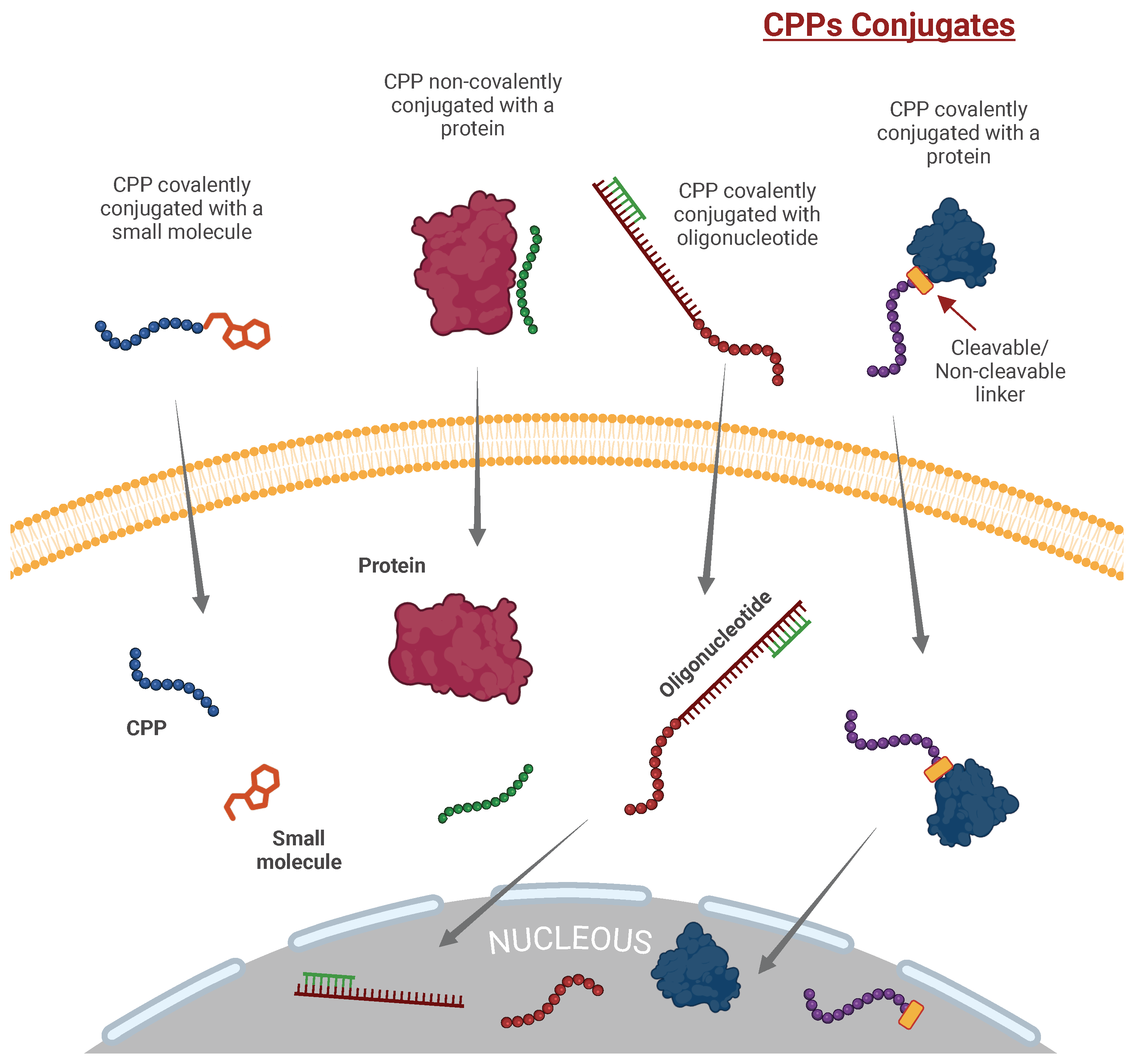
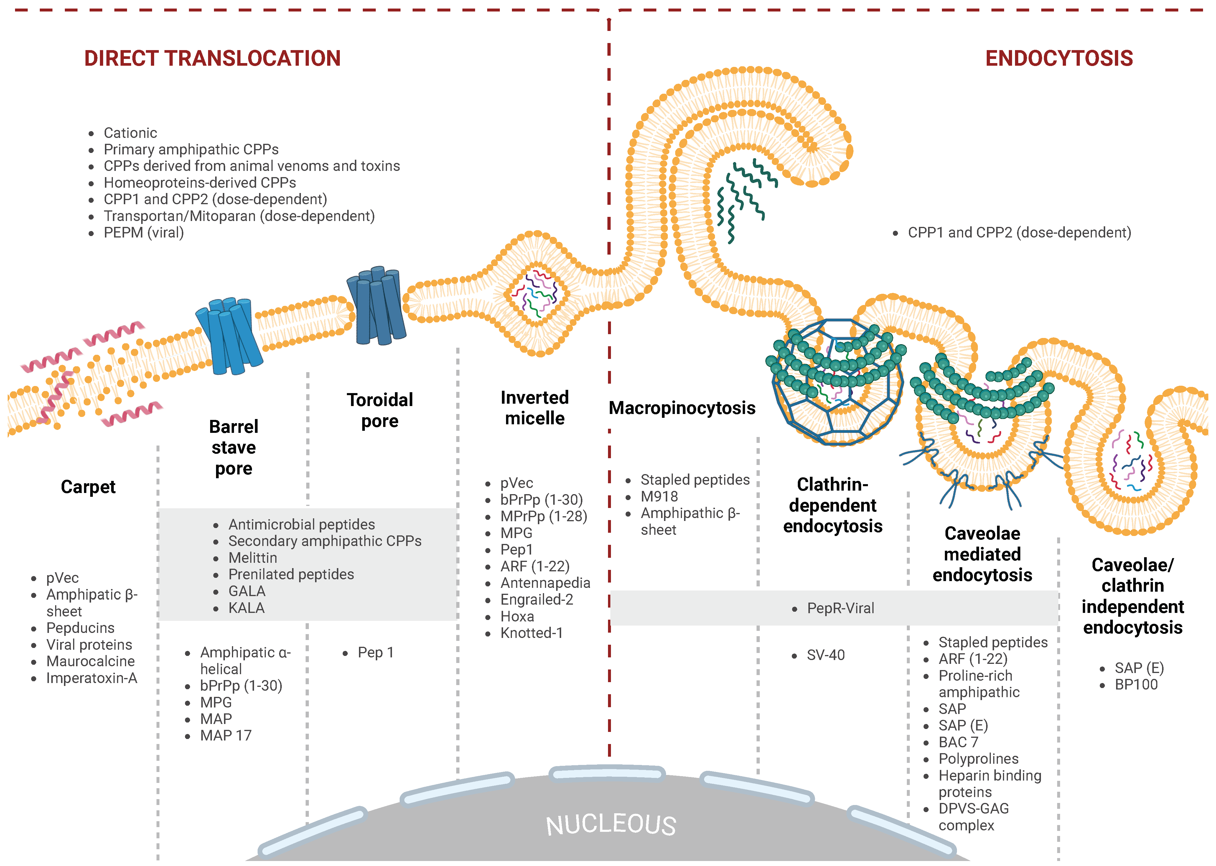
Figure 1. Classification of cell-penetrating peptides (cpps) by physicochemical properties and origin, with representative peptides for each category. This figure displays a selection of structurally diverse CPPs, classified into cationic (Penetratin, PDB ID: 1OMQ), amphipathic (Primary: bPrPp(1–30), PDB ID: 1SKH; Secondary: GALA; beta-sheet: RADA16; Proline-rich: SAP), hydrophobic (Linear: VPMLK (V5 antiapoptotic pentapeptide); Stapled: ALRN-6924, PDB ID: 8GJS; Prenylated: N-tert-prenylated tryptophan analogue [27]; Pepducins: p1pal-7 [28]) and cyclic (BT1718 [29]) peptides, as well as those derived from proteins (Homeoproteins: pIsl peptide; Heparin-binding proteins: CPPecp; Viral proteins: pepR), animal venoms, and toxins (Melittin (PDB ID: 6O4M) and, Pardaxin (PDB ID: 2KNS)). Each peptide is represented in its 3D conformation to enable the comparative visualization of structural motifs characteristic of each CPP type, which may impact their mechanisms of cellular uptake. AlphaFold [30] and PEP-FOLD3 [31] were used to predict the structures for those peptides without PDB ID. Notably, these configurations do not depict the spatial orientations involved in membrane interactions or the factors determining intracellular delivery efficiency.
Ijms 26 00059 g001
Figure 2. Attachment methods of CPPs to cargo molecules. This figure illustrates two main strategies for CPP–cargo attachment: covalent and non-covalent coupling. Covalent attachment, widely used for intracellular delivery, involves amide, disulfide, or triazole linkages, often with spacers to optimize CPP–cargo distance, and may include fusion proteins for delivering proteins or peptides. Non-covalent attachment leverages electrostatic interactions between positively charged CPPs and anionic cargo or polyanionic carriers. Although less stable in biological environments, non-covalent complexes form easily by simple mixing, as seen with CPP-siRNA interactions. Both approaches offer specific advantages and limitations in terms of stability, specificity, and intracellular delivery efficacy. [Created in BioRender. Moreno-Vargas, L. (2024) BioRender.com/j76w245 | CC-BY 4.0].
Ijms 26 00059 g002
Figure 3. Intracellular entry pathways for CPPs. CPPs utilize two primary mechanisms for cellular entry: energy-dependent endocytosis and energy-independent direct translocation across the lipid bilayer. Endocytosis in this context refers to various internalization processes that can be classified into four major pathways: macropinocytosis, clathrin-dependent endocytosis, caveolae-mediated endocytosis, and clathrin- and caveolae-independent endocytosis. In contrast, direct translocation occurs without energy input and involves mechanisms such as the toroidal or barrel-stave pore formation, inverted micelle, and the carpet model, where peptides disrupt the membrane to facilitate their entry. [Created in BioRender. Moreno-Vargas, L. (2024) BioRender.com/f68s318 | CC-BY 4.0].
Ijms 26 00059 g003
Table 1. CPP-based therapeutics in clinical trials.
CPPCargoCompoundApplicationStatusClinicalTrial.gov ID
Cationic CPPs
HIV-1 Tat-protein-derived Tat peptide δ PKC inhibitorKAI-9803Acute myocardial infarctionPhase II completed 2004NCT00093197
HIV-1 Tat-protein-derived Tat peptideMAGE-A3/HPV-16 Head and neck carcinomaPhase I completed 2005NCT00257738
HIV-1 Tat-protein-derived Tat peptide ϵ -PKC inhibitor Pain: postherpetic neuralgia, spinal cord injury, postoperativePhase II completed 2010NCT01106716
HIV-1 Tat-protein-derived Tat peptide δ PKC inhibitorKAI-9803Heart attackPhase II completed 2011NCT00785954
HIV-1 Tat-protein-derived Tat peptide ϵ -PKC inhibitor Pain: postherpetic neuralgia, spinal cord injury, postoperativePhase II completed 2011NCT01135108
HIV-1 Tat-protein-derived Tat peptideDextrogyre peptide Intraocular inflammation and painPhase I completed 2012NCT01570205
HIV-1 Tat-protein-derived Tat peptideBotulinum toxin A Cervical dystoniaPhase III completed 2012NCT01753310
HIV-1 Tat-protein-derived Tat peptide ϵ -PKC inhibitor Pain: postherpetic neuralgia, spinal cord injury, postoperativePhase II completed 2013NCT01015235
HIV-1 Tat-protein-derived Tat peptideD-JNKI-1 gel Hearing loss, idiopathic sudden sensorineuralPhase III completed 2015NCT02561091
HIV-1 Tat-protein-derived Tat peptideD-JNKI-1 gel Hearing loss, idiopathic sudden sensorineuralPhase III completed 2016NCT02809118
HIV-1 Tat-protein-derived Tat peptideJNK-1XG-102Intraocular inflammation and painPhase III completed 2016NCT02235272
HIV-1 Tat-protein-derived Tat peptidePSD-95 protein inhibitor Ischemic strokePhase III completed 2016NCT02930018
HIV-1 Tat-protein-derived Tat peptideBotulinum toxin A Cervical dystoniaPhase II completed 2016NCT02706795
HIV-1 Tat-protein-derived Tat peptideDextrogyre peptideXG-102Postoperative ocular inflammationPhase III completed 2017NCT02508337
HIV-1 Tat-protein-derived Tat peptideJNK-1AM-111Acute inner ear hearing lossPhase III completed 2017NCT02561091
ATX-101 Advanced dedifferentiated liposarcoma and leiomyosarcomaPhase II completed 2023NCT05116683
ATX-101Carboplatin Fallopian Tube and Primary Peritoneal CancerPhase I/II terminated 2024NCT04814875
(R-X-R)4, X = 6-Aminohexanoic acidPMO targeted to human c-MycAVI-5126Obstruction of vein graft after cardiovascular bypass surgeryPhase II completed 2009NCT00451256
TransMTSBotulinumtoxin A Cervical dystoniaPhase III completed 2022NCT03608397
MTSBotulinumtoxin A Lateral canthal linesPhase III completed 2016NCT02580370
MTSBotulinumtoxin A Primary Axillary HyperhidrosisPhase II completed 2016NCT02565732
AVB-620Tetramethylindo(di)-carbocyanines (Cy5 and Cy7) Breast cancerPhase I completed 2015NCT02391194
AVB-620Tetramethylindo(di)-carbocyanines (Cy5 and Cy7) Breast cancerPhase II completed 2021NCT03113825
Z12BI754091BI754091/ATP128/VSV-GP128Stage IV Colorectal CancerPhase I active, not recruiting, 2024NCT04046445
R7Cyclosporine APsorBanPsoriasisPhase II terminated 2003Not Applicable
PEP-010Paclitaxel Metastatic solid tumor cancerPhase I recruiting (2024)NCT04733027
Amphipatic CPPs
p28p28 Non-HDM2-mediated peptide inhibitor of p53p28Solid tumorsPhase I completed 2014NCT00914914
p28p28p28Central Nervous System TumorsPhase I completed 2017NCT01975116
p28Valganciclovir (VGCV)RZ-001GlioblastomaPhase I/II recruitingNCT06102525
p28SRF388Atezolizumab/Bevazizumab Phase II activeNCT05359861
PTD4AZX100 (a synthetic 24-amino acid peptide analog of heat shock protein 20 (Hsp20)) Excision of Keloid ScarsPhase II completed 2012NCT00825916
Hydrophobic CPPs: Stapled Peptides
Sulanemadlin (ALRN-6924)Cytarabine Acute Myeloid Leukemia or Advanced Myelodysplastic SyndromePhase I completed 2019NCT02909972
Sulanemadlin (ALRN-6924)Palbociclib Solid Tumor/Lymphoma/Peripheral T-Cell LymphomaPhase I/Phase II completed 2020NCT02264613
Sulanemadlin (ALRN-6924)Topotecan Small cell lung cancerPhase I active (Study completion (Actual) 2022)NCT04022876
Sulanemadlin (ALRN-6924)Cytarabine Solid tumor, brain tumor, leukemia or pediatric lymphomaPhase I active (Study completion (Actual) 2023)NCT03654716
Sulanemadlin (ALRN-6924)Doxorubicin/Cyclophosphamide/Docetaxel TP53-Mutant Breast CancerPhase I active (Study completion (Actual) 2023)NCT05622058
Sulanemadlin (ALRN-6924)Palbociclib Advanced, Metastatic, or Unresectable Solid TumorsPhase I active (Study completion (Estimated) 2025)NCT02264613
Pepducins
Pepducin PZ-128 (P1pal-7) Multiple Coronary Artery Disease Risk FactorsPhase I completed 2016NCT01806077
Pepducin PZ-128 (P1pal-7) Coronary artery diseasePhase II completed 2021NCT02561000
Cyclic CPPs
177Lu-DOTA0-Tyr3-octreotateLutetium Lu 177 Neuroendocrine TumorsNot Applicable completed 2019NCT02125474
177Lu-DOTA0-Tyr3-octreotateLutetium Lu 177 Neuroendocrine CarcinomaPhase II active 2023NCT02236910
BT1718 Solid tumorsPhase I/Phase II completed 2024NCT03486730
Disclaimer/Publisher’s Note: The statements, opinions and data contained in all publications are solely those of the individual author(s) and contributor(s) and not of MDPI and/or the editor(s). MDPI and/or the editor(s) disclaim responsibility for any injury to people or property resulting from any ideas, methods, instructions or products referred to in the content.

