Article

Systems Modeling Reveals Shared Metabolic Dysregulation and Potential Treatments in ME/CFS and Long COVID

1
State Key Laboratory of Genetic Evolution and Animal Models, Key Laboratory of Healthy Aging Research of Yunnan Province, Kunming Key Laboratory of Healthy Aging Study, KIZ/CUHK Joint Laboratory of Bioresources and Molecular Research in Common Diseases, Kunming Institute of Zoology, Chinese Academy of Sciences, Kunming 650201, China
2
Computational Research Center for Complex Chronic Diseases, Massachusetts General Hospital, Harvard Medical School, Boston, MA 02114, USA
3
Independent Researcher, 185 37 Pireas, Greece
4
CAS Center for Excellence in Animal Evolution and Genetics, Chinese Academy of Sciences, Kunming 650223, China
*
Authors to whom correspondence should be addressed.
These authors contributed equally to this work.
Int. J. Mol. Sci. 2025, 26(13), 6082; https://doi.org/10.3390/ijms26136082
Submission received: 21 May 2025 / Revised: 15 June 2025 / Accepted: 21 June 2025 / Published: 25 June 2025

Abstract

Myalgic Encephalomyelitis/Chronic Fatigue Syndrome (ME/CFS) and Long COVID are complex multisystem conditions that pose significant challenges in healthcare. Accumulated research evidence suggests that ME/CFS and Long COVID exhibit overlapping metabolic symptoms, indicating potential shared metabolic dysfunctions. This study aims to systematically explore shared metabolic disturbances in the muscle tissue of patients. Utilizing genome-wide metabolic modeling, we identified key metabolic irregularities in the muscle of patients with ME/CFS, notably the downregulation of the alanine and aspartate metabolism pathway and the arginine and proline metabolism pathway. Further, in silico knockout analyses suggested that supplementation with aspartate (ASP) or asparagine (ASN) could potentially ameliorate these metabolic deficiencies. In addition, assessments of metabolomic levels in Long COVID patients also showed the significant downregulation of ASP during post-exertional malaise (PEM) in both muscle and blood. Consequently, we propose that a combination of l-ornithine and l-aspartate (LOLA) is a potential candidate to alleviate metabolic symptoms in ME/CFS and Long COVID for future clinical trials.

1. Introduction

The emergence of Myalgic Encephalomyelitis/Chronic Fatigue Syndrome (ME/CFS) and Long COVID as prominent health crises has underscored the need for a deeper understanding of these complex multisystemic conditions. ME/CFS, a debilitating chronic condition that has been historically under-studied, is characterized by persistent and unexplained fatigue, post-exertional malaise (PEM), orthostatic intolerance, unrefreshing sleep, brain fog, muscle pain, and other symptoms [1,2,3]. While Long COVID represents a spectrum of symptoms severely impacting the quality of life of patients, persisting months beyond the acute phase of SARS-CoV-2 infection [4,5], most of the symptoms are similar to those of ME/CFS, with few exceptions [6,7,8].
The similar clinical presentations of ME/CFS and Long COVID have prompted researchers to hypothesize and explore shared underlying mechanisms, particularly within metabolic functions. Metabolic dysfunction has been increasingly recognized as a potential contributor to the symptomatology of these conditions, with disturbances noted in energy metabolism, amino acid and lipid profiles, and mitochondrial function [9,10,11,12,13,14]. Skeletal muscle, together with the liver and the brain, consumes the most energy at rest and dramatically increases its energy consumption during physical exertion [15]. Investigations of muscle tissues are likely critical in understanding the metabolic mechanisms underlying the key symptoms of these diseases and identifying candidates for potential treatments.
Two recently published studies included omics analyses of muscle biopsies of patients with ME/CFS [16] and Long COVID [17]. The NIH deep phenotyping of post-infectious ME/CFS study by Walitt et al. [16] included RNA sequencing data of the muscle biopsies of 13 ME/CFS patients and 12 healthy controls (Gene Expression Omnibus (GEO), GSE245661). The other study by Appelman et al. [17] included metabolomics measurements of skeletal muscle biopsies and blood samples from 25 Long COVID patients and 21 age- and sex-matched healthy controls (recovered from mild SARS-CoV-2 infection) taken before and one day after exercise testing, along with the metabolomics measurements (MetaboLights, MTBLS9103). These omics data provide the opportunity to systematically examine the metabolic network in the skeletal muscle of these diseases.
The metabolic network of a human tissue contains thousands of reactions and metabolites, and computational modeling using constraint-based reconstruction and analysis (COBRA) [18] has become an important tool for modeling cellular metabolisms [19,20] and systematically identifying metabolic changes in diseases [21,22,23,24]. Built on these, we recently developed an improved method for genome-wide precision metabolic modeling (GPMM), which can quantitatively integrate transcriptome, proteome, and kinetome data in the predictive modeling of metabolic networks [25].
In this study, we investigate the shared metabolic alterations in the muscle of ME/CFS and Long COVID patients (Figure 1). We first analyzed the transcriptomic data of muscle tissues in Walitt et al. using GPMM to identify metabolic fluxes changed in the muscle of ME/CFS patients and associated pathways. Genome-wide in silico knockout analyses were performed to identify metabolites whose modulation could potentially rescue the metabolic changes seen in patients. We then analyzed the metabolomics data of the samples of muscle and blood in Appelman et al. to determine whether the findings identified in ME/CFS can also be observed in Long COVID patients. We found that the most significant metabolic change is the downregulation of alanine and aspartate metabolism. We also propose the combination of l-ornithine and l-aspartate (LOLA) as a potential therapeutic candidate to replenish these deficient metabolic pathways.

2. Results

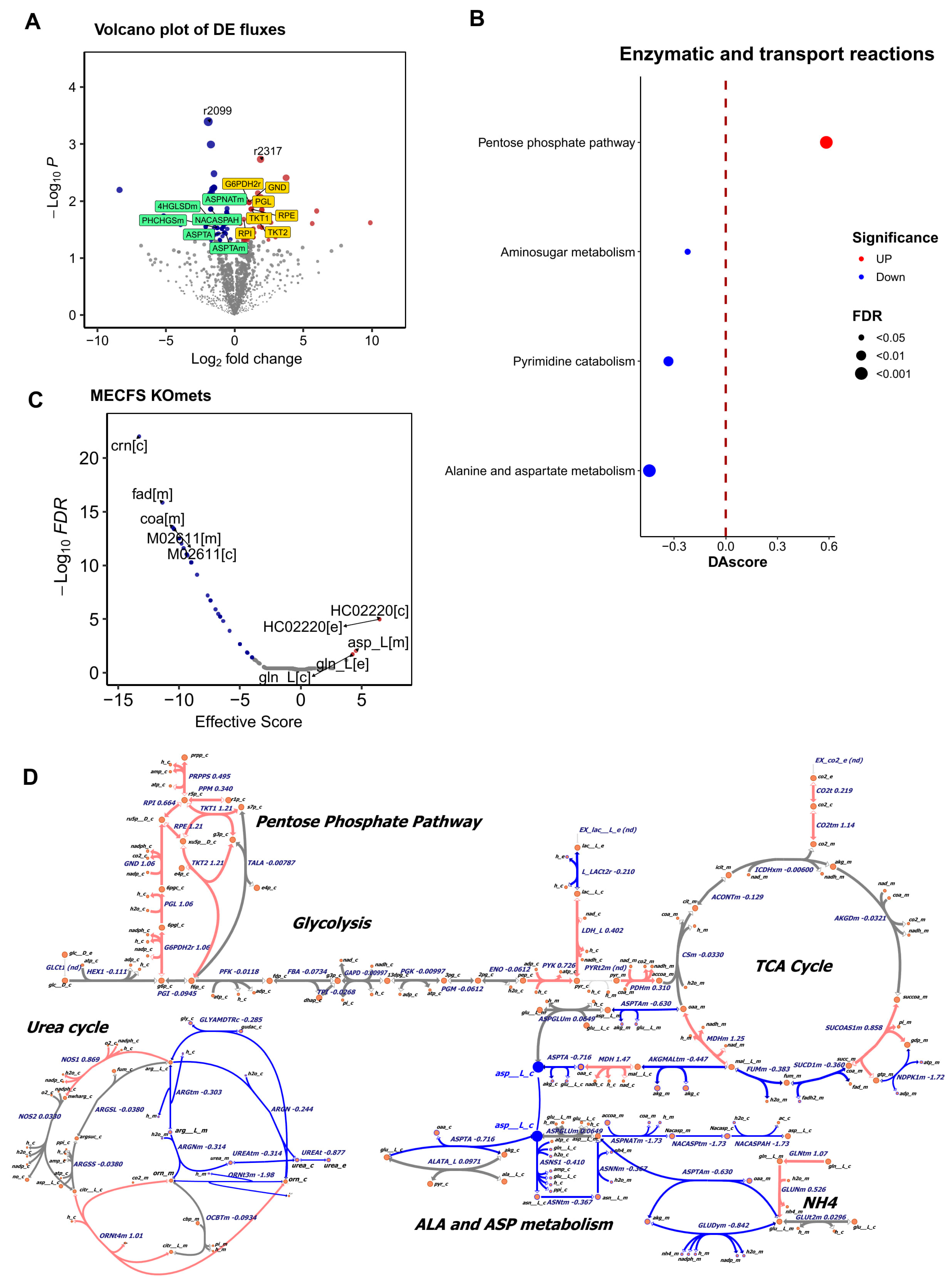
2.1. Metabolic Modeling Reveals Altered Metabolism in the Muscle of ME/CFS Patients

To investigate metabolic changes in ME/CFS, we conducted metabolic modeling using muscle samples from ME/CFS patients and controls from the NIH deep phenotyping of ME/CFS study [16].
Comparing the flux of each metabolic reaction between patients and controls identified 65 reactions showing significant up-regulation and 50 reactions showing significant down-regulation in the muscle of patients (Figure 2A, p < 0.05, |log2fc| > 0.2) (Supplemental Data S1). Notably, among the downregulated fluxes, four (ASPNATm, ASPTAm, NACASPAH, ASPTA) were related to alanine and aspartate metabolism, and two (4HGLSDm and PHCHGSm) were related to arginine and proline metabolism. Among the upregulated fluxes, seven (G6PDH2r, PGL, RPE, RPI, TKT1, TKT2, GND) were related to the pentose phosphate pathway.
Pathway enrichment analysis further revealed that several metabolic pathways were affected. Specifically, pathways of alanine and aspartate metabolism, pyrimidine catabolism, and aminosugar metabolism were significantly down-regulated, and conversely, the pentose phosphate pathway was significantly up-regulated (FDR < 0.05, |DAscore| > 0.2). Notably, the most prominently down-regulated pathway was alanine and aspartate metabolism in the muscle of ME/CFS patients (Figure 2B).
We next examined the metabolic network associated with glucose and amino acid metabolism. Intriguingly, most of the reactions in alanine and aspartate metabolism are down-regulated (log2FC < −0.2) in the muscle sample of ME/CFS patients (Figure 2D and Supplemental Figure S1). Additional flux changes (e.g., urea_c and urea_e in the urea cycle) indicated reduced urea secretion and a potential increase in NH4 secretion (labeled as NH4). On the other hand, the up-regulated reactions were clearly enriched on the pentose phosphate pathway.
We further investigated whether increasing the metabolites on alanine and aspartate metabolism pathway can potentially rescue the metabolic changes seen in patients toward the metabolic state of the controls. Our all-against-all knockout analysis highlighted that asparagine (ASN) and aspartate (ASP) are also among the top agonist metabolites (Figure 2C) (Supplemental Data S2), suggesting that administering these two amino acids could potentially rescue the metabolic changes observed in ME/CFS patients. This knockout result aligned with the modeling findings and supported the notion of dysfunction in alanine and aspartate metabolism among ME/CFS patients [10].

2.2. Metabolomics Measurements of Long COVID Reveals Down-Regulated Asparagine (ASN) During PEM in Muscle and the Blood

Given the similarity in symptoms between long COVID and ME/CFS, we analyzed the metabolic profile of the muscle in Long COVID patients. Since PEM is a cardinal symptom of ME/CFS and common in Long COVID, we performed pairwise comparisons between pre- and post-exercise samples from Long COVID patients and healthy controls using data from a study of muscle tissue during post-exertional malaise in Long COVID [17] (Supplementary Data S3). As shown in Figure 3A, significant changes were noted in muscle tissue, particularly the significantly lower level of asparagine and dihydroxyacetone-P during PEM. Similarly, in blood samples, asparagine also ranked highest among down-regulated metabolites following PEM in Long COVID patients (Figure 3B).
Collectively, these consistent findings from the ME/CFS and the Long COVID studies highlighted the significant down-regulation of ASN/ASP metabolism in the muscle tissue of both conditions.

3. Discussion

This study provides an analysis of the metabolic disruptions found in both ME/CFS and Long COVID, offering new insights into their pathophysiology and highlighting potential treatment avenues. Our findings reveal significant metabolic commonalities between these conditions, particularly in the down-regulation of amino acid metabolism pathways such as ASN/ASP and arginine/proline in the muscles. These findings not only help us to better understand the systemic impact of these conditions, but also highlight potential targets for therapeutic intervention.
The consistent down-regulation of specific metabolic pathways across both indications suggests a fundamental disruption in amino acid metabolism and energy metabolism, which could be contributing to the severity and persistence of patients’ symptoms [14]. In addition, asparagine provides key sites for N-linked glycosylation, which is required for proper protein folding in the endoplasmic reticulum (ER) [26], and otherwise may induce ER stress, which likely takes place in ME/CFS [27].
Here, we propose that L-ornithine and L-aspartate (LOLA) might hold the potential of intervening in these metabolic pathways (Figure 4). L-Aspartate aligns with the commonly observed down-regulation of ASN/ASP in both ME/CFS and Long COVID [28], suggesting it could help counteract this deficiency by directly replenishing the decreased aspartate pool. As the metabolic product of arginine, L-ornithine corresponds to the down-regulated pathway of arginine and proline metabolism in ME/CFS, potentially restoring balance in this pathway [29]. Additionally, the combined use of these amino acids in LOLA could enhance the efficiency of the urea cycle [30], thereby improving overall metabolic balance and reducing symptoms such as fatigue and cognitive dysfunction [31,32].
Moreover, emerging evidence suggests that supplementation with LOLA can improve mitochondrial function [33,34], thereby potentially enhancing energy metabolism. Mitochondrial dysregulation has been shown as a key factor in the pathogenesis of chronic diseases, including those characterized by post-exertional malaise, a hallmark of both ME/CFS and Long COVID [35]. By enhancing mitochondrial function and energy production, LOLA could potentially mitigate some of the core symptoms of these conditions.
Furthermore, ammonia dysregulation has also been implicated in fatigue and cognitive dysfunction observed in both ME/CFS [36,37,38] and Long COVID patients [39,40]. Hyperammonemia is known to occur after intense or exhausting exercise and in pathological liver disorders [41], and it can affect energy production [42] and potentially induce neuroinflammation [43]. LOLA has been studied clinically in these conditions to facilitate ammonia detoxification in the liver by replenishing intermediates of the urea cycle [30,44]. It may help alleviate the metabolic symptoms associated with ammonia dysregulation in ME/CFS and Long COVID.
This study highlights the importance of investigating skeletal muscle, a major metabolic organ system, to better understand the metabolic dysfunctions in complex multisystem conditions. Further research of major metabolic tissues could pave the road for personalized medicine strategies in treating ME/CFS and Long COVID, where treatments are tailored based on the specific metabolic profiles of patients.
Clinical trials are essential for evaluating the long-term efficacy and safety of LOLA in these conditions, and longitudinal tracking of metabolic alterations and key symptoms in patients over time will be invaluable in validating and further elucidating its potential effects. Notably, this study focuses on analyses of skeletal muscles of patients and the potential impact of LOLA on the metabolic system. Exploring the interactions between the metabolic and other physiological systems, such as the immune, endocrine, and neurological systems, is crucial to developing a deeper understanding of these complex conditions and discovering better treatments.
In addition to LOLA, sulfochenodeoxycholate (HC02220), a sulfoconjugated chenodeoxycholic acid (CDCA), was also identified as an agonist (Figure 2C). CDCA is a precursor in the formation of taurochenodeoxycholic acid (TUDCA), which potentially improves insulin sensitivity and supports mitochondrial function in the skeletal muscle [45]. Further study is required to further investigate these metabolites in ME/CFS and long COVID.

4. Materials and Methods

4.1. Dataset Collection

The transcriptome dataset of muscle tissues of ME/CFS and controls was accessed from GEO (GSE245661) [16], the Gene Expression Omnibus, a public functional genomics data repository from NCBI (https://www.ncbi.nlm.nih.gov/geo/, accessed on 20 March 2024). This was part of the NIH deep phenotyping of post-infectious ME/CFS study [16], and comprises the RNA sequencing data of 13 ME/CFS patients and 12 healthy controls. The metabolomics dataset of the muscle and blood samples of Long COVID was obtained from MetaboLights (MTBLS9103, accessed on 15 May 2024) [17], a database for metabolomics experiments and derived information held by EMBL/EBI (https://www.ebi.ac.uk/metabolights). The dataset we analyzed includes the metabolomics measurements of skeletal muscle biopsies of 46 long COVID patients taken before and 1 day after exercise testing along with the metabolomics measurements of the blood samples. Detailed protocols of muscle biopsies, exercise testing, and genomic and metabolomic measurements are in the respective publications [16,17].

4.2. Genome-Wide Precision Modeling of Metabolic Fluxes in the Muscle of Patients and Controls

4.2.1. Metabolic Modeling

We used our recently developed genome-wide precision metabolic modeling method, GPMM, to perform the genome-wide metabolic modeling [25]. Briefly, GPMM integrates protein abundance estimates from gene expression data with enzymatic kinetic parameters and uses these as upper bounds in a generic human metabolic model based on Michaelis–Menten kinetics. Nutrient uptake fluxes for cell lines were referenced from existing literature [46], and the lower bounds for other exchanges were set as zero. The metabolic model in Recon3 (version 1) [47] was utilized as the initial general model of human metabolism. We conducted Flux Variability Analysis (FVA) using the FastMM algorithm to construct tissue-specific models for each sample [48]. The metabolic model of the muscle tissue involved 2841 metabolic reactions, 2248 enzymes, and 1519 metabolites.

4.2.2. Identification of Significantly Changed Fluxes

For case-control design, the flux changes between patients and controls were calculated using the Limma package [49] based on the resulting values of fluxes from the metabolic modeling. Differentially expressed (DE) fluxes with p value < 0.05 were identified as significantly changed.

4.2.3. Identification of Significantly Changed Metabolic Pathways

To ascertain changes in metabolic pathways, we calculated the Differential Abundance Score (DA score) employing a method previously described in the literature [50]. For each metabolic pathway (i), the DA score (DAi) is computed as follows:
D A i = #   u p   r e g u l a t e d   f l u x e s     #   d o w n   r e g u l a t e d   f l u x e s T o t a l   r e a c t i o n s i n   i t h   s u b s y s t e m
The statistical significance (p-values) of the Differential Abundance (DA) scores was determined using a “bootstrap without replacement” method, as detailed in our previous study [25].

4.2.4. All-Against-All Knockout Analysis

We performed an all-against-all metabolite knockout analysis using the FastMM [48] algorithm to generate a comprehensive metabolite knockout matrix (M(KO)). In this matrix, rows represent metabolites, while columns correspond to reactions. The Metabolite Effective Score (MES) was calculated in a manner analogous to gene knockout analysis, as follows:
M E S i = j = 1 n M i , j ( K O ) × s i g n ( l o g 2 F C j ) × L j
Here, M i , j ( E M ) denotes the effect score of the ith metabolite on the jth reaction. The significance (p-value) of each MES was determined using a norm-background method, employing the pnorm function in R. FDR adjustments were made using the p.adjust function with the ‘FDR’ method in R. An agonist, identified as its MES value being greater than 0 and an FDR less than 0.05, is a metabolite the knockout of which exacerbates the flux changes seen in patients, suggesting that administering the metabolite could potentially rescue the changes observed in patients. On the other hand, an antagonist, identified as its MES value lower than 0 and an FDR less than 0.05, is a metabolite the knockout of which reduces the flux changes seen in patients, suggesting that reducing the metabolite could potentially rescue the changes observed in patients.

4.3. Metabolomics Data Analysis

In MTBLS9103, 116 metabolites in skeletal muscle and 83 metabolites in venous blood were annotated [17]. For both tissues, we calculated the Differential Metabolite (DM) using Equation (3) to quantitate the difference of metabolite concentration change after PEM between Long COVID patients and controls. The significance was calculated by employing the eBayes method using the Limma package in R [49].
D M i = l o g 2 P E M p i l o g 2 b a s e l i n e p i l o g 2 P E M c i l o g 2 b a s e l i n e c i
where p denotes patients while c means controls; i is the index of each metabolite in this study.

5. Conclusions

In conclusion, by systematically exploring the metabolic mechanism in the muscles of patients, our study contributes to an understanding of the underlying mechanism of complex conditions and candidates for potential treatments. Future clinical trials of LOLA will evaluate its safety and efficacy in alleviating metabolic symptoms in ME/CFS and Long COVID patients.

Supplementary Materials

The following supporting information can be downloaded at: https://www.mdpi.com/article/10.3390/ijms26136082/s1.

Author Contributions

All authors made substantial contributions to the conception or design of the work; or the acquisition, analysis, or interpretation of data for the work, and drafting the work or reviewing it critically for important intellectual content. Specifically, W.X. and G.-H.L. conceived and designed the study. W.X., G.-H.L. and Q.-P.K. supervised the project. G.-H.L. designed and developed the computational method. G.-H.L., F.-F.H. and W.X. collected the datasets. G.-H.L. and F.-F.H. performed the data analysis and conducted the statistical analysis. G.-H.L., F.-F.H., E.K. and W.X. interpreted the results. G.-H.L., F.-F.H. and W.X. drafted the manuscript. G.-H.L., F.-F.H., E.K. and W.X. revised the manuscript. All authors have read and agreed to the published version of the manuscript.

Funding

This work was supported by grants from Open Medicine Foundation (W.X.). Yunnan Ten Thousand Talents Plan Young & Elite Talents Project (G.-H.L.), Yunnan Fundamental Research Projects (202101AS070058).

Institutional Review Board Statement

Not applicable.

Informed Consent Statement

Not applicable.

Data Availability Statement

Metabolic modeling, flux analysis and visualization, the metabolic pathway analysis, all-against-all knock out analysis, and key metabolite identification were all performed by rGPMM (https://github.com/GonghuaLi/rGPMM, accessed on 15 May 2024) with version 1.0.0 on the metabolic models Recon3, version 1 using the CPLEX solver. The rGPMM package for non-commercial research may request access by contacting the developer author at ligonghua@mail.kiz.ac.cn. Individual metabolic flux can be queried online at http://bigg.ucsd.edu/models/Recon3D/reactions (accessed on 15 May 2024).

Acknowledgments

We would also like to express our sincere gratitude to the patients who provided valuable feedback to us on the results of this study.

Conflicts of Interest

The authors declare no conflict of interest.

References

  1. Board on the Health of Select Populations, Committee on the Diagnostic Criteria for Myalgic Encephalomyelitis/Chronic Fatigue Syndrome. Beyond Myalgic Encephalomyelitis/Chronic Fatigue Syndrome: Redefining an Illness; National Academies Press: Washington, DC, USA, 2015. [Google Scholar] [CrossRef]
  2. Cortes Rivera, M.; Mastronardi, C.; Silva-Aldana, C.T.; Arcos-Burgos, M.; Lidbury, B.A. Myalgic Encephalomyelitis/Chronic Fatigue Syndrome: A Comprehensive Review. Diagnostics 2019, 9, 91. [Google Scholar] [CrossRef] [PubMed]
  3. Valdez, A.R.; Hancock, E.E.; Adebayo, S.; Kiernicki, D.J.; Proskauer, D.; Attewell, J.R.; Bateman, L.; DeMaria, A., Jr.; Lapp, C.W.; Rowe, P.C.; et al. Estimating Prevalence, Demographics, and Costs of ME/CFS Using Large Scale Medical Claims Data and Machine Learning. Front. Pediatr. 2018, 6, 412. [Google Scholar] [CrossRef]
  4. Ladds, E.; Darbyshire, J.L.; Bakerly, N.D.; Falope, Z.; Tucker-Bell, I. Cognitive dysfunction after covid-19. BMJ 2024, 384, e075387. [Google Scholar] [CrossRef] [PubMed]
  5. Thaweethai, T.; Jolley, S.E.; Karlson, E.W.; Levitan, E.B.; Levy, B.; McComsey, G.A.; McCorkell, L.; Nadkarni, G.N.; Parthasarathy, S.; Singh, U.; et al. Development of a Definition of Postacute Sequelae of SARS-CoV-2 Infection. JAMA 2023, 329, 1934–1946. [Google Scholar] [CrossRef]
  6. Komaroff, A.L.; Lipkin, W.I. ME/CFS and Long COVID share similar symptoms and biological abnormalities: Road map to the literature. Front. Med. 2023, 10, 1187163. [Google Scholar] [CrossRef]
  7. Davis, H.E.; McCorkell, L.; Vogel, J.M.; Topol, E.J. Long COVID: Major findings, mechanisms and recommendations. Nat. Rev. Microbiol. 2023, 21, 133–146. [Google Scholar] [CrossRef]
  8. National Academies of Sciences, Engineering, and Medicine; Health and Medicine Division; Board on Health Sciences Policy; Board on Global Health; Forum on Neuroscience and Nervous System Disorders; Forum on Microbial Threats. Toward a Common Research Agenda in Infection-Associated Chronic Illnesses: Proceedings of a Workshop; Snair, M., Liao, J., Ashby, E., Biffl, C., Eds.; The National Academies Collection: Washington, DC, USA, 2024. [Google Scholar]
  9. Saito, S.; Shahbaz, S.; Luo, X.; Osman, M.; Redmond, D.; Cohen Tervaert, J.W.; Li, L.; Elahi, S. Metabolomic and immune alterations in long COVID patients with chronic fatigue syndrome. Front. Immunol. 2024, 15, 1341843. [Google Scholar] [CrossRef] [PubMed]
  10. Domingo, J.C.; Battistini, F.; Cordobilla, B.; Zaragoza, M.C.; Sanmartin-Sentanes, R.; Alegre-Martin, J.; Cambras, T.; Castro-Marrero, J. Association of circulating biomarkers with illness severity measures differentiates myalgic encephalomyelitis/chronic fatigue syndrome and post-COVID-19 condition: A prospective pilot cohort study. J. Transl. Med. 2024, 22, 343. [Google Scholar] [CrossRef]
  11. Naviaux, R.K.; Naviaux, J.C.; Li, K.; Bright, A.T.; Alaynick, W.A.; Wang, L.; Baxter, A.; Nathan, N.; Anderson, W.; Gordon, E. Metabolic features of chronic fatigue syndrome. Proc. Natl. Acad. Sci. USA 2016, 113, E5472–E5480. [Google Scholar] [CrossRef]
  12. Rutherford, G.; Manning, P.; Newton, J.L. Understanding Muscle Dysfunction in Chronic Fatigue Syndrome. J. Aging. Res. 2016, 2016, 2497348. [Google Scholar] [CrossRef]
  13. Huth, T.K.; Eaton-Fitch, N.; Staines, D.; Marshall-Gradisnik, S. A systematic review of metabolomic dysregulation in Chronic Fatigue Syndrome/Myalgic Encephalomyelitis/Systemic Exertion Intolerance Disease (CFS/ME/SEID). J. Transl. Med. 2020, 18, 198. [Google Scholar] [CrossRef]
  14. Hoel, F.; Hoel, A.; Pettersen, I.K.; Rekeland, I.G.; Risa, K.; Alme, K.; Sorland, K.; Fossa, A.; Lien, K.; Herder, I.; et al. A map of metabolic phenotypes in patients with myalgic encephalomyelitis/chronic fatigue syndrome. JCI Insight 2021, 6, e149217. [Google Scholar] [CrossRef] [PubMed]
  15. Wang, Z.; Ying, Z.; Bosy-Westphal, A.; Zhang, J.; Heller, M.; Later, W.; Heymsfield, S.B.; Muller, M.J. Evaluation of specific metabolic rates of major organs and tissues: Comparison between men and women. Am. J. Hum. Biol. 2011, 23, 333–338. [Google Scholar] [CrossRef] [PubMed]
  16. Walitt, B.; Singh, K.; LaMunion, S.R.; Hallett, M.; Jacobson, S.; Chen, K.; Enose-Akahata, Y.; Apps, R.; Barb, J.J.; Bedard, P.; et al. Deep phenotyping of post-infectious myalgic encephalomyelitis/chronic fatigue syndrome. Nat. Commun. 2024, 15, 907. [Google Scholar] [CrossRef] [PubMed]
  17. Appelman, B.; Charlton, B.T.; Goulding, R.P.; Kerkhoff, T.J.; Breedveld, E.A.; Noort, W.; Offringa, C.; Bloemers, F.W.; van Weeghel, M.; Schomakers, B.V.; et al. Muscle abnormalities worsen after post-exertional malaise in long COVID. Nat. Commun. 2024, 15, 17. [Google Scholar] [CrossRef]
  18. Orth, J.D.; Thiele, I.; Palsson, B.O. What is flux balance analysis? Nat. Biotechnol. 2010, 28, 245–248. [Google Scholar] [CrossRef]
  19. Heirendt, L.; Arreckx, S.; Pfau, T.; Mendoza, S.N.; Richelle, A.; Heinken, A.; Haraldsdóttir, H.S.; Wachowiak, J.; Keating, S.M.; Vlasov, V. Creation and analysis of biochemical constraint-based models using the COBRA Toolbox v. 3.0. Nat. Protoc. 2019, 14, 639–702. [Google Scholar] [CrossRef]
  20. Zhang, H.; Li, X.; Tseyang, L.T.; Giese, G.E.; Wang, H.; Yao, B.; Zhang, J.; Neve, R.L.; Shank, E.A.; Spinelli, J.B.; et al. A systems-level, semi-quantitative landscape of metabolic flux in C. elegans. Nature 2025, 640, 194–202. [Google Scholar] [CrossRef]
  21. Mardinoglu, A.; Boren, J.; Smith, U.; Uhlen, M.; Nielsen, J. Systems biology in hepatology: Approaches and applications. Nat. Rev. Gastroenterol. Hepatol. 2018, 15, 365–377. [Google Scholar] [CrossRef]
  22. Li, G.H.; Li, Y.H.; Yu, Q.; Zhou, Q.Q.; Zhang, R.F.; Weng, C.J.; Ge, M.X.; Kong, Q.P. Unraveling the metabolic heterogeneity and commonality in senescent cells using systems modeling. Life Med. 2025, 4, lnaf003. [Google Scholar] [CrossRef]
  23. Shen, Q.; Yang, H.; Kong, Q.P.; Li, G.H.; Li, L. Metabolic Modeling Identifies a Novel Molecular Type of Glioblastoma Associated with Good Prognosis. Metabolites 2023, 13, 172. [Google Scholar] [CrossRef]
  24. Mardinoglu, A.; Palsson, B.O. Genome-scale models in human metabologenomics. Nat. Rev. Genet. 2025, 26, 123–140. [Google Scholar] [CrossRef] [PubMed]
  25. Li, G.H.; Han, F.; Xiao, F.H.; Gu, K.S.Y.; Shen, Q.; Xu, W.; Li, W.X.; Wang, Y.L.; Liang, B.; Huang, J.F. System-level metabolic modeling facilitates unveiling metabolic signature in exceptional longevity. Aging Cell 2022, 21, e13595. [Google Scholar] [CrossRef] [PubMed]
  26. Reily, C.; Stewart, T.J.; Renfrow, M.B.; Novak, J. Glycosylation in health and disease. Nat. Rev. Nephrol. 2019, 15, 346–366. [Google Scholar] [CrossRef]
  27. Wang, P.Y.; Ma, J.; Kim, Y.C.; Son, A.Y.; Syed, A.M.; Liu, C.; Mori, M.P.; Huffstutler, R.D.; Stolinski, J.L.; Talagala, S.L.; et al. WASF3 disrupts mitochondrial respiration and may mediate exercise intolerance in myalgic encephalomyelitis/chronic fatigue syndrome. Proc. Natl. Acad. Sci. USA 2023, 120, e2302738120. [Google Scholar] [CrossRef] [PubMed]
  28. Holecek, M. Aspartic Acid in Health and Disease. Nutrients 2023, 15, 4023. [Google Scholar] [CrossRef]
  29. Butterworth, R.F.; McPhail, M.J.W. L-Ornithine L-Aspartate (LOLA) for Hepatic Encephalopathy in Cirrhosis: Results of Randomized Controlled Trials and Meta-Analyses. Drugs 2019, 79 (Suppl. 1), 31–37. [Google Scholar] [CrossRef]
  30. Canbay, A.; Sowa, J.-P. L-Ornithine L-Aspartate (LOLA) as a novel approach for therapy of non-alcoholic fatty liver disease. Drugs 2019, 79 (Suppl. 1), 39–44. [Google Scholar] [CrossRef]
  31. Kokubo, T.; Ikeshima, E.; Kirisako, T.; Miura, Y.; Horiuchi, M.; Tsuda, A. A randomized, double-masked, placebo-controlled crossover trial on the effects of L-ornithine on salivary cortisol and feelings of fatigue of flushers the morning after alcohol consumption. Biopsychosoc. Med. 2013, 7, 6. [Google Scholar] [CrossRef]
  32. Sugino, T.; Shirai, T.; Kajimoto, Y.; Kajimoto, O. L-ornithine supplementation attenuates physical fatigue in healthy volunteers by modulating lipid and amino acid metabolism. Nutr. Res. 2008, 28, 738–743. [Google Scholar] [CrossRef]
  33. Sisalli, M.J.; Della Notte, S.; Secondo, A.; Ventra, C.; Annunziato, L.; Scorziello, A. L-ornithine L-aspartate restores mitochondrial function and modulates intracellular calcium homeostasis in Parkinson’s disease models. Cells 2022, 11, 2909. [Google Scholar] [CrossRef] [PubMed]
  34. Das, A.; Fröhlich, D.; Achanta, L.B.; Rowlands, B.D.; Housley, G.D.; Klugmann, M.; Rae, C.D. L-aspartate, L-ornithine and L-ornithine-L-aspartate (LOLA) and their impact on brain energy metabolism. Neurochem. Res. 2020, 45, 1438–1450. [Google Scholar] [CrossRef] [PubMed]
  35. Tate, W.; Walker, M.; Sweetman, E.; Helliwell, A.; Peppercorn, K.; Edgar, C.; Blair, A.; Chatterjee, A. Molecular Mechanisms of Neuroinflammation in ME/CFS and Long COVID to Sustain Disease and Promote Relapses. Front. Neurol. 2022, 13, 877772. [Google Scholar] [CrossRef] [PubMed]
  36. Wilkinson, D.J.; Smeeton, N.J.; Watt, P.W. Ammonia metabolism, the brain and fatigue; revisiting the link. Prog. Neurobiol. 2010, 91, 200–219. [Google Scholar] [CrossRef]
  37. Glass, K.A.; Germain, A.; Huang, Y.V.; Hanson, M.R. Urine Metabolomics Exposes Anomalous Recovery after Maximal Exertion in Female ME/CFS Patients. Int. J. Mol. Sci. 2023, 24, 3685. [Google Scholar] [CrossRef]
  38. Germain, A.; Giloteaux, L.; Moore, G.E.; Levine, S.M.; Chia, J.K.; Keller, B.A.; Stevens, J.; Franconi, C.J.; Mao, X.; Shungu, D.C.; et al. Plasma metabolomics reveals disrupted response and recovery following maximal exercise in myalgic encephalomyelitis/chronic fatigue syndrome. JCI Insight 2022, 7, e157621. [Google Scholar] [CrossRef]
  39. Bobermin, L.D.; Quincozes-Santos, A. COVID-19 and hyperammonemia: Potential interplay between liver and brain dysfunctions. Brain Behav. Immun.-Health 2021, 14, 100257. [Google Scholar] [CrossRef]
  40. Lopez-Hernandez, Y.; Monarrez-Espino, J.; Lopez, D.A.G.; Zheng, J.; Borrego, J.C.; Torres-Calzada, C.; Elizalde-Diaz, J.P.; Mandal, R.; Berjanskii, M.; Martinez-Martinez, E.; et al. The plasma metabolome of long COVID patients two years after infection. Sci. Rep. 2023, 13, 12420. [Google Scholar] [CrossRef]
  41. Ali, R.; Nagalli, S. Hyperammonemia. In StatPearls [Internet]; StatPearls Publishing: Tampa, FL, USA, 2023. [Google Scholar]
  42. Bosoi, C.R.; Rose, C.F. Identifying the direct effects of ammonia on the brain. Metab. Brain Dis. 2009, 24, 95–102. [Google Scholar] [CrossRef]
  43. Hernandez-Rabaza, V.; Cabrera-Pastor, A.; Taoro-Gonzalez, L.; Malaguarnera, M.; Agusti, A.; Llansola, M.; Felipo, V. Hyperammonemia induces glial activation, neuroinflammation and alters neurotransmitter receptors in hippocampus, impairing spatial learning: Reversal by sulforaphane. J. Neuroinflamm. 2016, 13, 41. [Google Scholar] [CrossRef]
  44. Kircheis, G.; Lüth, S. Pharmacokinetic and pharmacodynamic properties of L-ornithine L-aspartate (LOLA) in hepatic encephalopathy. Drugs 2019, 79 (Suppl. 1), 23–29. [Google Scholar] [CrossRef] [PubMed]
  45. Kars, M.; Yang, L.; Gregor, M.F.; Mohammed, B.S.; Pietka, T.A.; Finck, B.N.; Patterson, B.W.; Horton, J.D.; Mittendorfer, B.; Hotamisligil, G.S.; et al. Tauroursodeoxycholic Acid may improve liver and muscle but not adipose tissue insulin sensitivity in obese men and women. Diabetes 2010, 59, 1899–1905. [Google Scholar] [CrossRef]
  46. Jain, M.; Nilsson, R.; Sharma, S.; Madhusudhan, N.; Kitami, T.; Souza, A.L.; Kafri, R.; Kirschner, M.W.; Clish, C.B.; Mootha, V.K. Metabolite profiling identifies a key role for glycine in rapid cancer cell proliferation. Science 2012, 336, 1040–1044. [Google Scholar] [CrossRef] [PubMed]
  47. Brunk, E.; Sahoo, S.; Zielinski, D.C.; Altunkaya, A.; Dräger, A.; Mih, N.; Gatto, F.; Nilsson, A.; Preciat Gonzalez, G.A.; Aurich, M.K. Recon3D enables a three-dimensional view of gene variation in human metabolism. Nat. Biotechnol. 2018, 36, 272–281. [Google Scholar] [CrossRef] [PubMed]
  48. Li, G.-H.; Dai, S.; Han, F.; Li, W.; Huang, J.; Xiao, W. FastMM: An efficient toolbox for personalized constraint-based metabolic modeling. BMC Bioinform. 2020, 21, 67. [Google Scholar] [CrossRef]
  49. Ritchie, M.E.; Phipson, B.; Wu, D.; Hu, Y.; Law, C.W.; Shi, W.; Smyth, G.K. limma powers differential expression analyses for RNA-sequencing and microarray studies. Nucleic Acids Res. 2015, 43, e47. [Google Scholar] [CrossRef]
  50. Hakimi, A.A.; Reznik, E.; Lee, C.-H.; Creighton, C.J.; Brannon, A.R.; Luna, A.; Aksoy, B.A.; Liu, E.M.; Shen, R.; Lee, W.; et al. An Integrated Metabolic Atlas of Clear Cell Renal Cell Carcinoma. Cancer Cell 2016, 29, 104–116. [Google Scholar] [CrossRef]
Figure 1. The workflow of omics analyses of the transcriptome data of muscle biopsies in ME/CFS and metabolomics data of muscle and blood in Long COVID. The left panel shows genome-wide metabolic modeling (GPMM) using transcriptomic data of muscle tissues from an ME/CFS study (GSE245661). The right panel shows the analysis of metabolomics data of muscle and blood from a Long COVID study (MTBLS9103). Common changes in metabolism identified in these two patient datasets suggest potential targets for modulation for the diseases.
Ijms 26 06082 g001
Figure 2. Metabolic modeling of muscle samples of ME/CFS patients. (A) Volcano plot of differentially changed metabolic fluxes between ME/CFS patients and controls (p-value < 0.05, |log2fc| > 0.2). Red, blue, and gray points represent significantly increased, decreased, and non-significant pathways, respectively. (B) Pathway analysis differential changed metabolic fluxes between ME/CFS patients and controls (FDR < 0.05 and |DAscroe| > 0.2). Red, blue, and gray points represent significantly up-regulated, down-regulated, and non-significant pathways, respectively. (C) Metabolite knockout analysis in ME/CFS. Effective scores greater than 0 indicate agonist metabolites, while scores less than 0 indicate antagonist metabolites. Administering an agonist metabolite, e.g., ASP, could potentially rescue the changes observed in patients. (D) Metabolic map of representative pathways and metabolic fluxes. The down-regulated reactions are enriched in alanine and aspartate metabolism in patients. In addition, the down-regulated reactions include reduced urea secretion and a potential increase in NH4 secretion.
Ijms 26 06082 g002
Figure 3. Pair-wise changes of metabolite levels in Long COVID after post-exertional malaise (PEM). (A). Volcano plot of pair-wise metabolic changes in the muscle of Long COVID patients after PEM. (B). Boxplot of changes of asparagine level in the blood between Long COVID and healthy controls. (C). Volcano plot of pair-wise metabolic changes in Long-COVID patient blood after PEM. (D). Boxplot of changes of asparagine in the muscle between Long COVID and Healthy controls.
Ijms 26 06082 g003
Figure 4. L-ornithine and L-aspartate (LOLA) as a Potential Treatment Candidate for ME/CFS and Long COVID. L-aspartate potentially can help counteract the observed decrease in asparagine/aspartate in both ME/CFS and Long COVID. L-ornithine, as the metabolic product of arginine, potentially can help restore the balance of the down-regulated pathway of arginine and proline metabolism in ME/CFS. Additionally, the combined use of these amino acids could enhance urea cycle efficiency, which is critical in removing ammonia and reducing fatigue symptoms commonly associated with these conditions. Besides, emerging evidence suggests LOLA can improve mitochondrial function, thereby potentially enhancing energy metabolism in patients with ME/CFS and Long COVID. Red arrows denote the increase or upregulation of the corresponding metabolites or pathways, and blue arrows denote the decrease or downregulation.
Ijms 26 06082 g004
Disclaimer/Publisher’s Note: The statements, opinions and data contained in all publications are solely those of the individual author(s) and contributor(s) and not of MDPI and/or the editor(s). MDPI and/or the editor(s) disclaim responsibility for any injury to people or property resulting from any ideas, methods, instructions or products referred to in the content.

