Article

Functional Characterization of OsCSN1 in the Agronomic Trait Control of Rice Seedlings Under Far-Red Light

1
College of Life Sciences, Jilin Agricultural University, Changchun 130118, China
2
School of Life Sciences, Northeast Normal University, Changchun 130024, China
*
Authors to whom correspondence should be addressed.
Int. J. Mol. Sci. 2025, 26(2), 522; https://doi.org/10.3390/ijms26020522
Submission received: 16 December 2024 / Revised: 4 January 2025 / Accepted: 7 January 2025 / Published: 9 January 2025
(This article belongs to the Special Issue Plant Resilience: Insights into Abiotic and Biotic Stress Adaptations)

Abstract

:
The COP9 signalosome (CSN) is a highly conserved multi-subunit protein complex, with CSN1 being its largest and most conserved subunit. The N-terminal function of CSN1 plays a pivotal and intricate role in plant photomorphogenesis and seedling development. Moreover, CSN is essential for far-red light-mediated photomorphogenesis in seedlings, but the function of OsCSN1 in seedling growth and development under far-red light conditions has not been determined. This study investigates the function of OsCSN1 under far-red light through phenotypic analysis of wild type and OsCSN1 mutant seedlings. Additionally, the effect of the N-terminal region of OsCSN1 on rice seedling growth and development was examined. The addition of exogenous hormone gibberellin (GA3) and gibberellin synthesis inhibitor paclobutrazol (PAC) resulted in notable changes in phenotypes and the expression of key proteins, including CUL4 and SLR1. The findings indicate that OsCSN1 functions as a positive regulator of plant height under far-red light and inhibits root elongation. Under far-red light, OsCSN1 integrates into the COP9 complex and regulates the nuclear localization of COP1. Through its interaction with CUL4 in the CULLIN-RING family, OsCSN1 facilitates the ubiquitin-mediated degradation of SLR1, thereby influencing the growth of rice seedlings. The regulatory function of OsCSN1 in seedling growth and development under far-red light predominantly relies on the 32 amino acids of its N-terminal region. The results of this study can provide new ideas for rice breeding and genetic improvement. Based on the study of key regulatory factors such as OsCSN1, new varieties that can make better use of far-red light signals can be cultivated to enhance crop adaptability and productivity.

1. Introduction

The COP9 signalosome (CSN) was initially found to be a negative regulator of photomorphogenesis in Arabidopsis [1]. Moreover, a mutant of the CSN exhibits a constitutive photomorphogenesis phenotype under dark conditions [2]. Biochemical studies in plant and animal systems have shown that the CSN is a highly conserved nucleoprotein complex [3,4,5], with its structural integrity being essential for its biological activity [6,7,8]. The CSN shares significant structural and amino acid sequence homology with the “lid” subcomplex of the 19S regulatory particle in the 26S proteasome [3,4,5]. This similarity suggests that the CSN’s regulatory functions may be closely linked to ubiquitin/26S proteasome system-mediated protein degradation. The CSN interacts with various CULLIN-RING E3 ubiquitin ligases (CRLs) to affect various developmental processes in plants [9]. The COP9 signaling complex (CSN), as a highly conserved protein complex, plays an important role in the growth and development of rice. The CSN regulates intracellular protein levels by regulating the ubiquitin–proteasome system (UPS) to affect protein degradation. The CSN is involved in the regulation of hormone signal transduction pathways and promotes rice growth and development.
CSN1 is the largest and most conserved subunit in the CSN complex, plays a critical role in complex assembly through its C-terminal PCI domain, and supports essential functions via its N-terminal region [10]. The N-terminal domain is vital for plant growth and development, where its deletion leads to seedling lethality, floral organ abnormalities, and photomorphogenesis defects. CSN1 interacts with other subunits to restore the NEDD8/RUB1 deconjugation activity of CULLINs in csn1 mutants of Arabidopsis [11]. Additionally, mutants such as csn5a-1 and csn1-10 show delayed germination and accumulation of germination inhibitors RGL2 and ABI5, highlighting the CSN’s regulatory role in seed germination [12]. Overall, the N-terminal domain of CSN1 is indispensable for photomorphogenesis, seedling development, gene expression regulation, subcellular localization, and enzymatic activity within the complex. Its integration into the CSN complex is mediated by the PCI domain [10].
Light is a critical regulator of plant growth and development, primarily through photoreceptor-mediated signaling pathways that control photomorphogenesis and coordinate various developmental processes [13,14]. Far-red light (720–750 nm) influences key physiological processes, including seed germination, photomorphogenesis, and flowering, with its signaling pathway having complex effects on different physiological responses [15].
Phytochrome is the main receptor for plants to sense light and is widely present in higher plants, existing in two interconvertible forms: the far-red light-absorbing form (Pfr, 730 nm) and the red light-absorbing form (Pr, 660 nm) [16,17]. Phytochrome A (phyA) specifically senses and responds to far-red light, playing a key role in the transition from skotomorphogenesis to photomorphogenesis under far-red-rich light conditions [16]. Under far-red light conditions, the expression of the PHYA gene in rice is upregulated and regulates the photomorphogenesis of rice [18]. Far-red light rapidly induces phyA translocation to the nucleus within minutes [19], where it is subsequently degraded upon light exposure [20]. Far-red light inhibits photomorphogenesis by promoting the activity of SPA1, a negative regulator, which collaborates with COP1 to form an E3 ubiquitin ligase complex. COP1 mediates the light-induced degradation of phyA through the ubiquitin/26S-proteasome pathway, suppressing photomorphogenesis by targeting phosphorylated phyA and the positive regulator HY5 for degradation [21,22,23,24]. Consequently, plants exhibit elongated hypocotyls and increased height under far-red light [21,25].
Plant hormones play a crucial role in regulating plant growth and development by stimulating various physiological processes. Gibberellins (GAs) promote cell division and elongation during seed germination and are involved in flower development and fruit ripening [26,27]. Conversely, paclobutrazol, a growth regulator, inhibits endogenous gibberellin biosynthesis, thereby enhancing plant stress resistance and increasing crop yield [28]. DELLA proteins act as negative regulators of the GA signaling pathway [29]. During seed germination, DELLA proteins function as repressors, with GAs alleviating this repression by inhibiting DELLA protein activity to regulate germination and stem elongation [30]. In rice, SLR1, the sole DELLA protein, plays a central role in the GA signaling pathway, where its activity suppresses growth, leading to dwarfism during development [31].
Light is a critical environmental factor influencing rice seed germination and seedling development. Previous studies have demonstrated that OsCSN1 mediates the blue light-regulated inhibition of shoot growth in rice seedlings by affecting the degradation of SLR1, thereby reducing endogenous GA synthesis [32]. In contrast, OsCSN1 acts as a negative regulator of shoot elongation under red light. Red light promotes the degradation of SLR1 via neddylation and the ubiquitin/26S proteasome pathway, ultimately suppressing stem and coleoptile elongation [33]. These findings underscore the distinct effects of different light spectra on rice seedling growth and development.
This study focuses on OsCSN1 to investigate its role in regulating rice seedling growth under far-red light. The results reveal that far-red light, in combination with exogenous hormones such as GA3 and PAC, induces distinct phenotypes and alters protein and gene expression levels between wild type and OsCSN1 mutants. Adjusting light and hormone treatments effectively promotes rice seedling growth and offers potential for enhancing agronomic traits in rice cultivation. Understanding the regulation mechanism of far-red light on rice gene expression is crucial for improving agronomic traits. Investigating the function of OsCSN1 may uncover its role in modulating gene expression, providing insights for the selection and cultivation of rice varieties adapted to diverse light conditions. Far-red light significantly influences the physiological and ecological characteristics of rice seedlings, impacting key agronomic traits such as yield, quality, and stress resistance. Therefore, elucidating these mechanisms will offer a scientific basis for optimizing rice cultivation practices and enhancing both yield and quality.

2. Results

2.1. OsCSN1 Is a Positive Regulator of Plant Height in Rice Seedlings Under Far-Red Light

Under far-red light, the height of the oscsn1-2 and OsCSN1DN32-GFP-OE was significantly reduced (Figure 1C,D). Exogenous gibberellin (GA3) treatment notably increased the height of OsCSN1DN32-GFP-OE, while the height of OsCSN1DN102-GFP-OE remained inhibited compared to far-red light (Figure 1E,F). After the addition of the exogenous hormone PAC, the height of the wild type and mutants was lower than those under far-red light and the height of the OsCSN1-GFP-OE was significantly lower than that of the wild type (Figure 1G,H).
Compared with natural light, far-red light had certain effects on plant height (Figure 1A–D). Under far-red light, the OsCSN1DN32-GFP-OE exhibited a notable decrease in plant height, while oscsn1-1 showed no significant change (Figure 1A–D). GA3 treatment under far-red light inhibited plant height in the knockout and N-terminal deletion mutants, but OsCSN1-GFP-OE treated with both far-red light and GA3 showed increased height compared to those grown under natural light (Figure 1A,B,E,F). PAC treatment suppressed endogenous GA3 synthesis, resulting in reduced plant height in wild type and mutants compared to those grown under natural light. Notably, OsCSN1-GFP-OE experienced a significant reduction in height (Figure 1A,B,G,H). Therefore, OsCSN1 may be a positive regulator of far-red light. The N-terminal of OsCSN1 may be an important key domain in the regulation of far-red light.

2.2. OsCSN1 Affects the Length of the Coleoptile by Far-Red Light Through the N-Terminal

Under far-red light, the coleoptiles of the oscsn1-2, OsCSN1DN102-GFP-OE, and OsCSN1-GFP-OE increased significantly compared with those of the wild type, whereas the coleoptile length of OsCSN1DN32-GFP-OE was inhibited (Figure 1C,D). Exogenous gibberellin (GA3) treatment further promoted coleoptile elongation in wild type and mutants under far-red light. Notably, oscsn1-1 and OsCSN1DN32-GFP-OE exhibited significantly longer coleoptiles compared to those under far-red light (Figure 1E,F). In contrast, treatment with the exogenous hormone PAC inhibited coleoptile growth in wild type and mutants, with the most significant inhibition observed in oscsn1-1 (Figure 1G,H).
Compared with the coleoptile of wild type and mutants under natural light, the coleoptile of the OsCSN1DN32-GFP-OE under far-red light did not significantly change compared with that of the wild type, while the coleoptile of the wild type and the remaining mutants increased (Figure 1A–D). After the addition of the exogenous hormone GA3, the coleoptile of the wild type and mutants increased compared to that under natural light, and the coleoptile of the oscsn1-1 significantly increased compared to that of those under natural light (Figure 1A,B,E,F), as they are sensitive to the effect of the exogenous hormone GA3. After the addition of the exogenous hormone PAC, the growth of the coleoptiles of the wild type and mutants were inhibited (Figure 1A,B,G,H).
The results showed that, because the far-red light elongated the coleoptiles of the mutants, the coleoptiles of the plants under the combined treatment of the exogenous hormone GA3 and the far-red light also grew, while the addition of exogenous hormone PAC inhibited the synthesis of endogenous gibberellin in the plants and the coleoptile elongation of the plants showed a significant inhibitory effect. The coleoptile of the OsCSN1-reduced mutant and OsCSN1DN102-GFP-OE changed significantly under far-red light and hormones. Therefore, it was speculated that the N-terminal deletion of OsCSN1 might affect coleoptile length under far-red light.

2.3. OsCSN1 Inhibits Root Length Elongation Mediated by Far-Red Light Through the N-Terminal

The root length of the mutants decreased under far-red light and the root lengths of the oscsn1-1 and OsCSN1DN32-GFP-OE were significantly inhibited, while the root length of the wild type under far-red light was significantly increased (Figure 1C,D). The root length of the OsCSN1DN102-GFP-OE was significantly inhibited after the addition of the exogenous hormone GA3 (Figure 1E,F). After the addition of the exogenous hormone PAC, the root length of the oscsn1-1 increased and the root length of the OsCSN1-GFP-OE was significantly inhibited under far-red light (Figure 1G,H).
The root length of the mutants under far-red light was shorter than that under natural light (Figure 1A–D). After the addition of the exogenous hormone GA3, the root length of wild type and mutants were inhibited compared with natural light (Figure 1A,B,E,F). After the addition of the exogenous hormone PAC, the root length of the wild type and mutants were inhibited compared to that under natural light, and the root length of OsCSN1-GFP-OE was significantly inhibited (Figure 1A,B,G,H).
The results show that far-red light-mediated inhibition of root length is associated with OsCSN1, particularly its N-terminal domain. Exogenous GA3 could not rescue this inhibition, indicating that OsCSN1 acts as an inhibitor of root growth under far-red light, with its primary regulatory domain located at the N-terminal.

2.4. Far-Red Light Signals and Hormone Signals Affect the Expression of Proteins and Genes in Mutants

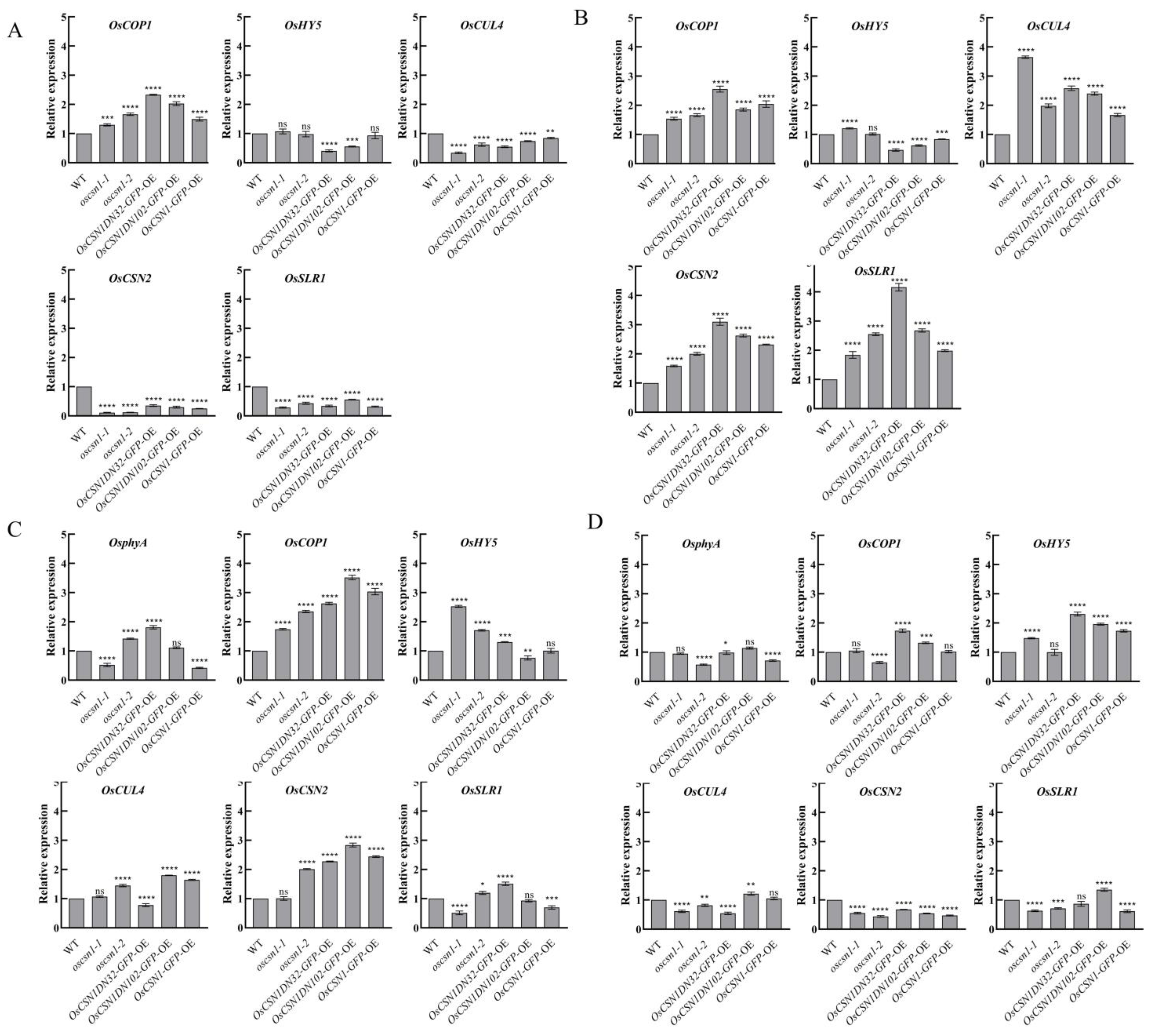
Under far-red light, the expression level of the OsSLR1 protein in oscsn1-1 was significantly upregulated. On the other hand, the expression level of the OsSLR1 protein in the oscsn1-2 and OsCSN1DN102-GFP-OE was significantly upregulated compared with that in the control group under natural light (Figure 2A,B). The expression level of the OsSLR1 gene was significantly upregulated in the oscsn1-2 (Figure 3A,B). After the addition of the exogenous hormone GA3, the OsSLR1 protein expression level in the wild type, oscsn1-2, and OsCSN1DN102-GFP-OE was significantly upregulated (Figure 2A,C). After the addition of the exogenous hormone PAC, the expression level of the OsSLR1 protein in the wild type, oscsn1-2, and OsCSN1DN102-GFP-OE was significantly downregulated (Figure 2A,D), while the expression level of the OsSLR1 gene in the oscsn1-2 was significantly downregulated (Figure 3A,D).
Under far-red light, the OsCSN2 expression levels of the OsCSN1DN32-GFP-OE and OsCSN1-GFP-OE were significantly upregulated (Figure 2A,B and Figure 3A,B). After the addition of the exogenous hormone GA3, the expression level of the OsCSN2 in the OsCSN1DN32-GFP-OE and OsCSN1-GFP-OE was significantly downregulated (Figure 2A,C). Similarly, the expression level of the OsCSN2 gene was significantly downregulated in the OsCSN1DN32-GFP-OE (Figure 3A,C). After the addition of the exogenous hormone PAC, the OsCSN2 expression level was upregulated in wild type, while the OsCSN2 expression levels in the mutants were downregulated. In addition, the OsCSN2 expression level was significantly downregulated in the OsCSN1DN32-GFP-OE and OsCSN1-GFP-OE (Figure 2A,D) and the expression level of the OsCSN2 gene was significantly downregulated in the mutants (Figure 3A,D).
Under far-red light, the OsCUL4 expression levels were upregulated in the mutants and the expression level in the oscsn1-1 plants was significant upregulated (Figure 2A,B and Figure 3A,B). After the addition of the exogenous hormone GA3, the OsCUL4 expression level in oscsn1-1 was significant upregulated (Figure 2A,C and Figure 3A,C). After the addition of the exogenous hormone PAC, the OsCUL4 protein expression level in oscsn1-1 and OsCSN1DN32-GFP-OE was significantly downregulated (Figure 2A,D and Figure 3A,D).
Under far-red light, neither the wild type’s nor the mutants’ OsphyA was expressed (Figure 2A,B). After the addition of the exogenous hormone GA3, the OsphyA and OsphyA gene expression levels in the wild type and mutants were upregulated compared to those in the far-red light, with the OsCSNDN32-GFP-OE showing a significant upregulation in the OsphyA protein and OsphyA gene expression levels (Figure 2A,C and Figure 3A,C). After the addition of the exogenous hormone PAC, the expression of the OsphyA protein and OsphyA gene in the wild type and mutants was upregulated compared with that under far-red light and the expression of OsphyA protein and OsphyA gene in OsCSN1DN102-GFP-OE was upregulated significantly (Figure 2A,D and Figure 3A,D).
Under far-red light, the expression level of the OsCOP1 gene was downregulated compared with that under natural light, while the expression level of the OsHY5 gene was upregulated (Figure 3A,B). The expression level of the OsCOP1 gene was significantly downregulated in the OsCSN1DN32-GFP-OE and OsCSN1DN102-GFP-OE (Figure 3A,B). After the addition of the exogenous hormones GA3 and PAC, the gene expression of OsCOP1 and OsHY5 was upregulated compared with that in the far-red light. The gene expression levels of OsCOP1 and OsHY5 in the OsCSN1DN32-GFP-OE were significantly upregulated (Figure 3A,C,D).

3. Discussion

3.1. OsCSN1 Localization Affects Rice Seedling Growth

Under far-red light, GFP expression was absent in the leaf tips and coleoptile veins of the OsCSN1DN32-GFP-OE and OsCSN1DN102-GFP-OE (Figure S1B). In contrast, the OsCSN1-GFP-OE exhibited extensive GFP expression in both leaves and coleoptiles, with limited expression in roots (Figure S1B). Overexpression of OsCSN1 inhibited both the aboveground and underground parts of the OsCSN1-GFP-OE (Figure 1A,B). In contrast, the aboveground height of the OsCSN1DN32-GFP-OE was significantly inhibited, the plant height of the OsCSN1DN102-GFP-OE was inhibited, and the coleoptiles were elongated under far-red light (Figure 1A,B). These observations suggest that deletions of 32 or 102 amino acids in the N-terminal region of OsCSN1 may induce conformational changes that impair far-red light signal reception, leading to distinct phenotypic differences.
Under the combined regulation of far-red light and exogenous GA3, OsCSN1-GFP-OE displayed abundant GFP expression in both leaves and roots (Figure S1C). In the OsCSN1DN32-GFP-OE, the addition of the exogenous hormone GA3 promoted the elongation of the aboveground and underground parts. However, in OsCSN1DN102-GFP-OE, exogenous GA3 reduced the overall plant height while promoting coleoptile elongation (Figure 1A,C). For OsCSN1-GFP-OE, GA3 application stimulated shoot growth but suppressed root growth. Furthermore, in OsCSN1-GFP-OE roots, GFP protein expression was significantly reduced, correlating with inhibited root elongation and protein expression.
Under the synergistic regulation of far-red light and exogenous hormone PAC, the OsCSN1DN32-GFP-OE and OsCSN1DN102-GFP-OE produced GFP proteins in the leaf tips, but a small amount of GFP was fused in the roots of the OsCSN1DN32-GFP-OE. Protein expression analysis revealed that there was a small amount of GFP protein in the coleoptiles of the OsCSN1DN102-GFP-OE (Figure S1D). The OsCSN1-GFP-OE expressed a large amount of the GFP protein in the leaves, coleoptiles, and roots (Figure S1D). The aboveground and underground parts of the OsCSN1DN32-GFP-OE, OsCSN1DN102-GFP-OE, and OsCSN1-GFP-OE all exhibited inhibition (Figure 1A,D). These results suggest that far-red light and GA signaling pathways jointly regulate seedling growth via OsCSN1, with far-red light signaling predominantly influencing plant height, while GA signaling has a stronger effect on coleoptile and root elongation. The phenotypic differences between the aerial and underground parts may result from differential signal reception, warranting further investigation into the exact regulatory mechanisms.

3.2. OsCSN1 Is Involved in the Regulation of Rice Plant Phenotype Through N-Terminal Under Far-Red Light and There Are Different Sensitivities and Synergistic Effects with Hormone Signals

The COP9 signalosome (CSN) is a highly conserved multi-subunit protein complex, with CSN1 being its largest and most conserved subunit. The N-terminal region of CSN1 plays a pivotal role in photomorphogenesis and seedling development, making it critical for plant survival [11]. Structural analysis of OsCSN1 revealed that the deletion of 32 amino acids from the N-terminal did not significantly affect its tertiary structure. However, the deletion of 102 amino acids from the N-terminal led to a substantial conformational change [32]. This study demonstrated that both OsCSN1DN32 and OsCSN1DN102 mutants exhibited normal growth during the seedling stage under far-red light but displayed varying degrees of phenotypic alterations. These findings highlight the N-terminal region of OsCSN1 as a critical domain for regulating rice seedling development under far-red light.
OsCSN1 is a positive regulator of seedling growth regulated by far-red light and its main domain is located at the N-terminal. The OsCSN1 N-terminal deletion mutants are sensitive to far-red light and the effect of far-red light is more prominent. After the addition of exogenous hormones and far-red light, OsCSN1 was found to have a deletion of 32 amino acids at the N-terminal. The far-red light signal and hormone signal responses showed significant sensitivity. The overexpression of OsCSN1 allowed far-red light and hormone signals to regulate plant height increases in rice seedlings through OsCSN1. Plants with reduced OsCSN1 expression exhibit coleoptile growth under far-red light and the coleoptile elongation in OsCSN1 mutants is more sensitive to hormonal signals (Figure 1).
Far-red light influenced the root length of the OsCSN1 N-terminal deletion mutants, with the root showing heightened sensitivity to far-red light signals. The addition of the hormones GA3 and PAC inhibited the root length of the mutants. Notably, GA3 did not mitigate the detrimental effects of far-red light on the root (Figure 1).

3.3. OsCSN1 Responds to Far-Red Light by Regulating the SLR1 Factor in the GA Pathway

Hormone signaling plays a crucial role in regulating seed germination and seedling growth. Under far-red light, OsSLR1 protein and gene expression was upregulated in both wild type and mutants (Figure 2A,B and Figure 3A,B). However, when far-red light was combined with exogenous GA3 or PAC, both OsSLR1 protein and gene expression were downregulated in the mutants (Figure 2A,C,D and Figure 3A,C,D). Under far-red light, the plant height and root length were inhibited in OsCSN1 mutants (Figure 1A,B). After the addition of the exogenous hormone GA3, the plant height of the OsCSN1DN102-GFP-OE was significantly inhibited (Figure 1A,C). Similarly, the addition of PAC resulted in inhibited plant height and root length in the mutants (Figure 1A,D).
OsCSN1 exhibited sensitivity to far-red light and GA signals in the aboveground and underground parts of the N-terminal deletion mutants. These findings suggest that far-red light may regulate OsSLR1 expression through OsCSN1, thereby influencing GA signaling pathways and modulating rice seedling growth. The primary functional domain of OsCSN1 resides in its N-terminal region.

3.4. OsCSN1 Senses Far-Red Light Signals Through phyA to Regulate Seedling Growth

Light plays a critical role in plant growth and development. Under far-red light, the expression levels of OsCOP1, OsHY5, OsCSN2, and OsCUL4 in the OsCSN1DN32-GFP-OE were upregulated (Figure 3A,B). However, when far-red light was combined with exogenous hormones, OsCSN2 and OsCUL4 gene expression was significantly downregulated, while OsHY5 gene expression was upregulated (Figure 3A,B). Additionally, when far-red light and the exogenous hormone GA3 were applied together, the expression of the OsCOP1 gene was significantly increased in both the OsCSN1DN32-GFP-OE and OsCSN1DN102-GFP-OE (Figure 3A,C).
PhyA, which shifts from its active Pfr form to the inactive Pr form under far-red light, was not expressed in the mutants under far-red light exposure (Figure 2A,B and Figure 3A,B). Under the synergistic regulation of far-red light and an exogenous hormone, the expression of the OsphyA and OsphyA gene was upregulated in the mutants. Under the synergistic regulation of far-red light and exogenous hormone, the growth of the underground part of the mutants was inhibited and the expression of OsphyA increased. The OsCSN1 deletion N-terminal 32 amino acid (OsCSN1DN32-GFP-OE) mutant also showed inhibition due to the increased expression of OsphyA. This finding is consistent with the finding that rice senses far-red light signals through phyA to inhibit coleoptile and root elongation [34,35]. Under far-red light, the phytochrome Pr and Pfr types switch and thus regulate the growth and development of rice plants through the metabolism of various hormones, affecting the growth of rice plants [36].
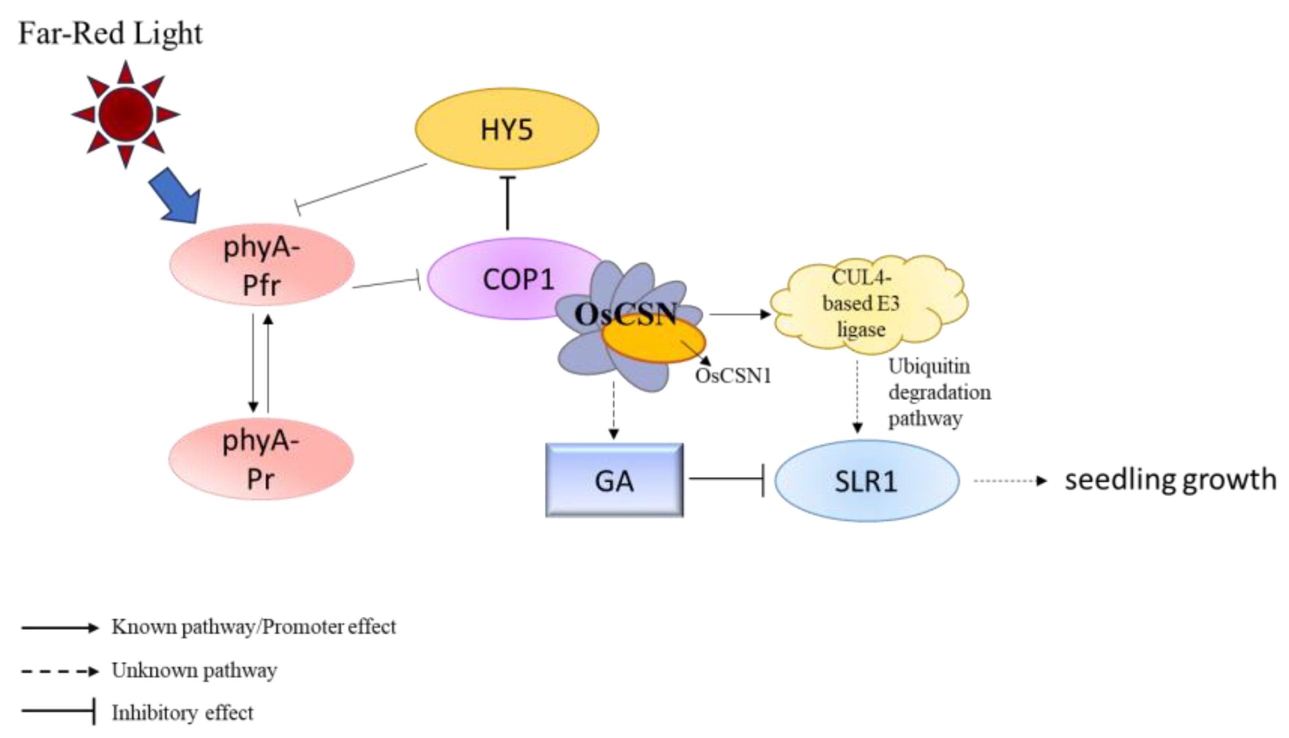
Previous studies have shown that after receiving a far-red light signal, phyA binds to FHY1/FHL, is transported into the nucleus, and is converted to the inactive Pr form [36,37]. phyA-Pfr inhibits the function of COP1, thereby promoting the accumulation of HY5. HY5 inhibits the transcriptional activation of FHY1/FHL via FHY3/FAR1 [25] and negatively regulates the entry of phyA into the nucleus. Moreover, phyA can also inhibit the transcription of FHY3/FAR1, thereby inhibiting FHY1/FHL in the entry and exit cores [38]. Therefore, phyA may sense the far-red light signal and convert it to the inactive Pr form to increase the biological activity of GA, thereby inhibiting the expression of SLR1.
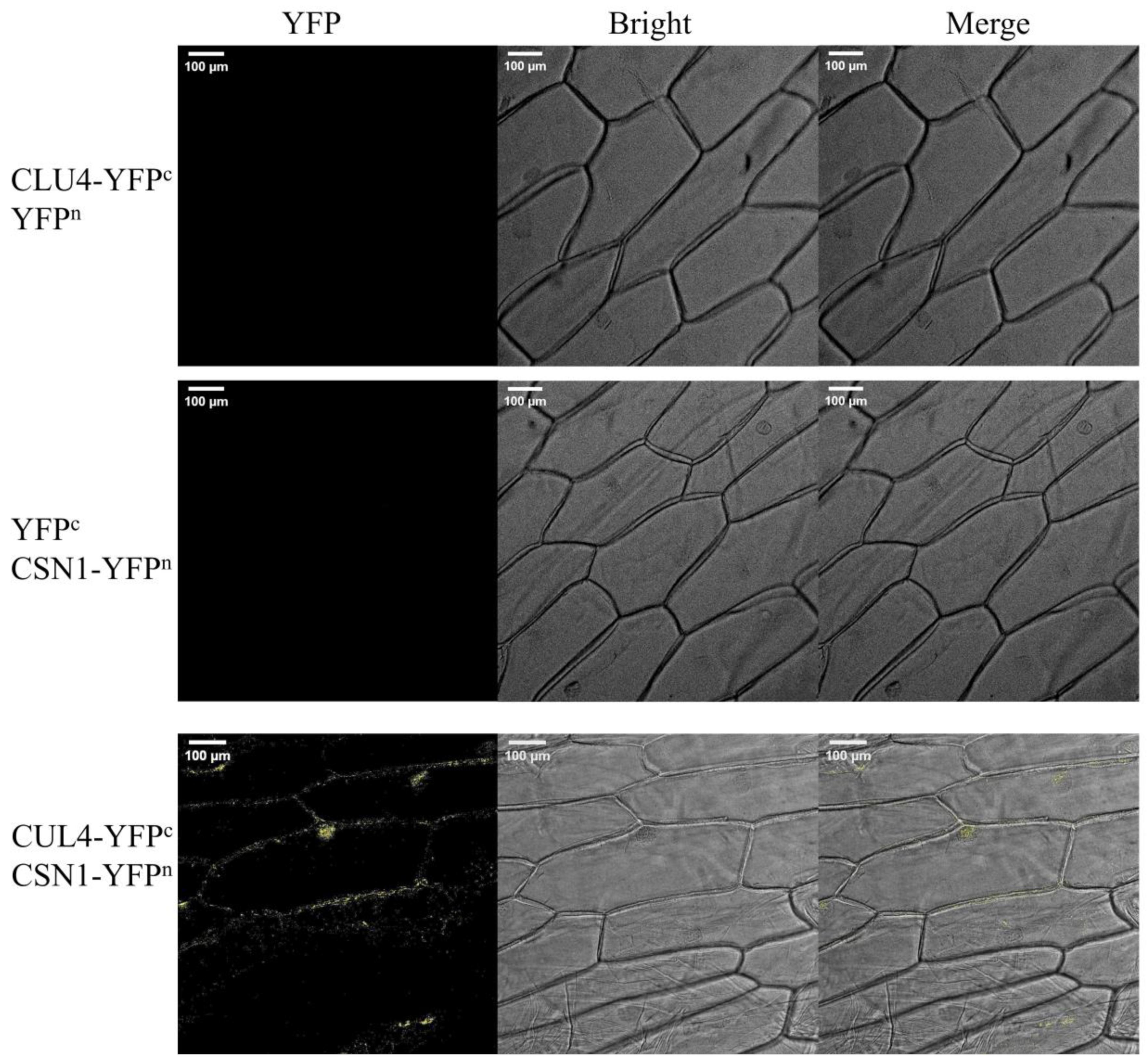
Far-red light also upregulated the expression of OsCSN2 and OsCUL4. It is proposed that OsCSN1, upon sensing far-red light, forms part of the COP9 complex, interacting with COP1 to regulate OsSLR1 degradation via OsCUL4-mediated ubiquitination in the CULLIN family. The results showed that the interaction between OsCUL4 and OsCSN1 was proved by a yeast two-hybrid experiment [32] and BiFC experiment (Figure 4). Far-red light signal activates phyA and converts it into the inactive Pr form. After sensing far-red light, OsCSN1 interacts with COP1 by forming a COP9 complex and then regulates the degradation of OsSLR1 through OsCUL4-mediated ubiquitination, while OsSLR1 is a negative regulator of GA signaling. This process affects the growth and development of rice seedlings, especially the elongation of seedling height, by regulating the GA signaling pathway (Figure 5).

3.5. Limitations and Potential of Far-Red Light in Regulating Rice Growth

Previous studies have shown that, while far-red light can promote growth, promote flowering and the chlorophyll fluorescence response, etc. [39], far-red light does have some limitations in regulating rice growth and development which may affect its effectiveness as an independent agricultural management tool. Plant growth requires a variety of light qualities, including blue light, red light, and far-red light. The application of a single type of far-red light may lead to spectral imbalance, thus affecting the overall health and productivity of plants. For example, excessive far-red light may lead to the overgrowth of plants, reduced mechanical strength, and reduced photosynthetic efficiency of leaves. Under natural conditions, the light conditions are complex and changeable and a with a single far-red light treatment it is difficult to achieve the desired effect stably. Environmental factors such as light intensity, photoperiod, and temperature may affect the interaction between far-red light and hormone pathways. Although far-red light has its unique advantages in the regulation of rice growth and development, it has many limitations as the only means of regulation. In order to overcome these problems, it is usually recommended to adopt a comprehensive light management strategy, combined with a variety of light quality and other agronomic measures, to ensure that rice can obtain balanced and suitable light conditions throughout the growth cycle, taking into account economic benefits and ecological adaptability. Although far-red light has some limitations in regulating the growth and development of rice, it also has unique application potential which can bring significant benefits to agricultural production. LED lights or other artificial light sources have been used to supplement or adjust the proportion of far-red light to optimize the growth environment of rice seedlings. By controlling the daily light duration and intensity, the diurnal variation found under natural conditions is simulated, especially the low-angle sunlight in the morning and evening, which is rich in far-red light. This helps to enhance the shade avoidance response of plants. Based on the research results of key regulatory factors such as OsCSN1, new varieties that can respond more effectively to far-red light signals were developed through gene editing, providing innovative solutions for modern agricultural practice.
In general, OsCSN1 plays a key role in regulating plant growth and development in response to far-red light. Future studies should continue to explore the specific mechanisms of far-red light on the regulation of other subunits of the COP9 signaling complex in rice and how to improve the productivity and adaptability of rice through these mechanisms. This is not only of great significance to basic research, but also brings potential benefits to the practice of agricultural production.

4. Materials and Methods

4.1. Plant Material and Culture Conditions

The construction method and source of the mutants were provided by our laboratory [32,33,40]. The seeds of the wild type (Oryza sativa subsp. japonica), OsCSN1 knockout (oscsn1-1) mutant, OsCSN1-reduced (oscsn1-2) mutant, OsCSN1 overexpression (OsCSN1-GFP-OE) mutant, and OsCSN1 deletion N-terminal (OsCSN1DN32-GFP-OE and OsCSN1DN102-GFP-OE) mutant plants were divided into four groups. Then, 0.8% (w/v) agar medium was prepared, 10 mM GA3 and 100 mM endogenous gibberellin inhibitor paclobutrazol (PAC) were added to the medium, respectively, and the 5 seeds of the wild type and mutants were sown on the medium. The seeds in the experimental group underwent light irradiation with LED-FR (10 μmol m−2s−1) (Shenzhen Lvheng Lighting Technology Co., Ltd., Shenzhen, China) for 24 h in a light incubator. Another group of seeds, as the control group, was sown on 0.8% (w/v) agar medium and positioned in a light incubator (Ningbo Jiangnan Instrument Factory, Ningbo, China) with a light intensity of 203 μmol m−2s−1 in a 12 L:12 D light cycle. The temperature during incubation was maintained at 28 ± 1 °C and the relative humidity during incubation was maintained at 95%.

4.2. Phenotypic Analysis of Rice Seedlings

Seeds of the wild type and mutants were cultured under the above conditions for 9 days. Phenotypic data were measured, including plant height, coleoptile length, and root length. All measurements for each group were repeated 5 times (n = 5).

4.3. Protein Extraction and Western Blot Analysis

A total of 0.5 g sample was ground in liquid nitrogen, mixed with Extraction Buffer (PI, PMSF, and DTT were added to the plant RIPA lysate in proportion), And vortexed and centrifuged at 4 °C for 10 min. The supernatant was taken and 5 × SDS-PAGE sample buffer was added and mixed. After boiling for 10 min, western blot testing was performed.
For western blotting, the modified Mahmood method was used [41]. Each sample was separated via 10% SDS-PAGE. The SDS-PAGE gels were subsequently transferred to polyvinylidene fluoride (PVDF) membranes using the Bio-Rad Trans-Blot Turbo blotting system. The proteins on the PVDF membranes were blocked with 4% skim milk. After incubation with the appropriate antibody, the target protein bands were visualized via chemiluminescence. The rabbit polyclonal antibodies used in this experiment included OsCUL4, OsCSN2, OsCSN5, OsSLR1, and OsphyA and an anti-plant actin mouse polyclonal antibody. The following secondary antibodies were used: goat anti-rabbit and goat anti-mouse IgG secondary antibody (HRP). The antibodies used in this study were purchased from Wuhan ABclonal Biotechnology Co., Ltd. (Wuhan, China).

4.4. Total RNA Extraction and Real-Time Quantitative Polymerase Chain Reaction

Total RNA was extracted from the seedlings using a Spectrum Plant Total RNA Kit (Sigma-Aldrich, Darmstadt, Germany). About 0.5 mg of cDNA was synthesized using StarScript II first-strand cDNA synthesis mix (GenStar, Beijing, China). Rapid quantitative polymerase chain reaction (PCR) was completed using a mixture of 2x RealStar Green Fast and ROX (GenStar, Beijing, China) on a StepOnePlusTM real-time PCR device (Thermo Fisher Scientific, Applied Biosystems, Waltham, MA, USA). The blank control gene GAPDH (JN848809) was used to quantify the relative mRNA levels calculated by averaging three replicates. The primers used in the experiment are shown in Table 1.

4.5. Spatiotemporal Positioning

Three proteins, OsCSN1DN32, OsCSN1DN102, and OsCSN1, were constructed as fusion proteins with green fluorescent protein (GFP) and were effectively expressed; subsequently, the proteins were observed and analyzed via laser scanning confocal microscopy. The GFP was observed in the leaves, coleoptiles, and roots of OsCSN1-overexpressing plants that grew for 9 days. Images were obtained using a Leica TCS SP8 CARS confocal microscope (Leica, Wetzlar, Germany) with a 10 × 0.40 eyepiece and 20× and observed with an objective lens of 0.70. The selected positioning point was excited with a 488 nm laser and we detected GFP fluorescence with a 500–530 nm band.

4.6. Bimolecular Fluorescence Complementation

PCR amplification was performed by using designed primers and the target gene as templates. The primers used in the experiment are shown in Table 2. The vectors pSAT4A-cEYFP-N1 and pSAT4A-nEYFP-N1 were linearized by EcoRI (Thermo Fisher Scientific, Waltham, MA, USA). OsCUL4-pSAT4A-cEYFP-N1 and OsCSN1-pSAT4A-nEYFP-N1 were constructed using a ClonExpress II One Step Cloning Kit-C112 (Vazyme, Nanjing, China). They were transferred into onion epidermal cells by an Agrobacterium-mediated method and cultured in the dark for 24 h for observation. Images were obtained using a Leica TCS SP8 CARS confocal microscope (Leica, Wetzlar, Germany) with a 10× eyepiece and 0.70 objective lens for observation. YFP fluorescence with an excitation wavelength of 514 nm and emission wavelength of 527 nm was detected.

4.7. Data Processing and Analysis

The data are expressed as the standard error of the mean for the control group and the experimental group. p ≤ 0.05 indicates statistical significance. ImageJ (1.52V) software was used to analyze the results of western blot. The experimental data were analyzed using Excel 2019 and SPSS 28.0 and the graphs were drawn using GraphPad Prism 8.

5. Conclusions

This study shows that OsCSN1 plays a crucial role in regulating the growth of rice seedlings under far-red light. The N-terminal region of OsCSN1 is essential for its function and this domain is an important site in the response to far-red light and hormone signals. When a far-red light signal is perceived, phyA is converted to its inactive Pr form, which promotes COP1 and inhibits HY5 accumulation. After sensing the far-red light signal, OsCSN1 forms a COP9 signaling complex and interacts with COP1, then regulates the degradation of the key regulator OsSLR1 in the GA signaling pathway through OsCUL4-mediated ubiquitination. This process affects aboveground and underground growth. Far-red light mainly regulates stem elongation and GA signals affect coleoptile elongation more strongly. In addition, the interaction between OsCSN1 and OsCUL4 in the far-red light signaling pathway highlights its central role in coordinating light and hormone signals to regulate rice growth. These findings highlight the importance of the N-terminus of OsCSN1 in mediating complex interactions between light and hormone regulation during plant development. Based on this mechanism, new rice varieties that can use far-red light signals more efficiently can be cultivated in the future, thereby improving the growth rate, adaptability, and productivity of rice. By optimizing the sensing mechanism of crop light signals, this approach can not only improve the yield of crops but also provide a more sustainable development plan for agricultural production.

Supplementary Materials

The following supporting information can be downloaded at: https://www.mdpi.com/article/10.3390/ijms26020522/s1.

Author Contributions

Data curation, Y.L., H.Z. (Hua Zeng), Y.S., H.Z. (Hexin Zhang), T.J., L.Y. and J.Y.; formal analysis, Y.L., H.Z. (Hua Zeng), Y.S., H.Z. (Hexin Zhang), T.J. and M.X.; funding acquisition, J.L., M.W. and L.G.; investigation, Y.L., H.Z. (Hua Zeng), Y.S., H.Z. (Hexin Zhang), T.J. and J.Y.; methodology, Y.L., M.W. and L.G.; resources, M.W. and L.G.; supervision, M.W. and L.G.; writing—original draft, Y.L., H.Z. (Hua Zeng), Y.S.; writing—review and editing, M.W. and L.G. All authors have read and agreed to the published version of the manuscript.

Funding

This research was funded by the Department of Jilin Province Science and Technology [grant numbers 20230402020GH, 20230203162SF, 20220402060GH, 20220203054SF].

Institutional Review Board Statement

Not applicable.

Informed Consent Statement

Not applicable.

Data Availability Statement

The data presented in this study are available in the article.

Acknowledgments

We thank the Department of Jilin Province Science and Technology. In addition, we thank editors and reviewers for critical and helpful comments for the manuscript.

Conflicts of Interest

The authors declare no conflicts of interest.

References

  1. Wei, N.; Chamovitz, D.A.; Deng, X.W. Arabidopsis COP9 is a component of a novel signaling complex mediating light control of development. Cell 1994, 78, 117–124. [Google Scholar] [CrossRef] [PubMed]
  2. Wei, N.; Deng, X. COP9: A new genetic locus involved in light-regulated development and gene expression in arabidopsis. Plant Cell 1992, 4, 1507–1518. [Google Scholar] [CrossRef] [PubMed]
  3. Bech-Otschir, D.; Seeger, M.; Dubiel, W. The COP9 signalosome: At the interface between signal transduction and ubiquitin-dependent proteolysis. J. Cell Sci. 2002, 115 Pt 3, 467–473. [Google Scholar] [CrossRef] [PubMed]
  4. Schwechheimer, C.; Deng, X. COP9 signalosome revisited: A novel mediator of protein degradation. Trends Cell Biol. 2001, 11, 420–426. [Google Scholar] [CrossRef] [PubMed]
  5. Serino, G.; Deng, X. The COP9 signalosome: Regulating plant development through the control of proteolysis. Annu. Rev. Plant Biol. 2003, 54, 165–182. [Google Scholar] [CrossRef]
  6. Sharon, M.; Mao, H.; Boeri Erba, E.; Stephens, E.; Zheng, N.; Robinson, C. Symmetrical modularity of the COP9 signalosome complex suggests its multifunctionality. Structure 2009, 17, 31–40. [Google Scholar] [CrossRef] [PubMed]
  7. Kotiguda, G.; Weinberg, D.; Dessau, M.; Salvi, C.; Serino, G.; Chamovitz, D.; Hirsch, J. The organization of a CSN5-containing subcomplex of the COP9 signalosome. J. Biol. Chem. 2012, 287, 42031–42041. [Google Scholar] [CrossRef] [PubMed]
  8. Hofmann, K.; Bucher, P. The PCI domain: A common theme in three multiprotein complexes. Trends Biochem. Sci. 1998, 23, 204–205. [Google Scholar] [CrossRef] [PubMed]
  9. Chen, H.; Huang, X.; Gusmaroli, G.; Terzaghi, W.; Lau, O.S.; Yanagawa, Y.; Zhang, Y.; Li, J.; Lee, J.H.; Zhu, D.; et al. Arabidopsis CULLIN4-damaged DNA binding protein 1 interacts with CONSTITUTIVELY PHOTOMORPHOGENIC1-SUPPRESSOR OF PHYA complexes to regulate photomorphogenesis and flowering time. Plant Cell 2010, 22, 108–123. [Google Scholar] [CrossRef]
  10. Tsuge, T.; Matsui, M.; Wei, N. The subunit 1 of the COP9 signalosome suppresses gene expression through its N-terminal domain and incorporates into the complex through the PCI domain. J. Mol. Biol. 2001, 305, 1–9. [Google Scholar] [CrossRef]
  11. Wang, X.; Kang, D.; Feng, S.; Serino, G.; Schwechheimer, C.; Wei, N. CSN1 N-terminal-dependent activity is required for Arabidopsis development but not for Rub1/Nedd8 deconjugation of cullins: A structure-function study of CSN1 subunit of COP9 signalosome. Mol. Biol. Cell 2002, 13, 646–655. [Google Scholar] [CrossRef] [PubMed]
  12. Jin, D.; Wu, M.; Li, B.; Bücker, B.; Keil, P.; Zhang, S.; Li, J.; Kang, D.; Liu, J.; Dong, J.; et al. The COP9 Signalosome regulates seed germination by facilitating protein degradation of RGL2 and ABI5. PLoS Genet. 2018, 14, e1007237. [Google Scholar] [CrossRef] [PubMed]
  13. Wei, N.; Deng, X. The role of the COP/DET/FUS genes in light control of arabidopsis seedling development. Plant Physiol. 1996, 112, 871–878. [Google Scholar] [CrossRef]
  14. Osterlund, M.; Hardtke, C.; Wei, N.; Deng, X. Targeted destabilization of HY5 during light-regulated development of Arabidopsis. Nature 2000, 405, 462–466. [Google Scholar] [CrossRef] [PubMed]
  15. Lee, K.; Lopez-Molina, L. Control of seed germination in the shade. Cell Cycle 2012, 11, 4489–4490. [Google Scholar] [CrossRef]
  16. Bae, G.; Choi, G. Decoding of light signals by plant phytochromes and their interacting proteins. Annu. Rev. Plant Biol. 2008, 59, 281–311. [Google Scholar] [CrossRef] [PubMed]
  17. Takano, M.; Inagaki, N.; Xie, X.; Yuzurihara, N.; Hihara, F.; Ishizuka, T.; Yano, M.; Nishimura, M.; Miyao, A.; Hirochika, H.; et al. Distinct and cooperative functions of phytochromes A, B, and C in the control of deetiolation and flowering in rice. Plant Cell 2005, 17, 3311–3325. [Google Scholar] [CrossRef] [PubMed]
  18. Kneissl, J.; Shinomura, T.; Furuya, M.; Bolle, C. A rice phytochrome A in Arabidopsis: The Role of the N-terminus under red and far-red light. Mol. Plant 2008, 1, 84–102. [Google Scholar] [CrossRef] [PubMed]
  19. Kircher, S.; Kozma-Bognar, L.; Kim, L.; Adam, E.; Harter, K.; Schafer, E.; Nagy, F. Light quality-dependent nuclear import of the plant photoreceptors phytochrome A and B. Plant Cell 1999, 11, 1445–1456. [Google Scholar] [CrossRef] [PubMed]
  20. Sharrock, R.; Clack, T. Heterodimerization of type II phytochromes in Arabidopsis. Proc. Natl. Acad. Sci. USA 2004, 101, 11500–11505. [Google Scholar] [CrossRef] [PubMed]
  21. Seo, H.; Watanabe, E.; Tokutomi, S.; Nagatani, A.; Chua, N. Photoreceptor ubiquitination by COP1 E3 ligase desensitizes phytochrome A signaling. Genes Dev. 2004, 18, 617–622. [Google Scholar] [CrossRef] [PubMed]
  22. Clough, R.; Jordan-Beebe, E.; Lohman, K.; Marita, J.; Walker, J.; Gatz, C.; Vierstra, R. Sequences within both the N- and C-terminal domains of phytochrome A are required for PFR ubiquitination and degradation. Plant J. 1999, 17, 155–167. [Google Scholar] [CrossRef]
  23. Hoecker, U.; Xu, Y.; Quail, P. SPA1: A new genetic locus involved in phytochrome A-specific signal transduction. Plant Cell 1998, 10, 19–33. [Google Scholar] [CrossRef] [PubMed]
  24. Deng, X.; Matsui, M.; Wei, N.; Wagner, D.; Chu, A.; Feldmann, K.; Quail, P. COP1, an Arabidopsis regulatory gene, encodes a protein with both a zinc-binding motif and a G beta homologous domain. Cell 1992, 71, 791–801. [Google Scholar] [CrossRef] [PubMed]
  25. Sheerin, D.; Menon, C.; zur Oven-Krockhaus, S.; Enderle, B.; Zhu, L.; Johnen, P.; Schleifenbaum, F.; Stierhof, Y.; Huq, E.; Hiltbrunner, A. Light-activated phytochrome A and B interact with members of the SPA family to promote photomorphogenesis in Arabidopsis by reorganizing the COP1/SPA complex. Plant Cell 2015, 27, 189–201. [Google Scholar] [CrossRef]
  26. Yamaguchi, S. Gibberellin metabolism and its regulation. Annu. Rev. Plant Biol. 2008, 59, 225–251. [Google Scholar] [CrossRef] [PubMed]
  27. Bao, S.; Hua, C.; Shen, L.; Yu, H. New insights into gibberellin signaling in regulating flowering in Arabidopsis. J. Integr. Plant Biol. 2020, 62, 118–131. [Google Scholar] [CrossRef]
  28. Chen, S.; Wang, X.; Tan, G.; Zhou, W.; Wang, G. Gibberellin and the plant growth retardant Paclobutrazol altered fruit shape and ripening in tomato. Protoplasma 2020, 257, 853–861. [Google Scholar] [CrossRef] [PubMed]
  29. Dill, A.; Thomas, S.; Hu, J.; Steber, C.; Sun, T. The Arabidopsis F-box protein SLEEPY1 targets gibberellin signaling repressors for gibberellin-induced degradation. Plant Cell 2004, 16, 1392–1405. [Google Scholar] [CrossRef]
  30. Peng, J.; Harberd, N. The role of GA-mediated signalling in the control of seed germination. Curr. Opin. Plant Biol. 2002, 5, 376–381. [Google Scholar] [CrossRef]
  31. Dai, C.; Xue, H. Rice early flowering1, a CKI, phosphorylates DELLA protein SLR1 to negatively regulate gibberellin signalling. EMBO J. 2010, 29, 1916–1927. [Google Scholar] [CrossRef]
  32. Han, S.; Liu, Y.; Bao, A.; Zeng, H.; Huang, G.; Geng, M.; Zhang, C.; Zhang, Q.; Lu, J.; Wu, M.; et al. OsCSN1 regulates the growth of rice seedlings through the GA signaling pathway in blue light. J. Plant Physiol. 2023, 280, 153904. [Google Scholar] [CrossRef] [PubMed]
  33. Han, S.; Liu, Y.; Bao, A.; Jiao, T.; Zeng, H.; Yue, W.; Yin, L.; Xu, M.; Lu, J.; Wu, M.; et al. A Characterization of the Functions of OsCSN1 in the Control of Sheath Elongation and Height in Rice Plants under Red Light. Agronomy 2024, 14, 572. [Google Scholar] [CrossRef]
  34. Wang, J.; Chen, C.; Chien, C.; Hsieh, H. FAR-RED INSENSITIVE219 modulates CONSTITUTIVE PHOTOMORPHOGENIC1 activity via physical interaction to regulate hypocotyl elongation in Arabidopsis. Plant Physiol. 2011, 156, 631–646. [Google Scholar] [CrossRef]
  35. Takano, M.; Inagaki, N.; Xie, X.; Kiyota, S.; Baba-Kasai, A.; Tanabata, T.; Shinomura, T. Phytochromes are the sole photoreceptors for perceiving red/far-red light in rice. Proc. Natl. Acad. Sci. USA 2009, 106, 14705–14710. [Google Scholar] [CrossRef] [PubMed]
  36. Shimizu, H.; Tanabata, T.; Xie, X.; Inagaki, N.; Takano, M.; Shinomura, T.; Yamamoto, K.T. Phytochrome-mediated growth inhibition of seminal roots in rice seedlings. Physiol. Plant. 2009, 137, 289–297. [Google Scholar] [CrossRef] [PubMed]
  37. Andel, F.; Lagarias, J.; Mathies, R. Resonance raman analysis of chromophore structure in the lumi-R photoproduct of phytochrome. Biochemistry 1996, 35, 15997–16008. [Google Scholar] [CrossRef]
  38. Hiltbrunner, A.; Tscheuschler, A.; Viczián, A.; Kunkel, T.; Kircher, S.; Schäfer, E. FHY1 and FHL act together to mediate nuclear accumulation of the phytochrome A photoreceptor. Plant Cell Physiol. 2006, 47, 1023–1034. [Google Scholar] [CrossRef] [PubMed]
  39. Jang, I.; Lee, J.; Shin., E.; Nam, S. Evaluation of Growth, Flowering, and Chlorophyll Fluorescence Responses of Viola cornuta cv. Penny Red Wing according to Spectral Power Distributions. J. People Plants Environ. 2023, 26, 335–349. [Google Scholar] [CrossRef]
  40. Musazade, E.; Liu, Y.; Ren, Y.; Wu, M.; Zeng, H.; Han, S.; Gao, X.; Chen, S.; Guo, L. OsCSN1 Regulates the Growth and Development of Rice Seedlings through the Degradation of SLR1 in the GA Signaling Pathway. Agronomy 2022, 12, 2946. [Google Scholar] [CrossRef]
  41. Mahmood, T.; Yang, P.C. Western blot: Technique, theory, and trouble shooting. N. Am. J. Med. Sci. 2012, 4, 429–434. [Google Scholar] [CrossRef] [PubMed]
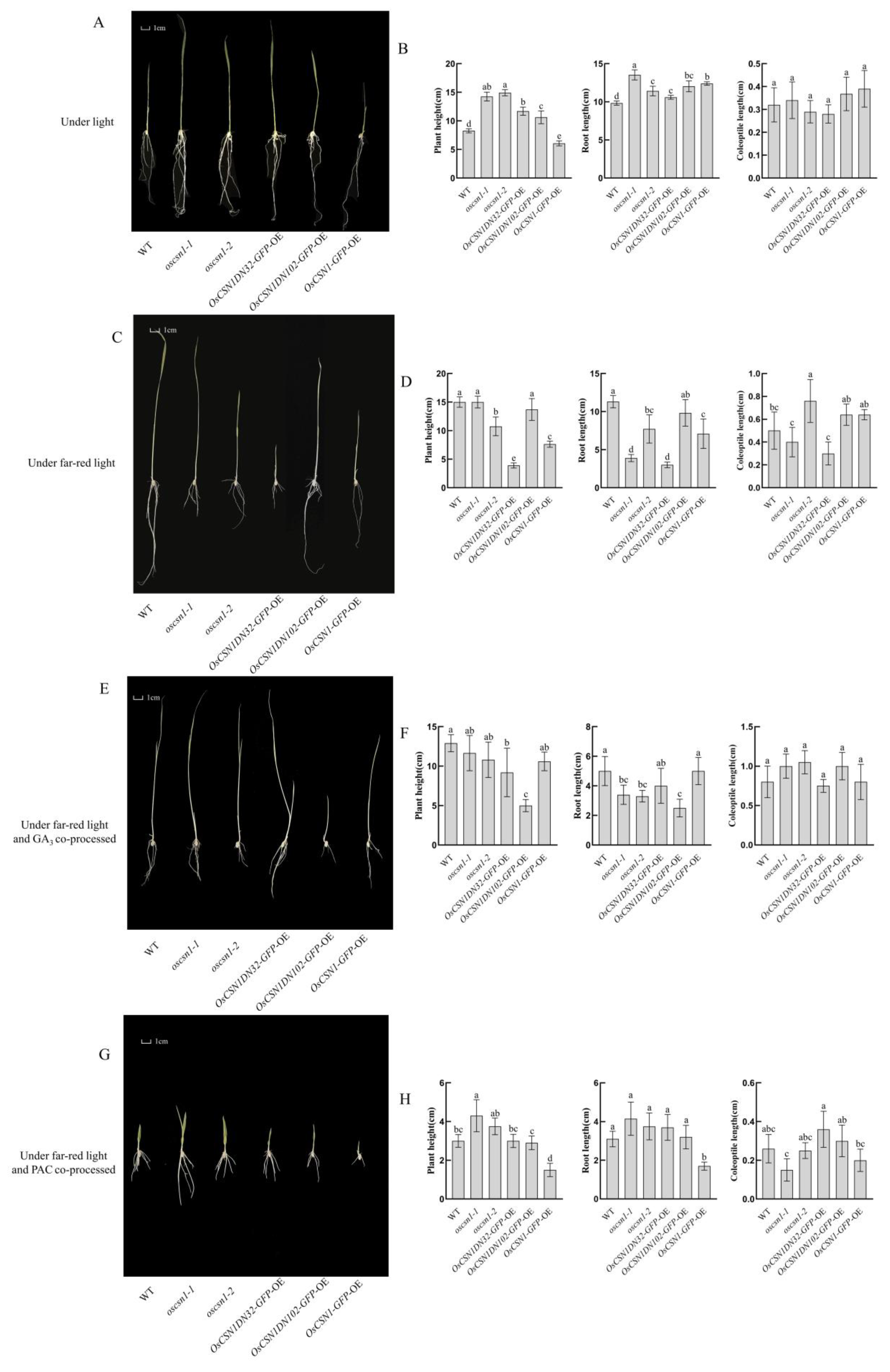
Figure 1. Phenotypic changes of wild type and OsCSN1 mutants under natural light, far-red light, and exogenous GA3 and PAC hormone treatments. Data are mean ± SD (n = 5). Different letters represent significant differences between wild type and mutants. One-way analysis of variance (ANOVA) test followed by Duncan’s post hoc test (p < 0.05). (A) Phenotype of WT and OsCSN1 mutants grown 9d under natural light. (B) Plant height, root length, and coleoptile length data graph of WT and OsCSN1 mutants grown for 9d under natural light. (C) Phenotype of WT and OsCSN1 mutants grown for 9d under far-red light. (D) Plant height, root length, and coleoptile length data graph of WT and OsCSN1 mutants grown for 9d under far-red light. (E) Phenotype of WT and OsCSN1 mutants grown for 9d under far-red light and GA3 co-processed. (F) Plant height, root length, and coleoptile length data graph of WT and OsCSN1 mutants grown for 9d under far-red light and GA3 co-processed. (G) Phenotype of WT and OsCSN1 mutants grown for 9d under far-red and PAC co-processed. (H) Plant height, root length, and coleoptile length data graph of WT and OsCSN1 mutants grown for 9d under far-red and PAC co-processed.
Ijms 26 00522 g001
Figure 2. OsCSN1 regulates seedling growth under far-red light by regulating the GA pathway. Protein expression of wild type and OsCSN1 mutants grown 9d under different conditions. Detection of OsSLR1, OsCSN2, OsCUL4, and OsphyA protein levels in samples lines using a western blot. (A) Under natural light. (B) Under far-red light. (C) Under far-red and GA3 co-processed. (D) Under far-red and PAC co-processed.
Ijms 26 00522 g002
Figure 3. Changes and interactions of OsCSN1-related genes during seedling growth under far-red light. The average values (±SD) from three biological repeats are shown. For each gene, different letters indicate significant differences in expression according to Duncan’s multiple range test, * p < 0.05; ** p < 0.01; *** p < 0.001; **** p < 0.0001; ns stands for no significant difference. Expression of genes associated with wild type and OsCSN1 mutants grown for 9d under different conditions of treatment at the mRNA level. Expression of OsSLR1, OsCSN2, OsCUL4, OsCOP1, OsHY5, and OsphyA genes in the sample lines was examined using qPCR. (A) Under natural light. (B) Under far-red light. (C) Under far-red light and GA3 co-processed. (D) Under far-red light and PAC co-processed.
Ijms 26 00522 g003
Figure 4. The interaction between OsCSN1 and OsCUL4 was detected by bimolecular fluorescence complementation. “CUL4-YFPc + YFPn” and “YFPc + CSN1-YFPn” were single-molecule control groups and no fluorescence was detected. “CUL4-YFPc + CSN1-YFPn” was the experimental group. Under the conditions of the normal negative and positive control, fluorescence could be detected in the experimental group, indicating that the two proteins could interact with each other. Bar = 100 μm.
Ijms 26 00522 g004
Figure 5. Presumed far-red light mediates the signaling pathway of OsCSN1 in regulating seedlings.
Ijms 26 00522 g005
Table 1. All the primers that were used for qRT-PCR.
PrimerSequence (5′-3′)
GAPDHFAAGCCAGCATCCTATGATCAGATT
GAPDHRCGTAACCCAGAATACCCTTGAGTTT
Dye-SLR1FCATGCTTTCCGAGCTCAACG
Dye-SLR1RTGACAGTGGACGAGGTGGAA
Dye-CUL4FAGGACAGACAGTATCAGGTGGATGC
Dye-CUL4RTCCGATGGCTTGATTGGGAACTTG
Dye-COP1FCATCTCAGCCACAAGAGCGACTG
Dye-COP1RGGTCTATCGGTGATGCTGTCTTCG
Dye-CSN2FGAGCAGCTCTTGGTCTCACTCATTC
Dye-CSN2RCGACCTGTCACCACGTTCTAGTAAC
Dye-phyAFGATGGTGCTCTGAGTGGAATGC
Dye-phyARACAGGAGGCGTTGGTGCTATC
Dye-HY5FAGGTGAAGGTGAAGGACTTGGAG
Dye-HY5RGAGCATCTGGTTCTCATTCTGTAGG
Table 2. All the primers that were used to BiFC.
PrimerSequence (5′-3′)
CSN1-c/nEYFP-FGATCTCGAGCTCAAGCTTCGAATTCATGGACGTCGAGGGCGAGGTCCCGG
CSN1-nEYFP-RTCGCCCTTGCTCACCATCAGGATCCCATCTTCCTTTGTCCAGCTCTTTGG
CUL4-c/nEYFP-FGATCTCGAGCTCAAGCTTCGAATTCATGCACAAAAACTAAGCTTC
CUL4-cEYFP-RGCGAGCTGCACGCTGCCCAGGATCCAGCCAGGTAATTGTAGATCT
Disclaimer/Publisher’s Note: The statements, opinions and data contained in all publications are solely those of the individual author(s) and contributor(s) and not of MDPI and/or the editor(s). MDPI and/or the editor(s) disclaim responsibility for any injury to people or property resulting from any ideas, methods, instructions or products referred to in the content.