Article

Zongertinib, a Novel HER2 Tyrosine Kinase Inhibitor, Maintains an Anticancer Activity for Trastuzumab Deruxtecan-Resistant Cancers Harboring HER2-Overexpression

1
Department of Medical Oncology, Kindai University Faculty of Medicine, Sakai 590-0197, Japan
2
Division of Cell Biology for Regenerative Medicine, Institute of Advanced Clinical Medicine, Kindai University Faculty of Medicine, Sakai 590-0197, Japan
3
Department of Genome Biology, Kindai University Faculty of Medicine, Sakai 590-0197, Japan
4
Center for Genomics, Life Science Research Institute, Kindai University, Sakai 590-0197, Japan
*
Author to whom correspondence should be addressed.
These authors contributed equally to this work.
Int. J. Mol. Sci. 2025, 26(21), 10515; https://doi.org/10.3390/ijms262110515
Submission received: 29 September 2025 / Revised: 25 October 2025 / Accepted: 27 October 2025 / Published: 29 October 2025
(This article belongs to the Special Issue Ongoing Anticancer Agents)

Abstract

Trastuzumab deruxtecan (T-DXd), an antibody–drug conjugate comprising trastuzumab linked to a payload DXd, has been extensively used to treat various cancers harboring HER2 overexpression. However, resistance development has been a major challenge to T-DXd treatment. To explore treatment strategies for T-DXd-resistant cancers, we examined two T-DXd-resistant cells named DSR32 and DSR4, which were obtained from HER2 gene-amplified H2170 lung cancer and N87 gastric cancer cells, respectively. The uptake of T-DXd and its transport to lysosomes in DSR32 cells were reduced. Subsequently, HER2 gene copy number (from 54 to 12) was reduced, which decreased HER2 expression on the cell surface. Thus, T-DXd-resistance might be observed due to the reduced T-DXd uptake caused by decreased HER2 expression in DSR32 cells. In DSR4 cells, no change was observed in HER2 expression and in the uptake and transport of T-DXd. The reduced linker cleavage activity may be associated with T-DXd resistance in DSR4 cells. Meanwhile, both DSR32 and DSR4 cells maintained HER2 activation; thus, zongertinib, a HER2-selective tyrosine kinase inhibitor, blocked the HER2 pathway, induced apoptosis, and inhibited colony formation. Overall, zongertinib can provide therapeutic relief to patients with HER2-overexpressing cancer who have developed resistance to T-DXd.

1. Introduction

HER2 is a member of the epidermal growth factor receptor (EGFR) family along with HER1, HER3, and HER4. Dimerization of HER2 with other members of the EGFR family, typically owing to the overexpression of HER2, results in the autophosphorylation or transphosphorylation of tyrosine residues within the cytoplasmic domain of the heterodimer, initiating cell signaling cascades in multiple pathways, such as the mitogen-activated protein kinase and phosphatidylinositol-3-kinase pathways. The activation of these cascades results in cell proliferation, survival, and drug resistance in cancer cells [1,2]. Abnormalities in the HER2 gene, such as mutations or amplification, act as oncogenic drivers, and a number of HER2-targeted therapies have already been implemented in clinical practice [2,3].
Trastuzumab deruxtecan (T-DXd) is a HER2-targeted antibody–drug conjugate (ADC) composed of an antibody, a cleavable linker, and a topoisomerase I inhibitor payload, DXd [4]. The advantage of this unique structure is widening the therapeutic window of payloads by selectively delivering the ADC to target tumor cells [5]. To date, T-DXd has become the standard therapy for several kinds of malignancies [3,6,7,8,9,10,11]. In HER2-overexpressing breast cancer, T-DXd exhibited an improvement in progression-free survival compared to trastuzumab emtansine (T-DM1), another trastuzumab-based ADC harboring a non-cleavable linker and a maytansine derivative as its payload, in the second-line setting [7,8]. T-DXd also achieved an improvement in survival outcomes of HER2-low breast cancer compared with conventional therapies after progression on one or two prior lines of chemotherapy [9]. Regarding HER2-overexpressing gastric cancer, T-DXd significantly improved response and overall survival in comparison with standard chemotherapies among patients who had progressed during at least two prior therapies, including trastuzumab [10]. In non-small-cell lung cancer, a single-arm, double-cohort phase II trial showed durable anticancer activity of T-DXd in patients with HER2 mutations who had relapsed following standard therapies, leading to the US Food and Drug Administration approval [11]. The promising results of HER2-overexpressing cohort support further investigation of T-DXd in this setting [12].
However, most advanced-stage malignancies treated with T-DXd remain incurable because of the development of resistance or a lack of initial response. Thus, exploring the mechanisms of resistance to T-DXd and developing strategies to overcome them are urgently needed. Recently, zongertinib, a novel HER2 tyrosine kinase inhibitor (TKI), has been reported to show a promising response in solid tumors harboring HER2 alterations in a phase Ia study [13]. A key advantage of zongertinib is its ability to circumvent EGFR inhibition due to its high selectivity for HER2, thereby reducing associated toxicities and providing a superior anticancer effect compared with that of conventional HER2-TKIs [14]. However, its efficacy against tumors acquiring resistance to T-DXd remains insufficiently explored.
In the present study, we established T-DXd-resistant cells from two HER2-overexpressing tumor cell lines, H2170 lung cancer and N87 gastric cancer cells. We evaluated the effectiveness of zongertinib against T-DXd-resistant cells in addition to exploring their resistance mechanisms.

2. Results

2.1. T-DXd-Resistance and Its Relationship to Internalization and Lysosomal Transport

We established T-DXd-resistant cell lines derived from the H2170 lung cancer and N87 gastric cancer cell lines by exposing them to increasing concentrations of T-DXd (1 ng/mL–1 μg/mL) for up to 9 months, as described previously [15,16]. From the pooled T-DXd-resistant cells of H2170 and N87, multiple clones were isolated, and a single clone for each (designated DSR32 and DSR4) was used in subsequent experiments. A growth inhibition assay showed that T-DXd decreased the viability of parent H2170 and N87 cells in a dose-dependent manner, whereas DSR32 and DSR4 cells maintained high viability even at high T-DXd concentrations (Figure 1A,B). The half-maximal inhibitory concentration (IC50) of DSR32 cells was higher than that of the parent H2170 cells (>10,000 ng/mL vs. 33.1 ng/mL, respectively; Figure 1A). Similarly, DSR4 cells showed a greater IC50 than that of the parental N87 cells (8383.0 vs. 75.7 ng/mL, respectively; Figure 1B). Apoptosis was assessed by detecting cleaved PARP [17] and cleaved caspase-3 [18] using Western blot (Figure 1C,D). Treatment with 1 μg/mL T-DXd induced PARP and caspase-3 cleavage in parent cells; however, these effects were not observed in DSR32 and DSR4 cells.
The anticancer effects of T-DXd depend on its internalization and transport to lysosomes, so we evaluated these processes using pH-sensitive red fluorescent dye–labeled T-DXd. In DSR32 cells, intracellular uptake and delivery of T-DXd to lysosomes were significantly reduced compared with those in the parental H2170 cells (two-way ANOVA, p < 0.0001; Figure 1E). In contrast, T-DXd delivery to lysosomes in DSR4 cells was not significantly different from that in the parental N87 cells (p = 0.25; Figure 1F). These results suggest that T-DXd resistance in DSR32 cells arises from impaired drug-induced apoptosis, potentially caused by the inhibition of T-DXd internalization and transport to lysosomes (Figure 1G).

2.2. HER2 Gene Copy Number Reduction Leads to Impaired T-DXd Internalization in Resistant Cells

Next, we analyzed gene signatures and expression alterations to investigate the causes of T-DXd resistance in DSR32 cells (Figure 2A,B). The significance score for clathrin-mediated endocytosis was not among the top 50, and clathrin gene (CLTA, CLHC1) expression showed no significant decrease (Figure 2A,B). The expression of caveolin gene (CAV1), which is involved in the formation and maintenance of caveolae related to endocytosis, was increased (Figure 2B). No significant changes were observed in the expression levels of the cathepsin B gene (CTSB), the cleavage enzyme for the T-DXd linker. Regarding genes associated with the efficacy of DXd, the payload of T-DXd, no variation in the expression level of its target topoisomerase 1 gene (TOP1) was observed in resistant DSR32 cells. Furthermore, no significant differences were detected between these cells in the expression levels of ATP-binding cassette transporters (such as ABCB1 and ABCG2), which are potentially involved in the efflux of DXd from cells [19] (Figure 2B, Supplementary Data S1).
The signature score for the RAF/MAP kinase cascade was the eighth highest among 1264 signatures (Figure 2A). Next-generation sequencing (NGS)–based gene panel was performed to explore the gene alterations affecting sensitivity to T-DXd involved in the RAF/MAP kinase cascade. No secondary mutations were detected in T-DXd-resistant DSR32 cells. Notably, the HER2 gene copy number decreased from 53.9 in the parental H2170 cells to 11.5 in DSR32 cells (Figure 2C). When evaluating HER2 protein levels using whole-cell lysates, HER2 protein levels were found to be reduced in DSR32 cells and another resistant clone DSR26 cells compared with those in H2170 cells (Figure 2D). Furthermore, HER2 expression levels on the cell membrane were evaluated using a flow cytometry. The number of HER2 molecules per cell in DSR32 cells was reduced to approximately 25% of that of the parental H2170 cells (Figure 2E). We evaluated whether HER2 protein reduction correlated with T-DXd resistance not only in vitro but also in vivo using a cell line–xenografted mouse model. In tumors of H2170 cells, T-DXd rapidly induced tumor shrinkage, with all six tumors completely disappearing at 3 weeks after exposure (Figure 2F). In contrast, tumors derived from DSR32 cells continued to grow for 3 weeks after T-DXd exposure (Figure 2G).
Collectively, in T-DXd-resistant DSR32 cells, reduced HER2 gene copy number led to decreased HER2 expression levels on the cell surface. Consequently, the amount of T-DXd binding to HER2 on the cell surface decreased, and the total amount of T-DXd internalized into the cells also decreased (Figure 1G). This may have been the cause of T-DXd resistance.

2.3. Resistance to T-DXd May Not Be Solely Due to Impaired Internalization or Transport to Lysosomes

Next, we investigated the resistance mechanism of T-DXd in DSR4 cells. DSR4 cells showed no reduction in HER2 gene copy number compared with the parental N87 cells (Figure 3A). Furthermore, HER2 expression levels on the cell surface were comparable between DSR4 cells and the parental N87 cells (Figure 3B). The signature score for the RAF/MAP kinase cascade was the ninth highest among the 998 signatures (Figure 3C), but no new genetic mutations were detected in the NGS-based panel. DSR4 cells harbored single nucleotide variants in the HER2 gene, such as S413L, F425L, or L436V, while the allele frequency of these variants was comparable to that observed in the parent N87 cells (Supplementary Table S1). Regarding endocytosis, there were no significant variations in clathrin-mediated endocytosis signature in DSR4 cells (Figure 3C), and clathrin gene (CLTA) expression levels also showed no significant changes (Figure 3D, Supplementary Data S2). In contrast, the expression levels of the caveolin gene (CAV1) and its protein were significantly higher in the DSR4 cells than those in the parental N87 cells (Figure 3D, Supplementary Figure S1). However, the transport level of T-DXd to lysosomes was greater in the DSR4 cells than that in the parental N87 cells (Figure 1F).
Regarding genes associated with the efficacy of the payload DXd, no variation was observed in the expression levels of its target TOP1 and its efflux pumps (ABCB1 and ABCG2) between these cells (Figure 3D). Additionally, we performed a growth inhibition assay of DXd to compare its sensitivity between N87 and DSR4 cells. Both cell lines showed decreased viability in a dose-dependent manner with comparable IC50 values for the parental N87 and DSR4 cells (105.4 and 45.3 nM, respectively; Figure 3E).
Given that HER2 expression was maintained in T-DXd-resistant DSR4 cells, we hypothesized that another HER2-targeted ADC, T-DM1, may be effective against them. That is because T-DM1 uses the same antibody as T-DXd, trastuzumab; however, it has a different linker and payload system. Growth inhibition assays showed that T-DM1 reduced cell viability in T-DXd-resistant DSR4 cells similarly to parental N87 cells in a dose-dependent manner (IC50: 2.7 and 4.1 ng/mL, respectively; Figure 3F). Additionally, a colony-formation assay was performed to evaluate the effects of long-term T-DM1 exposure. Ten days of exposure to either 1 μg/mL T-DM1, almost completely inhibited colony formation in both the parental N87 and resistant DSR4 cells (Figure 3G).
These results suggest that the cause of T-DXd resistance in DSR4 cells may lie not in the impaired internalization or transport to lysosomes, but rather in the linker cleavage process within lysosomes (Figure 1G).

2.4. T-DXd-Resistant Cells Demonstrated Susceptibility to Zongertinib

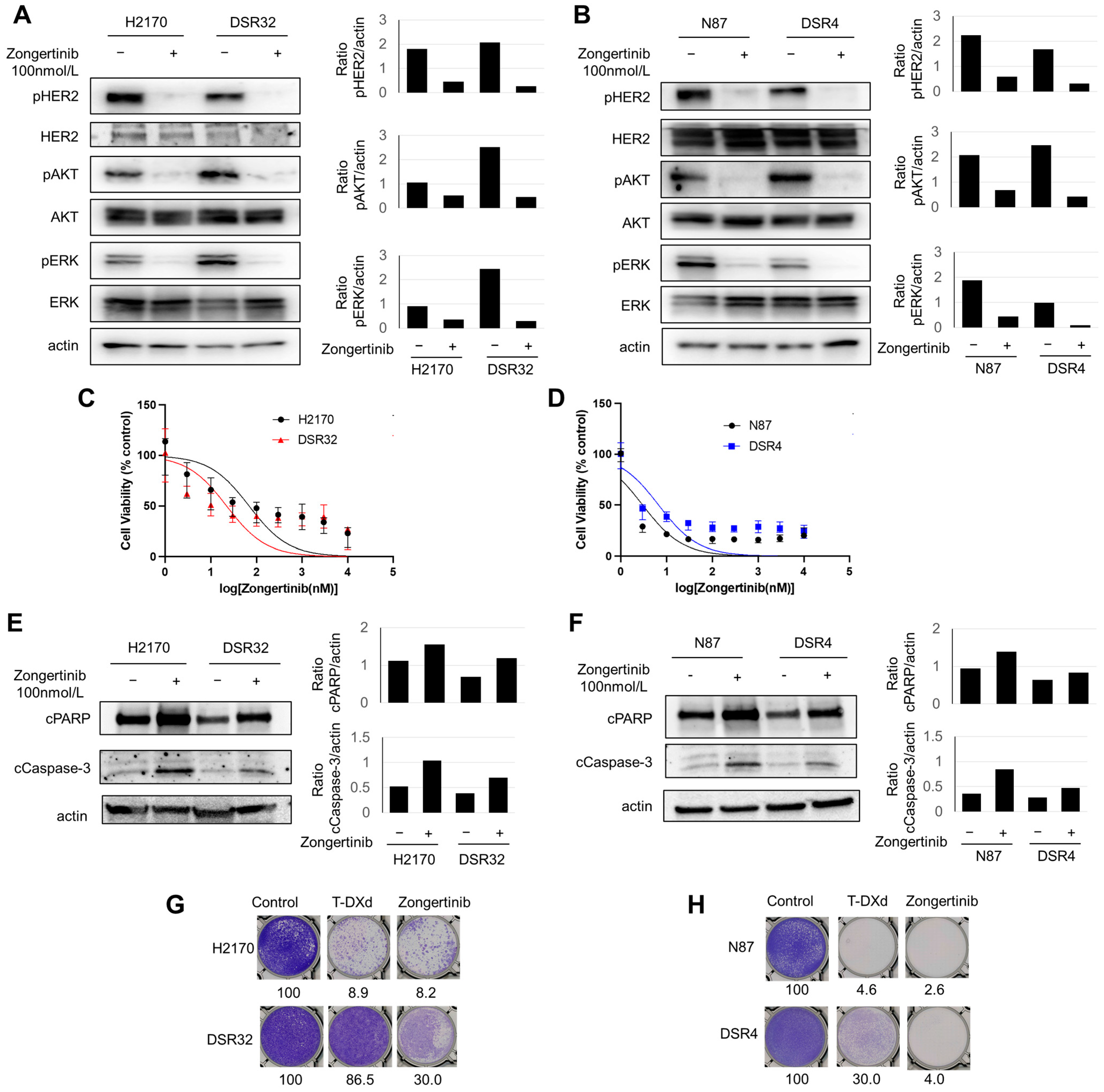
Next, we evaluated the activity of HER2 and its downstream pathways, AKT and ERK, following the acquisition of T-DXd resistance. Both DSR32 and DSR4 cells maintained a phosphorylation of HER2 and its downstream AKT and ERK even after developing resistance (Figure 4A,B). Exposure to zongertinib, an irreversible HER2-selective TKI, resulted in the significant decrease of HER2 and its downstream AKT and ERK phosphorylation in both DSR32 and DSR4 cells as well as in their parent cells (Figure 4A,B). Then, a cell growth inhibition assay was performed to evaluate sensitivity to zongertinib for 3 days. Zongertinib reduced cell viability of parent H2170 and N87 cells in a dose-dependent manner (Figure 4C,D). Furthermore, zongertinib reduced cell viability of both T-DXd-resistant DSR32 and DSR4 cells to the same degree as their parent cells (Figure 4C,D). Their IC50 values are 69.0, 22.2, 3.0, and 6.7 nmol/L for H2170, DSR32, N87, and DSR4 cells, respectively. Furthermore, to assess apoptosis caused by zongertinib, Western blot was performed for evaluating cleaved caspase 3 and cleaved PARP using these cells exposed to 100 nmol/L zongertinib for 24 h. Zongertinib induced apoptosis equivalent to that of the parental cells in resistant DSR32 and DSR4 cells (Figure 4E,F). The effects of extended zongertinib exposure were additionally evaluated using a colony formation assay. In parent H2170 and N87 cells, zongertinib caused colony formation inhibition comparable to that of T-DXd (Figure 4G,H). In DSR32 cells, colony formation was not inhibited by exposure to T-DXd, but zongertinib was able to inhibit it to 30.0% of the control (Figure 4G). In DSR4 cells, approximately 45.1% colony formation was observed even after T-DXd exposure, but zongertinib strongly inhibited this (Figure 4H).
Based on the above, conceivably even T-DXd-resistant cells might depend on HER2 signaling for their survival. Although the degree varied among cells, zongertinib may induce cancer cell death even in T-DXd-resistant cells by inhibiting the HER2/AKT/ERK signaling pathway.

3. Discussion

In this study, we established resistant clones from two HER2-overexpressing cancer cell lines. Although exploratory, our analyses suggested that reduced HER2 expression in one clone and impaired linker cleavage in the other may underlie the resistance mechanisms. Even after developing T-DXd resistance, these cells maintained HER2 expression and downstream signaling activity essential for their survival, thereby retaining sensitivity to the irreversible HER2-TKI zongertinib. Given the lack of established treatment options for T-DXd-resistant cancers, the findings of the present study provide valuable insights that may guide future therapeutic strategies for T-DXd-resistant cancers.
The mechanism of action of ADCs is complex and involves antibody binding to the target antigen, internalization, intracellular trafficking to lysosomes, cleavage of ADC, and cytotoxicity mediated by the released payload (Figure 1G) [5,20,21]. This complexity may promote the diversification of resistance mechanisms to ADCs [22]. We investigated which stage of the mechanism of action was impaired in the two established resistant cancer cells. First, we evaluated the internalization of T-DXd into cells and its transport to lysosomes, revealing that this process is impaired in DSR32. While multiple factors can cause ADC internalization defects [22,23], in this case, it is conceivable that the primary cause is reduced HER2 expression on the cell surface, which depends on HER2 gene copy number. The efficacy of trastuzumab-based ADCs depends on the expression level of HER2 in tumor cells [24,25]. The loss of HER2, particularly the extracellular domain, has been predominantly observed in cancer cells resistant to non-ADC anti-HER2 antibody therapeutics because of reduced dependence on HER2 signaling and weakened immunological effects such as impaired antibody-dependent cellular cytotoxicity [26]. However, in T-DXd-resistant cells, reduced uptake levels of T-DXd may exert a significant impact on resistance [26]. Indeed, numerous cancer cell lines that developed T-DM1 resistance through prolonged drug exposure showed diminished HER2 expression compared with that in their parent cells [27,28,29]. In terms of clinical settings, the DAISY trial, a prospective study evaluating the efficacy of T-DXd across different levels of HER2 expression in metastatic breast cancer, reported that HER2 status evaluated by IHC was reduced at the time of resistance acquisition in 65% of patients [30]. Currently, T-DXd is clinically employed as a therapeutic agent for HER2-overexpressing tumors across multiple cancer types [31,32]. A reduction in T-DXd internalization due to decreased HER2 expression levels may contribute to T-DXd resistance in various cancer types.
Unlike DSR32 cells, DSR4 cells do not exhibit reduced HER2 expression as a mechanism of resistance. However, in clinical practice, resistance to T-DXd has been observed in some patients with preserved HER2 expressions in tumor tissue. This is supported by the aforementioned DAISY trial, in which 35% of cases maintained HER2 expression even after T-DXd resistance [33], and another study reviewed tumor tissues of paired pre- and post-T-DXd treatment showed that HER2 status remained unchanged or was increased in 38% of patients [33]. These clinical findings underscore the significance of the current preclinical study. DSR4 cells had T-DXd internalization levels similar to those of the parental cells and remained highly susceptible to DXd, the payload component of T-DXd, without obvious resistance factors such as increased expression of ABC transporters or loss of TOP1. Although we did not perform direct assays for drug-efflux measurements, this result suggests that the cause of resistance in DSR4 cells lie in the process from lysosomal transport to cleavage of T-DXd. Specifically, T-DXd employs a cleavable tetrapeptide linker, which has been shown to be degraded by certain lysosomal enzymes such as cathepsin B and L [4,34]. In this study, there were no fluctuations in the expression levels of cathepsin B or L in DSR4 cells; however, considering the presence of other cathepsin members and factors affecting cleavage, an evaluation of cleavage efficiency may be necessary. On the other hand, T-DM1 utilizes a non-cleavable linker; thus, its antibody component undergoes proteolytic degradation in the lysosome, generating lysine-conjugated DM1 as the major active metabolite [35]. Therefore, T-DM1 may serve as a useful treatment option for T-DXd-resistant tumors.
This study demonstrated that T-DXd-resistant cells retained sensitivity to the HER2-TKI zongertinib in vitro. Although DSR32 cells exhibited reduced HER2 expression compared with parental cells, they maintained higher HER2 copy numbers than those in normal human dermal fibroblast cells. Intracellular signaling analysis further confirmed that DSR32 cell survival remained dependent on HER2 signaling. To date, several HER2-directed TKIs have been introduced into clinical practice for patients with HER2-positive breast cancer; however, their efficacy has been modest. The effectiveness of HER2-TKIs is often limited by dose-limiting toxicity resulting from the potential inhibition of other kinases. In particular, diarrhea and skin rash are major concerns, often hindering long-term treatment continuation [36,37]. In the present study, IC50 values for zongertinib were estimated to be below a clinically achievable plasma concentration of zongertinib (Figure 4C,D) [38]. A recent dose-expansion phase Ia study investigated the safety and efficacy of zongertinib in patients with HER2-altered solid tumors refractory to standard therapies. The objective response rate was 30.5%, and the most common treatment-related adverse events (any grade/grade ≥3) included diarrhea (50.5/1.0%), rash (16.2/1.9%), anemia (10.5/0%), decreased appetite (9.6/1%), and increased alanine transaminase (9.6/3.8%), indicating that both efficacy and tolerability were superior to those of conventional HER2-TKIs [13]. Our in vitro study suggests the potential of zongertinib as a treatment option for patients with HER2-overexpressing solid tumors who are resistant to T-DXd, warranting further investigation.
There are certain limitations in the present study. First, this study has not fully elucidated the detailed resistance mechanisms in the current resistant clones. In particular, live-cell imaging, endosomal or lysosomal co-localization studies are needed for further clarifying the resistance mechanism. Second, sensitization or synergy of combining HER2-TKI with T-DXd was not evaluated in this study. Further investigation is needed to determine whether HER2-TKI monotherapy can achieve sufficient therapeutic efficacy against T-DXd-resistant tumors, or whether combination therapy with T-DXd yields superior outcomes. Third, our T-DXd-resistant cells were experimentally generated. Only two types of resistant cell lines were used, each representing a different cancer type. Further translational studies are warranted to evaluate HER2 expression and the efficacy of zongertinib using patient-derived cells following the development of T-DXd resistance.

4. Materials and Methods

4.1. Cells and Reagents

HER2-expressing NCI-H2170 lung cancer cells and HER2-expressing NCI-N87 gastric cancer cells were obtained from the American Type Culture Collection. To generate T-DXd-resistant cells, H2170 and N87 cells were cultured in the presence of T-DXd with gradually increasing concentrations from 1 ng/mL to a maximum of 1 μg/mL for up to 9 months. The cells were maintained in a humidified atmosphere of 5% CO2 at 37 °C in RPMI 1640 medium (FUJIFILM Wako Pure Chemical Corporation, Osaka, Japan) supplemented with 10% fetal bovine serum (FBS) and 1% penicillin–streptomycin. Mycoplasma-free conditions were periodically tested using MycoAlert Mycoplasma Detection Kit (Lonza, Basel, Switzerland), and all experiments were performed with mycoplasma-free cells. All parent and T-DXd-resistant cell lines were authenticated using short tandem repeats profiling within the last 3 years. T-DXd, T-DM1, DXd, and zongertinib were purchased from Daiichi Sankyo Co., Ltd. (Tokyo, Japan), Chugai Pharmaceutical Co., Ltd. (Tokyo, Japan), MedChemExpress (Monmouth Junction, NJ, USA), and Selleck Chemicals (Houston, TX, USA), respectively.

4.2. In Vitro Growth Inhibition Assay

Cells were seeded in 96-well flat-bottom plates at a density of 1000 cells per well in RPMI 1640 medium supplemented with 10% FBS. After incubation for 24 h, cells were exposed to various concentrations of T-DXd, DXd, T-DM1, or zongertinib. Following further incubation for 6 days (for T-DXd and T-DM1) or 3 days (for DXd and zongertinib), cell viability was assessed using the CellTiter-Glo luminescence assay (Promega K.K., Tokyo, Japan) according to the manufacturer’s instructions. Luminescence was measured using a microplate reader (ARVO MX 1420; PerkinElmer, Shelton, CT, USA). Luminescence values were expressed as a percentage of those observed in untreated cells, and the IC50 for each drug was calculated.

4.3. ADC Internalization Detection Assay

pH-sensitive red fluorescent dye was conjugated to T-DXd using an antibody internalization detection reagent (Acrobiosystems, Newark, DE, USA) according to the manufacturer’s instructions. Briefly, the detection reagent was mixed with T-DXd and incubated for 10 min at room temperature. For the internalization detection assays, 3 × 10⁵ cells per well were seeded in 6-well plates containing RPMI 1640 medium supplemented with 10% FBS. The following day, cells were exposed to labeled T-DXd and harvested at 2, 4, 8, 16, and 24 h after exposure. Harvested cells were washed with phosphate-buffered saline (PBS), and fluorescence intensity was measured with an LSRFortessa X-20 (BD Biosciences, San Jose, CA, USA). Data were analyzed using FlowJo software version 10.7.2 (FlowJo, Ashland, OR, USA).

4.4. Cell Surface HER2 Measuring Assay

Quantification of cell surface HER2 was performed using QIFIKIT (Dako, Tokyo, Japan). Each cell line was treated with a saturating concentration of mouse anti-HER2 antibody (clone 9G6) or mouse IgG2a isotype control, followed by staining with an FITC-conjugated anti-mouse IgG (Dako). Each sample was analyzed by LSRFortessa X-20 (BD Biosciences), and the number of binding sites per cell was calculated according to the manufacturer’s instructions.

4.5. Colony-Formation Assay

Cells were seeded in 6-well plates at a density of 3.0  ×  104 cells/well. After 24 h, cells were treated with 0.5, 1 µg/mL T-DXd, 1, 10 µg/mL T-DM1, or 100 nM zongertinib. The medium containing each drug was changed every 3 days. After 10 days of incubation, the plates were gently washed with PBS and fixed with a fixation solution (acetic acid/methanol, 1:7) for 5 min. Colonies were stained with 0.5% crystal violet for 3 h at room temperature. The percentage of colony area was automatically calculated using ImageJ 1.52a (National Institutes of Health, Bethesda, MD, USA).

4.6. Western Blotting

Cells were seeded at 2 × 10⁶ cells per plate and allowed to grow overnight in RPMI 1640 medium containing 1% FBS before being harvested. Western blotting was performed as described previously [39]. Proteins were probed using antibodies specific for phospho-HER2 (#2243, 1:1000 dilution; Cell Signaling Technology, Danvers, MA, USA), HER2 (#2165, 1:1000 dilution; Cell Signaling Technology), phospho-AKT (#4060, 1:1000 dilution; Cell Signaling Technology), AKT (#4691, 1:1000 dilution; Cell Signaling Technology), phospho-ERK (#4376, 1:1000 dilution; Cell Signaling Technology), ERK (#9102, 1:1000 dilution; Cell Signaling Technology), cPARP (#5625, 1:1000 dilution; Cell Signaling Technology), cCaspase-3 (#9661, 1:1000 dilution; Cell Signaling Technology), and actin (A2066, 1:100 dilution; Sigma-Aldrich, St. Louis, MO, USA). The actin antibody was used as a loading control.

4.7. NGS

We evaluated HER2 gene mutations and copy number using the Oncomine Tumor Mutation Load Assay (Thermo Fisher Scientific, Waltham, MA, USA), a polymerase chain reaction–based NGS assay.

4.8. Microarray

The microarray gene expression analysis was performed using the Affymetrix Clariom™ S Human (Thermo Fisher Scientific) and the Affymetrix Transcriptome Analysis Console software version 4.0.3 (Thermo Fisher Scientific) according to the manufacturer’s instructions. All cells were cultured without T-DXd exposure in the culture medium for one week and were then harvested. Gene expression levels and the significance scores from WikiPathways were calculated using Transcriptome Analysis Console Ver4.0 (Thermo Fisher Scientific).

4.9. In Vivo Tumor Growth Inhibition Assay

All animal experiments were performed in accordance with the Recommendations for Handling of Laboratory Animals for Biomedical Research compiled by the Committee on Safety and Ethical Handling Regulations for Laboratory Animal Experiments of Kindai University. The study was also reviewed and approved by the Animal Ethics Committee of Kindai University. H2170 and DSR32 cells (5 × 10⁶ cells/mouse) were subcutaneously injected into the right flanks of female BALB/cAJcl-nu/nu mice (CLEA Japan, Tokyo, Japan). Once tumors reached the target volume (0.2 cm3), mice were randomly assigned to treatment and control groups. Mice received a single i.p. injection of PBS (200 μL; control) or T-DXd (10 mg/kg body weight in 200 μL PBS). Tumor volumes and mouse body weights were measured twice per week. Mice were sacrificed if tumors became necrotic or grew to a volume of 2.0 cm3. Tumor volume was defined as 1/2 × length × width2.

4.10. Statistical Analysis

The growth inhibition assay was analyzed using the GraphPad Prism 9.0 software (GraphPad, Boston, MA, USA). The curves were fitted using a nonlinear regression model, and IC50 values were calculated accordingly. Statistical analysis was performed using the SPSS version 22.0 (IBM Corp., Armonk, NY, USA). Statistical tests included two-sided, unpaired t-tests, one-way ANOVA, and two-way ANOVA, with p < 0.05 considered statistically significant.

Supplementary Materials

The following supporting information can be downloaded at: https://www.mdpi.com/article/10.3390/ijms262110515/s1.

Author Contributions

Conceptualization, S.S. and H.H.; Methodology, T.K., S.S., K.Y., Y.K., T.T. (Toshiyuki Takehara), T.T. (Takeshi Teramura), K.S., K.N. and H.H.; Resources, T.K., S.S. and K.Y.; Data curation, T.K., S.S. and K.Y.; Investigation, T.K., S.S., K.Y., Y.K., T.T. (Toshiyuki Takehara), T.T. (Takeshi Teramura), K.S. and K.N.; Writing—original draft, T.K., S.S. and K.Y.; Writing—review and editing, T.K., S.S., K.Y., Y.K., T.T. (Toshiyuki Takehara), T.T. (Takeshi Teramura), K.S., K.N. and H.H.; Funding acquisition, S.S. and H.H.; Project administration, H.H.; Supervision, H.H. All authors have read and agreed to the published version of the manuscript.

Funding

This work was supported by Nippon Boehringer Ingelheim Co., Ltd.

Institutional Review Board Statement

The animal study protocol was approved by the Animal Ethics Committee of Kindai University (protocol code: KAME-2024-016; date of approval: 21 February 2024).

Informed Consent Statement

Not applicable.

Data Availability Statement

The data generated in this study are available upon request from the corresponding author.

Acknowledgments

The authors thank Haruka Sakamoto, Yume Shinkai and Michiko Kitano for technical support.

Conflicts of Interest

T.K. reports grants from Boehringer Ingelheim and Chugai Pharmaceutical Co., Ltd. S.S. reports grants from Nippon Boehringer-Ingelheim Co., Ltd. and Boehringer Ingelheim. K.Y. reports grants from Boehringer Ingelheim.; personal fees from Boehringer Ingelheim Japan, and Daiichi Sankyo Co., Ltd. K.S. reports personal fees from Qiagen, Inc., Takeda Pharmaceutical Co., Ltd., Nippon Kayaku Co., Ltd., and Life Technologies Japan Ltd. K.N. reports grants from Nippon Boehringer Ingelheim, West Japan Oncology Group, Nichirei Biosciences Inc., Eli Lilly Japan, Hitachi, Sysmex, Otsuka Pharmaceutical, Thoracic Oncology Research Group, Okayama University, and Japan Breast Cancer Research Group.; personal fees from SymBio Pharmaceuticals, ELI LILLY JAPAN, Otsuka Pharmaceutical, Chugai, MSD, Novartis Pharma, AstraZeneca, Guardant Health, Janssen Pharmaceutical, DAIICHI SANKYO, Ono Pharmaceutical, Invitae Japan, Nichirei, and Maruho. H.H. reports grants from Boehringer Ingelheim, IQVIA Services JAPAN K.K., Eisai Co., Ltd., SYNEOS HEALTH CLINICAL K.K., Shionogi & Co., Ltd., EP-CRSU CO., LTD., Nippon Kayaku Co., Ltd., EPS Corporation., GlaxoSmithKline K.K., Otsuka Pharmaceutical Co., Ltd., Sanofi K.K., Takeda Pharmaceutical Co., Ltd., Taiho Pharmaceutical Co., Ltd., MSD K.K., Bristol Myers Squibb Company, Amgen Inc., SRL Medisearch Inc., Chugai Pharmaceutical Co., Ltd., PRA Health Sciences Inc., Nippon Boehringer Ingelheim Co., Ltd., CMIC CO., Ltd., Janssen Pharmaceutical K.K., Pfizer R&D Japan G.K., Astellas Pharma Inc., Ascent Development Services, Labcorp Development Japan K.K., Eisai Inc., Kobayashi Pharmaceutical Co., Ltd., Bayer Yakuhin, Ltd., Pfizer Japan Inc., AstraZeneca K.K., AbbVie Inc., Daiichi Sankyo Co., Ltd., A2 Healthcare Corp., Novartis Pharma K.K., Eli Lilly Japan K.K., Merck Biopharma Co., Ltd., Kyowa Kirin Co., Ltd., Medpace Japan KK, Sysmex Corporation, ICON Japan K.K., Clinical Research Support Center Kyushu, PPD-SNBL K.K, Kyushu Study group of Clinical Cancer, Mochida Pharmaceutical Co., Ltd., Public Health Research Foundation, Japanese Gastric Cancer Association, Thoracic Oncology Research Group, West Japan Oncology Group, Japan Clinical Cancer Research Organization, and Comprehensive Support Project for Oncological Research of Breast Cancer.; personal fees from Ono Pharmaceutical Co., Ltd., Eli Lilly Japan K.K., AstraZeneca K.K., Bristol Myers Squibb Company, MSD K.K., Takeda Pharmaceutical Co., Ltd., Sysmex Corporation, Novartis Pharma K.K., Nippon Boehringer Ingelheim Co., Ltd. Pfizer Japan Inc., AbbVie Inc., Chugai Pharmaceutical Co., Ltd., Kyowa Kirin Co., Ltd., Amgen Inc., Reno. Medical K.K., Eisai Co., Ltd., Nippon Kayaku Co., Ltd., Janssen Pharmaceutical K.K., Hisamitsu Pharmaceutical Co., Inc., Care Net, Inc., Novocure K.K., Taiho Pharmaceutical Co., Ltd., Merck Biopharma Co., Ltd., Daiichi Sankyo Co., Ltd., 3H Clinical Trial Inc., Guardant Health Japan Corp, Ishiyaku Publishers, Inc., and Medical Review Co., Ltd.; participation on a data safety monitoring board or advisory board for Bristol Myers Squibb Company, AstraZeneca K.K., Janssen Pharmaceutical K.K., Daiichi Sankyo Co., Ltd., AbbVie Inc., Novocure K.K., and Chugai Pharmaceutical Co., Ltd. No disclosures were reported by the other authors. The funders had no role in study design, data collection and analysis, decision to publish, or preparation of the manuscript.

Abbreviations

The following abbreviations are used in this manuscript:
EGFRepidermal growth factor receptor
ADCAntibody–drug conjugate
PBSPhosphate-buffered saline
FBSFetal bovine serum
NGSNext-generation sequencing
IC50Inhibitory concentration value
TKITyrosine kinase inhibitor
T-DXdTrastuzumab deruxtecan
T-DM1trastuzumab emtansine
CIconfidence interval
MFImean fluorescence intensity
SDstandard deviation
SEMstandard error of the mean

References

  1. Rubin, I.; Yarden, Y. The basic biology of HER2. Ann. Oncol. 2001, 12 (Suppl. S1), S3–S8. [Google Scholar] [CrossRef] [PubMed]
  2. Yoon, J.; Oh, D.Y. HER2-targeted therapies beyond breast cancer—An update. Nat. Rev. Clin. Oncol. 2024, 21, 675–700. [Google Scholar] [CrossRef] [PubMed]
  3. Swain, S.M.; Shastry, M.; Hamilton, E. Targeting HER2-positive breast cancer: Advances and future directions. Nat. Rev. Drug Discov. 2023, 22, 101–126. [Google Scholar] [CrossRef]
  4. Ogitani, Y.; Aida, T.; Hagihara, K.; Yamaguchi, J.; Ishii, C.; Harada, N.; Soma, M.; Okamoto, H.; Oitate, M.; Arakawa, S.; et al. DS-8201a, A novel HER2-targeting ADC with a novel DNA topoisomerase I inhibitor, demonstrates a promising antitumor efficacy with differentiation from T-DM1. Clin. Cancer Res. 2016, 22, 5097–5108. [Google Scholar] [CrossRef]
  5. Drago, J.Z.; Modi, S.; Chandarlapaty, S. Unlocking the potential of antibody-drug conjugates for cancer therapy. Nat. Rev. Clin. Oncol. 2021, 18, 327–344. [Google Scholar] [CrossRef]
  6. Modi, S.; Saura, C.; Yamashita, T.; Park, Y.H.; Kim, S.B.; Tamura, K.; Andre, F.; Iwata, H.; Ito, Y.; Tsurutani, J.; et al. Trastuzumab deruxtecan in previously treated HER2-positive breast cancer. N. Engl. J. Med. 2020, 382, 610–621. [Google Scholar] [CrossRef]
  7. Cortés, J.; Kim, S.B.; Chung, W.P.; Im, S.A.; Park, Y.H.; Hegg, R.; Kim, M.H.; Tseng, L.-M.; Petry, V.; Chung, C.-F.; et al. Trastuzumab Deruxtecan versus trastuzumab Emtansine for Breast Cancer. N. Engl. J. Med. 2022, 386, 1143–1154. [Google Scholar] [CrossRef]
  8. Liang, Y.; Zhang, P.; Li, F.; Lai, H.; Qi, T.; Wang, Y. Advances in the study of marketed antibody-drug conjugates (ADCs) for the treatment of breast cancer. Front. Pharmacol. 2024, 14, 1332539. [Google Scholar] [CrossRef]
  9. Modi, S.; Jacot, W.; Yamashita, T.; Sohn, J.; Vidal, M.; Tokunaga, E.; Tsurutani, J.; Ueno, N.T.; Prat, A.; Chae, Y.S.; et al. Trastuzumab deruxtecan in previously treated HER2-low advanced breast cancer. N. Engl. J. Med. 2022, 387, 9–20. [Google Scholar] [CrossRef]
  10. Shitara, K.; Bang, Y.J.; Iwasa, S.; Sugimoto, N.; Ryu, M.H.; Sakai, D.; Chung, H.-C.; Kawakami, H.; Yabusaki, H.; Lee, J.; et al. Trastuzumab deruxtecan in previously treated HER2-positive gastric cancer. N. Engl. J. Med. 2020, 382, 2419–2430. [Google Scholar] [CrossRef] [PubMed]
  11. Li, B.T.; Smit, E.F.; Goto, Y.; Nakagawa, K.; Udagawa, H.; Mazières, J.; Nagasaka, M.; Bazhenova, L.; Saltos, A.N.; Felip, E.; et al. Trastuzumab deruxtecan in HER2-mutant non-small-cell lung cancer. N. Engl. J. Med. 2022, 386, 241–251. [Google Scholar] [CrossRef] [PubMed]
  12. Smit, E.F.; Felip, E.F.; Uprety, D.; Nagasaka, M.; Nakagawa, K.; Rodríguez, P.A.; Pacheco, J.M.; Li, B.T.; Planchard, D.; Baik, C.; et al. Trastuzumab deruxtecan in patients with metastatic non-small-cell lung cancer (DESTINY-Lung01): Primary results of the HER2-overexpressing cohorts from a single-arm, phase 2 trial. Lancet Oncol. 2024, 25, 439–454. [Google Scholar] [CrossRef]
  13. Heymach, J.V.; Opdam, F.; Barve, M.; Tu, H.Y.; Wu, H.Y.; Berz, D.; Schröter, L.; Botilde, Y.; Sadrolhefazi, B.; Serra, J.; et al. HER2-selective tyrosine kinase inhibitor, zongertinib (BI 1810631), in patients with advanced/metastatic solid tumors with HER2 alterations: A phase Ia dose-escalation study. J. Clin. Oncol. 2025, 43, 1337–1347. [Google Scholar] [CrossRef] [PubMed]
  14. Wilding, B.; Woelflingseder, L.; Baum, A.; Chylinski, K.; Vainorius, G.; Gibson, N.; Waizenegger, I.C.; Gerlach, D.; Augsten, M.; Spreitzer, F.; et al. Zongertinib (BI 1810631), an irreversible HER2 TKI, spares EGFR signaling and improves therapeutic response in preclinical models and patients with HER2-driven cancers. Cancer Discov. 2025, 15, 119–138. [Google Scholar] [CrossRef]
  15. Yonesaka, K.; Zejnullahu, K.; Okamoto, I.; Satoh, T.; Cappuzzo, F.; Souglakos, J.; Ercan, D.; Rogers, A.; Roncalli, M.; Takeda, M.; et al. Activation of ERBB2 signaling causes resistance to the EGFR-directed therapeutic antibody cetuximab. Sci. Transl. Med. 2011, 3, 99ra86. [Google Scholar] [CrossRef]
  16. Suzuki, S.; Yonesaka, K.; Teramura, T.; Takehara, T.; Kato, R.; Sakai, H.; Haratani, K.; Tanizaki, J.; Kawakami, H.; Hayashi, H.; et al. KRAS inhibitor resistance in MET-amplified KRASG12C non-small cell lung cancer induced by RAS- and non-RAS-mediated cell signaling mechanisms. Clin. Cancer Res. 2021, 27, 5697–5707, Erratum in Clin. Cancer Res. 2022, 28, 428. [Google Scholar] [CrossRef] [PubMed]
  17. Kaufmann, S.H.; Desnoyers, S.H.; Ottaviano, Y.; Davidson, N.E.; Poirier, G.G. Specific proteolytic cleavage of poly(ADP-ribose) polymerase: An early marker of chemotherapy-induced apoptosis. Cancer Res. 1993, 53, 3976–3985. [Google Scholar]
  18. Liu, X.; Zou, H.; Slaughter, C.; Wang, X. DFF, a heterodimeric protein that functions downstream of caspase-3 to trigger DNA fragmentation during apoptosis. Cell 1997, 89, 175–184. [Google Scholar] [CrossRef]
  19. Nakagawa, H.; Saito, H.; Ikegami, Y.; Aida-Hyugaji, S.; Sawada, S.; Ishikawa, T. Molecular modeling of new camptothecin analogues to circumvent ABCG2-mediated drug resistance in cancer. Cancer Lett. 2006, 234, 81–89. [Google Scholar] [CrossRef]
  20. Fu, Z.; Li, S.; Han, S.; Shi, C.; Zhang, Y. Antibody drug conjugate: The “biological missile” for targeted cancer therapy. Signal Transduct. Target. Ther. 2022, 7, 93. [Google Scholar] [CrossRef]
  21. Grinda, T.; Rassy, E.; Pistilli, B. Antibody-drug conjugate revolution in breast cancer: The road ahead. Curr. Treat. Options Oncol. 2023, 24, 442–465, Erratum in Curr. Treat. Options Oncol. 2023, 24, 466–467. [Google Scholar] [CrossRef]
  22. Hunter, F.W.; Barker, H.R.; Lipert, B.; Rothé, F.; Gebhart, G.; Piccart-Gebhart, M.J.; Sotiriou, C.; Jamieson, S.M.F. Mechanisms of resistance to trastuzumab emtansine (T-DM1) in HER2-positive breast cancer. Br. J. Cancer 2020, 122, 603–612. [Google Scholar] [CrossRef] [PubMed]
  23. Chang, H.L.; Schwettmann, B.; McArthur, H.L.; Chan, I.S. Antibody-drug conjugates in breast cancer: Overcoming resistance and boosting immune response. J. Clin. Investig. 2023, 133, e172156. [Google Scholar] [CrossRef]
  24. Yamashita-Kashima, Y.; Yoshimura, Y.; Fujimura, T.; Shu, S.; Yanagisawa, M.; Yorozu, K.; Furugaki, K.; Higuchi, R.; Shoda, J.; Harada, N. Molecular targeting of HER2-overexpressing biliary tract cancer cells with trastuzumab emtansine, an antibody-cytotoxic drug conjugate. Cancer Chemother. Pharmacol. 2019, 83, 659–671. [Google Scholar] [CrossRef]
  25. Vasalou, C.; Proia, T.A.; Kazlauskas, L.; Przybyla, A.; Sung, M.; Mamidi, S.; Maratea, K.; Griffin, M.; Sargeant, R.; Urosevic, J.; et al. Quantitative evaluation of trastuzumab deruxtecan pharmacokinetics and pharmacodynamics in mouse models of varying degrees of HER2 expression. CPT Pharmacomet. Syst. Pharmacol. 2024, 13, 994–1005. [Google Scholar] [CrossRef] [PubMed]
  26. Wu, X.; Huang, S.; He, W.; Song, M. Emerging insights into mechanisms of trastuzumab resistance in HER2-positive cancers. Int. Immunopharmacol. 2023, 122, 110602. [Google Scholar] [CrossRef] [PubMed]
  27. Loganzo, F.; Tan, X.; Sung, M.; Jin, G.; Myers, J.S.; Melamud, E.; Wang, F.; Diesl, V.; Follettie, M.T.; Musto, S.; et al. Tumor cells chronically treated with a trastuzumab-maytansinoid antibody-drug conjugate develop varied resistance mechanisms but respond to alternate treatments. Mol. Cancer Ther. 2015, 14, 952–963. [Google Scholar] [CrossRef]
  28. Sabbaghi, M.; Gil-Gómez, G.; Guardia, C.; Servitja, S.; Arpí, O.; García-Alonso, S.; Menendez, S.; Arumi-Uria, M.; Serrano, L.; Salido, M.; et al. Defective cyclin B1 induction in trastuzumab-emtansine (T-DM1) acquired resistance in HER2-positive breast cancer. Clin. Cancer Res. 2017, 23, 7006–7019. [Google Scholar] [CrossRef]
  29. Li, G.; Guo, J.; Shen, B.Q.; Yadav, B.Q.; Sliwkowski, M.X.; Crocker, L.M.; Lacap, J.A.; Phillips, G.D.L. Mechanisms of acquired resistance to trastuzumab emtansine in breast cancer cells. Mol. Cancer Ther. 2018, 17, 1441–1453. [Google Scholar] [CrossRef]
  30. Mosele, F.; Deluche, E.; Lusque, A.; Le Bescond, L.; Filleron, T.; Pradat, Y.; Ducoulombier, A.; Pistilli, B.; Bachelot, T.; Viret, F.; et al. Trastuzumab deruxtecan in metastatic breast cancer with variable HER2 expression: The phase 2 DAISY trial. Nat. Med. 2023, 29, 2110–2120. [Google Scholar] [CrossRef]
  31. Tsurutani, J.; Iwata, H.; Krop, I.; Jänne, P.A.; Doi, T.; Takahashi, S.; Park, H.; Redfern, C.; Tamura, K.; Wise-Draper, T.M.; et al. Targeting HER2 with trastuzumab deruxtecan: A dose-expansion, Phase I study in multiple advanced solid tumors. Cancer Discov. 2020, 10, 688–701, Erratum in Cancer Discov. 2020, 10, 1078. [Google Scholar] [CrossRef]
  32. Meric-Bernstam, F.; Makker, V.; Oaknin, A.; Oh, D.Y.; Banerjee, S.; González-Martín, A.; Jung, K.H.; Ługowska, I.; Manso, L.; Manzano, A.; et al. Efficacy and safety of trastuzumab deruxtecan in patients with HER2-expressing solid tumors: Primary results from the DESTINY-PanTumor02 Phase II trial. J. Clin. Oncol. 2024, 42, 47–58. [Google Scholar] [CrossRef]
  33. Gouda, M.A.; Gonugunta, A.; Dumbrava, E.E.; Yap, T.A.; Rodon, J.; Piha-Paul, S.A.; Pohlmann, P.R.; Damodaran, S.; Murthy, R.; Valero, V.; et al. Human epidermal growth factor receptor 2 loss following treatment with trastuzumab deruxtecan in patients with metastatic breast cancer. Clin. Cancer Res. 2025, 31, 1268–1274. [Google Scholar] [CrossRef]
  34. Shiose, Y.; Ochi, Y.; Kuga, H.; Yamashita, F.; Hashida, M. Relationship between drug release of DE-310, macromolecular prodrug of DX-8951f, and cathepsins activity in several tumors. Biol. Pharm. Bull. 2007, 30, 2365–2370. [Google Scholar] [CrossRef]
  35. Lewis Phillips, G.D.; Li, G.D.; Dugger, D.L.; Crocker, L.M.; Parsons, K.L.; Mai, E.; Blättler, W.A.; Lambert, J.M.; Chari, R.V.J.; Lutz, R.J.; et al. Targeting HER2-positive breast cancer with trastuzumab-DM1, an antibody-cytotoxic drug conjugate. Cancer Res. 2008, 68, 9280–9290. [Google Scholar] [CrossRef] [PubMed]
  36. Le Du, F.; Diéras, V.; Curigliano, G. The role of tyrosine kinase inhibitors in the treatment of HER2+ metastatic breast cancer. Eur. J. Cancer 2021, 154, 175–189, Erratum in Eur. J. Cancer 2021, 158, 255. [Google Scholar] [CrossRef]
  37. Duchnowska, R.; Loibl, S.; Jassem, J. Tyrosine kinase inhibitors for brain metastases in HER2-positive breast cancer. Cancer Treat. Rev. 2018, 67, 71–77. [Google Scholar] [CrossRef] [PubMed]
  38. Tian, X.; Esmaeili, H.; Minich, D.; Seitz, F.; Roessner, P.M.; Wind, S.; Grempler, R.; Gan, G.; Chan, T.S.; Mahmoudi, M.; et al. The effect of carbamazepine, a strong CYP3A inducer, on the pharmacokinetics of zongertinib in healthy male volunteers. Pharmacotherapy 2025, 45, 94–103. [Google Scholar] [CrossRef] [PubMed]
  39. Yonesaka, K.; Kudo, K.; Nishida, S.; Takahama, T.; Iwasa, T.; Yoshida, T.; Tanaka, K.; Takeda, M.; Kaneda, H.; Okamoto, I.; et al. The pan-HER family tyrosine kinase inhibitor afatinib overcomes HER3 ligand heregulin-mediated resistance to EGFR inhibitors in non-small cell lung cancer. Oncotarget 2015, 6, 33602–33611. [Google Scholar] [CrossRef]
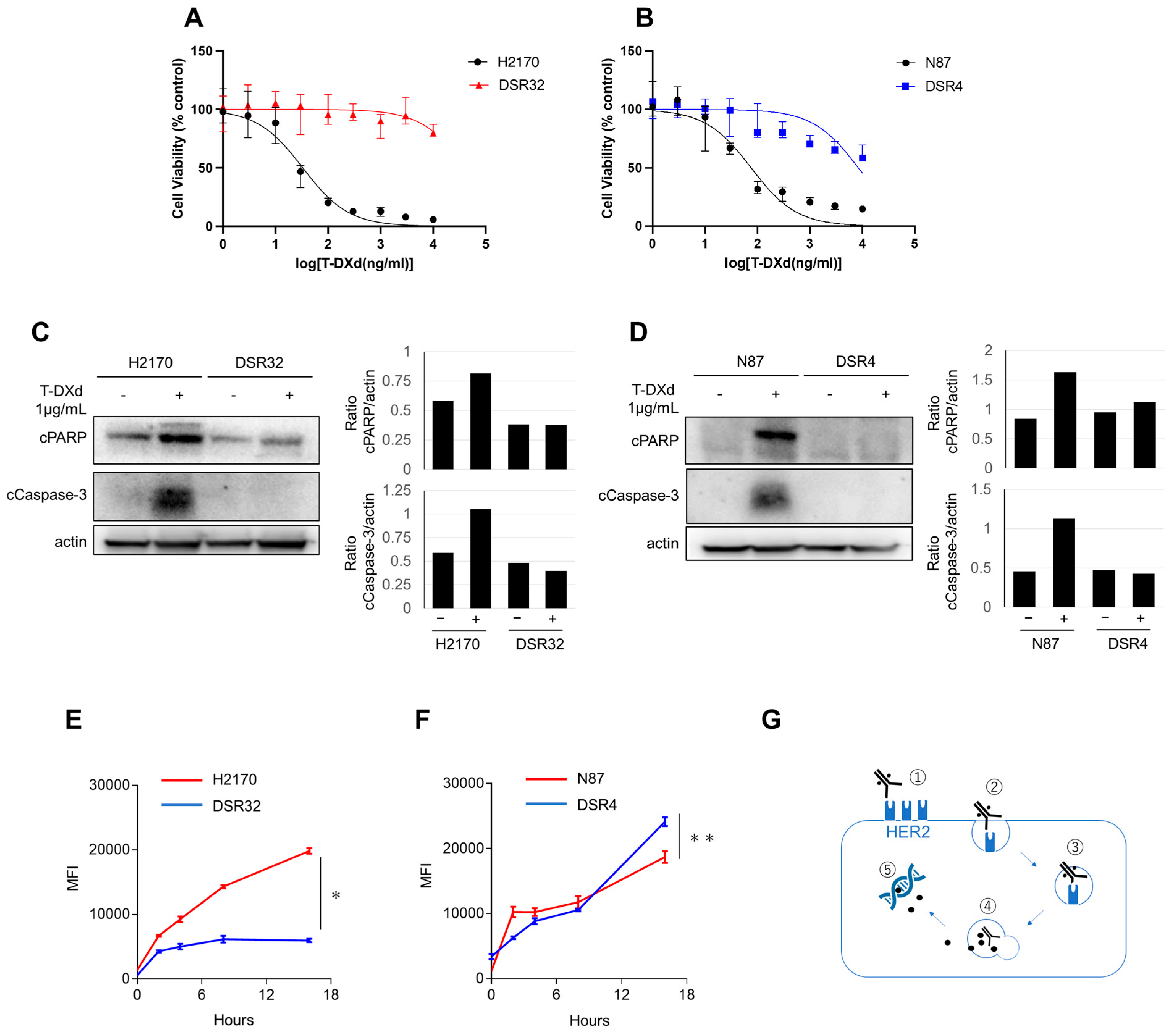
Figure 1. DSR32 and DSR4 cells are resistant to T-DXd. (A) HER2-expressing H2170 cells and their clone DSR32 cells were treated with T-DXd at the indicated concentrations. The percentage of viable cells is shown relative to untreated controls (median with a 95% CI of six independent experiments). (B) HER2-expressing N87 cells and their clone DSR4 cells were treated with T-DXd at the indicated concentrations. The percentage of viable cells is shown relative to untreated controls (median with a 95% CI of six independent experiments). (C,D) H2170 and DSR32 cells were treated with T-DXd (1 μg/mL) for 5 days. (C) N87 and DSR4 cells were treated with T-DXd (1 μg/mL) for 5 days. (D) Proteins were detected by Western blot. The pixel density of the indicated protein was shown as bar graphs relative to actin expression. (E,F) T-DXd internalization was measured by labeling with pH-sensitive fluorescent dye. H2170 and DSR32 cells (E) or N87 and DSR4 cells (F) were treated with T-DXd (1 μg/mL) for the indicated times and evaluated at each point. MFI with a 95% CI of three independent experiments is shown. Two-way ANOVA; * p < 0.0001, ** p = 0.25. (G) The schematic diagram of the intracellular signaling pathway associated with T-DXd is shown. 1. Binding to HER2; 2. Internalization; 3. Transport to lysosome; 4. Cleavage of linker in lysosomes; 5. Topoisomerase 1 inhibition.
Ijms 26 10515 g001
Figure 2. Decreased HER2 gene copy number in DSR32 cells. (A) A ranked pathway with significance score is shown comparing DSR32 vs. H2170 cells. The significance score was calculated by the Affymetrix Transcriptome Analysis Console software version 4.0.3. (B) Volcano plot of differential gene expression of DSR32 versus H2170 cells. Each point represents the average value of one transcript in two replicate experiments as revealed by microarray analysis. The X axis represents the linear fold change; Y axis represents −log10 p-values from the ANOVA. The expression difference is considered significant for a 2-fold change and a p-value of 0.05. Red-colored points indicate significantly enriched genes in DSR32 cells, and green-colored points indicate those in H2170 cells. Gray-colored points indicate no significant difference. (C) HER2 copy numbers of H2170 and DSR32 cells, determined by NGS, are shown. (D) HER2 expression of whole-cell lysate of H2170, DSR26, and DSR32 cells was determined by a Western blot assay. The pixel densities of HER2 are shown as bar graphs relative to parental H2170 cells. (E) HER2 expression on cell surfaces of H2170 and DSR32 cells, measured by a flow cytometry. Mean HER2 molecule count per cell with the SD of three independent experiments is shown. t-test; * p < 0.05. (F,G) In xenograft mouse models of H2170 (F) and DSR32 (G), tumors were treated with T-DXd (10 mg/kg triweekly) or vehicle. Each treatment group consisted of six mice. Data represent the mean ± SEM. Two-way ANOVA; ** p = 0.0002, *** p = 0.181.
Ijms 26 10515 g002
Figure 3. HER2 expression maintained in T-DXd-resistant DSR4 cells. (A) HER2 copy numbers of N87 and DSR4 cells, determined by NGS, are shown. (B) HER2 expression on the cell surfaces of N87 and DSR4 cells was measured by flow cytometry. Mean HER2 molecule count per cell with SD of three independent experiments is shown. t-test; * p = 0.003, (C) A ranked pathway with significance scores for DSR4 vs. N87 cells is shown. Significance score was calculated by the Affymetrix Transcriptome Analysis Console software version 4.0.3. (D) Volcano plot of differential gene expression of DSR4 versus N87 cells. Each point represents the average value of one transcript in two replicate experiments as revealed by microarray analysis. The X axis represents the linear fold change; Y axis represents −log10 p-values from the ANOVA. The expression difference is considered significant for a 2-fold change and a p-value of 0.05. Red colored points are significantly enriched genes in DSR4 cells, and green colored points are in N87 cells. Gray-colored points indicate no significant difference. (E) N87 and its clone DSR4 cells were treated with various concentrations of DXd. The percentage of viable cells is shown relative to untreated controls (median with 95% CI of six independent experiments). (F) N87 and DSR4 cells were treated with varying concentrations of T-DM1. The percentage of viable cells is shown relative to untreated controls (median with 95% CI of six independent experiments). (G) Clonogenic assay for N87 and DSR4 cells treated with/without T-DXd, or T-DM1 at indicated concentrations for 10 days The colony-formation area was measured and quantified using ImageJ, and the measurement is shown.
Ijms 26 10515 g003
Figure 4. DSR32 and DSR4 cells are susceptible to zongertinib. (A,B) Either H2170 and DSR32 cells (A) or N87 and DSR4 cells (B) were treated with zongertinib (100 nmol/L) for 24 h. The indicated proteins were detected by Western blot. The pixel density of the indicated protein was shown as bar graphs relative to actin expression. (C,D) Either H2170 and DSR32 cells (C) or N87 and DSR4 cells (D) were treated with zongertinib at indicated concentrations. The percentage of viable cells is shown relative to untreated controls (median with 95% CI of six independent experiments). (E,F) Either H2170 and DSR32 cells (E) or N87 and DSR4 cells (F) were treated with zongertinib (100 nmol/L). Indicated proteins were detected by Western blot. The pixel density of the indicated protein was shown as bar graphs relative to actin expression. (G,H) Clonogenic assay for either H2170 and DSR32 cells (G) or N87 and DSR4 cells (H) treated with 1 μg/mL T-DXd, 100 nM zongertinib, or vehicle for 10 days. The colony-formation area was measured and quantified using ImageJ, and the measurement is shown.
Ijms 26 10515 g004
Disclaimer/Publisher’s Note: The statements, opinions and data contained in all publications are solely those of the individual author(s) and contributor(s) and not of MDPI and/or the editor(s). MDPI and/or the editor(s) disclaim responsibility for any injury to people or property resulting from any ideas, methods, instructions or products referred to in the content.