Article

Effects of Alexandrium pacificum Exposure on Exopalaemon carinicauda: Hepatopancreas Histology, Antioxidant Enzyme Activity, and Transcriptome Analysis

1
Jiangsu Key Laboratory of Marine Bioresources and Environment, Jiangsu Ocean University, Lianyungang 222005, China
2
Co-Innovation Center of Jiangsu Marine Bio-industry Technology, Jiangsu Ocean University, Lianyungang 222005, China
3
The Jiangsu Provincial Infrastructure for Conservation and Utilization of Agricultural Germplasm, Nanjing 210014, China
*
Authors to whom correspondence should be addressed.
Int. J. Mol. Sci. 2025, 26(4), 1605; https://doi.org/10.3390/ijms26041605
Submission received: 21 January 2025 / Revised: 8 February 2025 / Accepted: 11 February 2025 / Published: 13 February 2025
(This article belongs to the Special Issue Molecular Insights into Zoology)

Abstract

:
Alexandrium pacificum, a dinoflagellate known for causing harmful algal blooms (HABs), has garnered significant attention due to its potential toxicity to marine ecosystems, fisheries, and human health. However, the effects of this toxin-producing alga on shrimp are not yet comprehensively understood. This study aimed to assess the hepatopancreas damage induced by A. pacificum in the economically important shrimp species E. carinicauda and to elucidate the underlying molecular mechanisms through histology, antioxidant enzyme activity, and transcriptome analysis. The shrimp were assigned to either a control group or an exposed group, with the latter involving exposure to A. pacificum at a concentration of 1.0 × 10⁴ cells/mL for 7 days. A histological analysis subsequently revealed pathological changes in the hepatopancreas tissue of the exposed group, including lumen expansion and the separation of the basement membrane from epithelial cells, while antioxidant enzyme activity assays demonstrated that exposure to A. pacificum weakened the antioxidant defense system, as evidenced by the reduced activities of catalase, superoxide dismutase, and glutathione, along with increased malondialdehyde levels. Transcriptome analysis further identified 663 significantly upregulated genes and 1735 significantly downregulated ones in the exposed group, with these differentially expressed genes being primarily associated with pathways such as protein processing in the endoplasmic reticulum, mitophagy, glycolysis/gluconeogenesis, sphingolipid metabolism, and glycerophospholipid metabolism. This study provides novel insights into the toxicological effects of A. pacificum on aquatic organisms and enhances the current understanding of the ecotoxicological risks posed by HABs.

1. Introduction

Exopalaemon carinicauda, a member of the suborder Pleocyemata and family Palaemonidae, is an economically significant pond-reared shrimp species in China [1,2]. Indeed, it is highly valued in the aquaculture industry due to its rapid growth, short breeding cycle, good adaptability, and long reproductive season which have led to a significant expansion in the species’ breeding area [3,4]. However, the growing scale of shrimp farming has also brought challenges, including threats from vibrio [5,6], viruses [7,8], and even dinoflagellates [9,10]. Dinoflagellates are single-celled microeukaryotes, of which approximately half have photosynthetic abilities, thereby contributing significantly to the primary productivity of aquatic ecosystems [11]. However, the rapid proliferation of certain dinoflagellate species can also lead to harmful algal blooms (HABs) that negatively impact both aquatic environments and aquaculture operations [12]. HABs have been reported in shrimp ponds across various regions worldwide [13,14], and different studies have highlighted the negative impact of harmful dinoflagellates on shrimp mobility, survival, oxidative stress responses, tissue health, growth, and disease resistance [10,15,16], with these effects ultimately leading to significant economic losses for the aquaculture industry.
The armored dinoflagellate genus Alexandrium is one of the most widespread HAB species [15]. Previous research indicated that Alexandrium tamarense can adversely affect the early developmental stages of scallops, resulting in developmental abnormalities and reduced survival rates [16]. Furthermore, toxic algae have been found to exert significant lethal and sub-lethal effects on fish larvae, whether in isolation or in mixtures [17]. In particular, exposure to sub-lethal levels of the toxic dinoflagellate Alexandrium fundyense was shown to significantly reduce reproduction and hatching success in the copepod Calanus finmarchicus, although it had minimal effects on survival rates and feeding behavior [18]. Similarly, studies revealed that the toxic dinoflagellate Alexandrium catenella could disrupt energy metabolism, alter the composition of osmotic pressure-regulating substances in blood cells, and modify lipid profiles in the blue mussel Mytilus edulis [19]. Collectively, these findings highlight the significant impact of harmful dinoflagellates on aquatic organisms. A. pacificum is a notable dinoflagellate species recognized for its harmful effects, particularly its ability to produce paralytic shellfish toxins [20]. However, its negative impact on cultured shrimp, especially E. carinicauda, remains poorly understood. It is, therefore, crucial to investigate the effects of A. pacificum on E. carinicauda in order to gain a comprehensive understanding of how harmful microalgae affect shrimp and to develop strategies for mitigating these risks in aquaculture systems.
The hepatopancreas is the primary organ responsible for nutrient absorption, metabolism, digestion, and storage in decapod crustaceans. It also plays a crucial role in energy metabolism under environmental stress [21,22]. Hence, the dysfunction of the hepatopancreas can result in metabolic disorders, immunosuppression, and even mortality [23]. Consequently, the hepatopancreas serves as an optimal tissue for examining the impacts of environmental stressors on shrimp. However, the specific effects of harmful algal exposure on the hepatopancreas of E. carinicauda have not yet been elucidated. This study, therefore, concentrated on the hepatopancreas as the focal organ to assess the impact of A. pacificum exposure on E. carinicauda. Notably, E. carinicauda is both an important species in aquaculture and a model organism for investigating the physiological responses of crustaceans to environmental stressors. [24,25]. The main aim of this study was to examine A. pacificum-induced hepatopancreas damage in E. carinicauda. The findings of the current study provide novel insights into the toxicity mechanisms of harmful toxin-producing dinoflagellates in aquatic animals and contribute to an ecological risk assessment of harmful algal blooms in aquaculture systems.

2. Results

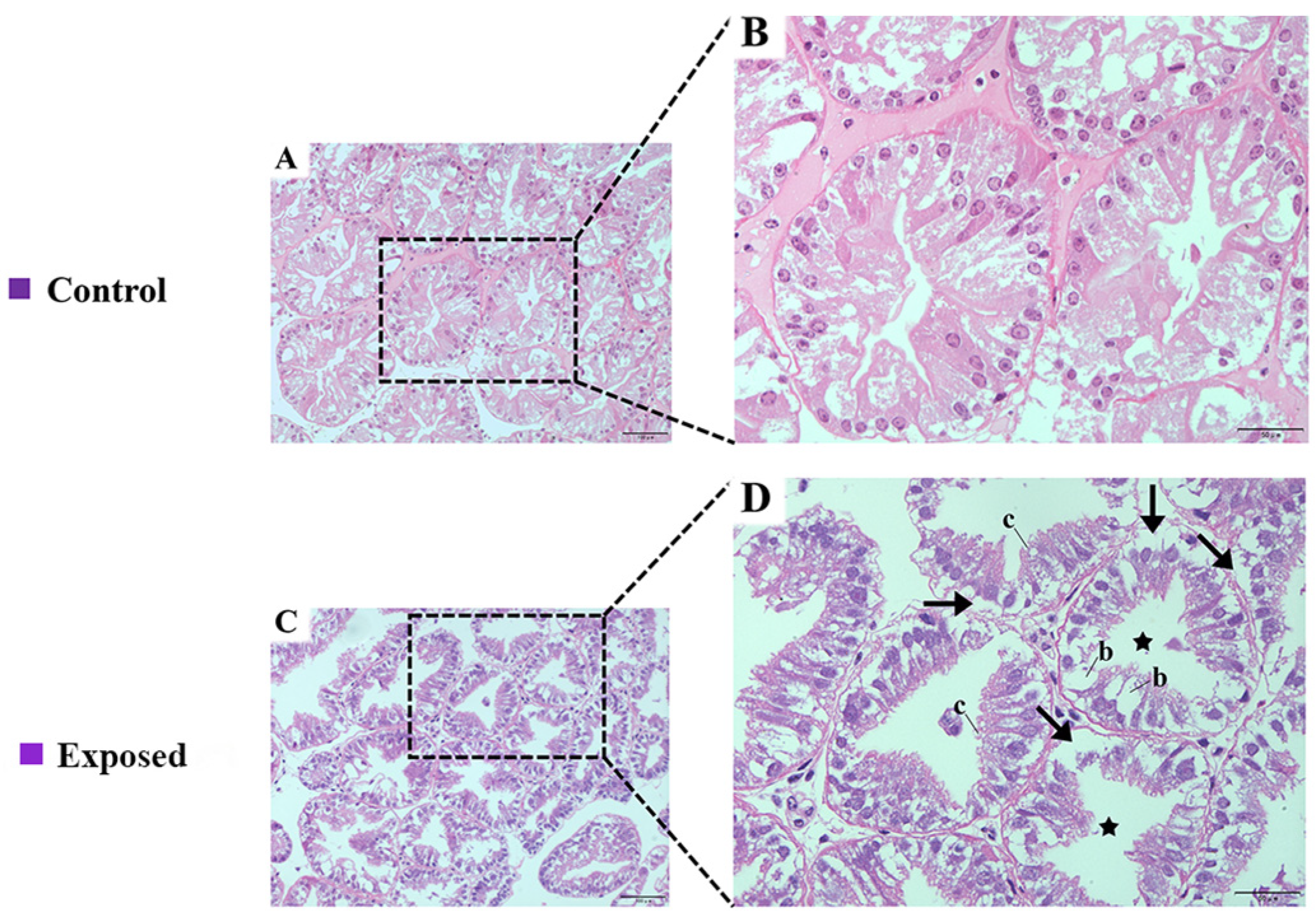
2.1. Histological Assays

Significant histological alterations were observed in the hepatopancreas of E. carinicauda following exposure to A. pacificum. In the control group, the hepatopancreas exhibited well-organized, rounded glandular tubes with an intact cellular morphology. The glandular ducts were also tightly arranged, while the lumen displayed a stellate structure (Figure 1A,B). In the exposed group, after exposing the shrimp to A. pacificum for 7 days, the hepatopancreas showed increased numbers and sizes of B cells, lumen dilation, a higher abundance of R cells, and the separation of the basement membrane from the epithelial layer (Figure 1C,D).

2.2. Antioxidant Parameters

In the exposed group, A. pacificum significantly decreased the levels of glutathione (GSH), catalase (CAT), and superoxide dismutase (SOD) in E. carinicauda, while the malondialdehyde (MDA) content showed a significant increase compared with the control group (p < 0.05; Figure 2A–D). These results indicated that A. pacificum exposure can induce oxidative stress, leading to the depletion of antioxidant defenses and enhanced lipid peroxidation in E. carinicauda.

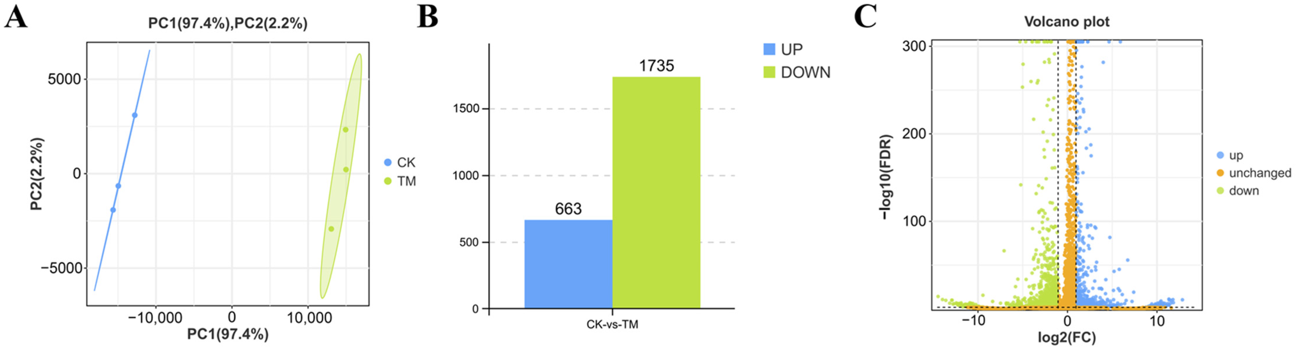
2.3. DEGs in the Hepatopancreas After A. pacificum Exposure

Transcriptome sequencing generated clean reads ranging from 6,322,223,082 to 7,749,082,188 bp, with Q20 and Q30 values reaching 98.26% and 94.78%, respectively. In addition, the GC content ranged from 44.64% to 45.31% (Table S1), hence confirming the reliability of the transcriptome data. PCA further revealed a distinct separation between the samples from the exposed and control groups (Figure 3A), indicating significant differences in the gene expression patterns of their hepatopancreas tissues after being exposed to A. pacificum. Finally, 2398 DEGs, including 663 upregulated and 1735 downregulated genes, were identified in the hepatopancreas (Figure 3B,C). Raw data were uploaded to the National Center for Biotechnology Information (NCBI) (accession number is PRJNA1212343).

2.4. GO Enrichment Analysis

To explore the biological responses of E. carinicauda’s hepatopancreas after being exposed to A. pacificum, a GO enrichment analysis was performed, with the focus being on biological processes, cellular components, and molecular function (Figure 4A). Within the biological process category, 21 level 2 GO terms were enriched following exposure, and these included regulation of the biological process, biological regulation, metabolic process, and cellular process. Similarly, DEGs were primarily associated with protein-containing complex and cellular anatomical entities in the cellular component category, while for molecular function, the top two enriched level 2 GO terms were binding and catalytic activity. Further analysis revealed significant enrichment in 10 GO terms related to protein trafficking and metabolism (Figure 4B), including lipid transporter activity, xenobiotic transmembrane transporter activity, isoprenoid binding, monocarboxylic acid transmembrane transporter activity, xenobiotic transport, transition metal ion transmembrane transporter activity, late endosome, ABC-type transporter activity, neutral amino acid transport, and phosphate ion transport. Finally, the results of the GSEA revealed significant downregulation of the genes involved in these processes in the exposed group (Figure 4C).

2.5. KEGG Enrichment Analysis

A KEGG enrichment analysis identified 1007 DEGs mapped to 5 KEGG Class A and 26 KEGG Class B categories (Figure 5A). Notably, exposure to A. pacificum significantly impacted 20 pathways, including lysosome, endocytosis, metabolic pathways, and the MAPK signaling pathway (Figure 5B). Furthermore, a network plot highlighted the key pathways altered by A. pacificum exposure (Figure 5C), and these included protein processing in the ER (ko04141), lysosome (ko04142), glycolysis/gluconeogenesis (ko00010) and the MAPK signaling pathway (ko04010).

2.6. Differentially Expressed Genes Involved in Different Functions

Table 1 summarizes the genes involved in key metabolic and functional pathways in the hepatopancreas, such as protein processing, mitochondrial function, sphingolipid metabolism, glycerophospholipid metabolism, and glycolysis/gluconeogenesis. Among the 18 genes associated with protein processing, only sec61 translocon subunit beta (SEC61B); lectin, mannose binding 1 (LMAN1); and crystallin, alpha B (CRYAB) were upregulated. In contrast, 11 genes related to mitochondrial function in the hepatopancreas were significantly downregulated. Furthermore, six genes were linked to sphingolipid metabolism in the hepatopancreas, with all but galactose-3-O-sulfotransferase 1 (GAL3ST1) showing downregulation, while among the 11 genes associated with glycerophospholipid metabolism, all, except for n-myristoyltransferase 2 (NMT2), were downregulated. Finally, nine genes related to glycolysis/gluconeogenesis were identified, and these included triosephosphate isomerase 1a (tpi1a), aldehyde dehydrogenase 3 family member A2 (Aldh3a2), enolase (Eno), fructose-1,6-bisphosphatase 1 (FBP1), alcohol dehydrogenase 5 (Class III), chi polypeptide (ADH5), phosphoglycerate kinase (Pgk), lactate dehydrogenase (LDH), aldehyde dehydrogenase 3 family member a1 (Aldh3a1), and phosphoenolpyruvate carboxykinase 2, mitochondrial (PCK2).

2.7. Validation of Gene Expression Through Quantitative Reverse-Transcription PCR (qRT-PCR)

To validate the RNA-seq results, 20 DEGs involved in protein processing, mitochondrial function, sphingolipid metabolism, glycerophospholipid metabolism, and glycolysis/gluconeogenesis were selected. The expression levels of these genes were then analyzed by qRT-PCR, with the results being consistent with those observed in the RNA-seq analysis (Figure 6).

3. Discussion

The dinoflagellate A. pacificum, a typical HAB species, leads to significant environmental issues, substantial economic losses in the aquaculture industry, and potential threats to human health [15,26]. Crustaceans are particularly sensitive to environmental changes, thus making shrimp ponds effective early warning signs for HABs. However, limited information is available regarding the detrimental effects of A. pacificum on E. carinicauda. Therefore, the current work investigated the adverse effects of A. pacificum on the hepatopancreas of E. carinicauda by examining the organ’s histological structure, antioxidant enzyme activity, and transcriptomic profile.
The hepatopancreas is a vital organ in shrimp as it serves as the primary site for metabolic processes and detoxification while being also a potential target for diseases [27]. Research has shown that environmental stress can induce oxidative damage in the hepatopancreas, thereby increasing its susceptibility to apoptosis [28]. For instance, acute nitrite stress in Litopenaeus vannamei caused abnormal star-shaped tubular structures in its hepatopancreas, with hepatic tubules also undergoing atrophy and detachment from the basement membrane [29]. Similarly, a 21-day exposure to nanopolystyrene and phosphorus oxide in Eriocheir sinensis compromised the mechanical barrier of the hepatopancreas [30]. In the exposed group, E. carinicauda exhibited pathological hepatopancreatic damage, including lumen expansion and the detachment of the epithelial cells from the basement membrane. It was, therefore, hypothesized that A. pacificum can disrupt physiological homeostasis in the hepatopancreas of E. carinicauda, with the observed tissue damage likely due to its role as a primary target organ for toxic dinoflagellates.
Environmental stressors, including toxic contaminants, are well-documented inducers of oxidative stress in aquatic organisms [31]. Antioxidant enzymes, such as SOD, CAT, and glutathione-dependent ones, play a crucial role in mitigating oxidative damage caused by reactive oxygen species (ROS). Thus, under biotic and abiotic stress conditions, the overexpression of these enzymes often serves as a defense mechanism [32]. However, this study showed that the activities of SOD, CAT, and GSH in the hepatopancreas of E. carinicauda were significantly reduced following exposure to A. pacificum. Similar findings, notably a significant decrease in SOD and CAT activities, were reported in L. vannamei which had been exposed to nitrite and alkaline environmental stress [28,33]. In the exposed group, the observed reduction in antioxidant enzyme activity could be attributed to the inhibitory effects of high concentrations of toxic A. pacificum on enzymatic function. This inhibition subsequently compromises the antioxidant defense system, resulting in increased levels of lipid peroxidation. The damage to hepatopancreatic tissues also likely disrupts cellular defense mechanisms, including antioxidant pathways [34].

3.1. Key Genes Associated with Protein Processing and Mitochondrial Function

Environmental stressors are known to impair the structure and function of protein-folding enzymes, thus reducing their capacity to achieve the proper folding of newly synthesized proteins. This dysfunction also results in the accumulation of misfolded proteins within the endoplasmic reticulum [35]. In this study, significant alterations were observed in the genes associated with protein processing. Among these, dnaJ heat shock protein family (Hsp40) member A2 (DNAJA2) and dnaJ heat shock protein family (Hsp40) member C10 (DNAJC10) are well-characterized molecular chaperones that play crucial roles in protein folding and serve as biomarkers of environmental stress sensitivity [36]. Specifically, DNAJA2 is involved in protein folding and the mitochondrial import of proteins, thus ensuring the proper folding of proteins and preventing the accumulation of misfolded variants. Conversely, DNAJC10 is a part of the endoplasmic reticulum-associated degradation complex which identifies and degrades misfolded proteins, thereby reducing the formation of improper disulfide bonds in misfolded glycoproteins. Studies on Procambarus clarkii have shown that high-temperature stress can downregulate genes involved in protein folding and processing in the hepatopancreas, thus leading to the accumulation of misfolded proteins [37]. In this study, the expression levels of DNAJA2 and DNAJC10 were significantly downregulated in the hepatopancreas of E. carinicauda. In this context, it was hypothesized that exposure to the toxic dinoflagellates A. pacificum may lead to the accumulation of misfolded proteins, disruption of the regulatory mechanisms of E. carinicauda, damage to hepatopancreatic tissues, and impaired physiological functions of hepatopancreatic cells [38].
Environmental stress often triggers the accumulation of ROS in the hepatopancreas, with this process potentially leading to mitochondrial dysfunction [39]. The ubiquitin c (UBC) modifies mitochondrial proteins through ubiquitination, thereby playing a critical role in mitochondrial quality control and mitophagy [40]. Similarly, ubiquitin-specific protease 8 (USP8) can remove K6-linked ubiquitin chains formed by Parkin and regulate the subsequent mitophagy process. Without USP8, aggregated ubiquitin chains hinder Parkin’s interactions with other receptors, such as sequestosome 1 (SQSTM1) and microtubule-associated protein 1 light chain 3 alpha (MAP1LC3A), thereby disrupting mitochondrial autophagy [41]. Previous studies have highlighted that an imbalance in mitochondrial homeostasis can exacerbate oxidative stress and apoptosis while disrupting overall organismal homeostasis [42]. For instance, in L. vannamei, reduced SQSTM1 levels under low-temperature stress can intensify hepatopancreas damage [43]. Additionally, in mitten crabs, increased MAP1LC3A protein levels have been observed following exposure to microcystin-LR, hence underscoring the crucial role of mitochondrial regulation in stress responses [44]. In this study, the expression levels of UBC, USP8, SQSTM1, and MAP1LC3A, which are involved in mitochondrial function, were all significantly downregulated in the exposed group. It was speculated that oxidative stress induced by A. pacificum could compromise the cells’ ability to effectively manage environmental stressors, leading to hepatopancreatic damage. Impaired mitochondrial function may also hinder cellular adaptation to changes in the external environment, potentially activating apoptotic pathways and increasing cell death. This cascade of events may adversely impact the growth and development of shrimp. Furthermore, damage to hepatopancreatic tissues can disrupt regulatory mechanisms within this organ, thereby affecting its normal physiological functions [45].

3.2. Key Genes Related to Energy Metabolism

Energy is essential for sustaining normal life activities, and in the case of crustaceans, regulating energy metabolism enables their adaptation to environmental changes [46]. In this context, the hepatopancreas is a crucial organ for energy storage and metabolism, providing the necessary energy for stress responses [47]. Glycolysis, a vital pathway for carbohydrate metabolism, is widespread among animals, with its key purpose being the production of ATP under hypoxic conditions to fulfill the energy demands of cells devoid of mitochondria or during environmental stress [48]. Among the key components in this process, Pgk is the first significant metabolic enzyme responsible for ATP generation during glycolysis. A transcriptome analysis undertaken in this study revealed that the expression levels of tpi1a, Eno, and Pgk, all of which are involved in the glycolytic pathway, were upregulated, while LDH expression was downregulated. LDH is a critical enzyme in the glycolytic pathway as it facilitates the interconversion between pyruvate and lactate. Previous studies have demonstrated that the upregulation of LDH activated the glycolysis pathway under environmental stress [49]. Conversely, in this study, the downregulation of LDH in the hepatopancreas of the exposed group could be attributed to a cellular shift towards the gluconeogenesis pathway as a means of adapting to environmental changes. This adjustment to maintain the balance of energy and metabolites could also be linked to structural damages and impaired physiological functions observed in the hepatopancreatic cells after exposure to A. pacificum. Gluconeogenesis is another critical pathway that generates ATP by converting non-carbohydrate substances into glucose under aerobic conditions [50]. PCK2, a key enzyme in this process, serves as a central molecule linking the TCA cycle, glycolysis, and gluconeogenesis [51]. Meanwhile, fructose-1,6-bisphosphatase 1 (FBP1) acts as a crucial regulator of the gluconeogenesis pathway, facilitating the hydrolysis of fructose 1,6-bisphosphate into fructose 6-phosphate and inorganic phosphate, thereby controlling the rate of gluconeogenesis [52]. The current findings suggested that the downregulation of PCK2 could be attributed to the disruption of hepatopancreatic energy metabolism caused by exposure to A. pacificum, while FBP1 upregulation may result from the activation of the gluconeogenesis pathway during glycolysis to adapt to environmental changes. Previous studies suggested that low temperature stress may downregulate FBP1 expression in the hepatopancreas of L. vannamei, thereby inhibiting the gluconeogenesis pathway while activating the glycolysis pathway to meet energy demands under such conditions [53]. Additionally, research indicated that the increased expression of the genes related to energy metabolism in the shrimp hepatopancreas was essential for maintaining normal physiological functions and enhancing tolerance to environmental changes [54].
This study observed a significant downregulation of ceramide synthase 6 (CERS6), 1-acylglycerol-3-phosphate o-acyltransferase 1 (AGPAT1), and 1-acylglycerol-3-phosphate o-acyltransferase 4 (AGPAT4), the genes involved in sphingolipid and glycerophospholipid metabolism. Sphingolipid metabolism, a crucial component of lipid metabolism, plays a key role in maintaining cellular homeostasis and responding to environmental stressors [55]. The hepatopancreas, the site of ceramide production, is rich in sphingolipids and highly sensitive to environmental changes. For instance, studies have shown that a reduction in CERS6 expression directly reduced the activity of the encoded enzyme, thereby diminishing the synthesis of specific chain-length ceramides [56]. Pauletto et al. (2018) further found that after aeration stress, CERS6 expression in Pecten maximus larvae was lower than that of the control group, suggesting that stress conditions may impair the efficiency of lipid metabolism and lead to delayed growth [57]. Animal cell membranes are also rich in glycerophospholipids which are critical to their structure and function [58], with their metabolism being closely related to biological functions and oxidative stress [59]. Furthermore, under oxidative stress, glycerophospholipid metabolism helps to maintain the cell membrane’s stability, and this process is associated with lipid peroxidation and stress mechanisms [60]. Notably, phosphatidic acid (PA), produced by AGPATs, serves as a key metabolite in glycerophospholipid metabolism, with AGPATs being central to this process [61]. The hepatopancreas is vital for crustacean functions, such as lipid metabolism which is essential for growth [62]. The current study found that CERS6, AGPAT1 and AGPAT4 were all downregulated in the hepatopancreas of E. carinicauda following exposure to the toxic dinoflagellates A. pacificum. In particular, the downregulation of CERS6 is speculated to be due to an imbalance in sphingolipid levels which disrupts the equilibrium between cholesterol and sphingolipids in the plasma membrane. Such disruption may further alter nutrient availability, thereby affecting the overall cellular homeostasis and function, potentially leading to metabolic disorders. Concurrently, the downregulation of AGPAT1 and AGPAT4 expression was expected for lowering the synthesis of PA. This may impact phospholipid synthesis and lead to changes in intracellular lipid storage, which may further affect cell growth and survival. Collectively, these findings suggest that the toxin-producing dinoflagellates A. pacificum may induce energy metabolism disorders in E. carinicauda, thereby interfering with their normal growth and development. This process may also be associated with the production of ROS [63].

4. Materials and Methods

4.1. Shrimp Selection and Culture Conditions

Experimental E. carinicauda, with an average weight and body length of 0.8 ± 0.02 g and 5.14 ± 0.38 cm, respectively, were selected from a healthy population sourced from a farm in Lianyungang city, Jiangsu Province, China. Before the experiment, the shrimp were allowed to acclimatize for one week in 40 L plastic tanks with continuous aeration, and during this period, one-third to one-half of the water was replaced daily using a drip method. In addition, the culture conditions were maintained at a pH, salinity, and temperature of 8.0–8.2, 24–26, and 25 ± 1 °C, respectively. The shrimp were fed a commercial diet at regular intervals each day, and the dead ones or waste materials were promptly removed.

4.2. Alexandrium Pacificum Culture

A. pacificum was cultured in 5 L Erlenmeyer flasks containing f/2 medium without silicate (Si). Batch cultivation was then conducted at a temperature of 20 ± 1 °C and under a light intensity of 60 ± 10 μEm−2 s−1 with a 14 h:10 h light-to-dark photoperiod. The culture medium was prepared using natural seawater (salinity of 25) which was collected from the Lianyungang area, filtered through a 0.5 μm membrane, and eventually sterilized under high temperature and pressure before use. Prior to the experiment, the algae were fixed with Lugol’s fixative and counted using a 1 mL plankton counting chamber.

4.3. Experimental Design and Sample Collection

After one week of acclimation, the shrimp were randomly assigned to one of the following two groups: one exposed to A. pacificum at a concentration of 1.0 × 10⁴ cells/mL (exposed group) and control group. Each group consisted of three replicates, with 40 E. carinicauda individuals randomly assigned per replicate. Every 12 h, the algal cells were fixed using Lugol’s fixative and counted under a microscope with a 1 mL plankton counting chamber. In addition, the culture medium was replaced every 24 h to maintain the concentration of A. pacificum at the designated level. The experiment lasted 7 days, during which the concentration in the exposed group was consistently maintained at 1.0 × 10⁴ cells/mL. At the end of the experimental period, hepatopancreas tissues were collected from nine randomly selected E. carinicauda individuals from each group. These tissues were then homogenized with 9 volumes (w/v) of physiological saline at 4 °C for the subsequent biochemical analysis. Similarly, hepatopancreas tissues obtained from nine randomly selected shrimp from each group were used for the transcriptome analysis. In this case, tissues from three shrimp were pooled into a single sample, with all the samples eventually stored at −80 °C for further analyses.

4.4. Histopathological Analyses

After the seven-day exposure, hepatopancreas tissues were collected from nine E. carinicauda individuals from each group. These tissue samples were preserved in paraformaldehyde for 24 h, after which dehydration was performed using 70% ethanol to remove residual water. This was followed by xylene treatment to facilitate paraffin infiltration, with the tissues eventually embedded in melted paraffin for thorough impregnation. The paraffin-embedded samples were then sliced into 4–6 μm thick sections using a microtome and mounted onto glass slides for drying. These sections were subsequently stained with hematoxylin and eosin (H&E) in order to visualize tissue alterations under an optical microscope.

4.5. Measurement of Hepatopancreas Antioxidant Parameters

Hepatopancreas tissues were accurately weighed and homogenized (1:9, v/w) in 0.85% ice-cold saline solution [64] prior to a 10 min centrifugation at 3000 rpm and 4 °C. The resulting supernatant was then stored at −80 °C until required for the subsequent determination of the total protein content. Additionally, malondialdehyde (MDA, A003-1-2, TBA) and glutathione (GSH, A006-2-1) levels as well as the activities of catalase (CAT, A007-1-1, visible light) and superoxide dismutase (SOD, A001-3-2, WST-1) in the hepatopancreas were determined, with commercial kits (Jiancheng Bioengineering Institute, Nanjing, China) used to measure all the enzyme activities.

4.6. Transcriptome Sequencing in Hepatopancreas

A UNlQ-10 Column Trizol Total RNA Isolation Kit was used to extract total RNA from the hepatopancreas tissues of E. carinicauda individuals of the control and exposed groups. The RNA quality was then assessed before synthesizing cDNA through reverse transcription. Following the purification of the double-stranded cDNA, a library was constructed prior to sequencing on an Illumina Nova 6000 system (Gene Denovo Biotechnology Co., Ltd., Guangzhou, China). Quality control of the raw sequences was performed using FASTP (version 0.18.0) to remove low-quality reads [65]. Clean reads were aligned to the reference genome assembly [66] using the HISAT v2.0 program. The resulting high-quality data were then subjected to a principal component analysis (PCA) to evaluate the relationship among the samples. Additionally, the DESeq2 software (version 1.20.0) was used to identify differentially expressed genes (DEGs) between the exposed and control groups, with an absolute log2 fold change (log2 FC) of ≥2 and a false discovery rate (FDR) of <0.05 selected as screening thresholds [67]. Biological functions and key signaling pathways were also explored through Gene Ontology (GO) (http://www.geneontology.org/, accessed on 23 October 2024) and Kyoto Encyclopedia of Genes and Genomes (KEGG) enrichment analyses (http://www.kegg.jp/, accessed on 23 October 2024) before using the Gene Set Enrichment Analysis (GSEA) (version 2.2.4) for identifying distinct GO terms and pathways between the exposed and control groups. In this case, an FDR of <0.05, a p-value of <0.05, and a normalized enrichment score (NES) of >1 were selected as the threshold for significance [68].

4.7. Quantitative Reverse-Transcription PCR (qRT-PCR)

The qRT-PCR analysis was performed to validate the accuracy of the transcriptome data. For this purpose, RNA was extracted and subsequently reverse transcribed into cDNA using the UNlQ-10 column-based Trizol Total RNA Extraction Kit (Shenggong Biological Engineering Co., Ltd., Shanghai, China) and the Trans Script® One-Step gDNA Removal and cDNA Synthesis Super Mix (TransGen Biotech Co., Ltd., Beijing, China), respectively. This was followed by qPCR, performed on a Multiwell Plate 96 reaction system (LightCycler® 96, Roche, Basel, Switzerland) using the TransGen Biotech Perfect Start® Green qPCR Super Mix kit (TransGen Biotech Co., Ltd., Beijing, China) according to the provided instructions. The 2−ΔΔCT method was eventually used to assess the relative expression levels of each target gene, with 18S selected as the reference [69]. All the primers used in this study were designed using Primer Premier 5.0 and are listed in Table S2.

4.8. Statistical Analysis

The results were statistically analyzed in the SPSS 27.0 software using one-way analysis of variance (ANOVA) and Duncan’s multiple range test to determine their significance at p < 0.05.

5. Conclusions

In this study, the toxicological effects of A. pacificum on the hepatopancreas of E. carinicauda were thoroughly examined through histological analysis, antioxidant enzyme activity assays, and transcriptomic profiling. The findings indicated that exposure to A. pacificum resulted in significant histological damage along with significant changes in antioxidant parameters, such as SOD, CAT, GSH, and MDA, which were indicative of hepatic dysfunctions. The transcriptomic analyses further identified 2398 differentially expressed genes that were primarily involved in protein processing, mitophagy, mitochondrial function, sphingolipid metabolism, and glycerophospholipid metabolism. From the results, it was hypothesized that A. pacificum likely exerted its toxicological effects by disturbing hepatic protein processing, mitophagy, mitochondrial function, and energy metabolism. Altogether, these findings offer new insights into the adverse impact of harmful toxin-producing dinoflagellates on aquatic animals while also assisting in the ecological risk assessment of harmful algal blooms in aquaculture.

Supplementary Materials

The following supporting information can be downloaded at https://www.mdpi.com/article/10.3390/ijms26041605/s1.

Author Contributions

Conceptualization, W.H. and W.C.; methodology, W.H., W.C., D.L., Y.C. and J.W.; software, W.H. and W.C.; validation, X.M. and J.W.; formal analysis, M.F., D.L. and Y.C.; investigation, M.F.; resources, M.F. and X.M.; data curation, W.C., D.L. and Y.C.; writing—original draft preparation, W.H. and G.H.; writing—review and editing, W.H. and G.H.; visualization, G.H., N.J. and H.G.; supervision, N.J. and H.G.; project administration, G.H., N.J. and H.G.; funding acquisition, G.H., N.J. and H.G. All authors have read and agreed to the published version of the manuscript.

Funding

This research was funded by the Jiangsu Provincial Qinglan Project (2024-2027), the Lianyungang Key Research and Development Program (No. SF2303, JCYJ2419), the Jiangsu Ocean Resources Development Technology Innovation Center Open Fund (No. LWJJ-03), the Lianyungang 521 Talent Projects (#2021-1021), the “JBGS” Project of Seed Industry Revitalization in Jiangsu Province (JBGS (2021)124), the Priority Academic Program Development of Jiangsu Higher Education Institutions (PAPD), and the Jiangsu Students’ innovation and entrepreneurship training program (No. 202311641038Z).

Institutional Review Board Statement

All the procedures were approved by the Animal Care and Use Committee of Jiangsu Ocean University (protocol No. 2020-37; approval date: 1 September 2019).

Informed Consent Statement

Not applicable.

Data Availability Statement

In this study, all the data generated are included in this article and Supplementary Materials. Further enquiries can be directed to the corresponding author.

Conflicts of Interest

The authors declare no conflicts of interest.

References

  1. Li, J. Comparative Transcriptomic and Proteomic Analysis of Exopalaemon carinicauda in Response to Alkalinity Stress. Front. Mar. Sci. 2021, 8, 759923. [Google Scholar]
  2. Feng, Y.; Zhai, Q.; Wang, J.; Li, J.; Li, J. Comparison of florfenicol pharmacokinetics in Exopalaemon carinicauda at different temperatures and administration routes. J. Vet. Pharmacol. Therap. 2019, 42, 230–238. [Google Scholar] [CrossRef]
  3. Ge, Q.; Li, J.; Wang, J.; Li, Z.; Li, J. Characterization, functional analysis, and expression levels of three carbonic anhydrases in response to pH and saline–alkaline stresses in the ridgetail white prawn Exopalaemon carinicauda. Cell Stress Chaperones 2019, 24, 503–515. [Google Scholar] [CrossRef] [PubMed]
  4. Li, J.; Ma, P.; Liu, P.; Chen, P.; Li, J. The roles of Na+/K+-ATPase α-subunit gene from the ridgetail white prawn Exopalaemon carinicauda in response to salinity stresses. Fish Shellfish. Immunol. 2015, 42, 264–271. [Google Scholar] [CrossRef]
  5. Zhang, X.-W. Cloning and characterization of two different ficolins from the giant freshwater prawn Macrobrachium rosenbergii. Dev. Comp. Immunol. 2014, 44, 359–369. [Google Scholar] [CrossRef] [PubMed]
  6. Li, L.; Meng, H.; Gu, D.; Li, Y.; Jia, M. Molecular mechanisms of Vibrio parahaemolyticus pathogenesis. Microbiol. Res. 2019, 222, 43–51. [Google Scholar] [CrossRef]
  7. Lee, C.; Kim, J.H.; Choi, S.-K.; Jeon, H.J.; Lee, S.H.; Kim, B.K.; Kim, Y.K.; Lee, K.-J.; Han, J.E. Detection of infectious white spot syndrome virus in red claw crayfish (Cherax quadricarinatus) and red swamp crayfish (Procambarus clarkii) imported into Korea. Aquaculture 2021, 544, 737117. [Google Scholar] [CrossRef]
  8. Wang, L.; Li, F.; Wang, B.; Xiang, J. A new shrimp peritrophin-like gene from Exopalaemon carinicauda involved in white spot syndrome virus (WSSV) infection. Fish Shellfish. Immunol. 2013, 35, 840–846. [Google Scholar] [CrossRef]
  9. Mu, C.; Ren, X.; Ge, Q.; Wang, J.; Li, J. Antioxidant response of ridgetail white prawn Exopalaemon carinicauda to harmful dinoflagellate Prorocentrum minimum exposure and its histological change. J. Ocean Univ. China 2017, 16, 285–293. [Google Scholar] [CrossRef]
  10. Mu, C.; Ren, X.; Li, J. Immune response of the ridgetail white prawn Exopalaemon carinicauda after exposure to the dinoflagellate Prorocentrum minimum. J. Ocean Univ. China 2023, 22, 821–830. [Google Scholar] [CrossRef]
  11. Taylor, F.J.R.; Hoppenrath, M.; Saldarriaga, J.F. Dinoflagellate diversity and distribution. Biodivers. Conserv. 2008, 17, 407–418. [Google Scholar]
  12. Wang, H.; Kim, H.; Ki, J.-S. Transcriptome survey, molecular identification, and expression analysis of stress-responsive genes in the toxic dinoflagellate Alexandrium pacificum under algicidal agents and metal stresses. J. Appl. Phycol. 2021, 33, 3139–3151. [Google Scholar]
  13. Alonso-Rodrıguez, R.; Paez-Osuna, F. Nutrients, phytoplankton and harmful algal blooms in shrimp ponds: A review with special reference to the situation in the Gulf of California. Aquaculture 2003, 219, 317–336. [Google Scholar] [CrossRef]
  14. Delgado, J.M.V.; Pólit, P.A.; Panta-Vélez, R.P.; Rodríguez-Díaz, J.M.; Dapena, J.D.; Lozano, A.L.; Maddela, N.R. Identification and composition of Cyanobacteria in ecuadorian shrimp farming ponds—Possible risk to human health. Curr. Microbiol. 2024, 81, 237. [Google Scholar]
  15. Hadjadji, I. A comparative analysis of Alexandrium catenella/tamarense blooms in Annaba Bay (Algeria) and Thau Lagoon (France); phosphorus limitation as a trigger. Comptes Rendus Biol. 2014, 337, 117–122. [Google Scholar]
  16. Yan, T.; Zhou, M.; Fu, M.; Yu, R.; Wang, Y.; Li, J. Effects of the dinoflagellate Alexandrium tamarense on early development of the scallop Argopecten irradians concentricus. Aquaculture 2003, 217, 167–178. [Google Scholar]
  17. Rolton, A.; Rhodes, L.; Hutson, K.S.; Biessy, L.; Bui, T.; MacKenzie, L.; Symonds, J.E.; Smith, K.F. Effects of harmful algal blooms on fish and shellfish species: A case study of New Zealand in a Changing environment. Toxins 2022, 14, 341. [Google Scholar] [CrossRef] [PubMed]
  18. Roncalli, V. The effect of the toxic dinoflagellate Alexandrium fundyense on the fitness of the calanoid copepod Calanus finmarchicus. Harmful Algae 2016, 51, 56–66. [Google Scholar] [CrossRef]
  19. Beauclercq, S.; Grenier, O.; Arnold, A.A.; Warschawski, D.E.; Wikfors, G.H.; Genard, B.; Tremblay, R.; Marcotte, I. Metabolomics and lipidomics reveal the effects of the toxic dinoflagellate Alexandrium catenella on immune cells of the blue mussel, mytilus edulis. Harmful Algae 2023, 129, 102529. [Google Scholar]
  20. Zhu, Z.; Qi, J.; Liu, Y.; Sui, Z. The H3K79 methylase DOT1, unreported in photosynthetic plants, exists in Alexandrium pacificum and participates in its growth regulation. Mar. Pollut. Bull. 2023, 190, 114867. [Google Scholar] [CrossRef]
  21. Li, Q.; Liu, X.; Li, L.; Ge, C.; Jian, L. A comprehensive analysis of hepatopancreas metabolomics and transcriptomics provides insights into the growth of three-year-old crabs (Eriocheir sinensis) under low temperature. Comp. Biochem. Physiol. Part D Genom. Proteom. 2024, 49, 101182. [Google Scholar]
  22. Jiang, Y.; Liu, X.; Han, H.; Shang, Y.; Li, J.; Gao, B.; Ren, Y.; Meng, X. Temporal and tissue-specific regulation of energy metabolism in the swimming crab Portunus trituberculatus during nitrite stress and recovery. Mar. Pollut. Bull. 2024, 208, 117024. [Google Scholar] [PubMed]
  23. Vogt, G. Functional cytology of the hepatopancreas of decapod crustaceans. J. Morphol. 2019, 280, 1405–1444. [Google Scholar] [CrossRef]
  24. Shi, K.; Li, J.; Lv, J.; Liu, P.; Li, J.; Li, S. Full-length transcriptome sequences of ridgetail white prawn Exopalaemon carinicauda provide insight into gene expression dynamics during thermal stress. Sci. Total. Environ. 2020, 747, 141238. [Google Scholar]
  25. Li, W.; Wang, J.; Li, J.; Liu, P.; Fei, F.; Liu, B.; Li, J. The effect of astaxanthin on the alkalinity stress resistance of Exopalaemon carinicauda. Sci. Total. Environ. 2024, 917, 170415. [Google Scholar]
  26. Anderson, D.M.; Alpermann, T.J.; Cembella, A.D.; Collos, Y.; Masseret, E.; Montresor, M. The globally distributed genus Alexandrium: Multifaceted roles in marine ecosystems and impacts on human health. Harmful Algae 2012, 14, 10–35. [Google Scholar] [PubMed]
  27. Ning, M.; Wei, P.; Shen, H.; Wan, X.; Jin, M.; Li, X.; Shi, H.; Qiao, Y.; Jiang, G.; Gu, W.; et al. Proteomic and metabolomic responses in hepatopancreas of whiteleg shrimp Litopenaeus vannamei infected by microsporidian Enterocytozoon hepatopenaei. Fish Shellfish. Immunol. 2019, 87, 534–545. [Google Scholar] [CrossRef]
  28. Lin, L.; Zhang, Y.; Zhuo, H.; Li, J.; Fu, S.; Zhou, X.; Wu, G.; Guo, C.; Liu, J. Integrated histological, physiological, and transcriptome analysis reveals the post-exposure recovery mechanism of nitrite in Litopenaeus vannamei. Ecotoxicol. Environ. Saf. 2024, 281, 116673. [Google Scholar]
  29. Duan, Y.; Zhong, G.; Xiao, M.; Yang, Y.; Wang, Y.; Nan, Y. Integrated physiological, energy metabolism, and metabonomic responses indicate the stress response in the hepatopancreas of Litopenaeus vannamei to nitrite stress. Aquat. Toxicol. 2024, 277, 107164. [Google Scholar] [PubMed]
  30. Huang, M.; Ma, Y.; Che, S.; Shen, L.; Wan, Z.; Su, S.; Ding, S.; Li, X. Nanopolystyrene and phoxim pollution: A threat to hepatopancreas toxicity in chinese mitten crab (Eriocheir sinensis). Aquat. Toxicol. 2024, 276, 107124. [Google Scholar]
  31. Pinto, E.; Sigaud-Kutner, T.C.S.; Leitao, M.A.S.; Okamoto, O.K.; Morse, D. Heavy metal–induced oxidative stress in algae. J. Phycol. 2003, 39, 1008–1018. [Google Scholar] [CrossRef]
  32. Leng, X. Comparative transcriptome analysis of grapevine in response to copper stress. Sci. Rep. 2015, 5, 17749. [Google Scholar] [CrossRef] [PubMed]
  33. Zhang, R.; Shi, X.; Guo, J.; Mao, X.; Fan, B. Acute stress response in gill of pacific white shrimp Litopenaeus vannamei to high alkalinity. Aquaculture 2024, 586, 740766. [Google Scholar] [CrossRef]
  34. Chen, Y.-T.; Xu, R.-Q.; Cheng, J.-W.; Singhania, R.R.; Chen, C.-W.; Dong, C.-D.; Hsieh, S.-L. Immunotoxicity and oxidative damage in Litopenaeus vannamei induced by polyethylene microplastics and copper co-exposure. Mar. Pollut. Bull. 2024, 205, 116683. [Google Scholar] [CrossRef] [PubMed]
  35. Urade, R. Oxidative protein folding in the plant endoplasmic reticulum. Biosci. Biotechnol. Biochem. 2019, 83, 781–793. [Google Scholar] [CrossRef] [PubMed]
  36. Gupta, S.C.; Sharma, A.; Mishra, M.; Mishra, R.K.; Chowdhuri, D.K. Heat shock proteins in toxicology: How close and how far? Life. Sci. 2010, 86, 377–384. [Google Scholar] [CrossRef] [PubMed]
  37. Zou, Y.; Cao, P.; Bao, Z.; Xu, Y.; Xu, Z.; Guo, H. Histological, physiological and transcriptomic analysis in hepatopancreas of Procambarus clarkii under heat stress. Ecotoxicol. Environ. Saf. 2024, 289, 117459. [Google Scholar] [CrossRef]
  38. Zeng, Y.; Deng, B.; Kang, Z.; Araujo, P.; Mjøs, S.A.; Liu, R.; Lin, J.; Yang, T.; Qu, Y. Tissue accumulation of polystyrene microplastics causes oxidative stress, hepatopancreatic injury and metabolome alterations in Litopenaeus vannamei. Ecotoxicol. Environ. Saf. 2023, 256, 114871. [Google Scholar] [CrossRef]
  39. Pu, C.; Liu, Y.; Zhu, J.; Ma, J.; Cui, M.; Mehdi, O.M.; Wang, B.; Wang, A.; Zhang, C. Mechanisms insights into bisphenol S-induced oxidative stress, lipid metabolism disruption, and autophagy dysfunction in freshwater crayfish. J. Hazard. Mater. 2024, 479, 135704. [Google Scholar] [CrossRef]
  40. Ravanelli, S.; den Brave, F.; Hoppe, T. Mitochondrial quality control governed by ubiquitin. Front. Cell Dev. Biol. 2020, 8, 270. [Google Scholar] [CrossRef]
  41. Durcan, T.M.; Tang, M.Y.; Pérusse, J.R.; Dashti, E.A.; Aguileta, M.A.; McLelland, G.; Gros, P.; Shaler, T.A.; Faubert, D.; Coulombe, B.; et al. USP8 regulates mitophagy by removing K6-linked ubiquitin conjugates from parkin. EMBO. J. 2014, 33, 2473–2491. [Google Scholar] [CrossRef]
  42. Liu, Y.; Cao, Y.; Li, H.; Liu, H.; Chen, T.; Lin, Q.; Gong, C.; Yu, F.; Cai, H.; Jin, L.; et al. Mitochondrial homeostatic imbalance-mediated developmental toxicity to H2S in embryonic zebrafish. Environ. Pollut. 2024, 367, 125588. [Google Scholar] [CrossRef]
  43. Dong, W.; Liao, M.; Zhuang, X.; Huang, L.; Liu, C.; Wang, F.; Yin, X.; Liu, Y.; Liang, Q.; Wang, W. MYC drives autophagy to adapt to stress in Penaeus vannamei. Fish Shellfish. Immunol. 2022, 126, 187–196. [Google Scholar] [CrossRef]
  44. Zhang, Y.; Gao, J.; Cao, L.; Du, J.; Xu, G.; Xu, P. Microcystin-LR-induced autophagy via miR-282–5p/PIK3R1 pathway in Eriocheir sinensis hepatopancreas. Ecotoxicol. Environ. Saf. 2023, 267, 115661. [Google Scholar] [CrossRef] [PubMed]
  45. Tan, Y.; Dong, J.; Wang, L.; Li, W.; Bao, J.; Jiang, H. Chronic chlorpyrifos exposure induces oxidative stress, neurological damage, and hepatopancreas enrichment in chinese mitten crab (Eriocheir sinensis). Comp. Biochem. Physiol. Part C Toxicol. Pharmacol. 2024, 289, 110111. [Google Scholar] [CrossRef]
  46. Sun, S. Transciptomic and histological analysis of hepatopancreas, muscle and gill tissues of oriental river prawn (Macrobrachium nipponense) in response to chronic hypoxia. BMC Genom. 2015, 16, 491. [Google Scholar] [CrossRef]
  47. Shang, X.; Geng, L.; Che, X.; Li, W.; Liu, Y.; Li, J.; Teng, X.; Xu, W. Analysis revealed the molecular mechanism of oxidative stress- autophagy-induced liver injury caused by high alkalinity: Integrated whole hepatic transcriptome and metabolome. Front. Immunol. 2024, 15, 1431224. [Google Scholar] [CrossRef]
  48. Chandel, N.S. Glycolysis. Cold. Spring. Harb. Perspect. Biol. 2021, 13, a040535. [Google Scholar] [CrossRef] [PubMed]
  49. Wang, Z. Histology, physiology, and glucose and lipid metabolism of Lateolabrax maculatus under low temperature stress. J. Therm. Biol. 2022, 104, 103161. [Google Scholar] [CrossRef] [PubMed]
  50. Holeček, M. Roles of malate and aspartate in gluconeogenesis in various physiological and pathological states. Metabolism 2023, 145, 155614. [Google Scholar] [CrossRef] [PubMed]
  51. Shang, Z.; Zhao, H.; Yang, T.; Xue, X.; Zhao, B.; Sun, Y. Prognostic and immunological significance of key gluconeogenesis regulators, PCK1 and PCK2, in human cancers especially kidney renal clear cell carcinoma: Insights from pan-cancer analysis, PCKs signature construction, and in vitro experiments. J. Radiat. Res. Appl. Sci. 2023, 16, 100614. [Google Scholar] [CrossRef]
  52. Gu, L.; Zhu, Y.; Watari, K.; Lee, M.; Liu, J.; Perez, S.; Thai, M.; Mayfield, J.E.; Zhang, B.; Cunha E Rocha, K.; et al. Fructose-1,6-bisphosphatase is a nonenzymatic safety valve that curtails AKT activation to prevent insulin hyperresponsiveness. Cell Metab. 2023, 35, 1009–1021.e9. [Google Scholar] [CrossRef]
  53. Zhu, J.; Shi, W.; Zhao, R.; Gu, C.; Shen, H.; Li, H.; Wang, L.; Cheng, J.; Wan, X. Integrated physiological, transcriptome, and metabolome analyses of the hepatopancreas of Litopenaeus vannamei under cold stress. Comp. Biochem. Physiol. Part. D Genomics Proteomics. 2024, 49, 101196. [Google Scholar] [CrossRef] [PubMed]
  54. Zhu, X.; Liu, X.; Liu, T.; Ren, X.; Bai, X. Sex differences in antioxidant ability and energy metabolism level resulting in the difference of hypoxia tolerance in red swamp crayfish (Procambarus clarkii). Comp. Biochem. Physiol. Part D Genomics Proteomics. 2023, 48, 101136. [Google Scholar] [CrossRef] [PubMed]
  55. Gault, C.R.; Obeid, L.M.; Hannun, Y.A. An overview of sphingolipid metabolism: From synthesis to breakdown. In Sphingolipids as Signaling and Regulatory Molecules; Advances in Experimental Medicine and Biology; Springer: New York, NY, USA, 2010; Volume 688, pp. 1–23. [Google Scholar]
  56. Hammerschmidt, P. CerS6-dependent ceramide synthesis in hypothalamic neurons promotes ER/mitochondrial stress and impairs glucose homeostasis in obese mice. Nat. Commun. 2023, 14, 7824. [Google Scholar] [CrossRef] [PubMed]
  57. Pauletto, M.; Di Camillo, B.; Miner, P.; Huvet, A.; Quillien, V.; Milan, M.; Ferraresso, S.; Pegolo, S.; Patarnello, T.; Bargelloni, L. Understanding the mechanisms involved in the high sensitivity of Pecten maximus larvae to aeration. Aquaculture 2018, 497, 189–199. [Google Scholar] [CrossRef]
  58. Zhu, T.; Li, S.; Wang, J. Induced sputum metabolomic profiles and oxidative stress are associated with Chronic Obstructive Pulmonary Disease (COPD) severity: Potential use for predictive, preventive, and personalized medicine. EPMA. J. 2020, 11, 645–659. [Google Scholar] [CrossRef]
  59. Hishikawa, D.; Hashidate, T.; Shimizu, T.; Shindou, H. Diversity and function of membrane glycerophospholipids generated by the remodeling pathway in mammalian cells. J. Lipid. Res. 2014, 55, 799–807. [Google Scholar] [CrossRef] [PubMed]
  60. Zhang, Q. Lipopolysaccharide binding protein resists hepatic oxidative stress by regulating lipid droplet homeostasis. Nat. Commun. 2024, 15, 3213. [Google Scholar] [CrossRef]
  61. Mak, H.Y.; Ouyang, Q.; Tumanov, S.; Xu, J.; Rong, P.; Dong, F. AGPAT2 interaction with CDP-diacylglycerol synthases promotes the flux of fatty acids through the CDP-diacylglycerol pathway. Nat. Commun. 2021, 12, 6877. [Google Scholar] [CrossRef]
  62. Yu, K.; Xu, H.; Shi, C.; Wang, C.; Mu, C.; Ye, Y.; Chen, S.; Li, R.; Wu, Q. Overwintering temperature affects lipid and fatty acid metabolism in hepatopancreas and ovary of female mud crab Scylla paramamosain. Aquacult. Rep. 2025, 40, 102563. [Google Scholar]
  63. Gaetke, L.M.; Chow, C.K. Copper toxicity, oxidative stress, and antioxidant nutrients. Toxicology 2003, 189, 147–163. [Google Scholar] [PubMed]
  64. Yang, Y. Acute thiamethoxam exposure induces hepatotoxicity and neurotoxicity in juvenile chinese mitten crab (Eriocheir sinensis). Ecotoxicol. Environ. Saf. 2023, 249, 114399. [Google Scholar]
  65. Chen, S.; Zhou, Y.; Chen, Y.; Gu, J. Fastp: An ultra-fast all-in-one FASTQ preprocessor. Bioinformatics 2018, 34, i884–i890. [Google Scholar]
  66. Yuan, J.; Gao, Y.; Zhang, X.; Wei, J.; Liu, C.; Li, F.; Xiang, J. Genome sequences of marine shrimp Exopalaemon carinicauda holthuis provide insights into genome size evolution of caridea. Mar. Drugs 2017, 15, 213. [Google Scholar] [CrossRef]
  67. Love, M.I.; Huber, W.; Anders, S. Moderated estimation of fold change and dispersion for RNA-seq data with DESeq2. Genome Biol. 2014, 15, 550. [Google Scholar]
  68. Yu, G.; Wang, L.-G.; Han, Y.; He, Q.-Y. clusterProfiler: An R package for comparing biological themes among gene clusters. OMICS 2012, 16, 284–287. [Google Scholar] [CrossRef] [PubMed]
  69. Dhar, A.K.; Bowers, R.M.; Licon, K.S.; Veazey, G.; Read, B. Validation of reference genes for quantitative measurement of immune gene expression in shrimp. Mol. Immunol. 2009, 46, 1688–1695. [Google Scholar]
Figure 1. Histopathological effects of A. pacificum exposure on the hepatopancreas of E. carinicauda. Control group (A,B); exposed group (C,D). Note: (A,C) 200 × magnification; (B,D) 400× magnification. The part circled in the black box is enlarged. Black arrow: separation of the basement membrane from epithelial cells and cell shedding; black star: lumen dilatation; b: B cells; c: R cells.
Ijms 26 01605 g001
Figure 2. Effects of A. pacificum on antioxidant parameter in the hepatopancreas of E. carinicauda. CK: control group; TM: exposed group; (A) Superoxide dismutase (SOD) activity; (B) Catalase (CAT) activity; (C) Glutathione (GSH) content; (D) Malondialdehyde (MDA) content.
Ijms 26 01605 g002
Figure 3. DEGs between the hepatopancreas of E. carinicauda from the exposed and control groups. (A) Correlations between the exposure and control samples. (B) Number of significantly upregulated and downregulated genes in the two groups. (C) Volcano plot highlighting the upregulated and downregulated genes identified via RNA-seq. CK: control group; TM: exposed group.
Ijms 26 01605 g003
Figure 4. GO enrichment analysis of DEGs in the hepatopancreas of E. carinicauda from the exposed and control groups. (A) GO terms at level 1 and level 2. (B) Differential GO terms associated with protein trafficking and metabolism. (C) The GSEA results for protein trafficking and metabolism-related differential GO terms. CK: control group; TM: exposed group.
Ijms 26 01605 g004
Figure 5. KEGG enrichment analysis of DEGs in the hepatopancreas of E. carinicauda from the exposed and control groups. (A) DEGs mapped to KEGG Class A and Class B categories. (B) Significantly enriched KEGG pathways. (C) Network plot illustrating interactions among key pathways. CK: Control group; TM: Exposed group.
Ijms 26 01605 g005
Figure 6. Validation of the expression levels of 20 DEGs from the transcriptome using qRT-PCR (A,B).
Ijms 26 01605 g006
Table 1. Summary of DEGs in significantly enriched pathways in the hepatopancreas of E. carinicauda.
KEGG PathwayGene IDGene NameRegulationLog2FC
Protein processingUnigene0004773SEC61 translocon subunit beta (SEC61B)1.0581
Unigene0035173Lectin, Mannose Binding 1 (LMAN1)1.2096
Unigene0011351DnaJ heat shock protein family (Hsp40) member C3 (DNAJC3)−1.3528
Unigene0018843Cullin 1 (CUL1)−1.6686
Unigene0019622DnaJ heat shock protein family (Hsp40) member C5 (DNAJC 5)−2.2786
Unigene0021323ER Degradation Enhancing Alpha-Mannosidase Like Protein 3 (edem3)−1.6412
Unigene0017803DnaJ heat shock protein family (Hsp40) member C10 (DNAJC10)−3.2146
Unigene0006430B Cell Receptor-Associated Protein 29 (Bcap29)−1.0944
Unigene0020150ubiquitination factor E4B (UBE4B)−1.5901
Unigene0006758DnaJ heat shock protein family (Hsp40) member A2 (DNAJA2)−1.1873
Unigene0006469crystallin, alpha B (CRYAB)1.8274
Unigene0024253Thioredoxin Domain Containing 5 (TXNDC5)−1.1330
Unigene0042968Membrane-bound transcription factor peptidase, site 1 (MBTPS1)−1.5759
Unigene0018951NSFL1 cofactor (NSFL1C)−1.1385
Unigene0039288Endoplasmic Reticulum Lectin 1 (Erlec1)−1.9039
Unigene0008319lethal (2) essential for life [l(2)efl]−2.1341
Unigene0014712Membrane-associated ring-CH-type finger 6 (MARCHF6)−1.0324
Unigene0017804Ubiquitin Recognition Factor In ER-Associated Degradation 1 (Ufd1)−1.2287
MitophagyUnigene0041625Sequestosome 1 (SQSTM1)−1.8847
Unigene0033333Ubiquitin C (UBC)−1.4974
Unigene0038070Autophagy-related protein 9A (ATG9A)−2.2367
Unigene0034577Muscle RAS Oncogene Homolog (MRAS)−1.5686
Unigene0009963Jun Proto-Oncogene, AP-1 Transcription Factor Subunit (JUN)−1.3923
Unigene0038584GABA type A receptor-associated protein (GABARAP)−1.3066
Unigene0019289TANK binding kinase 1 (tbk1)−1.5371
Unigene0003988Microtubule-associated protein 1 light chain 3 alpha (MAP1LC3A)−1.3475
Unigene0007421Ras oncogene at 85D (Ras85D)−1.1482
Unigene0043075Ubiquitin Specific Peptidase 8 (Usp8)−1.0614
Unigene0032676TBC1 Domain Family Member 15 (TBC1D15)−1.3495
Sphingolipid
metabolism
Unigene0039670Sphingomyelin Phosphodiesterase 1 (SMPD1)−1.2652
Unigene0041427UDP-glucose ceramide glucosyltransferase (ugcg)−1.9397
Unigene0000281sphingosine-1-phosphate lyase (Sply)−1.3089
Unigene0020466galactose-3-O-sulfotransferase 1 (GAL3ST1)1.0295
Unigene0032984Ceramide Synthase 6 (CERS6)−1.2039
Unigene0016910Burrows-Wheeler Aligner (bwa)−2.1083
Glycerophospholipid metabolismUnigene0003027N-myristoyltransferase 2 (NMT2)1.7817
Unigene0001693lipin 2 (LPIN2)−2.5149
Unigene0005731Phospholipase D beta 1 (PLDBETA1)−1.7478
Unigene00402711-acylglycerol-3-phosphate O-acyltransferase 4 (AGPAT4)−1.1435
Unigene0033720glycerol-3-phosphate acyltransferase 4 (Gpat4)−1.4497
Unigene0039712Glycerophosphocholine Phosphodiesterase 1 (Gpcpd1)−2.1396
Unigene0019010glycerol-3-phosphate dehydrogenase 2 (Gpd2)−2.2879
Unigene0019229Glycerol-3-phosphate dehydrogenase 1 (Gpdh1)−1.1828
Unigene0012708lysophosphatidylglycerol acyltransferase 1 (LPGAT1)−1.5238
Unigene00013741-Acylglycerol-3-Phosphate O-Acyltransferase 1 (AGPAT1)−1.6105
Unigene0008571Selenoprotein I (SELENOI)−1.4981
Glycolysis/GluconeogenesisUnigene0001370Triosephosphate Isomerase 1a (tpi1a)1.1835
Unigene0004971Aldehyde Dehydrogenase 3 Family Member A2 (Aldh3a2)−1.8675
Unigene0010840Enolase (Eno)1.0917
Unigene0039462Fructose-1,6-bisphosphatase 1 (FBP1)1.5572
Unigene0033523Alcohol Dehydrogenase 5 (Class III), Chi Polypeptide (ADH5)1.4259
Unigene0013435Phosphoglycerate Kinase (Pgk)1.0731
Unigene0000916Lactate Dehydrogenase (LDH)−1.3764
Unigene0037089Aldehyde Dehydrogenase 3 Family Member A1 (Aldh3a1)−1.4323
Unigene0002179Phosphoenolpyruvate Carboxykinase 2, Mitochondrial (PCK2)−1.6471
Note: “↓” indicate downregulation; “↑” indicate upregulation.
Disclaimer/Publisher’s Note: The statements, opinions and data contained in all publications are solely those of the individual author(s) and contributor(s) and not of MDPI and/or the editor(s). MDPI and/or the editor(s) disclaim responsibility for any injury to people or property resulting from any ideas, methods, instructions or products referred to in the content.

