Article

Bacterial Communities and Resistance and Virulence Genes in Hospital and Community Wastewater: Metagenomic Analysis

by
Maria Elena Velazquez-Meza
1,*,†,
Miguel Galarde-López
2,†,
Patricia Cornejo-Juárez
3,
Miriam Bobadilla-del-Valle
4,
Ernestina Godoy-Lozano
1,
Edgar Aguilar-Vera
1,
Berta Alicia Carrillo-Quiroz
1,
Alfredo Ponce de León-Garduño
4,
Consuelo Velazquez Acosta
3 and
Celia Mercedes Alpuche-Aranda
1,*
1
Centro de Investigación Sobre Enfermedades Infecciosas, Instituto Nacional de Salud Pública, Morelos 62100, Mexico
2
Centro Nacional de Investigación Disciplinaria en Salud Animal e Inocuidad, Instituto Nacional de Investigaciones Forestales, Agrícolas y Pecuarias, Cuajimalpa, Mexico City 05110, Mexico
3
Departamento de Infectología, Instituto Nacional de Cancerología, Tlalpan, Mexico City 14080, Mexico
4
Laboratorio Nacional de Máxima Seguridad para el Estudio de Tuberculosis y Enfermedades Emergentes, Instituto Nacional de Ciencias Médicas y Nutrición “Salvador Zubirán”, Mexico City 14080, Mexico
*
Authors to whom correspondence should be addressed.
These authors contributed equally to this work.
Int. J. Mol. Sci. 2025, 26(5), 2051; https://doi.org/10.3390/ijms26052051
Submission received: 8 January 2025 / Revised: 21 February 2025 / Accepted: 24 February 2025 / Published: 26 February 2025

Abstract

:
Metagenomic studies have made it possible to deepen the analysis of the abundance of bacterial populations that carry resistance and virulence determinants in the wastewater environment. In this study, a longitudinal collection of samples of community and hospital wastewater from August 2021 to September 2022 was obtained. Shotgun metagenomic sequencing and bioinformatic analysis were performed to characterize the bacterial abundance, antimicrobial resistance genes (ARGs), plasmids, and virulence factor genes (VFGs) contained in the wastewater. The microbial composition of the community and hospital wastewater showed that the most abundant bacterial phyla detected in all samples were: Proteobacteria, Bacteroides, Firmicutes, Campylobacterota, and Actinobacteria. Seasonal differences in the relative abundances of species, ARGs, plasmids, and VFGs were observed. In this study, a total of 270 ARGs were detected, and it was found that the absolute abundance of ARGs only showed a 39% reduction in the treated wastewater. Furthermore, the ARGs detected in this study were found to encode resistance to antibiotics of the last choice. Our results showed that plasmids carrying resistance genes were more abundant in raw wastewater, and 60% more abundant in hospital wastewater compared to community wastewater. Several of the VFGs detected in this study encode for adhesion, motility, and biofilm formation, which likely allows bacteria to remain and persist in the wastewater environment and survive WWTP treatment systems, thus managing to escape into the environment via treated wastewater.

1. Introduction

Antimicrobial resistance (AMR) currently represents one of the most important public health problems, and carries serious health and socioeconomic consequences [1]. Infections caused by resistant bacteria currently cause at least 700,000 deaths per year worldwide. This problem could rise to 10 million per year by 2050 in all income regions. It is estimated that, by 2050, 300 million people will die from AMR if this problem is not overcome [2]. Currently, most of the AMR studies span humans, animals, plants, and the environment, so the detection of bacterial communities, ARGs, plasmids, and GFVs in community and hospital wastewater systems can be an early warning tool for the potential spread of AMR to the environment [3,4].
Wastewater treatment plants (WWTPs) play a crucial role in decreasing the bacterial load of water, and they have been described as hot spots that promote AMR by transferring genetic material [5,6]. Although WWTPs reduce the abundance of bacteria released into the environment, current wastewater treatment processes do not eliminate all antibiotic-resistant bacteria (ARBs), ARGs, plasmids, VFGs, and other determinants such as trace antibiotics, heavy metals, biocides, and detergents. Wastewater monitoring, combined with the use of metagenomics, has been proposed as a tool capable of analyzing the microbiomes of these environments at a deeper level. The analysis of wastewater samples using metagenomics further offers the advantage that it can be used for the relative quantification of a wide variety of bacterial species, ARGs, plasmids, and VFGs present in the sample [7,8]. In this study, wastewater samples were collected from two community and two hospital treatment plants. Three are in Mexico City and the other is in Cuernavaca, Morelos. The community WWTPs have pretreatment and primary and secondary treatment systems; in the hospital WWTPs, one has up to tertiary treatment and the other only pretreatment.
Recent studies have applied metagenomic analysis to identify the resistome in wastewater [9,10]. Pärnänen et al. demonstrated that the ARGs present in wastewater samples collected in several European countries were correlated with resistomes observed in hospitals [9], while Raza et al. found a high abundance of ARGs in effluents from 12 WWTPs in Korea [10]. The viruloma present in wastewater has been addressed in other works, e.g., Mao G et al., who evaluated the role of biofilms as a reservoir and vehicle for virulence factors in wastewater, demonstrating that bacteria in wastewater increased their biofilm production and that the mobility of virulence factors is crucial for biofilm formation [11]. A previous study by our working group evidenced the presence and persistence of bacteria of the ESKAPEE group in raw and treated hospital wastewater [12], setting a precedent for metagenomic studies in wastewater in our country and establishing a standard to deepen and broaden the knowledge of AMR in these ecosystems through the longitudinal analysis of community and hospital wastewater samples.

2. Results

2.1. DNA Extraction and Sequencing

Of the 56 wastewater samples collected during the study period, 31 were sent for metagenomic sequencing; the reason for this selection was that the DNA concentration in several samples was insufficient for metagenomic analysis, in addition to the fact that we were within the budget available for this analysis. The wastewater samples from all the WWTPs studied were considered by their type of water (raw and treated) and seasonality. The average number of readings per sample was 21.4 million readings (range: 12.1 million−31.1 million). The average quality (Q) across all sample bases was 33.6.

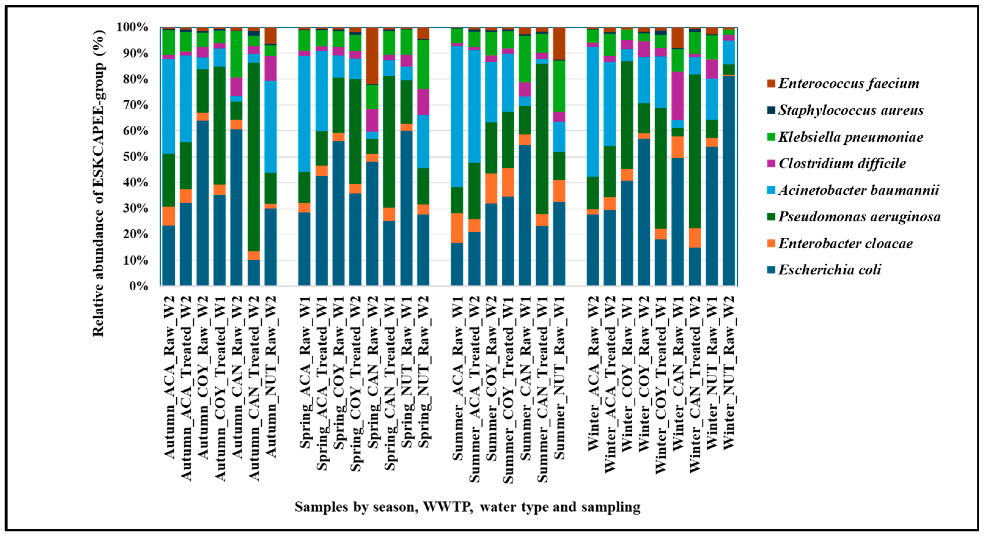
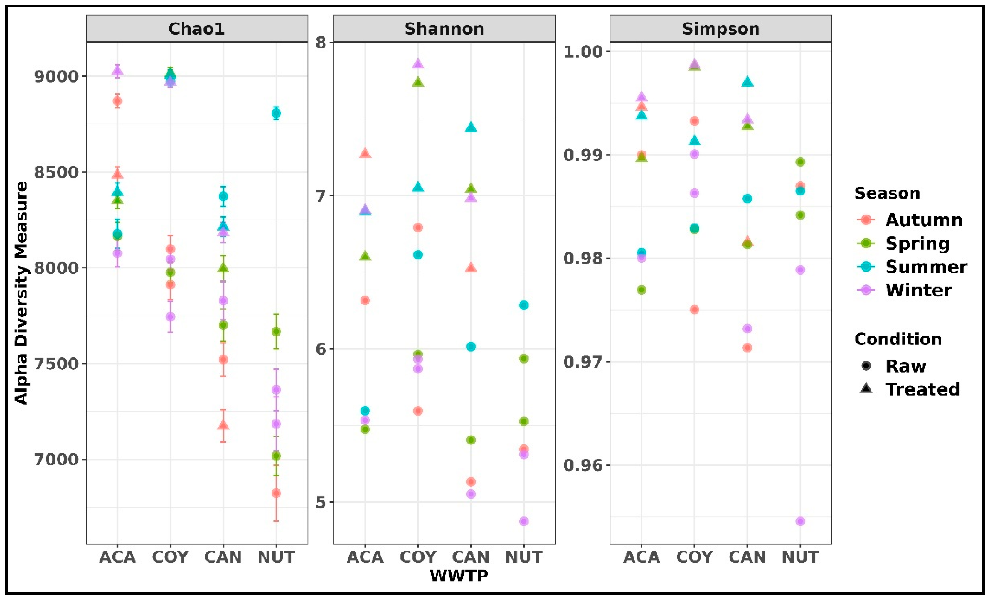
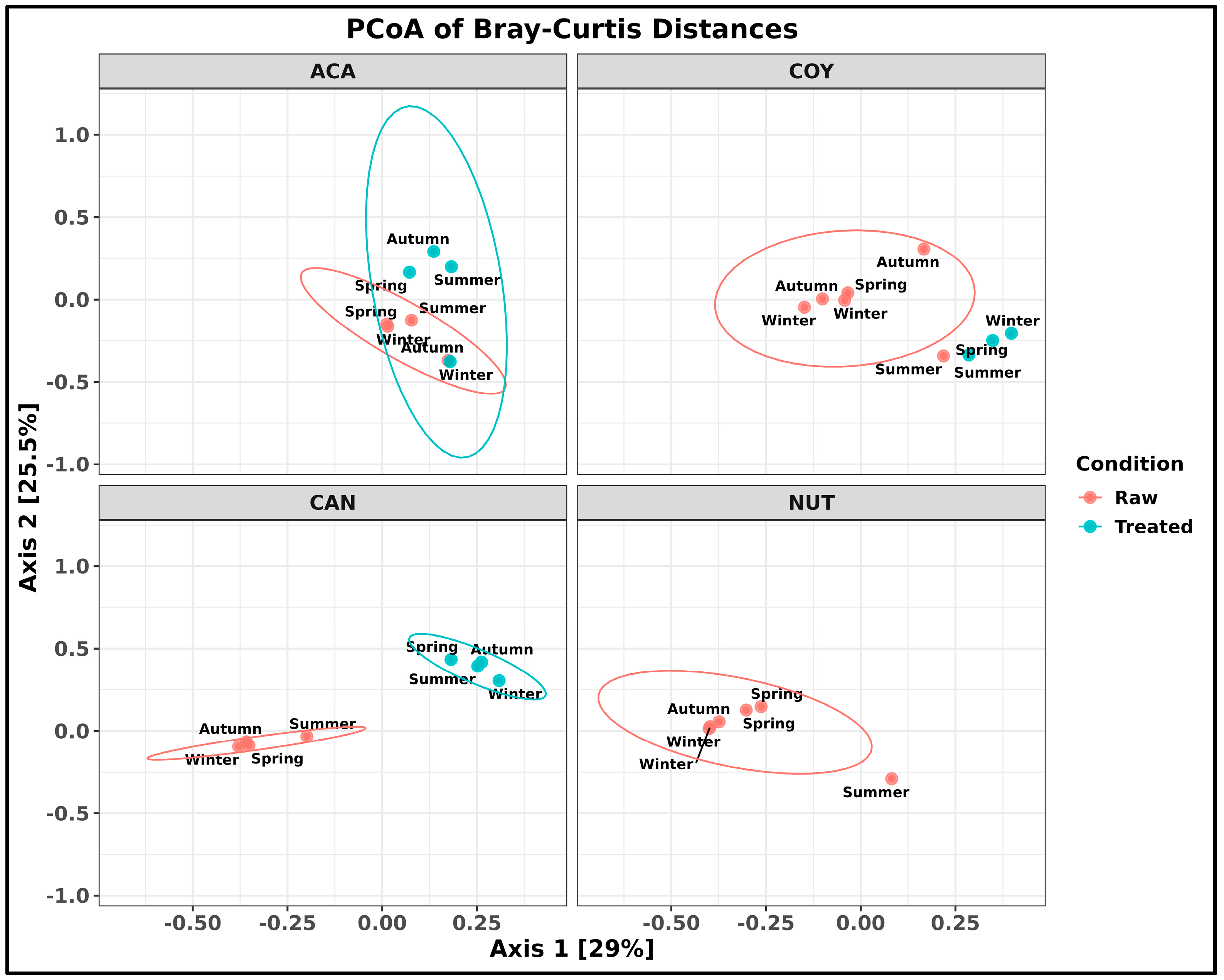
2.2. Taxonomic Composition of Bacteria in Community and Hospital Wastewater

A total of 43 phyla, 95 classes, 205 orders, 498 families, 1855 genera, and 9276 species were detected in the wastewater samples that were analyzed. The main bacterial phyla detected were: Protobacteria (35.03 ± 14.72% raw/64.29 ± 9.40% treated), Bacteroidetes (23.78 ± 7.53% raw/11.62 ± 5.28% treated), Firmicutes (25.81 ± 15.38% raw/4.69 ± 2.82% treated), Campylobacterota (8.74 ± 7.16% raw/5.70 ± 4.87% treated), and Actinobacteria (2.63 ± 1.17% raw/7.36 ± 2.48% treated). The relative abundance per phylum in the community WWTPs was as follows: Protobacteria (47.20 ± 20.82% raw/59.19 ± 5.96% treated), Bacteroidetes (20.01 ± 7.42% raw/14.32 ± 4.2% treated), Firmicutes (12.58 ± 6.76% raw/5.49 ± 3.04% treated), Campylobacterota (13.61 ± 7.80% raw/7.82 ± 4.63% treated), and Actinobacteria (2.03 ± 1.06% raw/7.17 ± 2.88% treated). The values for the hospital WWTP were as follows: Protobacteria (24.08 ± 6.94% raw/74.50 ± 5.73% treated), Bacteroidetes (27.18 ± 6.10% raw/6.22 ± 1.66% treated), Firmicutes (37.72 ± 10.01% raw/3.11 ± 1.61% treated), Campylobacterota (4.36 ± 1.88% raw/1.47 ± 1.13% treated), and Actinobacteria (3.16 ± 1.04% raw/7.75 ± 1.71% treated) (Figure 1).
The seasonal distribution of the relative abundance of the phylum Protobacteria was very similar in all seasons and ranged from 48.45 ± 16.71% in summer to 43.20 ± 22.18% in winter; the distribution of the relative abundance by WWTP showed that the community WWTPs ACA and COY had the highest abundance of the phylum Protobacteria. The results by type of wastewater (raw and treated) showed that the relative abundance of the phylum Protobacteria was higher in treated wastewater: 61.89 ± 10.86% in summer; 62.36 ± 10.24% in winter; 64.47 ± 7.96% in spring and 68.45 ± 12.62% in autumn.
For the phylum Bacteroidetes, the relative abundance by season was very similar, fluctuating between 20.22 ± 10.06% in summer and 18.13 ± 9.25% in autumn; the analysis by WWTP showed that the relative abundance was homogeneous within each WWTP, but different between WWTPs, being higher in the hospital WWTPs (CAN and NUT). For this phylum, the relative abundance by wastewater type was higher in raw wastewater: 22.27 ± 3.69% in spring; 23.06 ± 8.48% in autumn; 23.73 ± 10.42% in winter; and 26.48 ± 7.40% in summer.
For the phylum Firmicutes it was observed that the relative abundance by season was higher in winter, 22.09 ± 17.88%, lower in summer, 11.88 ± 10.48%, and very similar in spring and autumn. The relative abundance within each WWTP was very similar except in the ACA WWTP, where, in winter, this abundance increased (52.19 ± 0.29%) compared to the values observed in spring, summer, and autumn. The results of the relative abundance by wastewater type showed that, for this phylum, it was higher in raw wastewater: 17.08 ± 1.62% in summer, 26.19 ± 17.39% in spring, 27.51 ± 17.29% in autumn, and 30.18 ± 16.22% in winter.
For the phylum Actinobacteria, the seasonal relative abundance was homogeneous, fluctuating between 4.24 ± 2.99% in spring and 4.67 ± 3.50% in winter. This phylum presented a higher relative abundance in treated wastewater: 6.87 ± 2.57% in summer, 7.01 ± 3.00% in spring, 7.28 ± 0.02% in autumn, and 8.30 ± 3.91% in winter; in general, for this phylum, the relative abundance per WWTP was lower compared to the results observed for Protobacteria, Bacteroidetes, and Firmicutes. All results of relative abundance by phylum are shown in Supplementary Materials Table S1.
Of the 1855 bacterial genera detected in the wastewater samples that were analyzed, it was observed that 114 genera presented the highest relative abundances in almost all the stations and WWTPs studied, including Acidovorax, Acinetobacter, Aeromonas, Aliarcobacter, Arcobacter, Bacteroides, Blautia, Burkholderia, Chryseobacterium, Cloacibacterium, Clostridiodes, Comamonas, Coprococcus, Enterobacter, Escherichia, Faecalibacterium, Flavobacterium, Hydrogenophaga, Klebsiella, Parabacteroides, Phocaeicola, Prevotella, Pseudomonas, Streptococcus, Streptomyces, Sulfurospirillum, Thauera, Tolumonas, and Vibrio. (Supplementary Materials Table S2). In this study, 9276 species were detected, of which 110 showed the highest relative abundances in almost all the stations and WWTPs studied, including Acidovorax temperans, Acinetobacter johnsonii, Acinetobacter baumannii, Aeromonas caviae, Aeromonas hydrophila, Bacteroides fragilis, Acinetobacter johnsonii, and Pseudomonas aeruginosa, among others (Supplementary Materials Table S3).
The relative abundance of eight bacterial species included in the ESCKAPEE group (Enterococcus faecium, Staphylococcus aureus, Clostridioides difficile, Klebsiella pneumoniae, Acinetobacter baumannii, Pseudomonas aeruginosa, Enterobacter cloacae, and Escherichia coli) was analyzed, considering that these species are clinically relevant.
The results of the relative abundance of the ESCKAPEE-group bacteria detected in the community and hospital wastewater showed that the highest abundances were found in E. coli (0.51 ± 0.45%), A. baumannii (0.25 ± 0.26%), P. aeruginosa (0.22 ± 0.10%), and K. pneumoniae (0.10 ± 0.07%), while, for E. cloacae and the Gram-positive species, the relative abundance was lower.
The seasonal distribution of the relative abundance by species showed that, for E. faecium, it was higher in spring (0.06 ± 0.12%) and summer (0.04 ± 0.08%); while, for C. difficile, no marked seasonal differences were observed. S. aureus remained the same in all seasons (0.01 ± 0.01%). The seasonal distribution of the relative abundance of the Gram-negative bacteria was higher in summer, except for that of E. coli, which presented high values in all seasons, fluctuating between 0.39 ± 0.17% in summer and 0.63 ± 0.74% in winter (Figure 2).
The relative abundance by species between the WWTPs showed that the wastewater from the hospital WWTPs (CAN and NUT) had a higher abundance of E. faecium (CAN 0.06 ± 0.12%/NUT 0.07 ± 0.08%) and C. difficile (CAN 0.06 ± 0.06%/NUT 0.09 ± 0.02%) compared to the wastewater from the community WWTPs (ACA and COY), and that the relative abundance of S. aureus was similar in all WWTPs (0.01 ± 0.01%). In wastewater from the community WWTPs (ACA and COY), A. baumannii, P. aeruginosa, and E. coli were more abundant, while K. pneumoniae presented similar abundances, except in the samples from the COY WWTP, which had a lower abundance (0.05 ± 0.02%).
The relative abundance of these species according to the type of wastewater (raw/treated) showed that, in raw wastewater, this abundance was higher, except for S. aureus, for which it was similar (0.01 ± 0.01%), and for P. aeruginosa, where the relative abundance was higher in the treated wastewater (0.26 ± 0.06%) (Figure 2).
From the taxonomic annotations at the species level, the alpha diversity was determined with a value of p < 0.05 using three indices. The mean richness observed using the chao1 index in the analyzed wastewater samples was 8102 ± 619.77 unique species, showing a different richness between treatments and between each WWTP (8000–8500 ACA, 7750–8250 COY, 7400–8400 CAN, and 6250–7750 NUT), with an inter quartile interval (IIQ) of 7723–8440; in relation to seasonal abundance, a higher richness was observed in spring and summer. The Shannon index showed a higher richness of bacterial species in the treated wastewater samples (6.5–7.8), indicating that the bacterial diversity of the raw and treated wastewater samples was in the range of 4.87–7.85, with an IIQ 5.530–6.905. Simpson’s alpha diversity index showed that the most diverse bacterial communities were those of the raw wastewater samples (0.95–0.99), compared to the treated wastewater samples (0.99–1.00) (Figure 3).
A principal coordinate analysis (PCoA) was performed at the species level to determine the type of wastewater condition (raw and treated), the relationship between samples at the species level, and the sampling time for each sample. In this analysis, it was observed that the wastewater samples from the CAN WWTP were perfectly grouped according to wastewater type (raw and treated), but this was not the case for the samples from the ACA and COY WWTPs, where a treated wastewater sample overlapped within the universe of raw wastewater samples. Despite this occurrence, the results suggest differences in species composition between the two types of wastewaters that were analyzed. Between sampling time intervals in the treated wastewater, stational differences in the bacterial species composition were observed (p-value < 0.05). In the comparison of the wastewater samples, principal components 1 and 2 explained 54.5% of the variation after the samples underwent the processes in the WWTPs (Figure 4).

2.3. Analysis of Antimicrobial Resistance Genes

The CARD database was used to determine the presence, identity, relative abundance, absolute abundance, and persistence of ARGs in each sample and at each station. A total of 270 ARGs were detected in the wastewater samples that were analyzed, showing an absolute abundance of 95.1 ± 20.9 in the raw wastewater and 56.5 ± 25.8 in the treated wastewater. The absolute abundance of ARGs by WWTP and wastewater type was as follows: communities: ACA 98 ± 11.2 raw/70.3 ± 28.7 treated; COY 99.3 ± 18.6 raw/55.5 ± 33.7 treated; hospitals: CAN 99.5 ± 19.9 raw/43.7 ± 4.4 treated; NUT 83.7 ± 33.1 raw. The seasons where the highest abundances of ARGs were detected were spring and summer, coinciding with the seasons with the highest abundances of bacterial species.
In the wastewater samples analyzed in this study, the qacH, aadS, InuD, APH(3′)-IIIa, catB3, msrE, and OXA-2 genes were the most frequent. The number of ARGs detected by WWTP varied: ACA n = 162, COY n = 182, CAN n = 185, and NUT n = 163. The number of ARGs present at all stations was different at each WWTP: ACA/n = 23 (qacH, catB3, msrE, OXA-2, mphA, tet(C), mphE, QnrVC1, aadA2, APH(3″)-Ib, dfrA1, dfrA15, ANT(3″)-IIa, OXA-10, OXA-427, tet(A), tet(39), aadA11, ErmG, QnrB5, QnrS2, QnrVC4, and tet(E); COY/n = 10 (qacH, catB3, msrE, OXA-2, mphA, QnrVC1, APH(3″)-Ib, dfrA15, tet(X), and ANT(3″)-IIa); CAN/n = 12 (qacH, catB3, msrE, OXA-2, cmlA5, tet(C), dfrA1, sul1, TLA-1, AAC(6′)-Il, sul2, and floR), and NUT/n = 28 (qacH, aadS, APH(3′)-IIIa, catB3, msrE, OXA-2, cmlA5, mphA, tet(C), APH(6)-Id, mphE, QnrVC1, TLA-2, aadA2, APH(3″)-Ib, dfrA15, OXA-427, tet(A), APH(3′)-Ia, AAC(6′)-33, AAC(6′)-Il, dfrA12, ErmG, Mef(En2), aadA10, OXA-58, tet(40), and OXA-372). The seasonal distribution, persistence, and frequency of the ARGs detected in the community and hospital wastewater samples that were studied are shown in Supplementary Materials Figures S1 and S2.
The most abundant and persistent ARGs detected in the wastewater samples analyzed were those that confer resistance to aminoglycosides (APH(3′)-IIIa, APH(3″)-Ib, APH(6)-Id, APH(3′)-Ia, aadA2, aadA11, aadA10, AAC(6′)-Il, AAC(6′)-33, and ANT(3″)-IIa), beta-lactams (OXA-2, OXA-10, OXA-427, TLA-1, TLA-2, OXA-58, and OXA-372), macrolides lincosamines and streptogramins type B (MLS) (mphA-E, msrE, Mef(En2), ErmG, and InuC-D), and tetracyclines (tet(X), tet(C), tet(A), tet(39), tet(E), and tet(40)). Genes conferring resistance to other groups of antimicrobials: quinolones (QnrVC, QnrVC1, QnrB5, QnrS2, and QnrVC4), polymyxins (mcr-3.3, and mcr-5), glycopeptides (vanUG, vanXYG, vanTG, vanSB, vanB, vanHB, vanRB, vanWB, vanWG, vanXB,and vanG), and others, were found in lower abundance (Figure 5 and Supplementary Materials Figures S1 and S2).
The PlasmidFinder database was used to determine the presence, identity, absolute abundance, and persistence of plasmids carrying ARGs. A total of 60 plasmids were detected in the studied samples of raw (39.1 ± 23.4) and treated (12.8 ± 8.4) wastewater collected from community and hospital WWTPs. The absolute abundances of plasmids per WWTP and wastewater type were as follows: community: ACA 38.2 ± 19.1 raw/20.5 ± 7.7 treated and COY 22.2 ± 4.8 raw/11.0 ± 7.6 treated; hospital: CAN 59.2 ± 14.8 raw/7.0 ± 4.1 treated and NUT 37.0 ± 34.7 raw. The seasons with the highest detected plasmid abundances were spring and summer.
The most abundant plasmids detected in the wastewater samples analyzed were: Col440I_1, ColRNAI_1, Col440II_1, ColKP3_1, IncQ2_1, ColE10_1, IncQ1_1, repUS2_1_repA(pBI143), and Col156_1. These plasmids carry different ARGs: Col440I_1 carries NDM-1, blaampH, blaCTX-M-15, and blaTEM-1; ColRNAI_1 carries fosA; Col440II_1 carries qnrB, cmlA1, and fosA7; ColKP3_1 carries blaOXA-131 and blaOXA-232; IncQ2_1 and IncQ1_1-2 carry qnrB77, qnrB2, qnrS1, qnrS2, and aac-(6)-lb′-cr; and repUS2_1_repA(pBI143) carryies optrA. The number of plasmids detected per WWTP varied: ACA n = 34, COY n = 32, CAN n = 45, and NUT n = 45. The number of plasmids present at all stations was different in each WWTP: ACA/n = 13 (Col440I_1, ColRNAI_1, Col440II_1, ColKP3_1, IncQ2_1, IncQ1_1, ColE10_1, IncA/C2_1, Col156_1, IncP6_1, IncP6_1, repUS2_1_repA(pBI143), IncFIB(K)_1_Kpn3, and IncU_1); COY/n = 11 (IncQ2_1, ColRNAI_1, Col440I_1, ColKP3_1, repUS2_1_repA(pBI143), Col156_1, IncP6_1, IncQ1_1, Col(MG828)_1, Col440II_1, and IncA/C2_1); CAN/n = 15 (ColRNAI_1, Col440I_1, Col440II_1, ColKP3_1, ColE10_1, IncQ2_1, Col(MG828)_1, Col8282_1, IncA/C2_1, IncFIA(HI1)_1_HI1, repUS2_1_repA(pBI143), ColpVC_1, IncFIB(K)_1_Kpn3, and IncFIB(pB171)_1_pB171); and NUT/n = 8 (Col440I_1, ColRNAI_1, IncQ2_1, Col440II_1, Col156_1, ColKP3_1, repUS2_1_repA(pBI143), and IncU_1) (Supplementary Materials Figures S3 and S4).
The plasmids that persisted in the raw and treated wastewater, compared between each sampling (W1 and W2) and each station, varied by WWTP: ACA/n = 17 (Col440I_1, ColRNAI_1, Col440II_1, ColKP3_1, IncQ2_1, IncQ1_1, ColE10_1, IncA/C2_1, Col156_1, IncP6_1, IncP6_1, repUS2_1_repA(pBI143), IncFIB(K)_1_Kpn3, IncU_1, Col(MG828)_1, Col(MGD2)_1, Col8282_1, and ColpVC_1); COY/n = 3 (IncQ2_1, Col440I_1, and repUS2_1_repA(pBI143); CAN/n = 9 (Col440I_1, Col440II_1, ColKP3_1, ColE10_1, IncQ2_1, IncA/C2_1, IncFIA(HI1)_1_HI1, IncP6_1, and repA_2_pKPC-2). In NUT this persistence was not estimated because only raw water samples were available (Supplementary Materials Figures S3 and S4).

2.4. Analysis of Virulence Factor Genes

The virulence factor database (VFDB) was used to determine the presence, identity, absolute abundance, and persistence of genes that encode VFGs. A total of 149 virulence factor genes were detected in the wastewater samples collected from community and hospital WWTPs: raw: 36.1 ± 32.8; and treated: 17.4 ± 15.7. The absolute frequency of plasmids per WWTP and wastewater type was as follows: community: ACA 43.7 ± 20.9 raw/24.5 ± 19.1 treated and COY 50.2 ± 45.1 raw/24.0 ± 12.8 treated; hospital: CAN 16.0 ± 10.1 raw/3.7 ± 2.5 treated and NUT 34.2 ± 44.1 raw (Supplementary Materials Figures S5 and S6).
The virulence genes pilG, pilH, pilT, xcpT, flG, algU, csgB, and csgF were the most abundant in the wastewater samples analyzed; the pilG, pilH, pilT, and xcpT genes are associated with adhesion and motility in several enterobacteria; flG is present in S. aureus and is associated with alteration of the cutaneous microbiome and cutaneous immune response; algU is involved in alginate synthesis, producing an alginate capsule in P. auruginosa, which favors its pathogenicity; and csgB and csgF are associated with biofilm formation in enterobacteria. The number of VFGs varied in each WWTP: ACA n = 89, COY n = 83, CAN n = 39, and NUT n = 103. The number of VFGs that persisted across seasons was different in each WWTP: ACA/n = 10 (pilG, pilT, pilH, algU, hsiB1/vipA, algR, hsiC1/vipB, algW, fiml, and pilU); COY/n = 4 (pilG, pilT, xcpT, and csgB); CAN/n = 1 (csgB); in NUT no seasonal persistence was observed (Supplementary Materials Figures S5 and S6).
The VFGs that persisted in the raw and treated wastewater, compared between each sampling (W1 and W2) and each station, varied by WWTP: ACA/n = 38 (pilG, pilT, pilH, algU, hsiB1/vipA, algR, flgC, fliP, hsiC1/vipB, algW, fimI, fleN, fliN, waaF, yagZ/ecpA, algC, csgF, fepC, fimC, fimH, flgG, fliR, pilI, xcpT, csgB, fleQ, gspI, xcpS, yagV/ecpE, ykgK/ecpR, algA, espR1, flgI, fliE, fliG, gspJ, gspL, motA, and motC); COY/n = 3 (pilG, pilT, and xcpT); CAN/n = 1 (pilT); in NUT this persistence was not estimated because only raw water samples were available (Supplementary Materials Figures S5 and S6).

3. Discussion

Currently, the study of wastewater has attracted considerable interest, especially as a model for AMR surveillance, since water is a medium through which ARBs, ARGs, plasmids, and VFGs are disseminated from community and hospital settings to the environment. To this end, metagenomic studies have been crucial in detecting the presence, identity, abundance, and persistence of ARGs, plasmids, and VFGs in bacterial populations present in the wastewater environment. In this study, the most abundant phyla were Protobacteria, Bacteroidetes, Firmicutes, and Actinobacteria (Figure 1); the predominance of these phyla in community and hospital wastewater are the same as those described before [12,13].

3.1. Taxonomic Composition of Bacteria in Community and Hospital Wastewater

The microbial composition of the hospital and community wastewater at the genus level showed that, within the genera presenting the highest relative abundances, several genera associated with human and animal infections were found: Acinetobacter, Aeromonas, Burkholderia, Clostridiodes, Enterobacter, Escherichia, Flavobacterium, Klebsiella, Pseudomonas, Streptococcus, Streptomyces, and Vibrio, as well as those related to intestinal microbiota: Bacteroides, Blautia, Cloacibacterium, Comamonas, Coprococcus, Escherichia, Faecalibacterium, Parabacteroides, Phocaeicola, and Prevotella, and those found in soil and water: Chryseobacterium, Flavobacterium, Hydrogenophaga, Sulfurospirillum, Thauera, and Tolumonas (Supplementary Materials Table S2). The presence of these genera has been reported in other studies, i.e., Lu X et al., who reported high abundances of Aeromonas, Arcobacter, Clostridiodes, and Pseudomonas in wastewater samples [14]; Poopedi E et al. detected high abundances of the genera Escherichia, Shigella, Arcobacter, Acinetobacter, Streptococcus, and Aeromonas [15]. The abundant presence of bacterial genera associated with the intestinal microbiota in wastewater samples was reported by Zhang D et al., who detected, in their samples, Bacteroides, Faecalibacterium, Parabacteroides, and Prevotella [16].
The microbial compositions of the hospital and community wastewater at the species level showed that, within the species that had the highest relative abundances, several medically important species were found Escherichia coli, Enterobacter cloacae, Klebsiella pneumoniae, Pseudomonas aeruginosa, Acinetobacter baumannii, Clostridioides difficile, and Acidovorax temperans, among others (Supplementary Materials Table S3). Some of these species were also reported by Lepper H et al., who detected Flavobacterium spp., Acinetobacter, Wuhouensis, Kinneretia, Asaccharophila, Acidovorax, Konjaci, and Pseudomonas spp. as the most abundant species in the wastewater samples analyzed in their study [17]. Furthermore, Fresia P et al. detected significant abundances of Klebsiella, Pseudomonas, Acinetobacter, Enterobacter, and Streptococcus in wastewater samples [18].
Bacteria of the ESCKAPEE group are included within the phyla Proteobacteria and Firmicutes; these phyla have been reported in other studies as the most abundant [12,19,20]. Our results showed that E. coli, A. baumannii, P. aeruginosa, and K. pneumoniae were the most abundant species, with all having a marked seasonality in summer except for E. coli, which presented high abundances in all seasons. For E. faecium, the relative abundance was higher in spring and summer, while, for C. difficile, no marked seasonal changes were observed, and, for S. aureus, the abundance remained the same in all seasons (Figure 2). The seasonal distribution of some microorganisms has been pointed out in other works, in which several contributing factors are highlighted. He Y et al. found that the temperature and chemical oxygen demand were the main environmental factors affecting the bacterial community structure of the wastewater that they tested, with temperature having the most significant effect on the species composition [21]. In another work, Kang et al. reported that the diversity and relative abundance of bacterial communities in wastewater collected in winter were significantly lower compared to summer samples, and that the temperature and dissolved oxygen were the main factors driving seasonal changes in the bacterial diversity, richness, and community structure in the WWTPs that they studied [22].
The relative abundance by species between WWTPs showed that the wastewater of CAN and NUT had a higher abundance of E. faecium and C. difficile than the wastewater from ACA and COY. The abundance of E. faecium and C. difficile in the hospital wastewater could be related to the hospital origin of these species. Sanderson H et al. mentioned that E. faecium may be more adapted to the clinical environment than to the wastewater environment [23], while Moradigaravand D et al. found that clinical strains of C. difficile were closely related to isolates present in hospital wastewater [24]. The relative abundance of S. aureus remained the same in the wastewater samples collected from all WWTPs, similar to what we observed before, with no differences between the frequency of staphylococcal isolates in community wastewater compared to hospital wastewater [25]; this may demonstrate the ubiquitous capacity of this species to remain in both environments. The same behavior was observed for K. pneumoniae, demonstrating the ability of this species to stay in both community and hospital wastewater. A. baumannii, P. aeruginosa, and E. coli were more abundant in the wastewater from the community WWTPs (ACA and COY) (Figure 2). Two of these species (E. coli and P. aeruginosa) were reported by Galarde L et al. as the most abundant in hospital wastewater [12], while Hubeny J et al. reported that A. baumannii and E. coli were the dominant species in community wastewater [26], species that we also detected in the community wastewater of this study.
When the relative abundances of the ESCKAPEE-group bacteria were compared by type of wastewater (raw/treated), it was observed that E. faecium, C. difficile, E. coli, A. baumannii, and K. pneumoniae were more abundant in the raw wastewater, and the high abundance of these species in raw wastewater samples has been associated with human feces [27,28]. While S. aureus remained constant, for P. aeruginosa, the relative abundance was higher in treated wastewater (Figure 2). The ability of this species to increase in abundance in treated wastewater is probably related to its ability to form biofilms [29]. The abundance of species of this group in wastewater was also described by our work group, with E. coli, Klebsiella spp., and Enterobacter spp. being the predominant genera in the raw wastewater, while Enterococcus spp., Staphylococcus spp., Acinetobacter spp., and Pseudomonas spp. being more abundant in the treated wastewater [12].
Principal coordinate analysis (PCoA) showed that the wastewater samples from the CAN WWTP were perfectly grouped according to wastewater type (raw and treated), but this was not the case for the samples from the ACA and COY WWTPs, where a treated wastewater sample overlapped with raw wastewater samples (Figure 4). According to the records of each sampling, it was documented that, in these collections, the ACA and COY WWTPs had mechanical failures in pumping, which probably impacted the correct treatment of raw wastewater, resulting in an output of untreated wastewater, which would explain this result; the discharge of untreated wastewater can have an ecological impact when it is reused due to its potential for the dissemination of ARBs and ARGs in the environment.

3.2. Analysis of Antimicrobial Resistance Genes

Wastewater is increasingly recognized as an important reservoir of ARGs, allowing conditions that are conducive to selection pressure and the horizontal transfer of resistance genes (HTGs). Wastewater is rich in partially metabolized antibiotics, heavy metals, detergents, and biocides. Together, these compounds can exert selective pressure on the proliferation of ARBs. The high microbial load of these environments may be an ideal component for the proliferation of ARBs through the HTG process [30]. Furthermore, Karkman A et al. and Pilmis B et al. report that the human and animal gut microbiomes contain a wide variety of ARBs; thus, wastewater discharge and fecal contamination have been associated with increased abundances of ARBs in the aquatic environment [31,32]. In this study, a total of 270 ARGs were detected, and it was observed that the absolute abundance of ARGs showed only a 39% reduction in treated wastewater; but this reduction seems to vary in different studies, as some have reported a 70% reduction of ARGs in treated hospital wastewater [12], while Yang Y. et al. and Gupta S. et al., reported that wastewater treatment removed more than 99% of ARGs in community WWTPs in Hong Kong and South Korea, respectively [33,34], and Szczepanowski R et al. reported a reduction of 13% in treated wastewater from a WWTP in Germany [35]. The differences in the reduction percentages of each WWTP could probably be related to the types of treatment within each plant.
When comparing the absolute abundances of ARGs from community WWTPs (ACA/COY) versus hospital WWTPs (CAN/NUT), it was observed that, regardless of the WWTP, the abundances of ARGs were very similar in the raw wastewater samples, (ACA 98 ± 11.2, COY 99.3 ± 18.6, CAN 99.5 ± 19.9, and NUT 83.7 ± 33.1. These results are interesting because hospital WWTPs have been documented to contribute a higher load of ARGs to wastewater compared to community WWTPs due to antibiotic use within hospitals, the discharge of antibiotic residues to hospital effluent, and trace antibiotics in patient excreta [36,37]. However, our results show that, in community wastewater, ARGs are also abundant, which could possibly reflect the consumption of antibiotics in the community or the arrival of ARGs to community WWTPs through wastewater discharges from private health clinics.
The reduction in ARGs abundance by WWTPs was 28% in ACA, 43.8% in COY, and 55.8% in CAN, and it was observed that the hospital WWTP (CAN) had a higher reduction of ARGs, i.e., the abundance of ARGs in the treated wastewater from this WWTP was lower (43.7 ± 4.4) compared to that detected in ACA (70.3 ± 28. 7) and COY (55.5 ± 33.7). One explanation for this result may be the fact that the CAN WWTP has a tertiary treatment system, which would have favored the reduction of ARGs, while the community WWTPs (ACA and COY) both only have a secondary treatment system.
Our results showed that the qacH, aadS, InuD, aph(3′)-IIIa, catB3, msrE, and blaOXA genes were the most abundant in the wastewater samples analyzed (Supplementary Materials Figures S1 and S2). Some of these genes were previously reported with high abundance and persistence in wastewater samples from two hospital WWTPs in Mexico [12]. The qacH gene has been documented to encode for resistance to quaternary ammonium salts, heavy metal, and disinfectants and has been detected in several bacterial species (Staphylococcus spp. E. faecalis, E. coli, Listeria monocytogenes, Klebsiella, Enterobacter, Citrobacter, and Pseudomonas) [38,39,40,41,42,43].
The aadS gene confers resistance to aminoglycosides and has been described in Elizabethkingia meningoseptica [44] and in Chryseobacteria. Pham D. et al. found that aadS is found in mobile genetic elements, suggesting its high transmissibility across the phylum Bacteroidetes, including its transfer between different species [45]. The aph(3′)-IIIa gene encodes aminoglycoside resistance and has been described in methicillin-resistant S. aureus strains isolated from hospital wastewater in Portugal [46], as well as in Enterococcus spp. isolated from various sources, including wastewater [47,48]. Other ARGs were detected that also encode aminoglycoside resistance were detected in this study, including aads, aadA2, and aac3, which have been reported in other work [9,10,12].
The catB3 gene, which confers resistance to chloramphenicol, has been detected in Aeromonas, Bordetella, Pseudomonas, and Enterorobacteriaceae and is considered an abundant gene in wastewater [49,50]. Other genes abundantly detected in our study were msrE, mph(A, E, G), mef(B,C), Inu(C,D), and erm(B,F,G), which encode resistance to MLS. Pallares-Vega R et al. detected these same genes in wastewater from 62 WWTPs in the Netherlands [51]. The erm(B) gene was originally detected in Gram-positive bacteria (Enterococcus spp., Staphylococcus spp., and Streptococcus spp.), and this gene can be transferred to Gram-negative bacteria through a conjugative transposon [9,52]. In our study, the blaOXA genes were widely represented. All these genes have been detected in hospital and community wastewater from several countries [12,49,53,54,55,56,57]. Ramos et al. observed that the diversity and abundance of ARGs were not limited to the ESKAPE group, but that these genes predominated in bacteria such as Aeromonas, Aliarcobacter, and Acidovorax, finding that Aliarcobacter accumulated a high abundance of genes for resistance to sulfonamides and polymyxins, while Acinetobacter and Aeromonas harbored the highest abundances of ARGs against beta-lactams [58]. These four species were also abundant in our study, which may imply important reservoirs of ARGs in the analyzed wastewater (Supplementary Materials Tables S1 and S2).
It was observed that the ARGs detected in this study in hospital and community wastewater encode resistance to surveillance and reserve antibiotics, according to the WHO AWaRe classification [59]. These included carbapenems (Imipenem) (blaOXA-58, blaOXA-372, and bla-OXA-427); aminoglycosides (kanamycin, neomycin, streptomycin, ribostamycin), (aph(3″-IIIa, aph(3″)-Ib, aph(3″)-Ia, ant(3″)-IIa, aac(6″)-Il, aac(6″)-33, aadA2, aadA11, and aadA10); and the MLS group (erythromycin, ciprofloxacin, clindamycin), (mphA, mphE msrE, Mef(En2), and ErmG). All these genes have been detected in wastewater in other studies [60,61,62,63,64,65,66,67,68,69,70]. Also, genes encoding resistance to quinolones (ciprofloxacin), polymyxins (colistin), and glycopeptides (vancomycin) were also detected (Figure 5 and Supplementary Materials Figures S1 and S2). In this study, the mcr-5 and mcr-3.3 genes were detected in raw wastewater from NUT and treated wastewater from COY, respectively (Supplementary Materials Figures S1 and S2). The mcr gene was not previously detected in hospital wastewater in Mexico [12]; however, it has been detected in community wastewater in other countries, including Spain, France, Germany, and Tunisia [55,71,72,73]. The presence of genes encoding resistance to antibiotics of the last choice in hospital and community wastewater in our country represents a problem that requires surveillance.
Our results showed that plasmids carrying resistance genes were more abundant in raw wastewater, and 60% more abundant in hospital wastewater compared to community wastewater. Col440I_1, ColRNAI_1, Col440II_1, ColKP3_1, IncQ2_1, ColE10_1, IncQ1_1, repUS2_1_repA(pBI143), and Col156_1 were the most abundant plasmids (Supplementary Materials Figures S3 and S4). These plasmids carry genes encoding resistance to beta-lactams, quinolones, fosfomycin, phenicols, oxazilidones, lincosamides, aminoglycosides, sulfonamides, and tetracyclines. The study by Chukamnerd A et al. identified a high frequency of Col440I and ColKP3 plasmids in K. pneumoniae [74], while the works by Bönemann, G et al., Loftie-Eaton W et al., and Piotrowska M et al. demonstrated that IncQ-group plasmids carry several ARGs, which confer resistance to beta-lactams, aminoglycosides, carbapenems, phenicols, lincosamides, quinolones, tetracyclines, and sulfonamides [75,76,77]. Importantly, the presence of plasmids carrying ARGs in community and hospital wastewater favors the spread of antimicrobial resistance and enhances the transfer of resistance between bacteria of the same or different genera, representing an important public health problem.

3.3. Analysis of Virulence Factor Genes

Several of the VFGs detected in this study encode for adhesion, motility, and biofilm formation, allowing bacteria to remain and persist in the wastewater environment and survive WWTP treatment systems, thus managing to escape into the environment via treated wastewater. Mao G et al. evaluated the role of biofilms as a reservoir and vehicle for virulence factors in wastewater, demonstrating that bacteria in this environment increased their biofilm production and that the mobility of virulence factors is crucial for biofilm formation [11].
The present work has some limitations. This study included a reduced number of WWTPs, and the wastewater sampling was only carried out in the influent and effluent. Considering these points, we propose that future work should include a larger number of WWTPs, which would allow us to have geographical representativeness throughout the country, in addition to evaluating the dynamics of bacterial populations, ARGs, plasmids, and VFGs in the different stages of treatment within WWTPs, to evaluate the effectiveness of the elimination in each process.

4. Materials and Methods

4.1. Study Design and Sample Collection

This descriptive longitudinal study was conducted in Mexico City and Cuernavaca City from August 2021 to September 2022. Samples of raw (100 mL) and treated (200 mL) wastewater were collected in each season of the year; two samples of treated wastewater and two samples of raw wastewater in each included WWTP were collected. Samples were taken at one-month intervals between each sample. The WWTPs studied were the community WWTPs ACA with a 18.95666 N latitude and 99.23411 W longitude in Cuernavaca City and COY with a 19.36392 N latitude and 99.11668 W longitude in Mexico City, the hospital WWTP CAN with a 19.17128 N latitude and 99.06314 W longitude, and a second hospital, NUT, with a single sump with a 19.28814 N latitude and 99.15654 W longitude, from which raw wastewater samples were taken, both located in Mexico City. The samples were transported to the laboratory at 4 °C within less than two hours of collection [78,79].

4.2. Wastewater Treatment Plant Characteristics

The design of the three WWTPs (ACA, COY, and CAN) starts with a pretreatment process using a griller that eliminates large materials. Subsequently, the water flow passes through an aeration process mediated by regulating valves in different chambers. The treatment process continues with a sedimentation phase, precipitating the sludge. Tertiary treatment includes granular activated carbon filters, zeolite filters, ultraviolet light (UVL), and calcium hypochlorite tablets in the effluent (only CAN WWTP has tertiary treatment). The NUT hospital has only one sump system (pretreatment).

4.3. DNA Extraction

All aliquots collected (100 mL of raw wastewater and 200 mL of treated wastewater) were centrifuged at 5000× g for 20 min at 4 °C. The supernatant was decanted and one milliliter of EC lysis solution (1M Tris pH 8.0, EDTA, sodium deoxycholate, N-lauryl sarcosyl, RNAase, lysozyme, and lysostaphin) was added to the pellet and incubated at 37 °C for four hours. Then, the ESP solution (EDTA + N-lauryl sarcosyl + proteinase K) was added and incubated at 50 °C overnight [80]. The samples were purified with the Wizard® Genomic DNA Purification Kit (PROMEGA Corp., Madison, WI, USA) according to the manufacturer’s instructions. DNA was quantified by fluorometry using the Qubit 4 Fluorometer (Thermo Fisher Scientific, Waltham, MA, USA). The DNA samples were stored at −20 °C until sequencing.

4.4. Sequencing and Bioinformatics Analysis

The 31 samples were sequenced by Illumina HiSeq (Illumina, Inc., San Diego, CA, USA), with 2 × 150 configuration (21.4 GB). Libraries were performed using the Nextera DNA protocol. The reads are available from NCBI SRA under BioProject ID1197482.
Adapters were removed for raw reads and a Q > 20 was considered with Trimmomatic (v0.39) [81]. Quality control statistics were performed with FastQC (v0.12.1) [82]. Metagenome assemblies were performed with IDBA-UD (v1.1) [83,84]. Mapping statistics were performed with Bowtie 2 (v2.5.4) [85]. For functional annotation, the Trinotate (v3.0.1) pipeline was employed [86,87]. Gene abundance was estimated as coverage by mapping reads to contigs using BWA (v0.7.12-r1039) and the coverBed function of bedtools (v2.25.0) [88,89]. Kraken (v2.1.3) was used for taxonomic profiling [90,91]. Alpha and beta diversity analyses were performed with the vegan (v2.4-6) and phyloseq libraries (v1.42.0) [92,93]. The distance matrix for beta diversity was performed with the Bray–Curtis index at the species level. Comparisons between groups were determined with an analysis of similarity (ANOSIM) [93,94]. Abundance plots and histograms were performed with the ggplot2 (v3.5.1) and ggpubr (v0.6.0) library of R [92,95,96].

4.5. Analysis of Antimicrobial Resistance Genes and Virulence Genes

The resistome and virulome of the samples were determined using contigs of lengths of >150 bp using ABRicate (v1.0.1) [97] with the Comprehensive Antibiotic Resistance Database (CARD) (v3.2.9) [98] and PlasmidFinder (v2.0) [99]. The identified ARGs were grouped into classes according to the type of antibiotic to which they confer resistance, and quantified by absolute abundance, relative abundance, and normalized abundance expressed as percentages. The virulence factor database (VFDB) and the E. coli O-groups and H-types database (EcOH) were used for virulence gene detection [100,101].

5. Conclusions

This study reveals the predominant bacterial phyla in wastewater, including clinically relevant bacteria such as Klebsiella spp., E. coli, Acinetobacter spp., and Pseudomonas spp., among others, in both community and hospital samples, in function of the treatment and with seasonal variations in abondance. A significant variety of ARGs encoding antibiotics of the last choice were detected in the tested community and hospital wastewater. In addition, this study showed that community wastewater also carries a significant burden of ARGs, suggesting widespread antibiotic use and the spread of AMR beyond hospitals. The results showed that having a tertiary treatment system favors a reduction in ARGs, which could be considered as a proposal aimed at improving WWTP treatment systems in our country. The presence of plasmids carrying ARGs that encode surveillance and reserve antibiotics in wastewater could favor the transfer and spread of resistance between bacteria, which represents an important public health problem. The presence of ARGs, plasmids, and VFGs in treated wastewater may have an ecological impact through their spreading in the environment, through their use in the irrigation of green areas of cities and hospitals and their discharge to municipal sewers. Further analysis of these environments will allow us to generate more scientific evidence to propose public policies for environmental protection and public health. Finally, we consider that the results of this study allowed us to develop the first evidence of the seasonal behavior of bacterial populations, ARGs, plasmids, and VFGs in community and hospital wastewater in our country.

Supplementary Materials

The following supporting information can be downloaded at: https://www.mdpi.com/article/10.3390/ijms26052051/s1.

Author Contributions

Conceptualization, M.E.V.-M.; Methodology, M.E.V.-M., M.G.-L., P.C.-J., M.B.-d.-V., E.G.-L., E.A.-V., B.A.C.-Q., A.P.d.L.-G. and C.V.A.; Validation, M.E.V.-M.; Formal analysis, M.E.V.-M.; Investigation, M.E.V.-M.; Resources, M.E.V.-M.; Data curation, M.E.V.-M. and M.G.-L.; Writing—original draft, M.E.V.-M.; Writing—review & editing, M.E.V.-M., P.C.-J. and C.M.A.-A.; Visualization, M.E.V.-M.; Supervision, M.E.V.-M.; Project administration, M.E.V.-M.; Funding acquisition, M.E.V.-M. All authors have read and agreed to the published version of the manuscript.

Funding

This work was supported by a grant from FORDECY-PRONACES/682339/2019 CONAHCYT, Mexico. Miguel Galarde López was a postdoctoral student financed by this grant.

Institutional Review Board Statement

Ethical approval was obtained from the Ethic Committee of the Instituto Nacional de Salud Pública. CI/2020/651.

Informed Consent Statement

Not applicable.

Data Availability Statement

Data is contained within the article.

Acknowledgments

The authors wish to thank the personnel responsible for the wastewater treatment plants for their collaboration in the water sampling, as well as Alfredo Mendoza and Dora Garnica from the Instituto Nacional de Medicina Genómica (INMEGEN) for the sequencing and processing of the samples. We also thank Ariadna Nava-Mercado and Lorena Guerrero for their administrative support in this project.

Conflicts of Interest

The authors declare no conflicts of interest.

References

  1. World Health Organization. Global Antimicrobial Resistance Surveillance System (GLASS) Report: Early Implementation 2020, 1st ed.; World Health Organization: Geneva, Switzerland, 2020; ISBN 978-92-4-000558-7. [Google Scholar]
  2. O’Neill, J. Antimicrobial Resistance: Tackling a Crisis for the Health and Wealth of Nations. 2014. Available online: https://www.who.int/news/item/29-04-2019-new-report-calls-for-urgent-action-to-avert-antimicrobial-resistance-crisis (accessed on 7 January 2025).
  3. Sinclair, R.G.; Choi, C.Y.; Riley, M.R.; Gerba, C.P. Pathogen Surveillance Through Monitoring of Sewer Systems. In Advances in Applied Microbiology; Academic Press Inc.: Cambridge, MA, USA, 2008; Volume 65, pp. 249–269. ISBN 9780123744296. [Google Scholar]
  4. Daughton, C.G. Wastewater Surveillance for Population-Wide COVID-19: The Present and Future. Sci. Total Environ. 2020, 736, 139631. [Google Scholar] [CrossRef] [PubMed]
  5. Manaia, C.M.; Rocha, J.; Scaccia, N.; Marano, R.; Radu, E.; Biancullo, F.; Cerqueira, F.; Fortunato, G.; Iakovides, I.C.; Zammit, I.; et al. Antibiotic Resistance in Wastewater Treatment Plants: Tackling the Black Box. Environ. Int. 2018, 115, 312–324. [Google Scholar] [CrossRef] [PubMed]
  6. Warnes, S.L.; Highmore, C.J.; Keevil, C.W. Horizontal Transfer of Antibiotic Resistance Genes on Abiotic Touch Surfaces: Implications for Public Health. MBio 2012, 3, 1010–1128. [Google Scholar] [CrossRef] [PubMed]
  7. Aarestrup, F.M.; Woolhouse, M.E.J. Using Sewage for Surveillance of Antimicrobial Resistance. Science 2020, 367, 630–632. [Google Scholar] [CrossRef]
  8. Miłobedzka, A.; Ferreira, C.; Vaz-Moreira, I.; Calderón-Franco, D.; Gorecki, A.; Purkrtova, S.; Bartacek, J.; Dziewit, L.; Singleton, C.M.; Nielsen, P.H.; et al. Monitoring Antibiotic Resistance Genes in Wastewater Environments: The Challenges of Filling a Gap in the One-Health Cycle. J. Hazard. Mater. 2022, 424, 127407. [Google Scholar] [CrossRef]
  9. Pärnänen, K.M.M.; Narciso-da-Rocha, C.; Kneis, D.; Berendonk, T.U.; Cacace, D.; Do, T.T.; Elpers, C.; Fatta-Kassinos, D.; Henriques, I.; Jaeger, T.; et al. Antibiotic Resistance in European Wastewater Treatment Plants Mirrors the Pattern of Clinical Antibiotic Resistance Prevalence. Sci. Adv. 2019, 5, eaau9124. [Google Scholar] [CrossRef]
  10. Raza, S.; Shin, H.; Hur, H.G.; Unno, T. Higher Abundance of Core Antimicrobial Resistant Genes in Effluent from Wastewater Treatment Plants. Water Res. 2022, 208, 117882. [Google Scholar] [CrossRef]
  11. Mao, G.; Liang, J.; Wang, Q.; Zhao, C.; Bai, Y.; Liu, R.; Liu, H.; Qu, J. Epilithic Biofilm as a Reservoir for Functional Virulence Factors in Wastewater-Dominant Rivers after WWTP Upgrade. J. Environ. Sci. 2021, 101, 27–35. [Google Scholar] [CrossRef]
  12. Galarde-López, M.; Velazquez-Meza, M.E.; Godoy-Lozano, E.E.; Carrillo-Quiroz, B.A.; Cornejo-Juárez, P.; Sassoé-González, A.; Ponce-de-León, A.; Saturno-Hernández, P.; Alpuche-Aranda, C.M. Presence and Persistence of ESKAPEE Bacteria before and after Hospital Wastewater Treatment. Microorganisms 2024, 12, 1231. [Google Scholar] [CrossRef]
  13. Numberger, D.; Ganzert, L.; Zoccarato, L.; Mühldorfer, K.; Sauer, S.; Grossart, H.P.; Greenwood, A.D. Characterization of Bacterial Communities in Wastewater with Enhanced Taxonomic Resolution by Full-Length 16S RRNA Sequencing. Sci. Rep. 2019, 9, 9673. [Google Scholar] [CrossRef]
  14. Lu, X.M.; Lu, P.Z. Characterization of Bacterial Communities in Sediments Receiving Various Wastewater Effluents with High-Throughput Sequencing Analysis. Microb. Ecol. 2014, 67, 612–623. [Google Scholar] [CrossRef] [PubMed]
  15. Poopedi, E.; Singh, T.; Gomba, A. Potential Exposure to Respiratory and Enteric Bacterial Pathogens among Wastewater Treatment Plant Workers, South Africa. Int. J. Environ. Res. Public Health 2023, 20, 4338. [Google Scholar] [CrossRef] [PubMed]
  16. Zhang, D.; Peng, Y.; Chan, C.L.; On, H.; Wai, H.K.F.; Shekhawat, S.S.; Gupta, A.B.; Varshney, A.K.; Chuanchuen, R.; Zhou, X.; et al. Metagenomic Survey Reveals More Diverse and Abundant Antibiotic Resistance Genes in Municipal Wastewater Than Hospital Wastewater. Front. Microbiol. 2021, 12, 712843. [Google Scholar] [CrossRef] [PubMed]
  17. Lepper, H.C.; Perry, M.R.; Wee, B.A.; Wills, D.; Nielsen, H.; Otani, S.; Simon, M.; Aarestrup, F.M.; Woolhouse, M.E.J.; van Bunnik, B.A.D. Distinctive Hospital and Community Resistomes in Scottish Urban Wastewater: Metagenomics of a Paired Wastewater Sampling Design. Sci. Total Environ. 2023, 902, 165978. [Google Scholar] [CrossRef]
  18. Fresia, P.; Antelo, V.; Salazar, C.; Giménez, M.; D’Alessandro, B.; Afshinnekoo, E.; Mason, C.; Gonnet, G.H.; Iraola, G. Urban Metagenomics Uncover Antibiotic Resistance Reservoirs in Coastal Beach and Sewage Waters. Microbiome 2019, 7, 35. [Google Scholar] [CrossRef]
  19. Berglund, F.; Ebmeyer, S.; Kristiansson, E.; Larsson, D.G.J. Evidence for Wastewaters as Environments Where Mobile Antibiotic Resistance Genes Emerge. Commun. Biol. 2023, 6, 321. [Google Scholar] [CrossRef]
  20. Lin, Q.; Xavier, B.B.; Alako, B.T.F.; Mitchell, A.L.; Rajakani, S.G.; Glupczynski, Y.; Finn, R.D.; Cochrane, G.; Malhotra-Kumar, S. Screening of Global Microbiomes Implies Ecological Boundaries Impacting the Distribution and Dissemination of Clinically Relevant Antimicrobial Resistance Genes. Commun. Biol. 2022, 5, 1217. [Google Scholar] [CrossRef]
  21. He, Y.; Li, K.X.; Wang, J.W.; Wang, W.; Fan, P.C.; Chen, H.H.; Wang, J.J. Microbial Community Structure of Wastewater Treatment Plants in Different Seasons. Environ. Sci. 2021, 42, 1488–1495. [Google Scholar] [CrossRef]
  22. Kang, X.H.; Leng, Y.; Macdonald, O.M.; Zeng, X.Y.; Li, S.W. The Seasonal Changes of Core Bacterial Community Decide Sewage Purification in Sub-Plateau Municipal Sewage Treatment Plants. Bioprocess Biosyst. Eng. 2020, 43, 1609–1617. [Google Scholar] [CrossRef]
  23. Sanderson, H.; Ortega-Polo, R.; Zaheer, R.; Goji, N.; Amoako, K.K.; Brown, R.S.; Majury, A.; Liss, S.N.; McAllister, T.A. Comparative Genomics of Multidrug-Resistant Enterococcus spp. Isolated from Wastewater Treatment Plants. BMC Microbiol. 2020, 20. [Google Scholar] [CrossRef]
  24. Moradigaravand, D.; Gouliouris, T.; Ludden, C.; Reuter, S.; Jamrozy, D.; Blane, B.; Naydenova, P.; Judge, K.; Aliyu, S.H.; Hadjirin, N.F.; et al. Genomic Survey of Clostridium difficile Reservoirs in the East of England Implicates Environmental Contamination of Wastewater Treatment Plants by Clinical Lineages. Microb. Genom. 2018, 4, e000162. [Google Scholar] [CrossRef] [PubMed]
  25. Velazquez-Meza, M.E.; Galarde-López, M.; Cornejo-Juárez, P.; Carrillo-Quiroz, B.A.; Velázquez-Acosta, C.; Bobadilla-del-Valle, M.; Ponce-de-León, A.; Alpuche-Aranda, C.M. Multidrug-Resistant Staphylococcus sp. and Enterococcus sp. in Municipal and Hospital Wastewater: A Longitudinal Study. Microorganisms 2024, 12, 645. [Google Scholar] [CrossRef] [PubMed]
  26. Hubeny, J.; Korzeniewska, E.; Ciesielski, S.; Płaza, G.; Harnisz, M. The Resistome of ESKAPEE Pathogens in Untreated and Treated Wastewater: A Polish Case Study. Biomolecules 2022, 12, 1160. [Google Scholar] [CrossRef] [PubMed]
  27. Dijkshoorn, L.; van Aken, E.; Shunburne, L.; van der Reijden, T.J.K.; Bernards, A.T.; Nemec, A.; Towner, K.J. Prevalence of Acinetobacter baumannii and Other Acinetobacter spp. in Faecal Samples from Non-Hospitalised Individuals. Clin. Microbiol. Infect. 2005, 11, 329–332. [Google Scholar] [CrossRef]
  28. Ma, X.; Dong, X.; Cai, J.; Fu, C.; Yang, J.; Liu, Y.; Zhang, Y.; Wan, T.; Lin, S.; Lou, Y.; et al. Metagenomic Analysis Reveals Changes in Bacterial Communities and Antibiotic Resistance Genes in an Eye Specialty Hospital and a General Hospital Before and After Wastewater Treatment. Front. Microbiol. 2022, 13, 848167. [Google Scholar] [CrossRef]
  29. Gholami, S.; Tabatabaei, M.; Sohrabi, N. Comparison of Biofilm Formation and Antibiotic Resistance Pattern of Pseudomonas aeruginosa in Human and Environmental Isolates. Microb. Pathog. 2017, 109, 94–98. [Google Scholar] [CrossRef]
  30. Auguet, O.; Pijuan, M.; Borrego, C.M.; Rodriguez-Mozaz, S.; Triadó-Margarit, X.; Della Giustina, S.V.; Gutierrez, O. Sewers as Potential Reservoirs of Antibiotic Resistance. Sci. Total Environ. 2017, 605–606, 1047–1054. [Google Scholar] [CrossRef]
  31. Karkman, A.; Pärnänen, K.; Larsson, D.G.J. Fecal Pollution Can Explain Antibiotic Resistance Gene Abundances in Anthropogenically Impacted Environments. Nat. Commun. 2019, 10, 80. [Google Scholar] [CrossRef]
  32. Pilmis, B.; Le Monnier, A.; Zahar, J.R. Gut Microbiota, Antibiotic Therapy and Antimicrobial Resistance: A Narrative Review. Microorganisms 2020, 8, 269. [Google Scholar] [CrossRef]
  33. Yang, Y.; Li, B.; Zou, S.; Fang, H.H.P.; Zhang, T. Fate of Antibiotic Resistance Genes in Sewage Treatment Plant Revealed by Metagenomic Approach. Water Res. 2014, 62, 97–106. [Google Scholar] [CrossRef]
  34. Gupta, S.K.; Shin, H.; Han, D.; Hur, H.G.; Unno, T. Metagenomic Analysis Reveals the Prevalence and Persistence of Antibiotic- and Heavy Metal-Resistance Genes in Wastewater Treatment Plant. J. Microbiol. 2018, 56, 408–415. [Google Scholar] [CrossRef] [PubMed]
  35. Szczepanowski, R.; Linke, B.; Krahn, I.; Gartemann, K.H.; Gützkow, T.; Eichler, W.; Pühler, A.; Schlüter, A. Detection of 140 Clinically Relevant Antibiotic-Resistance Genes in the Plasmid Metagenome of Wastewater Treatment Plant Bacteria Showing Reduced Susceptibility to Selected Antibiotics. Microbiology 2009, 155, 2306–2319. [Google Scholar] [CrossRef] [PubMed]
  36. Rowe, W.P.M.; Baker-Austin, C.; Verner-Jeffreys, D.W.; Ryan, J.J.; Micallef, C.; Maskell, D.J.; Pearce, G.P. Overexpression of Antibiotic Resistance Genes in Hospital Effluents over Time. J. Antimicrob. Chemother. 2017, 72, 1617–1623. [Google Scholar] [CrossRef] [PubMed]
  37. Zhang, S.; Huang, J.; Zhao, Z.; Cao, Y.; Li, B. Hospital Wastewater as a Reservoir for Antibiotic Resistance Genes: A Meta-Analysis. Front. Public Health 2020, 8, 679. [Google Scholar] [CrossRef] [PubMed]
  38. Heir, E.; Sundheim, G.; Holck, A.L. The Staphylococcus QacH Gene Product: A New Member of the SMR Family Encoding Multidrug Resistance. FEMS Microbiol. Lett. 1998, 163, 49–56. [Google Scholar] [CrossRef]
  39. Jiang, X.; Xu, Y.; Li, Y.; Zhang, K.; Liu, L.; Wang, H.; Tian, J.; Ying, H.; Shi, L.; Yu, T. Characterization and Horizontal Transfer of QacH-Associated Class 1 Integrons in Escherichia coli Isolated from Retail Meats. Int. J. Food Microbiol. 2017, 258, 12–17. [Google Scholar] [CrossRef]
  40. Correa, J.E.; De Paulis, A.; Predari, S.; Sordelli, D.O.; Jeric, P.E. First Report of QacG, QacH and QacJ Genes in Staphylococcus haemolyticus Human Clinical Isolates. J. Antimicrob. Chemother. 2008, 62, 956–960. [Google Scholar] [CrossRef]
  41. Bischoff, M.; Bauer, J.; Preikschat, P.; Schwaiger, K.; Mölle, G.; Hölzel, C. First Detection of the Antiseptic Resistance Gene QacA/B in Enterococcus faecalis. Microb. Drug Resist. 2012, 18, 7–12. [Google Scholar] [CrossRef]
  42. Hefzy, E.M.; Radwan, T.E.E.; Hozayen, B.M.M.; Mahmoud, E.E.; Khalil, M.A.F. Antiseptics and Mupirocin Resistance in Clinical, Environmental, and Colonizing Coagulase Negative Staphylococcus Isolates. Antimicrob. Resist. Infect. Control 2023, 12, 110. [Google Scholar] [CrossRef]
  43. Xia, R.; Ren, Y.; Guo, X.; Xu, H. Molecular Diversity of Class 2 Integrons in Antibiotic-Resistant Gram-Negative Bacteria Found in Wastewater Environments in China. Ecotoxicology 2013, 22, 402–414. [Google Scholar] [CrossRef]
  44. Morgado, S.M.; Fonseca, É.L.; Vicente, A.C.P. Prevalence and Characterization of an Integrative and Conjugative Element Carrying Tet(X) Gene in Elizabethkingia meningoseptica. J. Glob. Antimicrob. Resist. 2024, 38, 227–230. [Google Scholar] [CrossRef] [PubMed]
  45. Pham, D.N.; Li, M. Comparative Resistomics Analysis of Multidrug-Resistant Chryseobacteria. Environ. Microbiol. Rep. 2024, 16, e13288. [Google Scholar] [CrossRef] [PubMed]
  46. Silva, V.; Ribeiro, J.; Rocha, J.; Manaia, C.M.; Silva, A.; Pereira, J.E.; Maltez, L.; Capelo, J.L.; Igrejas, G.; Poeta, P. High Frequency of the EMRSA-15 Clone (ST22-MRSA-IV) in Hospital Wastewater. Microorganisms 2022, 10, 147. [Google Scholar] [CrossRef] [PubMed]
  47. Zaidi, S.E.Z.; Zaheer, R.; Poulin-Laprade, D.; Scott, A.; Rehman, M.A.; Diarra, M.; Topp, E.; Van Domselaar, G.; Zovoilis, A.; McAllister, T.A. Comparative Genomic Analysis of Enterococci across Sectors of the One Health Continuum. Microorganisms 2023, 11, 727. [Google Scholar] [CrossRef]
  48. Ben Said, L.; Klibi, N.; Lozano, C.; Dziri, R.; Ben Slama, K.; Boudabous, A.; Torres, C. Diversity of Enterococcal Species and Characterization of High-Level Aminoglycoside Resistant Enterococci of Samples of Wastewater and Surface Water in Tunisia. Sci. Total Environ. 2015, 530–531, 11–17. [Google Scholar] [CrossRef]
  49. Surleac, M.; Barbu, I.C.; Paraschiv, S.; Popa, L.I.; Gheorghe, I.; Marutescu, L.; Popa, M.; Sarbu, I.; Talapan, D.; Nita, M.; et al. Whole Genome Sequencing Snapshot of Multidrug Resistant Klebsiella Pneumoniae Strains from Hospitals and Receiving Wastewater Treatment Plants in Southern Romania. PLoS ONE 2020, 15, e0228079. [Google Scholar] [CrossRef]
  50. Moura, A.; Pereira, C.; Henriques, I.; Correia, A. Novel Gene Cassettes and Integrons in Antibiotic-Resistant Bacteria Isolated from Urban Wastewaters. Res. Microbiol. 2012, 163, 92–100. [Google Scholar] [CrossRef]
  51. Pallares-Vega, R.; Blaak, H.; van der Plaats, R.; de Roda Husman, A.M.; Hernandez Leal, L.; van Loosdrecht, M.C.M.; Weissbrodt, D.G.; Schmitt, H. Determinants of Presence and Removal of Antibiotic Resistance Genes during WWTP Treatment: A Cross-Sectional Study. Water Res. 2019, 161, 319–328. [Google Scholar] [CrossRef]
  52. Rafraf, I.D.; Lekunberri, I.; Sànchez-Melsió, A.; Aouni, M.; Borrego, C.M.; Balcázar, J.L. Abundance of Antibiotic Resistance Genes in Five Municipal Wastewater Treatment Plants in the Monastir Governorate, Tunisia. Environ. Pollut. 2016, 219, 353–358. [Google Scholar] [CrossRef]
  53. Sekizuka, T.; Itokawa, K.; Tanaka, R.; Hashino, M.; Yatsu, K.; Kuroda, M. Metagenomic Analysis of Urban Wastewater Treatment Plant Effluents in Tokyo. Infect. Drug Resist. 2022, 15, 4763–4777. [Google Scholar] [CrossRef]
  54. Liang, Z.; Yao, J.; Ma, H.; Peng, W.; Xia, X.; Chen, Y. A Sludge Bulking Wastewater Treatment Plant with an Oxidation Ditch-Denitrification Filter in a Cold Region: Bacterial Community Composition and Antibiotic Resistance Genes. Environ. Sci. Pollut. Res. 2023, 30, 33767–33779. [Google Scholar] [CrossRef] [PubMed]
  55. Donchev, D.; Ivanov, I.N.; Stoikov, I.; Ivanova, M. Metagenomic Investigation of the Short-Term Temporal and Spatial Dynamics of the Bacterial Microbiome and the Resistome Downstream of a Wastewater Treatment Plant in the Iskar River in Bulgaria. Microorganisms 2024, 12, 1250. [Google Scholar] [CrossRef] [PubMed]
  56. Smith, A.M.; Ramudzulu, M.; Munk, P.; Avot, B.J.P.; Esterhuyse, K.C.M.; van Blerk, N.; Kwenda, S.; Sekwadi, P. Metagenomics Analysis of Sewage for Surveillance of Antimicrobial Resistance in South Africa. PLoS ONE 2024, 19, e0309409. [Google Scholar] [CrossRef] [PubMed]
  57. Agramont, J.; Gutiérrez-Cortez, S.; Joffré, E.; Sjöling, Å.; Toledo, C.C. Fecal Pollution Drives Antibiotic Resistance and Class 1 Integron Abundance in Aquatic Environments of the Bolivian Andes Impacted by Mining and Wastewater. Microorganisms 2020, 8, 1122. [Google Scholar] [CrossRef] [PubMed]
  58. Ramos, B.; Lourenço, A.B.; Monteiro, S.; Santos, R.; Cunha, M.V. Metagenomic Profiling of Raw Wastewater in Portugal Highlights Microbiota and Resistome Signatures of Public Health Interest beyond the Usual Suspects. Sci. Total Environ. 2024, 946, 174272. [Google Scholar] [CrossRef]
  59. WHO. The 2019 WHO AWaRe Classification of Antibiotics for Evaluation and Monitoring of Use. Available online: https://www.who.int/publications/i/item/WHOEMPIAU2019.11 (accessed on 18 July 2024).
  60. Xin, R.; Zhang, K.; Wu, N.; Zhang, Y.; Niu, Z. The Pollution Level of the BlaOXA-58 Carbapenemase Gene in Coastal Water and Its Host Bacteria Characteristics. Environ. Pollut. 2019, 244, 66–71. [Google Scholar] [CrossRef]
  61. Desmet, S.; Nepal, S.; van Dijl, J.M.; Van Ranst, M.; Chlebowicz, M.A.; Rossen, J.W.; Van Houdt, J.K.J.; Maes, P.; Lagrou, K.; Bathoorn, E. Antibiotic Resistance Plasmids Cointegrated into a Megaplasmid Harboring the Bla OXA-427 Carbapenemase Gene. Antimicrob. Agents Chemother. 2018, 62, 1010–1128. [Google Scholar] [CrossRef]
  62. Begmatov, S.; Beletsky, A.V.; Dorofeev, A.G.; Pimenov, N.V.; Mardanov, A.V.; Ravin, N.V. Metagenomic Insights into the Wastewater Resistome before and after Purification at Large-scale Wastewater Treatment Plants in the Moscow City. Sci. Rep. 2024, 14, 6349. [Google Scholar] [CrossRef]
  63. Woegerbauer, M.; Zeinzinger, J.; Springer, B.; Hufnagl, P.; Indra, A.; Korschineck, I.; Hofrichter, J.; Kopacka, I.; Fuchs, R.; Steinwider, J.; et al. Prevalence of the Aminoglycoside Phosphotransferase Genes Aph(39)-IIIa and Aph(39)-IIa in Escherichia coli, Enterococcus faecalis, Enterococcus faecium, Pseudomonas aeruginosa, Salmonella enterica Subsp. enterica and Staphylococcus aureus Isolates in Aust. J. Med. Microbiol. 2014, 63, 210–217. [Google Scholar] [CrossRef]
  64. Salgueiro, V.; Manageiro, V.; Rosado, T.; Bandarra, N.M.; Botelho, M.J.; Dias, E.; Caniça, M. Snapshot of Resistome, Virulome and Mobilome in Aquaculture. Sci. Total Environ. 2023, 905, 166351. [Google Scholar] [CrossRef]
  65. Zhang, G.; Leclercq, S.O.; Tian, J.; Wang, C.; Yahara, K.; Ai, G.; Liu, S.; Feng, J. A New Subclass of Intrinsic Aminoglycoside Nucleotidyltransferases, ANT(3″)-II, Is Horizontally Transferred among Acinetobacter spp. by Homologous Recombination. PLoS Genet. 2017, 13, e1006602. [Google Scholar] [CrossRef]
  66. Zhang, G.; Zhang, L.; Sha, Y.; Chen, Q.; Lin, N.; Zhao, J.; Zhang, Y.; Ji, Y.; Jiang, W.; Zhang, X.; et al. Identification and Characterization of a Novel 6′-N-Aminoglycoside Acetyltransferase AAC(6′)-Va from a Clinical Isolate of Aeromonas hydrophila. Front. Microbiol. 2023, 14, 1229593. [Google Scholar] [CrossRef]
  67. Asad, A.; Jahan, I.; Munni, M.A.; Begum, R.; Mukta, M.A.; Saif, K.; Faruque, S.N.; Hayat, S.; Islam, Z. Multidrug-Resistant Conjugative Plasmid Carrying MphA Confers Increased Antimicrobial Resistance in Shigella. Sci. Rep. 2024, 14, 6947. [Google Scholar] [CrossRef]
  68. Chen, Q.; Lu, W.; Zhou, D.; Zheng, G.; Liu, H.; Qian, C.; Zhou, W.; Lu, J.; Ni, L.; Bao, Q.; et al. Characterization of Two Macrolide Resistance-Related Genes in Multidrug-Resistant Pseudomonas aeruginosa Isolates. Pol. J. Microbiol. 2020, 69, 349–356. [Google Scholar] [CrossRef]
  69. Xiang, Y.; Wu, F.; Chai, Y.; Xu, X.; Yang, L.; Tian, S.; Zhang, H.; Li, Y.; Yang, C.; Liu, H.; et al. A New Plasmid Carrying MphA Causes Prevalence of Azithromycin Resistance in Enterotoxigenic Escherichia coli Serogroup O6. BMC Microbiol. 2020, 20, 247. [Google Scholar] [CrossRef]
  70. Marutescu, L.G.; Popa, M.; Gheorghe-Barbu, I.; Barbu, I.C.; Rodríguez-Molina, D.; Berglund, F.; Blaak, H.; Flach, C.F.; Kemper, M.A.; Spießberger, B.; et al. Wastewater Treatment Plants, an “Escape Gate” for ESCAPE Pathogens. Front. Microbiol. 2023, 14, 1193907. [Google Scholar] [CrossRef]
  71. Ovejero, C.M.; Delgado-Blas, J.F.; Calero-Caceres, W.; Muniesa, M.; Gonzalez-Zorn, B. Spread of Mcr-1-Carrying Enterobacteriaceae in Sewage Water from Spain. J. Antimicrob. Chemother. 2017, 72, 1050–1053. [Google Scholar] [CrossRef]
  72. Bréchet, C.; Plantin, J.; Sauget, M.; Thouverez, M.; Talon, D.; Cholley, P.; Guyeux, C.; Hocquet, D.; Bertrand, X. Wastewater Treatment Plants Release Large Amounts of Extended-Spectrum β-Lactamase-Producing Escherichia coli into the Environment. Clin. Infect. Dis. 2014, 58, 1658–1665. [Google Scholar] [CrossRef]
  73. Hembach, N.; Schmid, F.; Alexander, J.; Hiller, C.; Rogall, E.T.; Schwartz, T. Occurrence of the mcr-1 Colistin Resistance Gene and Other Clinically Relevant Antibiotic Resistance Genes in Microbial Populations at Different Municipal Wastewater Treatment Plants in Germany. Front. Microbiol. 2017, 8, 267477. [Google Scholar] [CrossRef]
  74. Chukamnerd, A.; Pomwised, R.; Jeenkeawpiam, K.; Sakunrang, C.; Chusri, S.; Surachat, K. Genomic Insights into BlaNDM-Carrying Carbapenem-Resistant Klebsiella pneumoniae Clinical Isolates from a University Hospital in Thailand. Microbiol. Res. 2022, 263, 127136. [Google Scholar] [CrossRef]
  75. Bönemann, G.; Stiens, M.; Pühler, A.; Schlüter, A. Mobilizable IncQ-Related Plasmid Carrying a New Quinolone Resistance Gene, QnrS2, Isolated from the Bacterial Community of a Wastewater Treatment Plant. Antimicrob. Agents Chemother. 2006, 50, 3075–3080. [Google Scholar] [CrossRef]
  76. Loftie-Eaton, W.; Rawlings, D.E. Diversity, Biology and Evolution of IncQ-Family Plasmids. Plasmid 2012, 67, 15–34. [Google Scholar] [CrossRef]
  77. Piotrowska, M.; Dziewit, L.; Ostrowski, R.; Chmielowska, C.; Popowska, M. Molecular Characterization and Comparative Genomics of Incq-3 Plasmids Conferring Resistance to Various Antibiotics Isolated from a Wastewater Treatment Plant in Warsaw (Poland). Antibiotics 2020, 9, 613. [Google Scholar] [CrossRef]
  78. Galarde-López, M.; Velazquez-Meza, M.E.; Bobadilla-del-Valle, M.; Carrillo-Quiroz, B.A.; Cornejo-Juárez, P.; Ponce-de-León, A.; Sassoé-González, A.; Alpuche-Aranda, C.M. Surveillance of Antimicrobial Resistance in Hospital Wastewater: Identification of Carbapenemase-Producing Klebsiella spp. Antibiotics 2022, 11, 288. [Google Scholar] [CrossRef]
  79. Galarde-López, M.; Velazquez-Meza, M.E.; Bobadilla-del-Valle, M.; Cornejo-Juárez, P.; Carrillo-Quiroz, B.A.; Ponce-de-León, A.; Sassoé-González, A.; Saturno-Hernández, P.; Alpuche-Aranda, C.M. Antimicrobial Resistance Patterns and Clonal Distribution of E. coli, Enterobacter spp. and Acinetobacter spp. Strains Isolated from Two Hospital Wastewater Plants. Antibiotics 2022, 11, 601. [Google Scholar] [CrossRef]
  80. Chung, M.; De Lencastre, H.; Matthews, P.; Tomasz, A.; Adamsson, I.; Aires de Sousa, M.; Camou, T.; Cocuzza, C.; Corso, A.; Couto, I.; et al. Molecular Typing of Methicillin-Resistant Staphylococcus aureus by Pulsed-Field Gel Electrophoresis: Comparison of Results Obtained in a Multilaboratory Effort Using Identical Protocols and MRSA Strains. Microb. Drug Resist. 2009, 6, 189–198. [Google Scholar] [CrossRef]
  81. Bolger, A.M.; Lohse, M.; Usadel, B. Trimmomatic: A Flexible Trimmer for Illumina Sequence Data. Bioinformatics 2014, 30, 2114–2120. [Google Scholar] [CrossRef]
  82. Andrews, S. Babraham Bioinformatics-FastQC a Quality Control Tool for High Throughput Sequence Data 2010. 2023. Available online: https://www.bioinformatics.babraham.ac.uk/projects/fastqc/ (accessed on 6 July 2024).
  83. Peng, Y.; Leung, H.C.M.; Yiu, S.M.; Chin, F.Y.L. IDBA—A practical iterative de bruijn graph de novo assembler. In Proceedings of the Research in Computational Molecular Biology: 14th Annual International Conference, RECOMB 2010, Lisbon, Portugal, 25–28 April 2010; Volume 6044 LNBI, pp. 426–440. [Google Scholar]
  84. Peng, Y.; Leung, H.C.M.; Yiu, S.M.; Chin, F.Y.L. IDBA-UD: A de Novo Assembler for Single-Cell and Metagenomic Sequencing Data with Highly Uneven Depth. Bioinformatics 2012, 28, 1420–1428. [Google Scholar] [CrossRef]
  85. Langmead, B.; Salzberg, S.L. Fast Gapped-Read Alignment with Bowtie 2. Nat. Methods 2012, 9, 357–359. [Google Scholar] [CrossRef]
  86. Bryant, D.M.; Johnson, K.; DiTommaso, T.; Tickle, T.; Couger, M.B.; Payzin-Dogru, D.; Lee, T.J.; Leigh, N.D.; Kuo, T.H.; Davis, F.G.; et al. A Tissue-Mapped Axolotl De Novo Transcriptome Enables Identification of Limb Regeneration Factors. Cell Rep. 2017, 18, 762–776. [Google Scholar] [CrossRef]
  87. Camacho, C.; Coulouris, G.; Avagyan, V.; Ma, N.; Papadopoulos, J.; Bealer, K.; Madden, T.L. BLAST+: Architecture and Applications. BMC Bioinform. 2009, 10, 421. [Google Scholar] [CrossRef] [PubMed]
  88. Li, H.; Durbin, R. Fast and Accurate Short Read Alignment with Burrows-Wheeler Transform. Bioinformatics 2009, 25, 1754–1760. [Google Scholar] [CrossRef] [PubMed]
  89. Quinlan, A.R.; Hall, I.M. BEDTools: A Flexible Suite of Utilities for Comparing Genomic Features. Bioinformatics 2010, 26, 841. [Google Scholar] [CrossRef] [PubMed]
  90. Truong, D.T.; Tett, A.; Pasolli, E.; Huttenhower, C.; Segata, N. Microbial Strain-Level Population Structure and Genetic Diversity from Metagenomes. Genome Res. 2017, 27, 626–638. [Google Scholar] [CrossRef]
  91. Lu, J.; Rincon, N.; Wood, D.E.; Breitwieser, F.P.; Pockrandt, C.; Langmead, B.; Salzberg, S.L.; Steinegger, M. Metagenome Analysis Using the Kraken Software Suite. Nat. Protoc. 2022, 17, 2815–2839. [Google Scholar] [CrossRef]
  92. Oksanen, A.J.; Blanchet, F.G.; Kindt, R.; Legen, P.; Minchin, P.R.; Hara, R.B.O.; Simpson, G.L.; Solymos, P.; Stevens, M.H.H. Community Ecology Package. r-project.org 2022. Available online: https://github.com/vegandevs/vegan (accessed on 18 July 2024).
  93. McMurdie, P.J.; Holmes, S. Phyloseq: An R Package for Reproducible Interactive Analysis and Graphics of Microbiome Census Data. PLoS ONE 2013, 8, e61217. [Google Scholar] [CrossRef]
  94. Clarke, K.R.; Gorley, R.N.; Sommerfield, P.J.; Warwick, R.M. Change in Marine Communities—Statistical Analysis, 3rd ed.; Plymouth Marine Laboratory: Plymouth, UK, 2014. [Google Scholar]
  95. Valero-Mora, P.M. Ggplot2: Elegant Graphics for Data Analysis. J. Stat. Softw. 2010, 35, 180–185. [Google Scholar] [CrossRef]
  96. RStudio Team. RStudio: Integrated Development for R; RStudio, Inc.: Boston, MA, USA, 2015; Available online: https://www.rstudio.com/ (accessed on 18 July 2024).
  97. Seemann, T. Abricate. Github. 2023. Available online: https://github.com/tseemann/abricate (accessed on 18 July 2024).
  98. Jia, B.; Raphenya, A.R.; Alcock, B.; Waglechner, N.; Guo, P.; Tsang, K.K.; Lago, B.A.; Dave, B.M.; Pereira, S.; Sharma, A.N.; et al. CARD 2017: Expansion and Model-Centric Curation of the Comprehensive Antibiotic Resistance Database. Nucleic Acids Res. 2017, 45, D566–D573. [Google Scholar] [CrossRef]
  99. Carattoli, A.; Zankari, E.; Garciá-Fernández, A.; Larsen, M.V.; Lund, O.; Villa, L.; Aarestrup, F.M.; Hasman, H. In Silico Detection and Typing of Plasmids Using Plasmidfinder and Plasmid Multilocus Sequence Typing. Antimicrob. Agents Chemother. 2014, 58, 3895–3903. [Google Scholar] [CrossRef]
  100. Ingle, D.J.; Valcanis, M.; Kuzevski, A.; Tauschek, M.; Inouye, M.; Stinear, T.; Levine, M.M.; Robins-Browne, R.M.; Holt, K.E. In Silico Serotyping of E. coli from Short Read Data Identifies Limited Novel o-Loci but Extensive Diversity of O:H Serotype Combinations within and between Pathogenic Lineages. Microb. Genom. 2016, 2, e000064. [Google Scholar] [CrossRef]
  101. Liu, B.; Zheng, D.; Zhou, S.; Chen, L.; Yang, J. VFDB 2022: A General Classification Scheme for Bacterial Virulence Factors. Nucleic Acids Res. 2022, 50, D912–D917. [Google Scholar] [CrossRef]
Figure 1. Phylum-level composition of the bacterial population in community and hospital wastewater samples. W1 (sampling 1), W2 (sampling 2).
Ijms 26 02051 g001
Figure 2. Relative abundance of ESCKAPEE-group bacteria at the species level from both community and hospitals wastewater.
Ijms 26 02051 g002
Figure 3. Alpha diversity indexes of each treatment at the species level, Chao1 diversity index, Shannon diversity index, and Simpson diversity index.
Ijms 26 02051 g003
Figure 4. Analysis of similarity (ANOSIM). Principal coordinate analysis between wastewater samples using the Bray–Curtis distance matrix at the species level.
Ijms 26 02051 g004
Figure 5. Relative abundance of resistance genes encoding antibiotic classes in wastewater samples. Drug classes: Agly (aminoglycoside), Bla (betalactam), Col (colistina), Fcyn (fosfomycin), Flq (fluoroquinolana), Gly (glycopeptides), MLS (macrolides–lincosamides–streptogramines), Ntmdz (nitroimidazole), Phe (phenicol), Rif (rifampicin), Sul (sulfonamides), Tet (tetracycline), and Tmt (trimethoprim).
Ijms 26 02051 g005
Disclaimer/Publisher’s Note: The statements, opinions and data contained in all publications are solely those of the individual author(s) and contributor(s) and not of MDPI and/or the editor(s). MDPI and/or the editor(s) disclaim responsibility for any injury to people or property resulting from any ideas, methods, instructions or products referred to in the content.

