Article

Gene Expression Changes in the Spleen, Lungs, and Liver of Wistar Rats Exposed to β-Emitted 31SiO2 Particles

1
Institute for Radiation Biology and Medicine, Hiroshima University, Hiroshima 734-8553, Japan
2
Center for Life Sciences, National Laboratory Astana, Nazarbayev University, Astana Z05H0P9, Kazakhstan
3
B. Atchabarov Scientific Research Institute of Fundamental and Applied Medicine, Asfendiyarov Kazakh National Medical University, Tole Bi Street 94, Almaty 050012, Kazakhstan
4
Institute of Nuclear Physics of the Ministry of Energy of the Republic of Kazakhstan, Almaty 050032, Kazakhstan
5
Faculty of Health Sciences, Ibaraki Prefectural University of Health Sciences, Ami-machi, Inashiki-gun, Ibaraki 300-0394, Japan
6
Department of Nuclear Physics, L.N. Gumilyov Eurasian National University, Astana 010000, Kazakhstan
7
A. Tsyb Medical Radiological Research Center, Branch of the National Medical Research Radiological Centre of the Ministry of Health of the Russian Federation, 4 Koroleva St., Obninsk, Kaluga 249036, Russia
8
National Medical Research Radiological Centre of the Ministry of Health of the Russian Federation, Koroleva Str., 4., Obninsk, Kaluga 249036, Russia
9
Peoples’ Friendship University of Russia, 6 Miklukho-Maklaya St., Moscow 117198, Russia
10
P.A. Hertzen Moscow Oncology Research Institute-Branch of the National Medical Research Radiological Centre of the Ministry of Health of the Russian Federation, 2nd Botkinsky Drive 3, Moscow 125284, Russia
11
The Center for Peace, Hiroshima University, Higashisenda-machi 1-1-89, Naka-ku, Hiroshima 730-0053, Japan
*
Author to whom correspondence should be addressed.
Int. J. Mol. Sci. 2025, 26(6), 2693; https://doi.org/10.3390/ijms26062693
Submission received: 7 February 2025 / Revised: 12 March 2025 / Accepted: 15 March 2025 / Published: 17 March 2025
(This article belongs to the Section Molecular Toxicology)

Abstract

:
To understand the biological effects of residual radioactivity after the atomic bomb explosion in Hiroshima and Nagasaki, we previously investigated the effects of 56Mn, a major residual radioisotope. Our rat study demonstrated that inhalation exposure to 56MnO2 microparticles affected gene expression in the lungs, testes, and liver, despite the low radiation doses. Because 56Mn is a β- and γ-emitter, the differential effects between β- and γ-rays should be clarified. In this study, 31Si, a β-emitter with a radioactive half-life similar to that of 56Mn, was used to determine its effects. Male Wistar rats were exposed to sprayed neutron-activated 31SiO2 microparticles, stable SiO2 microparticles, or X-rays. The animals were examined on days 3 and 14 after irradiation. The expression of radiation-inducible marker genes, including Ccng1, Cdkn1a, and Phlda3, was measured in the spleen, lungs, and liver. Furthermore, the expressions of pathophysiological marker genes, including Aqp1, Aqp5, and Smad7 in the lungs and Cth, Ccl2, and Nfkb1 in the liver, were determined. Impacts of 31SiO2 exposure were observed mainly in the liver, where the expression of Cth markedly increased on post-exposure days 3 and 14. Our data suggest that internal exposure to β-emitted microparticles has significant biological effects and its possible roles as residual radiation after atomic bombing.

1. Introduction

To evaluate the biological effects of the atomic bombings on Hiroshima and Nagasaki, the radiation doses directly from the detonated bombs were the most significant parameters. However, individuals who entered these cities after the explosion also had symptoms similar to acute radiation syndrome and a later increase in solid cancer mortality risk [1,2]. The residual radioactive dust in the area may cause these effects. Because 56Mn is a radioisotope produced in soil by the neutron beams from atomic explosions, we postulated that insoluble 56MnO2 is a critical source of residual radiation that victims may have inhaled and affected [3]. Our previous investigations in laboratory rodents revealed that internal exposure to 56MnO2 microparticles significantly affected the expression of genes and proteins in the examined organs even though the absorbed radiation dose in each organ was low [4].
These studies may contribute to understanding the potential health hazards to the people exposed to radioactive particles, including nuclear industry workers and the general public-enouncing nuclear accidents. In the case of the evacuees of the Chornobyl accident, the inhalation radiation doses were about 10 times higher than the external doses, although individual dose estimation is challenging to execute [5]. In the Fukushima Daiichi Nuclear accident, the atmospherically released radionuclides were estimated to go offshore and have less impact on human exposure [6]. Studies of the biological impacts of low-dose exposure to radioactive particles are required to evaluate these events.
Our theoretical analysis in the lung model, in which 56MnO2 microparticles were postulated to attach randomly to the alveolus epithelial cells, suggested that exposure to β-particles out of 56Mn decays produced a sharp distance-dependent absorbed dose gradient [7]. The tissues located close to 56Mn particles could receive intensively higher radiation doses than the average for the entire tissue. These high radiation doses in the tissue may be key to understanding our previous findings on gene/protein expression changes following low radiation doses from 56MnO2. To isolate the effects of β-emission, we used 31Si, an almost pure β-emitter, in this study. 31Si is also an isotope created in soil by neutron beams from the atomic bomb.
Since we found the significant effects of 56MnO2 exposure in the lungs and the liver, the gene expressions of these organs were examined for comparison in the present study. In addition, the spleen, whose loss in weight is a well-known marker for radiation exposure, was examined. Male Wistar rats were exposed to sprayed 31SiO2 microparticles in the air, and gene expression in the spleen, lungs, and liver was determined on post-exposure days 3 and 14. We examined the mRNA expression of p53-regulated genes, including Cdkn1a, Ccng1, and Phlda3, because they are useful indicators of radiation responses [8,9,10]. We further examined the expression of pathophysiologically relevant genes, such as Aqp1, Aqp5, and Smad7 in the lungs and Cth, Ccl2, and Nfkb1 in the liver [4,11]. Aquaporins control the water permeability of the plasma membrane [12]. It was reported that irradiation increased the Aqp5 and reduced Aqp1 mRNA expressions, probably protecting the radiation-induced inflammation and edema [13]. Smad7, a component of the TGFβ-Smad pathway, could increase in response to radiation-induced tissue injuries [14]. In the liver, irradiation increases all three marker gene expressions to protect the tissue against oxidative stress and inflammatory changes.

2. Results

2.1. Radiation Doses

The absorbed dose of inhaled irradiation from 31SiO2 in each organ was previously reported [15]. The absorbed doses in the spleen, lungs, and liver were 0.46 ± 0.2 mGy, 11 ± 4 mGy, and 0.28 ± 0.1 mGy, respectively. The external irradiation dose of X-irradiation with a linear accelerator (LINAC) was 2.0 Gy.

2.2. Body, Spleen, Lung, and Liver Weights

Table 1 presents body and organ weights at necropsy on days 3 or 14 after exposure (Individual data are shown in Supplementary Table S1). Body weight steadily increased during the 14 days of the study in every group. No significant changes in body weight were observed among the groups. On post-exposure day 3, spleen weight was significantly reduced in the 31Si group, whereas it recovered on day 14. No significant changes in lung and liver weights were observed among the groups on either day.

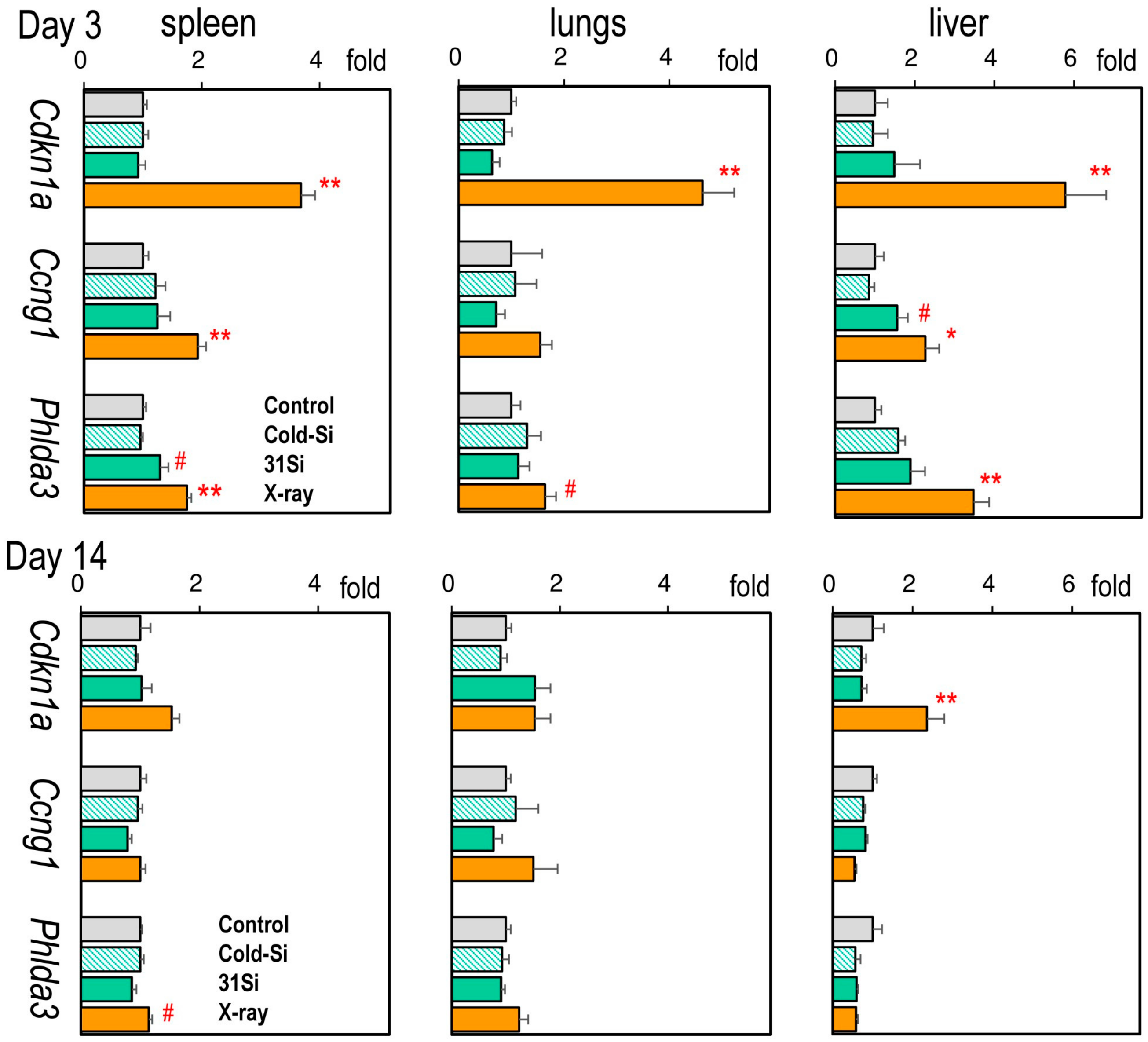
2.3. mRNA Expression of the Radiation Dose–Response Marker Genes Cdkn1a, Ccng1, and Phlda3

Figure 1 presents the gene expression levels of Cdkn1a, Ccng1, and Phlda3 in the spleen, lungs, and liver (Individual data are shown in Supplementary Tables S2–S4). On post-exposure day 3, the expression of all three marker genes was induced by external X-irradiation in every organ examined, except for Ccng1 in the lung. In contrast, only splenic Phlda3 and hepatic Cdkn1a mRNA levels were significantly high on day 14. 31SiO2 exposure significantly induced the expressions of splenic Phlda3 and hepatic Ccng1 on day 3.

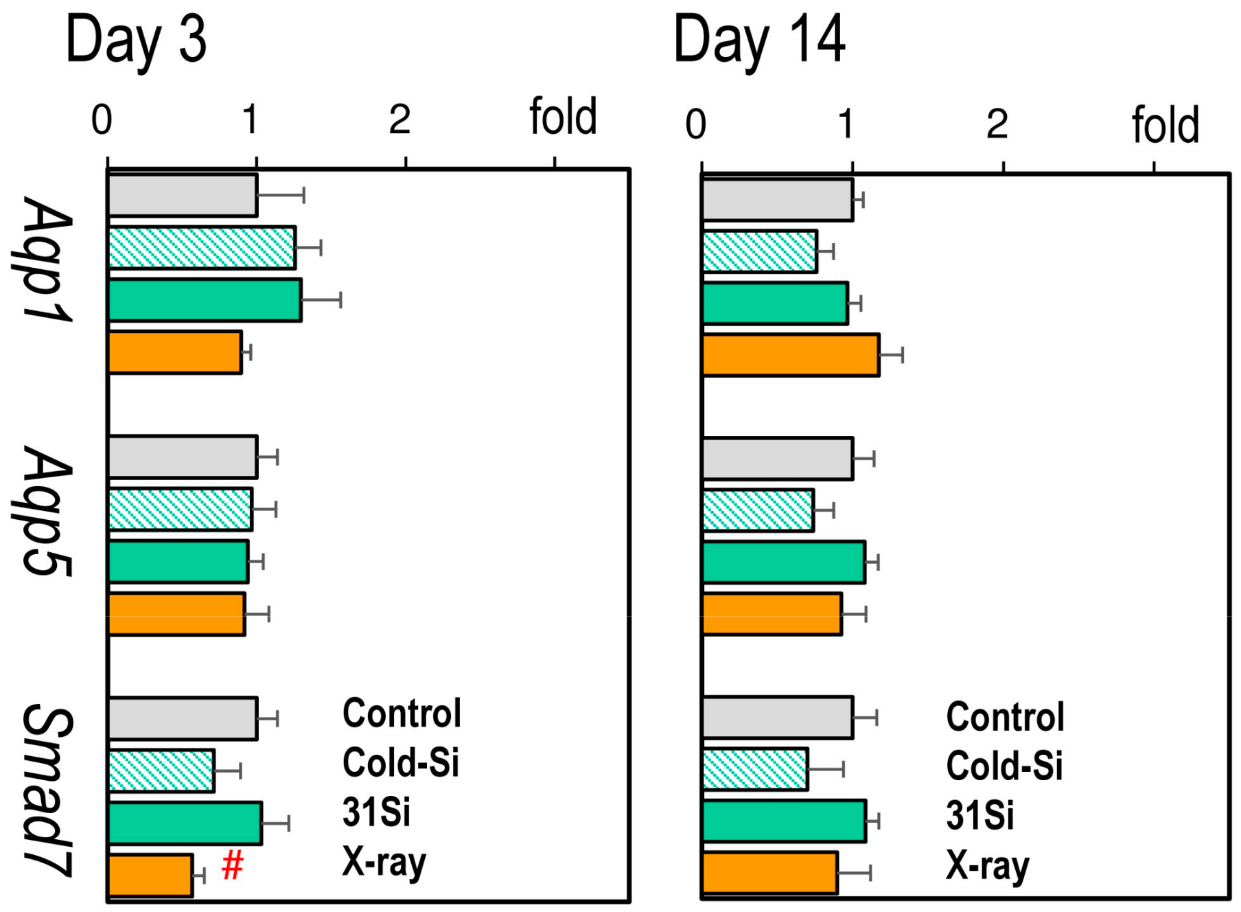
2.4. mRNA Expression of the Radiation-Sensitive Pathophysiological Marker Genes Aqp1, Aqp5, and Smad7 in the Lungs

Figure 2 presents the expression of pulmonary pathophysiological marker genes (Individual data are accessible in Supplementary Table S3). No changes in the expression of either Aqp1, Aqp5, or Smad7 were observed 3 or 14 days after exposure to external X-irradiation or 31SiO2, except for a reduction in the expression of Smad7 on day 3.

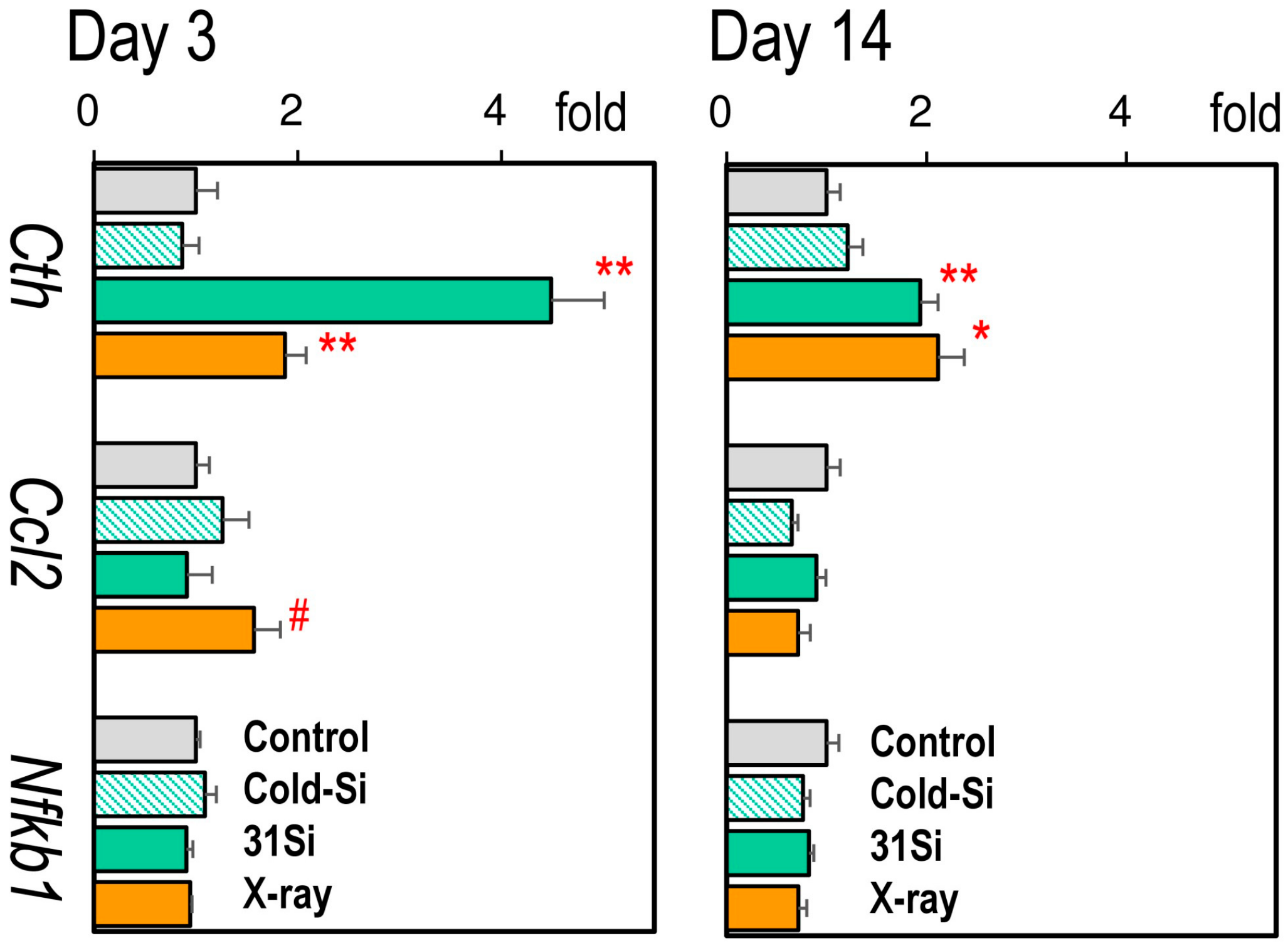
2.5. mRNA Expression of the Radiation-Sensitive Pathophysiological Marker Genes Cth, Ccl2, and Nfkb1 in the Liver

Figure 3 presents the expression of hepatic pathophysiological marker genes (Individual data are accessible in Supplementary Table S4). On post-exposure day 3, the mRNA levels of Cth were significantly increased by both external X-irradiation and 31SiO2 exposure, which interestingly continued to be high on day 14. The expression of Ccl2 was increased on day 3 in the X-ray group.

3. Discussion

This study investigated the effects of internal exposure to β-ray emitting 31SiO2 microparticles on the expression of marker genes in the spleen, lungs, and liver of rats. Exposure to 31SiO2 prominently increased the mRNA expression of Cth, even at low radiation doses in the liver. The results demonstrated the biological significance of exposure to β-emitting particles.
To fully understand the biological effects of atomic bombing, the residual radioactivity produced by the neutron beams from explosions should be considered [16]. Our previous studies involving rodents have reported that internal exposure to 56MnO2, a residual radiation source, significantly affected the expression of genes in organs [4,17]. Although 56Mn emits both β- and γ-rays, our theoretical study suggested the significance of β-rays, which are associated with high local radiation doses in the small tissue areas close to each radioactive particle and may result in biological consequences despite the low whole-organ doses [7]. In this study, 31SiO2 powders were used instead of 56MnO2 to examine the biological effects of internal β-rays. 31Si is also a residual radioisotope produced by the neutron beams of atomic bombs and primarily emits β-rays with a radioactive half-life of approximately 2.62 h [3].
This study focused on the spleen, lungs, and liver. Spleen weights are a radiation-dose-dependent index for whole-body irradiation [18]. In the X-ray group, relative spleen weights decreased on post-exposure day 3 and returned to the control level on day 14. A similar change was observed in the 31Si group, although the splenic radiation dose was only 0.46 mGy. Radiation-induced spleen weight loss is associated with lymphocyte apoptosis, not only in the spleen but also in the whole body [19,20]. Spleen weight loss might result from the systemic effects of 31Si exposure. However, no reduction in spleen weight was observed in our previous 56MnO2 study [4]. The discrepancy needs to be addressed in future studies.
Changes in the expression of p53-regulated genes could be used as radiation exposure signatures [21,22]. A microarray analysis identified highly sensitive expression marker genes, including Cdkn1a, Cccng1, and Phlda3 [10]. Although these markers were mainly found in studies of peripheral blood analysis, Some of them are also useful as exposure markers in solid tissues [23,24]. We then determined the expression of these genes to evaluate their responses to radiation exposure. As expected, external X-irradiation induced the expression of these marker genes on post-exposure day 3. Cdkn1a mRNA levels remained high on day 14, consistent with a previous finding involving mouse livers [25] and our previous study [17]. 31Si exposure increased the expression of Ccng1 in the liver, indicating that the liver responded to radiation from 31Si, although the gene was not radiation-inducible in the lung.
The lungs are the primary targets of 31Si because animals inhale sprayed 31SiO2 particles in the air. To examine the possible biological consequences of 31Si, three pathophysiological marker genes were examined: Aqp1, Aqp5, and Smad7. Aqp1 and Aqp5 encode water-selective channel proteins, aquaporins, that play critical roles in maintaining lung structure and function [12]. The altered expression in Aqp1 and Aqp5 represents physiological and pathological changes in the lungs exposed to radiation or infection [13,26]. The expression of Smad7 may increase during the healing process from radiation-induced tissue damage [14]. Our previous study demonstrated that exposure to 56MnO2 increased the expression of Aqp5 and Smad7 in the lungs, whereas 2 Gy of external irradiation did not [4]. However, in this study, no such changes were found in the lungs in the 31Si group, probably because of the differences in the lung doses between the two studies (48–68 mGy in the 56Mn study vs. 10 mGy in the present 31Si experiment).
Radiation exposure to the liver can cause hepatic injuries. Irradiation at high radiation doses (>30 Gy) results in the development of liver fibrosis, whereas lower doses affect hepatic function by inducing inflammatory and oxidative stresses, in which the expression of cytokines, such as IL-6 and CCL2, increases with the activation of the NFκb signaling [27,28]. Thus, these gene expression changes may serve as useful markers for evaluating radiation-induced hepatic damage; however, no indications were found in the 31Si group. An investigation of human hepatic cultured cells revealed that the expression of Cth was inducible by irradiation [11]. A protein encoded by Cth is an enzyme, cystathionine γ-lyase, producing hydrogen sulfide that protects hepatocytes against oxidative stress. This ability renders Cth an excellent expression marker for radiation-induced hepatic injuries. This study demonstrated that X-irradiation induced the expression of Cth in the liver not only in the cultured cells but also in vivo. The expression of Cth remained high on post-exposure day 14, which made it a more stable marker representing hepatic responses to radiation. Interestingly, 31SiO2 exposure also increased the expression of Cth in the liver, which was higher than that in the X-ray group, suggesting that 31SiO2 microparticles have more intensive biological effects on the liver. The exact physiological functions of Cth in the hepatocytes need to be defined in the future.
Our dosimetry results revealed a very low hepatic radiation dose averaged over whole-organ volume (0.28 mGy), calculated from the radioactivity measured 30 min after exposure. Most inhaled 31SiO2 particles rapidly moved from the bronchus to the digestive tract as the large intestinal dose reached 120 mGy [15]. An oral kinetics study showed that the plasma level of SiO2 started to increase 90 min after oral administration, suggesting that the hepatic dose of 31SiO2 through the portal vein system could elevate later than our timing of the radioactivity measurement, even considering the short radioactive half-life of 31Si. Furthermore, insoluble microparticles could be taken up across the mucosal barrier of the intestinal tract and transported to other organs, particularly the liver, where they are excreted or further distributed [29,30]. Thus, 31SiO2 microparticles can enter the liver and cause significant adverse effects on hepatocytes located close to the microparticles, as predicted by our previous theoretical analysis [8]. A study of 90Y microsphere treatment showed that administered microparticles were localized in the hepatic artery and trapped within the terminal arterioles [31]. A recent investigation of orally exposed polymer microparticles demonstrates their uniform distribution in the liver tissue [32]. Further studies are necessary to examine the details of 31SiO2 particle transport from the gastrointestinal tract and the distribution in the liver tissue and to estimate the peculiarities of local internal doses on the level of the liver’s microstructures.
The chemical toxicity of SiO2 has been extensively studied [33]. Its oral toxicity is considered to be very low because this compound exists ubiquitously in the environment. However, the health hazards of inhaled crystalline SiO2 are well documented, including silicosis and other pulmonary diseases [34,35]. The amorphous SiO2, used in this study possesses little toxicity, although chronic inhalation of amorphous SiO2 microparticles or nanoparticles may increase the expression of proinflammatory markers in the alveoli [35]. No reports have suggested that chemical toxicity could arise from the 1 h inhalation of SiO2 microparticles.
A recent study showed that 137Cs-bearing particles could cause DNA damage in the cells closely located to the particles using the cell culture [36]. A similar in vitro setup combined with a single-cell gene expression analysis will help examine our hypothesis of distant-dependent gene expression changes in the cells closely located to β-emitting particles in the future. It is unclear if our findings in gene expression changes have any specific implications for human health. The future global gene expression analysis in the 31Si exposed animals could provide a more complete view of the effects and possible relevance to human health.

4. Materials and Methods

4.1. Animals

The animal study was approved by the Animal Experiment Ethics Committee of Asfendiyarov Kazakh National Medical University, Almaty, Kazakhstan (document #1630, 7 September 2023). The study was conducted in accordance with the Animal Research: Reporting of In Vivo Experiments (ARRIVE) guidelines. Ten-week-old male Wistar rats were obtained from Asfendiyarov Kazakh National Medical University, Almaty, Kazakhstan. The animals were maintained in cages with free access to a basal diet and tap water. After a week of acclimatation, the rats were randomly divided into four groups: the control group (n = 10), Cold-Si group (n = 13), 31Si group (n = 13), and X-ray group (n = 10), with a total n = 46. The Cold-Si group was exposed to stable SiO2 microparticles. The 31Si group was exposed to 31SiO2 microparticles at an activity of 3.2 × 10⁷ Bq/g. Three rats from the 31Si group were used to estimate the absorbed radiation doses. The X-ray group received 2 Gy of external X-rays using a LINAC. Five rats from each group underwent necropsies on post-exposure days 3 and 14. The rats were euthanized by removal of whole blood under anesthesia with isoflurane (Fujifilm Wako Pure Chemical Co., Tokyo, Japan). The spleen, lungs, and liver were dissected, and tissue pieces were stored in Gene Keeper solution (Fujifilm Wako Pure Chemical Co.) for RNA and protein extraction.

4.2. Irradiation and Dosimetry

Exposure to 31SiO2 microparticles and internal dose estimation of internal doses averaged over whole organs’ volumes were performed as previously described [15]. In brief, SiO2 powder (Rare Metallic Co., Tokyo, Japan; average diameter of the particles: 2.4 µm, purity: >99.9%) was activated by a neutron beam in the WWR-K nuclear reactor at the Institute of Nuclear Physics, Almaty, Kazakhstan. The obtained radioactive 31SiO2 powder was sprayed over the rats for 1 h. Then, three rats of the 31Si group were euthanized for β-spectrometry, and each organ’s absorbed dose was estimated. All animals were brought to the exposure facility to maintain the same environment in each group. A medical LINAC, Clinac 2100C/D (Varian Medical Systems Inc., Palo Alto, CA, USA), was used for whole-body γ-irradiation at a dose rate of 6 Gy/min.

4.3. QRT Polymerase Chain Reaction (PCR)

Total RNA was isolated from tissues stored in Gene Keep solution with Isogen II (Nippon Gene Co., Tokyo, Japan). cDNA was obtained by incubating 3 µg of total RNA + RevaTra Ace 100 U (Toyobo Co., Osaka, Japan) + 20 pmol random hexamers/5 pmol oligo-dT(15) primers (Takara Bio Inc., Kusatsu, Japan). The quantitative PCR system StepOnePlus (Applied Biosystems, Life Technologies, Co., Carlsbad, CA, USA) was used for cDNA measurement with Thunderbird Next Sybr q-PCR Mix (Toyobo Co.). PCR was performed with a 2-min initial denaturation, followed by 40 cycles of 5 s at 95 °C and 35 s at 60 °C. Table 2 presents the specific primer sets for the genes. The nucleotide sequence of each PCR product was verified by Fasmac Co., Ltd. (Atsugi, Japan). The mRNA levels were normalized with reference to the β-actin mRNA levels.

4.4. Statistical Analysis

For multiple comparisons among the groups, Dunnett’s test was used with R’s package “SimComp” version 3.3 (http://cran.r-project.org, accessed on 10 December 2024). Paired comparisons were performed with the t-test function of the Microsoft Excel 2019 program.

5. Conclusions

This study investigated the effects of exposure to 31SiO2 microparticles emitting predominantly β-rays on the gene expression in the spleen, lungs, and liver of Wistar rats on post-exposure days 3 and 14. 31SiO2 exposure induced little expression change in the spleen or lungs. However, it markedly increased the expression of Cth in the liver on postexposure days 3 and 14, even at very low values of internal radiation doses averaged over whole organs’ volume. This indicates the significant biological effects of β-emitting particles and their possible roles as residual radiation of atomic bombs. In order to better understanding these effects, the estimations of local values of internal dose from exposure to 31SiO2 microparticles on the liver’s microstructure are ongoing.

Supplementary Materials

The following supporting information can be downloaded at: https://www.mdpi.com/article/10.3390/ijms26062693/s1.

Author Contributions

Conceptualization, M.H. and V.S.; methodology, N.F., M.H., H.S., V.S. and A.K. (Almagul Kushugulova); investigation and analysis, N.M., S.Z., L.C., T.F., M.M., H.S., K.Z., A.K. (Almagul Kushugulova), and N.F.; writing—original draft preparation, N.F.; supervision, A.K. (Andrey Kaprin), S.I., P.S. and A.K. (Almagul Kushugulova); funding acquisition, A.K. (Almagul Kushugulova), M.H. and N.F. All authors have read and agreed to the published version of the manuscript.

Funding

This work was supported by JSPS Kakenhi Grants, Japan (19H01149 and 19KK0266) and JSPS Bilateral Exchange Program (JPJSBP 120249929).

Institutional Review Board Statement

The animal study protocol was approved by the Animal Experiment Ethics Committee of Asfendiyarov Kazakh National Medical University, Almaty, Kazakhstan (document #1630, 7 September 2023).

Informed Consent Statement

Not applicable.

Data Availability Statement

All data are presented in the article and the Supplementary Materials.

Acknowledgments

The authors appreciate Samat Kozhakhmetov and Shynggys Sergazy of Center for Life Sciences, National Laboratory Astana, Nazarbayev University, for their excellent technical assistance in exposing animals to 31SiO2. The authors thank Bagdaulet Mukhametuly and Murat Kenessarin of Institute of Nuclear Physics of the Ministry of Energy of the Republic of Kazakhstan, for their exceptional technical assistance in the irradiation with a LINAC.

Conflicts of Interest

The authors declare no conflicts of interest.

References

  1. Imanaka, T.; Endo, S.; Tanaka, K.; Shizuma, K. Gamma-ray exposure from neutron-induced radionuclides in soil in Hiroshima and Nagasaki based on DS02 calculations. Radiat. Environ. Biophys. 2008, 47, 331–336. [Google Scholar] [CrossRef]
  2. Otani, K.; Ohtaki, M.; Yasuda, H. Solid cancer mortality risk among a cohort of Hiroshima early entrants after the atomic bombing, 1970–2010: Implications regarding health effects of residual radiation. J. Radiat. Res. 2022, 63, i45–i53. [Google Scholar] [CrossRef] [PubMed]
  3. Tanaka, K.; Endo, S.; Imanaka, T.; Shizuma, K.; Hasai, H.; Hoshi, M. Skin dose from neutron-activated soil for early entrants following the A-bomb detonation in Hiroshima: Contribution from β and γ rays. Radiat. Environ. Biophys. 2008, 47, 323–330. [Google Scholar] [CrossRef] [PubMed]
  4. Fujimoto, N.; Ruslanova, B.; Abishev, Z.; Chaizhunussova, N.; Shabdarbayeva, D.; Amantayeva, G.; Farida, R.; Sandybayev, M.; Nagano, K.; Zhumadilov, K.; et al. Biological impacts on the lungs in rats internally exposed to radioactive 56MnO2 particle. Sci. Rep. 2021, 11, 11055. [Google Scholar] [CrossRef] [PubMed]
  5. Mück, K.; Pröhl, G.; Likhtarev, I.; Kovgan, L.; Golikov, V.; Zeger, J. Reconstruction of the inhalation dose in the 30-km zone after the Chernobyl accident. Health Phys. 2002, 82, 157–172. [Google Scholar] [CrossRef]
  6. Steinhauser, G.; Brandl, A.; Johnson, T.E. Comparison of the Chernobyl and Fukushima nuclear accidents: A review of the environmental impacts. Sci. Total Environ. 2014, 470–471, 800–817. [Google Scholar] [CrossRef]
  7. Stepanenko, V.; Kaprin, A.; Ivanov, S.; Shegay, P.; Bogacheva, V.; Sato, H.; Shichijo, K.; Toyoda, S.; Kawano, N.; Ohtaki, M.; et al. Microdistribution of internal radiation dose in biological tissues exposed to 56Mn dioxide microparticles. J. Radiat. Res. 2022, 63, i21–i25. [Google Scholar] [CrossRef]
  8. Kabacik, S.; MacKay, A.; Tamber, N.; Manning, G.; Finnon, P.; Paillier, F.; Ashworth, A.; Bouffler, S.; Badie, C. Gene expression following ionising radiation: Identification of biomarkers for dose estimation and prediction of individual response. Int. J. Radiat. Biol. 2011, 87, 115–129. [Google Scholar] [CrossRef]
  9. Li, M.J.; Wang, W.W.; Chen, S.W.; Shen, Q.; Min, R. Radiation dose effect of DNA repair-related gene expression in mouse white blood cells. Med. Sci. Monit. 2011, 17, 290–297. [Google Scholar] [CrossRef]
  10. Paul, S.; Smilenov, L.B.; Elliston, C.D.; Amundson, S.A. Radiation Dose-Rate Effects on Gene Expression in a Mouse Biodosimetry Model. Radiat. Res. 2015, 184, 24–32. [Google Scholar] [CrossRef]
  11. Pan, Y.; Ye, S.; Yuan, D.; Zhang, J.; Bai, Y.; Shao, C. Radioprotective role of H2S/CSE pathway in Chang liver cells. Mutat. Res. 2012, 738–739, 12–18. [Google Scholar] [CrossRef] [PubMed]
  12. Verkman, A.S. Role of aquaporins in lung liquid physiology. Respir. Physiol. Neurobiol. 2007, 159, 324–330. [Google Scholar] [CrossRef] [PubMed]
  13. Sun, C.Y.; Zhao, Y.X.; Zhong, W.; Liu, D.W.; Chen, Y.Z.; Qin, L.L.; Bai, L.; Liu, D. The expression of aquaporins 1 and 5 in rat lung after thoracic irradiation. J. Radiat. Res. 2014, 55, 683–689. [Google Scholar] [CrossRef] [PubMed]
  14. Chen, J.; Zhang, W.; Zhang, L.; Zhang, J.; Chen, X.; Yang, M.; Chen, T.; Hong, J. Glycyrrhetinic acid alleviates radiation-induced lung injury in mice. J. Radiat. Res. 2017, 58, 41–47. [Google Scholar] [CrossRef]
  15. Stepanenko, V.; Sato, H.; Kaprin, A.; Fujimoto, N.; Kushugulova, A.; Ivanov, S.; Shegay, P.; Bogacheva, V.; Petukhov, A.; Zhumadilov, K.; et al. Internal radiation dose estimates in organs of Wistar rats exposed to sprayed neutron-activated 31SiO2 microparticles: First results of international multicenter study. J. Radiat. Res. 2024, 65, 744–751. [Google Scholar] [CrossRef]
  16. Kerr, G.D.; Egbert, S.D.; Al-Nabulsi, I.; Bailiff, I.K.; Beck, H.L.; Belukha, I.G.; Cockayne, J.E.; Cullings, H.M.; Eckerman, K.F.; Granovskaya, E.; et al. Workshop Report on Atomic Bomb Dosimetry—Review of Dose Related Factors for the Evaluation of Exposures to Residual Radiation at Hiroshima and Nagasaki. Health Phys. 2015, 109, 582–600. [Google Scholar] [CrossRef]
  17. Ruslanova, B.; Abishev, Z.; Chaizhunussova, N.; Shabdarbayeva, D.; Tokesheva, S.; Amantayeva, G.; Kairkhanova, Y.; Stepanenko, V.; Hoshi, M.; Fujimoto, N. Hepatic Gene Expression Changes in Rats Internally Exposed to Radioactive 56MnO2 Particles at Low Doses. Curr. Issues Mol. Biol. 2021, 43, 758–766. [Google Scholar] [CrossRef]
  18. Li, W.; Jiang, L.; Lu, X.; Liu, X.; Ling, M. Curcumin protects radiation-induced liver damage in rats through the NF-κB signaling pathway. BMC Complement. Med. Ther. 2021, 21, 10. [Google Scholar] [CrossRef]
  19. Fujikawa, K.; Hasegawa, Y.; Matsuzawa, S.; Fukunaga, A.; Itoh, T.; Kondo, S. Dose and dose-rate effects of X rays and fission neutrons on lymphocyte apoptosis in p53(+/+) and p53(-/-) mice. J. Radiat. Res. 2000, 41, 113–127. [Google Scholar] [CrossRef]
  20. Raventos, A. An abscopal effect of x-ray upon mouse spleen weight. Radiat. Res. 1954, 1, 381–387. [Google Scholar] [CrossRef]
  21. Daino, K.; Ichimura, S.; Nenoi, M. Early induction of CDKN1A (p21) and GADD45 mRNA by a low dose of ionizing radiation is due to their dose-dependent post-transcriptional regulation. Radiat. Res. 2002, 157, 478–482. [Google Scholar] [CrossRef] [PubMed]
  22. Meadows, S.K.; Dressman, H.K.; Muramoto, G.G.; Himburg, H.; Salter, A.; Wei, Z.; Ginsburg, G.S.; Ginsburg, G.; Chao, N.J.; Nevins, J.R.; et al. Gene expression signatures of radiation response are specific, durable and accurate in mice and humans. PLoS ONE 2008, 3, e1912. [Google Scholar] [CrossRef]
  23. Aryankalayil, M.J.; Bylicky, M.A.; Martello, S.; Chopra, S.; Sproull, M.; May, J.M.; Shankardass, A.; MacMillan, L.; Vanpouille-Box, C.; Dalo, J.; et al. Microarray analysis identifies coding and non-coding RNA markers of liver injury in whole body irradiated mice. Sci. Rep. 2023, 13, 200. [Google Scholar] [CrossRef]
  24. Lee, W.-J.; Majumder, Z.R.; Jeoung, D.-I.; Lee, H.-j.; Kim, S.-H.; Bae, S.; Lee, Y.-S. Organ-specific gene expressions in C57BL/6 mice after exposure to low-dose radiation. Radiat. Res. 2006, 165, 562–569. [Google Scholar] [CrossRef]
  25. Pawlik, A.; Delmar, P.; Bosse, S.; Sainz, L.; Petat, C.; Pietu, G.; Thierry, D.; Tronik-Le Roux, D. Changes in transcriptome after in vivo exposure to ionising radiation reveal a highly specialised liver response. Int. J. Radiat. Biol. 2009, 85, 656–671. [Google Scholar] [CrossRef]
  26. Towne, J.E.; Harrod, K.S.; Krane, C.M.; Menon, A.G. Decreased expression of aquaporin (AQP)1 and AQP5 in mouse lung after acute viral infection. Am. J. Respir. Cell Mol. Biol. 2000, 22, 34–44. [Google Scholar] [CrossRef]
  27. Ali, O.S.M.; Amin, N.E.D.; Abdel Fattah, S.M.; Abd El-Rahman, O. Ameliorative effect of kefir against γ-irradiation induced liver injury in male rats: Impact on oxidative stress and inflammation. Environ. Sci. Pollut. Res. Int. 2020, 27, 35161–35173. [Google Scholar] [CrossRef]
  28. Eassawy, M.M.T.; Salem, A.A.; Ismail, A.F.M. Biochemical study on the protective effect of curcumin on acetaminophen and gamma-irradiation induced hepatic toxicity in rats. Environ. Toxicol. 2020, 36, 748–763. [Google Scholar] [CrossRef]
  29. Paul, M.B.; Stock, V.; Cara-Carmona, J.; Lisicki, E.; Shopova, S.; Fessard, V.; Braeuning, A.; Sieg, H.; Böhmert, L. Micro- And nanoplastics-current state of knowledge with the focus on oral uptake and toxicity. Nanoscale Adv. 2020, 2, 4350–4367. [Google Scholar] [CrossRef]
  30. McMinn, L.H.; Hodges, G.M.; Carr, K.E. Gastrointestinal uptake and translocation of microparticles in the streptozotocin-diabetic rat. J. Anat. 1996, 189, 553–559. [Google Scholar]
  31. Gulec, S.A.; Sztejnberg, M.L.; Siegel, J.A.; Jevremovic, T.; Stabin, M. Hepatic structural dosimetry in 90Y microsphere treatment: A Monte Carlo modeling approach based on lobular microanatomy. J. Nucl. Med. 2010, 51, 301–310. [Google Scholar] [CrossRef] [PubMed]
  32. Garcia, M.M.; Romero, A.S.; Merkley, S.D.; Meyer-Hagen, J.L.; Forbes, C.; El Hayek, E.; Sciezka, D.P.; Templeton, R.; Gonzalez-Estrella, J.; Jin, Y.; et al. In Vivo Tissue Distribution of Polystyrene or Mixed Polymer Microspheres and Metabolomic Analysis after Oral Exposure in Mice. Environ. Health Perspect. 2024, 132, 47005. [Google Scholar] [CrossRef] [PubMed]
  33. Younes, M.; Aquilina, G.; Castle, L.; Degen, G.; Engel, K.H.; Fowler, P.; Frutos Fernandez, M.J.; Furst, P.; Gurtler, R.; Husoy, T.; et al. Re-evaluation of silicon dioxide (E 551) as a food additive in foods for infants below 16 weeks of age and follow-up of its re-evaluation as a food additive for uses in foods for all population groups. EFSA J. 2024, 22, e8880. [Google Scholar]
  34. Marques Da Silva, V.; Benjdir, M.; Montagne, P.; Pairon, J.-C.; Lanone, S.; Andujar, P. Pulmonary Toxicity of Silica Linked to Its Micro- or Nanometric Particle Size and Crystal Structure: A Review. Nanomaterials 2022, 12, 2392. [Google Scholar] [CrossRef] [PubMed]
  35. Merget, R.; Bauer, T.; Küpper, H.U.; Philippou, S.; Bauer, H.D.; Breitstadt, R.; Bruening, T. Health hazards due to the inhalation of amorphous silica. Arch. Toxicol. 2002, 75, 625–634. [Google Scholar] [CrossRef]
  36. Matsuya, Y.; Satou, Y.; Hamada, N.; Date, H.; Ishikawa, M.; Sato, T. DNA damage induction during localized chronic exposure to an insoluble radioactive microparticle. Sci. Rep. 2019, 9, 10365. [Google Scholar] [CrossRef]
Figure 1. Relative mRNA levels of Cdkn1a, Ccng1, and Phlda3 in the spleen, lungs, and liver 3 and 14 days after exposure to nonradioactive SiO2 particles (Cold-Si), 31SiO2 particles (31Si), or 2 Gy of X-rays (X-ray). Each bar indicates mean ± standard error of the mean (n = 5, each group). * p < 0.05 or ** p < 0.01 versus control. # p < 0.05 versus control or Cold-Si (paired comparison). X-rays increased the expression of all three marker genes on day 3, except for Ccng1 in the lung. 31SiO2 exposure increased splenic Phlda3 and hepatic Ccng1 expressions on day 3.
Ijms 26 02693 g001
Figure 2. Relative mRNA levels of Aqp1, Aqp5, and Smad7 in the lungs 3 and 14 days after exposure to nonradioactive SiO2 particles (Cold-Si), 31SiO2 particles (31Si), or 2 Gy of X-rays (X-ray). Each bar indicates mean ± standard error of the mean (n = 5, each group). # p < 0.05 versus control (paired comparison). X-rays reduced the expression of Smad7.
Ijms 26 02693 g002
Figure 3. Relative mRNA levels of Cth, Ccl2, and Nfkb1 in the liver 3 and 14 days after exposure to nonradioactive SiO2 particles (Cold-Si), 31SiO2 particles (31Si), or 2 Gy of X-rays (X-ray). Each bar indicates mean ± standard error of the mean (n = 5, each group). * p < 0.05 or ** p < 0.01 versus control. # p < 0.05 versus control (paired comparison). The expression of Cth was increased in the 31Si and X-ray groups on post-exposure days 3 and 14. The expression of Ccl2 was increased in the X-ray group on day 3.
Ijms 26 02693 g003
Table 1. Body and the relative organ weights in rats exposed to cold SiO2, 31SiO2, or γ-rays.
GroupsInitial Body Weight (g)Body Weight (g)Spleen
(g/kg bw)
Lung
(g/kg bw)
Liver
(g/kg bw)
Day 3Control271 ± 30277 ± 294.8 ± 0.848.4 ± 0.4628 ± 1.9
Cold-Si298 ± 27297 ± 224.2 ± 0.309.9 ± 2.4731 ± 1.7
31Si303 ± 26309 ± 263.2 ± 0.36 #8.4 ± 1.3829 ± 1.1
X-ray283 ± 27288 ± 263.6 ± 0.411.6 ± 4.1729 ± 1.1
Day 14Control293 ± 33314 ± 293.5 ± 0.228.5 ± 1.0526 ± 0.7
Cold-Si291 ± 39300 ± 394.1 ± 0.6010.2 ± 0.8530 ± 1.0
31Si299 ± 37319 ± 374.3 ± 0.649.2 ± 0.7531 ± 1.9
X-ray295 ± 34319 ± 383.3 ± 0.2512.9 ± 3.8432 ± 1.7
Control: untreated group; Cold-Si: exposed to stable SiO2 particles; 31Si: exposed to 31SiO2 particles at the activity of 4.9 × 10⁷ Bq/g; X-ray: exposed to X-rays (2 Gy) with an LINAC. Initial body weights are the body weights a day before exposure. Each value shows mean ± standard error of the mean (n = 5, each group). # indicates a significant difference from the Cold-Si at p < 0.05 (paired comparison).
Table 2. Q-PCR primers.
GeneGenBank Accession #Q-PCR Primer Sequences (5′ -> 3′)
ForwardReverse
Cdkn1aNM_080782TGTCCGACCTGTTCCACACACGTCTCAGTGGCGAAGTCAA
Ccng1NM_012923CGTGCCACTGCAGGATCATAAAGGTCAGATCTCGGCCACTT
Phlda3NM_001012206AAGCCGTGGAGTGCGTGGAGAGGTCTGGATGGCCTGTTGATTC
Aqp1NM_012778CCACTGGAGAGAAACCAGACGCTGAGCAGAAGCCCCAGTGT
Aqp5NM_012779ATGCGCTGAACAACAACACAACGTGACAGACAAGCCAATGGATAAG
Smad7NM_030858TTGCTGTGAATCTTACGGGAAGGGTTTGAGAAAATCCATCGGGT
CthNM_017074TCCCCGGGTAGAAAAGGTTATTTTGAGGAAGACCTGAGCATGC
Ccl2NM_031530AAGCCAGATCTCTCTTCCTCCACAGCAACTGTGAACAACAGGC
NfkbNM_001276711GGGCTACACAGAGGCCATTGTCTCGGAGCTCATCTATGTGCT
ActbNM_031144.3TTGTCCCTGTATGCCTCTGGTCTGAGGTAGTCTGTCAGGTCCC
Disclaimer/Publisher’s Note: The statements, opinions and data contained in all publications are solely those of the individual author(s) and contributor(s) and not of MDPI and/or the editor(s). MDPI and/or the editor(s) disclaim responsibility for any injury to people or property resulting from any ideas, methods, instructions or products referred to in the content.

