Article

Structure and Optical Properties of New 2-N-Phenylamino-methyl-nitro-pyridine Isomers

1
Department of Bioorganic Chemistry, Faculty of Production Engineering, Wroclaw University of Economics and Business, 118-120 Komandorska Str., 53-345 Wrocław, Poland
2
Institute of Low Temperature and Structure Research, 2 Okólna Str., 50-422 Wrocław, Poland
*
Authors to whom correspondence should be addressed.
Int. J. Mol. Sci. 2025, 26(7), 2874; https://doi.org/10.3390/ijms26072874
Submission received: 8 February 2025 / Revised: 5 March 2025 / Accepted: 12 March 2025 / Published: 21 March 2025
(This article belongs to the Section Physical Chemistry and Chemical Physics)

Abstract

:
Two new 2-N-phenylamino-(4 or 6)-methyl-3-nitropyridine derivatives were synthesized. Their structures were characterized on the basis of X-ray diffraction, IR, and Raman spectra as well as electron UV-Vis and emission spectra measurements. The experimental results were analyzed in terms of theoretical data in which the quantum chemical DFT and NBO calculations were applied. To elucidate the relaxation pathways of electronically excited states, multiple excitation wavelengths were employed to probe energy dissipation mechanisms in the studied compounds. A systematic analysis was conducted to evaluate how variations in methyl substituent positioning modulate both the structural architecture and photophysical behavior of the isomeric systems. The spectroscopic, structural and theoretical considerations allow us to propose the potential technological applications derived from the unique properties of these isomers.

Graphical Abstract

1. Introduction

The synthesis and investigation of new 2-N-phenylamino-methyl-nitropyridines have garnered significant attention due to their versatile properties and potential applications across various industries. These compounds, characterized by their unique structural features and tunable chemical properties, have been explored extensively in the fields of material science, optoelectronics, and biochemistry. Additionally, they belong to a wide class of pyridine derivatives, which serve as a fundamental unit in many natural products, such as vitamins, alkaloids, coenzymes, drugs, and pesticides. Pyridine derivatives also exhibit a broad spectrum of biological activities, including anti-inflammatory [1,2], antiviral [3,4], anticancer [5,6,7,8,9], antimicrobial [10,11], antidiabetic [12,13], osteogenic [14,15], and antihypertensive [16] properties. Recent findings even highlight their potential antiviral activity against SARS-CoV-2 (COVID-19) and antifungal effects [17,18,19,20,21,22,23,24,25]. Due to their specific chemical properties, they were used as oxidizing reagents in organic syntheses [26,27] as well as the ligands for complexation of the metal ions [28,29].
Recent advancements have highlighted the potential use of dyes derived from pyridine compounds in the development of smart materials, particularly in packaging films. Laboratory experiments demonstrated that new derivatives of 2-N-phenylamino-3-nitro-6-methylpyridine exhibit promising properties when incorporated into chitosan-based polymers. These derivatives show excellent color intensity and stability, as well as low toxicity. Experimental trials produced chitosan films infused with these derivatives, showcasing not only their ease of integration into polymer matrices but also their ability to enhance the functional attributes of the final materials. The dyes dissolved well in chitosan gel, enabling efficient and economical coloring, which adds significant value for industrial applications.
In this study, we focus on the synthesis, structural characterization, and spectroscopic analysis of new pyridine derivatives, particularly 2-N-phenylamino-(4 or 6)-methyl-(3)-nitropyridine. The structural nuances of these compounds, elucidated through X-ray crystallography and supported by theoretical modeling, reveal insights into their bonding interactions and potential reactivity. Furthermore, the optical and electronic properties, explored through vibrational spectroscopy and UV-Vis analysis, underscore their versatility and suitability for incorporation into functional materials. The influence of methyl group substitution in the pyridine ring on the structure, electron level distribution, and spectroscopic properties was studied experimentally and theoretically using DFT and NBO quantum chemical calculations. These studies are a continuation of our works on a broad class of pyridine derivatives [30,31,32].
This research is a continuation of previous studies on pyridine derivatives [31,32], which have extensively characterized the vibrations and reactivity of compounds closely related to those investigated in this work. By aligning the findings of this research with practical applications, such as the incorporation of pyridine-derived dyes into chitosan films, this work bridges the gap between fundamental chemistry and applied science.

2. Results and Discussion

2.1. Structural Characterization

The effect of substituents in the pyridine ring of nitro and methyl groups in 2-N-phenylaminopyridine strongly affects the structural and optical properties of these derivatives. The red color is characteristic of isomers with a nitro group substituted in the 3-position of the pyridine ring. The position of substituents in these isomers is also reflected in their crystal structures.

2.1.1. Description of the Structure of 2-N-Phenylamino-3-nitro-4-methylpyridine

2-N-phenylamino-3-nitro-4-methylpyridine (PA3N4MP) crystallizes in the centrosymmetric space group of the triclinic system with two molecules per unit cell. Its X-ray molecular structure together with the optimized one, for comparison, is shown in Figure 1—selected geometrical parameters are listed in Table 1. The conformation of the 2-N-phenylamino-3-nitro-4-methylpyridine molecule is not planar. The planar NO2 group is slightly twisted in relation to the plane of the pyridine ring by 3.84(15)°, whereas the dihedral angle between the average plane of pyridine ring and the average plane of phenyl ring is 6.20(15)°. A weak intramolecular N–H∙∙∙O is observed. This is reflected in the N–O bond lengths of the NO2 group (Table 1).
The geometry of the 2-N-phenylamino-3-nitro-4-methylpyridine molecule in the gaseous phase obtained from the DFT calculations also shows a non-planar conformation. The dihedral angle between the plane of NO2 group and the plane of the pyridine ring of ~25° is significantly greater than that in the crystal. A quite similar correlation also occurs for the dihedral angle between the planes of the pyridine and phenyl rings between the conformations of the molecule in the gas phase and the solid phase. Selected geometrical DFT parameters are included in Table 1, but full details are listed in Table S1 (in the Supplementary Materials).
The arrangement of the molecules in the PA3N4MP crystal is mainly determined by electrostatic interactions and van der Waals forces because there are no strong directional interactions (except of intramolecular N–H∙∙∙O hydrogen bond) between the molecules (Figure 2). The inversion-related molecules along the stacks are separated by ~4.70 Å, which indicates a π-π interaction between the π-clouds of the pyridine and phenyl rings that is too weak. However, the electrostatic interactions between the molecules within the stacks, as can be seen from the three-dimensional electrostatic potential map (Figure 2b), take place. The three-dimensional molecular map of the electrostatic potential (3D-MESP) is related with the electron density within the molecule and is helpful in the nucleation and crystal growth processes. The calculated 3D MESP is mapped onto the total electron density isosurface (0.008 eÅ−3) and illustrates the size, shape, charge density, and the sites of the chemical reactivity. Arrangement of the neighboring molecules within the stacks as well as between the stacks results from electrostatic interactions between the fragments of molecules with opposite signs of EP (Figure 2b).

2.1.2. Description of the Structure of 2-N-Phenylamino-3-nitro-6-methylpyridine

The 2-N-phenylamino-3-nitro-6-methylpyridine, PA3N6MP, crystallizes in the centrosymmetric space group P21/n of the monoclinic system with four molecules per unit cell. Its asymmetric unit contains one 2-phenylamino-3-nitro-6-methylpyridine molecule. Its X-ray molecular structure together with the optimized one, for comparison, is shown in Figure 3, and selected geometrical parameters are listed in Table 2, but full details of the optimized DFT parameters are given in Table S2 (in Supplementary Materials). The conformation of the 2-N-phenylamino-3-nitro-6-methylpyridine molecule is not planar. The planar NO2 group is slightly twisted in relation to the plane of the pyridine ring by 10.30(12)°, whereas the dihedral angle between the average plane of pyridine ring and the average plane of phenyl ring is 2.90(14)°. A weak intramolecular N–H∙∙∙O is observed. This is reflected in the N–O bond lengths of the NO2 group (Table 2). In contrast to the X-ray structure, the gas-phase conformation obtained from DFT calculations shows its strictly planar arrangement. In general, the bond lengths and angles, without that described of the dihedral angles described above, correlate well with that in the crystal.
Arrangement of the molecules in the crystal are mainly determined by the van der Waals forces and by the stacking interactions along the direction between adjacent molecules (Figure 4). Within the stack there is an alternating arrangement of pyridine and benzene rings with a distance of ~3.670 Å between the centroids of the rings. This alternating arrangement of adjacent molecules is consistent with the 3D MESP, illustrated in Figure 4. This distance indicates quite weak π-π interactions along the stack.

2.1.3. Comparison of the Structures of PA3N4MP and PA3N6MP

Hirshfeld surface analysis, HS [33], and its corresponding two-dimensional fingerprint plot [34] provide a convenient method for comparing interactions within crystal structures for different isomers, revealing important similarities and differences between them [35,36]. The Hirsfeld surface mapped with dborm for the molecules of both isomers in the crystals and their 2D fingerprint plots are illustrated in Figure 5.
The normalized contact distance (dborm) is based on the distances from the nearest atom inside (di) and outside (de) the surface. In the HS, the de value corresponds to the distance between the external atom and the surface, and di value corresponds to the distance between the internal atom and the surface. The red spots in the HS correspond to the contact distances between the atoms inside and outside the surface, which are smaller than the sum of the van der Waals radii. Blue and white areas represent the distances longer and equal than the sum of the respective van der Waals radii. The deconvolution of the 2D-fingerprint plots for individual types of interactions is given in Figures S1 and S2 (in Supplementary Materials) for PA3N4MP and PA3N6MP, respectively. In the crystal of both isomers (PA3N4MP and PA3N6MP), the main interactions between the molecules are the H···H dispersive forces with the contribution of 43.8% and 42.7% in the HS of PA3N4MP and PA3N6MP, respectively. The second type of interactions between the molecules in the crystals are H···O/O···H, which constitute 21.2% and 23.7%, respectively, in the HS of PA3N4MP and PA3N6MP. The next types of intermolecular interactions in the crystals PA3N4MP and PA3N6MP are C···H/H···C, C···C, and C···N/N···C, which constitute 13.9%, 8.7%, and 5.0% for PA3N4MP and 14.4, 7.5, and 5.8% for PA3N6MP. The contribution of the respective interactions to the HS in the crystals of PA3N4MP and PA3N6MP is illustrated in Figure 5 on the right. In general, the contribution of the individual interactions is quite comparable in the crystals of both isomers, but there is a visible difference between them. In the PA3N4MP crystal, the contribution of the H···O/O···H interactions associated with the hydrogen bonds is about 2.5% smaller than in the crystal of the second isomer, PA3N6MP, while in the PA3N4MP crystal, the contribution of the C···C interactions is 8.7% and is almost 20% larger than in the PA3N6MP crystal, where it is 7.5%.

2.2. Vibration Characterization

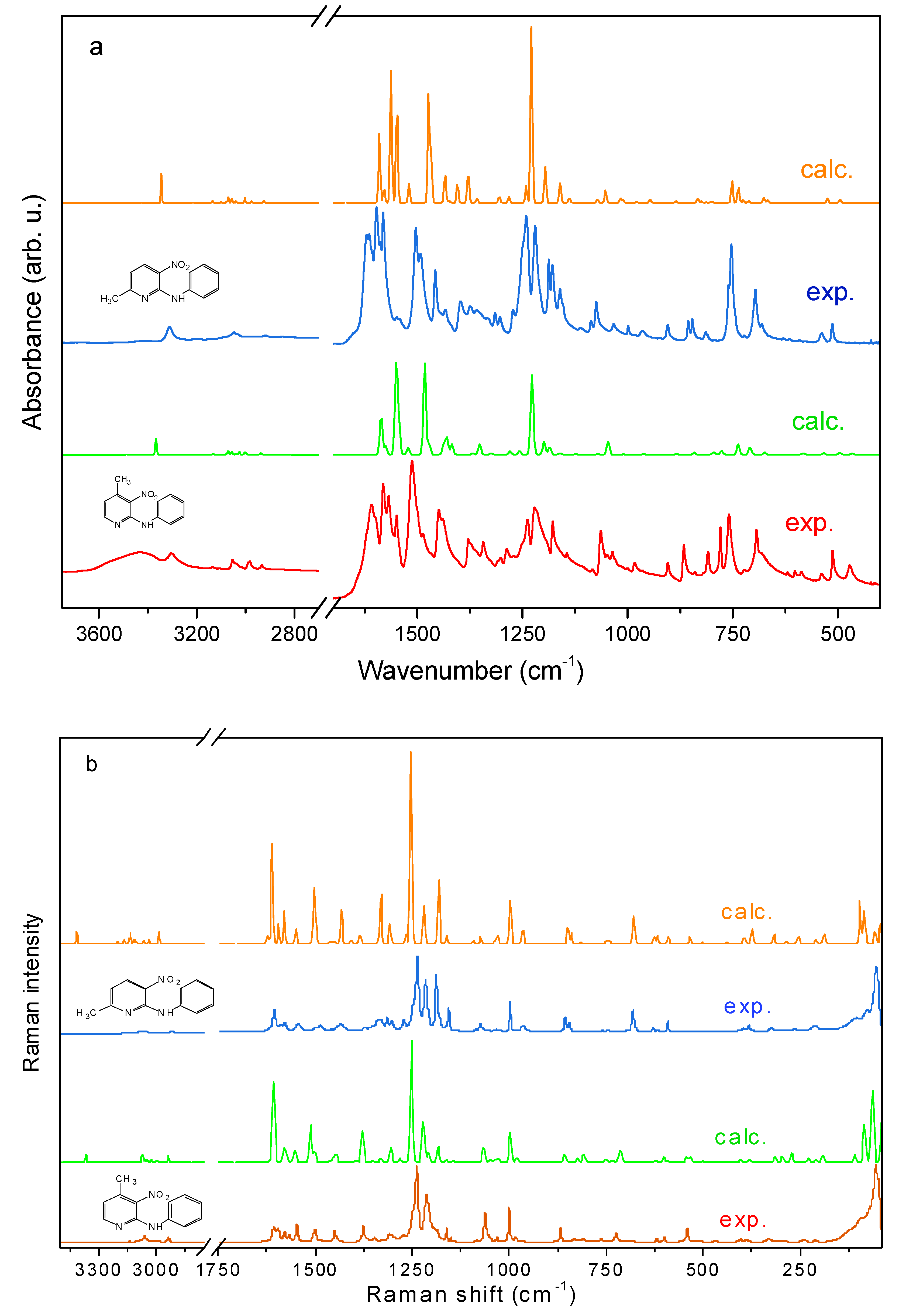
2.2.1. Vibrational Spectra

The IR and Raman spectra of the studied compounds are shown in Figure 6.

2.2.2. Vibrational Modes of Pyridine and Phenyl Systems

Given the structural homology between the investigated isomeric systems, characteristic vibrational signatures of the pyridine moiety are anticipated to fall within established spectral ranges for such aromatic frameworks. Vibrational mode assignments were performed using a dual approach: (1) guided by spectral benchmarks from pyridine derivatives documented in the classification schemes of Ureña et al. [37] and Wiberg et al. [38], complemented by comparative analysis of related systems [39,40,41]; (2) quantum chemical modeling of the target molecules.
Computational simulations at the DFT level reveal two distinct categories of vibrational behavior:
  • Localized modes arising from discrete atomic displacements, including the following:
    N–H stretching (ν(NH));
    Aromatic C–H stretching (ν(CH));
    Out-of-plane C–H bending (γ(CH));
    Methyl group vibrations (ν(CH3;), δ(CH3;), ρ(CH3;)).
  • Collective vibrational modes involving coupled displacements across multiple atomic coordinates. The following ranges are proposed as the pyridine ring vibrations for derivatives PA3N4MP and PA3N6MP (in parenthesis), respectively: ν(CH): 3036–3056, 3130 (3047–3146); ν(ϕ): 1148–1486 (1458–1491); ν(ϕ) + δ(CNH)ϕ: 1579–1596 (1580–1596); ν(ϕ) + δ(CϕNCΘ): 1500–1548 (1500–1548); δ(ϕ) and δ(ϕ) + δ(CH): 1238–1286 (1236–1271); δ(CH)ϕ: 1048–1063. 810 (1073, 813); τ(ϕ): 600 (592); γ(ϕ): 536–541 (534–536) and δ(ϕ) + ν(Cϕ-NO2): 386 (394–395) cm−1.
These assignments align with experimental observations and theoretical predictions for π-conjugated systems, confirming the minimal structural perturbation induced by methyl positional isomerism.
The vibrational signatures of the phenyl aromatic system are detected within their characteristic spectral regions [42]. A systematic correlation between the experimentally observed absorption bands and specific normal vibrational modes of the investigated isomeric systems is provided in Table 3.

2.2.3. Methyl Group Vibrational Modes

The methyl substituents, attached at 4- or 6-position of the pyridine ring, exhibit characteristic vibrational features. Mode assignments were guided by comparative DFT analysis and experimental benchmarks from structurally analogous pyridine derivatives [39,40,41,43,44]. Key vibrational parameters for PA3N4MP and PA3N6MP (in brackets) are as follows:
  • Asymmetric stretching (νas(CH3;)): 2981–3005 cm−1 (2980–3006 cm−1);
  • Symmetric stretching (νs(CH3;)): 2932–2935 cm−1 (2914–2915 cm−1);
  • Asymmetric bending (δas(CH3;)): 1429–1437 cm−1 (1430–1434 cm−1);
  • Symmetric bending (δs(CH3;)): 1326–1278 cm−1 (1315–1373 cm−1);
  • In-plane rocking (ρ(CH3;)): 1001–1027 cm−1 (1002–1025 cm−1);
  • Torsional modes (τ(CH3;)): ~270 cm−1 (264–278 cm−1).
These vibrational bands exhibit moderate intensity and frequently overlap with pyridine ring vibrational modes, reflecting coupling between the aromatic system and substituent dynamics.

2.2.4. Nitro Group Vibrations

The nitro substituent (–NO2) attached to the pyridine ring exhibits nine fundamental vibrational modes, categorized as follows:
  • Stretching vibrations:
    Asymmetric (νas(NO2));
    Symmetric (νs(NO2));
    C–N linkage stretching (ν(C–NO2)).
  • In-plane deformations:
    Bending (δ(NO2));
    Rocking (ρ(NO2));
    Pyridine ring–NO2 bending (δ(φ–NO2)).
  • Out-of-plane motions:
    Wagging (ω(NO2));
    Twisting (τ(NO2));
    Pyridine ring–NO2 torsional (γ(φ–NO2))
(where φ denotes the pyridine ring framework)
These vibrational modes demonstrate significant coupling with pyridine ring dynamics, producing hybrid spectral signatures. Nitro group vibrations are characterized by intense IR absorption bands (strong to very strong) and weak Raman activity. Key experimental frequencies for PA3N4MP and PA3N6MP (in parentheses) include the following:
  • νas(NO2): 1378–1567 cm−1 (1434–1578 cm−1);
  • νs(NO2): 1162–1378 cm−1 (1156–1373 cm−1);
  • Scissoring (δ(NO2)): 833–845 cm−1;
  • Wagging (ω(NO2)): 720–725 cm−1.
Additional in-plane and out-of-plane deformation modes reside within their characteristic frequency ranges, consistent with nitro-functionalized aromatic systems. The observed vibrational coupling underscores the electronic conjugation between the nitro group and the π-electron system of the pyridine ring.

2.2.5. Vibrations of the Amino Bridge

The vibrations of the amino-bridging moiety (-NH-) connecting the phenyl and pyridine rings were analyzed with emphasis on molecular conformation and its role in facilitating intramolecular hydrogen bonding. In the solid-state architecture, the NH group serves as the primary proton donor, critically stabilizing the molecular framework through moderate-strength N–H···O hydrogen bonds involving the nitro group’s oxygen atom.
Spectral features in the 3500–3100 cm−1 region are attributed to N–H···O interactions between the amino bridge and the 3-position nitro substituent on the pyridine ring. Distinct N–H stretching modes (ν(NH)) are observed:
  • PA3N4MP: 3433 cm−1 and 3304 cm−1;
  • PA3N6MP: 3430 cm−1 and 3311 cm−1.
These values align closely with reported data for analogous hydrogen-bonded systems [39,40,41]. The bifurcation of ν(NH) bands into doublet components arises from crystallographic packing differences: PA3N4MP contains two symmetry-independent molecules per unit cell, while PA3N6MP exhibits four discrete molecular units in its crystalline lattice. This structural dichotomy directly correlates with the observed splitting magnitude in vibrational spectra.

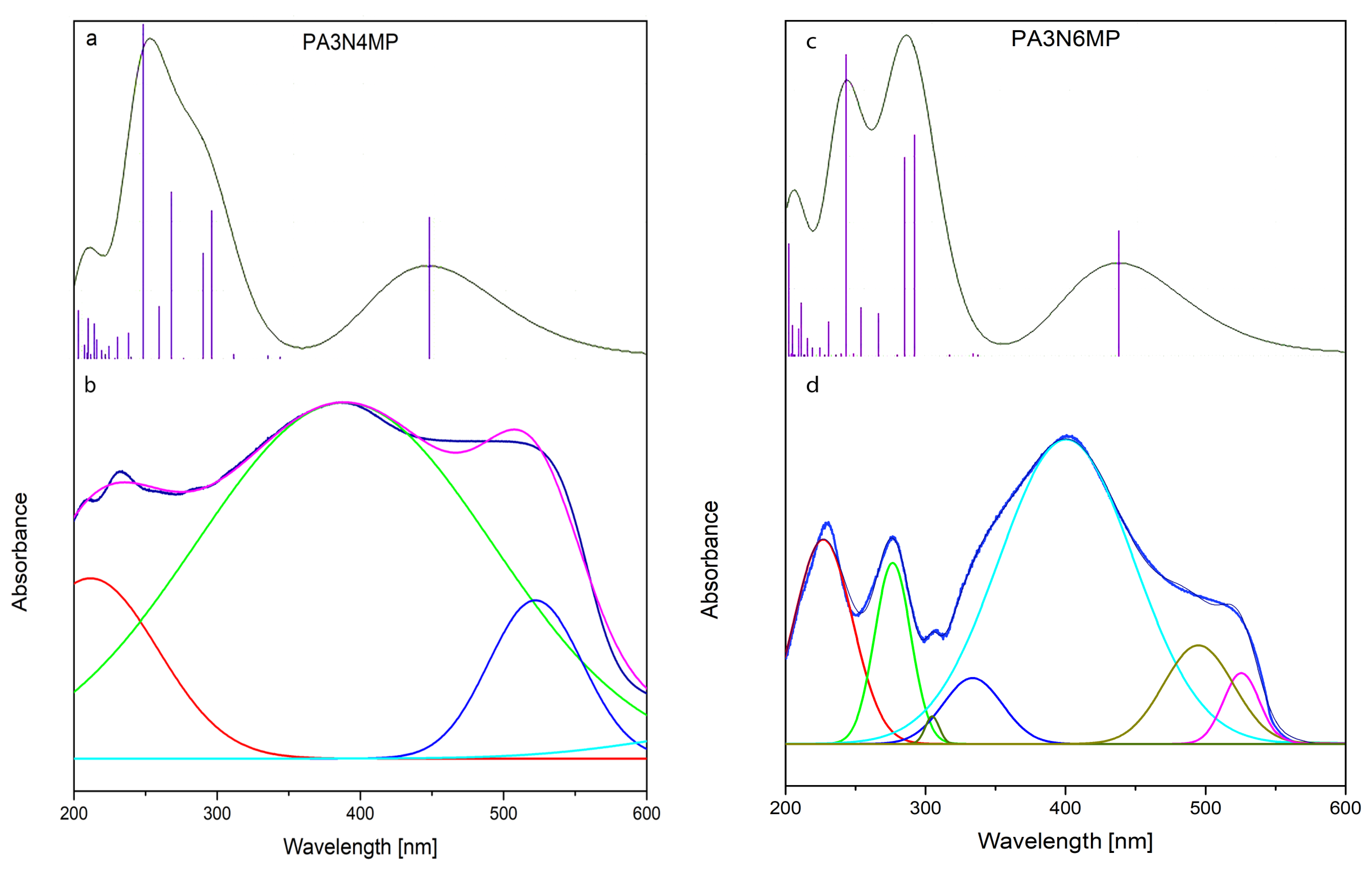
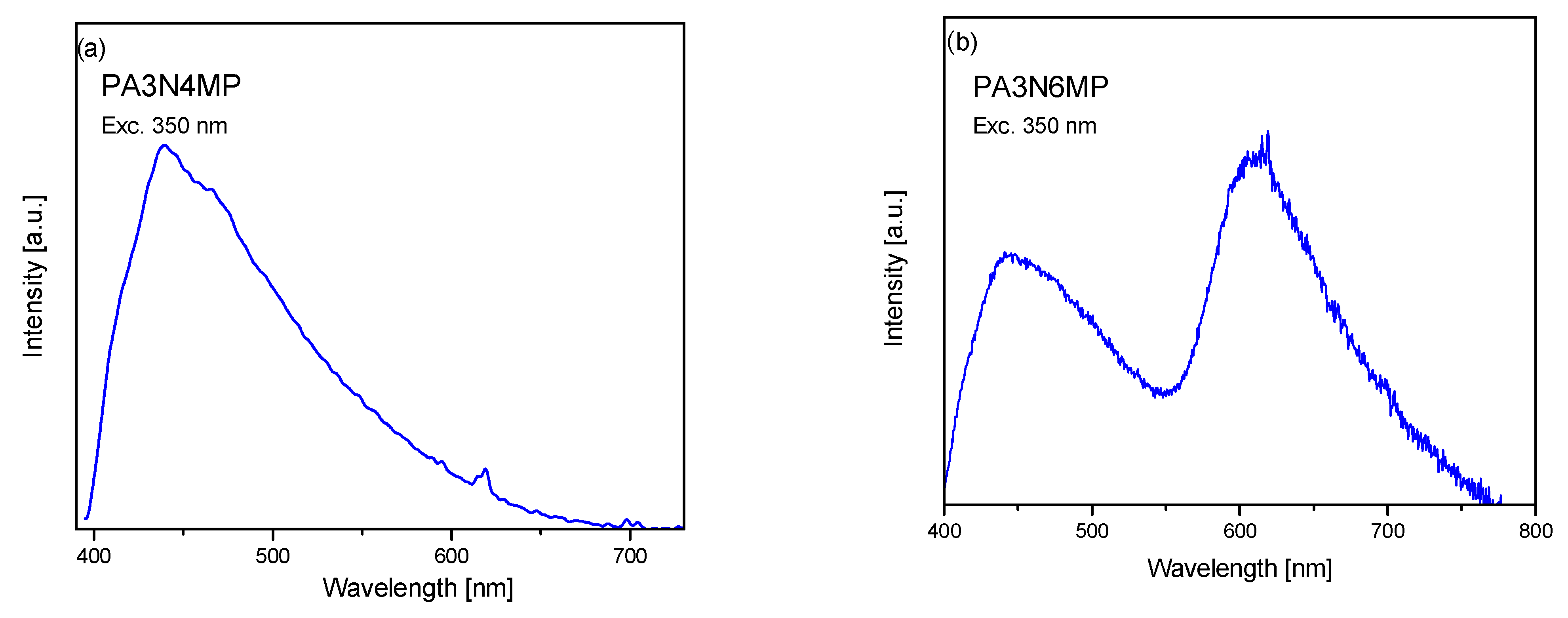
2.3. Electron Reflectance and Emission Spectra

Figure 7 displays the electronic reflectance profiles of the investigated compounds, which are characterized by prominent electronic transitions manifesting as broad absorption contour spanning the 200–600 nm wavelength region. These spectral features were assigned to specific electronic excitations through comparative analysis with prior investigations on structurally related pyridine-based systems [41,42,43,44,45,46], further supported by DFT computational modeling performed for the current derivatives. Table 4 summarizes the theoretically derived energy levels for both singlet and triplet excited states, along with their corresponding oscillator strength values (f). Notably, several singlet excitations exhibit exceptionally high oscillator strength values, indicating their pronounced contribution to the observed optical activity. For the PA3N4MP isomer, this appeared for the S5 (296 nm, 33,730 cm−1, S6 (292 nm, 34,260 cm−1), S8 (267 nm, 37,408 cm−1), and S10 (251 nm, 39,830 cm−1) states observed in the range 100–400 nm, and for the S1 state (464 nm, 21,552 cm−1) this appeared as the very broad band in the range 200–600. Similar spectroscopic contours appear for the PA3N6MP derivative. The calculated values of the singlet levels appear for the S5 (294 nm, 34,056 cm−1, S6 (288 nm, 34,739 cm−1), S8 (264 nm, 37,890 cm−1), and S10 (245 nm, 40,853 cm−1) states–they should be assigned to the bands observed in the ranges 250–300 nm and 200–290 nm. The S1 state is located at 454 nm (22,031 cm−1), which corresponds to the broad band observed in the range 300–500 nm. These spectral contours originate from electronic transitions localized within the π-system of the pyridine ring, as well as charge-transfer interactions between the nitro-chromophore and the aromatic system. These absorption bands correlate with π→π* electronic excitations, which aligns with the calculated energy difference between the HOMO and LUMO orbitals derived from quantum chemical modeling. The atomic composition of the molecular orbitals obtained for the studied compounds is similar to those determined for other amino-methyl-nitro-pyridine derivatives repeated in our earlier works [30,31,32,43,44,45,46]. In the HOMO orbital, the electron distribution is mainly scattered over the N-amine group and pyridine C–C and C–N bonds, whereas the LUMO is mainly over the pyridine nitro group. The theoretical HOMO-LUMO energy gap equals 3.13 eV (25,245 cm−1, 396 nm) for the PA3N4PT isomer and 3.1617 eV (25,501 cm−1, 392 nm) for the PA3N6MP isomer. It fits well to the very strong and broad bands observed in the 200–600 nm range with the maximum at about 400 nm (25,000 cm−1)—see Figure 7.
The emission spectra were measured using excitation at 350 nm. This wavelength was chosen because it falls into the 290–450 nm range in which the singlets with greatest oscillator strengths appear. Such procedure allows to recognize the possibility of the triplet→singlet transition phosphorescence in the studied compounds which arises predominantly through intersystem crossing mechanism. Therefore, the complete DFT calculations should give the singlet and triplet transitions in which also are those with zero oscillator strengths. They are important for the discussion of the luminescence spectra activated by the inter-crossing energy transfer process.
The emission spectra of the derivatives studied are shown in Figure 8.
The emission spectra of the studied compounds excited at 350 nm (28,571 cm−1) contain a very broad spectral contour observed for PA3N4MP in the range 400–700 nm and two intense bands in the ranges 400–550 and 550–700 nm for PA3N6MP. Their positions are clearly shifted into red in comparison to the observed S0 Sm absorption transition at 200–600 nm (16,666–50,000 cm−1), because the depopulation T1,T2 S0 mechanism of the excited levels was probably proceeded by the inter-system crossing of S1 T1,T2. Room temperature phosphorescence is generally not observed from excited triplet states; however, keeping in mind the calculated energies of the singlet and triplet levels (Table 4), such an explanation of the nature of these transitions is reasonable. The calculated energies of the T1 and T2 triplet states are localized within the 13,000–14,000 cm−1 range, closely matching theoretical predictions for triplet-mediated luminescence in π-conjugated systems. The modest energetic separation between these states (~1000 cm−1) likely drives significant spectral overlap, manifesting experimentally as a broad composite T1–T2 emission band.
This observation agrees with a depopulation mechanism governed by spin–orbit coupling effects, where intersystem crossing (ISC) facilitates efficient energy transfer from singlet to triplet manifolds–a hallmark process in the Jablonski framework for luminescent organic systems. The near-degeneracy of T1 and T2 energies relative to the ground state (S0) results in quasi-simultaneous T1→S0 and T2→S0 transitions. Their energetic proximity (ΔE < kT at ambient conditions) prevents discrete spectral resolution, yielding the observed broadened emission profile characteristic of multi-state relaxation pathways.

2.4. 13C and 1H NMR Spectra Measurements

The 1H and 13C NMR spectra of the investigated derivatives are presented in Figure 9 and Figure 10, respectively (full-length spectra are available in the Supplementary Materials—Figure S3 for 1H and Figure S4 for 13C). The observed spectral pattern–specifically, the number of distinct resonance lines–agrees with predictions derived from the molecular architecture of these systems. Each chemically inequivalent nucleus generates a discrete resonance signal, enabling unambiguous assignment of proton and carbon environments. For the 2-N-phenylamino-4-methyl-3-nitro-, and 2-N-phenylamino-6-methyl-3-nitro-pyridines, 13C chemical shifts are expected for the carbon atoms constituting the pyridine ring, phenyl ring, and one from the methyl group.
The molecular composition of the studied isomers predicts ten distinct 13C NMR resonances:
  • Pyridine ring: five unique carbons;
  • methyl group: one resonance;
  • phenyl ring: four signals (with two symmetry-equivalent carbon pairs).
Experimental 13C NMR spectra for both PA3N4MP and PA3N6MP isomers corroborate this theoretical framework, exhibiting ten resolved signals that map directly to their respective chemical environments. This agreement validates the proposed structural symmetry and atomic degeneracy within the aromatic systems.
For PA3N4MP, the chemical shifts in the pyridine ring are observed at 151.76, 150.08, 131.92, 122.26, and 118.13 ppm but for PA3N6MP at 166.33, 149.664, 128.98, 122.01, and 140.19 ppm. The chemical shifts in the phenyl carbon atoms appear at 146.45, 138.60, 129.09, and 124.47 ppm for the PA3N4MP isomer and 138.41, 135.75, 126.67, and 124.36 ppm for the PA3N6MP isomer. The line at 21.51 ppm corresponds to the carbon atom of the methyl chromophore in the PA3N4MP derivative and at 25.30 ppm in the PA3N6NP isomer. These carbon atoms are located in the vicinity of three hydrogen atoms and, therefore, show the greatest chemical shifts.
Similar analysis can be carried out for the proton units. In the 1H NMR spectra of the studied isomers, nine lines are expected: two from the pyridine ring, five from the phenyl ring, one from the methyl group, and one of the NH chromophore. They are observed at the following chemical shifts: at 7.55, 7.37, 7.14, 6.85, and 6.67 ppm originated from the phenyl protons of the PA3N4MP isomer and at 7.73, 7.31, 7.15, 6.85, and 6.68 ppm in the PA3N6MP isomer. The chemical shifts in the pyridine protons appear at 9.14 and 8.20 ppm for PA3N4MP and at 10.25 and 8.41 ppm for PA3N6MP. The protons of the NH chromophores give rise to the peaks at 5.93 and 5.93 ppm for the PA3N4MP and PA3N6MP isomers, respectively, and those of methyl groups at 1.56 and 1.55 ppm.
Proposed here, assignment of the chemical shifts observed for the studied derivatives fit well to the reported earlier data reported for the similar compounds [47,48,49,50].

3. Materials and Methods
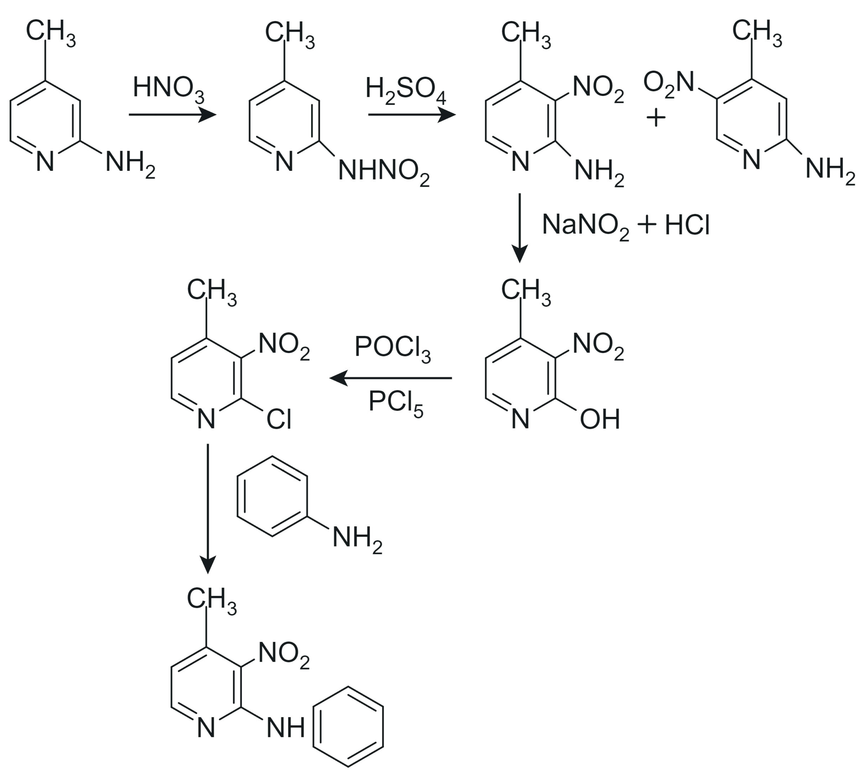
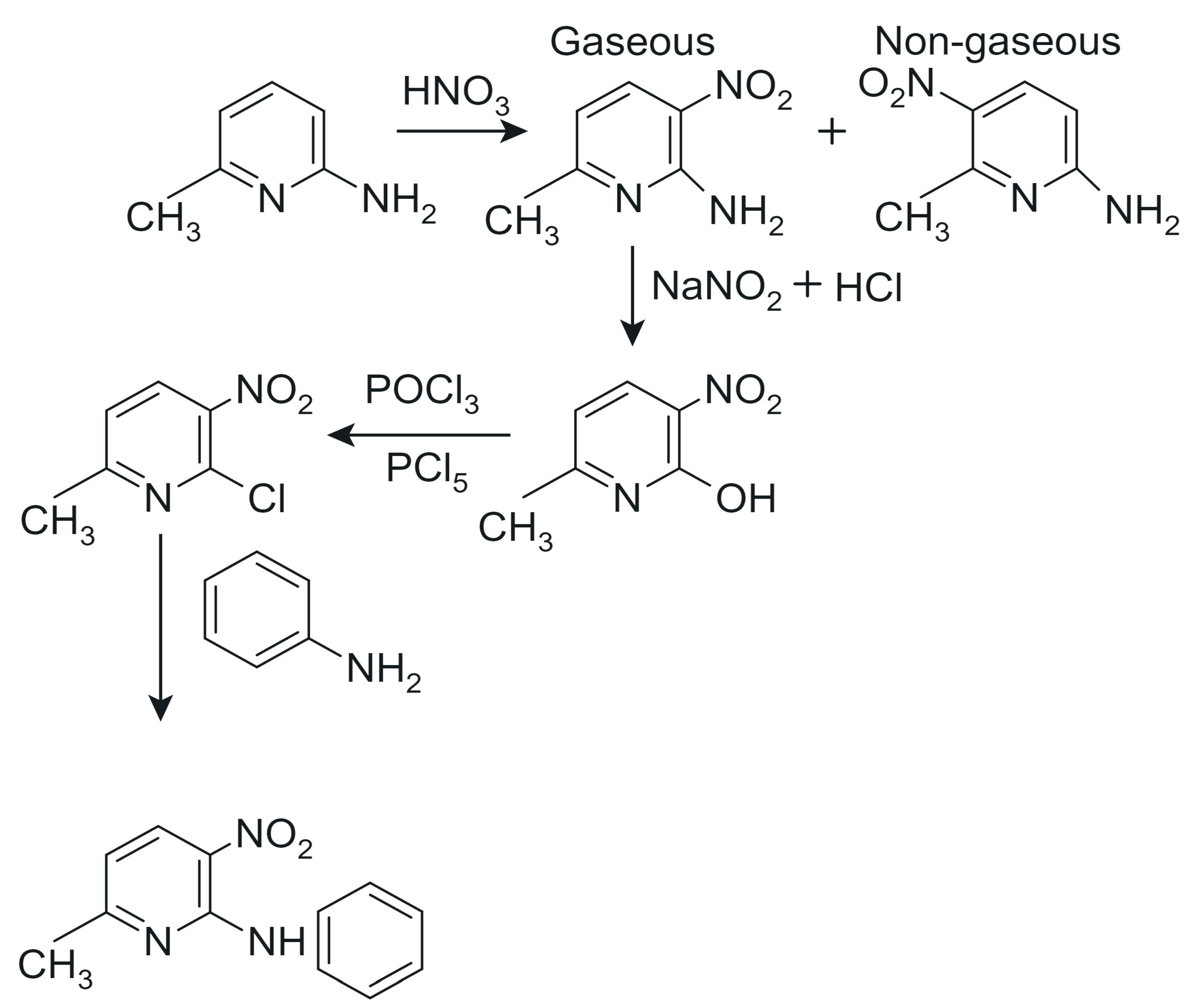
3.1. Synthesis

Two isomers:
  • 2-N-phenylamino-3-nitro-4-methylpyridine
  • 2-N-phenylamino-3-nitro-6-methylpyridine
The two isomers were synthesized following a general procedure inspired by the methodology outlined in [51]. However, due to the limited accessibility of this reference–it is neither available online nor widely distributed–an overview of the synthetic approach is provided here in the form of reaction schemes to aid reproducibility and comprehension.
The synthesis began with 2-amino-4-methylpyridine and 2-amino-6-methylpyridine, which were nitrated and subsequently converted into 2-hydroxy derivatives. These intermediates were then treated with phosphoryl chloride (POCl3) to yield the corresponding 2-chloro derivatives, which served as substrates for the final reaction with phenylamine to form the target compounds. The final products were purified via multiple recrystallizations from ethanol solutions to ensure high purity. Synthesis schemes are presented in Scheme 1 and Scheme 2.
The synthesis began with 2-amino-4-methylpyridine and 2-amino-6-methylpyridine, which were nitrated and subsequently converted into 2-hydroxy derivatives. These intermediates were then treated with phosphoryl chloride (POCl3) to yield the corresponding 2-chloro derivatives, which served as substrates for the final reaction with phenylamine to form the target compounds. The final products were purified via multiple recrystallizations from ethanol solutions to ensure high purity.
The chemical composition of the synthesized compounds was verified through microanalysis, which confirmed a good agreement with theoretical stoichiometry. The following are the chemical names, formulas, and abbreviations used in this study:
  • 2-N-phenylamino-3-nitro-4-methylpyridine
    • Chemical formula: C12H11N3O2
    • Abbreviation: PA3N4MP
    • Description: Red crystals
  • 2-N-phenylamino-3-nitro-6-methylpyridine
    • Chemical formula: C12H11N3O2
    • Abbreviation: PA3N6MP
    • Description: Red crystals
    • Elemental chemical analysis:
  • PA3N4MP (red crystals):
    Calculated: C 62.87%, H 4.84%, N 18.33%
    Experimental: C 62.51%, H 4.79%, N 18.17%
    Melting point: 395 K, 122 °C
    Synthetic yield: 79.0%
  • PA3N6MP (red crystals):
    Calculated: C 62.87%, H 4.84%, N 18.33%
    Experimental: C 62.55%, H 4.88%, N 18.12%
    Melting point: 405 K, 132 °C
    Synthetic yield: 71.0%
The inclusion of reaction schemes in this work ensures that the general synthetic route, as adapted from [51], is clearly understood and reproducible despite the restricted availability of the original reference. These schemes highlight the key transformations and provide a concise summary of the methodology employed.
The Figure 11 illustrates the uniform coloration of the gel, demonstrating the 2-N-phenylamino-3-nitro-6-methylpyridine compound excellent solubility and color intensity, confirming its potential application in the production of packaging films.

3.2. Single-Crystal X-Ray Diffraction 128–142

Single crystals of PA3N4MP and PA3N6MP suitable for diffraction studies were grown through slow solvent diffusion using a methanol/ethanol mixed system. X-ray intensity data for both compounds were acquired on a κ-geometry Xcalibur diffractometer equipped with a Sapphire2 CCD detector, employing graphite-monochromated Mo Kα radiation (λ = 0.71073 Å). Data acquisition and initial processing, including frame integration and absorption corrections, were executed using the CrysAlis CCD and CrysAlis RED ver. 1.171.35.11 software suite [52].
Structural solutions were derived via direct methods (SHELXT-2014/7 [53]), followed by iterative full-matrix least-squares refinement against F2 values (SHELXL-2018/3 [54]). Hydrogen atoms bonded to carbon were geometrically positioned and constrained using a riding model, while those participating in hydrogen bonds underwent unrestrained refinement. Post-refinement difference Fourier analysis revealed no residual electron density features exceeding 0.3 eÅ−3, confirming structural validity.
Critical experimental parameters, crystallographic metrics, and refinement statistics are tabulated in Table 5. Molecular topology representations were generated using Diamond 3.0 [55], with thermal ellipsoids plotted at 50% probability.
Hirshfeld surface analyses and 2D fingerprint plots as well as percentage contributions for various intermolecular contacts in the investigated crystals were calculated using the Crystal Explorer Ver. 3.1 program package [56].

3.3. Infrared and Raman Studies

IR spectra were measured using a Nicolet iS50 FT-IR (Thermo Fisher Scientific Inc., Warsaw, Poland) spectrometer equipped with an Automated Beamsplitter exchange system (iS50 ABX containing DLaTGS KBr detector (Thermo Fisher Scientific Inc., Warsaw, Poland) and DLaTGS Solid Substrate detector (Thermo Fisher Scientific Inc., Warsaw, Poland) for mid-IR and far-IR regions, respectively). A built-in all-reflective diamond ATR module (iS50 ATR), Thermo Scientific PolarisTM (Thermo Fisher Scientific Inc., Warsaw, Poland), and a HeNe laser were used as an IR radiation source. Polycrystalline IR spectra were collected in the 4000–100 cm−1 range. The advanced ATR correction software, part of the OMNICTM 6.2 program attached to the NicoletTM FT-IR spectrometer (Thermo Fisher Scientific Inc., Warsaw, Poland), was used in the studies of polycrystalline samples. A spectral resolution of 4 cm−1 was applied in the measurements.
Raman spectra in the 4000–80 cm−1 range were measured in backscattering geometry with a FT Bruker 110/S spectrometer (Bruker, Warsaw, Poland). The resolution was 2.0 cm−1. The YAG:Nd (excitation wavelength 1064 nm) laser was used as an excitation source.

3.4. Electronic Absorption Spectroscopy

Electronic absorption spectra in 200–1500 nm spectral range were measured at ambient temperature using a Cary Varian 5E UV-Vis-NIR spectrophotometer (SpectraLab Scientific Inc., Warsaw, Poland). For samples exhibiting low absorption intensity, diffuse reflectance spectroscopy was employed with a Praying Mantis accessory (Harrick Scientific Products Inc., Warsaw, Poland). Prior to sample measurements, a baseline correction was performed using high-purity Al2O3 powder as a reference standard, which was subsequently subtracted from the spectra to isolate specific absorption features, characteristic for the studied isomers.

3.5. Time-Resolved Emission Spectroscopy

Time-dependent emission profiles were determined using a Princeton Instruments Acton 2500i spectrograph interfaced with a Hamamatsu C5680 streak camera (HAMAMATSU Co., Ltd., Warsaw, Poland). Excitation of the spectra was achieved using a femtosecond Ti:sapphire laser (Coherent, Inc., Warsaw, Poland), generating 89 fs pulses at 800 nm (1 mJ/pulse, repetition rate adjustable up to 1 kHz). Wavelength tuning across the 230–2800 nm range was accomplished using an optical OPerA Solo parametric amplifier (Coherent Inc., Warsaw, Poland), enabling selective excitation at targeted energies.

3.6. Computational Methodology

Initial molecular geometries for quantum chemical simulations were derived from X-ray crystallographic data using Gaussian 03 [57]. Structural optimizations were executed at the B3LYP/6-311G(2d,2p) level of theory [58,59,60,61,62], yielding a single conformer with minimized vibrational energy. To facilitate comparison with prior studies on analogous pyridine derivatives [30,31,32], harmonic vibrational frequencies were scaled by empirical factors (Table 3, Table S3) compensating for anharmonicity and theoretical approximations.
Potential energy distribution (PED) analyses were performed using the BALGA algorithm [63], while molecular orbital visualizations (HOMO/LUMO) and vibrational mode animations were generated using GaussView 4.1 [64] and ChemCraft ver. 1.8 [65], respectively. Raman intensities were computed with the RAINT program ver. 2003 [66], employing a 0.965 scaling coefficient derived from established protocols [67].

3.7. NMR Spectroscopy

13C and 1H nuclear magnetic resonance spectra were acquired on a Bruker Avance III 600 MHz spectrometer fitted with a BBO Z-gradient probe. Liquid-state measurements utilized a 3.2 mm magic-angle spinning (MAS) probe ((Bruker, Warsaw, Poland)), optimizing resolution through high-speed sample rotation.

4. Conclusions

  • In the present work, two new heterocyclic isomers, 2-N-phenylamino-4-methyl-3-nitro- and 2-N-phenylamino-6-methyl-3-nitro-pyridines, were obtained. Their structures were characterized on the basis of X-ray diffraction, IR and Raman spectra, as well as electron UV-Vis and emission spectra measurements. They form new class of pyridine derivatives with several potential applications. This follows from their peculiar structural, electronic, and optical properties.
  • The specific structure of the studied compounds originates from the amino bridge between the phenyl and pyridine rings, and this conformation is stabilized by the intramolecular medium-strength hydrogen bond of N–H···O type formed by the O-atom of the nitro group and the NH group. The NH group plays an important role as the proton donor in the structural stabilization of the studied compounds in the solid state. Such structure predisposes these derivatives as precursors of antihistamine drugs, which can be used to oppose the activity of histamine receptors in the human body, forming antihistaminic pharmaceutics for allergic disorders. Such drugs contain the pyridine ring as an important part of their structure [68].
  • Integrated spectroscopic analysis and DFT computational modeling reveal distinct singlet-triplet state distributions governing the photophysical behavior of the investigated pyridine isomers. Key findings include the following:
    Phosphorescence mechanism: electronic absorption and emission spectral data confirm that triplet→singlet phosphorescence arises predominantly through intersystem crossing machanism (ISC), a well-documented relaxation pathway in nitro-functionalized pyridine systems.
    Structure–function correlation: unique electronic architecture of phenyl-aminopyridine derivatives enables their dual functionality as follows:
    Versatile synthetic building blocks for heterocyclic chemistry;
    Efficient ligands for d-/f-block metal coordination complexes;
    Tunable luminophores with tailorable ISC efficiency.
    Technological potential: engineered lanthanide complexes incorporating these scaffolds demonstrate promise as follows:
    Targeted bioimaging probes leveraging metal-centered luminescence;
    Photosensitizers for photodynamic therapy applications;
    Molecular sensors for environmental metal ion detection.
  • The 13C and 1H NMR spectra fully confirm the structural XRD data reported in the present work.
  • The intense red color of the obtained materials allow for their use as new dyes for the production of the color plastic foils for food packing.
  • This work establishes foundational structure–property relationships critical for rational design of next-generation luminescent materials in medicinal and materials chemistry.

Supplementary Materials

The following supporting information can be downloaded at: https://www.mdpi.com/article/10.3390/ijms26072874/s1.

Author Contributions

P.G.: Visualization, Writing–Original Draft, Resources; J.H.: Conceptualization, Writing–Original Draft, Supervision; A.Z.: Investigation; M.B.: Investigation; L.D.: Methodology, Supervision; J.J.: Investigation, Validation; R.L.: Investigation, Validation. All authors have read and agreed to the published version of the manuscript.

Funding

This research received no external funding.

Institutional Review Board Statement

Not applicable.

Informed Consent Statement

Not applicable.

Data Availability Statement

The data presented in this study are available on request from the corresponding author.

Conflicts of Interest

The authors declare no conflict of interest.

References

  1. El-Gazzar, A.-R.; Hussein, H.; Hafez, H. Synthesis and biological evaluation of thieno [2,3-d]pyrimidine derivatives for anti-inflammatory, analgesic and ulcerogenic activity. Acta Pharm. 2007, 57, 395–411. [Google Scholar] [CrossRef] [PubMed]
  2. Hsu, M.-H.; Savas, Ü.; Lasker, J.M.; Johnson, E.F. Genistein, Resveratrol, and 5-Aminoimidazole-4-carboxamide-1-β-d-ribofuranoside Induce Cytochrome P450 4F2 Expression through an AMP-Activated Protein Kinase-Dependent Pathway. J. Pharmacol. Exp. Ther. 2011, 337, 125–136. [Google Scholar] [CrossRef] [PubMed]
  3. Bernardino, A.M.R.; de Azevedo, A.R.; Pinheiro, L.C.d.S.; Borges, J.C.; Carvalho, V.L.; Miranda, M.D.; de Meneses, M.D.F.; Nascimento, M.; Ferreira, D.; Rebello, M.A.; et al. Synthesis and antiviral activity of new 4-(phenylamino)/4-[(methylpyridin-2-yl)amino]-1-phenyl-1H-pyrazolo[3,4-b]pyridine-4-carboxylic acids derivatives. Med. Chem. Res. 2007, 16, 352–369. [Google Scholar] [CrossRef]
  4. Wang, N.-Y.; Zuo, W.-Q.; Xu, Y.; Gao, C.; Zeng, X.-X.; Zhang, L.-D.; You, X.-Y.; Peng, C.-T.; Shen, Y.; Yang, S.-Y.; et al. Discovery and structure–activity relationships study of novel thieno[2,3-b]pyridine analogues as hepatitis C virus inhibitors. Bioorganic Med. Chem. Lett. 2014, 24, 1581–1588. [Google Scholar] [CrossRef]
  5. Elansary, A.K.; Moneer, A.A.; Kadry, H.H.; Gedawy, E.M. Synthesis and anticancer activity of some novel fused pyridine ring system. Arch. Pharmacal Res. 2012, 35, 1909–1917. [Google Scholar] [CrossRef]
  6. Lauria, A.; Abbate, I.; Patella, C.; Martorana, A.; Dattolo, G.; Almerico, A.M. New annelated thieno[2,3-e][1,2,3]triazolo[1,5-a]pyrimidines, with potent anticancer activity, designed through VLAK protocol. Eur. J. Med. Chem. 2013, 62, 416–424. [Google Scholar] [CrossRef]
  7. Mohareb, R.M.; Al-Omran, F.; Azzam, R.A. Heterocyclic ring extension of estrone: Synthesis and cytotoxicity of fused pyran, pyrimidine and thiazole derivatives. Steroids 2014, 84, 46–56. [Google Scholar] [CrossRef]
  8. Nikkhoo, A.R.; Miri, R.; Arianpour, N.; Firuzi, O.; Ebadi, A.; Salarian, A.A. Cytotoxic activity assessment and c-Src tyrosine kinase docking simulation of thieno[2,3-b] pyridine-based derivatives. Med. Chem. Res. 2013, 23, 1225–1233. [Google Scholar] [CrossRef]
  9. Ahmed, S.A.; Ahmed, O.M.; Elgendy, H.S. Novel synthesis of puriensanalougues and thieno [2, 3-b] pyridine derivatives with anticancer and antioxidant activity. J. Pharm. Res. 2014, 8, 1303–1313. [Google Scholar]
  10. Bakhite, E.A.; Abdel-Rahman, A.E.; Mohamed, O.S.; Thabet, E.A. Synthesis, reactions and antimicrobial activity of new cyclopenta [e] thieno [2, 3-b] pyridines and related heterocyclic systems. Pharmazie 2000, 55, 577–583. [Google Scholar] [CrossRef]
  11. Altalbawy, F.M.A. Synthesis and Antimicrobial Evaluation of Some Novel Bis-α,β-Unsaturated Ketones, Nicotinonitrile, 1,2-Dihydropyridine-3-carbonitrile, Fused Thieno [2,3-b]pyridine and Pyrazolo [3,4-b]pyridine Derivatives. Int. J. Mol. Sci. 2013, 14, 2967–2979. [Google Scholar] [CrossRef] [PubMed]
  12. Bumpus, N.N.; Johnson, E.F. 5-Aminoimidazole-4-carboxyamide-ribonucleoside (AICAR)-Stimulated Hepatic Expression of Cyp4a10, Cyp4a14, Cyp4a31, and Other Peroxisome Proliferator-Activated Receptor α-Responsive Mouse Genes Is AICAR 5′-Monophosphate-Dependent and AMP-Activated Protein Kinase-Independent. J. Pharmacol. Exp. Ther. 2011, 339, 886–895. [Google Scholar] [CrossRef] [PubMed]
  13. Peyton, K.J.; Liu, X.-M.; Yu, Y.; Yates, B.; Durante, W. Activation of AMP-Activated Protein Kinase Inhibits the Proliferation of Human Endothelial Cells. J. Pharmacol. Exp. Ther. 2012, 342, 827–834. [Google Scholar] [CrossRef]
  14. Nakajima, K.; Komiyama, Y.; Hojo, H.; Ohba, S.; Yano, F.; Nishikawa, N.; Aburatani, H.; Takato, T.; Chung, U.-I. Enhancement of bone formation ex vivo and in vivo by a helioxanthin-derivative. Biochem. Biophys. Res. Commun. 2010, 395, 502–508. [Google Scholar] [CrossRef]
  15. Maeda, Y.; Hojo, H.; Shimohata, N.; Choi, S.; Yamamoto, K.; Takato, T.; Chung, U.-I.; Ohba, S. Bone healing by sterilizable calcium phosphate tetrapods eluting osteogenic molecules. Biomaterials 2013, 34, 5530–5537. [Google Scholar] [CrossRef]
  16. Gemba, T.; Ninomiya, M.; Matsunaga, K.; Ueda, M.J. Effects of a novel calcium antagonist, S-(+)-methyl-4, 7-dihydro-3-isobutyl-6-methyl-4-(3-nitrophenyl) thieno [2, 3-b] pyridine-5-carboxylate (S-312-d), on ischemic amino acid release and neuronal injury in stroke-prone spontaneously hypertensive rats. Pharmacol. Exp. Ther. 1993, 265, 463–467. [Google Scholar]
  17. Balzarini, J.; Stevens, M.; Andrei, G.; Snoeck, R.; Strunk, R.; Pierce, J.B.; Lacadie, J.A.; De Clercq, E.; Pannecouque, C. Pridine oxide derivatives: Structure-activity relationship for inhibition of human immunodeficiency virus and cytomegalovirus replication in cell culture. Helv. Chim. Acta 2002, 85, 2961–2974. [Google Scholar] [CrossRef]
  18. Stevens, M.; Pannecouque, C.; De Clercq, E.; Balzarini, J. Inhibition of human immunodeficiency virus by a new class of pyridine oxide derivatives. Antimicrob. Agents Chemother. 2003, 47, 2951–2957. [Google Scholar] [CrossRef]
  19. Stevens, M.; Pannecouque, C.; De Clercq, E.; Balzarini, J. Novel human immunodeficiency virus (HIV) inhibitors that have a dual mode of anti-HIV action. Antimicrob. Agents Chemother. 2003, 47, 3109–3116. [Google Scholar] [CrossRef]
  20. Balzarini, J.; Stevens, M.; De Clercq, E.; Schols, D.; Pannecouque, C. Pyridine N-oxide derivatives: Unusual anti-HIV compounds with multiple mechanisms of antiviral action. J. Antimicrob. Chemother. 2005, 55, 135–138. [Google Scholar] [CrossRef]
  21. Scriven, E.F.V.; Murugan, R. Pyridine and pyridine derivatives. In Kirk-Othmer Encyclopedia of Chemical Technology; John Wiley & Sons, Inc.: Hoboken, NJ, USA, 2005; Volume 20, pp. 1–53. [Google Scholar] [CrossRef]
  22. Liu, C.; Luo, J.; Xu, L.; Huo, Z. Synthesis of 2-substituted pyridines from pyridine N-oxides. Arkivoc 2013, 2013, 154–174. [Google Scholar] [CrossRef]
  23. Dzierszinski, F.; Coppin, A.; Mortuaire, M.; Dewailly, E.; Slomianny, C.; Ameisen, J.-C.; DeBels, F.; Tomavo, S. Ligands of the Peripheral Benzodiazepine Receptor Are Potent Inhibitors of Plasmodium falciparum and Toxoplasma gondii In Vitro. Antimicrob. Agents Chemother. 2002, 46, 3197–3207. [Google Scholar] [CrossRef] [PubMed]
  24. Dressler, H. Pyridine and derivatives. In Van Nostrand’s Scientific Encyclopedia Van Nostrand’s Scientific Encyclopedia; Considine, D.M., Considine, G.D., Eds.; John Wiley & Sons, Inc.: Hoboken, NJ, USA, 2006. [Google Scholar] [CrossRef]
  25. Balzarini, J.; Keyaerts, E.; Vijgen, L.; Vandermeer, F.; Stevens, M.; De Clercq, E.; Egberink, H.; Van Ranst, M. Pyridine N-oxide derivatives are inhibitory to the human SARS and feline infectious peritonitis coronavirus in cell culture. J. Antimicrob. Chemother. 2005, 57, 472–481. [Google Scholar] [CrossRef] [PubMed]
  26. Youssif, S. Recent trends in the chemistry of pyridine N-oxides. Arkivoc 2001, 2001, 242–268. [Google Scholar]
  27. Kilenyi, S.N.; Mousseau, J.J. Pyridine N-oxide. In Encyclopedia of Reagents for Organic Synthesis; John Wiley & Sons, Ltd.: Hoboken, NJ, USA, 2015; p. 1. [Google Scholar] [CrossRef]
  28. Carlin, R.L. Transition Metal Complexes of Pyridine N-Oxide. J. Am. Chem. Soc. 1961, 83, 3773–3775. [Google Scholar] [CrossRef]
  29. Sarma, R.; Karmakar, A.; Baruah, J.B. Synthesis and characterization of pyridine N-oxide complexes of manganese, copper and zinc. Inorganica Chim. Acta 2008, 361, 2081–2086. [Google Scholar] [CrossRef]
  30. Lorenc, J. Dimeric structure and hydrogen bonds in 2-N-ethylamino-5-metyl-4-nitro-pyridine studied by XRD, IR and Raman methods and DFT calculations. Vib. Spectrosc. 2012, 61, 112–123. [Google Scholar] [CrossRef]
  31. Szatyłowicz, H. Structural aspects of the intermolecular hydrogen bond strength: H-bonded complexes of aniline, phenol and pyridine derivatives. J. Phys. Org. Chem. 2008, 21, 897–914. [Google Scholar] [CrossRef]
  32. Bryndal, I.; Kucharska, E.; Wandas, M.; Lorenc, J.; Hermanowicz, K.; Mączka, M.; Lis, T.; Marchewka, M.; Hanuza, J. Comprehensive physicochemical studies of a new hybrid material: 2-Amino-4-methyl-3-nitropyridinium hydrogen oxalate. Spectrochim. Acta A 2014, 117, 434–441. [Google Scholar] [CrossRef]
  33. Spackman, M.A.; Jayatilaka, D. Hirshfeld surface analysis. CrystEngComm 2009, 11, 19–32. [Google Scholar] [CrossRef]
  34. McKinnon, J.J.; Spackman, M.A.; Mitchell, A.S. Novel tools for visualizing and exploring intermolecular interactions in molecular crystals. Acta Crystallogr. Sect. B Struct. Sci. 2004, 60, 627–668. [Google Scholar] [CrossRef]
  35. McKinnon, J.J.; Mitchell, A.S.; Spackman, M.A. Hirshfeld Surfaces: A New Tool for Visualising and Exploring Molecular Crystals. Chem. Eur. J. 1998, 4, 2136–2141. [Google Scholar] [CrossRef]
  36. McKinnon, J.J.; Jayatilaka, D.; Spackman, M.A. Towards quantitative analysis of intermolecular interactions with Hirshfeld surfaces. Chem. Commun. 2007, 7, 3814–3816. [Google Scholar] [CrossRef]
  37. Ureña, F.; Gómez, M.; González, J.; Torres, E. A new insight into the vibrational analysis of pyridine. Acta Part A Mol. Biomol. Spectrosc. 2003, 59, 2815–2839. [Google Scholar] [CrossRef]
  38. Wiberg, K.B.; Walters, V.A.; Wong, K.N.; Colson, S.D. Azines: Vibrational force field and intensities for pyridine. J. Phys. Chem. 1984, 88, 6067–6075. [Google Scholar] [CrossRef]
  39. Lorenc, J.; Bryndal, I.; Syska, W.; Wandas, M.; Marchewka, M.; Pietraszko, A.; Lis, T.; Mączka, M.; Hermanowicz, K.; Hanuza, J. Order–disorder phase transitions and their influence on the structure and vibrational properties of new hybrid material: 2-Amino-4-methyl-3-nitropyridinium trifluoroacetate. Chem. Phys. 2010, 374, 1–14. [Google Scholar] [CrossRef]
  40. Lorenc, J.; Hanuza, J.; Janczak, J. Structural and vibrational study of 3- or 5-methyl substituted 2-N-ethylamino-4-nitropyridine N-oxides. Vib. Spectrosc. 2012, 59, 59–70. [Google Scholar] [CrossRef]
  41. Bryndal, I.; Lorenc, J.; Macalik, L.; Michalski, J.; Sąsiadek, W.; Lis, T.; Hanuza, J. Crystal structure, vibrational and optic properties of 2-N-methylamino-3-methylpyridine N-oxide–Its X-ray and spectroscopic studies as well as DFT quantum chemical calculations. J. Mol. Struct. 2019, 1195, 208–219. [Google Scholar] [CrossRef]
  42. Socrates, G. Infrared and Raman Characteristic Group Frequencies: Tables and Charts, 3rd ed.; John Wiley & Sons, Ltd.: Hoboken, NJ, USA, 2004. [Google Scholar]
  43. Bryndal, I.; Kucharska, E.; Sąsiadek, W.; Wandas, M.; Lis, T.; Lorenc, J.; Hanuza, J. Molecular and crystal structures, vibrational studies and quantum chemical calculations of 3 and 5-nitroderivatives of 2-amino-4-methylpyridine. Spectrochim. Acta Part A Mol. Biomol. Spectrosc. 2012, 96, 952–962. [Google Scholar] [CrossRef]
  44. Bryndal, I.; Marchewka, M.; Wandas, M.; Sąsiadek, W.; Lorenc, J.; Lis, T.; Dymińska, L.; Kucharska, E.; Hanuza, J. The role of hydrogen bonds in the crystals of 2-amino-4-methyl-5-nitropyridinium trifluoroacetate monohydrate and 4-hydroxybenzenesulfonate–X-ray and spectroscopic studies. Spectrochim. Acta Part A Mol. Biomol. Spectrosc. 2014, 123, 342–351. [Google Scholar] [CrossRef]
  45. Lorenc, J.; Kucharska, E.; Hanuza, J.; Chojnacki, H. Excited electronic states of 2-ethylamino-(3 or 5)-methyl-4-nitropyridine and 2-methylamino-(3 or 5)-methyl-4-nitropyridine. J. Mol. Struct. 2004, 707, 47–57. [Google Scholar] [CrossRef]
  46. Lorenc, J.; Zając, A.; Janczak, J.; Lisiecki, R.; Hanuza, J.; Hermanowicz, K. Structure and optical properties of new nitro-derivatives of 2-N-alkiloamino-picoline N-oxide isomers. J. Mol. Struct. 2022, 1265, 133372. [Google Scholar] [CrossRef]
  47. Barakat, A.; Al-Najjar, H.J.; Al-Majid, A.M.; Soliman, S.M.; Mabkhot, Y.N.; Shaik, M.R.; Ghabbour, H.A.; Fun, H.-K. Synthesis, NMR, FT-IR, X-ray structural characterization, DFT analysis and isomerism aspects of 5-(2,6-dichlorobenzylidene)pyrimidine-2,4,6(1H,3H,5H)-trione. Spectrochim. Acta Part A Mol. Biomol. Spectrosc. 2015, 147, 107–116. [Google Scholar] [CrossRef]
  48. Carthigayan, K.; Xavier, S.; Periandy, S. HOMO–LUMO, UV, NLO, NMR and vibrational analysis of 3-methyl-1-phenylpyrazole using FT-IR, FT-RAMAN FT-NMR spectra and HF-DFT computational methods. Spectrochim. Acta Part A Mol. Biomol. Spectrosc. 2015, 142, 350–363. [Google Scholar] [CrossRef]
  49. Puszko, A.; Wasylina, L. The influence of steric effect on 1H NMR, 13C NMR and IR spectra of methylated de-rivatives of 4-nitropyridine N-oxide. Chem. Pap. 1995, 49, 176–181. [Google Scholar]
  50. Kątcka, M.; Urbański, T. NMR spectra of pyridine, picolines and hydrochlorides and of their hydrochlorides and methiodides. Bull. Acad. Pol. Sci. 1968, 16, 347–350. [Google Scholar]
  51. Palasek, B.; Talik, Z. Synthesis of the 2-alcilonitramino-5-nitroderivatives of 6-methylpyridine. Pr. Nauk. AE Wrocław 1985, 291, 111–114. [Google Scholar]
  52. Rigaku Oxford Diffraction, CrysAlisPro 1.171.42.93a Software System, Oxford, UK. 2023. Available online: https://rigaku.com/products/crystallography/x-ray-diffraction/crysalispro (accessed on 28 February 2025).
  53. Sheldrick, G.M. SHELXT–Integrated space-group and crystal-structure determination. Acta Cryst. 2015, A71, 3–8. [Google Scholar] [CrossRef]
  54. Sheldrick, G.M. Crystal Structure of Refinement with SHELXL. Acta Cryst. 2015, C71, 3–8. [Google Scholar] [CrossRef]
  55. Brandenburg, K.; Putz, H. DIAMOND, Version 3.0; Crystal Impact GbR: Bonn, Germany, 2006.
  56. Wolf, S.W.; Grimwood, D.J.; MacKimon, J.J.; Turner, M.J.; Jayatilaka, D.; Spackman, A.M. Crystal Explorer, Ver. 3.1; University of Western Australia: Perth, Australia, 2013.
  57. Frisch, M.J.; Trucks, G.W.; Schlegel, H.B.; Scuseria, G.E.; Robb, M.A.; Cheeseman, J.R.; Montgomery, J.A.; Vreven, T., Jr.; Kudin, K.N.; Burant, J.C.; et al. Gaussian 03, Revision A.1; Gaussian Inc.: Pittsburgh, PA, USA, 2003. [Google Scholar]
  58. Becke, A.D. Density-functional thermochemistry. IV. A new dynamical correlation functional and implications for exact-exchange mixing. J. Chem. Phys. 1996, 104, 1040–1046. [Google Scholar] [CrossRef]
  59. Lee, C.; Yang, W.; Parr, R.G. Density-functional exchange-energy approximation with correct asymptotic behavior. Phys. Rev. 1988, B37, 785–789. [Google Scholar]
  60. Parr, R.G.; Yang, W. Density-Functional Theory of Atoms and Molecules; Oxford University Press: New York, NY, USA, 1989. [Google Scholar]
  61. McLean, A.D.; Chandler, G.S. Contracted Gaussian basis sets for molecular calculations. I. Second row atoms, Z = 11–18. J. Chem. Phys. 1980, 72, 5639–5648. [Google Scholar] [CrossRef]
  62. Krishnan, R.; Binkley, J.S.; Seeger, R.; Pople, J.A. Self-consistent molecular orbital methods. XX. A basis set for correlated wave functions. J. Chem. Phys. 1980, 72, 650–654. [Google Scholar] [CrossRef]
  63. Rostkowska, H.; Lapinski, L.; Nowak, M.J. Analysis of the normal modes of molecules with D3h symmetry: Infrared spectra of monomeric s-triazine and cyanuric acid. Vib. Spectrosc. 2009, 49, 43–51. [Google Scholar] [CrossRef]
  64. GaussView 4.1; Gaussian, Inc.: Wallingford, CT, USA, 2009.
  65. Zhurko, G.A.; Zhurko, D.A. Chemcraft Graphical Program for Visualization of Computed Results. Available online: http://www.chemcraftprog.com (accessed on 10 August 2024).
  66. Michalska, D. RAINT (Raman Intensities), Computer Program for Calculation of Raman Intensities from the Gaussian Outputs; Wrocław University of Technology: Wrocław, Poland, 2002. [Google Scholar]
  67. Palafox, M.A.; Rastogi, V. Quantum chemical predictions of the vibrational spectra of polyatomic molecules. The uracil molecule and two derivatives. Spectrochim. Acta 2002, A58, 411–440. [Google Scholar] [CrossRef]
  68. Joshi, K.K. Chemistry with Schiff bases of pyridine derivatives: Their potential as bioactive ligands and chemosensors. In The Edited Volume: Exploring Chemistry with Pyridine Derivatives; Pal, S., Ed.; IntechOpen: London, UK, 2022. [Google Scholar] [CrossRef]
Figure 1. View of the X-ray structure of PA3N4MP (a) and DFT optimized molecule (b).
Ijms 26 02874 g001
Figure 2. Arrangement of molecules in the unit cell showing the stacking structure along b-axis (a) and 3D MESP map for molecule PA3N4MP (b).
Ijms 26 02874 g002
Figure 3. (a) View of the X-ray structure of PA3N6MP, (b) DFT optimized molecule, and (c) 3D EP map; color coded as in Figure 2.
Ijms 26 02874 g003
Figure 4. Packing diagram of the PA3N6MP.
Ijms 26 02874 g004
Figure 5. HS (left), the 2D-fingerprint plots (middle), and the contributions of the respective interactions (right) for PA3N4MP (a) and PA3N6MP (b).
Ijms 26 02874 g005
Figure 6. Experimental and calculated IR (a) and Raman (b) spectra of the studied phenyl–aminopyridines in the range of 3500–100 cm−1.
Ijms 26 02874 g006
Figure 7. Reflectance UV-Vis spectra of the studied isomers. Calculated: PA3N4MP (a), PA3N6MP (c). Experimental: PA3N4MP (b), PA3N6MP (d).
Ijms 26 02874 g007
Figure 8. Emission spectra of measured for studied isomers under excitation at 350 nm: (a) PA3N4MP, (b) PA3N6MP.
Ijms 26 02874 g008
Figure 9. 1H NMR spectra of the studied isomers: (A) 2-N-phenylamino-4-methyl-3-nitro- and (B) 2-N-phenylamino-6-methyl-3-nitro-pyridines.
Ijms 26 02874 g009
Figure 10. 13C NMR spectra of the studied isomers: (A) 2-N-phenylamino-4-methyl-3-nitro- and (B) 2-N-pheny lamino-6-methyl-3-nitro-pyridines.
Ijms 26 02874 g010
Scheme 1. Synthesis of 2-N-phenylamino-3-nitro-4-methylpyridine.
Ijms 26 02874 sch001
Scheme 2. Synthesis of 2-N-phenylamino-3-nitro-6-methylpyridine.
Ijms 26 02874 sch002
Figure 11. Chitosan gel dyed with the isomer 2-N-phenylamino-3-nitro-6-methylpyridine.
Ijms 26 02874 g011
Table 1. Selected geometrical parameters (Å, °) for PA3N4MP.
X-RayDFT
N3–O1 1.2412(15)1.242
N3–O2 1.2272 (14)1.226
C3–N3 1.4508 (16)1.454
C2–N2–C7 131.01 (11)131.64
Dihedral angle between the planes:
NO2 (O1N3O2)/pyridine ring (N1,C2–C6)3.84(15)25.50
pyridine ring (N1,C2–C6)/phenyl ring (C7–C12)6.20(15)15.70
D–H···A D–H H···A D···A D–H···A
N2–H2···O1 0.900(16) 1.860(15) 2.5877(14)136.4(13)X-ray
N2–H2···O11.0141.8142.632135.17DFT
Table 2. Selected geometrical parameters (Å, °) for PA3N6MP.
X-RayDFT
N3–O1 1.249 (2)1.243
N3–O2 1.230 (2)1.226
C3–N3 1.447 (3)1.450
C2–N2–C7 132.0 (2)132.24
Dihedral angle between the planes:
NO2 (O1N3O2)/pyridine ring (N1,C2–C6)3.84 (15)0.00
pyridine ring (N1,C2–C6)/phenyl ring (C7–C12)6.20 (15)0.00
D–H···A D–H H···A D···A D–H···A
N2–H2···O1 0.84 (2) 1.96 (2) 2.649 (3) 139 (2)X-ray
N2–H2···O11.0161.8162.649136.58DFT
Table 3. Chosen experimental and calculated wavenumbers with assignment of the observed bands to respective normal modes for monomer.
PA3N4MPPA3N6MP
Calc.Exp.Calc.Exp.
IRRSIRRSIRRSIRRSAssignment
336733673433m 334433443311w νN–H····O
158515851579s1579w158915891580vs1585wδ(CNH)ϕ + ν(ϕ)
157415741566s1567w15781578 1578wδ(CNH)θ + ν(ϕ) + νas(NO2)
154915491548s1547m154815481548vw1545wνas(NO2) + ν(ϕ) + ν(θ) + δ(Cϕ NCθ)
135013501361 135713571357w ν(CN)ϕ + νs(NO2) + δ(ϕ) + δs(CH3)
122612261237m1212s122712271219vs1216sν(ϕ) + νs(NO) + ν(CN)ϕα + ν(CNH)ϕ
119711971220s1192sh119511951186s1188sνs(NO2) + (CNH)θ
118411841203sh νs(NO2) + ν(NCN)ϕ
807807807m ν(CNHC)
790790 ν(CNHC)ϕ+θ + δs(NO)ϕ
707707 710710 δ(NH)
699699691m 694m δ(CNHC)ϕ+θ
395395 386w386386394w395vwδ(CCH3) + δ(CNO)ϕ
290290 289w 295m295vwδ(CNHC)ϕ+θ
187187 184184188w δ(CNHC)ϕ+θ
107107 9494177w δ(CNHC)ϕ+θ
8383 8484150m δs(NO)ϕ + δ(CNHC)ϕ+θ
Table 4. Calculated singlet and triplet electron levels of the PA3N4MP (a) and PA3N6MP (b) molecules.
(a)
Electron levelseVnmcm−1Oscillator strength
singlets
(1)2.6717 464.06 21,5520.1230
(2)2.7783 446.25 22,4090.1648
(3)3.6225 342.26 29,2180.0008
(4)3.6351 341.07 29,3190.0009
(5)3.7309 332.32 30,0920.0022
(6)3.7385 331.64 30,1530.0026
(7)4.0091 309.26 32,3360.0038
(8)4.1820 296.47 33,7300.1643
(9)4.2399 292.42 34,1970.1730
(10)4.2476 291.89 34,2600.0935
(11)4.3350 286.00 34,9650.1234
(12)4.5328 273.53 36,5590.0007
(13)4.6380 267.32 37,4080.1533
(14)4.8088 257.83 38,7850.0237
(15)4.9383 251.07 39,8300.2944
(16)5.0938 243.40 41,0850.3909
(17)5.2701 235.26 42,5060.0019
(18)5.3092 233.53 42,8210.0295
(19)5.5492 223.43 44,7570.0007
(20)5.8961 210.28 47,5560.0212
triplets
(1)1.6711741.93 13,4780.0000
(2)1.7361 714.16 14,0020.0000
(3)1.8193 681.49 14,6740.0000
(4)1.9895 623.19 16,0460.0000
(5)2.1489 576.96 17,3320.0000
(6)2.7649 448.42 22,3000.0000
(b)
Electron levelseVnmcm−1Oscillator strength
singlets
(1)2.7315 453.91 22,0310.1285
(2)2.8418 436.28 22,9210.1693
(3)3.7076 334.40 29,9040.0000
(4)3.7191 333.37 29,9970.0001
(5)3.7537 330.30 30,0300.0007
(6)3.7585 329.88 30,3140.0010
(7)3.9573 313.31 31,9170.0000
(8)3.9688 312.40 32,0100.0001
(9)4.2224 293.63 34,0560.2176
(10)4.3072 287.86 34,7390.2413
(11)4.5014 275.43 36,3070.0007
(12)4.6978 263.92 37,8900.0396
(13)4.9311 251.43 39,7730.0334
(14)5.0652 244.78 40,8530.2846
(15)5.2259 237.25 42,1500.4088
(16)5.3044 233.74 42,7830.0018
(17)5.5145 224.83 44,4780.0449
(18)5.6807 218.25 45,8190.0095
(19)5.8055 213.56 46,8250.0101
(20)5.9308 209.05 47,8350.0233
triplets
(1)1.6560 748.71 13,3560.0000
(2)1.7222 719.90 13,8910.0000
(3)1.7516 707.82 14,1280.0000
(4)1.9444 637.65 15,6830.0000
(5)2.0811 595.77 16,7850.0000
(6)2.2669 546.94 18,2840.0000
(7)2.5069 494.58 20,2190.0000
(8)2.5736 481.76 20,7570.0000
(9)2.8381 436.86 22,8910.0000
(10)3.0912 401.08 24,9330.0000
Table 5. Crystal data and refinement parameters for the studied crystals.
PA3N4MPPA3N6MP
FormulaC12H11N3O2C12H11N3O2
Mol. weight229.24229.24
Temperature (K)100(2)100(2)
Crystal systemtriclinicmonoclinic
Crystal colorredred
Space groupP-1P21/n
a (Å)7.3285(5)7.8874(5)
b (Å)7.4403(6)17.3681(11)
c (Å)10.9340(11)8.4396(7)
α (°)101.781(8)
β (°)102.348(7)111.309(9)
γ (°)109.981(7)
V3)521.79(8)1077.09(15)
Z24
Dcal1.4591.414
θ range (°)3.056–29.3122.844–29.331
μ (mm−1)0.1030.100
Crystal size0.32 × 0.24 × 0.130.27 × 0.09 × 0.08
Tmin./Tmax.0.9825/1.0000.9792/1.000
Total/unique/obs. refls.4590/2420/19557173/2565/1515
Rint0.01580.0545
R [F2 > 2σ(F2)] a0.04140.0650
wR [F2 all refls] a0.11290.1359
S0.9781.001
Δρmax, Δρmin (eÅ−3)+0.228; −0.324+0.244; −0.247
a R = Σ ||Fo|–|Fc||/ΣFo, wR = {Σ [w(Fo2Fc2)2]/ΣwFo4}½; w−1 = σ2(Fo2) + (aP)2 + bP where P = (Fo2 + 2Fc2)/3, and the a = 0.0578 and b = 0.1467 for crystal 1, a = 0.0424 and b = 0.0 for crystal 2; a = 0.0333 and b = 1.2084 for crystal 3 and a = 0.0481 and b = 0.1271 for crystal 4.
Disclaimer/Publisher’s Note: The statements, opinions and data contained in all publications are solely those of the individual author(s) and contributor(s) and not of MDPI and/or the editor(s). MDPI and/or the editor(s) disclaim responsibility for any injury to people or property resulting from any ideas, methods, instructions or products referred to in the content.

