Article

Genome-Wide Investigation of CPK-Related Kinase (CRK) Gene Family in Arabidopsis thaliana

Ministry of Education Key Laboratory of Cell Activities and Stress Adaptations, School of Life Sciences, Lanzhou University, Lanzhou 730000, China
*
Author to whom correspondence should be addressed.
These authors contributed equally to this work.
Int. J. Mol. Sci. 2025, 26(7), 3297; https://doi.org/10.3390/ijms26073297
Submission received: 14 October 2024 / Revised: 10 March 2025 / Accepted: 14 March 2025 / Published: 2 April 2025
(This article belongs to the Section Molecular Plant Sciences)

Abstract

:
Calcium-dependent protein kinase (CPK), representing a group of typical Ca2+ sensors in plants, has been well characterized in plants. CPK is capable of binding to Ca2+, which sequentially activates CPK. CPK-related kinase (CRK) shows protein structures similar to CPK but only contains degenerative EF-hands, which likely makes the activation of CRK Ca2+ independent. Compared with CPK, CRK is barely functionally analyzed. In this study, we systematically investigated CRK genes in the Arabidopsis genome. We found that CRK appeared to emerge in land plants, suggesting CPK and CRK are divided at very early stages during plant evolution. In Arabidopsis, the detailed analysis of the calmodulin-like domain of CRK indicated the substitutions of key amino acid residues in its EF-hands result in disrupted Ca2+ association. Next, by using a YFP tag, we found that all Arabidopsis CRK proteins were localized at the plasma membrane. After cloning the promoters of all eight CRK genes, we found that CRKs were widely expressed at all stages of Arabidopsis by using GUS staining. Furthermore, the kinase activity of CRK was examined by using phospho-antibody and Pro-Q staining. CRK was shown to possess high autophosphorylation, which was not affected by the presence of Ca2+. Moreover, we analyzed the cis-elements of CRK promoters and discovered that stress signals potentially regulate the expression of CRK genes. Consistently, by using quantitative real-time PCR (qPCR), we found a number of CRK genes were regulated by a variety of biotic and abiotic treatments such as flg22, ABA, drought, salt, and high and low temperatures. Furthermore, by utilizing proteomic approaches, we identified more than 100 proteins that interacted with CRK5 in planta. Notably, RLK and channels/transporters were found in CRK5-containing complexes, suggesting they function upstream and downstream of CRK, respectively.

1. Introduction

Plants employ multiple strategies to transduce external and internal cues into cellular signaling to coordinate growth/development and stress responses, in which protein modifications play a central role [1]. The first characterized protein modification was phosphorylation, which is mediated by protein kinases. Kinases transfer the γ-phosphate group of ATP to the threonine (Thr/T), serine (Ser/S), and tyrosine (Tyr/Y) residues of substrate proteins, resulting in the reforming of charge effects and subsequent conformational changes [2]. The phosphorylation made to proteins causes altered protein features such as enzymatic activities, stabilities, subcellular locations, and binding affinities, leading to reprogramming downstream signaling events and, ultimately, cellular responses [3].
In plants, Ca2+ not only serves as an essential micronutrient but also acts as a crucial signal molecule. Ca2+ signals are encoded by plasma membrane (PM) or endomembrane-localized Ca2+ channels and transporters. Specific stimuli cause unique Ca2+ oscillations, referred to as Ca2+ signatures. The Ca2+ signatures are next decoded by a group of Ca2+-binding proteins known as Ca2+ sensors, such as calmodulin (CaM), calcineurin B-like protein (CBL), and calcium-dependent protein kinases (CPK) [4]. CaM contains four EF-hands that are directly associated with Ca2+. Both CBL and CPK have CaM-like domains that possess Ca2+-binding affinity [5].
In plants, CPK belongs to a kinase super-family also containing sucrose non-fermenting 1 (SNF1)-related kinase 1 (SnRK1), SnRK2, SnRK3, and CPK-related kinase (CRK) [6]. The SnRK1 homologs are known as SNF1 and AMP-activated protein kinase (AMPK) in yeast and animals, respectively. SnRK1/SNF1/AMPK play central roles in sugar sensing and energy homeostasis [7,8]. In plants, SnRK1 is required for normal growth as the snrk1.1 snrk1.2 double mutant is lethal in Arabidopsis [9]. Moreover, SnRK1 engages in various stress responses [10]. In plants, SnRK1/SNF1/AMPK genes are expanded and form a super-family during evolution. SnRK2, SnRK3, CPK, and CRK are only found in plant genomes. SnRK2 was originally found to mediate abscisic acid (ABA) signaling [11]. SnRK2 is inhibited by protein phosphatase 2C (PP2C), while it is activated by the presence of ABA [12]. The activation of SnRK3, also known as CBL-interacting protein kinase (CIPK), depends on Ca2+. In the presence of Ca2+, CBL interacts with and activates CIPK. Unlike CIPK, which does not directly bind to Ca2+, CPK interacts with Ca2+ to release the inhibition caused by its autoinhibitory domain. This interaction allows CPK to activate its kinase domain. Thus, although both CIPK and CPK are involved in calcium signaling, they function by different mechanisms: CIPK relies on CBL for activation, whereas CPK binds directly to Ca2+ to reach its active form [13,14]. CPK phosphorylates downstream components to transduce cellular signaling.
CRK shares high similarities with CPK in terms of protein structures. However, degenerative EF-hands are found in the CaM-like domain of CRK, which makes it unlikely that CRK is activated by Ca2+ [15]. CRK was first cloned and named in maize and found in rice thereafter [16,17]. Biochemical assays demonstrated that the kinase activity of CRK in maize was not regulated by Ca2+ [18]. In tobacco, the overexpression of NtCBK1 (CRK) results in a late flowering phenotype [19]. In tomatoes, SlCRK1 plays a role in the ethylene and salicylic acid (SA) signaling pathways for mechanical and cold responses [20]. The slcrk6 mutant exhibits increased susceptibility to PstDC3000 [21]. A recent study revealed that ZmCRK1 phosphorylates H+-ATPase to regulate drought response in maize [22]. In Arabidopsis, CRK1 has been implicated in regulating salt adaption [23]. The crk1 mutant exhibits a semi-dwarf phenotype under continuous light conditions [24]. CRKs phosphorylate tyrosine residues to modulate SA, gibberellic acid (GA), and ABA signaling pathways [25,26,27]. CRK3 phosphorylates the cytoplasmic glutamine synthetase GLN1;1 to regulate leaf senescence [28]. Additionally, CRK3 phosphorylates HSFA1a, a heat shock protein, to be involved in heat responses [29]. CRK5 was shown to be localized to PM to phosphorylate PIN2, accelerating exocytosis through BFA-sensitive membrane recycling [30,31,32]. Moreover, the degradation of CRK5 is regulated by WDRP, a DDB1-binding WD40 protein, in a ubiquitin-dependent manner [33]. In summary, the studies of CRK genes indicate that CRKs are involved in plant growth/development and stress responses. However, compared to CPK, the detailed molecular mechanisms underlying CRK functions are far less studied. For instance, the upstream and downstream components of CRKs remain largely elusive and await further investigations.
In this study, we systematically investigated all eight CRK genes in Arabidopsis. By analyzing pCRK::GUS plants, we found CRK genes were widely expressed in all stages of a life span. CRK proteins appeared to be localized to PM, even though not all CRKs contain N-terminal lipidation sites. The D-x-D motif in EF-hands that is essential for Ca2+-binding is missing in CRK, which likely causes CRK to lose its Ca2+-association ability. The biochemical results confirmed that CRK activation is Ca2+-independent. CRK genes were found to respond to different stressed conditions using quantitative real-time PCR (qPCR). Over 100 candidate proteins were identified as CRK5-interacting components, including receptor-like kinase (RLK) and channels/transporters, such as SERK4 and STP1. This study aimed to provide a survey of CRK genes in Arabidopsis, which potentially contributes to the future functionality of CRK genes.

2. Results

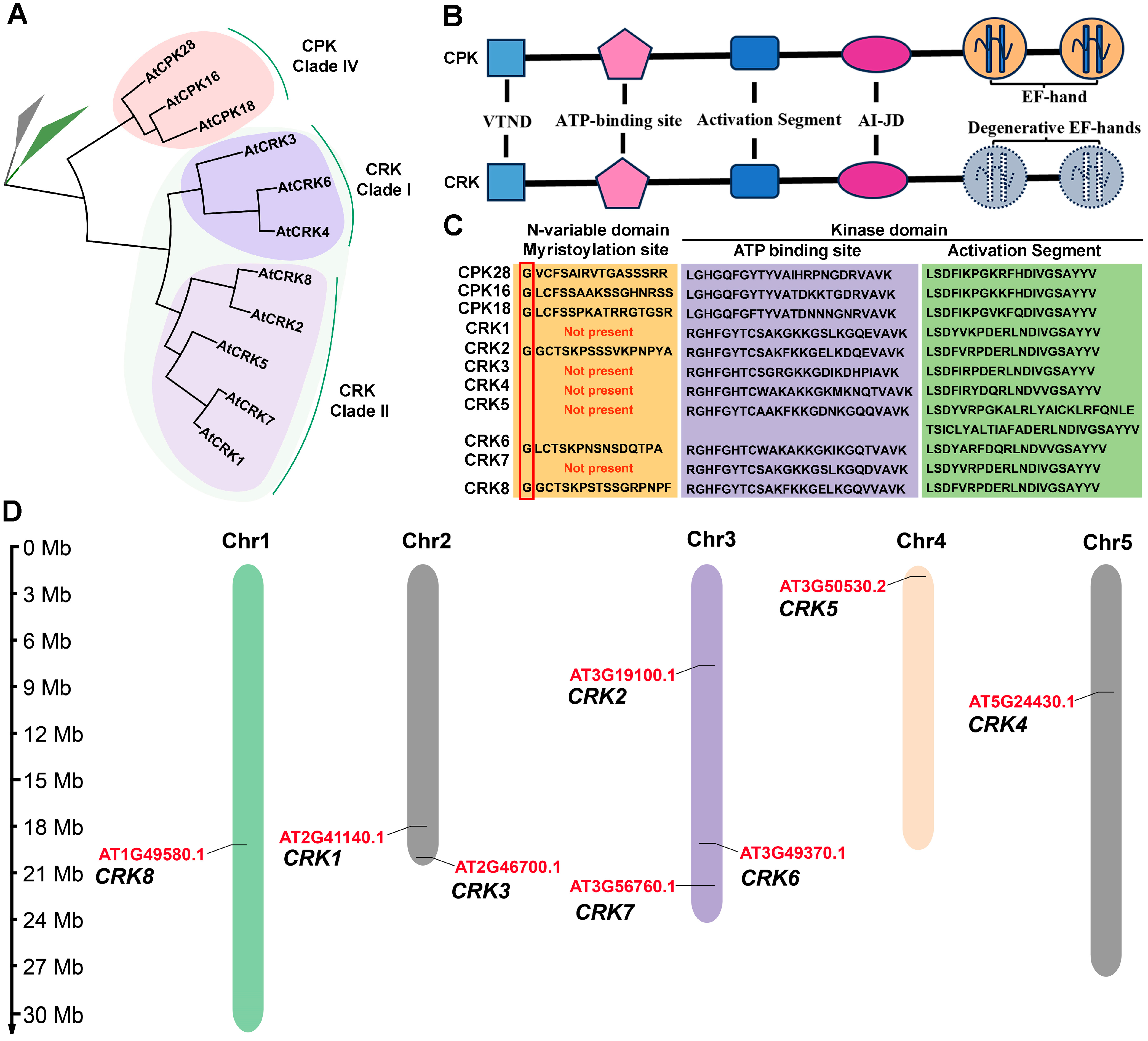
2.1. CRK Represents a Group of CPK-like Kinase That Contains Degenerative EF-Hands

Eight CRK genes, namely CRK1 to CRK8, are identified in the Arabidopsis genome. The CRK gene family is comprised of two subclades. Clade I consists of CRK3, 4, and 6, and Clade II contains CRK1, 2, 5, 7, and 8 (Figure 1A). CRK shows protein structures similar to CPK. The CPK subclade IV, consisting of CPK16, 18, and 28, is the closest paralog of CRK (Figure 1A). Both CRK and CPK contain a variable N-terminal domain (VTND), a kinase domain, an auto-inhibition junction domain (AI-JD), and a CaM-like domain (Figure 1B). The VTND of a number of CPKs and CRKs contains the G-x-x-x-S/T motif, which is the recognition site for N-myristoylation (Figure 1C). The N-terminal myristoylation often determines the membrane association of the modified proteins. The kinase domains of both CRK and CPK possess an ATP-binding site, associating with ATP, and an activation segment, which regulates the kinase activity (Figure S1). AI-JD functions to inhibit the kinase domain when Ca2+ is absent. When Ca2+ binds to EF-hands, the inhibition of AI-JD on the kinase domain is released by the interaction of AI-JD and EF-hands, resulting in CPK activation. Distinct from CPK, CRK merely contains degenerative EF-hands in its CaM-like domain, suggesting the kinase activation of CRK is Ca2+-independent.

2.2. Distribution of CRK Genes and Analysis of the Physicochemical Properties of CRK Proteins

We analyzed the chromosomal distribution of CRK genes. CRK8 is located on chromosome one, CRK1/3 are located on chromosome two, CRK2/6/7 are located on chromosome three, CRK5 is located on chromosome four, and CRK4 is located on chromosome five (Figure 1D). In addition, the physicochemical properties of CRK proteins were predicted. CRKs contain amino acid residues ranging from 500 to 600, with molecular weights ranging from 60 to 70 kDa (Table 1). The theoretical pIs, instability indexes, and aliphatic indexes of CRKs are also predicted (Table 1).

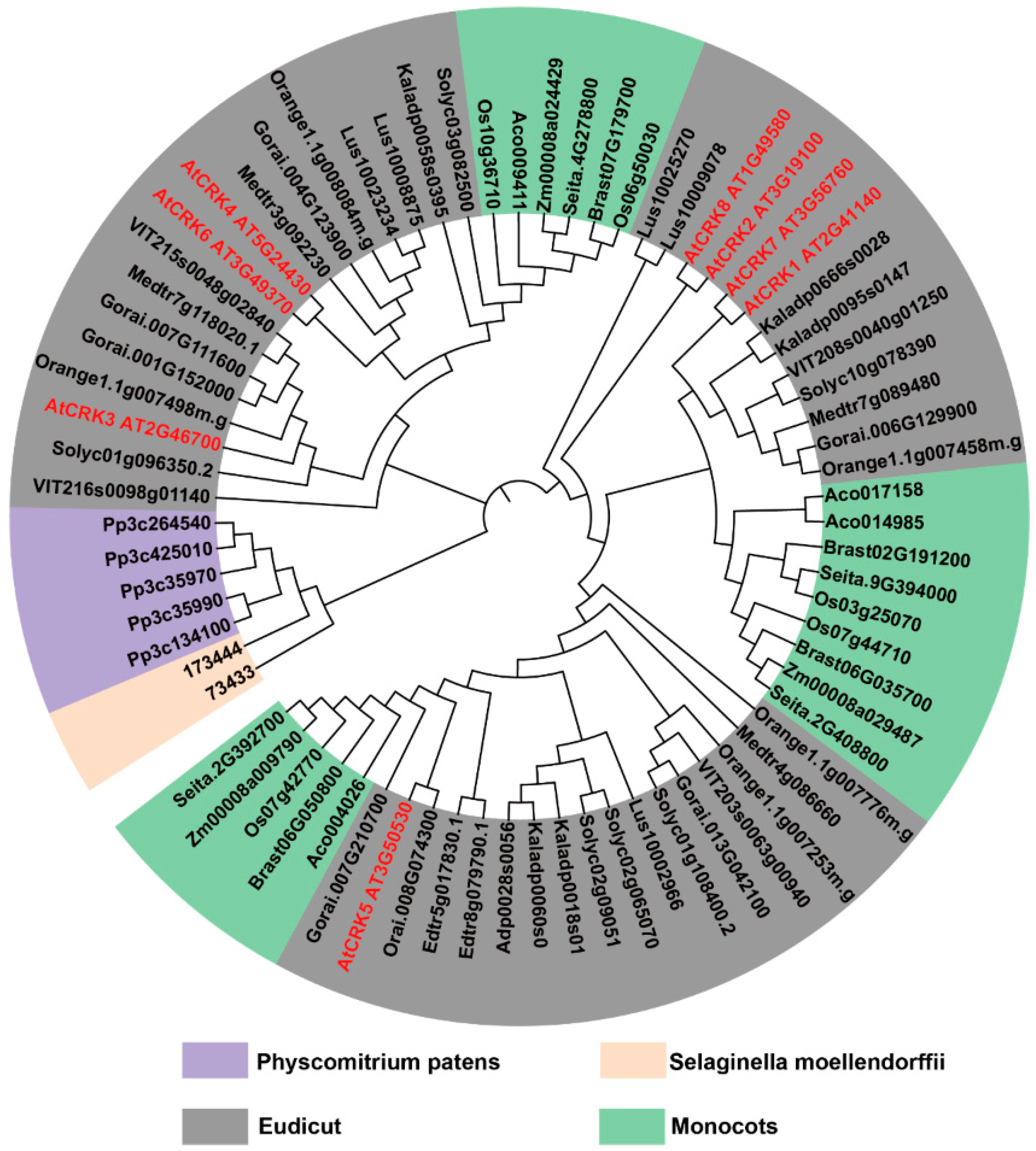
2.3. CRK Widely Exists in Land Plants

To resolve the evolutionary relationships among the CRK genes in diverse plants, we performed a maximum-likelihood analysis of the conserved CaM-like domains in CPK and CRK. CRK genes are found in land plants but not in algae Chromochloris zofingiensis and Chondrus crispus, while CPK is identified in plants, including algae (Figure 2 and Figure S2A,B). It is suggested that Ca2+-mediated regulations emerge at the early stages of plant evolution. In mosses and vascular plants, CRK clusters form a clade. In angiosperms, CRK does not show a trend of lower to higher evolution (Figure 2). These results suggest that the divergence of CRK and CPK likely occurred when ancient plants relocated from the oceans to the lands.

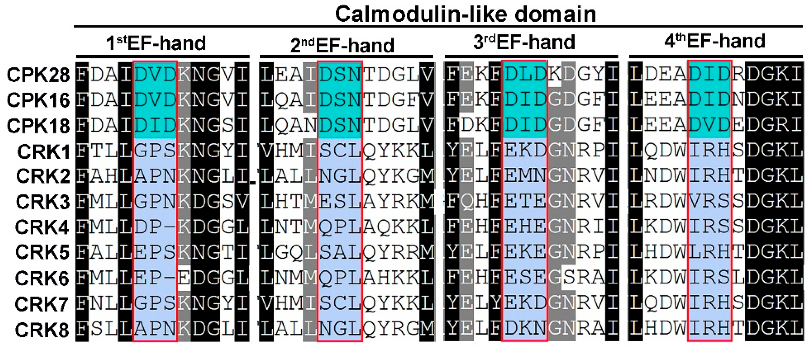
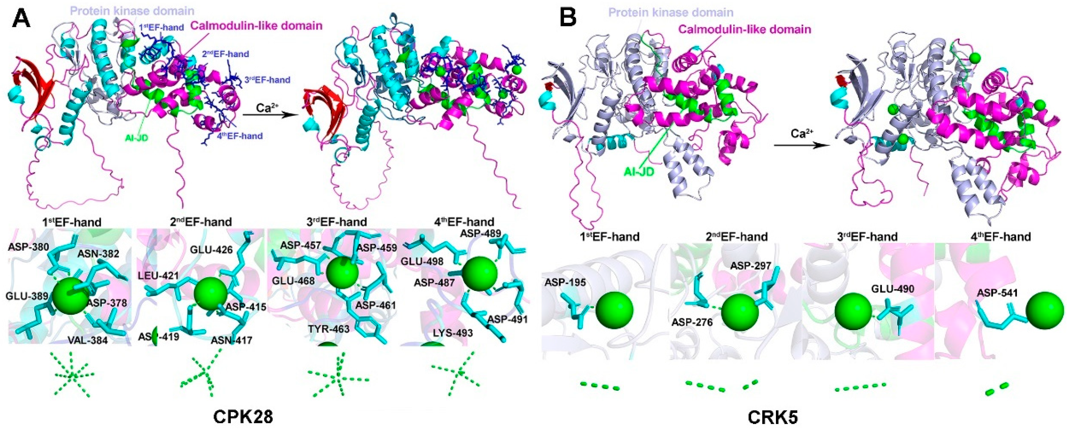
2.4. CRK Loses the Ability to Associate with Ca2+

We analyzed the CaM-like domain of CRK in Arabidopsis and compared it to CPK, which contains four EF-hands, in which the D-x-D motif contributes to direct Ca2+-binding (Figure S3). The amino acid substitutions in the D-x-D motif of CRK potentially disrupt Ca2+ association (Figure 3 and Table 2). The notion was further tested by using AlfaFold3. In CPK28, five to seven hydrogen (H)-bonds contribute to Ca2+-binding. For instance, the amino acid residues in the first EF-hand (D378, D380, N382, V384, E389), second EH-hand (D415, N417, D419, L421, E426), third EH-hand (D457, D459, D461, Y463, E468), and fourth EH-hand (D487, D489, D491, K493, E498) directly associated with Ca2+ via non-convent H-bonds (Figure 4A). By contrast, one to two hydrogen (H)-bonds were found to potentially associate with Ca2+ in each EF-hand of CRK5 (D195, D276, D279, E490, D541) (Figure 4B), indicating CRK is not likely to directly bind to Ca2+.

2.5. The Subcellular Localizations of CRK

Next, the subcellular locations of Arabidopsis CRK were analyzed. Except for CRK6, which was unable to be cloned yet, likely due to extremely low expression, all seven CRK genes were cloned and fused with a YFP tag. The CRK-YFP plasmids were transiently expressed in Nicotiana Benthamiana cells, followed by protoplast isolation. Even though only CRK2, 6, and 8 appeared to possess N-myristoylation site (Figure 1C), all CRKs-YFP were found to be PM-associated (Figure 5). This indicated that CRK, similar to CPK, likely functions as a modulator in regulating membrane proteins such as channels and transporters.

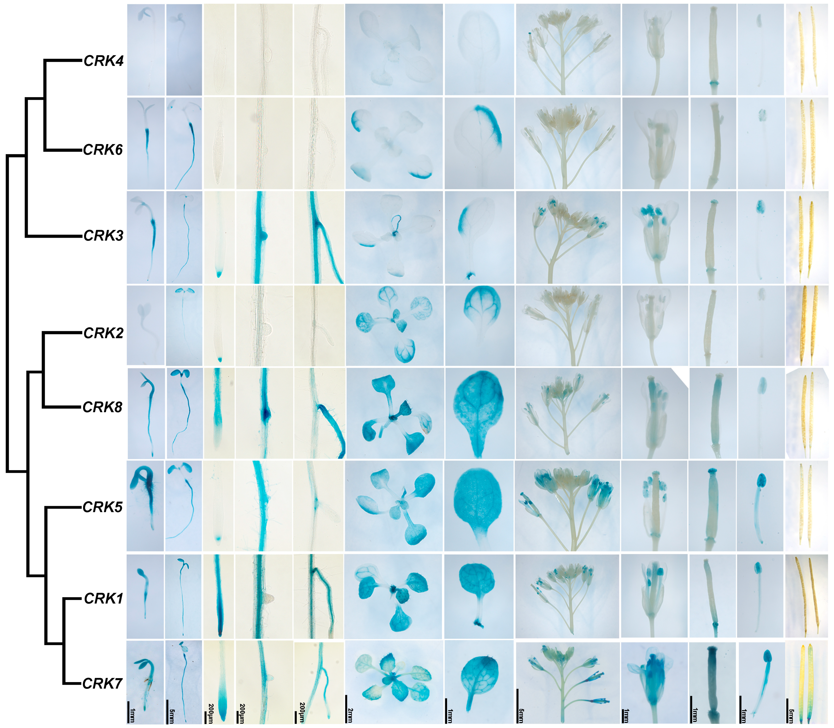
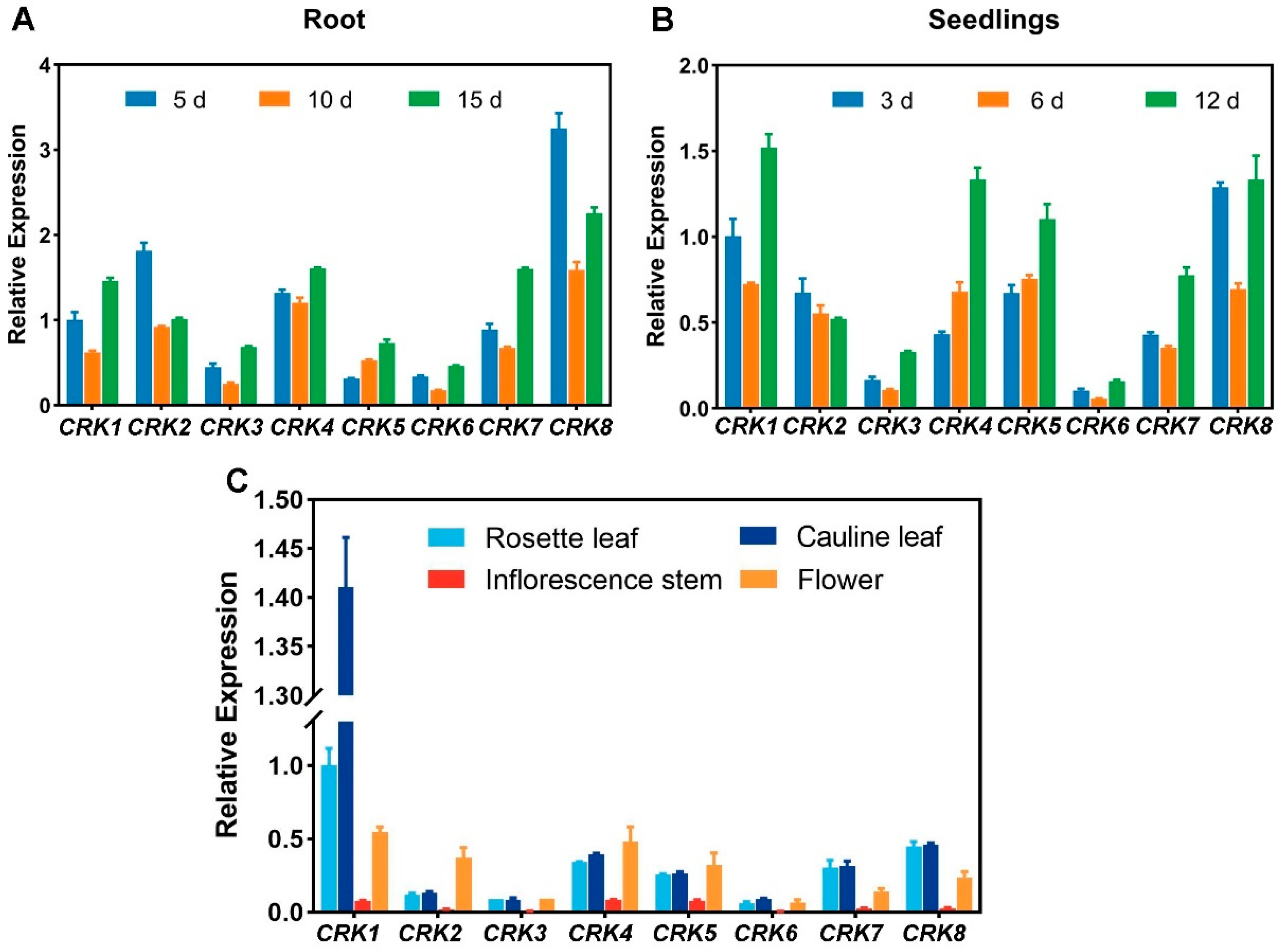
2.6. The Expression Patterns of CRK Genes in Arabidopsis

To analyze the expression patterns of CRK genes at the tissue levels, we cloned the promoter regions of all eight CRK genes. The transgenic plants harboring pCRK::GUS were generated. The expression patterns of CRK were examined by using GUS staining. Except for CRK4, which showed very low expression during all developmental stages, all other CRKs were found to be globally expressed (Figure 6). CRK2, 3, and 5 were expressed in the root tip, and CRK1, 3, 7, and 8 were expressed in the lateral root (Figure 6). CRK1, 2, 5, 7 and 8 showed strong expression in the leaf. CRK1, 3, 5, and 7 were obviously expressed in floral organs, including pollen tubes (Figure 6).
We also utilized RT-qPCR to verify the expression patterns of CRKs. Consistent with GUS staining results, CRK1 and 8 showed high expression in roots (Figure 7A). In seedlings, CRK1, 5, and 8 were highly expressed (Figure 7B). CRK1 displayed much higher expression levels than other CRKs in the leaf (Figure 7C). The expression of CRK1/7/8 showed a V-shaped pattern, suggesting these genes were finely regulated during these times of development and were potentially important for these stages of growth and development. High-order mutants of these CRK genes can be generated and analyzed in future studies.

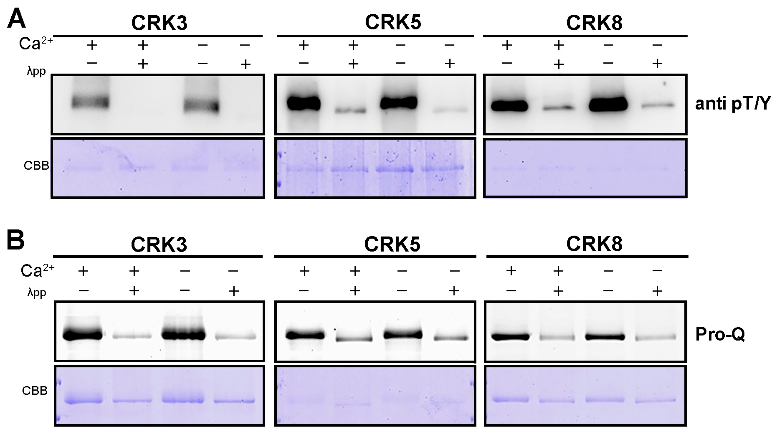
2.7. The Kinase Activity of Arabidopsis CRK Is Ca2+-Independent

It is well established that the kinase domain of Arabidopsis CPK is activated upon Ca2+ association. However, it was unknown whether Ca2+ is required for CRK activation in Arabidopsis. By using an α-phospho T/Y antibody, we were able to detect the autophosphorylation of CRK3, 5, and 8, which was eliminated by the addition of phosphatase λpp (Figure 8A). Of note, this phosphorylation of CRK was spotted without the presence of Ca2+, and adding Ca2+ failed to promote CRK phosphorylation (Figure 8A). Furthermore, the phosphorylation levels of CRK were examined by using Pro-Q. Consistent with Western immunoblotting results, the Pro-Q staining showed that the phosphorylation of CRK3, 5, and 8 was Ca2+-independent (Figure 8B). These results thus demonstrated that the kinase activity of Arabidopsis CRK is spontaneous, in which Ca2+ is not required.

2.8. The Analysis of Cis-Elements of the Promoters of CRK

We next analyzed the promoter regions of CRK genes. Among all the cis-elements, most CRK promoters contained conserved ABRE-motif and G-box cis-elements. A number of CRK promoters possessed a stress-related MBS-motif, CGTCA-motif, TGACG-motif regulatory element, and plant-immunity-related TC-rich element (Figure 9). In addition, specific cis-elements such as the LTR motif and TGA motif were found in the CRK5 promoter region (Figure 9), suggesting CRK5 is involved in low-temperature and phytohormone responses. Overall, the promoter sequences of CRK genes implied the involvement of CRK genes in various stress responses, for instance, ABA signal, low-temperature, and abiotic stimuli.

2.9. The Expression of CRK Genes Is in Response to Biotic and Abiotic Stresses

The cis-element analysis of CRK promoters suggested that CRKs are engaged in various stress responses. We thus examined how CRK expression responded to different stress conditions, such as flg22, ABA, drought, salt, and high/low temperatures, by using qPCR. Low temperature highly induced CRK1 and slight reduced CRK5 and 6 (Figure 10A). CRK2 was significantly prompted by high temperature (Figure 10B). ABA slightly enticed the expression of CRK1 and 2 (Figure 10C). Almost all CRK genes were upregulated by sorbitol or NaCl treatment (Figure 10D,E). The presence of pathogenic elicitor, flag22, appeared not to obviously affect the expression of CRKs (Figure 10F). Consistent with cis-element analysis of CRK promoters, the qPCR results indicated CRKs are regulated by various stressed conditions at transcriptional levels.

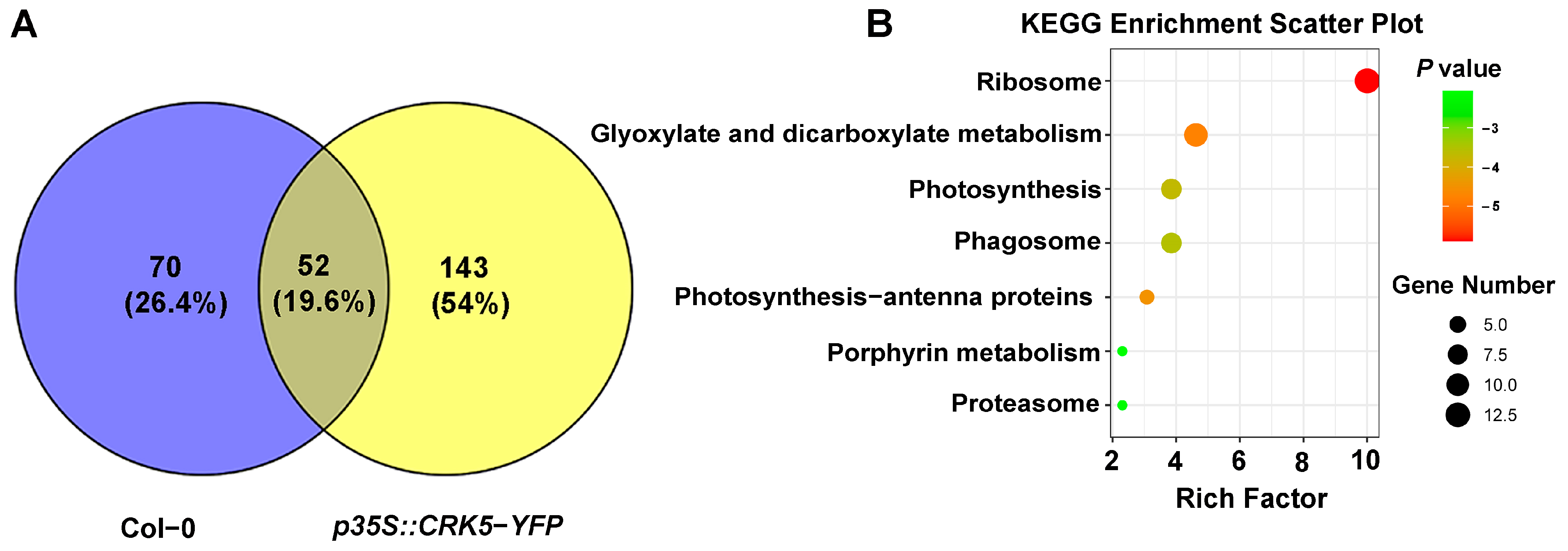
2.10. Identification of CRK-Interacting Proteins by IP-MS

To exert their functions, the kinases need to directly interact with their substrates for full phosphorylation. It, therefore, is important to identify the interacting components of the kinases to understand their functions. CRK5 is so far the most characterized CRK gene, which has been shown to regulate several signaling events such as PIN2 regulation, embryogenesis, and an ROS-NO balance [30,31,32]. We thus used CRK5 to screen its interacting proteins. By using CRK5-YFP transgenic plants, we screened the interacting proteins of CRK5 through an IP-MS approach. In total, 143 proteins were identified (Figure 11A). Analysis using the Kyoto Encyclopedia of Genes and Genomes (KEGG) indicated that CRK5 is likely involved in metabolism, genetic information processing, and cell processes (Figure 11B). Gene ontology (GO) analysis suggested that the CRK5-interacting proteins are involved in biological processes, including low-temperature stress, plant immunity, and protein transportation (Figure 11C). CRK1, a homologous protein of CRK5, was identified in the CRK5-containing complex (Table S1), suggesting CRK kinases may form homo- or hetero-dimers to fulfill their functions. Moreover, an RLK, somatic embryogenesis receptor kinase 4 (SERK4), was found to be associated with CRK5 (Table S2). SERK4, together with its close homolog SERK4/BRI1-associated kinase 1 (BAK1), acts as a co-receptor for multiple ligand-binding receptors such as BRI1 and FLS2 to modulate plant growth and immunity [34]. Furthermore, several protein channels/transporters, such as plastid envelope ion channels 2 (PEC2), sugar transport protein 1 (STP1), ABC transporter of the mitochondrion 3 (ATM3), and karyopherin enabling the transport of the cytoplasmic hyl 1 (KETCH1), were found to interact with CRK5 (Table S2). The identification of RLK and channels/transporters as the components that bind to CRK5 dropped a hint that RLKs and transport proteins function upstream and downstream of CRK kinases, respectively.

3. Discussion

Despite numerous studies on CPK in regulating growth, development, and stress responses in plants, the functions of CRK, the closest homolog of CPK, are still largely unknown. In this study, we investigated CRK genes in plant genomes and found that CRK only exists in land plants, indicating CRK is likely divided from CPK at the early stages of plant evolution. In Arabidopsis, eight CRK genes were identified. We provided evidence that CRKs are broadly expressed during plant growth and stress adaptions. We also showed that Arabidopsis CRK possesses kinase activity and, importantly, is Ca2+-independent. By utilizing a proteomic approach, we identified a number of candidate proteins that potentially interact with CRK5. Among the CRK5-interacting proteins, RLK and transporters were pinpointed. It is thus conjectured that RLKs and channels/transporters function upstream and downstream of CRKs, respectively.
CRK is only found in land plants starting from moss but not in algae. In contrast, CPK is found in land plants and algae. This result demonstrated that CRK was originally derived from CPK, which likely occurred during the evolutionary stages when ancient plants relocated from the oceans to the lands. Future studies will focus on the potential selection pressures that drove CRKCPK separation and caused the origin of CRK. Our results indicated that CRK is spontaneously active regarding its autophosphorylation ability. The presence of Ca2+ was unable to affect the kinase activity of CRK. Even though this can be explicated by the degenerative EF-hands in CRK, it remains unknown whether the CaM-like domain of CRK still functions as a regulatory domain. Although the generative EF-hands are not capable of directly binding to Ca2+, it cannot be excluded that they can recruit additional components to form complexes or associate with the substrate proteins to determine CRK specificities. Considering that the CaM-like domains in a number of proteins were found to interact with CaM or CaM domain [35,36], the CaM-like domain of CRK may act as the target of Ca2+ decoding components such as CaM and CBL, which allows indirect involvement of CRK in Ca2+-mediated signaling.
The kinase domain of CPK appears to be repressed by the AI-JD via direct association under Ca2+-free conditions. Despite the lack of Ca2+ binding affinity, CRK still contains an AI-JD with unknown functions. CRK appears to be spontaneously active, in which Ca2+ is not involved. It thus will be interesting to explore whether the AI-JD of CRK still interacts with its kinase domain if AI-JD inhibits the kinase domain of CRK and whether the phosphorylation of AI-JD results in their disassociation. If the AI-JD of CRK fails to interact with the kinase domain, the comparison analysis of the AI-JDs between CPK and CRK will provide additional insights into the molecular mechanisms underlying the auto-inhibition and activation of CPK and CRK.
CPK is activated and regulated by Ca2+ oscillations. Moreover, CPK is activated by other kinases, such as RLKs and other CPKs, through phosphorylation [37,38,39,40,41]. Given that Ca2+ signals activate CPK but not CRK, it is important to investigate the upstream components that activate CRK in future studies. Of note, RLK SERK4 was found to interact with CRK5. SERK4 and SERK3/BAK1 were previously identified as essential co-receptors for multiple ligand-binding receptors-mediated pathways such as brassinosteroid (BR), pattern-triggered immunity (PTI), and effector-triggered immunity (ETI) [42,43,44,45,46,47]. The identification of RLK as the interacting component of CRK led to an assumption that RLK perceives extracellular ligands such as phytohormones and pathogen-associated molecular patterns (PAMPs) and subsequently activates CRKs to initiate intracellular signaling.
CPK activity is modulated by Ca2+ signatures, which often reflect instantaneous exogenous and endogenous events [48]. Thus, the substrates usually include transporters and channels that mediate rapid ion fluxes in response to acute intercellular and intracellular stimuli. The transphosphorylation of channels and transporters mediated by CPK causes conformational change, leading to activation/inactivation of their transport abilities. It has been well established that CPK phosphorylates a variety of channels and transporters that mediate the transport of cations, anions, H2O, and phytochromes [49,50]. Intriguingly, various transporters were identified to be associated with CRK5 during proteomic assay. It suggested CRK, similar to CPK, phosphorylates transporters and channels to manipulate ion/membrane potential/pH homeostasis. In this study, RLK and channels/transporters were identified to be associated with CRK5. Given that Ca2+ is not capable of directly activating CRK, it will be interesting to explore whether RLK can function upstream of CRK. We speculated that distinct from CPK, which is activated by Ca2+ signals, CRK can be activated by PM-localized RLK upon apoplastic ligand perceptions to regulate the activities of channels and transporters, leading to rapid cellular responses.
Most CPKs were found to play their roles under stressed conditions. Hence, very few cpk mutant plants display obvious developmental defects under normal growth conditions. Most cpk mutants exhibit phenotypes different from WT plants only when they are subjected to stress treatments. This reminds us that genetic analysis on CRK may require examining the crk mutant plants by using different abiotic and biotic conditions. In addition, due to gene redundancy, high-order mutant plants for CRK need to be generated and examined for genetic analysis. The expression patterns of CRK presented in this study may provide information for constructing crk multiple mutants in future studies. Furthermore, the functional analyses of CRK genes may rely on multiple strategies, including proteomic and genomic methods. For crk mutants, RNAseq can be utilized in different treatments, such as phytohormones and stressed conditions, to identify potential CRK-regulated genes, which may indicate the possible signaling pathways in which the CRKs are involved. In addition, proteomic approaches will help to screen CRK-interacting components.
Compared to CPK, which is extensively involved in all aspects of plant growth and stress adaptions, CRK is barely functionally analyzed. In this study, we systematically characterized Arabidopsis CRK genes in terms of genetic and biochemical futures, which provides an additional understanding of Ca2+-related and Ca2+-unrelated signaling pathways mediated by protein kinases in plants.

4. Materials and Methods

4.1. Plant Materials and Growth Conditions

The Col-0 accession of Arabidopsis thaliana was used as the wild type (WT) in all experiments conducted in this study. The plants were transformed using the Agrobacterium tumefaciens-mediated floral dip method [51]. Arabidopsis plants were cultivated in soil within a greenhouse under long-day conditions (16 h light/8 h dark, 22 °C). Nicotiana benthamiana was grown in soil in a plant incubator at 28 °C under a 16 h light/8 h dark photoperiod.

4.2. Molecular Cloning and Construction of Transgenic Plants

The sequences of primers used for vector construction in this study are listed in Supplemental Table S3. The PCR products were recombined into the pDONR vector via the Gateway BP reaction, followed by the Gateway LR reaction to construct the destination vectors (Invitrogen, Waltham, MA, USA). To generate of overexpression plants, the coding sequences (CDS) of CRK were recombined into the binary vector pBIB-BASTA-35S-GWR-YFP. For GUS staining, the promoter sequences of approximately 2.0 kb of CRK genes were cloned into the binary vector pBIB-BASTA-GWR-GUS. The destination vectors were then transformed into plants using the floral dip method with A. tumefaciens strain GV3101 [51].

4.3. Database Search and Sequence Retrieval

The protein sequences of AtCRK and AtCPK were downloaded from the Arabidopsis information resource (TAIR) (https://www.arabidopsis.org/) URL (accessed on 23 March 2025). Since the protein sequences of CRK and CPK are highly conserved in the N-segment and kinase domains, thus affecting the homologous protein search, we only used the more divergent C-terminal region for comparison. The protein sequences of CRK from other species were retrieved from Phytozome (https://phytozome-next.jgi.doe.gov/) URL (accessed on 23 March 2025) and National Center for Biotechnology Information (NCBI) (https://www.ncbi.nlm.nih.gov/) (E ≤ 10−5) URL (accessed on 23 March 2025). The CDS and promoter sequences of CRK were obtained from the TAIR (https://www.arabidopsis.org/) URL (accessed on 23 March 2025).

4.4. Phylogenetic Analysis and Chromosomal Distributions of CRK Genes

The phylogenetic tree was constructed to elucidate the evolutionary relationship of CRK proteins among different species. The protein sequences were aligned using ClustalW to generate a Clustal file [52]. The generated Clustal files were then converted to the MEGA format using MEGA11.0.10 software [53]. Unrooted phylogenetic trees were generated using the maximum likelihood method in MEGA11.0.10, with bootstrap tests conducted using 1000 replicates. The trees were visualized using the iTOL website (https://itol.embl.de/) URL (accessed on 23 March 2025). The chromosomal distribution of CRK genes in the Arabidopsis genome was analyzed by using the online tool MG2C (http://mg2c.iask.in/mg2c_v2.1/) URL (accessed on 23 March 2025) [54].

4.5. Multiple Sequence Alignment and Myristoylation Site Prediction

Multiple sequence alignments of CRK and CPK proteins were performed separately to identify conserved domains and motifs. GENEDOC 2.7.0 software was used to perform the multiple sequence alignment. The conserved domains and motifs were predicted using Motif Scan (https://myhits.sib.swiss/cgi-bin/motif_scan) URL (accessed on 28 March 2025). The myristoylation sites of CRK and CPK proteins were predicted using the NMT Predictor (https://mendel.imp.ac.at/myristate/SUPLpredictor.htm) URL (accessed on 28 March 2025).

4.6. Model Performance Analysis and Visualization

The prediction of CRK and CPK protein structures was performed using AlphaFold3 (https://golgi.sandbox.google.com/) URL (accessed on 28 March 2025) [55]. In addition, the differences in the binding sites of CRK and CPK to metal ions were analyzed. The structure visualizations were made in PyMOL 2.5.x software

4.7. Promoter Sequence Analysis and Physicochemical Properties of CRK Proteins

The promoter sequences of the CRK genes were downloaded from the TAIR (https://www.arabidopsis.org/) URL (accessed on 28 March 2025). The promoter sequences are approximately 2.0 kb. PlantCARE (https://bioinformatics.psb.ugent.be/webtools/plantcare/html/) URL (accessed on 28 March 2025). was used to analyze the promoter elements. The results are visualized using TBtools 2.0 software. In addition, the physicochemical properties of CRK proteins were analyzed using TBtools 2.0 software [56].

4.8. Subcellular Localization Assay

Subcellular localization of CRKs was determined by observing the YFP fluorescence signal. The CDS of CRKs were cloned into the pBIB-BASTA-35S-YFP vector with the cauliflower mosaic virus 35S (CaMV35S) promoter. The 35S::YFP control construct and 35S::CRK-YFP constructs were transiently expressed in N. benthamiana. Tobacco protoplasts were prepared as previously described [57]. The plasma membrane of chloroplasts was labeled with FM4-64, and YFP signals were detected using a confocal microscope (Leica/Stellaris 5) with laser excitation at a wavelength of 514 nm and between 520 and 550 nm and an excitation laser at a wavelength of 561 nm and between 570 and 630 nm to detect FM4-64 signals [57].

4.9. Expression and Purification of Recombinant CRK in Escherichia coli

Recombinant MBP-CRKs were expressed in E. coli (Rosetta). Protein induction and purification were performed as previously described [58]. The glutathione agarose beads (Sangon Biotech, Shanghai, China) were used according to the manufacturer’s manual.

4.10. In Vitro Kinase Assays

For phosphorylation assays using protein blotting, full-lengths of CRKs were PCR amplified and cloned into the pDEST15 vector with an MBP tag (Invitrogen, 11802014) to generate fusion proteins of MBP-CRK3, MBP-CRK5, and MBP-CRK8. Proteins expressed in E. coli were purified using glutathione agarose beads (Sangon Biotech, C600031) according to the manufacturer’s instructions. For the phosphorylation assay, reaction conditions reference [54]. For Pro-Q assays, for sample preparation and reaction conditions, please refer to Sun [59]. Imaging with a PharosFX molecular imager (Bio-Rad, Hercules, CA, USA) instrument.

4.11. GUS Staining

Different developmental stages and different tissues of transgenic plants were collected and stained. The tissues were first incubated in rinsing solution (34.2 mM Na2HPO4, 15.8 mM NaH2PO4, 0.5 mM K3Fe(CN)6, and 0.5 mM K4Fe(CN)6 3H2O) for 5 min, then incubated in staining solution (34.2 mM Na2HPO4, 15.8 mM NaH2PO4, 0.5 mM K3Fe(CN)6, 0.5 mM K4Fe(CN)6 3H2O, and 2 mM X-Gluc) at 37 °C for appropriate time. After staining, the plant tissues were immersed in 30%, 50%, 75%, and 95% ethanol for 1 h in succession and then immersed in 75% ethanol. After destaining, the tissues were observed under a stereomicroscope (Leica M165C).

4.12. RT-qPCR

To quantify CRK gene expression at different time points, total RNA extraction in Col-0 was performed with RNAprep Pure Plant Kit (TIANGEN, Beijing, China) according to the manufacturer’s instructions. Two micrograms of total RNA were used for reverse transcription using the Reverse Transcription System (Takara, Shiga, Japan). The PCR program was run on the Step One Plus Real Time PCR system (Applied Biosystems, Waltham, MA, USA). ACTIN2 (ACT2) expression was used as an internal control to normalize all data. Each experiment was performed with at least three independent biological replicates.

4.13. Mass Spectrometry Analysis

For the IP experiment, the transgenic seedlings were cultured in a liquid MS medium with gentle shaking for 10 d. The seedlings were ground in liquid nitrogen and lysed in extraction buffer, which includes 10 mM HEPES (pH 7.5), 100 mM NaCl, 1 mM EDTA, 10% glycerol, 1% TritonX-100, 20 mM NaF, 1 mM PMSF, 1 × protease inhibitor. The transparent supernatant was obtained by multiple centrifugation (6000× g, 4 °C, 10 min). The supernatant was incubated with anti-GFP beads (KTSM1301, Alpalife, Shenzhen, China) for 3 h at 4 °C with gentle shaking. The beads were washed five times using extraction buffer containing 0.2% TritonX-100 and then boiled with 2 × SDS loading buffer (325 mM Tris, pH 6.8, 10% (w/v) SDS, 50% (v/v) glycerol, 25% (v/v) β-mercaptoethanol, and 0.05% (w/v) bromophenol blue) for 5 min. The proteins were separated in 8% SDS-PAGE gel. The antibodies in this study were α-GFP (11814460001, Roche, Mannheim, Germany) antibodies. For the mass spectrometry experiment, the sample preparation is referred to as Sun [59]. The samples were analyzed using an Orbitrap Fusion Lumos mass spectrometer (Thermo Fisher Scientific, Waltham, MA, USA) connected to an EASY-nLC 1200 system. Proteome Discoverer Daemon 2.2 software (Thermo Fisher Scientific) was used for data analysis.

4.14. Data Acquisition and Statistics

To analyze the functional characteristics of CRK protein, GO functional annotation and protein enrichment analysis were performed using Metascape (https://metascape.org/gp/index.html#/main/step1) URL (accessed on 28 March 2025). For KEGG pathway analysis, KEGG (https://www.genome.jp/kegg/pathway.html) URL (accessed on 28 March 2025) was searched. Visualizations of the results were generated online (https://www.bioinformatics.com.cn/) URL (accessed on 28 March 2025).

Supplementary Materials

The following supporting information can be downloaded at https://www.mdpi.com/article/10.3390/ijms26073297/s1.

Author Contributions

S.Y., Y.F. and K.H. conceived the project. S.Y. and K.H. designed all experiments and analyzed the data. S.Y. and Y.F. conducted all experiments. X.F. and J.H. assisted in the experiments. S.Y. and K.H. wrote the manuscript. All authors have read and agreed to the published version of the manuscript.

Funding

This work was supported by National Natural Science Foundation of China (32370295, 32170280), and the Gansu Provincial Science and Technology Department (24JRRA392, 23JRRA1138), and the China Postdoctora Science Foundation (2024M751259, GZB20240285).

Institutional Review Board Statement

Not applicable.

Informed Consent Statement

Not applicable.

Data Availability Statement

The data that support the findings of this study are available from the corresponding author upon reasonable request.

Acknowledgments

We are grateful to Liang Peng, Li Xie, Liping Guan, Yahu Gao, and Yang Zhao (Core Facility for Life Science Research, Lanzhou University) for technical assistance.

Conflicts of Interest

The authors declare that they have no conflict of interest.

References

  1. Friso, G.; van Wijk, K.J. Posttranslational Protein Modifications in Plant Metabolism. Plant Physiol. 2015, 169, 1469–1487. [Google Scholar]
  2. Matte, A.; Tari, L.W.; Delbaere, L.T. How do kinases transfer phosphoryl groups? Structure 1998, 6, 413–419. [Google Scholar] [PubMed]
  3. Zhang, W.J.; Zhou, Y.; Zhang, Y.; Su, Y.H.; Xu, T. Protein phosphorylation: A molecular switch in plant signaling. Cell Rep. 2023, 42, 112729. [Google Scholar] [PubMed]
  4. Luan, S.; Wang, C. Calcium Signaling Mechanisms Across Kingdoms. Annu. Rev. Cell Dev. Biol. 2021, 37, 311–340. [Google Scholar]
  5. Mohanta, T.K.; Yadav, D.; Khan, A.L.; Hashem, A.; Abd Allah, E.F.; Al-Harrasi, A. Molecular Players of EF-hand Containing Calcium Signaling Event in Plants. Int. J. Mol. Sci. 2019, 20, 1476. [Google Scholar] [CrossRef]
  6. Hrabak, E.M.; Chan, C.W.; Gribskov, M.; Harper, J.F.; Choi, J.H.; Halford, N.; Kudla, J.; Luan, S.; Nimmo, H.G.; Sussman, M.R.; et al. The Arabidopsis CDPK-SnRK superfamily of protein kinases. Plant Physiol. 2003, 132, 666–680. [Google Scholar]
  7. Polge, C.; Thomas, M. SNF1/AMPK/SnRK1 kinases, global regulators at the heart of energy control? Trends Plant Sci. 2007, 12, 20–28. [Google Scholar]
  8. Jossier, M.; Bouly, J.P.; Meimoun, P.; Arjmand, A.; Lessard, P.; Hawley, S.; Grahame, H.D.; Thomas, M. SnRK1 (SNF1-related kinase 1) has a central role in sugar and ABA signalling in Arabidopsis thaliana. Plant J. 2009, 59, 316–328. [Google Scholar]
  9. Baena-Gonzalez, E.; Rolland, F.; Thevelein, J.M.; Sheen, J. A central integrator of transcription networks in plant stress and energy signalling. Nature 2007, 448, 938–942. [Google Scholar]
  10. Margalha, L.; Confraria, A.; Baena-Gonzalez, E. SnRK1 and TOR: Modulating growth-defense trade-offs in plant stress responses. J. Exp. Bot. 2019, 70, 2261–2274. [Google Scholar]
  11. Fujii, H.; Verslues, P.E.; Zhu, J.K. Identification of two protein kinases required for abscisic acid regulation of seed germination, root growth, and gene expression in Arabidopsis. Plant Cell 2007, 19, 485–494. [Google Scholar] [PubMed]
  12. Lin, Z.; Li, Y.; Wang, Y.; Liu, X.; Ma, L.; Zhang, Z.; Mu, C.; Zhang, Y.; Peng, L.; Xie, S.; et al. Initiation and amplification of SnRK2 activation in abscisic acid signaling. Nat. Commun. 2021, 12, 2456. [Google Scholar] [PubMed]
  13. Luan, S. The CBL-CIPK network in plant calcium signaling. Trends Plant Sci. 2009, 14, 37–42. [Google Scholar]
  14. Luan, S.; Kudla, J.; Rodriguez-Concepcion, M.; Yalovsky, S.; Gruissem, W. Calmodulins and calcineurin B-like proteins: Calcium sensors for specific signal response coupling in plants. Plant Cell 2002, 14, 389–400. [Google Scholar]
  15. Harper, J.F.; Breton, G.; Harmon, A. Decoding Ca2+ signals through plant protein kinases. Annu. Rev. Plant Biol. 2004, 55, 263–288. [Google Scholar]
  16. Lindzen, E.; Choi, J.H. A carrot cDNA encoding an atypical protein kinase homologous to plant calcium-dependent protein kinases. Plant Mol. Biol. 1995, 28, 785–797. [Google Scholar] [PubMed]
  17. Zhang, L.; Liu, B.F.; Liang, S.; Jones, R.L.; Lu, Y.T. Molecular and biochemical characterization of a calcium/calmodulin-binding protein kinase from rice. Biochem. J. 2002, 368, 145–157. [Google Scholar]
  18. Furumoto, T.; Ogawa, N.; Hata, S.; Izui, K. Plant calcium-dependent protein kinase-related kinases (CRKs) do not require calcium for their activities. FEBS Lett. 1996, 396, 147–151. [Google Scholar]
  19. Hua, W.; Zhang, L.; Liang, S.; Jones, R.L.; Lu, Y.T. A tobacco calcium/calmodulin-binding protein kinase functions as a negative regulator of flowering. J. Biol. Chem. 2004, 279, 31483–31494. [Google Scholar]
  20. Leclercq, J.; Ranty, B.; Sanchez-Ballesta, M.T.; Li, Z.; Jones, B.; Jauneau, A.; Pech, J.C.; Latche, A.; Ranjeva, R.; Bouzayen, M. Molecular and biochemical characterization of LeCRK1, a ripening-associated tomato CDPK-related kinase. J. Exp. Bot. 2005, 56, 25–35. [Google Scholar]
  21. Wang, J.P.; Xu, Y.P.; Munyampundu, J.P.; Liu, T.Y.; Cai, X.Z. Calcium-dependent protein kinase (CDPK) and CDPK-related kinase (CRK) gene families in tomato: Genome-wide identification and functional analyses in disease resistance. Mol. Genet. Genom. 2016, 291, 661–676. [Google Scholar]
  22. Liu, J.; Li, X.D.; Jia, D.; Qi, L.; Jing, R.; Hao, J.; Wang, Z.; Cheng, J.; Chen, L.M. ZmCRK1 negatively regulates maize’s response to drought stress by phosphorylating plasma membrane H+-ATPase ZmMHA2. N. Phytol. 2024, 244, 1362–1376. [Google Scholar]
  23. Tao, X.C.; Lu, Y.T. Loss of AtCRK1 gene function in Arabidopsis thaliana decreases tolerance to salt. J. Plant Biol. 2013, 56, 306–314. [Google Scholar]
  24. Baba, A.I.; Rigó, G.; Ayaydin, F.; Rehman, A.U.; Andrási, N.; Zsigmond, L.; Valkai, I.; Urbancsok, J.; Vass, I.; Pasternak, T.; et al. Functional Analysis of the Arabidopsis thaliana CDPK-Related Kinase Family: AtCRK1 Regulates Responses to Continuous Light. Int. J. Mol. Sci. 2018, 19, 1282. [Google Scholar] [CrossRef]
  25. Nemoto, K.; Takemori, N.; Seki, M.; Shinozaki, K.; Sawasaki, T. Members of the Plant CRK Superfamily Are Capable of Trans and Autophosphorylation of Tyrosine Residues. J. Biol. Chem. 2015, 290, 16665–16677. [Google Scholar]
  26. Nemoto, K.; Ramadan, A.; Arimura, G.I.; Imai, K.; Tomii, K.; Shinozaki, K.; Sawasaki, T. Tyrosine phosphorylation of the GARU E3 ubiquitin ligase promotes gibberellin signalling by preventing GID1 degradation. Nat. Commun. 2017, 8, 1004. [Google Scholar]
  27. Miyamoto, T.; Uemura, T.; Nemoto, K.; Daito, M.; Nozawa, A.; Sawasaki, T.; Arimura, G.I. Tyrosine Kinase-Dependent Defense Responses Against Herbivory in Arabidopsis. Front. Plant Sci. 2019, 10, 776. [Google Scholar]
  28. Li, R.J.; Hua, W.; Lu, Y.T. Arabidopsis cytosolic glutamine synthetase AtGLN1;1 is a potential substrate of AtCRK3 involved in leaf senescence. Biochem. Biophys. Res. Commun. 2006, 342, 119–126. [Google Scholar]
  29. Liu, H.T.; Gao, F.; Li, G.L.; Han, J.L.; Liu, D.L.; Sun, D.Y.; Zhou, R.G. The calmodulin-binding protein kinase 3 is part of heat-shock signal transduction in Arabidopsis thaliana. Plant J. 2008, 55, 760–773. [Google Scholar]
  30. Rigó, G.; Ayaydin, F.; Tietz, O.; Zsigmond, L.; Kovács, H.; Páy, A.; Salchert, K.; Darula, Z.; Medzihradszky, K.F.; Szabados, L.; et al. Inactivation of plasma membrane-localized CDPK-RELATED KINASE5 decelerates PIN2 exocytosis and root gravitropic response in Arabidopsis. Plant Cell. 2013, 25, 1592–1608. [Google Scholar]
  31. Baba, A.I.; Valkai, I.; Labhane, N.M.; Koczka, L.; Andrási, N.; Klement, É.; Darula, Z.; Medzihradszky, K.F.; Szabados, L.; Fehér, A.; et al. CRK5 Protein Kinase Contributes to the Progression of Embryogenesis of Arabidopsis thaliana. Int. J. Mol. Sci. 2019, 20, 6120. [Google Scholar] [CrossRef] [PubMed]
  32. Cséplő, Á.; Zsigmond, L.; Andrási, N.; Baba, A.I.; Labhane, N.M.; Pető, A.; Kolbert, Z.; Kovács, H.E.; Steinbach, G.; Szabados, L.; et al. The AtCRK5 Protein Kinase Is Required to Maintain the ROS NO Balance Affecting the PIN2-Mediated Root Gravitropic Response in Arabidopsis. Int. J. Mol. Sci. 2021, 22, 5979. [Google Scholar] [CrossRef]
  33. Teng, H.J.; Guo, Y.; Wang, J.Q.; Li, R.; Lu, Y.T.; Zhang, L. WDRP, a DWD protein component of CUL4-based E3 ligases, acts as a receptor of CDPK-related protein kinase 5 to mediate kinase degradation in Arabidopsis. J. Plant Biol. 2016, 59, 627–638. [Google Scholar] [CrossRef]
  34. Liu, J.; Li, J.; Shan, L. SERKs. Curr. Biol. 2020, 30, R293–R294. [Google Scholar] [CrossRef] [PubMed]
  35. Villalobo, A.; Gonzalez-Munoz, M.; Berchtold, M.W. Proteins with calmodulin-like domains: Structures and functional roles. Cell Mol. Life Sci. 2019, 76, 2299–2328. [Google Scholar] [CrossRef]
  36. Andrews, C.; Xu, Y.; Kirberger, M.; Yang, J.J. Structural aspects and prediction of calmodulin-binding proteins. Int. J. Mol. Sci. 2020, 22, 308. [Google Scholar] [CrossRef]
  37. Zhao, L.N.; Shen, L.K.; Zhang, W.Z.; Zhang, W.; Wang, Y.; Wu, W.H. Ca2+-dependent protein kinase11 and 24 modulate the activity of the inward rectifying K+ channels in Arabidopsis pollen tubes. Plant Cell. 2013, 25, 649–661. [Google Scholar] [CrossRef]
  38. Monaghan, J.; Matschi, S.; Shorinola, O.; Rovenich, H.; Matei, A.; Segonzac, C.; Malinovsky, F.G.; Rathjen, J.P.; MacLean, D.; Romeis, T.; et al. The calcium-dependent protein kinase CPK28 buffers plant immunity and regulates BIK1 turnover. Cell Host Microbe 2014, 16, 605–615. [Google Scholar] [CrossRef]
  39. Monaghan, J.; Matschi, S.; Romeis, T.; Zipfel, C. The calcium-dependent protein kinase CPK28 negatively regulates the BIK1-mediated PAMP-induced calcium burst. Plant Signal Behav. 2015, 10, e1018497. [Google Scholar] [CrossRef]
  40. Bredow, M.; Bender, K.W.; Dingee, J.A.; Holmes, D.R.; Thomson, A.; Ciren, D.; Tanney, C.A.S.; Dunning, K.E.; Trujillo, M.; Huber, S.C.; et al. Phosphorylation-dependent subfunctionalization of the calcium-dependent protein kinase CPK28. Proc. Natl. Acad. Sci. USA 2021, 118, e2024272118. [Google Scholar] [CrossRef]
  41. Hailemariam, S.; Liao, C.J.; Mengiste, T. Receptor-like cytoplasmic kinases: Orchestrating plant cellular communication. Trends Plant Sci. 2024, 29, 1113–1130. [Google Scholar] [PubMed]
  42. Li, J.; Wen, J.; Lease, K.A.; Doke, J.T.; Tax, F.E.; Walker, J.C. BAK1, an Arabidopsis LRR receptor-like protein kinase, interacts with BRI1 and modulates brassinosteroid signaling. Cell 2002, 110, 213–222. [Google Scholar] [PubMed]
  43. Nam, K.H.; Li, J. BRI1/BAK1, a receptor kinase pair mediating brassinosteroid signaling. Cell 2002, 110, 203–212. [Google Scholar] [CrossRef] [PubMed]
  44. He, K.; Gou, X.; Yuan, T.; Lin, H.; Asami, T.; Yoshida, S.; Russell, S.D.; Li, J. BAK1 and BKK1 Regulate Brassinosteroid-Dependent Growth and Brassinosteroid-Independent Cell-Death Pathways. Curr. Biol. 2007, 17, 1109–1115. [Google Scholar]
  45. Chinchilla, D.; Zipfel, C.; Robatzek, S.; Kemmerling, B.; Nurnberger, T.; Jones, J.D.; Felix, G.; Boller, T. A flagellin-induced complex of the receptor FLS2 and BAK1 initiates plant defence. Nature 2007, 448, 497–500. [Google Scholar]
  46. Heese, A.; Hann, D.R.; Gimenez-Ibanez, S.; Jones, A.M.; He, K.; Li, J.; Schroeder, J.I.; Peck, S.C.; Rathjen, J.P. The receptor-like kinase SERK3/BAK1 is a central regulator of innate immunity in plants. Proc. Natl. Acad. Sci. USA 2007, 104, 12217–12222. [Google Scholar] [CrossRef]
  47. Wu, Y.; Gao, Y.; Zhan, Y.; Kui, H.; Liu, H.; Yan, L.; Kemmerling, B.; Zhou, J.M.; He, K.; Li, J. Loss of the common immune coreceptor BAK1 leads to NLR-dependent cell death. Proc. Natl. Acad. Sci. USA 2020, 117, 27044–27053. [Google Scholar] [CrossRef]
  48. Liese, A.; Eichstädt, B.; Lederer, S.; Schulz, P.; Oehlschläger, J.; Matschi, S.; Feijó, J.A.; Schulze, W.X.; Konrad, K.R.; Romeis, T. Imaging of plant calcium-sensor kinase conformation monitors real time calcium-dependent decoding in planta. Plant Cell 2024, 36, 276–297. [Google Scholar]
  49. Delormel, Y.T.; Boudsocq, M. Properties and functions of calcium-dependent protein kinases and their relatives in Arabidopsis thaliana. N. Phytol. 2019, 224, 585–604. [Google Scholar] [CrossRef]
  50. Chen, X.; Ding, Y.; Yang, Y.; Song, C.; Wang, B.; Yang, S.; Guo, Y.; Gong, Z. Protein kinases in plant responses to drought, salt, and cold stress. J. Integr. Plant Biol. 2021, 63, 53–78. [Google Scholar]
  51. Clough, S.J.; Bent, A.F. Floral dip: A simplified method for Agrobacterium-mediated transformation of Arabidopsis thaliana. Plant J. 1998, 16, 735–743. [Google Scholar] [CrossRef] [PubMed]
  52. Sievers, F.; Wilm, A.; Dineen, D.; Gibson, T.J.; Karplus, K.; Li, W.; Lopez, R.; McWilliam, H.; Remmert, M.; Soding, J.; et al. Fast, scalable generation of high-quality protein multiple sequence alignments using Clustal Omega. Mol. Syst. Biol. 2011, 7, 539. [Google Scholar] [CrossRef] [PubMed]
  53. Tamura, K.; Stecher, G.; Peterson, D.; Filipski, A.; Kumar, S. MEGA6: Molecular Evolutionary Genetics Analysis version 6.0. Mol. Biol. Evol. 2013, 30, 2725–2729. [Google Scholar] [CrossRef] [PubMed]
  54. Chao, J.; Li, Z.; Sun, Y.; Aluko, O.O.; Wu, X.; Wang, Q.; Liu, G. MG2C: A user-friendly online tool for drawing genetic maps. Mol. Hortic. 2021, 1, 16. [Google Scholar] [CrossRef]
  55. Abramson, J.; Adler, J.; Dunger, J.; Evans, R.; Green, T.; Pritzel, A.; Ronneberger, O.; Willmore, L.; Ballard, A.J.; Bambrick, J. Accurate structure prediction of biomolecular interactions with AlphaFold3. Nature 2024, 630, 493–500. [Google Scholar] [CrossRef]
  56. Chen, C.; Wu, Y.; Li, J.; Wang, X.; Zeng, Z.; Xu, J.; Liu, Y.; Feng, J.; Chen, H.; He, Y.; et al. TBtools-II: A “one for all, all for one” bioinformatics platform for biological big-data mining. Mol. Plant 2023, 16, 1733–1742. [Google Scholar] [CrossRef]
  57. Fang, X.; Liu, B.; Kong, H.; Zeng, J.; Feng, Y.; Xiao, C.; Shao, Q.; Huang, X.; Wu, Y.; Bao, A.; et al. Two calcium sensor-activated kinases function in root hair growth. Plant Physiol. 2024, 196, 1534–1545. [Google Scholar] [CrossRef]
  58. Cui, Y.; Hu, C.; Zhu, Y.; Cheng, K.; Li, X.; Wei, Z.; Xue, L.; Lin, F.; Shi, H.; Yi, J.; et al. CIK Receptor Kinases Determine Cell Fate Specification during Early Anther Development in Arabidopsis. Plant Cell 2018, 30, 2383–2401. [Google Scholar] [CrossRef]
  59. Sun, D.; Fang, X.; Xiao, C.; Ma, Z.; Huang, X.; Su, J.; Li, J.; Wang, J.; Wang, S.; Luan, S.; et al. Kinase SnRK1.1 regulates nitrate channel SLAH3 engaged in nitrate-dependent alleviation of ammonium toxicity. Plant Physiol. 2021, 186, 731–749. [Google Scholar] [CrossRef]
Figure 1. Phylogenetic and domain structural analyses of CRK. (A) Maximum likelihood phylogeny of Arabidopsis CRK based on the protein sequences by using MEGA11. (B) The domain structures of CRK and CPK. CRK and CPK consist of a variable N-terminal domain (VNTD), an activation segment, an auto-inhibitory junction domain (AI-JD), and a C-terminal CaM-like domain with functional (CPK) or degenerative (CRK) EF-hands. (C) Multiple sequence alignment of CRK and CPK protein sequences. The myristoylation sites of CRK and CPK are highlighted in red. (D) Chromosomal distributions of CRK genes in the genome of Arabidopsis thaliana.
Ijms 26 03297 g001
Figure 2. The evolutionary tree generated based on plant CRK. Phylogenetic analysis of Arabidopsis CRK (red) and CRK in other plants through the maximum likelihood method with 1000 bootstraps by using MEGA11. The phylogenetic groups of CRK were marked in different colors.
Ijms 26 03297 g002
Figure 3. The EF-hands of CRK and CPK in Arabidopsis. Multiple sequence alignment of C-terminal sequences of CRK and CPK proteins in Arabidopsis. CPK contains EF-hand domains with conserved D-x-D motifs (x is any amino acid), which are crucial for calcium binding. CRK has no conserved D-x-D motifs in its predicted EF-hands. Medium turquoise indicates conserved D-x-D motifs in CPK, light steel blue indicates degenerative EF-hands in CRK.
Ijms 26 03297 g003
Figure 4. Molecular structures of CaM-like domains in CRK and CPK. (A) Potential H-bonds that bind to Ca2+ in the EF-hands of CPK28. Three-dimensional structure of the CPK28 protein with Ca2+ ligand binding to the EF-hands. Green broken lines denote H-bonds. (B) Potential H-bonds that bind to Ca2+ in the EF-hands of CRK5. Three-dimensional structure of the CRK5 protein with Ca2+ ligand binding to the EF-hands. Green broken lines denote H-bonds. The molecular models were constructed using AlphaFold3 (https://golgi.sandbox.google.com/) URL (accessed on 23 March 2025).
Ijms 26 03297 g004
Figure 5. Subcellular localizations of CRK proteins. The subcellular localizations of CRK proteins were detected by transiently expressed CRKs-YFP in N. benthamiana protoplasts. Scale bars, 25 μm. p35S::YFP was used as the control.
Ijms 26 03297 g005
Figure 6. Tissue-specific expression of CRK genes. Pro::GUS transgenic plants were generated for all eight Arabidopsis CRK genes. Seedlings and tissues were stained with X-Gluc for GUS expression analysis. From left to right, a 2-day-old seedling after germination, a 4-day-old seedling, a root tip from an 8-day-old seedling, an early lateral root from an 8-day-old seedling, a late lateral root from an 8-day-old seedling, a shoot from a 14-day-old seedling, a leaf from a 14-day-old seedling, an inflorescence from a 35-day-old plant, a mature flower from a 35-day-old plant, and a mature silique from a 35-day-old plant.
Ijms 26 03297 g006
Figure 7. RT-qPCR analysis of the expression of CRK genes in various developmental stages and different tissues. (A) Relative expression of CRK genes in the root of 5-, 10-, and 15-day-old plants was quantified by RT-qPCR. (B) Relative expression of CRK genes in 3-, 6-, and 12-day-old seedlings was quantified by RT-qPCR. (C) Relative expression of CRK genes in 21-day-old rosette leaf, cauline leaf, 35-day-old inflorescence stem, and flower was quantified by RT-qPCR.
Ijms 26 03297 g007
Figure 8. Arabidopsis CRK has Ca2+-independent kinase activity. (A) The autophosphorylation levels of CRKs. CRKs fused with MBP-tag were purified and examined. Phosphorylated proteins were separated by SDS-PAGE gels and detected by using a phospho-T/Y antibody (upper panels). Total proteins were Coomassie Brilliant Blue (CBB) stained in SDS-PAGE gels (bottom panels). (B) In vitro kinase assay using Pro-Q staining showed the autophosphorylation of CRKs.
Ijms 26 03297 g008
Figure 9. Analysis of cis-acting regulatory elements in the promoter regions of CRK genes. ABRE: cis-acting element involved in the abscisic acid responsiveness. CGTCA-motif and TGACG-motif: cis-acting regulatory element involved in the MeJA-responsiveness. G-box: cis-acting regulatory element involved in light responsiveness. TGA-box: part of an auxin-responsive element. LTR: cis-acting element involved in low-temperature responsiveness. TGA-element: auxin-responsive element. MBS: MYB binding site involved in drought-inducibility. TC-rich: cis-acting element involved in defense and stress responsiveness. AE-box: part of a module for light response.
Ijms 26 03297 g009
Figure 10. The expression of CRKs is in response to stress conditions. qPCR analysis was performed to assess expression of CRK genes under the following treatments: (A) 4 °C, (B) 43.5 °C, (C) 10 μM ABA, (D) 200 mM sorbitol, and (E) 200 mM NaCl for 24 h. (F) 1 μM flg22 in 1/2 MS liquid medium for 4 h. 1/2 MS liquid medium was used as control treatment.
Ijms 26 03297 g010
Figure 11. Mass spectrometry analysis of CRK5-interacting proteins. (A) Venn diagram showing the number of proteins enriched for 35S::YFP in Col-0 vs. 35S::CRK5-YFP in Col-0. (B) KEGG analysis of mass spectrometric data. (C) GO analysis and identification of mass spectrometric data. Predicting the most enriched molecular functions, biological processes, and cellular components. Y-axis: enrichment score, X-axis: enrichment factor.
Ijms 26 03297 g011aIjms 26 03297 g011b
Table 1. Physicochemical properties of CRK proteins.
Protein NameNumber of Amino AcidMolecular WeightTheoretical pIInstability IndexAliphatic IndexGrand Average of Hydropathicity
CRK157664,315.068.7647.8784.97−0.232
CRK259967,207.079.0653.9186.49−0.340
CRK359566,600.058.9647.0482.64−0.395
CRK459466,513.968.4649.5279.33−0.369
CRK563270,478.649.0147.3181.99−0.312
CRK659466,371.738.3352.6081.99−0.328
CRK757764,547.318.8451.5588.54−0.245
CRK860667,972.809.1551.5981.65−0.352
Table 2. Statistics of conserved motifs of EF-hands of CPK and CRK in Arabidopsis.
EF-HandCPKCRK
1stD-x-D, D-x-N, DNone
2ndD-x-D, D-x-N, DNone
3rdD-x-D, D-x-NNone
4thD-x-D, D-x-NNone
Disclaimer/Publisher’s Note: The statements, opinions and data contained in all publications are solely those of the individual author(s) and contributor(s) and not of MDPI and/or the editor(s). MDPI and/or the editor(s) disclaim responsibility for any injury to people or property resulting from any ideas, methods, instructions or products referred to in the content.

