Article

Mitigation of PFOA-Induced Developmental Toxicity in Danio rerio by Bacillus subtilis var. natto: Focus on Growth and Ossification

by
Christian Giommi
1,2,†,
Marta Lombó
1,2,3,†,
Francesca Francioni
4,
Fiorenza Sella
1,2,
Hamid R. Habibi
5,
Francesca Maradonna
1,2,* and
Oliana Carnevali
1,2,*
1
Department of Life and Environmental Sciences, Polytechnic University of Marche, 60131 Ancona, Italy
2
INBB—Biostructures and Biosystems National Institute, 00136 Roma, Italy
3
Department of Molecular Biology, Faculty of Biology and Environmental Sciences, University of León, 24071 León, Spain
4
Department of Agricultural, Food and Environmental Sciences, Marche Polytechnic University, 60131 Ancona, Italy
5
Department of Biological Sciences, University of Calgary, Calgary, AB T2N 1N4, Canada
*
Authors to whom correspondence should be addressed.
These authors contributed equally to this work.
Int. J. Mol. Sci. 2025, 26(9), 4261; https://doi.org/10.3390/ijms26094261
Submission received: 13 March 2025 / Revised: 24 April 2025 / Accepted: 28 April 2025 / Published: 30 April 2025

Abstract

:
Perfluorooctanoic acid (PFOA) is a persistent environmental contaminant that resists biological degradation and accumulates in organisms. It disrupts zebrafish embryo development, affecting their heartbeat rate and locomotion. Meanwhile, probiotics are known to enhance the development and ossification of zebrafish embryos. In this study, we examined the toxic effects of PFOA on growth and bone formation in zebrafish and the potential of the probiotic Bacillus subtilis var. natto to counteract its toxicity. Larvae were exposed to 0, 50, or 100 mg/L PFOA from hatching to 21 days post-fertilization (dpf), with or without dietary probiotic supplementation (107 CFU/larva/day), and they were sampled at 7, 14, and 21 dpf. PFOA exposure reduced standard length at 21 dpf, while the co-administration of probiotics mitigated these effects. Craniofacial cartilage defects appeared in larvae exposed to 50 mg/L PFOA at 7 and 14 dpf, while 100 mg/L PFOA impaired bone development at 7 dpf. Probiotics counteracted these abnormalities. PFOA also delayed ossification, correlating with the downregulation of col10a1a, runx2b, and cyp26b1, while the probiotic treatment restored normal ossification. These findings improve our understanding of PFOA’s detrimental effects on zebrafish growth and bone formation while demonstrating the protective role of probiotics against PFOA-induced developmental toxicity.

1. Introduction

Per- and poly-fluorinated substances (PFASs) are industrial chemicals widely used in everyday items, including nonstick cookware coatings, waterproof and stain-resistant materials, food packages, and firefighting foams. Among PFAS compounds, perfluorooctanoic acid (PFOA) was one of the most extensively produced until it was banned globally in 2019 by the Stockholm Convention on Persistent Organic Pollutants [1] and in 2020 in Europe by REACH regulations [2]. However, these restrictions did not stop the use of PFOA in certain medical devices, firefighting foams, and protective clothing coating. As a result, PFOA continues to pose environmental and public concerns due to its high persistence, resistance to degradation, and bioaccumulation capacity. It easily leaches from household items, leading to its widespread presence in air, water, soil, and biota. Notably, PFOA has been found to negatively impact growth [3], early development [4,5] and ossification [6] in zebrafish, raising significant concerns about its exposure, particularly in early life stages. In fact, PFOA gavage for 28 days at 1 mg/kg/day or 2 mg/kg/day has been associated with bone growth retardation and skeletal malformations in male BALB/c mice [7]. One of the key mechanisms through which PFOA exerts these effects is its endocrine-disrupting activity, particularly through interference with thyroid function, which regulates different developmental processes including ossification [8,9]. This endocrine disruptor activity was evidenced by in vitro studies on rat thyroid cells (FRTL-5) exposed to PFOA at concentrations of 0, 1.25, 5, 20, and 80 μM for 24 h and 48 h [10] or on FRTL-5 and primary normal human thyroid (NHT) cells exposed to 0.1, 1, 10, 100 and 1000 ng/mL PFOA for 24, 48, and 72 h [11].
Zebrafish represent a valuable experimental model for investigating the toxic effects of pollutants on development [12,13,14,15,16]. In zebrafish, intramembranous ossification (which is involved in skull bone ossification), perichondral ossification (which is involved in jaw ossification), and endochondral ossification (which is involved in the formation of vertebrae and fin bones) are concomitantly present [17]. Perichondral and endochondral ossification begins with the production of collagen by hypertrophic chondrocytes, followed by extracellular matrix mineralization carried out by osteoblasts. During this process, col10a1a, which encodes Collagen Type X Alpha 1a, plays a crucial role in collagen deposition by hypertrophic chondrocytes during osteogenesis [18]. In fact, col10a1a−/− mutant zebrafish larvae showed reduced chondrocranium size and impaired bone mineralization, effects that persisted into adulthood, leading to decreased vertebral thickness and tissue mineral density [19]. Another key gene, runx2b, which encodes Runt-related transcription factor 2, works alongside col10a1a to regulate cartilage-to-bone transition. Runx2b is expressed by hypertrophic chondrocytes to promote the differentiation of osteoblasts from mesenchymal cells, thereby facilitating extracellular matrix mineralization [20,21]. Additionally, cyp26b1, which encodes cytochrome P450 26B1, an enzyme responsible for retinoic acid (RA) metabolism, plays a critical role in skeletal development [22,23]. Cyp26b1 degrades RA, preventing its excessive accumulation, which could otherwise disrupt ossification. This gene is essential for proper craniofacial bone ossification, as it affects bones derived from neural crest cells [24,25] and is also involved in the ossification of the vertebral column and fin endoskeleton [25,26]. Overexpression of cyp26b1 can delay ossification due to excessive RA degradation, hindering chondrocyte maturation and osteoblast differentiation [23]. Conversely, cyp26b1 loss-of-function mutants display skeletal malformations due to premature cartilage resorption, defective vertebral ossification, and abnormal craniofacial development [27].
While the detrimental effects of PFOA and its substitutes on zebrafish development [28,29,30,31,32] have been reported, probiotic administration has been shown to enhance growth and ossification [33,34,35]. These effects have been thoroughly investigated in vitro [36,37] and using animal models [38,39,40,41] including Danio rerio [42,43,44]. Notably, Bacillus subtilis var. natto has been shown to enhance zebrafish larval growth and ossification [45] and to promote bone regeneration following injury in adult zebrafish specimens [26]. Moreover, probiotic administration has been reported to mitigate the adverse effects of endocrine-disrupting chemicals at different biological levels [46,47,48,49,50,51,52]. Specifically, another probiotic strain, Lactobacillus rhamnosus, has been shown to counteract the negative effects of another PFAS, perfluorobutanesulfonate (PFBS), on zebrafish larval growth [53,54,55]. On this basis, replacing PFOA with other chemicals is not an effective approach for reducing the environmental impact of PFAS. The strategies adopted so far by certain manufacturers have involved devising substitute compounds that have similar or even greater levels of toxicity. Therefore, probiotic administration could serve as a promising strategy to mitigate PFAS-induced toxicity.
In this context, the present study hypothesizes that the administration of the probiotic Bacillus subtilis var. natto could mitigate any potential negative effects induced by PFOA exposure on zebrafish development specifically, on bone formation and mineralization. To test this hypothesis, integrated morphological and molecular studies were conducted over different crucial developmental stages, encompassing different time points of ossification: early ossification, with most skeletal elements being cartilaginous (7 dpf), the onset of mineralization when jaw bones and vertebrae start to show mineralization (14 dpf), and advanced ossification, with most skeletal elements showing mineralization (21 dpf).

2. Results

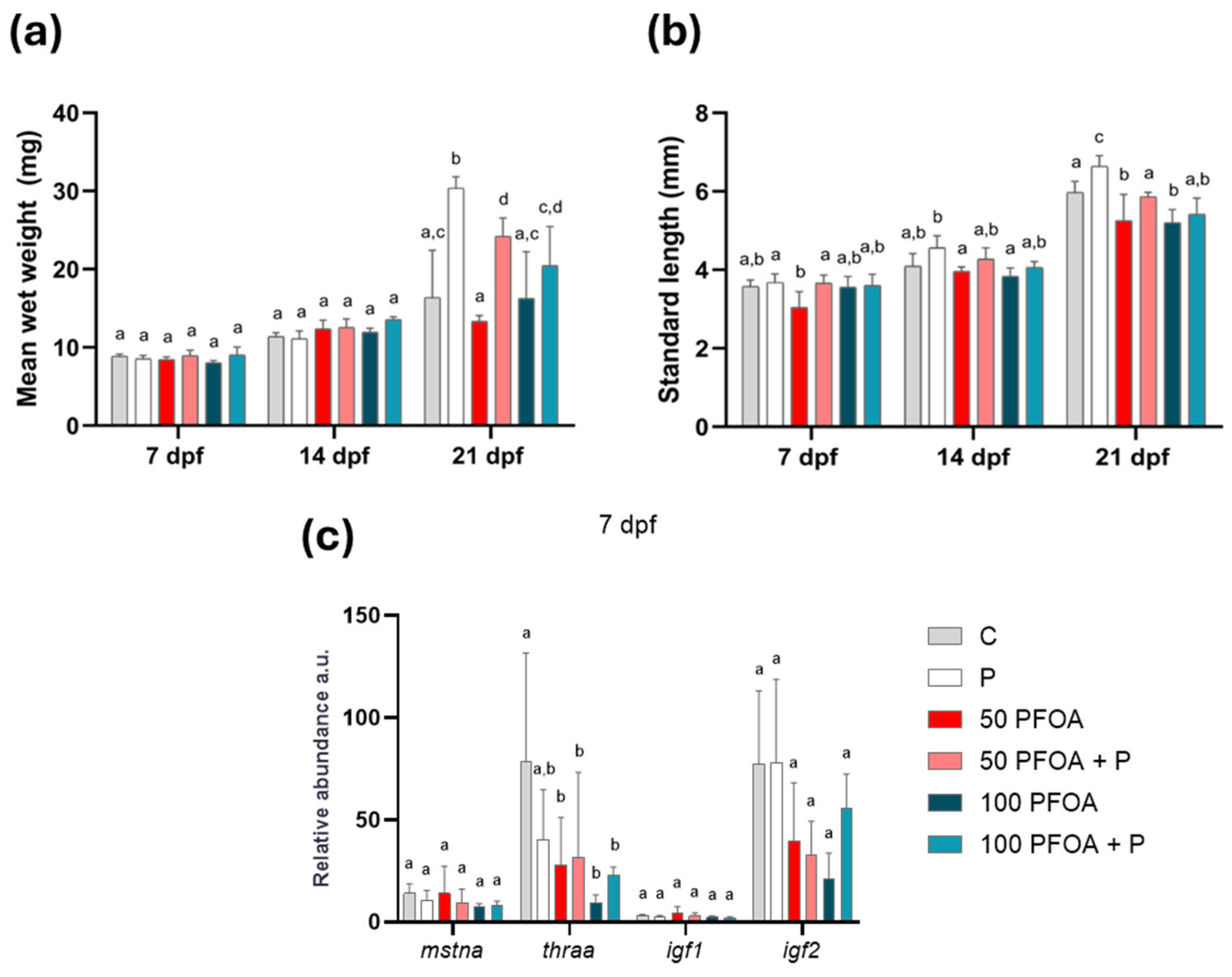
2.1. Impact of PFOA Exposure and Probiotic Treatment on Zebrafish Growth

Regarding larval growth, the most interesting results were observed at 21 dpf: the weight of the larvae was increased following treatment with the probiotic, either alone or with PFOA (Figure 1a); however, the standard length of the larvae only increased when the probiotic was administered alone (Figure 1b). Exposure to both concentrations of PFOA significantly reduced the standard length at 21 dpf (Figure 1b). The observed PFOA-induced response was completely reversed when the larvae were co-treated with B. subtilis var. natto (Figure 1b).
Focusing on genes involved in zebrafish growth and development, myostatin a (mstna), thyroid hormone receptor alpha a (thraa), and the insulin growth factors igf1 and igf2 were analyzed at each time point (Figure 1c and Figure S1). The results demonstrate that exposure to both concentrations of PFOA reduced the thraa transcript at 7 dpf. This alteration was partially counteracted by the probiotic treatment (Figure 1c). The mRNA levels of the other genes investigated were not affected following treatments at 7 (Figure 1c), 14 or 21 dpf (Supplementary Figure S1).

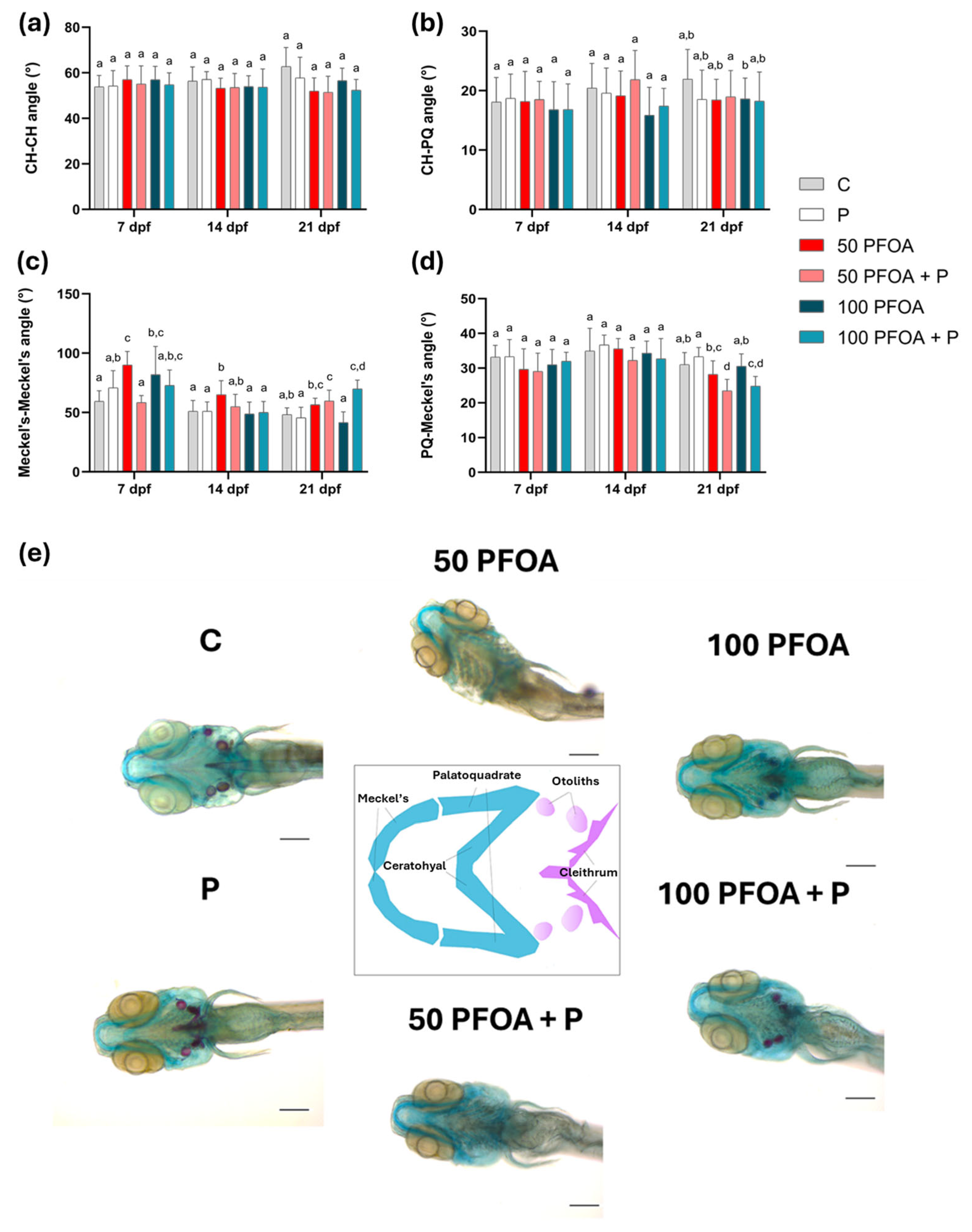
2.2. Effects of PFOA Exposure and Probiotic Treatments on Zebrafish Larval Morphology

The ocular morphometric assessment revealed that, at 14 dpf, the larvae exposed to 100 mg/L PFOA and treated with probiotic (100 PFOA + P) showed a significant increase in eye angle (Figure 2a) and eye distance (Figure 2b) compared to all other groups. At 21 dpf, both PFOA concentrations induced increases in eye diameter that were counteracted by concomitant treatment with the probiotic (Figure 2c). The eye depth did not change among the groups (Figure 2d).
To further investigate the capacity of PFOA to induce craniocephalic malformations, the length of head cartilages (Supplementary Figure S2) and the angles between these cartilages (Figure 3) were analyzed at all time points tested using alcian blue and alizarin red staining. Although the length of head cartilages was not affected by treatments (Supplementary Figure S2), exposure to both PFOA concentrations increased the Meckel’s-Meckel angle at 7 dpf, an effect mitigated by the co-administration of the probiotic. At 14 dpf, the Meckel’s-Meckel’s angle was only increased following exposure to 50 mg/L PFOA (50 PFOA), and this alteration was also counteracted by probiotic co-administration (Figure 3c). In 21-dpf larvae, an increase in the Meckel’s-Meckel’s angle and a decrease in the PQ–Meckel angle were observed following exposure to 50 mg/L PFOA concomitantly with probiotic administration (50 PFOA + P) and 100 PFOA + P groups (Figure 3c,d). Moreover, otolith and cleithrum formation was observed in the control group (C), the Bacillus subtilis var. natto administered group (P), and the 100 mg/L PFOA (100 PFOA) and 100 PFOA + P groups at 7 dpf (Figure 3e), although the ossification of these structures was absent in the 50 PFOA group, either alone or with a probiotic treatment.

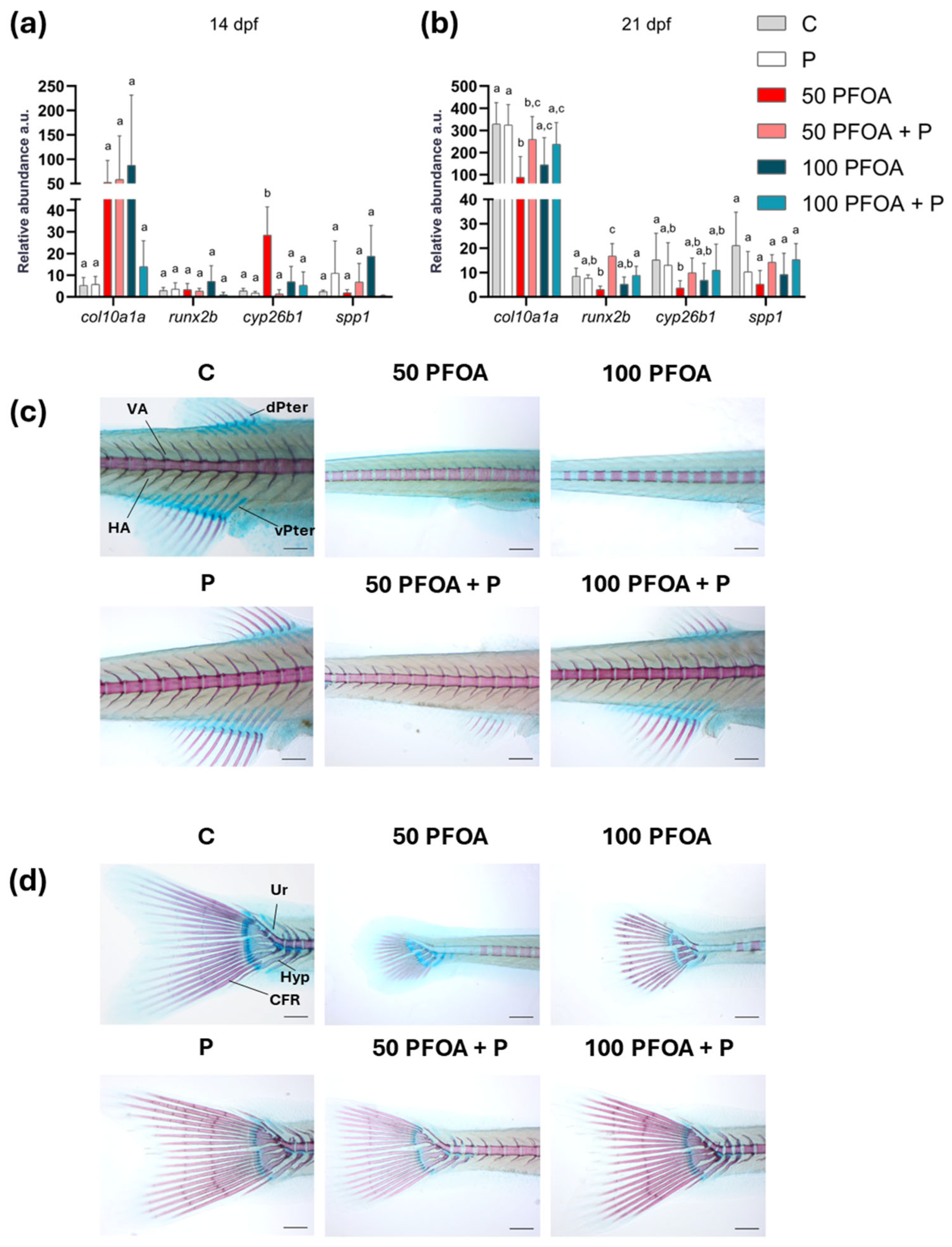
2.3. Effects of PFOA Exposure and Probiotic Treatment on Zebrafish Larval Ossification

To further investigate the adverse effects of PFOA on ossification, the mRNA levels of genes involved in ossification, including Collagen type X alpha 1a (col10a1a), RUNX family transcription factor b (runx2b), Cytochrome P450, family26 subfamily b polypeptide 1 (cyp26b1), and secreted phosphoprotein 1 (spp1), were analyzed at each time point (Figure 4a,b and Figure S3). Exposure to 50 PFOA increased the cyp26b1 mRNA level at 14 dpf compared to all other groups (Figure 4a) and decreased the cyp26b1 transcript level at 21 dpf compared to C (Figure 4b). In addition, 50 PFOA caused a decrease in both col10a1a and runx2b transcript levels compared to C (Figure 4b). After the co-administration of 50 PFOA and P, col10a1a expression remained decreased compared to C. In contrast, the runx2b transcript level increased compared to all the other groups, following an opposite trend to that of 50 PFOA alone (Figure 4b). Furthermore, the alizarin red/alcian blue staining conducted at 21 dpf showed similar ossification of the pterygiophore vertebral arches, hypurals, and urals in both the C and P groups. However, exposure to both concentrations of PFOA reduced ossification of these bones. Notably, the P completely rescued this phenotype when co-administered with 100 PFOA, while only partially rescuing it at the lowest concentrations (Figure 4c,d).

3. Discussion

Using a morphological and molecular approach, we provided clear evidence that the administration of a probiotic can counteract PFOA-induced skeletal abnormalities. The results obtained align well with previous observations regarding the ability of PFOA [3,4,56,57], as well as other PFASs [6,56,57,58,59], to affect growth and development, as well as heart and swim bladder defects [6]. The present study confirmed that exposure to PFOA alters larval growth by decreasing the standard length. Administration of probiotics for 21 days, however, resulted in an overall increase in the larval body growth, in agreement with a vast body of literature highlighting the ability of the probiotic Bacillus subtilis var. natto [45], as well as other probiotics such as L. rhamnosus [42,43,60,61,62], to enhance fish growth. The results were also supported by the increase in the wet weight of the larvae, which was also evident when this probiotic was co-administered with PFOA exposure. It should be noted that the standard-length alteration induced by PFOA was mitigated by the co-administration of a probiotic. To date, no data are reported in the literature concerning the mitigation effects of probiotics against PFOA-induced growth alterations. However, previous studies in zebrafish have demonstrated the capacity of L. rhamnosus to counteract developmental alterations induced by PFBS, an alternative PFAS, at concentrations of 1, 3.3, and 10 mg/L [53,54] and 10 µg/L [55], by restoring the larval length, weight, and growth rate to those of unexposed fish. To investigate the possible molecular mechanisms underlying the changes herein observed on larvae biometry, we conducted a deeper analysis focusing on key genes involved in growth. While alterations in growth were mainly observed at 21 dpf, these changes were not associated with a modulation of the transcript levels of the genes investigated. This is not surprising, as phenotypic changes may not always happen at the time of changes in gene expression. Nevertheless, considering the pivotal role of thyroid hormones in the regulation of growth [63,64,65,66] and the well-documented disruptive effects of PFAS, including PFOA, on the thyroid axis, and in turn on growth and development [4,56,58,59,67], we analyzed the mRNA levels of thraa at all of the selected time points. The thraa transcript level was reduced at 7 dpf in all the groups exposed to PFOA. Thus, we postulate that the alteration in growth observed may in part be caused by dysregulation of the thyroid pathway during early development. In this context, the thyroid system is critical to eye development and function [68,69,70], and alterations in the thyroid system could, to a certain extent, be one reason for the observed alterations in eye diameter. Indeed, eye morphology alterations were observed upon exposure to several EDCs [71]. Thyroid-hormone-disrupting chemicals such as propylthiouracil (PTU) and tetrabromobisphenol A (TBBPA) were reported to alter eye size and pigmentation and to induce changes in the retinal cellular structure in zebrafish embryos [72]. Similarly, exposure to bisphenol S (BPS) [73] altered retinoid metabolism and thyroid hormone homeostasis, and bisphenol A (BPA) [74] disrupted retinal layering in zebrafish larvae. In addition, triclosan and benzophenone-2 exposure led to thyroid follicle hyperplasia, leading to an alteration in the retina’s cellular structure in zebrafish larvae [75]. The increase in eye diameter observed in this study is consistent with similar changes reported in zebrafish exposed to a plasticizer, BPA, during development [76]. The mitigation of eye diameter exerted by the co-administration of Bacillus subtilis in PFOA-exposed larve adds new evidence to the well-established beneficial role of different probiotic strains, as well as Bacillus subtilis var. natto, in promoting developmental processes and in counteracting the detrimental effects of endocrine disruptors on larval development.
A similar alteration in jaw morphology to that observed in the present study following exposure to PFOA has previously been described in zebrafish exposed to BPA, increasing the Meckel–Meckel angle during early development until 120 h post fertilization [76]. The mitigation of PFOA toxicity following the administration of Bacillus subtilis var. natto highlights the ability to promote bone ossification, as previously observed in caudal fin regeneration [26] and in backbone ossification [34]. To deepen our knowledge regarding the possible mechanism of probiotic action, we evaluated the transcript levels of key genes involved in ossification. While the co-administration of probiotics partially mitigated the effects of PFOA on col10a1a, it led to an increased transcript level of runx2b at 21 dpf. Since runx2b directly regulates bone formation in the jaws via a cartilage precursor during perichondral ossification [20,21], this increase may underline the combined effects of PFOA and the probiotic on the Meckel’s-Meckel’s and PQ–Meckel’s angles observed at 21 dpf. This interaction could have contributed to the persistence of alterations induced earlier in development. Notably, in the present study, the lowest concentration of PFOA tested reduced both col10a1a and runx2b transcript levels, correlating with the reduced ossification observed via alcian blue and alizarin red staining at 21 dpf. These data suggest a non-monotonic response of PFOA, which has already been reported for other EDCs, such as BPA and phthalates [77,78]. Along with col10a1a and runx2b, cyp26b1, an additional master gene involved in chondrocyte maturation, osteoblast differentiation, and cartilage-to-bone transition, was deregulated. Given the role of this gene, the craniofacial malformations and ossification delay induced by the lowest PFOA concentration could be attributed to the increased cyp26b1 transcript levels found in this group at 14 dpf. This likely led to higher CYP26b1 and thus to a major RA catabolism, reducing its availability for ossification processes [27]. Furthermore, the decrease in cyp26b1 in the same group at 21 dpf could be responsible for prolonged cartilage maintenance and delayed ossification [23], as evidenced by the alizarin red/alcian blue staining histological analysis. Notably, probiotic administration was able to mitigate this effect, as was particularly evident at 14 dpf and partially at 21 dpf, when col10a1a, cyp26b1, and runx2b transcript levels became closer to those of the control group, further indicating the capacity of probiotic administration to mitigate PFOA-induced toxicity on ossification.
Preventing the toxic effects of PFOA on ossification is essential to reducing the risk of skeletal abnormalities and long-term health issues, particularly in developing individuals. Overall, this study not only advances knowledge about PFOA toxicity on ossification but also provides a promising foundation for developing intervention strategies to reduce pollutant-related developmental defects. This highlights the potential of probiotics as a natural strategy to counteract PFOA-induced toxicity and support proper bone development, offering a promising approach to mitigating the adverse effects of environmental contaminants on the skeletal system.

4. Materials and Methods

4.1. Zebrafish Husbandry and Treatment

Adult zebrafish (AB strain) were kept under controlled conditions (28 ± 1 °C with a 14:10 light:dark period) in a recirculation system and fed twice per day with an adult zebrafish complete diet (Zeigler Bross, Inc., Gardners, PA, USA, Aquamerik Inc., St. Nicolas, QC, Canada) and live brine shrimp nauplii, Artemia cysts. In total, 3000 embryos were obtained from synchronized breeding; they were selected and maintained under standard conditions until hatching (3 dpf) in fish water from a reverse osmosis system. At this point, the embryos were equally divided into 6 experimental groups in triplicate: (1) control (C), untreated; (2) probiotic Bacillus subtilis var. natto administration via water at a final concentration of 107 CFU/mL (P); (3) exposed to 50 mg/L PFOA (50 PFOA); (4) exposed to 50 mg/L PFOA plus the administration of Bacillus subtilis var. natto via water at a final concentration of 107 CFU/mL (50 PFOA + P); (5) exposed to 100 mg/L PFOA (100 PFOA); (6) exposed to 100 mg/L PFOA plus the administration of Bacillus subtilis var. natto via water at a final concentration of 107 CFU/mL (100 PFOA + P). Rearing water was completely changed daily, and PFOA and the probiotic were renewed during every water change, being handled identically across groups to minimize co-morbid conditions. The experiment lasted from hatching till 21 dpf. The PFOA concentrations were selected considering previous studies’ reports of its ability to alter zebrafish development [4] and zebrafish female fertility [79], to induce peroxisomal fatty acid oxidation, and to alter the immune system in Oryzias latipes adult liver [80], and considering the ability of this pollutant to alter the development and reproduction of both the amphipod Hyalella azteca and the fish Pimephales promelas [81]. Regarding the probiotic, the concentration was selected based on previous studies in zebrafish using other probiotic strains such as Lactobacillus acidophilus AC [82] and mice with Lactobacillus rhamnosus GG [83], Lactobacillus rhamnosus MTCC-5897 [84], Weissella cibaria [85], and Bifidobacterium breve [86]. Larvae were fed twice a day with rotifers starting from day 5 days post fertilization (dpf) until the end of the experiment, and the water was changed and renewed in all tanks every day. To follow the effects throughout the development, three sampling timings were established at 7, 14, and 21 dpf (Figure 5).
All procedures involving animals were conducted according to the University of Calgary animal care protocols (AC19-0160 approved on 8 March 2022) for the care and use of experimental animals. All efforts were made to minimize animal suffering. Larvae were euthanized in a solution containing 300 mg/L MS-222 (3-aminobenzoic acid ethyl ester; Sigma Aldrich, St. Louis, MO, USA) buffered to pH 7.4, according to the University of Calgary animal care protocol. For each time point, 30 larvae were fixed with 4% paraformaldehyde (PFA) (Bio-Optica, Milan, Italy) for 2 h, washed three times with 70% ethanol, and then stored at 4 °C until being processed for biometric evaluations and alcian blue and alizarin red staining. Additionally, five pools of 25 larvae each were sampled and stored at −80 °C until being processed for total RNA extraction.

4.2. Biometric Evaluation

Larvae length and wet weight were measured following the protocol of Zarantoniello et al. [87] to assess the presence of biometric changes among the experimental groups. The length of five larvae per group at each time point was measured using a caliber (Measy 2000 Typ 5921, Greifensee, Switzerland; precision: 0.1 mm). For the wet weight measure, three replicates of five larvae per group at each time point were used employing an OHAUS Explorer (OHAUS Europe GmbH, Greifensee, Switzerland) analytical balance (precision: 0.1 mg).

4.3. Alcian Blue and Alizarin Red Staining

To analyze both the cartilage and the bone, alcian blue/alizarin red staining was performed according to the protocol described by Walker and Kimmel [88] and as previously described [89] on 15 larvae per each group and time point.

4.4. Morphological Studies

Larval malformations, as well as ocular and jaw morphometrical measurements, were studied via stereomicroscopy (Leica microsystems, Wetzlar, Germany). The craniocephalic morphological measurements (ceratohyal (CH) length, palatoquadrate (PQ) length, CH–Meckel length, CH–CH angle, CH–PQ angle, PQ–Meckel angle and Meckel-Meckel angle) were conducted on larvae stained with alcian blue/alizarin red using ImageJ-win64 (Image J, version 1.54, NIH, Bethesda, MD, USA, https://imagej.nih.gov/ij/, accessed on 6 November 2023). An optical microscope (Zeiss imager M.2, Castiglione Orona, Italy) coupled with a photo camera (Axiocam 105) was used to obtain the representative images of each experimental group at the different time points.

4.5. Gene Expression Analysis

Total RNA was extracted using RNAzol® RT (Qiagen Science, Milano, Italy) following the manufacturer’s instructions from 7, 14, 21 dpf larvae from each experimental group using 5 pools of 25 larvae per group at each time point. Final RNA concentrations were determined using the Nanophotometer (Implen GmbH, Munich, Germany), and RNA integrity was verified on 1% agarose gel (Supplementary Figure S4). To avoid genomic DNA contamination, an additional step to selectively digest DNA was performed using a DNAse kit (Sigma-Aldrich, St. Louis, MO, USA). Then, cDNA synthesis was performed using an iScript cDNA Synthesis Kit (Bio-Rad Laboratories, Milano, Italy) according to the manufacturer’s protocol. Real-time PCR was performed in a thermal cycler (CFX Connect, Bio-Rad Laboratories, Milano, Italy) in duplicates. Forward and reverse primers were diluted at a final concentration of 10 pmol/mL. Ribosomal protein, large, P0 (rplp0) and ribosomal protein L13a (rpl13a) were used as endogenous reference genes. Primer sequences, accession numbers, and annealing temperatures are reported in Table 1. The data were analyzed using Bio-Rad CFX maestro software 2.2 (5.2.008.0222) RTq-PCR data were analyzed using the Gene Expression Analysis for iCycler iQ® Real-Time PCR Detection System provided by BioRad (Milan, Italy).

4.6. Statistical Analysis

All data were analyzed using a two-way ANOVA with treatment and developmental stage as factors, followed by Tukey’s multiple comparisons test (GraphPad Prism 8.0, San Diego, CA, USA). Post hoc comparisons were performed within each developmental stage to evaluate differences among treatments. Statistical significance was set at p < 0.05.

5. Conclusions

The present study demonstrates the toxicity of PFOA on the development of zebrafish and its harmful impact on bone formation through delayed bone ossification and skeletal malformations. Remarkably, three master genes involved in ossification were affected by the lowest concentration of PFOA, which emerged as the most detrimental, highlighting the non-monotonic response of this toxicant. The probiotic Bacillus subtilis var. natto counteracted PFOA-induced growth impairments and improved ossification, aligning with previous evidence on the benefits of probiotics in fish development. To the best of our knowledge, this is the first evidence of probiotic mitigation of the adverse impact of PFOA on development and ossification. Future research is needed to explore the potential mitigation capacity of different probiotic strains and formulations. Additionally, in vitro investigations using 3D models that mimic the bone microenvironment would provide valuable insight into the deleterious effects of PFAS on osteogenesis and their association with bone-related diseases.

Supplementary Materials

The following supporting information can be downloaded at: https://www.mdpi.com/article/10.3390/ijms26094261/s1.

Author Contributions

Conceptualization: O.C., C.G., M.L. and F.M.; data curation: C.G. and M.L.; formal analysis: C.G., M.L., F.F. and F.S.; funding acquisition: O.C. and H.R.H.; methodology: C.G. and M.L.; software: C.G. and M.L.; supervision: O.C. and H.R.H.; validation: O.C., H.R.H. and F.M.; writing—original draft: C.G. and M.L.; writing—review and editing: F.M., M.L., H.R.H. and O.C. All authors have read and agreed to the published version of the manuscript.

Funding

This research was funded by Fondi di Ateneo (2019–2020), Università Politecnica delle Marche, to O.C., by Fondi di Ateneo (2022–2023), Università Politecnica delle Marche, to F.M., and by the funding given by the Natural Sciences and Engineering Research Council of Canada to H.R.H. (NSERC Discovery Grant no. 2022-03560).

Institutional Review Board Statement

The animal study protocol was approved by Ethics Committee of the University of Calgary (AC19-0160).

Data Availability Statement

Data are contained within the article or Supplementary Materials.

Conflicts of Interest

The authors declare no conflicts of interest.

References

  1. PFOA. Available online: https://chm.pops.int/Implementation/Alternatives/AlternativestoPOPs/ChemicalslistedinAnnexA/PFOA/tabid/8292/Default.aspx?utm (accessed on 7 February 2025).
  2. Sostanze Per-e Polifluoroalchiliche (PFAS)—ECHA. Available online: https://echa.europa.eu/hot-topics/perfluoroalkyl-chemicals-pfas (accessed on 7 February 2025).
  3. Jantzen, C.E.; Toor, F.; Annunziato, K.A.; Cooper, K.R. Effects of chronic perfluorooctanoic acid (PFOA) at low concentration on morphometrics, gene expression, and fecundity in zebrafish (Danio rerio). Reprod. Toxicol. 2017, 69, 34. [Google Scholar] [CrossRef] [PubMed]
  4. Wang, J.; Shi, G.; Yao, J.; Sheng, N.; Cui, R.; Su, Z.; Guo, Y.; Dai, J. Perfluoropolyether carboxylic acids (novel alternatives to PFOA) impair zebrafish posterior swim bladder development via thyroid hormone disruption. Environ. Int. 2020, 134, 105317. [Google Scholar] [CrossRef] [PubMed]
  5. Gui, W.; Guo, H.; Wang, C.; Li, M.; Jin, Y.; Zhang, K.; Dai, J.; Zhao, Y. Comparative developmental toxicities of zebrafish towards structurally diverse per- and polyfluoroalkyl substances. Sci. Total Environ. 2023, 902, 166569. [Google Scholar] [CrossRef] [PubMed]
  6. Rericha, Y.; Simonich, M.T.; Truong, L.; Tanguay, R.L. Review of the zebrafish as a model to investigate per- and polyfluoroalkyl substance toxicity. Toxicol. Sci. 2023, 194, 138. [Google Scholar] [CrossRef]
  7. Wang, X.; Wang, K.; Mao, W.; Fan, Z.; Liu, T.; Hong, R.; Chen, H.; Pan, C. Emerging perfluoroalkyl substances retard skeletal growth by accelerating osteoblasts senescence via ferroptosis. Environ. Res. 2024, 258, 119483. [Google Scholar] [CrossRef]
  8. Duncan Bassett, J.H.; Williams, G.R. Role of Thyroid Hormones in Skeletal Development and Bone Maintenance. Endocr. Rev. 2016, 37, 135. [Google Scholar] [CrossRef]
  9. Zhu, S.; Pang, Y.; Xu, J.; Chen, X.; Zhang, C.; Wu, B.; Gao, J. Endocrine Regulation on Bone by Thyroid. Front. Endocrinol. 2022, 13, 873820. [Google Scholar] [CrossRef]
  10. Du, Y.; Chen, C.; Zhou, G.; Cai, Z.; Man, Q.; Liu, B.; Wang, W.C. Perfluorooctanoic acid disrupts thyroid-specific genes expression and regulation via the TSH-TSHR signaling pathway in thyroid cells. Environ. Res. 2023, 239, 117372. [Google Scholar] [CrossRef]
  11. Zhang, S.; Chen, K.; Li, W.; Chai, Y.; Zhu, J.; Chu, B.; Li, N.; Yan, J.; Zhang, S.; Yang, Y. Varied thyroid disrupting effects of perfluorooctanoic acid (PFOA) and its novel alternatives hexafluoropropylene-oxide-dimer-acid (GenX) and ammonium 4,8-dioxa-3H-perfluorononanoate (ADONA) in vitro. Environ. Int. 2021, 156, 106745. [Google Scholar] [CrossRef]
  12. Dietrich, K.; Fiedler, I.A.K.; Kurzyukova, A.; López-Delgado, A.C.; McGowan, L.M.; Geurtzen, K.; Hammond, C.L.; Busse, B.; Knopf, F. Skeletal Biology and Disease Modeling in Zebrafish. J. Bone Miner. Res. 2021, 36, 436–458. [Google Scholar] [CrossRef]
  13. Le Pabic, P.; Dranow, D.B.; Hoyle, D.J.; Schilling, T.F. Zebrafish endochondral growth zones as they relate to human bone size, shape and disease. Front. Endocrinol. 2022, 13, 1060187. [Google Scholar] [CrossRef] [PubMed]
  14. Bergen, D.J.M.; Kague, E.; Hammond, C.L. Zebrafish as an Emerging Model for Osteoporosis: A Primary Testing Platform for Screening New Osteo-Active Compounds. Front. Endocrinol. 2019, 10, 6. [Google Scholar] [CrossRef]
  15. Masiero, C.; Aresi, C.; Forlino, A.; Tonelli, F. Zebrafish Models for Skeletal and Extraskeletal Osteogenesis Imperfecta Features: Unveiling Pathophysiology and Paving the Way for Drug Discovery. Calcif. Tissue Int. 2024, 115, 931–959. [Google Scholar] [CrossRef] [PubMed]
  16. Veldman, M.B.; Lin, S. Zebrafish as a Developmental Model Organism for Pediatric Research. Pediatr. Res. 2008, 64, 470–476. [Google Scholar] [CrossRef]
  17. Tonelli, F.; Bek, J.W.; Besio, R.; De Clercq, A.; Leoni, L.; Salmon, P.; Coucke, P.J.; Willaert, A.; Forlino, A. Zebrafish: A Resourceful Vertebrate Model to Investigate Skeletal Disorders. Front. Endocrinol. 2020, 11, 489. [Google Scholar] [CrossRef]
  18. Heubel, B.P.; Bredesen, C.A.; Schilling, T.F.; Le Pabic, P. Endochondral growth zone pattern and activity in the zebrafish pharyngeal skeleton. Dev. Dyn. 2021, 250, 74–87. [Google Scholar] [CrossRef]
  19. Raman, R.; Antony, M.; Nivelle, R.; Lavergne, A.; Zappia, J.; Guerrero-Limón, G.; Caetano da Silva, C.; Kumari, P.; Sojan, J.M.; Degueldre, C.; et al. The Osteoblast Transcriptome in Developing Zebrafish Reveals Key Roles for Extracellular Matrix Proteins Col10a1a and Fbln1 in Skeletal Development and Homeostasis. Biomolecules 2024, 14, 139. [Google Scholar] [CrossRef]
  20. Nie, C.H.; Wan, S.M.; Chen, Y.L.; Huysseune, A.; Wu, Y.M.; Zhou, J.J.; Hilsdorf, A.W.S.; Wang, W.M.; Witten, P.E.; Lin, Q.; et al. Single-cell transcriptomes and runx2b−/− mutants reveal the genetic signatures of intermuscular bone formation in zebrafish. Natl. Sci. Rev. 2022, 9, nwac152. [Google Scholar] [CrossRef]
  21. Galea, G.L.; Zein, M.R.; Allen, S.; Francis-West, P. Making and shaping endochondral and intramembranous bones. Dev. Dyn. 2020, 250, 414. [Google Scholar] [CrossRef]
  22. Spoorendonk, K.M.; Peterson-Maduro, J.; Renn, J.; Trowe, T.; Kranenbarg, S.; Winkler, C.; Schulte-Merker, S. Retinoic acid and Cyp26b1 are critical regulators of osteogenesis in the axial skeleton. Development 2008, 135, 3765–3774. [Google Scholar] [CrossRef]
  23. Silveira, K.C.; Fonseca, I.C.; Oborn, C.; Wengryn, P.; Ghafoor, S.; Beke, A.; Dreseris, E.S.; Wong, C.; Iacovone, A.; Soltys, C.L.; et al. CYP26B1-related disorder: Expanding the ends of the spectrum through clinical and molecular evidence. Hum. Genet. 2023, 142, 1571. [Google Scholar] [CrossRef] [PubMed]
  24. Maclean, G.; Dollé, P.; Petkovich, M. Genetic disruption of CYP26B1 severely affects development of neural crest derived head structures, but does not compromise hindbrain patterning. Dev. Dyn. 2009, 238, 732–745. [Google Scholar] [CrossRef]
  25. Laue, K.; Jänicke, M.; Plaster, N.; Sonntag, C.; Hammerschmidt, M. Restriction of retinoic acid activity by Cyp26b1 is required for proper timing and patterning of osteogenesis during zebrafish development. Development 2008, 135, 3775. [Google Scholar] [CrossRef] [PubMed]
  26. Sojan, J.M.; Gioacchini, G.; Giorgini, E.; Orlando, P.; Tiano, L.; Maradonna, F.; Carnevali, O. Zebrafish caudal fin as a model to investigate the role of probiotics in bone regeneration. Sci. Rep. 2022, 12, 8057. [Google Scholar] [CrossRef]
  27. Laue, K.; Pogoda, H.M.; Daniel, P.B.; Van Haeringen, A.; Alanay, Y.; Von Ameln, S.; Rachwalski, M.; Morgan, T.; Gray, M.J.; Breuning, M.H.; et al. Craniosynostosis and Multiple Skeletal Anomalies in Humans and Zebrafish Result from a Defect in the Localized Degradation of Retinoic Acid. Am. J. Hum. Genet. 2011, 89, 595–606. [Google Scholar] [CrossRef]
  28. Li, J.; Hu, C.; Zhao, B.; Li, J.; Chen, L. Proteomic and cardiac dysregulation by representative perfluoroalkyl acids of different chemical speciation during early embryogenesis of zebrafish. Sci. Total Environ. 2024, 926, 172000. [Google Scholar] [CrossRef]
  29. Britton, K.N.; Judson, R.S.; Hill, B.N.; Jarema, K.A.; Olin, J.K.; Knapp, B.R.; Lowery, M.; Feshuk, M.; Brown, J.; Padilla, S. Using Zebrafish to Screen Developmental Toxicity of Per- and Polyfluoroalkyl Substances (PFAS). Toxics 2024, 12, 501. [Google Scholar] [CrossRef]
  30. Xiao, J.; Yang, D.; Hu, B.; Zai, W.; Li, W.; Wang, Y.; Liu, F.; Liao, X.; Li, H.; Tao, Q.; et al. Perfluorodecanoic acid induces the increase of innate cells in zebrafish embryos by upregulating oxidative stress levels. Comp. Biochem. Physiol. Part C Toxicol. Pharmacol. 2025, 287, 110037. [Google Scholar] [CrossRef]
  31. Zhang, J.; Zhang, S.; Lu, C.; Wang, X.; Du, Z.; Wang, J.; Li, B.; Wang, J.; Zhu, L. Comparison of the combined toxicity of PFOA and emerging alternatives: A comprehensive evaluation of oxidative damage, apoptosis and immunotoxicity in embryonic and adult zebrafish. Water Res. 2025, 273, 123028. [Google Scholar] [CrossRef]
  32. Wang, X.; Yang, X.; Lu, C.; Zhang, J.; Li, B.; Du, Z.; Wang, J.; Wang, J.; Juhasz, A.; Yang, Y.; et al. Are HFPO-TA and HFPO-DA safe substitutes for PFOA? A comprehensive toxicity study using zebrafish (Danio rerio) embryos and adults. J. Hazard. Mater. 2025, 484, 136718. [Google Scholar] [CrossRef]
  33. Seely, K.D.; Kotelko, C.A.; Douglas, H.; Bealer, B.; Brooks, A.E. The Human Gut Microbiota: A Key Mediator of Osteoporosis and Osteogenesis. Int. J. Mol. Sci. 2021, 22, 9452. [Google Scholar] [CrossRef] [PubMed]
  34. Schepper, J.D.; Irwin, R.; Kang, J.; Dagenais, K.; Lemon, T.; Shinouskis, A.; Parameswaran, N.; McCabe, L.R. Probiotics in Gut-Bone Signaling. Adv. Exp. Med. Biol. 2017, 1033, 225–247. [Google Scholar] [CrossRef] [PubMed]
  35. Ibáñez, L.; Rouleau, M.; Wakkach, A.; Blin-Wakkach, C. Gut microbiome and bone. Jt. Bone Spine 2019, 86, 43–47. [Google Scholar] [CrossRef] [PubMed]
  36. Goudarzi, F.; Kiani, A.; Nami, Y.; Shahmohammadi, A.; Mohammadalipour, A.; Karami, A.; Haghshenas, B. Potential probiotic Lactobacillus delbrueckii subsp. lactis KUMS-Y33 suppresses adipogenesis and promotes osteogenesis in human adipose-derived mesenchymal stem cell. Sci. Rep. 2024, 14, 9689. [Google Scholar] [CrossRef]
  37. Sojan, J.M.; Licini, C.; Marcheggiani, F.; Carnevali, O.; Tiano, L.; Mattioli-Belmonte, M.; Maradonna, F. Bacillus subtilis Modulated the Expression of Osteogenic Markers in a Human Osteoblast Cell Line. Cells 2023, 12, 364. [Google Scholar] [CrossRef]
  38. Parvaneh, K.; Ebrahimi, M.; Sabran, M.R.; Karimi, G.; Hwei, A.N.M.; Abdul-Majeed, S.; Ahmad, Z.; Ibrahim, Z.; Jamaluddin, R. Probiotics (Bifidobacterium longum) Increase Bone Mass Density and Upregulate Sparc and Bmp-2 Genes in Rats with Bone Loss Resulting from Ovariectomy. BioMed Res. Int. 2015, 2015, 897639. [Google Scholar] [CrossRef]
  39. Guo, M.; Liu, H.; Yu, Y.; Zhu, X.; Xie, H.; Wei, C.; Mei, C.; Shi, Y.; Zhou, N.; Qin, K.; et al. Lactobacillus rhamnosus GG ameliorates osteoporosis in ovariectomized rats by regulating the Th17/Treg balance and gut microbiota structure. Gut Microbes 2023, 15, 2190304. [Google Scholar] [CrossRef]
  40. Avella, M.A.; Olivotto, I.; Silvi, S.; Place, A.R.; Carnevali, O. Effect of dietary probiotics on clownfish: A molecular approach to define how lactic acid bacteria modulate development in a marine fish. Am. J. Physiol.-Regul. Integr. Comp. Physiol. 2010, 298, 359–371. [Google Scholar] [CrossRef]
  41. Palermo, F.A.; Mosconi, G.; Avella, M.A.; Carnevali, O.; Verdenelli, M.C.; Cecchini, C.; Polzonetti-Magni, A.M. Modulation of cortisol levels, endocannabinoid receptor 1A, proopiomelanocortin and thyroid hormone receptor alpha mRNA expressions by probiotics during sole (Solea solea) larval development. Gen. Comp. Endocrinol. 2011, 171, 293–300. [Google Scholar] [CrossRef]
  42. Maradonna, F.; Gioacchini, G.; Falcinelli, S.; Bertotto, D.; Radaelli, G.; Olivotto, I.; Carnevali, O. Probiotic Supplementation Promotes Calcification in Danio rerio Larvae: A Molecular Study. PLoS ONE 2013, 8, e83155. [Google Scholar] [CrossRef]
  43. Avella, M.A.; Place, A.; Du, S.J.; Williams, E.; Silvi, S.; Zohar, Y.; Carnevali, O. Lactobacillus rhamnosus accelerates zebrafish backbone calcification and gonadal differentiation through effects on the GnRH and IGF systems. PLoS ONE 2012, 7, e45572. [Google Scholar] [CrossRef] [PubMed]
  44. Carnevali, O.; Avella, M.A.; Gioacchini, G. Effects of probiotic administration on zebrafish development and reproduction. Gen. Comp. Endocrinol. 2012, 188, 297–302. [Google Scholar] [CrossRef] [PubMed]
  45. Sojan, J.M.; Raman, R.; Muller, M.; Carnevali, O.; Renn, J. Probiotics Enhance Bone Growth and Rescue BMP Inhibition: New Transgenic Zebrafish Lines to Study Bone Health. Int. J. Mol. Sci. 2022, 23, 4748. [Google Scholar] [CrossRef] [PubMed]
  46. Giommi, C.; Maradonna, F.; Ladisa, C.; Habibi, H.R.; Carnevali, O. Probiotics as Potential Tool to Mitigate Nucleotide Metabolism Alterations Induced by DiNP Dietary Exposure in Danio rerio. Int. J. Mol. Sci. 2024, 25, 11151. [Google Scholar] [CrossRef]
  47. Giommi, C.; Lombó, M.; Habibi, H.R.; Rossi, G.; Basili, D.; Mangiaterra, S.; Ladisa, C.; Chemello, G.; Carnevali, O.; Maradonna, F. The probiotic SLAB51 as agent to counteract BPA toxicity on zebrafish gut microbiota -liver-brain axis. Sci. Total Environ. 2024, 912, 169303. [Google Scholar] [CrossRef]
  48. Giommi, C.; Habibi, H.R.; Candelma, M.; Carnevali, O.; Maradonna, F. Probiotic Administration Mitigates Bisphenol A Reproductive Toxicity in Zebrafish. Int. J. Mol. Sci. 2021, 22, 9314. [Google Scholar] [CrossRef]
  49. Zang, L.; Ma, Y.; Huang, W.; Ling, Y.; Sun, L.; Wang, X.; Zeng, A.; Dahlgren, R.A.; Wang, C.; Wang, H. Dietary Lactobacillus plantarum ST-III alleviates the toxic effects of triclosan on zebrafish (Danio rerio) via gut microbiota modulation. Fish Shellfish Immunol. 2019, 84, 1157–1169. [Google Scholar] [CrossRef]
  50. Hu, C.; Liu, M.; Tang, L.; Liu, H.; Sun, B.; Chen, L. Probiotic intervention mitigates the metabolic disturbances of perfluorobutanesulfonate along the gut-liver axis of zebrafish. Chemosphere 2021, 284, 131374. [Google Scholar] [CrossRef]
  51. Chen, L.; Lam, J.C.W.; Tang, L.; Tang, L.; Hu, C.; Liu, M.; Liu, M.; Lam, P.K.S.; Zhou, B. Probiotic Modulation of Lipid Metabolism Disorders Caused by Perfluorobutanesulfonate Pollution in Zebrafish. Environ. Sci. Technol. 2020, 54, 7494–7503. [Google Scholar] [CrossRef]
  52. Liu, M.; Tang, L.; Hu, C.; Huang, Z.; Sun, B.; Lam, J.C.W.; Lam, P.K.S.; Chen, L. Antagonistic interaction between perfluorobutanesulfonate and probiotic on lipid and glucose metabolisms in the liver of zebrafish. Aquat. Toxicol. 2021, 237, 105897. [Google Scholar] [CrossRef]
  53. Sun, B.; Liu, M.; Tang, L.; Hu, C.; Huang, Z.; Chen, L. Probiotics inhibit the stunted growth defect of perfluorobutanesulfonate via stress and thyroid axes in zebrafish larvae. Environ. Pollut. 2021, 290, 118013. [Google Scholar] [CrossRef] [PubMed]
  54. Sun, B.; Liu, M.; Tang, L.; Hu, C.; Huang, Z.; Zhou, X.; Chen, L. Probiotic supplementation mitigates the developmental toxicity of perfluorobutanesulfonate in zebrafish larvae. Sci. Total Environ. 2021, 799, 149458. [Google Scholar] [CrossRef]
  55. Liu, M.; Tang, L.; Hu, C.; Sun, B.; Huang, Z.; Chen, L. Interaction between probiotic additive and perfluorobutanesulfonate pollutant on offspring growth and health after parental exposure using zebrafish. Ecotoxicol. Environ. Saf. 2021, 214, 112107. [Google Scholar] [CrossRef] [PubMed]
  56. Zhao, X.; Meng, X.; Yang, D.; Dong, S.; Xu, J.; Chen, D.; Shi, Y.; Sun, Y.; Ding, G. Thyroid disrupting effects and the developmental toxicity of hexafluoropropylene oxide oligomer acids in zebrafish during early development. Chemosphere 2024, 361, 142462. [Google Scholar] [CrossRef]
  57. Hamed, M.; Vats, A.; Lim, I.E.; Sapkota, B.; Abdelmoneim, A. Effects of developmental exposure to individual and combined PFAS on development and behavioral stress responses in larval zebrafish. Environ. Pollut. 2024, 349, 123912. [Google Scholar] [CrossRef] [PubMed]
  58. Zhang, S.; Guo, X.; Lu, S.; He, J.; Wu, Q.; Liu, X.; Han, Z.; Xie, P. Perfluorohexanoic acid caused disruption of the hypothalamus-pituitary-thyroid axis in zebrafish larvae. Ecotoxicol. Environ. Saf. 2022, 232, 113283. [Google Scholar] [CrossRef]
  59. Godfrey, A.; Hooser, B.; Abdelmoneim, A.; Horzmann, K.A.; Freemanc, J.L.; Sepúlveda, M.S. Thyroid disrupting effects of halogenated and next generation chemicals on the swim bladder development of zebrafish. Aquat. Toxicol. 2017, 193, 228–235. [Google Scholar] [CrossRef]
  60. Gioacchini, G.; Giorgini, E.; Merrifield, D.L.; Hardiman, G.; Borini, A.; Vaccari, L.; Carnevali, O. Probiotics Can Induce Follicle Maturational Competence: The Danio rerio Case. Biol. Reprod. 2012, 86, 1–11. [Google Scholar] [CrossRef]
  61. Lutfi, E.; Basili, D.; Falcinelli, S.; Morillas, L.; Carnevali, O.; Capilla, E.; Navarro, I. The probiotic Lactobacillus rhamnosus mimics the dark-driven regulation of appetite markers and melatonin receptors’ expression in zebrafish (Danio rerio) larvae: Understanding the role of the gut microbiome. Comp. Biochem. Physiol. Part B Biochem. Mol. Biol. 2021, 256, 110634. [Google Scholar] [CrossRef]
  62. Carnevali, O.; Miccoli, A.; Gioacchini, G.; Maradonna, F.; Benato, F.; Skobo, T. Beneficial Bacteria Affect Danio rerio Development by the Modulation of Maternal Factors Involved in Autophagic, Apoptotic and Dorsalizing Processes. Cell. Physiol. Biochem. 2015, 35, 1706–1718. [Google Scholar] [CrossRef]
  63. Power, D.M.; Llewellyn, L.; Faustino, M.; Nowell, M.A.; Björnsson, B.T.; Einarsdottir, I.E.; Canario, A.V.M.; Sweeney, G.E. Thyroid hormones in growth and development of fish. Comp. Biochem. Physiol. C Toxicol. Pharmacol. 2001, 130, 447–459. [Google Scholar] [CrossRef] [PubMed]
  64. Lazcano, I.; Pech-Pool, S.M.; Olvera, A.; García-Martínez, I.; Palacios-Pérez, S.; Orozco, A. The importance of thyroid hormone signaling during early development: Lessons from the zebrafish model. Gen. Comp. Endocrinol. 2023, 334, 114225. [Google Scholar] [CrossRef] [PubMed]
  65. Chang, J.; Wang, M.; Gui, W.; Zhao, Y.; Yu, L.; Zhu, G. Changes in Thyroid Hormone Levels during Zebrafish Development. Zool. Sci. 2012, 29, 181–184. [Google Scholar] [CrossRef] [PubMed]
  66. Lazcano, I.; Rodríguez-Ortiz, R.; Villalobos, P.; Martínez-Torres, A.; Solís-Saínz, J.C.; Orozco, A. Knock-down of specific thyroid hormone receptor isoforms impairs body plan development in zebrafish. Front. Endocrinol. 2019, 10, 441653. [Google Scholar] [CrossRef]
  67. Kim, J.; Lee, G.; Lee, Y.M.; Zoh, K.D.; Choi, K. Thyroid disrupting effects of perfluoroundecanoic acid and perfluorotridecanoic acid in zebrafish (Danio rerio) and rat pituitary (GH3) cell line. Chemosphere 2021, 262, 128012. [Google Scholar] [CrossRef]
  68. Deveau, C.; Jiao, X.; Suzuki, S.C.; Krishnakumar, A.; Yoshimatsu, T.; Hejtmancik, J.F.; Nelson, R.F. Thyroid hormone receptor beta mutations alter photoreceptor development and function in Danio rerio (zebrafish). PLOS Genet. 2020, 16, e1008869. [Google Scholar] [CrossRef]
  69. Farre, A.A.; Thomas, P.; Huang, J.; Poulsen, R.A.; Owusu Poku, E.; Stenkamp, D.L. Plasticity of cone photoreceptors in adult zebrafish revealed by thyroid hormone exposure. Sci. Rep. 2023, 13, 15697. [Google Scholar] [CrossRef]
  70. Cohen, A.; Popowitz, J.; Delbridge-Perry, M.; Rowe, C.J.; Connaughton, V.P. The Role of Estrogen and Thyroid Hormones in Zebrafish Visual System Function. Front. Pharmacol. 2022, 13, 837687. [Google Scholar] [CrossRef]
  71. Pearce, E.N. Endocrine Disruptors and Thyroid Health. Endocr. Pract. 2024, 30, 172–176. [Google Scholar] [CrossRef]
  72. Pannetier, P.; Poulsen, R.; Gölz, L.; Coordes, S.; Stegeman, H.; Koegst, J.; Reger, L.; Braunbeck, T.; Hansen, M.; Baumann, L. Reversibility of Thyroid Hormone System–Disrupting Effects on Eye and Thyroid Follicle Development in Zebrafish (Danio rerio) Embryos. Environ. Toxicol. Chem. 2023, 42, 1276–1292. [Google Scholar] [CrossRef]
  73. Qiu, L.; Wei, S.; Wang, Y.; Zhang, R.; Ru, S.; Zhang, X. Mechanism of thyroid hormone and its structurally similar contaminant bisphenol S exposure on retinoid metabolism in zebrafish larval eyes. Environ. Int. 2023, 180, 108217. [Google Scholar] [CrossRef] [PubMed]
  74. Volz, S.N.; Poulsen, R.; Hansen, M.; Holbech, H. Bisphenol A alters retinal morphology, visually guided behavior, and thyroid hormone levels in zebrafish larvae. Chemosphere 2023, 348, 140776. [Google Scholar] [CrossRef] [PubMed]
  75. Kraft, M.; Gölz, L.; Rinderknecht, M.; Koegst, J.; Braunbeck, T.; Baumann, L. Developmental exposure to triclosan and benzophenone-2 causes morphological alterations in zebrafish (Danio rerio) thyroid follicles and eyes. Environ. Sci. Pollut. Res. 2023, 30, 33711–33724. [Google Scholar] [CrossRef]
  76. Huang, W.; Wang, X.; Zheng, S.; Wu, R.; Liu, C.; Wu, K. Effect of bisphenol A on craniofacial cartilage development in zebrafish (Danio rerio) embryos: A morphological study. Ecotoxicol. Environ. Saf. 2021, 212, 111991. [Google Scholar] [CrossRef]
  77. Vandenberg, L.N. Non-Monotonic Dose Responses in Studies of Endocrine Disrupting Chemicals: Bisphenol A as a Case Study. Dose-Response 2013, 12, 259. [Google Scholar] [CrossRef]
  78. Astuto, M.C.; Benford, D.; Bodin, L.; Cattaneo, I.; Halldorsson, T.; Schlatter, J.; Sharpe, R.M.; Tarazona, J.; Younes, M. Applying the adverse outcome pathway concept for assessing non-monotonic dose responses: Biphasic effect of bis(2-ethylhexyl) phthalate (DEHP) on testosterone levels. Arch. Toxicol. 2022, 97, 313–327. [Google Scholar] [CrossRef]
  79. Zhang, H.; Han, L.; Qiu, L.; Zhao, B.; Gao, Y.; Chu, Z.; Dai, X. Perfluorooctanoic Acid (PFOA) Exposure Compromises Fertility by Affecting Ovarian and Oocyte Development. Int. J. Mol. Sci. 2023, 25, 136. [Google Scholar] [CrossRef]
  80. Yang, J.H. Perfluorooctanoic acid induces peroxisomal fatty acid oxidation and cytokine expression in the liver of male Japanese medaka (Oryzias latipes). Chemosphere 2010, 81, 548–552. [Google Scholar] [CrossRef]
  81. Bartlett, A.J.; De Silva, A.O.; Schissler, D.M.; Hedges, A.M.; Brown, L.R.; Shires, K.; Miller, J.; Sullivan, C.; Spencer, C.; Parrott, J.L. Lethal and sublethal toxicity of perfluorooctanoic acid (PFOA) in chronic tests with Hyalella azteca (amphipod) and early-life stage tests with Pimephales promelas (fathead minnow). Ecotoxicol. Environ. Saf. 2021, 207, 111250. [Google Scholar] [CrossRef]
  82. Qiu, H.; Huang, L.; Wang, H.; Tao, C.; Ran, Z.; Xu, J.; Sun, H.; Wang, P. Effects of Lactobacillus acidophilus AC on the growth, intestinal flora and metabolism of zebrafish (Danio rerio). Fish Shellfish Immunol. 2024, 149, 109570. [Google Scholar] [CrossRef]
  83. Liu, X.; Jin, G.; Tang, Q.; Huang, S.; Zhang, Y.; Sun, Y.; Liu, T.; Guo, Z.; Yang, C.; Wang, B.; et al. Early life Lactobacillus rhamnosus GG colonisation inhibits intestinal tumour formation. Br. J. Cancer 2022, 126, 1421. [Google Scholar] [CrossRef] [PubMed]
  84. Bhat, M.I.; Singh, V.K.; Sharma, D.; Kapila, S.; Kapila, R. Adherence capability and safety assessment of an indigenous probiotic strain Lactobacillus rhamnosus MTCC-5897. Microb. Pathog. 2019, 130, 120–130. [Google Scholar] [CrossRef] [PubMed]
  85. Kim, J.W.; Jung, B.H.; Lee, J.H.; Yoo, K.Y.; Lee, H.; Kang, M.S.; Lee, J.K. Effect of Weissella cibaria on the reduction of periodontal tissue destruction in mice. J. Periodontol. 2020, 91, 1367–1374. [Google Scholar] [CrossRef]
  86. Ren, J.; Zhao, Y.; Huang, S.; Lv, D.; Yang, F.; Lou, L.; Zheng, Y.; Zhang, J.; Liu, S.; Zhang, N.; et al. Immunomodulatory effect of Bifidobacterium breve on experimental allergic rhinitis in BALB/c mice. Exp. Ther. Med. 2018, 16, 3996. [Google Scholar] [CrossRef]
  87. Zarantoniello, M.; Bruni, L.; Randazzo, B.; Vargas, A.; Gioacchini, G.; Truzzi, C.; Annibaldi, A.; Riolo, P.; Parisi, G.; Cardinaletti, G.; et al. Partial Dietary Inclusion of Hermetia illucens (Black Soldier Fly) Full-Fat Prepupae in Zebrafish Feed: Biometric, Histological, Biochemical, and Molecular Implications. Zebrafish 2018, 15, 519–532. [Google Scholar] [CrossRef]
  88. Walker, M.B.; Kimmel, C.B. A two-color acid-free cartilage and bone stain for zebrafish larvae. Biotech. Histochem. 2007, 82, 23–28. [Google Scholar] [CrossRef]
  89. Damiani, E.; Sella, F.; Astolfi, P.; Galeazzi, R.; Carnevali, O.; Maradonna, F. First In Vivo Insights on the Effects of Tempol-Methoxycinnamate, a New UV Filter, as Alternative to Octyl Methoxycinnamate, on Zebrafish Early Development. Int. J. Mol. Sci. 2023, 24, 6767. [Google Scholar] [CrossRef]
Figure 1. Zebrafish growth assessment. (a) Mean wet body weight and (b) standard length in the different experimental groups at 7, 14 and 21 dpf. Data are reported as means ± SD. Different letters indicate statistically significant changes (p < 0.05) among the experimental groups at each time point (n = 3 for mean wet weight and 5 for standard length). (c) Histogram showing the mstna, thraa, igf1, and igf2 transcript levels in the different experimental groups at 7 dpf reported as relative abundance in arbitrary units (a.u). Data are reported as means ± SD. Different letters indicate statistically significant changes (p < 0.05) among the experimental groups (n = 5).
Ijms 26 04261 g001
Figure 2. Zebrafish ocular morphometric analysis. (a) Eye angle (degrees), (b) eye distance (µm), (c) eye diameter (µm), and (d) eye depth (µm) in the different experimental groups at 7, 14 and 21 dpf. Data are reported as means ± SD. Different letters indicate statistically significant changes (p < 0.05) among the experimental groups (n = 15) at each time point.
Ijms 26 04261 g002
Figure 3. Zebrafish craniocephalic morphometric analysis. (a) CH–CH angles (degrees), (b) CH–PQ angles (degrees), (c) PQ–Meckel angle (degrees) and (d) Meckel’s-Meckel’s angle (degrees) in the different experimental groups at 7, 14, and 21 dpf. Data are reported as means ± SD. Different letters indicate statistically significant changes (p < 0.05) among the experimental groups (n = 15) at each time point. (e) Representative microphotographs of Alcian-blue- and Alizarin-red-stained head at 7 dpf. Scale bar = 200 µm.
Ijms 26 04261 g003
Figure 4. Zebrafish ossification assessment. Histograms summarizing the col10a1a, runx2b, cyp26b1, and spp1 transcript levels in the different experimental groups at (a) 14 and (b) 21 dpf as relative abundance in arbitrary units (a.u). Data are reported as means ± SD. Different letters indicate statistically significant changes (p < 0.05) among the experimental groups (n = 5). Representative images or (c) pterygiophore and arch ossification and (d) hypural and ural ossification in the different experimental groups at 21 dpf by Alcian blue (cartilages stained in blue) and Alizarin red (bones stained in red) staining. Scale bar = 200 µm. Ur = ural; Hyp = hypural; CFR = caudal fin ray.
Ijms 26 04261 g004
Figure 5. Experimental design. At hatching, larvae were divided into 6 experimental groups: C, P, 50 PFOA, 50 PFOA + P, 100 PFOA, 100 PFOA + P. To trace the detrimental and/or beneficial effects during development, three sampling timings were established at 7, 14, and 21 dpf.
Ijms 26 04261 g005
Table 1. List of primers designed for the gene expression analysis via RT-qPCR.
Gene
Name
Gene
Symbol
Primer Sequence (5′-3′)Accession
Number
Tm (C°)
Ribosomal protein large P0rplp0F: CTGAACATCTCGCCCTTCTC
R: TAGCCGATCTGCAGACACAC
NM_131580.260
Ribosomal protein L13arpl13aF: TCTGGAGGACTGTAAGAGGTATGC
R: AGACGCACAATCTTGAGAGCAG
NM_212784.159
Insulin-like growth factor 1igf1F: GGCAAATCTCCACGATCTCTAC
R: GGCAAATCTCCACGATCTCTA
NM_131825.253
Insulin-like growth factor 2igf2F: TCCTTTGTTTGTTGCCATTTG
R: GAGTCCCATCCATTCTGTTG
NM_131433.159
Myostatin amstnaF: GGACTGGACTGCGATGAG
R: GATGGGTGTGGGGATACTTC
AF019626.158
Thyroid hormone receptor alpha athraaF: GGAAACAGAAGCGCAAGTTC
R: TCTTCACAAGGCAGCTCTGA
NM_131396.152
BCL2 associated X apoptosis regulator abaxaF: CAACAAGATGGCATCACACC
R: TGAACCCGCTCGTATATGAAA
NM_131562.260
Bcl-2 apoptosis regulator abcl2aF: CCTTCAATAAAGCAGTGGAGGAA
R: CGGGCTATCAGGCATTCAGA
NM_001030253.260
caspase 3, apoptosis-related cysteine peptidase acasp3aF: GTGCCAGTCAACAAACAAAG
R: CATCTCCAACCGCTTAACG
NM_131877.360
Collagen type X alpha 1acol10a1aF: CCCATCCACATCACATCAAA
R: GCGTGCATTTCTCAGAACAA
NM_001083827.160
Secreted phosphoprotein 1spp1F: GAGCCTACACAGACCACGCCAACAG
R: GGTAGCCCAAACTGTCTCCCCG
NM_001002308.160
RUNX family transcription factor brunx2bF: GTGGCCACTTACCACAGAGC
R: TCGGAGAGTCATCCAGCTT
NM_212862.260
Cytochrome P450, family 26 subfamily b polypeptide 1cyp26b1F: GCTGTCAACCAGAACATTCCC
R: GGTTCTGATTGGAGTCGAGGC
NM_212666.160
Disclaimer/Publisher’s Note: The statements, opinions and data contained in all publications are solely those of the individual author(s) and contributor(s) and not of MDPI and/or the editor(s). MDPI and/or the editor(s) disclaim responsibility for any injury to people or property resulting from any ideas, methods, instructions or products referred to in the content.

