Polymorphic Variant Associated with Sex Hormone-Binding Globulin Level Is a Risk Factor for Preeclampsia
 , , ,
, , ,  ,
,  and
and
Abstract
1. Introduction
2. Results
2.1. Analysis of Associations Between SHBG-Related Genetic Variants and PE
2.2. Replication of Associations of rs10454142 PPP1R21/KLRAQ1 and Its Proxy SNPs with PE Using GWAS Data (UK Biobank and FinnGen Database)
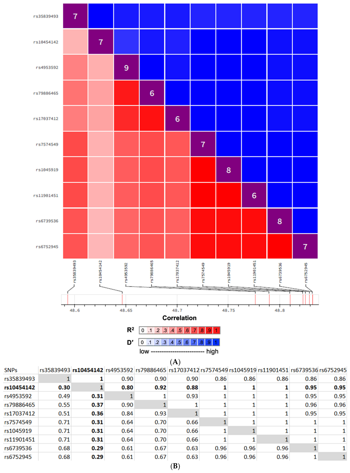
2.3. Predicted Functionality of the PE-Associated Locus rs10454142 PPP1R21/KLRAQ1 and Its Proxy SNPs
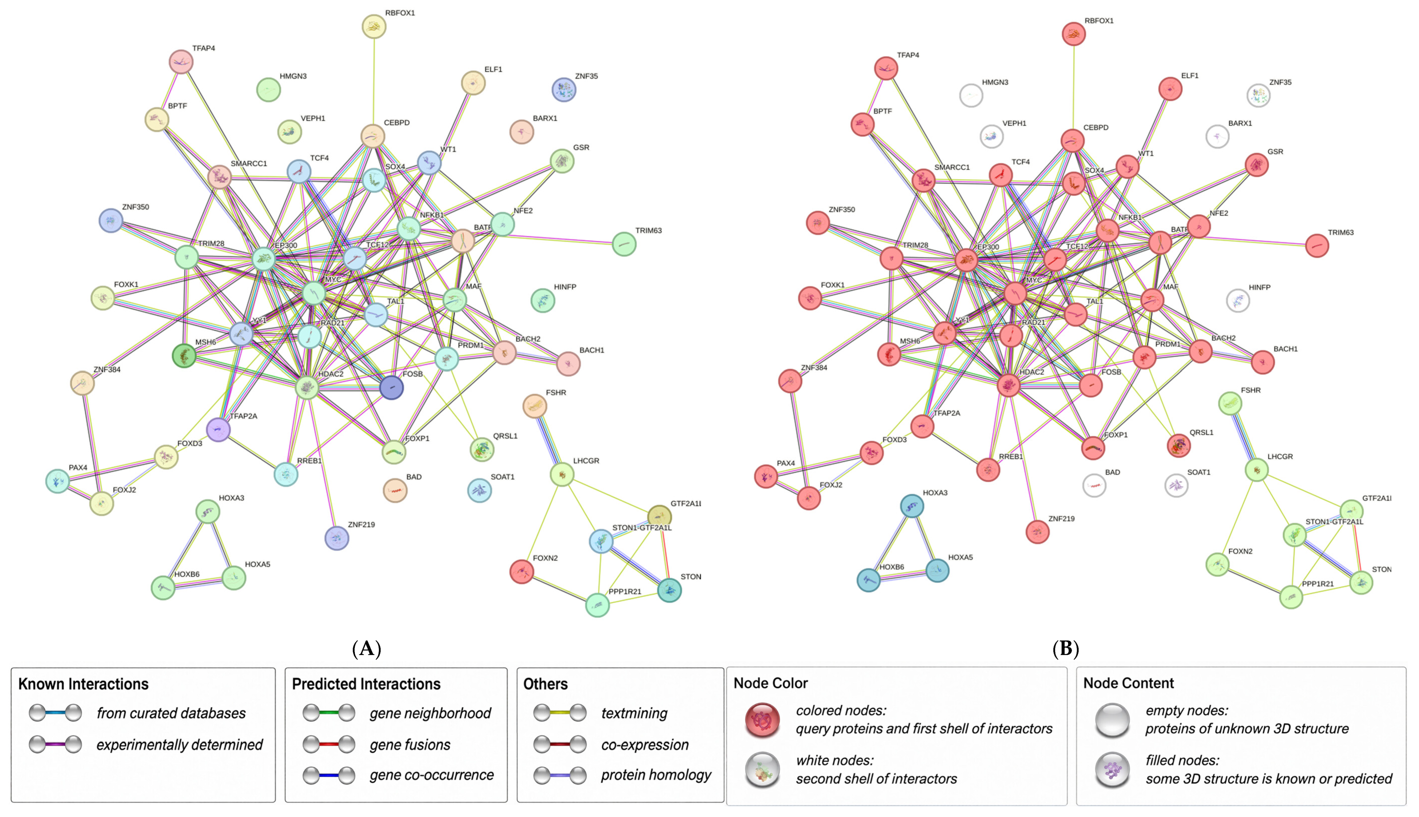
2.4. Identification of Possible PE-Related Biological Pathways
3. Discussion
4. Materials and Methods
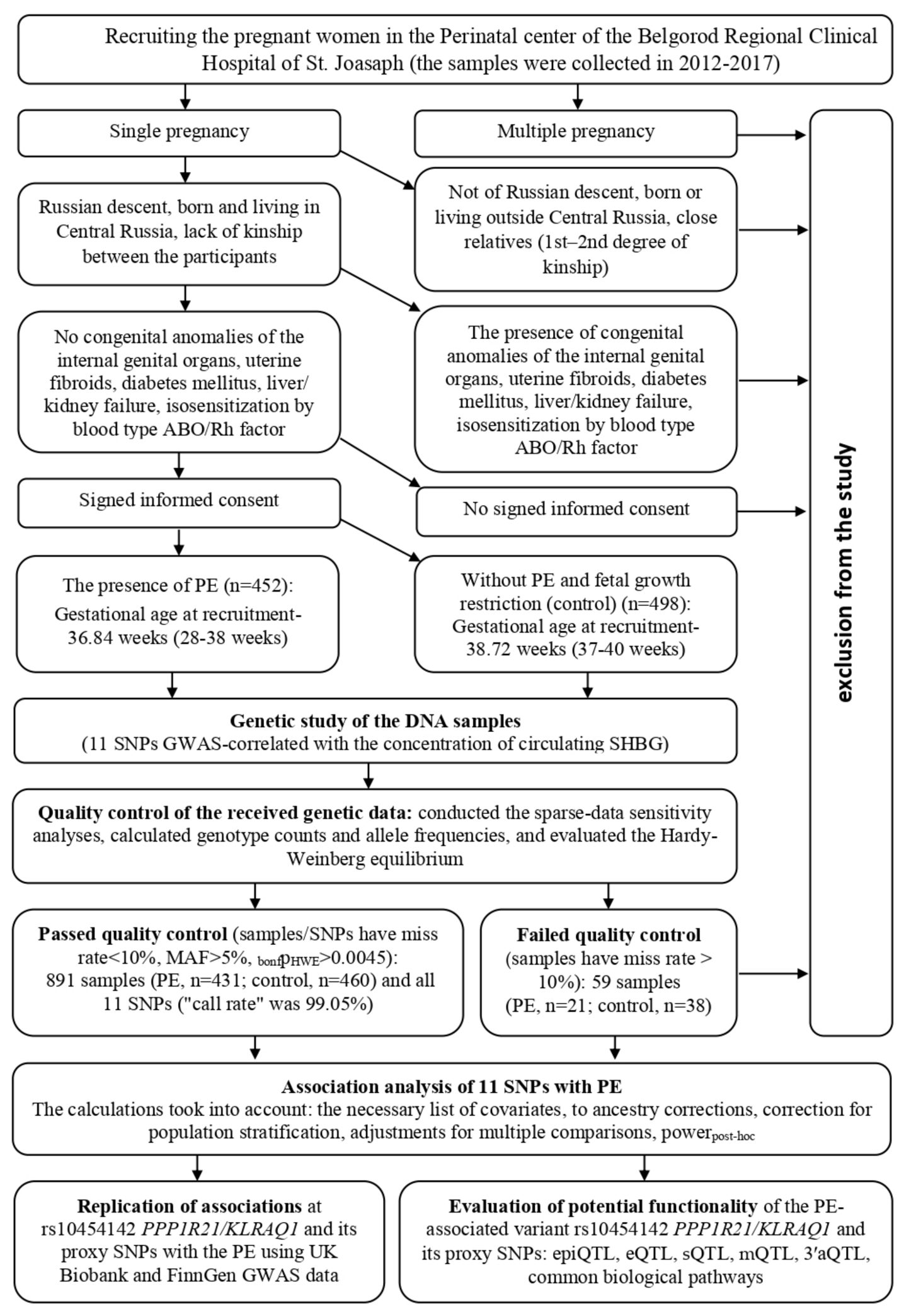
4.1. Study Subjects
4.2. DNA Study: Extraction and SNP Selection/Detection
4.3. Statistical Analysis
- (a)
- The necessary list of covariates (age and BMI/obesity before pregnancy, family history of PE, number of pregnancies/abortions/stillbirths (materials from Table 1));
- (b)
- Ancestry corrections in the studied sample; a genomic relationship matrix (GRM) based on the identity-by-similarity (IBS) was built and then taken into account when calculating the principal components;
- (c)
- The first principal component (the distribution graph of the variances of the principal components [scree plot] reaches a plateau after the first principal component) was used in calculating associations as a covariate (to exclude the factor of hidden stratification of the analyzed cohort, including those related to kinship);
- (d)
- Adjustments for multiple comparisons; a permutation test with the calculation of the pperm index was used to correct the number of studied SNPs (the permutation test effectively solves the problem of multiple comparisons even when analyzing GWAS data [92,93]); to correct the number of considered genetic models (n = 3), the Bonferroni amendment was additionally introduced, which was equal to 3 [79]; the association parameter corresponding to pperm < 0.017 (0.05/3) was recognized as statistically significant;
- (e)
- Mandatory assessment of the power (powerpost hoc) of the obtained associative relationship indicators (powerpost hoc calculations were performed in the Quanto_v.1.2.4 program [94]).
4.4. Replication of Association of the rs10454142 PPP1R21/KLRAQ1 and Its Proxy SNPs with the PE Using GWAS Data
4.5. Predictive Functions of PE-Associated Loci
5. Conclusions
Supplementary Materials
Author Contributions
Funding
Institutional Review Board Statement
Informed Consent Statement
Data Availability Statement
Conflicts of Interest
Abbreviations
| PE | Preeclampsia |
| SNP | Single nucleotide polymorphism |
| GWAS | Genome-wide association studies |
| SHBG | Sex hormone-binding globulin |
| LD | Linkage disequilibrium |
References
- Chiang, Y.T.; Seow, K.M.; Chen, K.H. The Pathophysiological, Genetic, and Hormonal Changes in Preeclampsia: A Systematic Review of the Molecular Mechanisms. Int. J. Mol. Sci. 2024, 25, 4532. [Google Scholar] [CrossRef] [Scilit]
- Ali, M.; Ahmed, M.; Memon, M.; Chandio, F.; Shaikh, Q.; Parveen, A.; Phull, A.R. Preeclampsia: A comprehensive review. Clin. Chim. Acta 2024, 563, 119922. [Google Scholar] [CrossRef] [Scilit]
- Dimitriadis, E.; Rolnik, D.L.; Zhou, W.; Estrada-Gutierrez, G.; Koga, K.; Francisco, R.P.V.; Whitehead, C.; Hyett, J.; da Silva Costa, F.; Nicolaides, K.; et al. Pre-eclampsia. Nat. Rev. Dis. Primers 2023, 9, 8, Correction in Nat. Rev. Dis. Primers 2023, 9, 35. [Google Scholar] [CrossRef] [Scilit]
- Torres-Torres, J.; Espino-Y-Sosa, S.; Martinez-Portilla, R.; Solis-Paredes, J.M.; Guzman-Huerta, M.; Castro-Santamaria, R.; Cruz-Martinez, R.; Estrada-Gutierrez, G.; Poon, L.C. A Narrative Review on the Pathophysiology of Preeclampsia. Int. J. Mol. Sci. 2024, 25, 7569. [Google Scholar] [CrossRef] [Scilit]
- Poon, L.C.; Shennan, A.; Hyett, J.A.; Kapur, A.; Hadar, E.; Divakar, H.; McAuliffe, F.; da Silva Costa, F.; von Dadelszen, P.; McIntyre, H.D.; et al. The International Federation of Gynecology and Obstetrics (FIGO) initiative on pre-eclampsia: A pragmatic guide for first-trimester screening and prevention. Int. J. Gynaecol. Obstet. 2019, 145, 1–33, Correction in Int. J. Gynaecol. Obstet. 2019, 146, 390–391. [Google Scholar] [CrossRef] [Scilit]
- Zhao, L.; Bracken, M.B.; DeWan, A.T. Genome-wide association study of pre-eclampsia detects novel maternal single nucleotide polymorphisms and copy-number variants in subsets of the Hyperglycemia and Adverse Pregnancy Outcome (HAPO) study cohort. Ann. Hum. Genet. 2013, 77, 277–287. [Google Scholar] [CrossRef] [Scilit]
- Serebrova, V.N.; Trifonova, E.A.; Stepanov, V.A. Evolutionary-genetic analysis of the role of regulatory regions in CORO2A gene in the development of hereditary predisposition to preeclampsia in Russian and Yakut ethnic groups. Res. Results Biomed. 2018, 4, 38–48. [Google Scholar] [CrossRef] [Scilit]
- Golovchenko, O.V. Molecular genetic determinants of pre-eclampsia. Res. Results Biomed. 2019, 5, 139–149. [Google Scholar] [CrossRef] [Scilit]
- Sakaue, S.; Kanai, M.; Tanigawa, Y.; Karjalainen, J.; Kurki, M.; Koshiba, S.; Narita, A.; Konuma, T.; Yamamoto, K.; Akiyama, M.; et al. A cross-population atlas of genetic associations for 220 human phenotypes. Nat. Genet. 2021, 53, 1415–1424. [Google Scholar] [CrossRef] [Scilit]
- Changalidis, A.I.; Maksiutenko, E.M.; Barbitoff, Y.A.; Tkachenko, A.A.; Vashukova, E.S.; Pachuliia, O.V.; Nasykhova, Y.A.; Glotov, A.S. Aggregation of Genome-Wide Association Data from FinnGen and UK Biobank Replicates Multiple Risk Loci for Pregnancy Complications. Genes 2022, 13, 2255. [Google Scholar] [CrossRef] [Scilit]
- Nieves-Colón, M.A.; Badillo Rivera, K.M.; Sandoval, K.; Villanueva Dávalos, V.; Enriquez Lencinas, L.E.; Mendoza-Revilla, J.; Adhikari, K.; González-Buenfil, R.; Chen, J.W.; Zhang, E.T.; et al. Clotting factor genes are associated with preeclampsia in high-altitude pregnant women in the Peruvian Andes. Am. J. Hum. Genet. 2022, 109, 1117–1139. [Google Scholar] [CrossRef] [Scilit]
- Cao, Z.; Ma, L.; Cai, W.; Niu, X.; Yang, N.; Ni, J.; Wang, X.; Wei, M.; Chen, S.; Li, Y. Genome-wide association study reveals HSF2, GJA1 and TRIM36 as susceptibility genes for preeclampsia: A community-based population study in Tianjin, China. Hypertens. Pregnancy 2023, 42, 2256863. [Google Scholar] [CrossRef] [Scilit]
- Yan, Q.; Guerrero, R.F.; Khan, R.R.; Pagel, K.A.; Chu, H.; Ramola, R.; Haas, D.M.; Chung, J.; Simhan, H.; Silver, R.M.; et al. Searching and visualizing genetic associations of pregnancy traits by using GnuMoM2b. Genetics 2023, 225, iyad151. [Google Scholar] [CrossRef] [Scilit]
- Golovchenko, O.V. Features of interlocus interactions in the formation of isolated and combined pregnancy complications. Res. Results Biomed. 2024, 10, 532–552. [Google Scholar] [CrossRef] [Scilit]
- Khan, R.R.; Guerrero, R.F.; Wapner, R.J.; Hahn, M.W.; Raja, A.; Salleb-Aouissi, A.; Grobman, W.A.; Simhan, H.; Silver, R.M.; Chung, J.H.; et al. Genetic polymorphisms associated with adverse pregnancy outcomes in nulliparas. Sci. Rep. 2024, 14, 10514. [Google Scholar] [CrossRef] [Scilit]
- Yang, M.L.; Xu, C.; Gupte, T.; Hoffmann, T.J.; Iribarren, C.; Zhou, X.; Ganesh, S.K. Sex-specific genetic architecture of blood pressure. Nat. Med. 2024, 30, 818–828. [Google Scholar] [CrossRef] [Scilit]
- Bordaeva, O.Y.; Alayasa, N.N.I.; Zezerova, Y.S.; Abramova, M.A.; Churnosova, M.I. Clinic-laboratory blood parameters and the contribution of renin-angiotensin system gene polymorphisms in pregnant women with preeclampsia. Res. Results Biomed. 2025, 11, 200–214. [Google Scholar] [CrossRef] [Scilit]
- Liu, T.Y.; Lu, H.F.; Chen, Y.C.; Liao, C.C.; Lin, Y.J.; Yang, J.S.; Liao, W.L.; Lin, W.D.; Chen, S.Y.; Huang, Y.C.; et al. Diversity and longitudinal records: Genetic architecture of disease associations and polygenic risk in the Taiwanese Han population. Sci. Adv. 2025, 11, eadt0539. [Google Scholar] [CrossRef] [Scilit]
- Pujol Gualdo, N.; Džigurski, J.; Rukins, V.; Pajuste, F.-D.; Wolford, B.N.; Võsa, M.; Golob, M.; Haug, L.; Alver, M.; Läll, K.; et al. Atlas of genetic and phenotypic associations across 42 female reproductive health diagnoses. Nat. Med. 2025, 31, 1626–1634. [Google Scholar] [CrossRef] [Scilit]
- Khanam, F.; Sattar, A.; Arif, M.H.; Islam, M.S. Impact of the angiotensin-converting enzyme 2 gene polymorphism on essential hypertension in the southern region of Bangladesh. Res. Results Biomed. 2026, 12, 355–376. [Google Scholar] [CrossRef]
- Kim, H.I.; DeBoever, C.; Walter, K.; Kalantzis, G.; Li, C.; Mozaffari, S.V.; Kundu, K.; Jacobs, B.M.; Mohammadi-Shemirani, P.; Musolf, A.M.; et al. Exome sequencing and analysis of 44,028 British South Asians enriched for high autozygosity. Nat. Genet. 2026, 58, 821–830. [Google Scholar] [CrossRef] [Scilit]
- Nilsson, E.; Salonen Ros, H.; Cnattingius, S.; Lichtenstein, P. The importance of genetic and environmental effects for pre-eclampsia and gestational hypertension: A family study. BJOG 2004, 111, 200–206. [Google Scholar] [CrossRef] [Scilit]
- Salonen Ros, H.; Lichtenstein, P.; Lipworth, L.; Cnattingius, S. Genetic effects on the liability of developing pre-eclampsia and gestational hypertension. Am. J. Med. Genet. 2000, 91, 256–260. [Google Scholar] [CrossRef]
- McGrath, I.M.; Mortlock, S.; Montgomery, G.W. Genetic Regulation of Physiological Reproductive Lifespan and Female Fertility. Int. J. Mol. Sci. 2021, 22, 2556. [Google Scholar] [CrossRef] [Scilit]
- Cnattingius, S.; Reilly, M.; Pawitan, Y.; Lichtenstein, P. Maternal and fetal genetic factors account for most of familial aggregation of preeclampsia: A population-based Swedish cohort study. Am. J. Med. Genet. A 2004, 130A, 365–371. [Google Scholar] [CrossRef] [Scilit]
- Steinthorsdottir, V.; McGinnis, R.; Williams, N.O.; Stefansdottir, L.; Thorleifsson, G.; Shooter, S.; Evison, B.; Bumpstead, S.; Plagnol, V.; Halldorsson, B.V.; et al. Genetic predisposition to hypertension is associated with preeclampsia in European and Central Asian women. Nat. Commun. 2020, 11, 5976. [Google Scholar] [CrossRef] [Scilit]
- Johnson, M.P.; Brennecke, S.P.; East, C.E.; Dyer, T.D.; Bufford, J.T.; Goring, H.H.; Kent, J.W., Jr.; Charlesworth, J.C.; Preston, B.R.; Drigalenko, E.I.; et al. Genome-wide association scan identifies a risk locus for preeclampsia on 2q14, near the inhibin, beta B gene. PLoS ONE 2012, 7, e33666. [Google Scholar] [CrossRef] [Scilit]
- Gray, K.J.; Kovacheva, V.P.; Mirzakhani, H.; Bjonnes, A.C.; Khan, R.R.; Wapner, R.J.; Marazita, M.L.; Chung, J.H.; Silver, R.M.; Simhan, H.N.; et al. Risk of pre-eclampsia in patients with a maternal genetic predisposition to common medical conditions: A case-control study. BJOG 2021, 128, 55–65. [Google Scholar] [CrossRef] [Scilit]
- Honigberg, M.C.; Truong, B.; Khan, R.R.; Xiao, B.; Bhatta, L.; Vy, H.M.T.; Guerrero, R.F.; Schuermans, A.; Selvaraj, M.S.; Patel, A.P.; et al. Polygenic prediction of preeclampsia and gestational hypertension. Nat. Med. 2023, 29, 1540–1549. [Google Scholar] [CrossRef] [Scilit]
- Tyrmi, J.S.; Kaartokallio, T.; Lokki, A.I.; Aalto, K.; Almqvist, C.; Arshad, S.H.; Barton, S.J.; Beilin, L.J.; Bennett, A.J.; Bønnelykke, K.; et al. Genetic Risk Factors Associated With Preeclampsia and Hypertensive Disorders of Pregnancy. JAMA Cardiol. 2023, 8, 674–683. [Google Scholar] [CrossRef] [Scilit]
- Prabaharan, E.; Armant, D.R.; Drewlo, S. Human placentation: Foundations and implications for reproductive endocrinology and infertility. Syst. Biol. Reprod. Med. 2025, 71, 277–306. [Google Scholar] [CrossRef] [Scilit]
- Kumar, S.; Gordon, G.H.; Abbott, D.H.; Mishra, J.S. Androgens in maternal vascular and placental function: Implications for preeclampsia pathogenesis. Reproduction 2018, 156, R155–R167. [Google Scholar] [CrossRef] [Scilit]
- Shu, C.; Han, S.; Xu, P.; Wang, Y.; Cheng, T.; Hu, C. Estrogen and Preeclampsia: Potential of Estrogens as Therapeutic Agents in Preeclampsia. Drug Des. Dev. Ther. 2021, 15, 2543–2550. [Google Scholar] [CrossRef] [Scilit]
- Fernandes, L.M.; Lorigo, M.; Cairrao, E. Relationship Between Androgens and Vascular and Placental Function During Pre-Eclampsia. Curr. Issues Mol. Biol. 2024, 46, 1668–1693. [Google Scholar] [CrossRef] [Scilit]
- Goldštajn, M.Š.; Toljan, K.; Grgić, F.; Jurković, I.; Baldani, D.P. Sex Hormone Binding Globulin (SHBG) as a Marker of Clinical Disorders. Coll. Antropol. 2016, 40, 199–209. [Google Scholar]
- Hammond, G.L. Plasma steroid-binding proteins: Primary gatekeepers of steroid hormone action. J. Endocrinol. 2016, 230, R13–R25. [Google Scholar] [CrossRef] [Scilit]
- Dunn, J.F.; Nisula, B.C.; Rodbard, D. Transport of steroid hormones: Binding of 21 endogenous steroids to both testosterone-binding globulin and corticosteroid-binding globulin in human plasma. J. Clin. Endocrinol. Metab. 1981, 53, 58–68. [Google Scholar] [CrossRef] [Scilit]
- Balogh, A.; Karpati, E.; Schneider, A.E.; Hetey, S.; Szilagyi, A.; Juhasz, K.; Szabo, G., Jr.; Rigó, J., Jr.; Bánrévi, Z.; Tulassay, Z.; et al. Sex hormone-binding globulin provides a novel entry pathway for estradiol and influences subsequent signaling in lymphocytes via membrane receptor. Sci. Rep. 2019, 9, 4. [Google Scholar] [CrossRef] [Scilit]
- Ohlsson, C.; Wallaschofski, H.; Lunetta, K.L.; Stolk, L.; Perry, J.R.B.; Koster, A.; Kraft, P.; Eriksson, J.; Eriksson, A.; Bhasin, S.; et al. Genetic determinants of serum testosterone concentrations in men. PLoS Genet. 2011, 7, e1002313. [Google Scholar] [CrossRef] [Scilit]
- Coviello, A.D.; Haring, R.; Wellons, M.; Vaidya, D.; Lehtimäki, T.; Keildson, S.; Lunetta, K.L.; He, C.; Fornage, M.; Lagou, V.; et al. A genome-wide association meta-analysis of circulating sex hormone-binding globulin reveals multiple Loci implicated in sex steroid hormone regulation. PLoS Genet. 2012, 8, e1002805. [Google Scholar] [CrossRef] [Scilit]
- Prescott, J.; Thompson, D.J.; Kraft, P.; Chanock, S.J.; Audley, T.; Brown, J.; Leyland, J.; Folkerd, E.; Doody, D.; Hankinson, S.E.; et al. Genome-wide association study of circulating estradiol, testosterone, and sex hormone-binding globulin in postmenopausal women. PLoS ONE 2012, 7, e37815. [Google Scholar] [CrossRef] [Scilit]
- Ruth, K.S.; Campbell, P.J.; Chew, S.; Almgren, P.; Baba, A.; Barrett, J.H.; Behagu, S.P.; Blackburn, H.; Boekstegers, P.; Brand, H.; et al. Genome-wide association study with 1000 genomes imputation identifies signals for nine sex hormone-related phenotypes. Eur. J. Hum. Genet. 2016, 24, 284–290. [Google Scholar] [CrossRef] [Scilit]
- Ruth, K.S.; Day, F.R.; Tyrrell, J.; Thompson, D.J.; Wood, A.R.; Mahajan, A.; Beaumont, R.N.; Tsilidis, K.K.; Martin, N.G.; Montgomery, G.W.; et al. Using human genetics to understand the disease impacts of testosterone in men and women. Nat. Med. 2020, 26, 252–258. [Google Scholar] [CrossRef] [Scilit]
- Barton, A.R.; Sherman, M.A.; Mukamel, R.E.; Loh, P.R. Whole-exome imputation within UK Biobank powers rare coding variant association and fine-mapping analyses. Nat. Genet. 2021, 53, 1260–1269. [Google Scholar] [CrossRef] [Scilit]
- Harrison, S.; Davies, N.M.; Howe, L.D.; Hughes, A. Testosterone and socioeconomic position: Mendelian randomization in 306,248 men and women in UK Biobank. Sci. Adv. 2021, 7, eabf8257. [Google Scholar] [CrossRef] [Scilit]
- Fantus, R.J.; Na, R.; Wei, J.; Meyer, M.S.; Jiang, H.Y.; Al-Agha, O.M.; Kundu, S.; Shah, T.A.; Helfand, B.T. Genetic Susceptibility for Low Testosterone in Men and Its Implications in Biology and Screening: Data from the UK Biobank. Eur. Urol. Open Sci. 2021, 29, 36–46. [Google Scholar] [CrossRef] [Scilit]
- Pietzner, M.; Wheeler, E.; Carrasco-Zanini, J.; Cortes, A.; Koprulu, M.; Wuggenig, M.A.; Ledo, J.H.; Franke, R.; Rodriguez-Martinez, A.; Lopez-Guydis, J.; et al. Mapping the proteo-genomic convergence of human diseases. Science 2021, 374, eabj1541. [Google Scholar] [CrossRef] [Scilit]
- Sinnott-Armstrong, N.; Naqvi, S.; Rivas, M.; Pritchard, J.K. GWAS of three molecular traits highlights core genes and pathways alongside a highly polygenic background. eLife 2021, 10, e58615. [Google Scholar] [CrossRef] [Scilit]
- Gudjonsson, A.; Gudmundsdottir, V.; Axelsson, G.T.; Gudmundsdottir, F.A.; Vestnes, J.K.; Jonsdottir, M.K.; Styrkarsdottir, U.; Halldorsson, B.V.; Saevarsdottir, S.; Thorsteinsdottir, U.; et al. A genome-wide association study of serum proteins reveals shared loci with common diseases. Nat. Commun. 2022, 13, 480. [Google Scholar] [CrossRef] [Scilit]
- Haas, C.B.; Hsu, L.; Lampe, J.W.; Wernli, K.J.; Lindström, S. Cross-ancestry Genome-wide Association Studies of Sex Hormone Concentrations in Pre- and Postmenopausal Women. Endocrinology 2022, 163, bqac020, Correction in Endocrinology 2023, 164, bqac207. [Google Scholar] [CrossRef] [Scilit]
- Katz, D.H.; Tahir, U.A.; Bick, A.G.; Pampana, A.; Ngo, D.; Benson, M.D.; Yu, Z.; Robbins, J.M.; Chen, Z.Z.; Cruz, D.E.; et al. Whole Genome Sequence Analysis of the Plasma Proteome in Black Adults Provides Novel Insights Into Cardiovascular Disease. Circulation 2022, 145, 357–370. [Google Scholar] [CrossRef] [Scilit]
- Leinonen, J.T.; Mars, N.; Lehtonen, L.E.; Ruotsalainen, S.E.; Ahola-Olli, A.V.; Palotie, A.; Widén, E.M.; Ripatti, S. Genetic analyses implicate complex links between adult testosterone levels and health and disease. Commun. Med. 2023, 3, 4. [Google Scholar] [CrossRef] [Scilit]
- Thareja, G.; Belkadi, A.; Arnold, M.; Albagha, O.M.E.; Graumann, J.; Schmidt, F.; Grallert, H.; Peters, A.; Gieger, C.; Consortium, T.Q.; et al. Differences and commonalities in the genetic architecture of protein quantitative trait loci in European and Arab populations. Hum. Mol. Genet. 2023, 32, 907–916. [Google Scholar] [CrossRef] [Scilit]
- Niu, L.; Stinson, S.E.; Holm, L.A.; Geyer, P.E.; Thomsen, H.S.; Marving, E.; Jensen, S.M.; Suvitaival, T.; Ahluwalia, T.S.; Rossing, P.; et al. Plasma proteome variation and its genetic determinants in children and adolescents. Nat. Genet. 2025, 57, 635–646. [Google Scholar] [CrossRef] [Scilit]
- Loya, H.; Kalantzis, G.; Cooper, F.; Palamara, P.F. A scalable variational inference approach for increased mixed-model association power. Nat. Genet. 2025, 57, 461–468. [Google Scholar] [CrossRef] [Scilit]
- Lin, C.; Chen, J.; Zhao, X. Genetic Causation Analysis of Hyperandrogenemia Testing Indicators and Preeclampsia. Sichuan Da Xue Xue Bao Yi Xue Ban 2024, 55, 566–573. [Google Scholar] [CrossRef] [Scilit]
- Wu, Z.; Chen, J.; Huang, K.; Li, Y.; Chen, J.; Lin, B.; Lin, C.; Pan, G. Causal association of sex hormone-binding globulin on gestational hypertension and pre-eclampsia: A two-sample Mendelian randomization study. J. Matern. Fetal Neonatal Med. 2024, 37, 2389979. [Google Scholar] [CrossRef] [Scilit]
- Malvi, A.; Shabil, M.; Khatib, M.N.; Khan, Z.; Bushi, G.; Rajput, P.; Satapathy, P.; Barwal, A.; Srivastava, M.; Mehta, R. Association Between SHBG Levels and Hypertensive Disorders of Pregnancy: A Systematic Review and Meta-Analysis. J. Clin. Hypertens. 2025, 27, e14977. [Google Scholar] [CrossRef] [Scilit]
- Gan, Y.; Tan, X.; Tang, Y.; Shi, Q.; Qi, H. Causal Relationship Between Sex Hormone Binding Globulin and Obstetrical Disorders: A Two-Sample Bidirectional Mendelian Randomization Study. Int. J. Women’s Health 2025, 17, 1985–1999. [Google Scholar] [CrossRef] [Scilit]
- Pazoki, R.; Vujkovic, M.; Elliott, J.; Evangelou, E.; Gill, D.; Ghanbari, M.; van der Most, P.J.; Pinto, R.C.; Wielscher, M.; Farlik, M.; et al. Genetic analysis in European ancestry individuals identifies 517 loci associated with liver enzymes. Nat. Commun. 2021, 12, 2579. [Google Scholar] [CrossRef] [Scilit]
- Chen, V.L.; Du, X.; Chen, Y.; Kuppa, A.; Handelman, S.K.; Vohnoutka, R.B.; Peyser, P.A.; Palmer, N.D.; Bielak, L.F.; Halligan, B.; et al. Genome-wide association study of serum liver enzymes implicates diverse metabolic and liver pathology. Nat. Commun. 2021, 12, 816, Correction in Nat. Commun. 2023, 14, 3356. [Google Scholar] [CrossRef] [Scilit]
- Xargay-Torrent, S.; Carreras-Badosa, G.; Borrat-Padrosa, S.; Torrent-Pallàs, M.; Alvarez-Pitti, S.; Bassols, J.; López-Bermejo, A.; Ortega, F.J. Circulating sex hormone binding globulin: An integrating biomarker for an adverse cardio-metabolic profile in obese pregnant women. PLoS ONE 2018, 13, e0205592. [Google Scholar] [CrossRef] [Scilit]
- Li, M.Y.; Rawal, S.; Hinkle, S.N.; Tsai, M.Y.; Zhang, C. Sex Hormone-binding Globulin, Cardiometabolic Biomarkers, and Gestational Diabetes: A Longitudinal Study and Meta-analysis. Matern. Fetal Med. 2020, 2, 2–9. [Google Scholar] [CrossRef] [Scilit]
- Udagani, P. Study of Sex Hormone Binding Globulin and High Sensitive C-Reactive Protein in Preeclampsia. Master’s Thesis, Rajiv Gandhi University of Health Sciences, Bengaluru, India, 2014. [Google Scholar]
- Simó, R.; Sáez-López, C.; Barbosa-Desongles, A.; Hernández, C.; Selva, D.M. Novel Insights in SHBG Regulation and Clinical Implications. Trends Endocrinol. Metab. 2015, 26, 376–383. [Google Scholar] [CrossRef] [Scilit]
- Sinnott-Armstrong, N.; Tanigawa, Y.; Amar, D.; Mars, N.J.; Benner, C.; Aguirre, M.; Venkataraman, G.R.; Wainberg, M.; Ollila, H.M.; Kiiskinen, T.; et al. Genetics of 35 blood and urine biomarkers in the UK Biobank. Nat. Genet. 2021, 53, 185–194, Correction in Nat. Genet. 2021, 53, 1622. [Google Scholar] [CrossRef] [Scilit]
- Cantonwine, D.E.; McElrath, T.F.; Trabert, B.; Fichorova, R.N.; James-Todd, T.M.; O’Tierney-Ginn, P.F.; Zhang, C.; Robinson, J.N.; Buring, J.E.; Ridker, P.M.; et al. Estrogen metabolism pathways in preeclampsia and normal pregnancy. Steroids 2019, 144, 8–14. [Google Scholar] [CrossRef] [Scilit]
- Lee, S.B.; Wong, A.P.; Kanasaki, K.; Xu, Y.; Shenoy, V.K.; McElrath, T.F.; Karumanchi, S.A.; Kalluri, R. 2-Methoxyestradiol induces cytotrophoblast invasion and vascular development specifically under hypoxic conditions. Am. J. Pathol. 2010, 176, 710–720. [Google Scholar]
- Churnosov, M.; Abramova, M.; Reshetnikov, E.; Lyashenko, I.V.; Efremova, O.; Churnosova, M.; Ponomarenko, I. Polymorphisms of hypertension susceptibility genes as a risk factors of preeclampsia in the Caucasian population of central Russia. Placenta 2022, 129, 51–61. [Google Scholar] [CrossRef] [Scilit]
- Abramova, M.; Churnosova, M.; Efremova, O.; Aristova, I.; Reshetnikov, E.; Polonikov, A.; Churnosov, M.; Ponomarenko, I. Effects of pre-pregnancy over-weight/obesity on the pattern of association of hypertension susceptibility genes with preeclampsia. Life 2022, 12, 2018. [Google Scholar] [CrossRef] [Scilit]
- Reshetnikov, E.; Churnosova, M.; Reshetnikova, Y.; Abramova, M.; Ponomarenko, I.; Churnosov, M. Maternal Age at Menarche Genes Determines Fetal Growth Restriction Risk. Int. J. Mol. Sci. 2024, 25, 2647. [Google Scholar] [CrossRef] [Scilit]
- Reshetnikova, Y.N. Investigation of SNP-SNP interactions of maternal organism in matrix metalloproteinases genes and folate cycle genes and their influence on birth weight. Res. Results Biomed. 2026, 12, 183–201. [Google Scholar] [CrossRef]
- Brown, M.A.; Magee, L.A.; Kenny, L.C.; Karumanchi, S.A.; McCarthy, F.P.; Saito, S.; Hall, D.R.; Warren, C.E.; Adoyi, G.; Ishaku, S.; et al. Hypertensive Disorders of Pregnancy: ISSHP Classification, Diagnosis, and Management Recommendations for International Practice. Hypertension 2018, 72, 24–43. [Google Scholar]
- Ivanova, T.; Churnosova, M.; Abramova, M.; Ponomarenko, I.; Reshetnikov, E.; Aristova, I.; Sorokina, I.; Churnosov, M. Risk Effects of rs1799945 Polymorphism of the HFE Gene and Intergenic Interactions of GWAS-Significant Loci for Arterial Hypertension in the Caucasian Population of Central Russia. Int. J. Mol. Sci. 2023, 24, 8309. [Google Scholar] [CrossRef] [Scilit]
- Novakov, V.; Novakova, O.; Churnosova, M.; Sukacheva, E.; Yakunchenko, T.; Churnosov, M. Polymorphism rs143384 GDF5 reduces the risk of knee osteoarthritis development in obese individuals and increases the disease risk in non-obese population. Arthroplasty 2024, 6, 12. [Google Scholar] [CrossRef] [Scilit]
- Pasenov, K.N. Features of associations of SHBG-related genes with breast cancer in women, depending on the presence of hereditary burden and mutations in the BRCA1/CHEK2 genes. Res. Results Biomed. 2024, 10, 69–88. [Google Scholar] [CrossRef] [Scilit]
- Ponomarenko, I.; Pasenov, K.; Churnosova, M.; Sorokina, I.; Aristova, I.; Churnosov, V. Sex-Hormone-Binding Globulin Gene Polymorphisms and Breast Cancer Risk in Caucasian Women of Russia. Int. J. Mol. Sci. 2024, 25, 2182. [Google Scholar] [CrossRef] [Scilit]
- Ponomarenko, I.; Pasenov, K.; Churnosova, M.; Sorokina, I.; Aristova, I.; Churnosov, V.; Ponomarenko, M.; Reshetnikova, Y.; Reshetnikov, E.; Churnosov, M. Obesity-Dependent Association of the rs10454142 PPP1R21 with Breast Cancer. Biomedicines 2024, 12, 818. [Google Scholar] [CrossRef] [Scilit]
- Ponomareva, T.A. Genetic variants of sex hormone-binding globulin and hormonal profile in patients with genital endometriosis. Res. Results Biomed. 2025, 11, 75–90. [Google Scholar] [CrossRef] [Scilit]
- Ponomarenko, M.; Reshetnikov, E.; Churnosova, M.; Sorokina, I.; Aristova, I.; Churnosov, M. Genetic Variants Linked with the Concentration of Sex Hormone-Binding Globulin Correlate with Uterine Fibroid Risk. Life 2025, 15, 1150. [Google Scholar] [CrossRef] [Scilit]
- Ponomarenko, M.; Reshetnikov, E.; Churnosova, M.; Aristova, I.; Abramova, M.; Novakov, V.; Churnosov, V.; Polonikov, A.; Churnosov, M.; Ponomarenko, I. Obesity/Overweight as a Meaningful Modifier of Associations Between Gene Polymorphisms Affecting the Sex Hormone-Binding Globulin Content and Uterine Myoma. Life 2025, 15, 1459. [Google Scholar] [CrossRef] [Scilit]
- Ponomarenko, M.S. The relationship between the genetic determinants of SHBG and the hormonal profile of patients with uterine fibroids. Res. Results Biomed. 2025, 11, 628–642. [Google Scholar] [CrossRef] [Scilit]
- Ward, L.D.; Kellis, M. HaploReg v4: Systematic mining of putative causal variants, cell types, regulators and target genes for human complex traits and disease. Nucleic Acids Res. 2016, 44, D877–D888. [Google Scholar] [CrossRef] [Scilit]
- Novakov, V.; Novakova, O.; Churnosova, M.; Sorokina, I.; Aristova, I.; Polonikov, A.; Reshetnikov, E.; Churnosov, M. Intergenic Interactions of SBNO1, NFAT5 and GLT8D1 Determine the Susceptibility to Knee Osteoarthritis among Europeans of Russia. Life 2023, 13, 405. [Google Scholar] [CrossRef] [Scilit]
- Pavlova, N.; Demin, S.; Churnosov, M.; Reshetnikov, E.; Aristova, I.; Churnosova, M.; Ponomarenko, I. Matrix Metalloproteinase Gene Polymorphisms Are Associated with Breast Cancer in the Caucasian Women of Russia. Int. J. Mol. Sci. 2022, 23, 12638. [Google Scholar] [CrossRef] [Scilit]
- Nielsen, D.M.; Ehm, M.G.; Weir, B.S. Detecting marker-disease association by testing for Hardy-Weinberg disequilibrium at a marker locus. Am. J. Hum. Genet. 1998, 63, 1531–1540. [Google Scholar] [CrossRef] [Scilit]
- Polonikov, A.; Kharchenko, A.; Bykanova, M.; Sirotina, S.; Ponomarenko, I.; Bocharova, A.; Vagaytseva, K.; Stepanov, V.; Bushueva, O.; Churnosov, M.; et al. Polymorphisms of CYP2C8, CYP2C9 and CYP2C19 and risk of coronary heart disease in Russian population. Gene 2017, 627, 451–459. [Google Scholar] [CrossRef] [Scilit]
- Golovchenko, I.; Aizikovich, B.; Golovchenko, O.; Reshetnikov, E.; Churnosova, M.; Aristova, I.; Ponomarenko, I.; Churnosov, M. Sex Hormone Candidate Gene Polymorphisms Are Associated with Endometriosis. Int. J. Mol. Sci. 2022, 23, 13691. [Google Scholar] [CrossRef] [Scilit]
- Ivanova, T.; Churnosova, M.; Abramova, M.; Plotnikov, D.; Ponomarenko, I.; Reshetnikov, E.; Aristova, I.; Sorokina, I.; Churnosov, M. Sex-Specific Features of the Correlation between GWAS-Noticeable Polymorphisms and Hypertension in Europeans of Russia. Int. J. Mol. Sci. 2023, 24, 7799. [Google Scholar] [CrossRef] [Scilit]
- Pavlova, N.; Demin, S.; Churnosov, M.; Reshetnikov, E.; Aristova, I.; Churnosova, M.; Ponomarenko, I. The Modifying Effect of Obesity on the Association of Matrix Metalloproteinase Gene Polymorphisms with Breast Cancer Risk. Biomedicines 2022, 10, 2617. [Google Scholar] [CrossRef] [Scilit]
- Purcell, S.; Neale, B.; Todd-Brown, K.; Thomas, L.; Ferreira, M.A.; Bender, D.; Maller, J.; Sklar, P.; de Bakker, P.I.; Daly, M.J.; et al. PLINK: A tool set for whole-genome association and population-based linkage analyses. Am. J. Hum. Genet. 2007, 81, 559–575. [Google Scholar] [CrossRef] [Scilit]
- Che, R.; Jack, J.R.; Motsinger-Reif, A.A.; Brown, C.C. An adaptive permutation approach for genome-wide association study: Evaluation and recommendations for use. BioData Min. 2014, 7, 9. [Google Scholar] [CrossRef] [Scilit]
- Guo, Y.F.; Li, J.; Chen, Y.; Zhang, L.S.; Deng, H.W. A new permutation strategy of pathway-based approach for genome-wide association study. BMC Bioinform. 2009, 10, 429. [Google Scholar] [CrossRef] [Scilit]
- Gauderman, W.; Morrison, J. QUANTO 1.1: A Computer Program for Power and Sample Size Calculations Genetic–Epidemiology Studies. 2006. Available online: https://www.semanticscholar.org/paper/QUANTO-1.1%3A-A-computer-program-for-power-and-sample-Gauderman-Morrison/09a1e2267f90d8ddc9a05400807ec4feccc832fa (accessed on 15 August 2025).
- Bycroft, C.; Freeman, C.; Petkova, D.; Band, G.; Elliott, L.T.; Sharp, K.; Motyer, A.; Vukcevic, D.; Delaneau, O.; O’Connell, J.; et al. The UK Biobank Resource with Deep Phenotyping and Genomic Data. Nature 2018, 562, 203–209. [Google Scholar] [CrossRef] [Scilit]
- Kurki, M.I.; Karjalainen, J.; Palta, P.; Sipilä, T.P.; Kristiansson, K.; Donner, K.M.; Reeve, M.P.; Laivuori, H.; Aavikko, M.; Kaunisto, M.A.; et al. FinnGen Provides Genetic Insights from a Well-Phenotyped Isolated Population. Nature 2023, 613, 508–518, Correction in Nature 2023, 615, E19. [Google Scholar] [CrossRef] [Scilit]
- Machiela, M.J.; Chanock, S.J. LDlink: A web-based application for exploring population-specific haplotype structure and linking correlated alleles of possible functional variants. Bioinformatics 2015, 31, 3555–3557. [Google Scholar] [CrossRef] [Scilit]
- Reshetnikova, Y.; Churnosova, M.; Stepanov, V.; Bocharova, A.; Serebrova, V.; Trifonova, E.; Ponomarenko, I.; Sorokina, I.; Efremova, O.; Orlova, V.; et al. Maternal Age at Menarche Gene Polymorphisms Are Associated with Offspring Birth Weight. Life 2023, 13, 1525. [Google Scholar] [CrossRef] [Scilit]
- Churnosov, V.; Churnosova, M.; Reshetnikov, E.; Aristova, I.; Tsoy, K.; Sorokina, I.; Polonikov, A.; Solodilova, M.; Churnosov, M.; Ponomarenko, I. Polymorphism of the FSHB Gene Is Associated with Endometrial Hyperplasia. Life 2026, 16, 782. [Google Scholar] [CrossRef] [Scilit]
- GTEx Consortium. The GTEx Consortium atlas of genetic regulatory effects across human tissues. Science 2020, 369, 1318–1330. [Google Scholar] [CrossRef] [Scilit]
- Zheng, Z.; Zhang, L.; Cui, X.L.; Yu, X.; Hsu, P.J.; Lyu, R.; Tan, H.; Mandal, M.; Zhang, M.; Sun, H.-L.; et al. Control of Early B Cell Development by the RNA N6-Methyladenosine Methylation. Cell Rep. 2020, 31, 107819. [Google Scholar] [CrossRef] [Scilit]
- Cui, Y.; Peng, F.; Wang, D.; Li, Y.; Li, J.S.; Li, L.; Li, W. 3’aQTL-atlas: An atlas of 3’UTR alternative polyadenylation quantitative trait loci across human normal tissues. Nucleic Acids Res. 2022, 50, D39–D45. [Google Scholar] [CrossRef] [Scilit]
- Szklarczyk, D.; Kirsch, R.; Koutrouli, M.; Nastou, K.; Mehryary, F.; Hachilif, R.; Gable, A.L.; Fang, T.; Doncheva, N.T.; Pyysalo, S.; et al. The STRING database in 2023: Protein-protein association networks and functional enrichment analyses for any sequenced genome of interest. Nucleic Acids Res. 2023, 51, D638–D646. [Google Scholar] [CrossRef] [Scilit]



| Parameters | Preeclampsia, ± SD/% (n) | Controls, ± SD/% (n) | p |
|---|---|---|---|
| N | 431 | 460 | - |
| Age, years (min–max) | 27.36 ± 5.06 (17–43) | 26.58 ± 4.94 (16–42) | 0.02 |
| Pre-pregnancy BMI, kg/m2 | 24.85 ± 5.23 | 23.56 ± 3.61 | 0.008 |
| Family history of preeclampsia | 23.43 (101) | 12.17 (56) | 0.0005 |
| Smoker (yes) | 46.17 (199) | 52.61 (242) | 0.06 |
| Alcohol consumption (yes) | 75.64 (326) | 79.35 (365) | 0.21 |
| Pre-pregnancy blood pressure (BP) | |||
| Systolic BP, mm Hg | 112.82 ± 9.85 | 111.23 ± 8.79 | 0.32 |
| Diastolic BP, mm Hg | 71.90 ± 7.31 | 72.26 ± 7.09 | 0.16 |
| Mean BP, mm Hg | 85.56 ± 7.86 | 85.23 ± 7.50 | 0.27 |
| Pulse BP, mm Hg | 39.92 ± 5.20 | 39.01 ± 3.69 | 0.14 |
| The indicators of this pregnancy | |||
| Gestational age at recruitment (mean, weeks) (min–max) | 36.84 ± 3.32 (28–38) | 38.72 ± 1.54 (37–40) | <0.0001 |
| Gestational age at onset of PE (mean, weeks) | 34.56 ± 3.67 | - | - |
| Gestational age at onset of PE, early onset/late onset | 14.15/85.85 (61/370) | - | - |
| Severity of PE, severe/mild | 16.70/83.30 (72/359) | - | - |
| Fetal growth restriction | 19.26 (83) | - | - |
| Age at menarche and menstrual cycle | |||
| Age at menarche (mean, years) | 12.45 ± 1.01 | 12.56 ± 1.02 | 0.62 |
| Duration of bleeding menstrual (mean, days) | 4.88 ± 0.96 | 5.00 ± 0.75 | 0.28 |
| Menstrual cycle length (mean, days) | 27.94 ± 2.93 | 28.39 ± 1.65 | 0.16 |
| Reproductive status | |||
| First pregnancy | 39.44 (170) | 42.39 (195) | 0.40 |
| No of gravidity (mean) | 1.37 ± 1.70 | 1.08 ± 2.00 | 0.04 |
| No of births (mean) | 0.50 ± 0.71 | 0.53 ± 0.78 | 0.53 |
| No of spontaneous abortions (mean) | 0.21 ± 0.47 | 0.14 ± 0.32 | 0.02 |
| No of induced abortions (mean) | 0.62 ± 1.08 | 0.40 ± 0.72 | 0.004 |
| No of stillbirths | 0.04 ± 0.19 | 0.01 ± 0.06 | 0.02 |
| Somatic pathologies | |||
| Cardiovascular | 13.69 (59) | 9.78 (45) | 0.09 |
| Kidney | 5.34 (23) | 3.69 (17) | 0.31 |
| Endocrine | 3.02 (13) | 1.96 (9) | 0.42 |
| Gastrointestinal | 2.32 (10) | 2.83 (13) | 0.79 |
| Obesity | 16.71 (72) | 6.52 (30) | 0.0005 |
| SNP (Alleles: ref>alt) Gene | N | Additive Model | Dominant Model | Recessive Model | |||||||||
|---|---|---|---|---|---|---|---|---|---|---|---|---|---|
| OR | 95% CI | p | OR | 95% CI | p | OR | 95% CI | p | |||||
| L95 | U95 | L95 | U95 | L95 | U95 | ||||||||
| rs17496332 (A>G) PRMT6 | 865 | 1.03 | 0.85 | 1.25 | 0.782 | 1.01 | 0.77 | 1.34 | 0.919 | 1.08 | 0.74 | 1.58 | 0.686 |
| rs780093 (C>T) GCKR | 889 | 0.99 | 0.82 | 1.20 | 0.945 | 0.94 | 0.71 | 1.24 | 0.671 | 1.08 | 0.75 | 1.54 | 0.675 |
| rs10454142 (T>C) PPP1R21/KLRAQ1 | 880 | 1.21 | 0.99 | 1.48 | 0.063 | 1.14 | 0.87 | 1.50 | 0.325 | 1.73 | 1.16 | 2.66 | 0.015 |
| rs3779195 (T>A) BAIAP2L1 | 878 | 1.10 | 0.86 | 1.41 | 0.436 | 1.14 | 0.85 | 1.52 | 0.394 | 1.07 | 0.53 | 2.14 | 0.855 |
| rs440837 (A>G) ZBTB10 | 869 | 1.02 | 0.81 | 1.29 | 0.866 | 1.00 | 0.76 | 1.31 | 0.996 | 1.18 | 0.61 | 2.29 | 0.621 |
| rs7910927 (G>T) JMJD1C | 883 | 0.84 | 0.70 | 1.02 | 0.082 | 0.82 | 0.61 | 1.11 | 0.200 | 0.78 | 0.57 | 1.07 | 0.119 |
| rs4149056 (T>C) SLCO1B1 | 851 | 1.21 | 0.96 | 1.53 | 0.102 | 1.17 | 0.88 | 1.55 | 0.278 | 1.83 | 0.99 | 3.36 | 0.053 |
| rs8023580 (T>C) NR2F2-AS1 | 882 | 1.21 | 0.99 | 1.48 | 0.069 | 1.29 | 0.99 | 1.69 | 0.063 | 1.24 | 0.79 | 1.95 | 0.354 |
| rs12150660 (G>T) SHBG | 888 | 0.77 | 0.57 | 1.05 | 0.096 | 0.73 | 0.51 | 1.04 | 0.084 | 0.89 | 0.49 | 1.61 | 0.696 |
| rs727428 (C>T) SHBG | 890 | 0.84 | 0.61 | 1.16 | 0.295 | 0.90 | 0.62 | 1.32 | 0.602 | 0.80 | 0.50 | 1.30 | 0.375 |
| rs1641549 (C>T) TP53 | 886 | 1.01 | 0.75 | 1.37 | 0.942 | 1.20 | 0.85 | 1.71 | 0.306 | 0.59 | 0.31 | 1.11 | 0.099 |
| SNP | Position (GRCh37) | Effect Allele | Gestational Hypertension/ Preeclampsia (UK Biobank GWAS Data) Case-1829 Control-243665 | Preeclampsia | ||||
|---|---|---|---|---|---|---|---|---|
| FinnGen GWAS Data, Case-9256 Control-95490 | Meta-Analysis of UK Biobank and FinnGen GWAS Data, Case-9465 Control-485003 | |||||||
| OR | p | β | p | β | p | |||
| rs35839493 | 48593077 | G | 0.86 | 0.041 | 0.05 | 0.025 | 0.05 | 0.023 |
| rs4953592 | 48776217 | A | 0.89 | 0.019 | ||||
| rs79886465 | 48789709 | C | 1.13 | 0.020 | ||||
| rs17037412 | 48809965 | T | 1.11 | 0.040 | ||||
| rs7574549 | 48822540 | C | 1.20 | 0.008 | 0.05 | 0.045 | 0.05 | 0.043 |
| rs1045919 | 48825109 | G | 1.19 | 0.011 | 0.04 | 0.050 | 0.04 | 0.048 |
| rs11901451 | 48829030 | G | 1.19 | 0.010 | 0.04 | 0.050 | 0.04 | 0.047 |
| rs6739536 | 48831901 | A | 1.13 | 0.047 | ||||
| rs6752945 | 48831931 | C | 1.13 | 0.047 | ||||
| SNP (Position hg38) (r2, D′) | Liver—The Organ in Which SHBG Is Synthesized (HaploReg, GTE-Portal and QTLbase Data) | PE-Targeted Organs and Cell Cultures | In the Organism (In Total) | |||||||
|---|---|---|---|---|---|---|---|---|---|---|
| H1 BMP4 Derived Trophoblast Cultured Cells (HaploReg Data) | Placenta (HaploReg Data) | Placenta Amnion (HaploReg Data) | Uterus (GTE-Portal Data) | eQTL (GTE-Portal Data) | sQTL (GTE- Portal Data) | 3′aQTL (3′aQTL- Atlas Data) | mQTL (QTLbase Data) | Transcription Factors (HaploReg Data) | ||
| rs17855177 (48375113) (r2 = 0.81, D′ = 0.99) | GTF2A1L (eQTL) PPP1R21 (sQTL) FOXN2 (mQTL) | PPP1R21 (sQTL) | MIR548BAHG, FOXN2, FSHR, GTF2A1L, LHCGR, MSH6, PPP1R21, PPP1R21-DT, STON1, STON1-GTF2A1L | FOXN2, PPP1R21, STON1, STON1-GTF2A1L | PPP1R21 | FOXN2, PPP1R21, KLRAQ1 | ||||
| rs78597273 (48380665) (r2 = 0.81, D′ = 0.99) | GTF2A1L (eQTL) PPP1R21 (sQTL) FOXN2 (mQTL) | PPP1R21 (sQTL) | MIR548BAHG, FOXN2, FSHR, GTF2A1L, LHCGR, MSH6, PPP1R21, PPP1R21-DT, STON1, STON1-GTF2A1L | FOXN2, PPP1R21, STON1, STON1-GTF2A1L | PPP1R21 | FOXN2, PPP1R21, KLRAQ1 | MIZF | |||
| rs11689645 (48381420) (r2 = 0.81, D′ = 0.99) | GTF2A1L (eQTL) PPP1R21 (sQTL) FOXN2 (mQTL) | PPP1R21 (sQTL) | MIR548BAHG, FOXN2, FSHR, GTF2A1L, LHCGR, MSH6, PPP1R21, PPP1R21-DT, STON1, STON1-GTF2A1L | FOXN2, PPP1R21, STON1, STON1-GTF2A1L | PPP1R21 | FOXN2, PPP1R21, KLRAQ1 | AP-1, AP-2, BAF155, BATF, GR, Myc, BCL, Bach1, Bach2, GATA, HMGN3, KAP1, Maf, NF-E2, STAT, PRDM1, TCF4, p300 | |||
| rs111960813 (48404376) (r2 = 0.80, D′ = 0.93) | PPP1R21 (sQTL) FOXN2 (mQTL) | PPP1R21 (sQTL) | MIR548BAHG, FOXN2, FSHR, GTF2A1L, LHCGR, MSH6, PPP1R21, PPP1R21-DT, STON1, STON1-GTF2A1L | FOXN2, PPP1R21, STON1, STON1-GTF2A1L | PPP1R21 | FOXN2, PPP1R21, KLRAQ1 | ELF1, Myc, ZBRK1 | |||
| rs56391806 (48404838) (r2 = 0.85, D′ = 0.98) | PPP1R21 (sQTL) FOXN2 (mQTL) | PPP1R21 (sQTL) | ELOBP3, MIR548BAHG, FOXN2, FSHR, GTF2A1L, LHCGR, MSH6, PPP1R21, PPP1R21-DT, STON1, STON1-GTF2A1L | FOXN2, PPP1R21, STON1, STON1-GTF2A1L | PPP1R21 | FOXN2, PPP1R21, KLRAQ1 | Fox, Hoxb6 | |||
| rs55744465 (48405316) (r2 = 0.85, D′ = 0.98) | PPP1R21 (sQTL) FOXN2 (mQTL) | PPP1R21 (sQTL) | MIR548BAHG, FOXN2, FSHR, GTF2A1L, LHCGR, MSH6, PPP1R21, PPP1R21-DT, STON1, STON1-GTF2A1L | FOXN2, PPP1R21, STON1, STON1-GTF2A1L | PPP1R21 | FOXN2, PPP1R21, KLRAQ1 | Hoxa5 | |||
| rs201414717 (48419259) (r2 = 1.00, D′ = 1.00) | * H3K4me1 H3K4me3 H3K27ac H3K9ac | H3K4me1 | H3K4me1 H3K27ac | H3K27ac | * | * | * | * | FOXN2, PPP1R21, KLRAQ1 | AP-4, CACD, WT1, YY1, TAL1, TCF12, Rad21, LBP-1, ZNF219 |
| rs10454142 (48419260) | GTF2A1L (eQTL) PPP1R21 (sQTL) FOXN2 (mQTL) H3K4me1 H3K4me3 H3K27ac H3K9ac | H3K4me1 | H3K4me H3K27ac | H3K27ac | PPP1R21 (sQTL) | MIR548BAHG, FOXN2, FSHR, GTF2A1L, LHCGR, PPP1R21, PPP1R21-DT, STON1, MSH6, STON1-GTF2A1L | FOXN2, PPP1R21, STON1, STON1-GTF2A1L | PPP1R21 | FOXN2, PPP1R21, KLRAQ1 | NF-kappaB |
| rs10454143 (48419261) (r2 = 1.00, D′ = 1.00) | GTF2A1L (eQTL) PPP1R21 (sQTL) FOXN2 (mQTL) H3K4me1 H3K4me3 H3K27ac H3K9ac | H3K4me1 | H3K4me1 H3K27ac | H3K27ac | PPP1R21 (sQTL) | MIR548BAHG, FOXN2, FSHR, GTF2A1L, LHCGR, MSH6, PPP1R21, PPP1R21-DT, STON1, STON1-GTF2A1L | FOXN2, PPP1R21, STON1, STON1-GTF2A1L | PPP1R21 | FOXN2, PPP1R21, KLRAQ1 | Barx1, CEBPD, Hoxa3 |
| rs13399936 (48426987) (r2 = 0.87, D′ = 0.96) | GTF2A1L (eQTL) PPP1R21 (sQTL) FOXN2 (mQTL) | H3K4me1 | PPP1R21 (sQTL) | MIR548BAHG, FOXN2, FSHR, GTF2A1L, LHCGR, MSH6, PPP1R21, PPP1R21-DT, STON1, STON1-GTF2A1L | FOXN2, PPP1R21, STON1, STON1-GTF2A1L | PPP1R21 | FOXN2, PPP1R21, KLRAQ1 | |||
| rs4638844 (48427445) (r2 = 0.81, D′ = 0.94) | * FOXN2 (mQTL) | H3K4me1 | * | * | * | * | FOXN2, PPP1R21, KLRAQ1 | CIZ, FAC1, Foxa, Foxd3, Foxj2, Foxo, Foxk1, Foxp1, HDAC2, Irf, Pax-4, Sox, RREB-1, Zfp105, p300 | ||
| SNP, Gene | Chromosome Position (hg38) | Phenotype | Association (Significance) (Effect Allele) | Reference |
|---|---|---|---|---|
| rs17496332 PRMT6 | 1p13.3 (107003753) | SHBG | β = −0.028 (p = 1 × 10⁻¹¹) (A) | [40] |
| rs780093 GCKR | 2p23.3 (27519736) | SHBG | β = −0.032 (p = 2 × 10⁻¹⁶) (T) | [40] |
| rs10454142 PPP1R21/ KLRAQ1 | 2p16.3(48419260) | SHBG | β = 0.026 (p = 1 × 10⁻⁷) (T) | [40] |
| rs3779195 BAIAP2L1 | 7q21.3 (98364050) | SHBG | β = −0.033 (p = 3 × 10⁻⁸) (A) | [40] |
| SHBG (pre-menopausal women) | β = −2.41 (p = 9 × 10⁻⁹) (A) | [45] | ||
| rs440837 ZBTB10 | 8q21.13 (80549739) | SHBG | β = −0.030 (p = 3 × 10⁻⁹) (A) | [40] |
| SHBG (post-menopausal women) | β = 1.43 (p = 1 × 10⁻¹²) (G) | [45] | ||
| rs7910927 JMJD1C | 10q21.3 (63379150) | SHBG | β = −0.048 (p = 6 × 10⁻³⁵) (T) | [40] |
| rs4149056 SLCO1B1 | 12p12.1 (21178615) | SHBG | β = 0.029 (p = 2 × 10⁻⁸) (T) | [40] |
| Total testosterone | β = 0.028 (p = 5 × 10⁻¹⁰) (C) | [50] | ||
| SHBG | β = −0.065 (p = 5 × 10−48) (C) | [50] | ||
| SHBG (pre-menopausal women) | β = −0.062 (p = 8 × 10⁻¹¹) (C) | [50] | ||
| SHBG (post-menopausal women) | β = −0.079 (p = 7 × 10⁻³⁴) (C) | [50] | ||
| Free testosterone | β = 0.02 (p = 2 × 10⁻¹⁶) (C) | [45] | ||
| SHBG | β = 0.030 (p = 1 × 10−73) (T) | [43] | ||
| Total testosterone | β = −0.029 (p = 1 × 10⁻¹⁴) (T) | [43] | ||
| Free testosterone | β = −0.043 (p = 3 × 10⁻³⁵) (T) | [43] | ||
| rs8023580 NR2F2-AS1 | 15q26.2 (96165062) | SHBG | β = −0.03 (p = 8 × 10⁻¹²) (T) | [40] |
| rs12150660 SHBG | 17p13.1 (7618597) | SHBG | β = 0.103 (p = 2 × 10−106) (T) | [40] |
| SHBG | β = 6.14 (p = 1 × 10−300) (T) | [45] | ||
| rs727428 SHBG | 17p13.1(7634474) | SHBG | β = −0.126 (p = 2.09 × 10⁻¹⁶) (T) | [41] |
| Free testosterone | β = 0.095 (p = 8.3 × 10−309) (T) | [43] | ||
| Estradiol | β = −0.041 (p = 2.10 × 10⁻¹⁰) (T) | [50] | ||
| Free testosterone | β = −0.001 (p = 5.00 × 10⁻²¹) (C) | [45] | ||
| SHBG (pre-menopausal women) | β = 7.25 (p = 5.43 × 10−109) (C) | [45] | ||
| Free testosterone (post-menopausal women) | β = −0.019 (p = 3.09 × 10⁻²⁵) (C) | [45] | ||
| Free testosterone | β = −0.021 (p = 1.03 × 10−48) (C) | [45] | ||
| rs1641549 SHBG | 17p13.1 (7671457) | SHBG | β = −0.127 (p = 1.21 × 10⁻¹⁵) (T) | [42] |
Disclaimer/Publisher’s Note: The statements, opinions and data contained in all publications are solely those of the individual author(s) and contributor(s) and not of MDPI and/or the editor(s). MDPI and/or the editor(s) disclaim responsibility for any injury to people or property resulting from any ideas, methods, instructions or products referred to in the content. |
© 2026 by the authors. Licensee MDPI, Basel, Switzerland. This article is an open access article distributed under the terms and conditions of the Creative Commons Attribution (CC BY) license.
Share and Cite
Churnosova, M.; Reshetnikov, E.; Sorokina, I.; Aristova, I.; Tsoy, K.; Voronin, M.; Abramova, M.; Polonikov, A.; Solodilova, M.; Churnosov, M.; et al. Polymorphic Variant Associated with Sex Hormone-Binding Globulin Level Is a Risk Factor for Preeclampsia. Int. J. Mol. Sci. 2026, 27, 8387. https://doi.org/10.3390/ijms27188387
Churnosova M, Reshetnikov E, Sorokina I, Aristova I, Tsoy K, Voronin M, Abramova M, Polonikov A, Solodilova M, Churnosov M, et al. Polymorphic Variant Associated with Sex Hormone-Binding Globulin Level Is a Risk Factor for Preeclampsia. International Journal of Molecular Sciences. 2026; 27(18):8387. https://doi.org/10.3390/ijms27188387
Chicago/Turabian StyleChurnosova, Maria, Evgeny Reshetnikov, Inna Sorokina, Inna Aristova, Kirill Tsoy, Mikhail Voronin, Maria Abramova, Alexey Polonikov, Maria Solodilova, Mikhail Churnosov, and et al. 2026. "Polymorphic Variant Associated with Sex Hormone-Binding Globulin Level Is a Risk Factor for Preeclampsia" International Journal of Molecular Sciences 27, no. 18: 8387. https://doi.org/10.3390/ijms27188387
APA StyleChurnosova, M., Reshetnikov, E., Sorokina, I., Aristova, I., Tsoy, K., Voronin, M., Abramova, M., Polonikov, A., Solodilova, M., Churnosov, M., & Ponomarenko, I. (2026). Polymorphic Variant Associated with Sex Hormone-Binding Globulin Level Is a Risk Factor for Preeclampsia. International Journal of Molecular Sciences, 27(18), 8387. https://doi.org/10.3390/ijms27188387