1. Introduction
Millions of lives are influenced by loss of neuronal function as a consequence of neurodegeneration or injury to the neuronal pathway of the central or peripheral nervous system. Neurons have the ability to repair and regenerate in case of minimal damages. But, if major damage occurs to the peripheral and central nervous systems, the neurons fail to restore themselves leading to a loss of memory, impaired sensory responses, muscle disfunctioning. Current efforts at solving this problem include nerve grafting and surgical suturing. These procedures are however limited by the availability of donor tissue, donor site morbidity, multiple surgeries and partial recovery [
1].
Tissue engineering offers various engineered biodegradable and non-biodegradable templates for regeneration of severed neurons. These engineered constructs are a promising alternative due to their mechanical strength, biocompatibility and chemical inertness. A variety of natural materials such as laminin, fibronectin and collagen are employed to improve the efficacy of these substrates [
2–
4]. However these materials induce undesirable immune response and lack mechanical stability.
The need of mechanically stable neural implant materials led researchers to explore the possibility of employing various metals and non-metals for nerve regeneration purposes. Till now, silicon is being widely explored for the continuous monitoring, diagnosis and regeneration of neurons. Silicone tubes have been used for nerve restoration applications. They are non-biodegradable and impermeable to large molecules and create an isolated environment for nerve regeneration [
5] Researchers have successfully shown the organization of neural networks on silicone tubes functionalized with laminin, collagen and fibronectin gels. Fromherz et al have been working on electrical interfacing between the individual nerve cells and have been working to control the mechanical and electrical quality of cell-semiconductor contact and signal processing between neurons [
6]. However, the non-biodegradable silicon imparts inflexibility and induces foreign body reaction resulting from excessive scar tissue.
Lack of sufficient bonding of silicon implant to desirable surrounding tissue and lack of inherent flexibility, mechanical stability and electrical conductivity has led investigations of novel biomaterials that can overcome the limitations of synthetic implant devices. There has been growing interest to use carbon for biomedical applications. Carbon, especially in its fiber form, has been successfully used in the orthopedic and neural implants due to its composition, high aspect ratio, electrical and physical properties [
7]. Previous studies have provided evidence that carbon fibers possess ability to be functionalized with biomolecules. This has made them successful candidates to form scaffolds to support neuron hybrids. Zhang et.al have demonstrated the capability of functionalized vertical nanotube arrays as support platform for guiding neurite growth and forming synaptic ally communicative networks [
8]. Pyrolzed carbon is also being investigated for its applications in artificial heart valves due to its ability to maintain a coherent interface with living tissue and its ability of being non-thrombogenic in blood [
9–
10].
In the present work, we investigated the potentiality of carbon as a resultant of pyrolysis of polymeric precursor- SPR 220.7 for
in vivo applications by culturing several human and rodent neuronal cells lines including neuroblastoma (SK-N-MC, SY5Y), carcinoma (P-19), and PC-12 (rat pheochromocytoma) cells
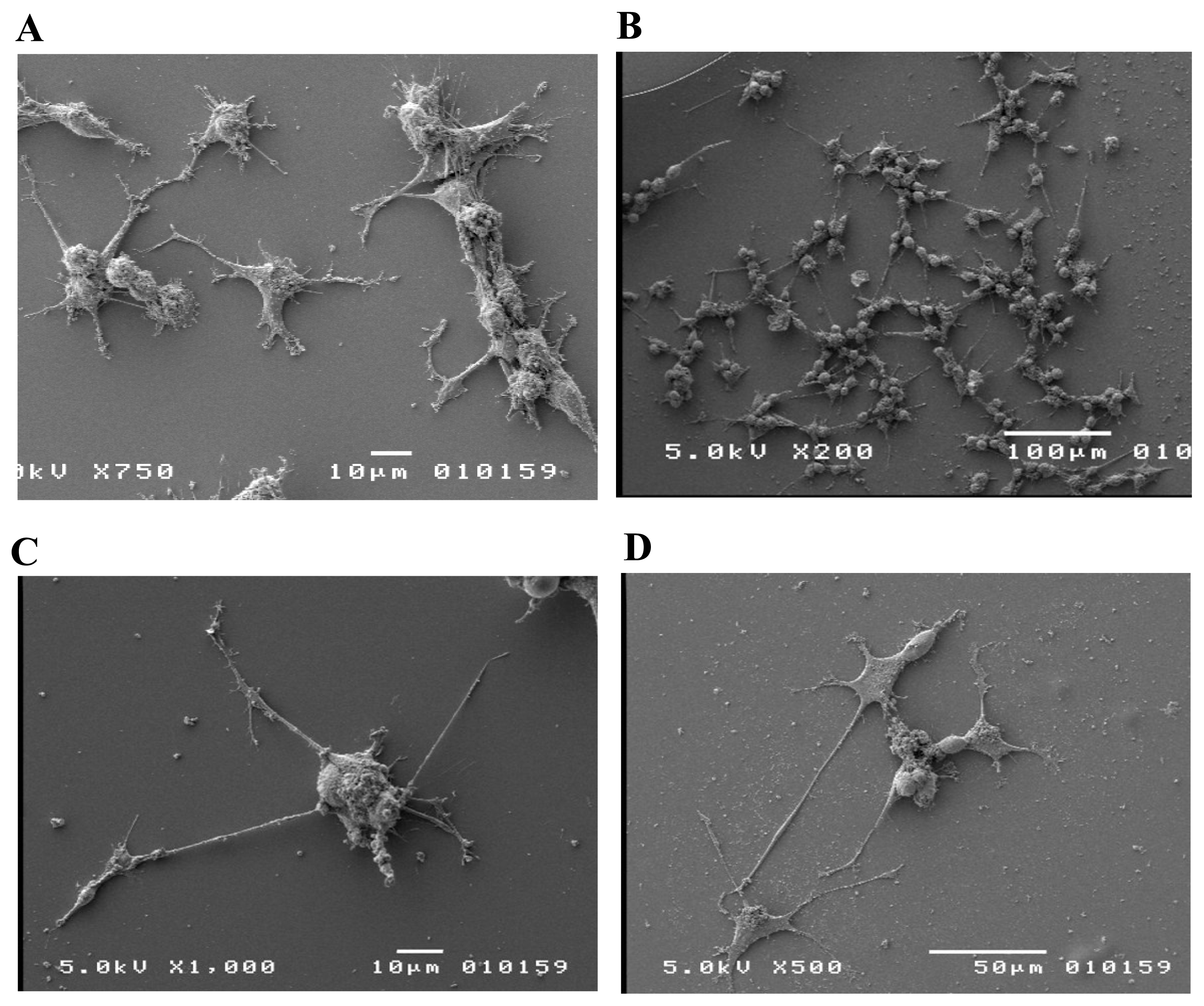
in vitro. We show the successful growth and proliferation of these cell lines on photoresist derived carbon films that were not functionalized with naturally occurring protein or synthetically modified biopolymers. More importantly, we demonstrated that PC12 cells, a model system for differentiation studies [
13], can be induced to differentiate on carbon surface when subjected to differentiation-inducing substance NGF. One advantage of carbon material without functionalized with other biomolecules is that it would considerably reduce the undesirable immune response, if used in
vivo. Other advantages of these carbon devices, derived from polymeric precursor, include excellent biocompatibility, wide electrochemical stability, and availability in high purity, reproducibility, chemical inertness, good thermal conductivity, dimensional and mechanical permanence [
11–
12]. Therefore, our results present evidence for further substantiation.
2. Materials and Methods
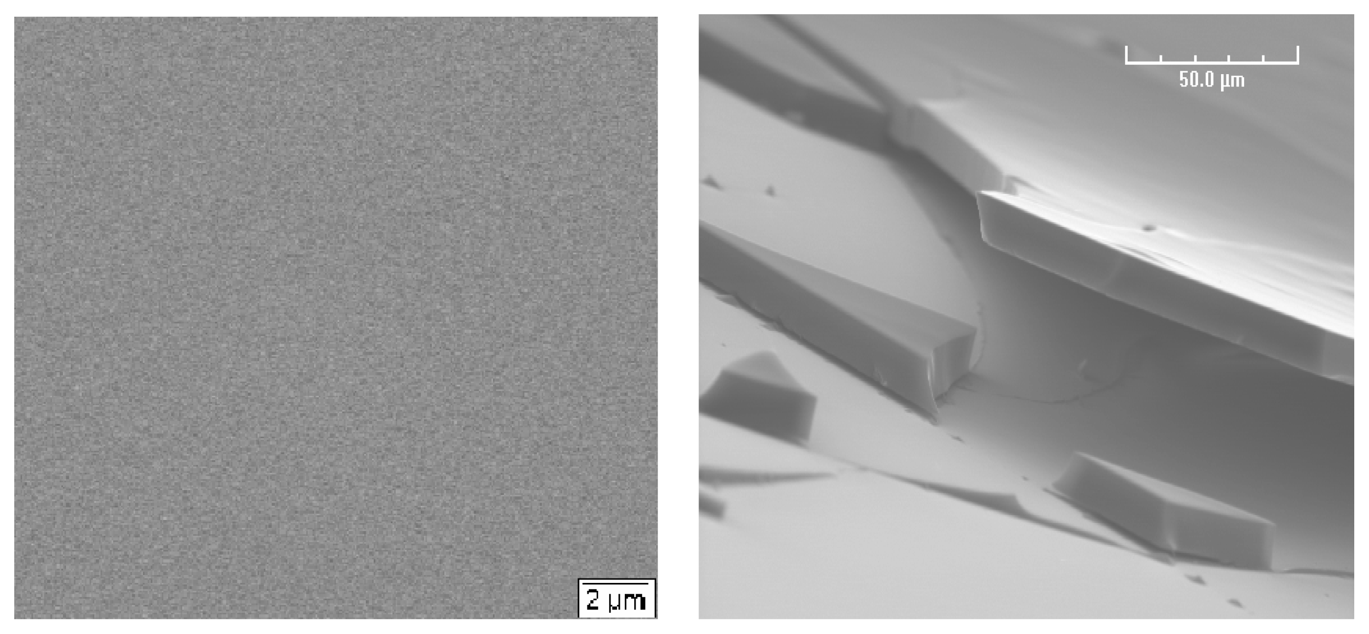
2.1 Fabrication of Carbon substrate
Carbon was derived from pyrolysis of photoresist coated on silicon wafer. A 2” diameter silicon wafer is cut into numerous chips of approximate length 15.6mm. Silicon chips were washed with 70% ethanol and then air dried. A clean silicon chip was placed in the spin coater. A layer of positive photoresist SPR 220-7.0 was applied to the silicon chip manually. The spin coater was run at 300 revolutions per minute (rpm) for 3 seconds to spread the photoresist on the wafer then run at 3000 rpm for 30 sec to fully situate the photoresist on the silicon chip. The photoresist coating process was repeated four times to ensure that the desired thickness range had been reached. Following the four coats, photoresist coated silicon chip was placed on a hot plate set at 95 °C for five minutes and was left to cool naturally to room temperature.
The chip was placed in a Nitrogen gas atmosphere. The gas atmosphere must be completely free of oxygen. The sample was heated at a rate of 10 °C per minute until the desired maximum temperature (700 °C to 1100 °C) had been reached. Once at the desired maximum temperature, the sample remained under heat at that temperature for one hour. The chip is allowed to cool in the nitrogen environment. Once at room temperature, the chip with a layer of carbon was ready to reenter an oxygenated atmosphere [
11–
12].
2.2 Cell Culture
Carbon chips were placed into wells of 6-well plate, washed with 70% v/v ethanol and sterilized under ultraviolet radiation for 45 minutes.
The rat pheochromocytoma PC12 cells were cultured in DMEM high glucose medium supplemented with 5% of FBS and 10% of Horse serum. The cells were induced to differentiate in DMEM high glucose medium (Gibco-11495) with 100 ng/mL of Nerve Growth Factor (Gibco 13257-019).
Human neuroblastoma cell lines (SK-N-MC, SY5Y), mouse teratocarcinoma cell line (P-19), were maintained in DMEM high glucose medium (Gibco 11495) supplemented with 10% fetal bovine serum (Gibco 26140-079) and penicillin-streptomycin (Gibco 15140-122). The cells were incubated at 37 °C with 5% CO2 in cell culture dishes. The cells were removed from the cell culture dish with 0.25% trypsin EDTA (Gibco-25200). Removed cells were centrifuged at 1000 rpm for 5 minutes. The pellet was suspended with fresh medium, and counted via a haemocytometer. Approximately 100,000 cells were transferred to the sterilized carbon chip placed in wells of 24-well cell culture plate.
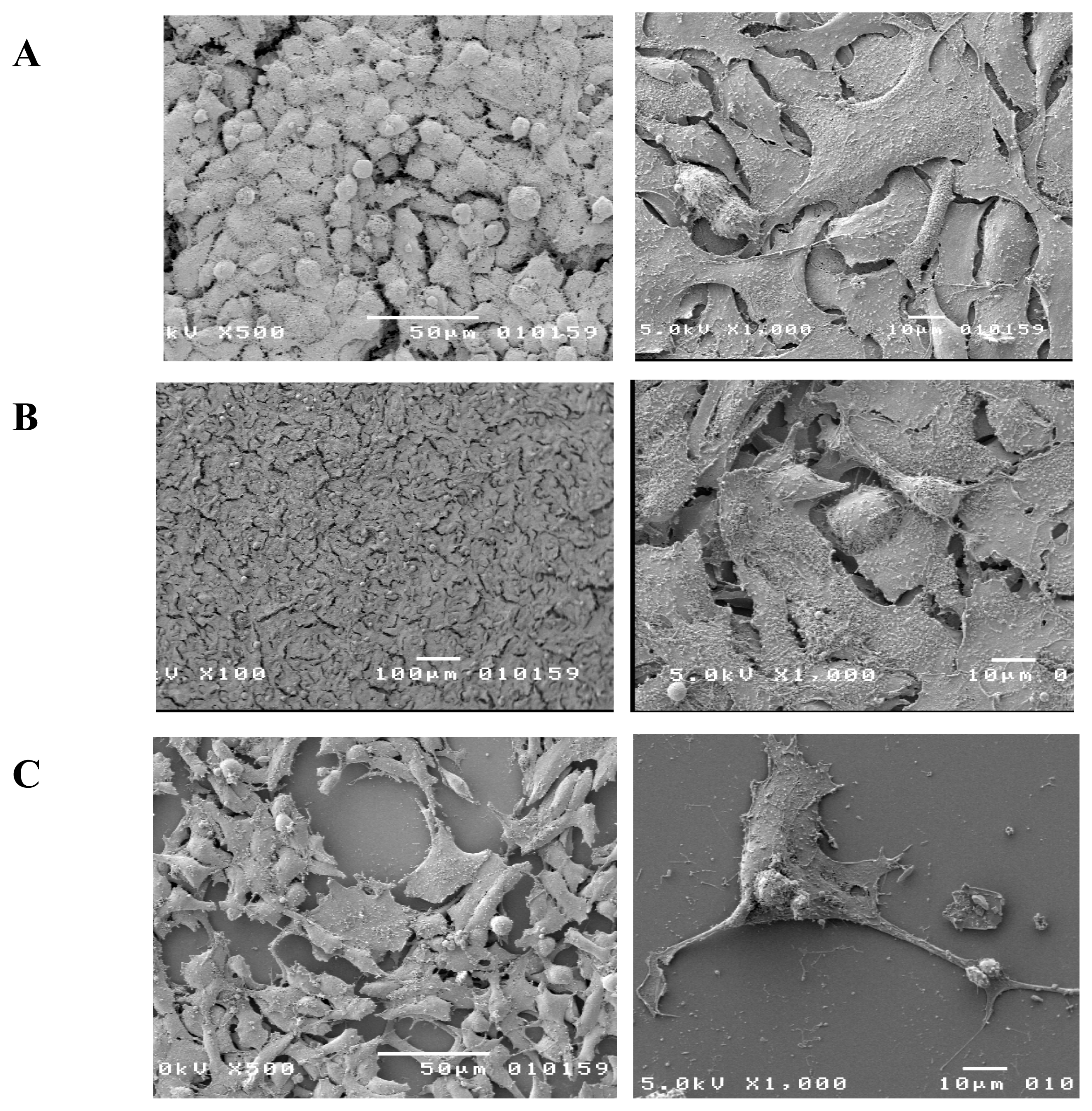
2.3 Scanning Electron Microscopy
Differentiated and undifferentiated cells on photoresist derived carbon were fixed by immersion in 2.5% (v/v) glutaraldehyde in 0.5 M Na cacodylate-HCl buffer (pH 7.2) for 1 hr at room temperature. The fixed samples were washed three times in the same buffer. Following the third wash the cells were post fixed for 1 hr in 1% osmium tetroxide (w/v) in the same buffer, washed three more times in the same buffer and left overnight at 4 °C in fresh buffer. The next morning the samples were dehydrated through a graded series of ethanol to 10% and then Critical Point Dried in liquid CO2.
The carbon chips with the cells attached on the surface were affixed to aluminum SEM studs and sputter coated with Au/Pd (80/20). The specimens were then examined using an ETEC autoscan scanning electron microscope at 20 kV accelerating voltage [
14].
5. Conclusion and Future Work
Conducted study provides the first implication that photoresist derived carbon may be explored as one of the options for promoting initial cell response that may lead to the future design of neural implants. The work presented in this paper demonstrates support for neural cell differentiation. The results represented meet the parameters for further investigation of photoresist derived carbon as a neural probe.
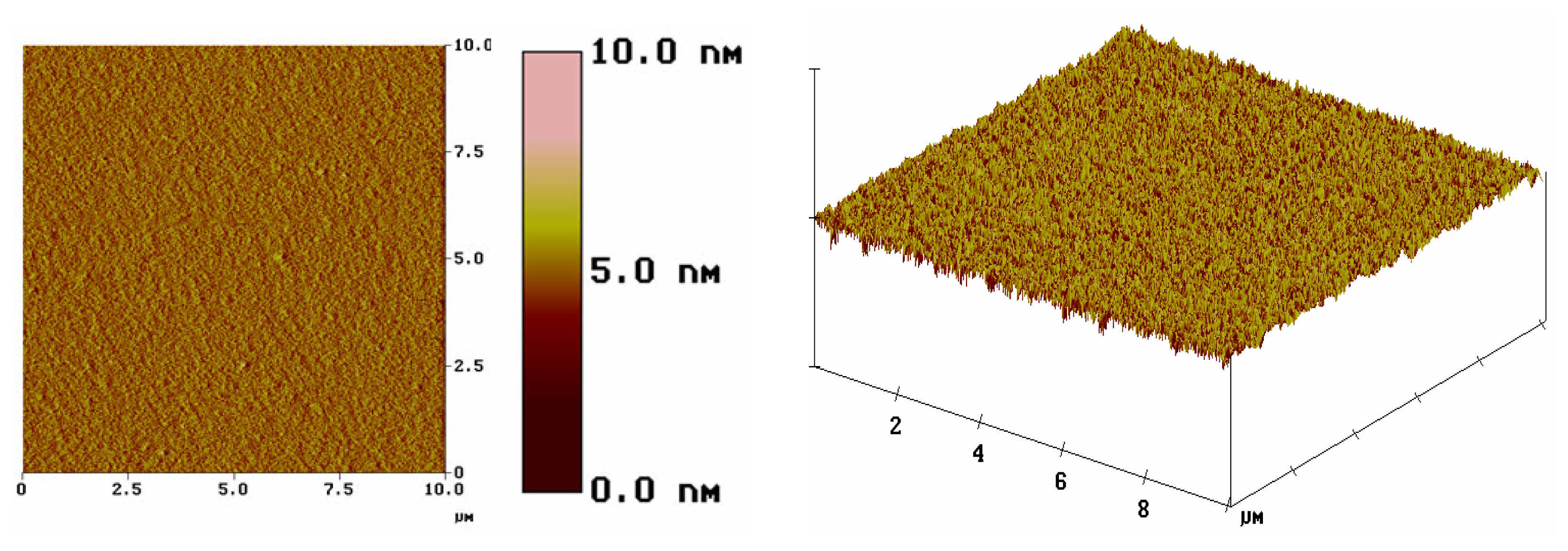
Topography of the substrate plays a vital role in determining cell response of cells to a structure. We can take the advantage of using photoresists as the starting material for carbon template, as the photoresists can be patterned by photolithography techniques. Once cells have been plated onto carbon structure in a specific pattern, we can selectively eliminate cells from the established network and study how cells reconnect after injury.
The ability to grow cultured neurons in a specific pattern and be able to record electrical activity will open up the area of study in neuronal networks and function. It would be then possible to record the extracellular potential from a cell body or the firing of an individual fiber to the outstanding electrical properties of photoresist derived carbon.


