Communication

Structural Elucidation of a New Puzzling Compound Emerged from Doebner Quinoline Synthesis

by
Cristina Maria Al-Matarneh
1 and
Alina Nicolescu
2,*
1
Center of Advanced Research in Bionanoconjugates and Biopolymers, “Petru Poni” Institute of Macromolecular Chemistry of Romanian Academy, 41A Grigore Ghica Voda Alley, 700487 Iasi, Romania
2
NMR Laboratory, “Petru Poni” Institute of Macromolecular Chemistry of Romanian Academy, 41A Grigore Ghica Voda Alley, 700487 Iasi, Romania
*
Author to whom correspondence should be addressed.
Molbank 2024, 2024(3), M1841; https://doi.org/10.3390/M1841
Submission received: 3 June 2024 / Revised: 18 June 2024 / Accepted: 21 June 2024 / Published: 24 June 2024
(This article belongs to the Section Structure Determination)

Abstract

:
The quinoline ring is found in many biologically active natural alkaloids and is still being highly exploited by researchers due to its numerous potential applications in fields ranging from pharmacology to material science. During our synthetic attempts for new quinoline-4-carboxylic acids, using an extended version of the Doebner reaction, a new puzzling compound emerged when para-iodine aniline was reacted with salicylaldehyde and pyruvic acid in acetic acid as a reaction medium. The chemical structure of this new compound was established based on the information obtained from 1D and 2D NMR experiments (1H-, 13C-, and 15N-NMR), corroborated with MS spectrometry and IR spectroscopy. The photophysical properties (UV–vis and fluorescence) were also investigated. The proposed structure contains as the main elements a 1,4-dioxane-2,5-dione core symmetrically substituted with a propylidene chain that has attached to it a salicylaldehyde fragment and a pyrrole-2-one ring containing two 4-iodophenyl fragments. The isolation of this compound, reported here for the first time, is direct evidence that unexpected compounds can emerge from “classical” synthetic pathways when the right components are combined.

Graphical Abstract

1. Introduction

Quinoline is a scaffold highly exploited by researchers due to its numerous pharmacological applications [1]. The quinoline ring is found in many biologically active natural alkaloids, and it has been used for the synthesis of a plethora of compounds, some of them approved as medications in different pathologies. The best-known quinoline derivatives are quinine and chloroquine, which have played historical roles in malaria treatment and eradication from endemic regions [2]. Other biological activities encountered in quinoline derivatives include anti-inflammatory [3,4,5], anticonvulsant [6,7], cardiovascular [8,9], anticancer [10,11,12], or antimycobacterial [13,14]. Quinoline derivatives have also found multiple applications in material science due to their optical and sensing properties. It has been demonstrated that quinoline derivatives can be useful agrochemicals [15,16], pH indicators and pigments [17,18,19,20], ligands in the synthesis of OLED [21,22], or used as selective chemosensors for different ions in conjunction with conjugated polymers [23,24].
Several classical synthesis protocols are presented in the literature for the construction of quinoline scaffolds like Doebner–von Miller, Pfitzinger, Gould–Jacob, Friedlander, Skraup, and Conrad–Limpach, their “strengths and weakness” being reviewed in several papers in comparison with newer eco-friendly methods [25,26,27,28].
Our group has extensive experience in the synthesis of N-heterocycles as many of them are interesting pharmacophores with a wide range of biological activities, such as antibacterial, anticancer, anti-inflammatory, or antimalarial [29,30,31,32,33,34,35].
Recently, we showed that, by using a Doebner synthesis protocol with acetic acid as a solvent and catalytic amounts of trifluoroacetic acid, substituted quinoline-4-carboxylic acids bearing an iodine atom could be obtained with yields between 50 and 90%. Aside from the main quinoline derivative, we isolated several reaction by-products, such as dihydro-pyrrol-2-ones and furan derivatives or 2-methyl-4-carboxyquinoline. In the same study, we showed that all the newly synthesized quinoline derivatives demonstrated good activity against S. epidermidis and C. parapsilosis. Due to their hydrophobic nature, these derivatives can easily penetrate the stratum corneum, increasing their bioavailability for skin infection [36].
Herein, we report the puzzling structure of a new by-product from the Doebner reaction, obtained during our synthetic attempts for new quinoline-4-carboxylic acids bearing an iodine atom. The chemical structure of this new compound, never reported in the literature, was established from different 1D and 2D nuclear magnetic resonance spectroscopy (NMR) techniques, corroborated with infrared (IR) and mass spectrometry (MS) data. The photophysical properties were also investigated.

2. Results and Discussion

2.1. Synthesis

The compound described in this study emerged during our attempts to synthesize new quinoline derivatives by reacting para-iodine aniline with various substituted aldehydes using an extended version of the Doebner reaction, wherein acetic acid served as the reaction medium [36]. Interestingly, in the presence of salicylaldehyde, which features an activating electron-donating group (hydroxyl) in the ortho position, compound 4 turned up as the primary product, accompanied by 2-methyl-4-carboxyquinoline (5) as a by-product (Scheme 1). The formation of by-product 5 was consistent across different starting aldehydes, as reported previously [36]; however, compound 4 was something that we never encountered in our previous synthetic attempts.
Going further, we reacted para-hydroxyl benzaldehyde with para-iodine aniline while keeping the rest of the experimental conditions constant and obtained the expected quinoline derivative. Thus, we suppose that the ortho position of the hydroxyl group favors the rapid bond of the aldehyde moiety with the reaction media because of the vicinity of both oxygen atoms with an attracting effect.

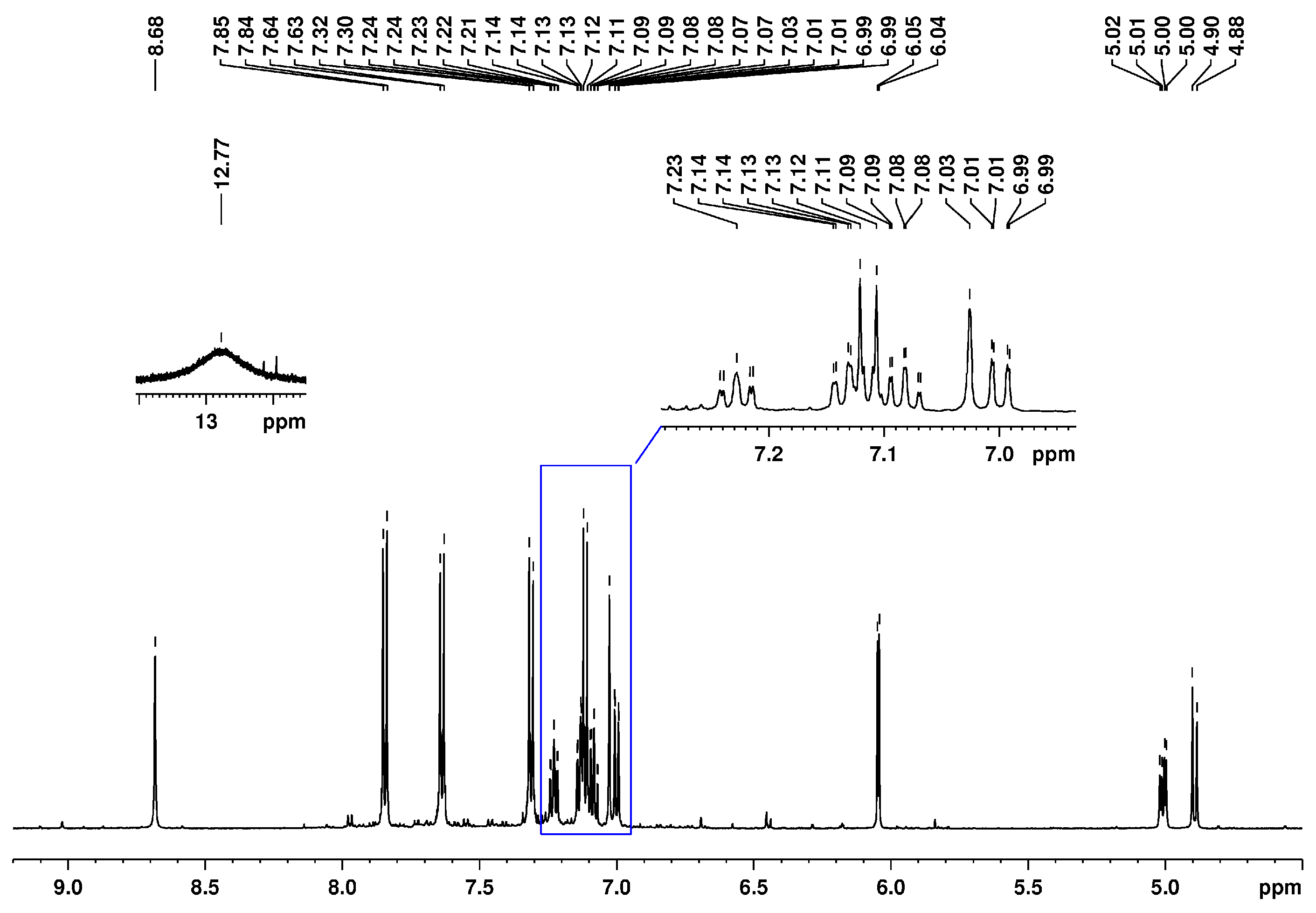
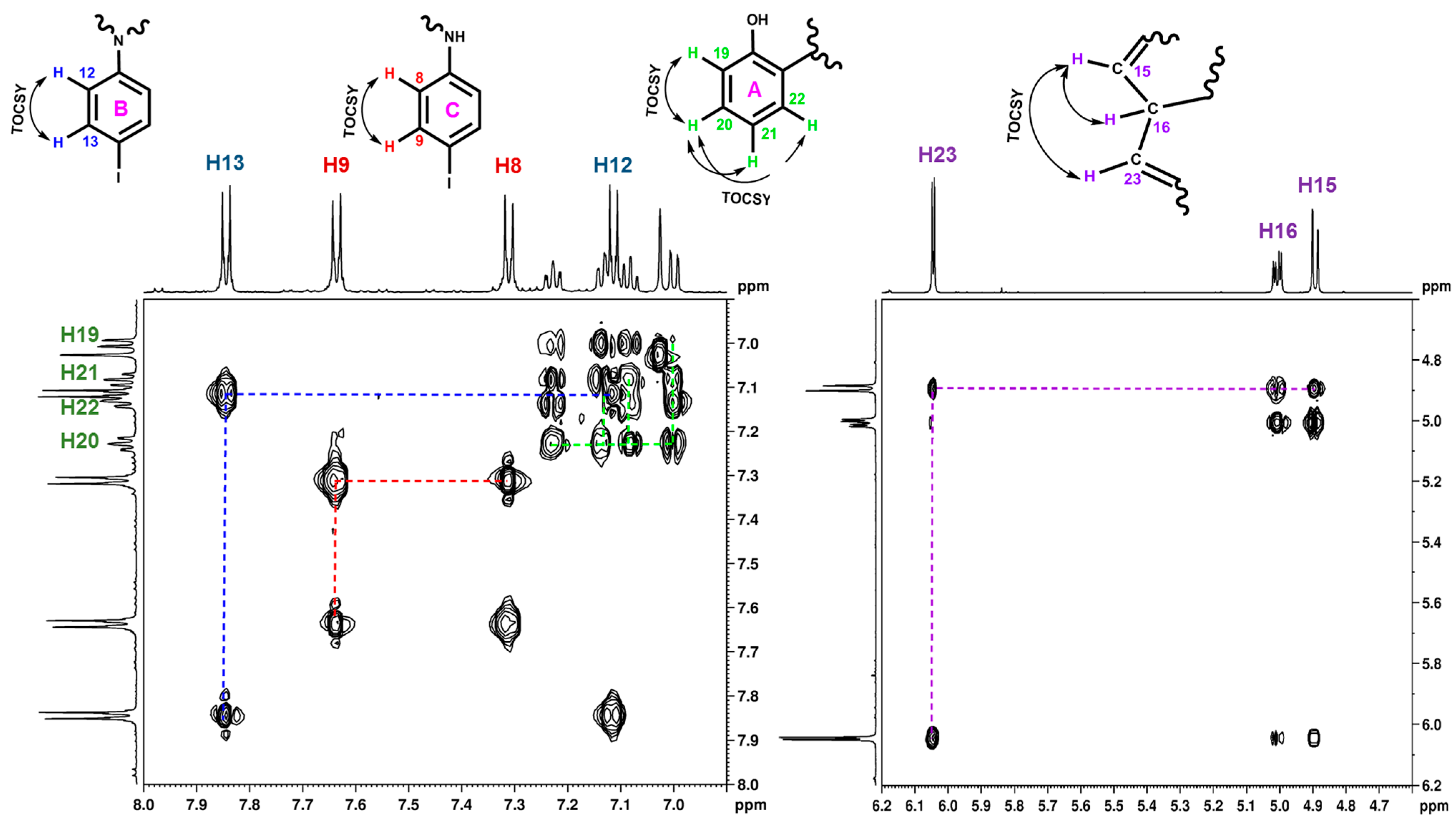
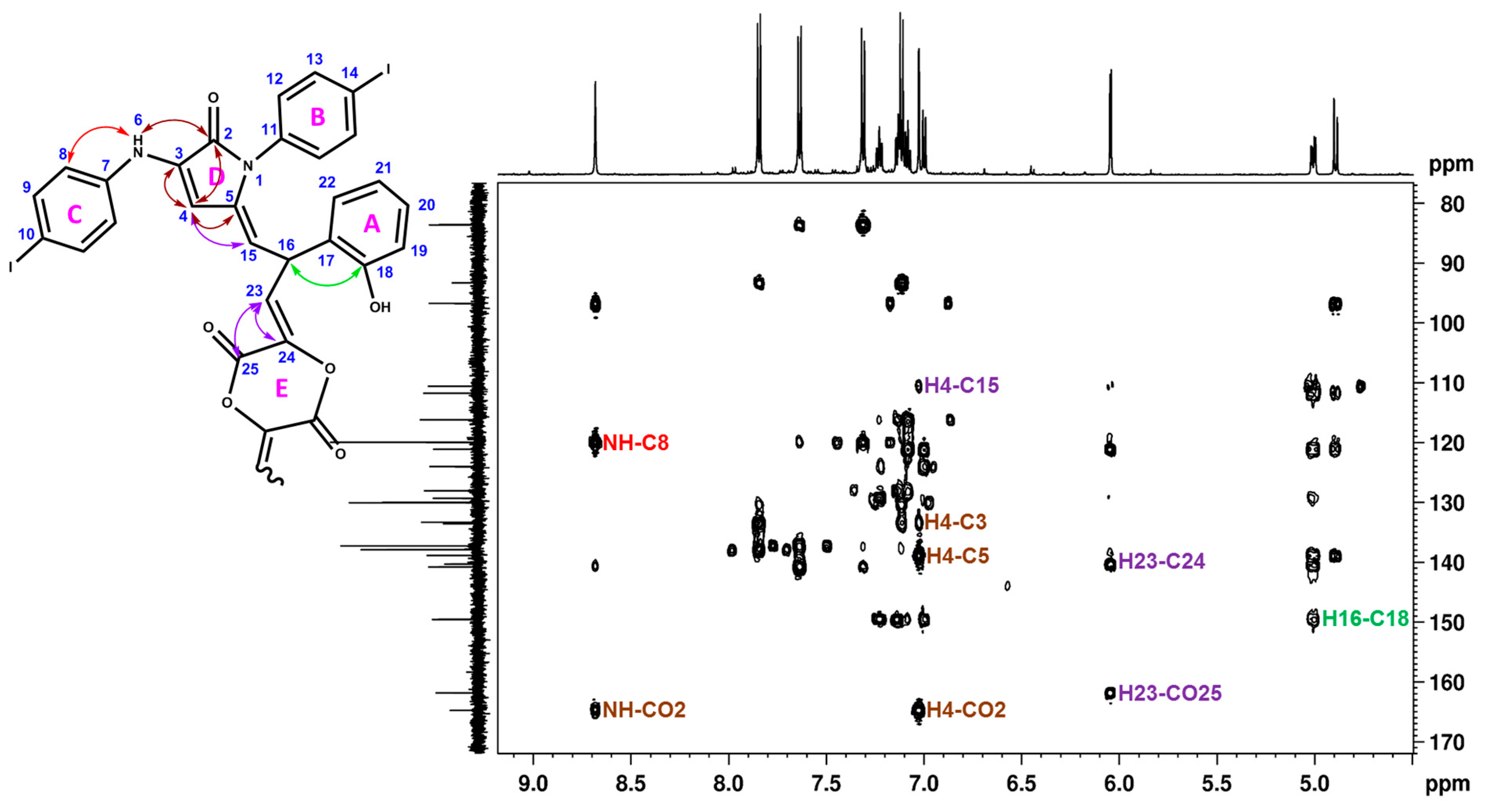
2.2. Spectral Analysis

The structure of the newly synthesized compound 4 (Scheme 2) was determined through spectral analyses, including IR and NMR spectroscopies and HR-MS spectrometry. The unambiguous proton and carbon signal assignments as obtained from bidimensional correlation experiments are presented in the Materials and Methods section. Because the new compound 4 was not crystalline, its chemical structure was deduced mainly from protons’ interactions with neighboring protons, carbon, and nitrogen atoms, followed by 2D NMR experiments.
The 1H-NMR spectrum corresponding to the new compound 4 presents several signals in the interval 4.80–13.50 ppm (Figure S1), most of them being separated and showing first-order proton–proton couplings. By heating the DMSO-d6 solution up to 333 K, we were able to obtain a better separation of the proton signals resonating in the interval 7.00–7.15 ppm (Figure 1); thus, all the 1D and 2D NMR experiments were recorded at this temperature. A better separation of the signals favors an unambiguous interpretation of the proton–carbon interactions that are crucial for solving the “puzzling” new structures.
In the 13C-NMR spectrum (Figure S2), 23 well-resolved signals were observed, only one resonating in the high-field region at 31.8 ppm, which can be associated with the presence of an aliphatic carbon atom. In the low-field region, three signals, resonating above 150 ppm, can be correlated with carbon atoms covalently bonded to oxygen.
The proton–proton correlations obtained in the 2D TOCSY spectrum (Figure 2) indicated four isolated spin systems: three aromatic and one containing aliphatic and olefinic protons. One of the aromatic spin systems was readily assigned to the salicylic residue (ring A) as it connected the doublet of doublets from 7.00 ppm (H19), triplet of doublets from 7.08 ppm (H21), doublet of doublets from 7.14 ppm (H22), and triplet of doublets from 7.23 ppm (H20). The other two aromatic spin systems, both corresponding to para-substituted iodo-phenyls, were differentiated based on the three-bond correlation between NH proton (8.68 ppm) and carbon C8 (119.9 ppm), identified in the HMBC spectrum. It was found that the doublet from 7.11 ppm (H12) and its coupling partner from 7.84 ppm (H13) belong to ring B, whereas the protons from ring C resonate at 7.31 ppm (H8) and 7.64 ppm (H9). The hybrid spin system contains a doublet at 4.89 ppm (H15), a doublet of doublets at 5.00 ppm (H16), and a second doublet at 6.04 ppm (H23).
The olefinic nature of the protons involved in this spin system was deduced based on the carbon chemical shift values. In the proton–carbon direct correlations’ HSQC spectrum (Figure 3), it was found that proton H15 is directly linked to a carbon atom at 110.6 ppm and proton H23 is directly linked to a carbon atom at 111.7 ppm. A three-bond correlation in the HMBC spectrum (Figure 4) between H16 (5.00 ppm) and C18 (149.6 ppm) pointed us to covalently link this propylidene-like spin system with the salicylic residue (ring A).
As in our previous attempts to obtain quinoline derivatives using Doebner-type reaction conditions, we obtained pyrrol-2-one derivatives as by-products [36]; we looked into the possibility that our unknown compound also contained this ring. Indeed, we identified in the proton–carbon and proton–nitrogen HMBC spectra several correlations that enabled us to find and assign all the carbons and nitrogen atoms belonging to the pyrrol-2-one residue (ring D). Thus, the carbonyl group CO-2 (165.0 ppm) was assigned from the proton–carbon HMBC spectrum where three-bond correlations with both NH (8.68 ppm) and H4 (7.03 ppm) protons were visible (Figure 4). The proton H4 resonates as a singlet, indicating that no other protons are in its immediate vicinity. It also has correlations with carbon atoms situated two (C3 (133.6 ppm) and C5 (138.9 ppm)) and three (C15 (110.6 ppm)) bonds apart, visible in the H,C-HMBC spectrum (Figure 4).
Finally, the 1,4-dioxane-2,5-dione-like residue (ring E) was deduced based on the H23 long-range interactions with C24 (140.3 ppm) and CO-25 (161.8 ppm) carbon atoms, visible in the HMBC spectrum (Figure 4). All the direct and long-range proton–carbon correlations visible in the HQSC and HMBC spectra, which allowed us to “assemble” the structure for compound 4, are centralized in Table 1. As there were no additional proton and carbon signals left unassigned, we duplicated the OC24-OOC25 “build-up” residue such that ring E was formed and a symmetrical structure was obtained.
Additional information to elucidate the structures of newly synthesized nitrogen-containing compounds can be obtained from 15N NMR spectroscopy [37]. In our previous studies, we used 15N NMR to highlight the structural modifications in pyrrolo [1,2-a]benzimidazole and pyrrolo[1,2-a]quinoxaline derivatives [38]. Thus, based on our previous experience, we recorded the proton–nitrogen HMBC spectrum for compound 4 (Figure 5) to obtain further insights into the elements that comprise this new structure. Several correlation signals were visible in the 2D proton–nitrogen HMBC spectrum, indicating the presence of two nitrogen atoms at 91.8 and 143.5 ppm. The nitrogen atom resonating at 91.8 ppm was assigned to the NH group, as indicated by the direct couplings (annotated on the spectrum in Figure 5) with the proton from 12.8 ppm. Another signal was generated by the three-bond interaction of this nitrogen atom with aromatic proton H8, confirming the residual 4-iodophenyl-amine fragment (ring C, Scheme 2). The second nitrogen atom from the structure, resonating at 143.5 ppm, generated three-bond correlation signals with protons H4, H15, and H12, supporting the presence of pyrrole ring D substituted with 4-iodophenyl (ring B) and an exo-cyclic double bond, as suggested in Scheme 2.
The presence of oxygen atoms was supported not only by the carbon chemical shift values but also by the molecular mass obtained through mass spectrometry. The exact molecular weight was determined by high-resolution mass spectrometry, and the consistency of the molecular formula has been assessed by comparison of the experimental and simulated isotopic patterns (Figure S6).
The supplementary information confirming mainly the functional groups proposed for compound 4 was obtained from IR analysis. Aside from the strong band from 1742 cm−1 attributed to carbonyl groups (C=O bond in ester and amide), other strong bands are visible at 1368 and 1211 cm−1 that can be associated with the different C-O bonds (carboxylic, ether, and alcohol) present in the structure of compound 4 (see Figure S7). The two weak bands from 3460 and 3300 cm−1 indicate the existence of N-H and O-H bonds. In the fingerprint region, the medium band from 519 cm−1 is an indication of a C-I bond. The aliphatic C-H bond can generate medium bands from 2972 and 1449 cm−1, whereas the medium band from 3015 cm−1 supports the aromatic C-H bonds.

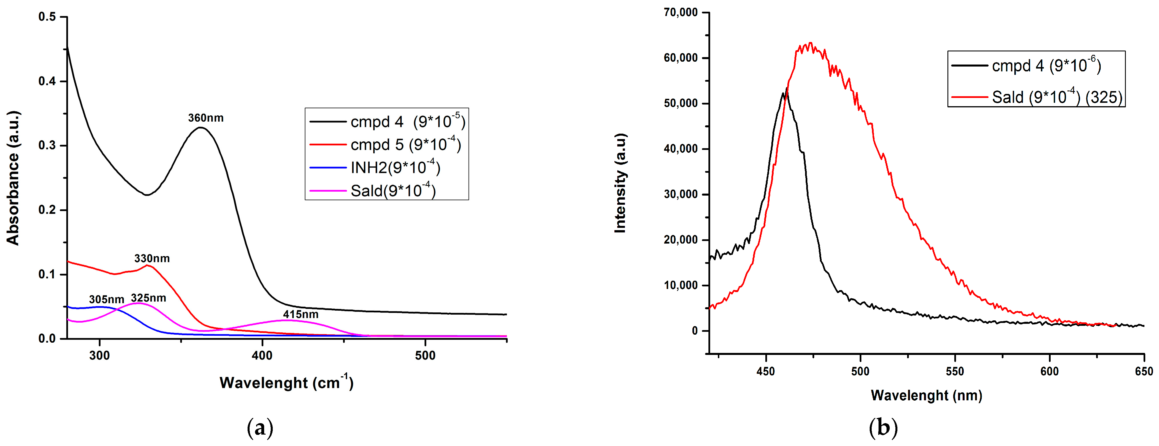
2.3. Photophysical Investigations

Our previous research on compounds with extended π conjugations that contain pyrrole and other nitrogen heterocycles revealed that they have intriguing photophysical characteristics that can make them useful as chemosensors [39,40]. This led us to investigate the photophysical properties of the recently obtained compounds 4 and 5.
The electronic absorption spectra of reaction products 4 and 5 along with the starting materials (iodaniline and salicylaldehyde) are presented in Figure 6a. For comparative analysis, the DMSO solutions’ concentrations were 9 × 10⁻⁵ M for 4 and 9 × 10⁻⁴ M for the rest of the compounds. The molecular architecture for compound 4 consists of a 1,4-dioxane-2,5-dione core symmetrically substituted with a propylidene chain that has attached to it a salicylaldehyde fragment and a pyrrole-2-one ring containing two 4-iodophenyl fragments, which leads to an extended electron-rich conjugated system. Compound 4 presents an intense absorption band at 360 nm, while compound 5 absorbs at 330 nm. The starting materials, with lower absorption bands at working concentrations, have characteristic absorption peaks at 305 nm for iodaniline and at 325 nm and 415 nm for salicylaldehyde. The broad bands visible in this region are usually attributed to a mixture of p→p* and n→p* electronic transitions [41,42].
The photophysical properties of the emissions for compound 4 compared to salicylaldehyde have been investigated by fluorescence spectroscopy at 360 and 325 nm excitation wavelengths, respectively, in the same solvent (DMSO) as with the UV–vis. The spectra displayed emission bands located at 460 nm for compound 4 and at 475 nm for salicylaldehyde (Figure 6b). Compound 5 and iodaniline had no fluorescence and thus are not shown in the figure. The emission band for salicylaldehyde is broad and structureless, while, for compound 4, it is well-defined. This difference can easily be explained by the extended conjugate system present in the compound 4 structure. For a better comparison of the peak shape, the fluorescence spectra of the two compounds were recorded at a concentration difference of 102 M.

3. Materials and Methods

All commercially available products were used without further purification unless otherwise specified. Analytical thin-layer chromatography was performed with commercial silica gel plates 60 F254 (Merck, Darmstadt, Germany) and visualized with UV light (λmax = 254 or 365 nm). The melting point of the compounds measured on a MEL-TEMP capillary melting point apparatus from ambient temperature up to 400 °C.
The NMR spectra included in this study were recorded on Bruker Avance NEO 600 MHz spectrometer (Bruker Biospin, Ettlingen, Germany) equipped with 5 mm inverse detection multinuclear z-gradient probe. Proton and carbon chemical shifts are reported in δ units (ppm) relative to the residual solvent signal (ref: DMSO-d6 1H, 2.51 ppm, and 13C, 39.47 ppm). H,H-TOCSY, H,C-HSQC, and H,C-HMBC experiments were recorded using standard pulse sequences as delivered by Bruker with TopSpin 4.0.8 spectrometer control and processing software. The 15N chemical shifts were obtained as projections from the 2D indirectly detected H,N-HMBC spectra and are referred to liquid ammonia (0.0 ppm) using nitromethane (380.2 ppm) as external standard.
The IR spectrum was recorded on a Shimadzu IRTracer-100 instrument (Shimadzu USA Manufacturing, Inc., Canby, OR, USA) in transmission mode, covering the spectral range 400–4,000 cm−1. The bands intensities are described as strong, medium, and weak.
The exact molecular weights of newly synthesized compounds were obtained on either Bruker Maxis II QTOF spectrometer ((Bruker Daltonics, Bremen, Germany) with electrospray ionization (ESI) in the negative mode, for compound 4, or on a Bruker RapifleX MALDI-TOF/TOF (Bruker Daltonics, Bremen, Germany) equipped with a Smartbeam 3D laser, in positive mode, for compound 5.
Spectroscopic-grade solvents were used for the photophysical characterization of the synthesized derivatives. UV–vis measurements were performed on a Lambda 35 device (Perkin Elmer, Shelton, CT, USA). The absorption spectra were measured in the 270–700 nm range for identical sample volumes (3 mL) with the following parameters: slit width 1 nm, scan speed 480 nm/min, and data interval 1 nm. The spectra of the samples were measured at room temperature using 1 cm path length quartz cuvettes. Fluorescence measurements were carried out using a FluoroMax-4 spectrophotometer (Horiba, Kyoto, Japan). The emission spectra were collected using excitation wavelengths of 360 and 325 nm.
General procedure for synthesis of compound 4
In the first step, salicylaldehyde (1 mmol, 122 mg) was dissolved in approximately 1 mL of acetic acid. To this solution, a mixture containing pyruvic acid (1.5 mmol, 132 mg) and TFA (20 µL) as a catalyst, in 0.5 mL acetic acid, was added and stirred for 10 min. In the second step, iodo-aniline (1 mmol, 219 mg) was dissolved in about 4 mL of acetic acid and added to the mixture, which was then heated to 80 °C for 24 h. The primary product 4 was obtained by filtering the resulting suspension and washing the solid with ethanol. The secondary product 5 precipitated until next day from the reaction mixture and was separated by filtration and washed with ethanol.
(3Z)-3,6-bis((E)-2-(2-hydroxyphenyl)-3-(1-(4-iodophenyl)-4-((4-iodophenyl)amino)-5-oxo-1H-pyrrol-2(5H)-ylidene)propylidene)-1,4-dioxane-2,5-dione (compound 4): crystallized from acetic acid; yellow powder; 70% yield (235 mg); mp 247–250 °C; IR ATR ν(cm−1): 3460, 3300, 3015, 2972, 1742, 1622, 1578, 1533, 1485, 1448, 1368, 1211, 903, 814, 756, 519.
1H NMR (DMSO-d6, 600.1 MHz, δ (ppm)): 4.89 (1H, d, 3J = 10 Hz, H-15), 5.00 (1H, dd, 3J = 10 Hz, 3J = 4 Hz, H-16), 6.05 (1H, d, 3J = 4 Hz, H-23), 7.00 (1H, dd, 3J = 8 Hz, 4J = 1 Hz, H-19), 7.03 (1H, s, H-4), 7.08 (1H, td, 3J = 8 Hz, 4J = 1 Hz, H-21), 7.11 (2H, d, 3J = 9 Hz, H-12), 7,14 (1H, dd, 3J = 8 Hz, 4J = 1 Hz, H-22), 7.23 (1H, td, 3J = 8 Hz, 4J = 1 Hz, H-20), 7.31 (2H, d, 3J = 9 Hz, H-8), 7.64 (2H, d, 3J = 9 Hz, H-9), 7.84 (7.31 (2H, d, 3J = 9 Hz, H-13), 8.68 (1H, s, NH), 12.77 (1H, bs, OH).
13C NMR (DMSO-d6, 150.9 MHz, δ (ppm)): 31.8 (CH-16), 83.6 (C-10), 93.3 (C-14), 96.7 (CH-4), 110.6 (CH-15), 111.8 (CH-23), 116.2 (CH-19), 120.0 (CH-8), 121.1 (C-17), 124.0 (CH-21), 128.1 (CH-20), 129.3 (CH-22), 130.0 (CH-12), 133.3 (C-11), 133.6 (C-3), 137.3 (CH-9), 137.9 (CH-13), 138.9 (C-5), 140.3 (C-24), 140.8 (C-7), 149.6 (C-18), 161.8 (COO-25), 164.7 (CO-2).
15N NMR (DMSO-d6, 60.8 MHz, δ (ppm)): 91.88 (NH), 143.49 (N).
HRMS-ESI (m/z): [M−H] C54H35I4N4O8, calcd. 1374.8639, found 1374.8650.
6-iodo-2-methylquinoline-4-carboxylic acid (compound 5): crystallized from acetic acid; brown powder; 27% yield (84 mg); mp 289–290 °C; IR ATR ν(cm−1): 3656, 3083, 2979, 2887, 1714, 1603, 1579, 1504, 1454, 1377, 1351, 1240, 1152, 1113, 1032, 937, 867, 810, 604, 519.
1H NMR (DMSO-d6, 600.1 MHz, δ (ppm)): 2.71 (3H, s, CH3), 7.79 (1H, d, 3J = 9 Hz, H-8), 7.89 (1H, s, H-3), 8.05 (1H, dd, 3J = 9 Hz, 4J = 2 Hz, H-7), 9.10 (1H, d, 4J = 2 Hz, H-5), 14.00 (1H, bs, OH).
13C NMR (DMSO-d6, 150.9 MHz, δ (ppm)): 24.7 (CH3), 93.7 (C-6), 123.8 (CH-3), 124.5 (C-10), 130.8 (CH-8), 133.9 (CH-5), 134.4 (C-4), 137.9 (CH-7), 147.0 (C-9), 159.6 (C-2), 167.1 (COOH).
HRMS (MALDI-TOF/TOF) m/z calcd for [M+H]+ C11H8INO2+ 313.96, found 313.95

4. Conclusions

An interesting puzzle between salicylaldehyde, iodo-aniline, pyruvic acid, and reaction media was obtained as an unexpected product during a Doebner-type synthetic protocol. The complex structure of this compound was established through conventional spectroscopic methods (NMR, IR, and MS). The synthesis method is reproducible, leading to an interesting compound possessing an extended conjugated system. Given the starting salicylaldehyde moiety present in the molecule and the extended conjugated system, we investigated the photoluminescent properties of the compound, and the results are promising.

Supplementary Materials

The following supporting information can be downloaded, Figure S1. The 1H-NMR spectrum corresponding to newly synthesized compound 4, recorded in DMSO-d6 at 297 K. A better visualization of the aromatic signals is presented in the insert. The signal resonating around 13 ppm, assigned to OH proton, is included in the left-side insert. Figure S2. The 13C-NMR spectrum corresponding to newly synthesized compound 4, recorded in DMSO-d6 at 333 K. A better visualization of the aromatic signals is presented in the insert. Figure S3. The H,H-TOCSY spectrum corresponding to newly synthesized compound 4, recorded at 333 K. Figure S4. The H,C-HSQC spectrum corresponding to newly synthesized compound 4, recorded at 333 K. Figure S5. The H,C-HMBC spectrum corresponding to newly synthesized compound 4, recorded at 333 K. Figure S6. HRMS-ESI spectrum with isotopic pattern in negative mode for compound 4. Upper trace (red)—simulated pattern; lower trace (black)—experimental spectrum for C54H35I4N4O8 [M−H]. Figure S7. FT-IR spectrum of compound 4. Figure S8. The 1H-NMR spectrum corresponding to synthesized compound 5, recorded in DMSO-d6. Figure S9. The 13C-NMR spectrum corresponding to synthesized compound 5, recorded in DMSO-d6. Figure S10. FT-IR spectrum of compound 5.

Author Contributions

Conceptualization, C.M.A.-M. and A.N.; methodology, C.M.A.-M. and A.N.; validation, C.M.A.-M. and A.N.; formal analysis, C.M.A.-M. and A.N.; investigation, C.M.A.-M. and A.N.; data curation, C.M.A.-M. and A.N.; writing—original draft preparation, C.M.A.-M.; writing—review and editing, C.M.A.-M. and A.N.; project administration, C.M.A.-M. All authors have read and agreed to the published version of the manuscript.

Funding

This research received no external funding.

Data Availability Statement

The original contributions presented in the study are included in the article and Supplementary Materials; further inquiries can be directed to the corresponding author.

Acknowledgments

C.M.A.-M. acknowledges the support provided by the ICUB Fellowship for Young Researchers (Contract no. 13461/14.12.2023).

Conflicts of Interest

The authors declare no conflicts of interest.

References

  1. Kumar, S.; Bawa, S.; Gupta, H. Biological activities of quinoline derivatives. Mini-Rev. Med. Chem. 2009, 9, 1648–1654. [Google Scholar] [CrossRef] [PubMed]
  2. Wiesner, J.; Ortmann, R.; Jomaa, H.; Schlitzer, M. New antimalarial drugs. Angew. Chem. Int. Ed. 2003, 42, 5274–5293. [Google Scholar] [CrossRef] [PubMed]
  3. Clemence, F.; Martret, O.L.; Delevallee, F.; Benzoni, J.; Jouanen, A.; Jouquey, S.; Mouren, M.; Deraedt, R. 4-Hydroxy-3-quinolinecarboxamides with antiarthritic and analgesic activities. J. Med. Chem. 1988, 31, 1453–1462. [Google Scholar] [CrossRef] [PubMed]
  4. Calhoun, W.; Carlson, R.P.; Crossley, R.; Datko, L.J.; Dietrich, S.; Heatherington, K.; Marshall, L.A.; Meade, P.J.; Opalko, A.; Shepherd, R.G. Synthesis and antiinflammatory activity of certain 5, 6, 7, 8-tetrahydroquinolines and related compounds. J. Med. Chem. 1995, 38, 1473–1481. [Google Scholar] [CrossRef] [PubMed]
  5. Kohno, Y.; Awano, K.; Miyashita, M.; Ishizaki, T.; Kuriyama, K.; Sakoe, Y.; Kudoh, S.; Saito, K.; Kojima, E. Synthesis and antirheumatic activity of novel tetrahydro-6-quinolineacetic acid derivatives. Bioorg. Med. Chem. Lett. 1997, 7, 1519–1524. [Google Scholar] [CrossRef]
  6. Guo, L.J.; Wei, C.X.; Jia, J.H.; Zhao, L.M.; Quan, Z.S. Design and synthesis of 5-alkoxy-[1,2,4]triazolo[4,3-a]quinoline derivatives with anticonvulsant activity. Eur. J. Med. Chem. 2009, 44, 954–958. [Google Scholar] [CrossRef] [PubMed]
  7. Shabana, K.; Shabana; Mazumder, A.; Singh, H.; Kumar, R.; Tyagi, S.; Datt, V.; Sharma, A.S.; Yar, M.S.; Ahsan, M.J.; et al. Synthesis, characterization, in silico and in vivo evaluation of benzimidazole-bearing quinoline schiff bases as new anticonvulsant agents. ChemistrySelect 2023, 8, e202300209. [Google Scholar] [CrossRef]
  8. Ferlin, M.G.; Chiarelotto, G.; Antonucci, F.; Caparrotta, L.; Froldi, G. Mannich bases of 3H-pyrrolo[3,2-f]quinoline having vasorelaxing activity. Eur. J. Med. Chem. 2002, 37, 427–434. [Google Scholar] [CrossRef] [PubMed]
  9. Sircar, I.; Haleen, S.J.; Burke, S.E.; Barth, H. Synthesis and biological activity of 4-(diphenylmethyl)-alpha-[(4-quinolinyloxy)methyl]-1-piperazineethanol and related compounds. J. Med. Chem. 1992, 35, 4442–4449. [Google Scholar] [CrossRef]
  10. Chaaban, I.; Hafez, H.; AlZaim, I.; Tannous, C.; Ragab, H.; Hazzaa, A.; Ketat, S.; Ghoneim, A.; Katary, M.; Abd-Alhaseeb, M.M.; et al. Transforming iodoquinol into broad spectrum anti-tumor leads: Repurposing to modulate redox homeostasis. Bioorg. Chem. 2021, 113, 105035. [Google Scholar] [CrossRef]
  11. Kemnitzer, W.; Kuemmerle, J.; Jiang, S.; Zhang, H.Z.; Sirisoma, N.; Kasibhatla, S.; Crogan-Grundy, C.; Tseng, B.; Drewe, J.; Cai, X.Z. Discovery of 1-benzoyl-3-cyanopyrrolo[1,2-a]quinolines as a new series of apoptosis inducers using a cell- and caspase-based high-throughput screening assay. Part 1: Structure–activity relationships of the 1- and 3-positions. Bioorg. Med. Chem. Lett. 2008, 18, 6259–6264. [Google Scholar] [CrossRef]
  12. Köprülü, T.K.; Ökten, S.; Tekin, S.; Çakmak, O. Biological evaluation of some quinoline derivatives with different functional groups as anticancer agents. J. Biochem. Mol. Toxicol. 2019, 33, e22260. [Google Scholar] [CrossRef]
  13. Bokosi, F.R.B.; Beteck, R.M.; Jordaan, A.; Seldon, R.; Warner, D.F.; Tshiwawa, T.; Lobb, K.; Khanye, S.D. Arylquinolinecarboxamides: Synthesis, in vitro and in silico studies against Mycobacterium tuberculosis. J. Heterocycl. Chem. 2021, 58, 2140–2151. [Google Scholar] [CrossRef]
  14. Santoso, K.T.; Menorcad, A.; Cheungd, C.Y.; Cook, G.M.; Stocker, B.L.; Timmer, M.S.M. The synthesis and evaluation of quinolinequinones as anti-mycobacterial agents. Bioorg. Med. Chem. 2019, 27, 3532–3545. [Google Scholar] [CrossRef]
  15. Ghareeb, E.A.; Mahmoud, N.F.H.; El-Bordany, E.A.; El-Helw, E.A.E. Synthesis, DFT, and eco-friendly insecticidal activity of some N-heterocycles derived from 4-((2-oxo-1,2-dihydroquinolin-3-yl)methylene)-2-phenyloxazol-5(4H)-one. Bioorg. Chem. 2021, 112, 104945. [Google Scholar] [CrossRef]
  16. Chen, Y.-J.; Ma, K.-Y.; Du, S.-S.; Zhang, Z.-J.; Wu, T.-L.; Sun, Y.; Liu, Y.-Q.; Yin, X.-D.; Zhou, R.; Yan, Y.-F.; et al. Antifungal exploration of quinoline derivatives against phytopathogenic fungi inspired by quinine alkaloids. J. Agric. Food Chem. 2021, 69, 12156–12170. [Google Scholar] [CrossRef]
  17. Li, G.; Zhu, D.; Xue, L.; Jiang, H. Quinoline-based fluorescent probe for ratiometric detection of lysosomal pH. Org. Lett. 2013, 15, 5020–5023. [Google Scholar] [CrossRef]
  18. Van den Berg, O.; Jager, W.F.; Picken, S.J. 7-Dialkylamino-1-alkylquinolinium salts: Highly versatile and stable fluorescent probes. J. Org. Chem. 2006, 71, 2666–2676. [Google Scholar] [CrossRef]
  19. Liu, X.; Su, Y.; Tian, H.; Yang, L.; Zhang, H.; Song, X.; Foley, J.W. Ratiometric fluorescent probe for lysosomal pH measurement and imaging in living cells using single-wavelength excitation. Anal. Chem. 2017, 89, 7038–7045. [Google Scholar] [CrossRef]
  20. El-Ghamaz, N.A.; Diab, M.A.; El-Bindary, A.A.; El-Sonbati, A.Z.; Nozha, S.G. Thermal, dielectric characteristics and conduction mechanism of azodyes derived from quinoline and their copper complexes. Spectrochim. Acta A Mol. Biomol. Spectrosc. 2015, 143, 200–212. [Google Scholar] [CrossRef]
  21. Jiang, B.; Ning, X.; Gong, S.; Jiang, N.; Zhong, C.; Lu, Z.-H.; Yang, C. Highly efficient red iridium(III) complexes cyclometalated by 4-phenylthieno[3,2-c]quinoline ligands for phosphorescent OLEDs with external quantum efficiencies over 20%. J. Mater. Chem. C 2017, 5, 10220–10224. [Google Scholar] [CrossRef]
  22. Sych, G.; Volyniuk, D.; Bezvikonnyi, O.; Lytvyn, R.; Grazulevicius, J.V. Dual interface exciplex emission of quinoline and carbazole derivatives for simplified nondoped white OLEDs. J. Phys. Chem. C 2019, 123, 2386–2397. [Google Scholar] [CrossRef]
  23. Aoki, S.; Sakurama, K.; Matsuo, N.; Yamada, Y.; Takasawa, R.; Tanuma, S.; Shiro, M.; Takeda, K.; Kimura, E. A new fluorescent probe for Zinc(II): An 8-hydroxy-5-N,N-dimethylaminosulfonylquinoline-pendant 1,4,7,10-Tetraazacyclododecane. Chem. Eur. J. 2006, 12, 9066–9080. [Google Scholar] [CrossRef] [PubMed]
  24. Liu, H.; Dong, Y.; Zhang, B.; Liu, F.; Tan, C.; Tan, Y.; Jiang, Y. An efficient quinoline-based fluorescence sensor for zinc(II) and its application in live-cell imaging. Sens. Actuators B-Chem. 2016, 234, 616–624. [Google Scholar] [CrossRef]
  25. Kouznetsov, V.V.; Mendez, L.Y.V.; Gomez, C.M.M. Recent progress in the synthesis of quinolines. Curr. Org. Chem. 2005, 9, 141–161. [Google Scholar] [CrossRef]
  26. Prajapati, S.M.; Patel, K.D.; Vekariya, R.H.; Panchal, S.N.; Pate, H.D. Recent advances in the synthesis of quinolines: A review. RSC Adv. 2014, 4, 24463–24476. [Google Scholar] [CrossRef]
  27. Weyesa, A.; Mulugeta, E. Recent advances in the synthesis of biologically and pharmaceutically active quinoline and its analogues: A review. RSC Adv. 2020, 10, 20784–20793. [Google Scholar] [CrossRef]
  28. Yamashkin, S.A.; Oreshkina, E.A. Traditional and modern approaches to the synthesis of quinoline systems by the Skraup and Doebner–Miller methods. Chem. Heterocycl. Compd. 2006, 42, 701–718. [Google Scholar] [CrossRef]
  29. Al Matarneh, C.M.; Rosca, I.; Apostu, M.O.; Danac, R. Synthesis, structure, characterization and antibacterial evaluation of new [1,10]phenanthroline derivatives. J. Serb. Chem. Soc. 2021, 86, 901–915. [Google Scholar] [CrossRef]
  30. Al Matarneh, C.M.; Ciobanu, C.; Mangalagiu, V.; Zbancioc, G.; Danac, R. Microwave assisted synthesis of six member ring azaheterocycles and their antimycobacterial and anticancer evaluation. Rev. Chim. 2020, 71, 287–293. [Google Scholar] [CrossRef]
  31. Al-Matarneh, C.M.; Sardaru, M.C.; Apostu, M.O.; Rosca, I.; Ciobanu, C.I.; Mangalagiu, I.I.; Danac, R. Synthesis and antibacterial evaluation of new pyrrolo [3′,4′:3,4]pyrrolo[1,2-a]Quinoline and pyrrolo[3′,4′:3,4]pyrrolo[2,1-a]Isoquinoline derivatives. Stud. Univ. Babes-Bolyai Chem. 2019, 64, 67–80. [Google Scholar] [CrossRef]
  32. Al Matarneh, C.M.; Apostu, M.O.; Mangalagiu, I.I.; Danac, R. Reactions of ethyl cyanoformate with cycloimmonium salts: A direct pathway to fused or substituted azaheterocycles. Tetrahedron 2016, 72, 4230–4238. [Google Scholar] [CrossRef]
  33. Zhang, J.; Wang, S.; Ba, Y.; Xu, Z. 1,2,4-Triazole-quinoline/quinolone hybrids as potential anti-bacterial agents. Eur. J. Med. Chem. 2019, 174, 1–8. [Google Scholar] [CrossRef]
  34. Mantu, D.; Antoci, V.; Nicolescu, A.; Delenu, C.; Vasilache, V.; Mangalagiu, I.I. Synthesis, stereochemical studies and antimycobacterial activity of new acetyl- hydrazine pyridazinones. Curr. Org. Synth. 2017, 14, 112–119. [Google Scholar] [CrossRef]
  35. Popovici, L.; Amarandi, R.M.; Mangalagiu, I.I.; Mangalagiu, V.; Danac, R. Synthesis, molecular modelling and anticancer evaluation of new pyrrolo[1,2-b]pyridazine and pyrrolo[2,1-a]phthalazine derivatives. J. Enz. Inhib. Med. Chem. 2019, 34, 230–243. [Google Scholar] [CrossRef] [PubMed]
  36. Al-Matarneh, C.M.; Nicolescu, A.; Marinaş, I.C.; Găboreanu, M.D.; Shova, S.; Dascălu, A.; Silion, M.; Pinteală, M. New library of iodo-quinoline derivatives obtained by an alternative synthetic pathway and their antimicrobial activity. Molecules 2024, 29, 772. [Google Scholar] [CrossRef] [PubMed]
  37. Marek, R.; Lykab, A. 15N NMR spectroscopy in structural analysis. Curr. Org. Chem. 2002, 6, 35–66. [Google Scholar] [CrossRef]
  38. Georgescu, E.; Nicolescu, A.; Georgescu, F.; Shova, S.; Teodorescu, F.; Macsim, A.M.; Deleanu, C. Novel one-pot multicomponent strategy for the synthesis of pyrrolo[1,2-a] benzimidazole and pyrrolo[1,2-a] quinoxaline derivatives. Synthesis 2015, 47, 1643–1655. [Google Scholar] [CrossRef]
  39. Airinei, A.; Tigoianu, R.; Danac, R.; Al Matarneh, C.M.; Isac, D.L. Steady state and time resolved fluorescence studies of new indolizine derivatives with phenanthroline skeleton. J. Lumin. 2018, 199, 6–12. [Google Scholar] [CrossRef]
  40. Doroftei, C.; Leontie, L.; Danac, R.; Al Matarneh, C.M.; Carlescu, A. Exploring pyrrolo-phenanthrolines as semiconductors for potential implementation in organic electronics. Materials 2023, 16, 3366. [Google Scholar] [CrossRef]
  41. Coroaba, A.; Al-Matarneh, M.C.; Vasiliu, T.; Ibanescu, S.-A.; Zonda, R.; Esanu, I.; Isac, D.-L.; Pinteala, M. Revealing the supramolecular interactions of the bis(azopyrenyl) dibenzo-18-crown-6-ether system. J. Mol. Liq. 2023, 374, 121298. [Google Scholar] [CrossRef]
  42. Coroaba, A.; Isac, D.L.; Al-Matarneh, M.C.; Vasiliu, T.; Ibanescu, S.A.; Zonda, R.; Ardeleanu, R.; Neamtu, A.; Timpu, D.; Nicolescu, A.; et al. Probing the supramolecular features: Via π-π interaction of a di-iminopyrene-di-benzo-18-crown-6-ether compound: Experimental and theoretical study. RSC Adv. 2020, 10, 38304–38315. [Google Scholar] [CrossRef] [PubMed]
Scheme 1. Reaction scheme for the synthesis of compound 4.
Molbank 2024 m1841 sch001
Scheme 2. The structure proposed for compound 4 based on spectral information with the numbering used for the NMR proton, carbon, and nitrogen signals’ assignments.
Molbank 2024 m1841 sch002
Figure 1. The 1H-NMR spectrum corresponding to newly synthesized compound 4, recorded in DMSO-d6 at 333 K. A better visualization of the aromatic signals separation with increasing temperature is presented in the insert. The signal resonating 12.77 ppm, assigned to OH proton, is included in the left-side insert.
Molbank 2024 m1841 g001
Figure 2. Detailed regions of H,H-TOCSY spectrum around the signals of interest, recorded for compound 4, showing the four isolated spin systems.
Molbank 2024 m1841 g002
Figure 3. Detailed regions of H,C-HSQC spectrum around the signals of interest, recorded for compound 4, showing the correlation signals generated by the direct proton–carbon couplings.
Molbank 2024 m1841 g003
Figure 4. Detailed region of the H,C-HMBC spectrum around the signals of interest, recorded for compound 4, showing the correlation signals generated by the two- or three-bond proton–carbon couplings. The correlations used to link the five rings from the structure are annotated on the 2D spectrum. The schematic representation of compound 4 structure with illustration of the most significant proton–carbon HMBC couplings is illustrated on the left.
Molbank 2024 m1841 g004
Figure 5. Detailed region of the 1H,15N-HMBC spectrum around the signals of interest, recorded for compound 4, showing the correlation signals generated by the three-bond proton–nitrogen couplings. The schematic representation of compound 4 structure with illustration of the proton–nitrogen HMBC couplings.
Molbank 2024 m1841 g005
Figure 6. (a) UV–vis absorption spectra in DMSO for compounds 4 and 5 along with the starting materials. (b) Fluorescence spectra in DMSO of compound 4 and salicylaldehyde.
Molbank 2024 m1841 g006
Table 1. Proton–carbon interactions obtained experimentally in HSQC and HMBC spectra.
HSQC Direct Correlations (Chemical Shift, ppm)
H16 (5.00)–C16 (31.8)H19 (7.00)–C19 (116.2)H22 (7.14)–C22 (129.3)
H4 (7.03)–C4 (96.7)H8 (7.31)–C8 (120.0)H12 (7.11)–C12 (130.0)
H15 (4.89)–C15 (110.6)H21 (7.08)–C21 (124.0)H9 (7.63)–C9 (137.3)
H23 (6.05)–C23 (111.7)H20 (7.23)–C20 (128.1)H13 (7.84)–C13 (137.9)
HMBC long range correlations
C16 (31.8)–H15, H22C8 (120.0)–NHC3 (133.6)–H4
C10 (83.6)–H8, H9C17 (121.1)–H15, H16, H19, H21, H23C5 (138.9)–H4, H15, H16
C14 (93.3)–H12, H13C24 (140.3)–H16, H23
C4 (96.7)–H15, NHC21 (124.0)–H19C7 (140.8)–H9
C15 (110.6)–H16, H4C20 (128.1)–H22C18 (149.6)–H19, H20, H22
C23 (111.7)–H15, H16C22 (129.3)–H20, H16C25 (161.8)–H23
C19 (116.2)–H21C11 (133.3)–H13C2 (164.7)–H4, NH
Disclaimer/Publisher’s Note: The statements, opinions and data contained in all publications are solely those of the individual author(s) and contributor(s) and not of MDPI and/or the editor(s). MDPI and/or the editor(s) disclaim responsibility for any injury to people or property resulting from any ideas, methods, instructions or products referred to in the content.

