Article

The Effect of UVB Irradiation and Oxidative Stress on the Skin Barrier—A New Method to Evaluate Sun Protection Factor Based on Electrical Impedance Spectroscopy

by
Aura Rocio Hernández
1,2,3,
Bibiana Vallejo
1,
Tautgirdas Ruzgas
2,3 and
Sebastian Björklund
2,3,*
1
Department of Pharmacy, Universidad Nacional de Colombia, Bogota 1101, Colombia
2
Department of Biomedical Science, Malmö University, SE-205 06 Malmö, Sweden
3
Biofilms—Research Center for Biointerfaces, Malmö University, SE-205 06 Malmö, Sweden
*
Author to whom correspondence should be addressed.
Sensors 2019, 19(10), 2376; https://doi.org/10.3390/s19102376
Submission received: 17 April 2019 / Revised: 16 May 2019 / Accepted: 21 May 2019 / Published: 23 May 2019
(This article belongs to the Special Issue Skin Sensors)

Abstract

:
Sunlight is vital for several biochemical processes of the skin organ. However, acute or chronic exposure to ultraviolet radiation (UVR) has several harmful effects on the skin structure and function, especially in the case of the failing function of antioxidative enzymes, which may lead to substantial tissue damage due to the increased presence of reactive oxygen species (ROS). The aim of this work was to investigate the combined effect of ultraviolet B (UVB) irradiation and oxidative stress on the skin barrier integrity. For this, we employed electrical impedance spectroscopy (EIS) to characterize changes of the electrical properties of excised pig skin membranes after various exposure conditions of UVB irradiation, oxidative stress, and the inhibition of antioxidative enzymatic processes. The oxidative stress was regulated by adding hydrogen peroxide (H2O2) as a source of ROS, while sodium azide (NaN3) was used as an inhibitor of the antioxidative enzyme catalase, which is naturally present throughout the epidermis. By screening for the combined effect of UVB and oxidative stress on the skin membrane electrical properties, we developed a new protocol for evaluating these parameters in a simple in vitro setup. Strikingly, the results show that exposure to extreme UVB irradiation does not affect the skin membrane resistance, implying that the skin barrier remains macroscopically intact. Likewise, exposure to only oxidative stress conditions, without UVB irradiation, does not affect the skin membrane resistance. In contrast to these observations, the combination of UVB irradiation and oxidative stress conditions results in a drastic decrease of the skin membrane resistance, indicating that the integrity of the skin barrier is compromised. Further, the skin membrane effective capacitance remained more or less unaffected by UVB exposure, irrespective of simultaneous exposure of oxidative stress. The EIS results were concluded to be associated with clear signs of macroscopic tissue damage of the epidermis as visualized with microscopy after exposure to UVB irradiation under oxidative stress conditions. Finally, the novel methodology was tested by performing an assessment of cosmetic sunscreen formulations with varying sun protection factor (SPF), with an overall successful outcome, showing good correlation between SPF value and protection capacity in terms of skin resistance change. The results from this study allow for the development of new skin sensors based on EIS for the detection of skin tissue damage from exposure to UVB irradiation and oxidative stress and provide a new, more comprehensive methodology, taking into account both the influence of UVB irradiation and oxidative stress, for in vitro determination of SPF in cosmetic formulations.

1. Introduction

The skin is the largest organ in the body and performs many important functions, such as being a transport barrier against water loss and the entrance of toxic xenobiotics, defending against microbial pathogens, and providing a general protection against injuries [1,2]. Considering the complexity of the skin organ, in combination with presence of several external parameters that may compromise the skin integrity, such as oxidative stress [3] and exposure to ultraviolet radiation (UVR) [4], it is inherently challenging to assign a precise mechanism why a particular defective skin condition develops. Progress is being made on how to use topical or clinical therapies to reverse or alleviate the symptoms of defective or diseased skin; still, establishing the evidence of beneficial effects from various therapies in human populations remains elusive [2,3]. To approach this challenging topic and advance our general knowledge of how to maintain healthy skin, it is important to have access to reliable in vitro methods that allow for simple, fast, and inexpensive evaluation of relevant mechanisms responsible for defective skin and how topical therapies can be beneficially implemented. The aim of this work is to investigate the combined effect of UVR and oxidative stress on the skin barrier integrity of excised pig skin membranes in vitro by electrical impedance spectroscopy (EIS) measurements. Furthermore, the protective capacity of cosmetic sunscreen formulations against the combined assault from UVR and oxidative stress is examined with the aim to illustrate that the proposed in vitro methodology can be used to evaluate the sun protection factor (SPF) of cosmetic sunscreens.
In general, photons reaching the earth consist of 56% of infrared light (wavelengths 780–5000 nm), 39% of visible light (400–780 nm), and 5% of UVR (100–400 nm) [5]. Of the UVR reaching the earth’s surface, 95% is UVA (320–400 nm) and 5% is UVB (290–320 nm), while 0% of UVC (100–280 nm) is transmitted due to absorption from atmospheric ozone [5]. Solar irradiation is the main source of UVR, but in recent decades artificial sources have been developed. One reason for this is that artificial UV light can be taken advantage of for inducing beneficial effects of UVR, such as production of the vitamin D3 precursor in the epidermis and dermis, which occurs via photochemical action of UVB [5]. UVR is also used for treatment of skin conditions such as psoriasis, atopic dermatitis, vitiligo, and eczema [6,7]. Nonetheless, uncontrolled exposure of skin to UVR is a frequent health problem and it is well known that UVR cause damage to skin molecules, including DNA [8], and alter the mechanical integrity of the skin barrier [9], as well inducing indirect genotoxic effects mediated by reactive oxygen species (ROS) [3,10]. In particular, UVB irradiation can photochemically produce ROS radicals, such as the superoxide anion radical ( O 2 ) and hydroxyl radical ( O H ) [4], which can cause significant oxidative damage of proteins and lipids of the skin barrier [8]. Taken together, when considering defective skin in general, it is difficult to disregard the fact that skin is a major target of UVR and oxidative stress from ROS.
The high exposure of skin towards oxidative stress is normally not a problem since the skin has a robust antioxidative system consisting of low molecular weight antioxidants [8,11] and antioxidative enzymes such as catalase, superoxide dismutase, glutathione peroxidase, peroxiredoxin, and heme oxygenase [3,12,13,14]. In particular, catalase is a principal enzyme of the antioxidative system of skin where it acts to detoxify hydrogen peroxide (H2O2) according to 2 H 2 O 2   C a t a l a s e 2 H 2 O + O 2 [12]. The importance of catalase in the skin organ is emphasized by its high expression in this tissue [10]; in particular, the expression of catalase is increasing in the skin towards the oxygen-rich atmosphere [13,14]. Further, the topical application of catalase of has been proposed to treat the inflammatory disease vitiligo, which is associated with reduced levels of catalase and increased concentrations of H2O2 in the epidermis of the depigmented skin site [15].
It is clear that acute, or chronic, exposure to combined assault from UVR and ROS can overwhelm the antioxidant defense mechanisms of the skin and contribute to the development of skin disorders, including skin cancer, skin aging, and dermatitis [3,4,8,10]. This issue is of particular relevance for the skin cosmetic field, where it is important to have simple and suitable methods for evaluating the performance of sunscreen formulations. At present, the only validated procedure for SPF determination involves in vivo measurements on human volunteers based on the generation of erythema from UVR, which is a biological end point mainly attributed to UVB irradiation [16]. More specifically, the in vivo method is based on the minimal erythema dose (MED), which is defined as the lowest dose of UVB irradiation that causes reddening and inflammation of the skin 24 to 48 h after exposure (i.e., the lowest UV dose that causes sunburn). The more sensitive an individual is to UVB exposure, the lower the MED of his/her skin and typical values are between approximately 15–150 mJ/cm2 [4]. From these measurements, the SPF value for a product is defined as the ratio of the MED measured with 2 mg/cm2 of applied sunscreen formulation to the MED corresponding to unprotected skin of the same subject [5]. In general, this in vivo method has the drawbacks of being expensive, time-consuming, and ethically questionable, besides being based on a subjective visual evaluation of skin redness. Therefore, there is considerable interest from the industry to develop new in vitro methods for SPF testing. Several in vitro techniques and protocols have been developed [17], but at present there is no broadly accepted method that can replace the in vivo method for SPF determination for labeling by authorities. Considering the strong connection between UVB irradiation, oxidative stress from ROS, and antioxidative enzyme function, as outlined above, it is clear that a more comprehensive methodology, taking into account these parameters, is highly relevant to develop. To approach this challenge, we have investigated the effect of UVB irradiation and oxidative stress on the electrical properties of excised pig skin membranes. In order to generate oxidative stress, the skin membrane was exposed to the ROS agent H2O2, which normally is detoxified by epidermal catalase (see above). Therefore, to simulate additional oxidative stress, the enzyme inhibitor NaN3 was employed to inhibit this detoxification process. This protocol is of particular biological relevance for the skin disorder vitiligo, which is associated with low levels of catalase and accumulation of H2O2 in the epidermis [15]. All experimental conditions were investigated with and without UVB exposure to secure proper reference values. Based on the results, a new simple in vitro methodology was developed, which was successfully verified by evaluating the protecting capacity of commercially available cosmetic sunscreen formulations with SPF values ranging from 10 to 70.

2. Materials and Methods

2.1. Materials

Hydrogen peroxide (H2O2, 30%, 9.8M), tablets for phosphate buffer saline (PBS, pH 7.4), sodium azide (NaN3), cetyl alcohol, mineral oil, and sodium dodecyl sulfate were purchased from Sigma Aldrich. All solutions were prepared from deionized water with resistivity of 18.2 Ωcm. Commercial sunscreens currently available on the market were selected on the basis of their SPF value (SPF 10, 20, 30, 50, and 70). The sunscreens contained different compositions of the same ingredients, without any antioxidants, which allows for a consistent comparison of the protecting capacity from each formulation under the present experimental conditions. The ingredients were: methylene-bis-benzotriazolyl tetramethylbutylphenol, ethylhexyl methoxycinnamate, diethylamino hydroxybenzoyl hexyl benzoate, ethylhexyl triazone, bisethylhexyloxyfenol methoxyphenyltriazine. A reference formulation with 0 SPF was prepared by mixing cetyl alcohol (8 wt.%), mineral oil (6 wt.%), sodium dodecyl sulfate (2 wt. %), and water (84 wt.%). This oil-in-water emulsion is referred to as cream (or 0 SPF) below.

2.2. Preparation of Skin Membranes

Fresh pig ears were obtained from a local abattoir and stored at −80 °C until use. To prepare skin membranes, the frozen ears were thawed and rinsed with cold water and cut into strips with a scalpel. Hair was removed by an electrical clipper. From the tissue strips, skin from the inside of the ear was sliced out with a dermatome (TCM 3000 BL, Nouvag AG, Goldach, Switzerland), giving approximately 0.5 mm thick skin pieces. From the skin pieces, circular membranes with a diameter of 16 mm were punched out to fit the Franz cell that was used for impedance measurements. Membranes, not immediately used, were kept at −20 °C on a filter paper soaked in PBS and used within two weeks.

2.3. Narrowband UVB Irradiation

The source of radiation was a narrowband UVB bulb (Philips model PL-9 9W/01/2P) emitting photons with wavelengths between 306 and 316 nm, with a peak at 312 nm (without any meaningful radiation at other wavelengths). The narrowband UVB bulb was operated by a handheld phototherapy device from Philips connected to a Kernel system (model KN-4003BL, Kernel Medical Equipment Company, Xuzhou, China). The system was turned on at least 10 min prior to the experiment to ensure a stable radiation flux. Control measurements were performed to confirm that the irradiation output from this particular setup was in line with the specifications by employing a UV meter (UV-340A, Lutron Electronic Company, Taipei, Taiwan). The radiance was determined to 0.01 W/cm2 at a distance of 2 cm, which corresponds to the distance consistently used between the skin membrane and the light source. The narrowband UVB irradiation from this setup after 4 h to 6 h (as used herein) correspond to dosages between 144 and 216 J/cm2. It should be pointed out that these dosages are extremely high; considerably higher as compared to the naturally occurring solar UVB irradiation of any biological skin organ. For example, the annual UVB irradiation dosage ranges between roughly 30–130 J/cm2 depending on latitude [5]. However, UVB irradiation dosages above those that are physiologically normal was selected to amplify the effects on the skin barrier impedance properties. Further, it can be noted that the SC cohesion and mechanical integrity has been investigated after UVB dosages up to 800 J/cm2 [9].

2.4. Electrical Impedance Spectroscopy (EIS) Measurements of Skin Membranes

EIS measurements were performed with a four-electrode setup mounted in a Franz cell (Ø = 0.90 cm, V = 6 mL, PermeGear Inc., see Figure 1A). The electrodes were connected to a potentiostat from Ivium Technologies. Platinum wires were employed as working and counter electrodes, while Ag/AgCl/3M KCl electrodes (World Precision Instruments) were used as sensing and reference electrodes. All measurements were conducted under temperature control at 20 °C. The frequency range was from 0.1 Hz to 1 MHz with six frequencies per decade. The amplitude of the applied voltage was 100 mV.
EIS is an established technique in electrochemistry that has gained attention as a tool for investigating the integrity and biophysical properties of biological tissues, such as the oral epithelium [18] or the skin organ [19]. In particular, EIS has been demonstrated to be a robust, simple and accurate method for characterization of skin cancer in human patients [19]. In addition, our previous work has also shown that EIS is very sensitive for detecting changes of the stratum corneum (SC) barrier properties of excised skin membranes in vitro, such as hydration-induced changes [20], which lead to significant changes of the resistive and capacitive currents [21]. Impedance in its simplest form describes the relation between voltage and current over a range of frequencies. Referring to Figure 1A, a measurement is performed by applying an alternating sinusoidal potential (voltage) between the working and counter electrodes so that the potential difference between the working and reference electrodes is equal to the set value of the potentiostat. The applied potential difference generates a response current between the counter and working electrodes, which is measured by the potentiostat. The impedance properties of the skin membrane contain both resistive and capacitive elements, which can be modeled with equivalent circuits of varying complexity. In this work, the EIS data were analyzed in accordance with an equivalent circuit consisting of a resistor (for solution resistance, R s o l ), in series with a parallel combination of a resistor (for skin membrane resistor, R m e m ) and a constant phase element ( C P E ) as shown in Figure 1A. This circuit is frequently used for analyzing skin impedance data [21,22,23]. The resistance values were obtained from the real part of the impedance in the frequency regions where the imaginary part gives minimum contribution to the total impedance. For R s o l , this region corresponds to high frequencies in the range of approximately 0.2–0.1 MHz. The corresponding frequency region for R m e m occurs at low frequencies close to direct current (DC) where R m e m = Z R E R s o l . In this analysis, all data were normalized with the skin membrane area (0.64 cm2) to get units in Ohm cm2. The complex nature of skin membranes results in deviations from ideal properties, which has been recognized in several EIS studies on excised skin [21,22]. To account for this deviation it is common to include the empirical C P E element, which can be used to derive C e f f [24]. For this, we followed a procedure in which the layers of epidermis are considered to have a distribution of time-constants [24]. The effective capacitance C e f f was derived from the high frequency region from the imaginary impedance data by a procedure described in detail in previous studies [21,23]. The EIS experiments were designed to avoid the natural variability of individual skin membrane previously reported [21,23]. This was achieved by analyzing impedance data from individual membranes in terms of the change over time (t) of R m e m and C e f f from their initial (i) values according to:
Δ R m e m = R m e m , t R m e m , i R m e m , i × 100 %
Δ C e f f = C e f f , t C e f f , i C e f f , i × 100 %

2.5. Experimental Design

To investigate the combined effect of UVB irradiation and oxidative stress from H2O2 and NaN3 on the electrical properties of skin, the following experimental design was used (see Figure 1). The receptor chamber of the Franz cell was filled with degassed PBS solution, after which the skin membrane was mounted and kept without donor solution and for 1h to reach an initially stable state in terms of temperature and hydration. Next, the different stress agents (i.e., NaN3 and/or H2O2) were added to both the donor and receptor solution (control experiments were performed without NaN3 and H2O2). Then, EIS measurements were performed every hour for 3 h without UVB irradiation in order to establish reference values of the effect of the oxidative stress agents by themselves (without UVB). Subsequently, the membrane was irradiated with UVB for 1h, corresponding to a dose 36 J/cm2, in the presence of oxidative stress conditions (control experiments were performed with NaN3 and H2O2 and without UVB irradiation). The donor and receptor media were always present during irradiation of UVB to assure full action of the oxidative stress agents and to avoid drying of the skin membrane. It should be pointed out that the heat generated by the UVB lamp was counteracted by cooling the system to assure a constant temperature of 20 °C. After UVB exposure, the EIS measurements were conducted again. This cycle was repeated to achieve 4 h in total of UVB exposure time (see Figure 2), which corresponds to a dosage of 144 J/cm2. In the evaluation of the sunscreen formulations it was decided to prolong the UVB exposure time to 6 h in total (see Figure 3 and Figure 4), which corresponds to a dosage of 216 J/cm2. All measurements were performed in triplicates (n = 3) under the following experimental conditions:
  • Exposure to UVB irradiation (without additional oxidative stress from H2O2 and NaN3)
  • Exposure to UVB irradiation with presence of 10 mM NaN3 in the donor and receptor solution
  • Exposure to UVB irradiation with presence of 1 mM H2O2 in the donor and receptor solution
  • Exposure to UVB irradiation with presence of 10 mM NaN3 and varying concentrations of H2O2 (i.e., 0.5, 1.0, 5.0, 50, 980 mM H2O2) in the donor and receptor solution
  • Exposure to UVB irradiation with presence of topically applied sunscreen formulation with SPF varying between 0, 10, 20 30, 50, 70.
For case E, a dose of 2 mg/cm2 of sunscreen formulation was applied topically (i.e., standard dose). Next, approximately 50 µl of PBS containing 10 mM NaN3 and 1 mM H2O2 was added on top of the formulation in the donor chamber as a source of oxidative stress. In addition, in this manner the possibility of drying of the membrane was avoided, which otherwise may occur in the case of surface regions with low or inadequate formulation coverage. Similarly, a receptor solution containing 10 mM NaN3 and 1 mM H2O2 in PBS was used in these experiments (i.e., case E).
In general, it should be pointed out that this study design includes control experiments, where the effect of the stress agents by themselves on each individual skin membrane is investigated for 3h without exposure to UVB, followed by 4 h or 6 h with exposure to UVB irradiation. In other words, this design enables us to distinguish between the effect of NaN3 and/or H2O2 per se and the combined effect of these oxidative stress agents and UVB exposure. In addition, control experiments without exposure to neither UVB nor oxidative stress were performed and included as reference.

2.6. Histology and Microscopy

Light microscopy was employed to investigate the combined effect of UVB irradiation and oxidative stress conditions on the macroscopic features of the skin membrane integrity. For these experiments, skin membranes were immersed in PBS solution containing 10 mM NaN3 and 1 mM H2O2 and exposed to UVB irradiation for 5h (corresponding to a dose of 180 mJ/cm2). As a reference, the skin membranes were immersed in PBS solution containing 10 mM NaN3 and 1 mM H2O2 for 5 h without UVB treatment. Next, the skin membranes were prepared by a standard staining procedure with hematoxylin and eosin. After the staining procedure, histological sections of 5 µm thickness were sliced from paraffin embedded samples and fixed with 10% formaldehyde, before light microscopy imaging (Leica DM500 light microscopy CH-9435).

2.7. Statistical Analysis

The differences in the mean values of Δ R m e m or Δ C e f f between groups were analyzed with 2-tailed two-sample t-tests, assuming equal variance, and p-values lower than 0.05 was considered as statistically significant.

3. Results

3.1. A New Protocol for Investigating UVB and Oxidative Stress with Electrical Impedance Spectroscopy

The aim of this work was to investigate the combined effect of UVB irradiation and oxidative stress on the electrical properties of the skin barrier and to develop a new methodology for evaluating the SPF of cosmetic sunscreen formulations. To achieve this, we developed a new simple in vitro method based on EIS measurements of excised pig skin membranes. The setup is presented in Figure 1A, together with the model circuit used to analyze the data (a detailed description of the analytical procedure is given elsewhere [21,23]). To illustrate the general experimental procedure, we present representative data in Figure 1B from reference experiments (Figure 1C) and experiments with oxidative stress conditions (Figure 1D).
A first conclusion from the results in Figure 1B is that Δ R m e m remains more or less unaffected for the first 3h when no UVB irradiation occurs. Notably, this conclusion is also true for Δ R m e m corresponding to the membranes exposed to oxidative stress conditions for the first 3h in Figure 1B (without UVB exposure). In fact, this observation is valid for all experimental conditions studied herein, irrespective of presence of the catalase inhibitor NaN3 and/or the ROS agent H2O2 (see below). The second conclusion from Figure 1B is that exposure to 4 h of UVB irradiation does not influence R m e m when no additional oxidative stress parameters are present (i.e., reference data in Figure 1B). This is a striking finding considering that the UVB dosage is extremely high (i.e., 144 J/cm2) and shows that Δ R m e m is virtually unaffected by UVB irradiation alone. This is in contrast to the combined exposure of UVB and oxidative stress from NaN3 and/or H2O2, which results in a clear decrement of Δ R m e m (e.g., oxidative stress data in Figure 1B).

3.2. The Combined Effect of UVB Irradiation And Oxidative Stress on the Skin Barrier Electrical Properties

To evaluate the combined effect of UVB irradiation and oxidative stress in more detail we performed additional experiments, in accordance to the general procedure illustrated in Figure 1. The results from these experiments are presented in Figure 2 where Δ R m e m after 3 h without UVB exposure are compared with Δ R m e m after 4 h of UVB irradiation.
The results from the experiments without UVB irradiation presented in Figure 2, (i.e., No UVB from all treatments) clearly show that exposure to 10 mM NaN3 and/or H2O2 at concentrations of 0.5, 1.0, 5.0, 50 and 980 mM does not markedly influence Δ R m e m . In fact, the results corresponding to these treatments, without UVB irradiation, are similar as compared to neat PBS solution (p-values > 0.05 between groups in all cases). This important observation proves that NaN3 and H2O2 do not influence Δ R m e m by themselves under the present experimental conditions. The next clear observation in Figure 2 is that Δ R m e m is drastically decreased after exposure to a combination of UVB irradiation and either NaN3 or H2O2, which is in contrast to the case of UVB irradiation with neat PBS. Further, the most drastic decrease of Δ R m e m was observed for the highest concentration of H2O2, which is perhaps not surprising considering that 980 mM H2O2 is a very high concentration. In summary, it is clear that Δ R m e m corresponding to case A (i.e., neat PBS) after UVB irradiation is significantly less affected as compared to Δ R m e m corresponding to all other treatments (i.e., cases B, C, D, E, F, G, and H) with p-values ranging between 0.000 and 0.008 based on two-sample t-tests between groups. In addition, Δ R m e m corresponding to treatment in 980 mM H2O2 (i.e., case H) is significantly more reduced as compared to Δ R m e m corresponding to cases C, E, and F (p-values between 0.006 and 0.035), while Δ R m e m from cases B, D, and G can be considered to be similar to case H (i.e., p-values above 0.05).
Taken together, the main result from Figure 2 is that the presence of NaN3 and/or H2O2, together with UVB irradiation, induces a significant decrease of Δ R m e m . On the other hand, there is no clear dose response with respect to increasing the concentration of H2O2 in the range between 0.5 and 50 mM (in the presence of 10 mM NaN3). Based on these results, it is clear that any of the oxidative stress conditions can be used, together with an acute dose of UVB irradiation, in order to induce a significant decrease of Δ R m e m . However, we decided to include both NaN3 (10 mM) and H2O2 (1 mM) in the treatment protocol for further experiments. The reason for this was to simulate oxidative stress by simultaneous inhibition of catalase (i.e., by NaN3) and to assure that the treatment included a known source of ROS (i.e., from H2O2). Also, H2O2 at high concentrations can be practically challenging due to formation of gas bubbles, which potentially may influence the measurement (for example if gas is trapped below the membrane). Thus, 1 mM H2O2 is a reasonable concentration in this regard and together with 10 mM NaN3 a significant decrease of Δ R m e m is ensured after and UVB irradiation (Figure 2E).

3.3. A New Method to Evaluate Sun Protection Factor (SPF) based on Electrical Impedance Spectroscopy (EIS)

Next, we investigated the possibility to employ EIS on excised skin membranes in vitro to evaluate the protecting capacity of cosmetic sunscreen formulations with varying degrees of SPF. For this, it was decided to employ experimental conditions that lead to a clear and drastic reduction of Δ R m e m , which is fulfilled by simulating oxidative stress with 1 mM H2O2 and 10 mM NaN3, together with UVB irradiation (see Figure 1B and Figure 2E). To evaluate the protection from this harsh experimental condition, a standard dose of sunscreen formulation (2 mg/cm2) was topically applied on the skin membrane. To establish reference values of Δ R m e m , the membranes were initially examined with EIS for 3 h without UVB irradiation in PBS containing 1 mM H2O2 and 10 mM NaN3. Thereafter, sunscreen protected membranes were exposed to UVB irradiation for 6 h in total (corresponding to a dosage of 216 J/cm2). As controls, both neat PBS (without any topical formulation) and a cream with 0 SPF were included in these experiments (with presence of 1 mM H2O2 and 10 mM NaN3 and UVB irradiation). Further, it can be noted that the exposure time for the UVB irradiation was extended with 2 h (from 4 h to 6 h) to obtain more challenging conditions. Other than these modifications, the experimental protocol was kept the same as for previous measurements in accordance with the procedure described in Figure 1. The results from these experiments are presented in Figure 3.
The results in Figure 3 illustrate, once again, that the combination of UVB irradiation and oxidative stress from NaN3 and H2O2 results in a significant decrease of Δ R m e m , which is not observed in the case of only exposure to NaN3 and H2O2 (i.e., the data corresponding to No UVB in Figure 3). Further, by comparing the results in Figure 2 and Figure 3 it is possible to conclude that the treatment of the skin membrane with 6 h of UVB irradiation, under immersion in PBS containing 10 mM NaN3 and 1.0 mM H2O2, results in Δ R m e m   = −48 ± 9% (Figure 3), which is in line with the results in Figure 2A from similar treatment, but after a shorter exposure time of 4 h UVB, where Δ R m e m   = −38 ± 14. Moreover, the results in Figure 3 show that increased SPF value results in a sequentially increased capacity to retain the integrity of the membrane, as judged by the fact that Δ R m e m is less affected for higher SPF values. This conclusion is clearly supported by the regression analysis presented in Figure 3B for the UVB treated samples (r2 = 0.87). It should be noted that the stress conditions were identical for all these experiments and that the only parameter that was varied was the SPF value of the sunscreen. In other words, the results in Figure 3 clearly illustrate that the proposed methodology successfully allows for evaluation of sunscreen formulations with different SPF values.
The EIS data corresponding to the experiments presented in Figure 3 were analyzed in terms of the effective capacitance of the skin membranes ( Δ C e f f ) to obtain a more complete picture of the protecting capacity of the sunscreen formulations. The results from this analysis are summarized in Figure 4.
Interestingly, Figure 4A shows that Δ C e f f remained less affected by exposure to the combination of UVB irradiation and oxidative stress from NaN3 and H2O2, as compared to Δ R m e m presented in Figure 3 from the corresponding skin membranes. In general, Δ C e f f increased about 10% after the UVB irradiation treatment (Figure 4A,B). However, this increase cannot be distinguished from the initial increase of Δ C e f f , which is likely due to skin hydration leading to increased skin membrane capacitance [21]. Therefore, the data of Δ C e f f in Figure 4 can be regarded as more or less constant, irrespective of SPF value (i.e., no correlation between these parameters as shown in Figure 4B). The only treatment that resulted in a statistically significant change of Δ C e f f was UVB irradiation with topical cream with 0 SPF (p-values between 0.002–0.008 when comparing Δ C e f f corresponding to this case with all other treatments, see Figure 4A,B). Considering that the composition of the cream with 0 SPF was different as compared to the commercially sunscreen products (see above), it is possible that some specific ingredient of this cream induces the observed change of Δ C e f f after UVB irradiation. However, it should be pointed out that the ingredients of this cream are commonly used in commercial skin care products (without sunscreen protection), which implies that this observation is of general relevance. Except for this significant result, no differences in Δ C e f f corresponding to different treatments were observed (i.e., p-values above 0.05).

3.4. UVB Irradiation in the Presence of Oxidative Stress Conditions leads to Substantial Damage of the Skin Membrane

Finally, light microscopy imaging was performed to investigate the combined effect of UVB and oxidative stress from NaN3 and H2O2 on the macroscopic integrity of the skin membrane. For this, the skin membrane was treated by the identical procedure as during the EIS measurements by exposing the membrane to UVB irradiation for 5 h (corresponding to a dosage of 180 J/cm2), while being immersed in PBS solution containing 10 mM NaN3 and 1 mM H2O2. In addition, another membrane was immersed in PBS solution containing 10 mM NaN3 and 1 mM H2O2 without UVB irradiation as reference. In other words, these experimental conditions correspond to the data presented in Figure 1B (with the exception of 5 h exposure time) under oxidative stress conditions without UVB (0–3 h) and with UVB irradiation (3–7 h). The results from these experiments are presented in Figure 5.
By comparing the histological images in Figure 5, it is clear that the combination of UVB irradiation and oxidative stress from NaN3 and H2O2 results in significant tissue damage and breakdown of the skin membrane integrity (Figure 5B). In particular, the epidermal layers beneath the SC are significantly damaged, while the staining of the SC barrier is clearly altered (Figure 5B). Taken together, it is likely that the status of the tissue sample presented in Figure 5B corresponds to an impaired skin barrier towards molecular transport. This conclusion is in line with the impedance results presented in Figure 1B showing a drastic decrease of Δ R m e m , which is initiated by UVB irradiation of the skin membrane in the presence of NaN3 and H2O2. On the other hand, Δ R m e m does not change during the first hours (Figure 1B, no UVB irradiation). This is supported by the image in Figure 5A showing that the membrane remains relatively intact with proper skin barrier towards molecular transport after treatment with NaN3 and H2O2 (without UVB irradiation).

4. Discussion

The skin barrier is directly exposed to UVR from sunlight and the oxygen-rich external environment; it is therefore a major target of photochemically damaging processes and oxidative stress from ROS. The epidermal antioxidant defense mechanisms can be depleted by acute or chronic UVR exposure and, together with oxidative stress, make the skin susceptible to various skin disorders [3,4,8,10]. To advance the understanding of this complex topic, it is important to have access to simple, fast, and inexpensive methods that allow for reliable evaluation of these stress parameters on the skin structure and function. However, at present there is a lack of in vitro methods that take into account the combined assault from UVR and ROS on the skin barrier integrity. Here, we introduce a new methodology to investigate the collective effect of these parameters by EIS measurements on excised pig skin in vitro (see Figure 1). To generate oxidative stress, the skin is exposed to the ROS H2O2, while the enzyme inhibitor NaN3 is used to inactivate the antioxidative enzyme catalase. The combined exposure of UVB, H2O2, and NaN3 is of particular biological relevance for the skin depigmentation disorder vitiligo, which is associated with low levels of catalase and accumulation of H2O2 in the epidermis [15].

4.1. The Skin Membrane Electrical Resistance Is not Influenced by UVB Irradiation

In general, the observed effects of the skin membrane electrical resistance ( R m e m ) are clear and can be rationalized in terms of the skin barrier towards electrical current. A relevant starting point for discussion of the present results is to consider the origin of the electrical resistive properties of the skin membrane. Several studies have proposed that ions, which represent the charge carriers of an electrical current, are primarily transported, and hence distributed, in the extracellular domains of the SC barrier [25,26]. The extracellular matrix consists primarily of stacked lipid lamellar structures and represents the only continuous element across the skin barrier, which therefore has to be permeated by ions to allow for electric currents [2]. In addition, there is strong evidence that tight junctions (TJs) represent a significant barrier towards diffusion of ions and low molecular weight molecules [27]. TJs are multiprotein structures that seal the intersections of adjacent keratinocytes in the stratum granulosum (SG), which is found below the SC [27]. Even though the assembly of these structures represent a robust barrier, it is a striking observation that Δ R m e m remains virtually unaffected after exposure to an extreme dosage of UVB irradiation of 144 J/cm2 (see Figure 2A). In fact, this dosage is about 100–1000 times higher than typical values of MED (minimal erythema dose) for human patients (e.g., 0.4–1.2 J/cm2 [28] or 0.1–0.8 J/cm2 [29]). This shows that the electrical resistance of the skin barrier is largely insensitive to UVB exposure per se under the conditions investigated herein (Figure 2A), implying that the macroscopic skin barrier remains intact. Similarly, a previous study showed that the stiffness of SC, which is mainly controlled by the keratin filaments of the corneocytes, remained virtually constant after exposure to an extreme UVB dosage of 800 J/cm2 [9]. It is important to note that there is a lag time, corresponding to days, between an acute UVB assault and the biological response that leads to inflammation and defective skin barrier integrity [28,29,30]. Therefore, there is no contradiction between the present results and previous reports showing that the SC barrier becomes reduced several days after an acute dose of UVB irradiation [30]. For example, based on measurements of the transepidermal water loss of hairless mice (TEWL) it has been shown that the SC barrier is significantly weakened three days after an acute UVB irradiation (0.15 J/cm2) [30]. However, it was also concluded that the TEWL values were not statistically different after one or two days following acute UVB treatment, as compared to the untreated control [30]. Taken together, it is reasonable to suggest that an acute and extreme dosage of UVB irradiation does not result in an immediate impairment of the skin barrier integrity, which explain why there is no observed significant reduction of Δ R m e m after UVB irradiation in the present work (Figure 2A).

4.2. The Combined Effect of UVB Irradiation and Oxidative Stress Results in a Significant Decrease of the Skin Membrane Electrical Resistance

The second clear observation is that Δ R m e m is significantly reduced after exposure to a combination of UVB irradiation and H2O2 and/or NaN3 (Figure 2B–H), which implies that the integrity of the skin barrier is compromised. In particular, it is likely that ROS radicals, such as the superoxide anion radical ( O 2 ) and hydroxyl radical ( O H ) [4], are generated by the these treatments (Figure 2B–H). It is known that these radicals cause oxidative damage of the proteins and lipids comprising the skin barrier [8]. In other words, it is probable that Δ R m e m is reduced due to alterations of the lipids of the lamellar matrix of the SC and the proteins of the TJs in SG, which effectively can introduce defective regions where ions can be transported with low resistance across the skin barrier. This is in line with the observed signs of macroscopic tissue damage of the epidermis after exposure to UVB radiation and oxidative stress (Figure 5B). Notably, there is no clear dose response with respect to an increasing concentration of H2O2 (Figure 2), implying that the induced conductive pathways across the skin barrier do not increase in size as a function of H2O2 concentration. Speculatively, this can, for example, be explained by breakdown of some structural element of the skin barrier, which is finite and therefore only leads to a finite decrease of Δ R m e m , independent of the concentration of H2O2 (between 0.5 and 50 mM).
It has been reported that a UVB irradiation dosage of 2.8 J/cm2 caused a significant decrease of the catalase activity in mice, as compared to the non-irradiated control [31]. Therefore, we hypothesized that UVB irradiation would lower the removal rate of H2O2 and lead to oxidative damage of the skin barrier from UVB irradiation alone, without any supplementary H2O2. However, the fact that Δ R m e m remains constant after UVB irradiation (see Figure 2A) implies that the concentration of naturally occurring H2O2 is too low to cause any detectable oxidative damage of the skin barrier from the present impedance measurements. Considering this, a question arises regarding the mechanism leading to the observed significant decrease of Δ R m e m after treatment with NaN3, without any additional H2O2 (see Figure 2B). If the protocol for UVB irradiation and NaN3 exposure used herein are equally efficient in terms of inhibiting catalase, these experiments are expected to generate similar values of Δ R m e m , which they do not (Figure 2A,B). Speculatively, these findings may be due to the fact that UVB irradiation does not inhibit epidermal catalase as efficiently as NaN3, or that UVB irradiation induces some unknown photochemical damage of the skin barrier in the presence of NaN3.

4.3. Comprehensive Evaluation of the Protecting Capacity of Sunscreen Formulations against the Combined Assault Of UVB Irradiation And Oxidative Stress

The third main finding of this work is that the significant decrease of Δ R m e m can be minimized by topical application of sunscreen formulation, which protects against the combined assault from UVB radiation and oxidative stress (Figure 3A). The results clearly demonstrate that the protecting effect of the applied sunscreen correlate well with the degree of SPF (Figure 3B). This new methodology is promising as a simple and relatively fast in vitro method for assessment of sunscreen cosmetic formulations.
One benefit of analyzing the impedance data in terms of resistance and capacitance can be illustrated by comparing the results in Figure 3 ( Δ R m e m ) and Figure 4 ( Δ C e f f ). In particular, the change of Δ C e f f in Figure 4B, after UVB irradiation and exposure to oxidative stress, is relatively weak as compared to the corresponding change of Δ R m e m in Figure 3B. This is in contrast to the change of Δ C e f f in Figure 4C (i.e., treatment with cream with 0 SPF) and the corresponding value of Δ R m e m in Figure 3B, which both changes significantly. In other words, both treatments lead to drastic decreases of Δ R m e m , but it is only the cream treatment that significantly alters Δ C e f f . To explain this, it is relevant to understand the source of the capacitive currents of the skin membrane, which is usually attributed to the dielectric nature of lipid lamellar structures that can build up capacitive currents by blocking transport of ions [21,32]. Thus, if Δ C e f f reflects alterations of the lipid lamellar matrix of the SC barrier, then these domains are significantly affected by the cream treatment, after UVB irradiation (Figure 4C). However, application of the cream alone, without UVB irradiation, does not affect the SC lipids in the same manner, as judged from the nearly constant value of Δ C e f f observed in Figure 4A. This implies that the increase of Δ C e f f (Figure 4C) is most likely related to the combination of UVB irradiation and some component of the oil-in-water emulsion. Interestingly, pretreatment with mineral oil, before UVB therapy, has been shown to significantly increase the plaque clearance in psoriasis, especially in severe psoriasis, where the scaling and infiltration were significantly improved [33]. Thus, it is possible that the significant increase of Δ C e f f observed in Figure 4C is related to presence of mineral oil, in combination of UVB treatment. However, it is difficult to rule out that this increase effect equally well could be due to the presence of cetyl alcohol or sodium dodecyl sulfate and UVB radiation. Even though the combined analysis of Δ R m e m and Δ C e f f is not fully conclusive, this complementary examination definitely provides a more comprehensive picture of the effects of various treatments on the skin membrane electrical properties.

5. Conclusions

The aim of this work was to investigate the combined effect of UVB radiation and oxidative stress on the electrical properties of the skin barrier. For this, EIS was employed to characterize changes of the skin membrane resistance ( Δ R m e m ) and effective capacitance ( Δ C e f f ) of excised pig skin. In particular, changes of skin electrical impedance induced by exposure to UVB irradiation in the presence, or absence, of oxidative stress parameters were investigated (see Figure 1). The oxidative stress was induced by adding H2O2 as a source of ROS, while NaN3 was supplemented to inhibit the antioxidative enzyme catalase, which is naturally present in epidermis (see Figure 2). The main conclusions from this work can be summarized by following points:
  • Δ R m e m and Δ C e f f remain largely unaffected by exposure to an extreme dosage of UVB irradiation (Figure 1B and Figure 2A and PBS control in Figure 3 and Figure 4).
  • If no UVB irradiation is applied to the skin membrane, Δ R m e m and Δ C e f f are not significantly affected by exposure to oxidative stress from 10 mM NaN3 and H2O2 in concentrations ranging between 0.5 mM and 980 mM (data without UVB irradiation in Figure 1, Figure 2, Figure 3 and Figure 4). This conclusion is supported by the relatively intact skin integrity observed by microscopy imaging after exposure to oxidative stress conditions (Figure 5A).
  • The combined assault from UVB irradiation and oxidative stress conditions results in a significant decrease of Δ R m e m (Figure 2). This conclusion is supported by the severe tissue damage observed by microscopy imaging after exposure to UVB irradiation in the presence of oxidative stress conditions (Figure 5B).
  • A new methodology is presented, based on EIS measurements, which successfully allows for the evaluation of the protecting capacity from topical sunscreen formulations against the combined assault from UVB irradiation and oxidative stress conditions (Figure 3 and Figure 4).
  • demonstration of the proposed methodology for in vitro testing of cosmetic sunscreen formulations with varying SPF values is presented, showing good correlation between Δ R m e m and SPF values (Figure 3B), while Δ C e f f is shown to be virtually constant irrespective of SPF value (Figure 4B).
Finally, it should be pointed out that there are many possibilities to adjust the protocol for optimization with respect to the research question that is addressed. For example, screening for beneficial and protecting effects from various relevant compounds, such as anti-inflammatory lipid species, vitamin C, vitamin E, ascorbate, tocopherol, and polyphenols [11,31,34], to mention a few, could be investigated with the proposed methodology. Further, the results from this study invite the development of novel skin sensors based on EIS for the detection of skin tissue damage due to exposure to UVB irradiation and oxidative stress.

Author Contributions

A.R.H., T.R. and S.B. designed the experiments and the methodology with input from B.V.; A.R.H. performed the experiments with assistance from T.R. and S.B.; A.R.H., T.R. and S.B. analyzed and interpreted the data. A.R.H. prepared the original draft of the manuscript. S.B. revised, edited, and wrote the final manuscript with contributions from A.R.H., B.V. and T.R.

Funding

This research was funded by Universidad Nacional de Colombia (A.R.H., B.V.: grant number 301010122), COLCIENCIAS (A.R.H., B.V.: grant number 617-2), The Crafoord Foundation (S.B.: grant number 20180740), Malmö University (S.B.: LED 1.3-2017/498), The Knowledge Foundation (T.R., S.B.: grant number 20170058), and The Gustaf Th. Ohlsson Foundation (T.R.).

Acknowledgments

A.R.H. is grateful to the Colombian Science Foundation for granting her participation in the PhD exchange program.

Conflicts of Interest

The authors declare no conflict of interest.

References

  1. Kupper, T.S.; Fuhlbrigge, R.C. Immune surveillance in the skin: Mechanisms and clinical consequences. Nat. Rev. Immunol. 2004, 4, 211. [Google Scholar] [CrossRef]
  2. Madison, K.C. Barrier Function of the Skin: “La Raison d’Être” of the Epidermis. J. Investig. Dermatol. 2003, 121, 231–241. [Google Scholar] [CrossRef] [PubMed]
  3. Bickers, D.R.; Athar, M. Oxidative Stress in the Pathogenesis of Skin Disease. J. Investig. Dermatol. 2006, 126, 2565–2575. [Google Scholar] [CrossRef] [PubMed] [Green Version]
  4. D’Orazio, J.; Jarrett, S.; Amaro-Ortiz, A.; Scott, T. UV radiation and the skin. Int. J. Mol. Sci. 2013, 14, 12222–12248. [Google Scholar] [CrossRef] [PubMed]
  5. IARC Monographs on the Evaluation of Carcinogenic Risks to Humans—Solar and Ultraviolet Radiation. Available online: https://monographs.iarc.fr/wp-content/uploads/2018/06/mono55.pdf (accessed on 2 March 2019).
  6. Bulat, V.; Situm, M.; Dediol, I.; Ljubicić, I.; Bradić, L. The mechanisms of action of phototherapy in the treatment of the most common dermatoses. Coll. Antropol. 2011, 35, 147–151. [Google Scholar]
  7. Benedix, F.; Berneburg, M.; Röcken, M. Phototherapy with Narrowband vs Broadband UVB. Acta Derm. Venereol. 2005, 85, 98–108. [Google Scholar] [CrossRef]
  8. Briganti, S.; Picardo, M. Antioxidant activity, lipid peroxidation and skin diseases. What’s new. J. Eur. Acad. Dermatol. Venereol. 2003, 17, 663–669. [Google Scholar] [CrossRef]
  9. Biniek, K.; Levi, K.; Dauskardt, R.H. Solar UV radiation reduces the barrier function of human skin. Proc. Acad. Sci. 2012, 109, 17111–17116. [Google Scholar] [CrossRef] [PubMed] [Green Version]
  10. Wagener, F.A.D.T.G.; Carels, C.E.; Lundvig, D.M.S. Targeting the Redox Balance in Inflammatory Skin Conditions. Int. J. Mol. Sci. 2013, 14, 9126–9167. [Google Scholar] [CrossRef]
  11. Podda, M.; Grundmann-Kollmann, M. Low molecular weight antioxidants and their role in skin ageing. Clin. Exp. Dermatol. 2001, 26, 578–582. [Google Scholar] [CrossRef] [PubMed]
  12. Glorieux, C.; Calderon, P.B. Catalase, a remarkable enzyme: Targeting the oldest antioxidant enzyme to find a new cancer treatment approach. Boil. Chem. 2017, 398, 1095–1108. [Google Scholar] [CrossRef] [PubMed]
  13. Hellemans, L.; Corstjens, H.; Neven, A.; Declercq, L.; Maes, D. Antioxidant Enzyme Activity in Human Stratum Corneum Shows Seasonal Variation with an Age-Dependent Recovery. J. Investig. Dermatol. 2003, 120, 434–439. [Google Scholar] [CrossRef] [PubMed] [Green Version]
  14. Nocchi, S.; Björklund, S.; Svensson, B.; Engblom, J.; Ruzgas, T. Electrochemical monitoring of native catalase activity in skin using skin covered oxygen electrode. Biosens. Bioelectron. 2017, 93, 9–13. [Google Scholar] [CrossRef] [PubMed] [Green Version]
  15. Schallreuter, K.U.; Moore, J.; Wood, J.M.; Beazley, W.D.; Gaze, D.C.; Tobin, D.J.; Marshall, H.S.; Panske, A.; Panzig, E.; Hibberts, N.A. In Vivo and In Vitro Evidence for Hydrogen Peroxide (H2O2) Accumulation in the Epidermis of Patients with Vitiligo and its Successful Removal by a UVB-Activated Pseudocatalase. J. Investig. Dermatol. Symp. Proc. 1999, 4, 91–96. [Google Scholar] [CrossRef] [PubMed]
  16. ISO 24444: 2010 Cosmetics—Sun Protection Test Methods—In Vivo Determination of the Sun Protection Factor (SPF); ISO: Geneva, Switzerland, 2010.
  17. Pelizzo, M.; Zattra, E.; Nicolosi, P.; Peserico, A.; Garoli, D.; Alaibac, M. In Vitro Evaluation of Sunscreens: An Update for the Clinicians. ISRN Dermatol. 2012, 2012, 1–4. [Google Scholar] [CrossRef] [Green Version]
  18. Tatullo, M.; Marrelli, M.; Amantea, M.; Paduano, F.; Santacroce, L.; Gentile, S.; Scacco, S. Bioimpedance Detection of Oral Lichen Planus Used as Preneoplastic Model. J. Cancer 2015, 6, 976–983. [Google Scholar] [CrossRef] [Green Version]
  19. Geladi, P.; Holmgren, U.; Ollmar, S.; Aberg, P.; Nicander, I.; Hansson, J. Skin cancer identification using multifrequency electrical impedance-a potential screening tool. IEEE Trans. Biomed. Eng. 2004, 51, 2097–2102. [Google Scholar]
  20. Björklund, S.; Nowacka, A.; Bouwstra, J.A.; Sparr, E.; Topgaard, D. Characterization of stratum corneum molecular dynamics by natural-abundance 13C solid-state NMR. PloS ONE 2013, 8, e61889. [Google Scholar] [CrossRef]
  21. Björklund, S.; Ruzgas, T.; Nowacka, A.; Dahi, I.; Topgaard, D.; Sparr, E.; Engblom, J. Skin Membrane Electrical Impedance Properties under the Influence of a Varying Water Gradient. Biophys. J. 2013, 104, 2639–2650. [Google Scholar] [CrossRef] [Green Version]
  22. Hirschorn, B.; Orazem, M.E.; Tribollet, B.; Vivier, V.; Frateur, I.; Musiani, M. Determination of effective capacitance and film thickness from constant-phase-element parameters. Electrochim. Acta 2010, 55, 6218–6227. [Google Scholar] [CrossRef]
  23. Björklund, S.; Pham, Q.D.; Jensen, L.B.; Knudsen, N.Ø.; Nielsen, L.D.; Ekelund, K.; Ruzgas, T.; Engblom, J.; Sparr, E. The effects of polar excipients transcutol and dexpanthenol on molecular mobility, permeability, and electrical impedance of the skin barrier. J. Colloid Interface Sci. 2016, 479, 207–220. [Google Scholar] [CrossRef] [Green Version]
  24. Orazem, M.E.; Pébère, N.; Tribollet, B. Enhanced Graphical Representation of Electrochemical Impedance Data. J. Electrochem. Soc. 2006, 153, B129–B136. [Google Scholar] [CrossRef]
  25. Potts, R. Routes of ionic permeability through mammalian skin. Solid State Ionics 1992, 53, 165–169. [Google Scholar] [CrossRef]
  26. Boddé, H.; Brink, I.V.D.; Koerten, H.; De Haan, F. Visualization of in vitro percutaneous penetration of mercuric chloride; transport through intercellular space versus cellular uptake through desmosomes. J. Control. Release 1991, 15, 227–236. [Google Scholar] [CrossRef]
  27. Brandner, J.; Zorn-Kruppa, M.; Yoshida, T.; Moll, I.; Beck, L.; De Benedetto, A. Epidermal tight junctions in health and disease. Tissue Barriers 2014, 3, e974451. [Google Scholar] [CrossRef]
  28. Jeon, S.-Y.; Lee, C.-Y.; Song, K.-H.; Kim, K.-H. Spectrophotometric Measurement of Minimal Erythema Dose Sites after Narrowband Ultraviolet B Phototesting: Clinical Implication of Spetrophotometric Values in Phototherapy. Ann. Dermatol. 2014, 26, 17–25. [Google Scholar] [CrossRef] [Green Version]
  29. Clayton, T.H.; Clark, S.M.; Turner, D.; Goulden, V. The treatment of severe atopic dermatitis in childhood with narrowband ultraviolet B phototherapy. Clin. Exp. Dermatol. Clin. Dermatol. 2007, 32, 28–33. [Google Scholar] [CrossRef]
  30. Jiang, S.J.; Chen, J.Y.; Lu, Z.F.; Yao, J.; Che, D.F.; Zhou, X.J. Biophysical and morphological changes in the stratum corneum lipids induced by UVB irradiation. J. Dermatol. Sci. 2006, 44, 29–36. [Google Scholar] [CrossRef] [PubMed]
  31. Cezar, T.L.C.; Martinez, R.M.; Da Rocha, C.; Melo, C.P.B.; Vale, D.L.; Borghi, S.M.; Fattori, V.; Vignoli, J.A.; Camilios-Neto, D.; Baracat, M.M.; et al. Treatment with maresin 1, a docosahexaenoic acid-derived pro-resolution lipid, protects skin from inflammation and oxidative stress caused by UVB irradiation. Sci. Rep. 2019, 9, 3062. [Google Scholar] [CrossRef]
  32. Kalia, Y.N.; Guy, R.H. The Electrical Characteristics of Human Skin in Vivo. Pharm. Res. 1995, 12, 1605–1613. [Google Scholar] [CrossRef]
  33. Penven, K.; Leroy, D.; Verneuil, L.; Faguer, K.; Dompmartin, A. Evaluation of vaseline oil applied prior to UVB TL01 phototherapy in the treatment of psoriasis. Photodermatol. Photoimmunol. Photomed. 2005, 21, 138–141. [Google Scholar] [CrossRef] [PubMed]
  34. Tatullo, M.; Simone, G.M.; Tarullo, F.; Irlandese, G.; De Vito, D.; Marrelli, M.; Santacroce, L.; Cocco, T.; Ballini, A.; Scacco, S. Antioxidant and Antitumor Activity of a Bioactive Polyphenolic Fraction Isolated from the Brewing Process. Sci. Rep. 2016, 6, 36042. [Google Scholar] [CrossRef] [PubMed] [Green Version]
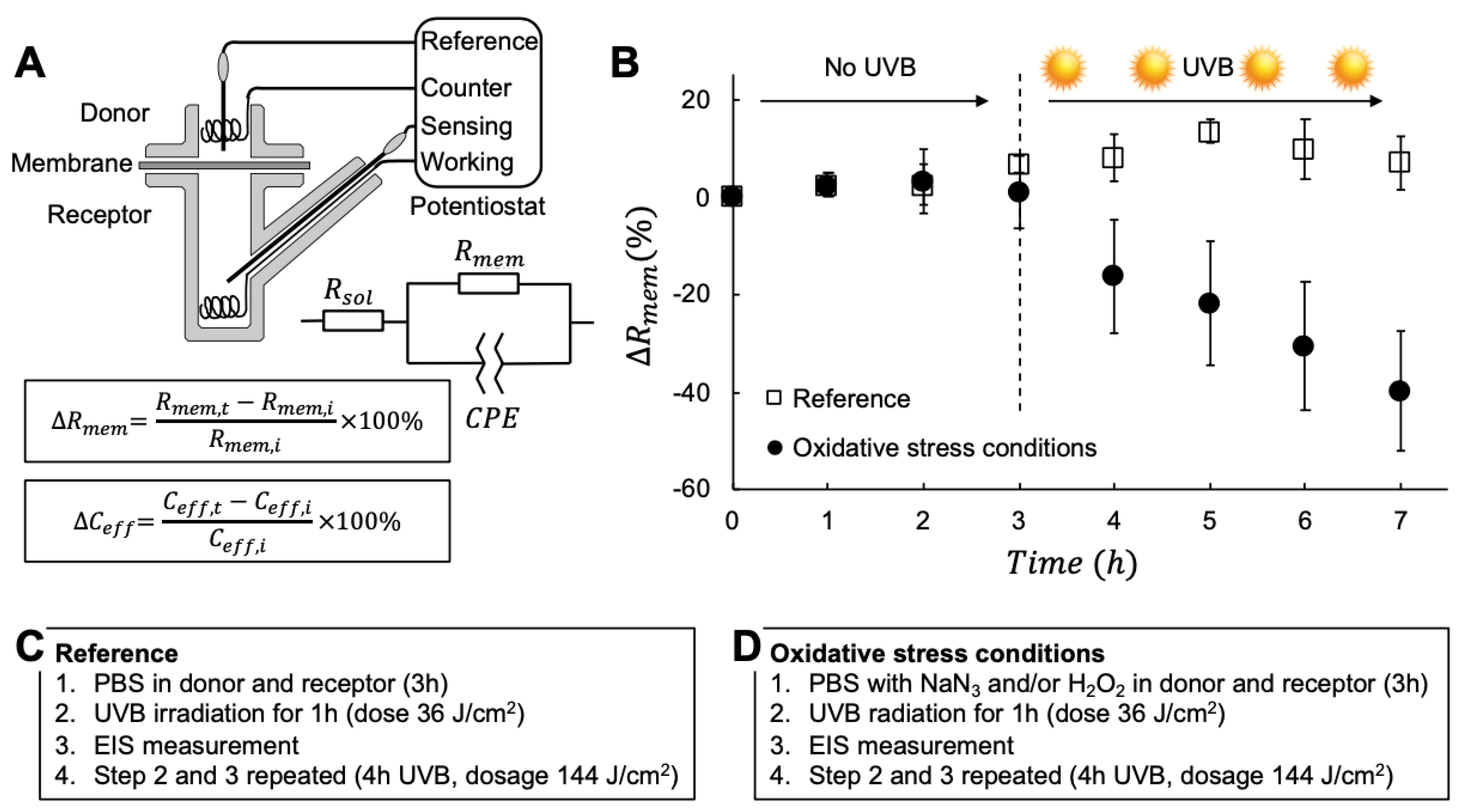
Figure 1. (A) Schematic representation of the 4-electrode EIS setup, equivalent circuit, and definitions of Δ R m e m and Δ C e f f . Two platinum wires served as working and counter electrodes and two Ag/AgCl/3M KCl electrodes were used as sensing and reference electrodes. R s o l is the resistance of the donor and receptor solution, R m e m is the membrane resistance, and C P E is a constant-phase element used to derive the effective capacitance of the membrane, C e f f . (B) Representative data (average values ± SD, n = 3) from reference experiments with no oxidative stress parameters (i.e., neat PBS) and with oxidative stress conditions (in this case PBS containing 10 mM NaN3 and 1 mM H2O2). The impedance properties of the membranes were examined for 3h without UVB irradiation, followed by 4 h of UVB irradiation. The experimental procedures used to generate the data in (B) are specified in (C) and (D).
Sensors 19 02376 g001
Figure 2. Summary of Δ R m e m (%) after 3 h without UVB irradiation and 4 h of total UVB irradiation (corresponding to 144 J/cm2) in combination with different stress parameters present in both the donor and receptor media. Data show average values (n = 3) with error bars showing either +SD (without UVB) or −SD (with UVB); n = 6 for A and n = 2 for F and G.
Sensors 19 02376 g002
Figure 3. (A) Summary of Δ R m e m (%) after 6 h UVB irradiation (216 J/cm2) and protection from topically applied sunscreen formulations. PBS with 10 mM NaN3 and 1 mM H2O2 was included as control, without and with UVB irradiation. Data show average values (n = 3) with error bars showing either +SD (without UVB) or −SD (with UVB). (B) Δ R m e m as a function of SPF value without and with UVB irradiation. The coefficient of determination for the regression line corresponding to the Δ R m e m after UVB irradiation was r2 = 0.87. (C) Schematic illustration of the experimental setup with presence of 10 mM NaN3 and 1 mM H2O2 in the donor and receptor media.
Sensors 19 02376 g003
Figure 4. (A) Summary of Δ C e f f (%) after 6 h UVB irradiation (216 J/cm2) and protection from topically applied sunscreen formulations. PBS with 10 mM NaN3 and 1 mM H2O2 was included as control, without and with UVB irradiation. Data show average values (n = 3) with error bars showing either +SD (without UVB) or −SD (with UVB). (B) Δ C e f f as a function of SPF value without and with UVB irradiation. (C) Schematic illustration of the experimental setup with presence of 10 mM NaN3 and 1 mM H2O2 in the donor and receptor media.
Sensors 19 02376 g004
Figure 5. Excised pig skin membrane soaked in 10 mM NaN3 and 1 mM H2O2 for 5 h without (A) and with (B) exposure to UVB irradiation (dosage corresponding to 180 J/cm2).
Sensors 19 02376 g005