Article

Surface-Electromyography-Based Co-Contraction Index for Monitoring Upper Limb Improvements in Post-Stroke Rehabilitation: A Pilot Randomized Controlled Trial Secondary Analysis

1
IRCCS Fondazione Don Carlo Gnocchi Onlus, Via Capecelatro 66, 20148 Milan, Italy
2
Department of Electronics, Information and Bioengineering (DEIB), Politecnico di Milano, Piazza Leonardo da Vinci 32, 20133 Milan, Italy
3
Department of Experimental Medicine, Section of Human Physiology, University of Genoa, 16132 Genoa, Italy
4
IRCCS Ospedale Policlinico San Martino, IRCCS, 16132 Genoa, Italy
5
Department of Neuroscience, Rehabilitation, Ophthalmology, Genetics and Maternal Child Health, University of Genoa, 16132 Genova, Italy
*
Author to whom correspondence should be addressed.
Sensors 2023, 23(17), 7320; https://doi.org/10.3390/s23177320
Submission received: 12 July 2023 / Revised: 28 July 2023 / Accepted: 4 August 2023 / Published: 22 August 2023
(This article belongs to the Section Biomedical Sensors)

Abstract

:
Persons post-stroke experience excessive muscle co-contraction, and consequently the arm functions are compromised during the activities of daily living. Therefore, identifying instrumental outcome measures able to detect the motor strategy adopted after a stroke is a primary clinical goal. Accordingly, this study aims at verifying whether the surface electromyography (sEMG)-based co-contraction index (CCI) could be a new clinically feasible approach for assessing and monitoring patients’ motor performance. Thirty-four persons post-stroke underwent clinical assessment and upper extremity kinematic analysis, including sEMG recordings. The participants were randomized into two treatment groups (robot and usual care groups). Ten healthy subjects provided a normative reference (NR). Frost’s CCI was used to quantify the muscle co-contraction of three different agonist/antagonist muscle pairs during an object-placing task. Persons post-stroke showed excessive muscle co-contraction (mean (95% CI): anterior/posterior deltoid CCI: 0.38 (0.34–0.41) p = 0.03; triceps/biceps CCI: 0.46 (0.41–0.50) p = 0.01) compared to NR (anterior/posterior deltoid CCI: 0.29 (0.21–0.36); triceps/biceps CCI: 0.34 (0.30–0.39)). After robot therapy, persons post-stroke exhibited a greater improvement (i.e., reduced CCI) in proximal motor control (anterior/posterior deltoid change score of CCI: −0.02 (−0.07–0.02) p = 0.05) compared to usual care therapy (0.04 (0.00–0.09)). Finally, the findings of the present study indicate that the sEMG-based CCI could be a valuable tool in clinical practice.

1. Introduction

Stroke is one of the major causes of long-term disability worldwide, and stroke survivors typically have difficulty performing activities of daily living (ADLs) [1]. The impairment of the paretic arm is one of the most disabling post-stroke motor consequences [2], which includes an exaggerated muscle co-contraction (simultaneous activity of agonist and antagonist muscles) [3,4,5,6,7,8].
Agonist-antagonist muscle co-contraction is an essential and common motor control strategy used by the central nervous system to regulate joint stability and provide movement accuracy [9,10]. After a stroke, there is an alteration in these coordination patterns, resulting in motor deficits of the arm function [4]. Specifically, excessive muscle co-contraction after neurological injuries can lead to impaired movement due to muscle weakness [10], and increased physiological, metabolic, and energetic costs [9,11,12,13]. These alterations consequently cause a reduction in the net moment and power development [11], which are functionally disadvantageous and result in the inefficacy of human movement [11]. Therefore, the reduction in the exaggerated muscle co-contraction and subsequent recovery of more physiological patterns of muscle activation might lead to an improvement in upper limb functionality of persons post-stroke [14].
The identification of instrumented outcome measures, able to characterize the neural strategy adopted after a stroke, can be useful in clinical practice for a better understanding of how the disease modifies muscle strategies during the performance of movements, allowing the planning of personalized training [12] and monitoring the effect of treatment.
Surface electromyography (sEMG) records the electrical activity of muscles via electrodes placed over the skin, and it is a suitable tool for detecting antagonist muscle co-contraction, allowing to distinguish between physiological and pathological patterns [15,16,17,18,19,20,21].
In fact, for more than five decades, sEMG has been employed in the neurorehabilitation field as a tool to assess muscle activation and improve neuromuscular rehabilitation treatments [17,18]. For example, the EMG-based analysis has been used to investigate interlimb coordination, provide biofeedback, and track rehabilitation responses in subjects with neurological impairments [22]. In addition, the potential of sEMG in prognosticating recovery is clinically recognized, as it provides specific quantitative evidence for treatment decisions [22].
However, although the literature has already provided substantial evidence that sEMG applications supply relevant information in the neurorehabilitation area to enhance motor recovery, sEMG-based outcome measures currently have limited use in clinical practice [15,22].
Appropriate interpretation of sEMG needs both technical and clinical expertise since sEMG parameters extracted from the signal are typically obtained by means of complex methods [23]. Therefore, the availability of simplified and intuitive EMG-based analysis, provided that it correctly represents muscle recruitment, could facilitate the clinical application of sEMG in clinical settings [22].
The sEMG-based co-contraction index (CCI), which is commonly used for the quantification of muscle co-contraction [4,8,9], can be an easy-to-use method for assessing motor control, having a quick implementation and easy interpretation even by non-technical experts, such as clinicians [9]. Previous studies have proposed Frost’s CCI as a quantitative, robust, and accurate outcome measure in pathological subjects, capable of providing a rapid indication of the simultaneous activation of antagonist muscles with respect to healthy subjects [4,13,24,25,26]. The use of such an instrumented index overcomes the limitations related to clinical scales as outcome measures, which may suffer from poor sensitivity to changes and floor/ceiling effects.
We have already investigated the changes induced by rehabilitation on the patterns of muscle synergies activation using complex techniques of clustering, which decompose multiple sEMG signals into a limited number of primitive patterns [2]. However, the interpretation of the muscle synergies parameters is quite difficult and requires a close collaboration between clinicians and engineers, which makes it difficult to transfer such a procedure to clinical practice [15,22]. Consequently, the development of easy-to-use methods for assessing motor control in a clinical setting could be valuable in tailoring the treatment of persons post-stroke. This is a topic of particular interest in the clinical field, but a recent review highlighted that sEMG is a promising tool to further improve functional recovery and stressed the need to develop specific sEMG-based interventions [17].
This study aims at investigating whether an sEMG-based CCI, namely Frost’s CCI, which is a direct expression of muscular activity, can be an outcome measure able to detect (1) the deviation of the pathological muscle activation pattern from the physiological one, and (2) the rehabilitation-induced changes in the upper limb of persons post-stroke, setting a new clinically feasible approach in the evaluation of patients’ motor performance.

2. Materials and Methods
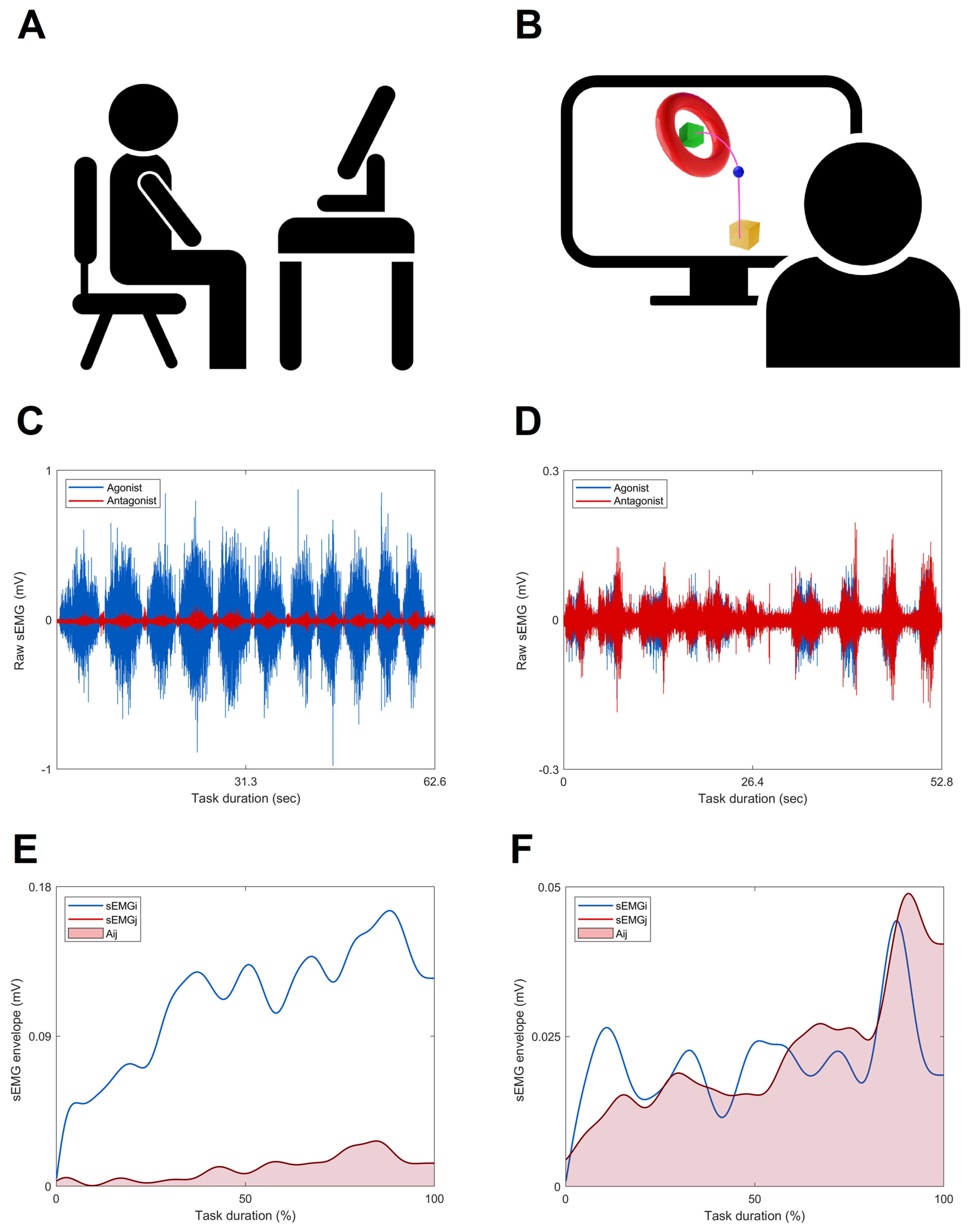
2.1. Participants

Persons post-stroke were recruited from the Neurorehabilitation Department of IRCCS Don C. Gnocchi Foundation (Milan, Italy) between March 2015 and November 2017. The inclusion criteria were as follows [1,2]: (i) age > 18 years, (ii) first-time ischemic or hemorrhagic stroke, (iii) a National Institute of Health Stroke Scale Motor Arm score ranging from 1 to 3, and (iv) a score higher than 6 out of 66 points on the Fugl-Meyer Motor Assessment of Upper Extremity (FM-UE) scale. The exclusion criteria were as follows [1,2]: (i) the presence of a moderate cognitive decline with a Mini-Mental State Examination score of <20 points, (ii) evidence of severe verbal comprehension deficit, apraxia, and/or visuospatial neglect (iii) evidence of behavioral disturbances (i.e., delusions, aggressiveness, and severe apathy/depression), which could affect compliance with the rehabilitation programs, (iv) presence of non-stabilized fractures, (v) presence of traumatic brain injury and (vi) presence of drug-resistant epilepsy.
A sample of 10 healthy subjects without any neurological or musculoskeletal disorders provided the normative reference.

2.2. Study Design

This study was a secondary analysis of the MOSE (Modularity for Sensory Motor Control) study (ClinicalTrial.gov, NCT03530358), a larger bi-center randomized controlled trial, and analyzed the data recorded at the IRCCS Don C. Gnocchi Foundation, Milan, Italy (Center 2), which tested the robot-assisted therapy for the upper limb rehabilitation after stroke [1,2].
The study design, in both centers, consisted of a single-blind two-arm randomized 1:1 controlled trial. The subjects’ randomization was performed using a computerized automated program and was stratified according to disease onset (≤3 months or >3 months) to ensure group comparability in terms of the number and chronicity of participants.

2.3. Rehabilitation Program

The rehabilitation program for the paretic arm for both treatment groups (robot group–RG and usual care group–UCG) consisted of 20 45 min sessions, carried out 5 times a week by trained physiotherapists. The interventions have been already described in previously published papers [1,2] and are briefly reported below.
The RG treatment consisted of a planar robotic manipulandum (Braccio Di Ferro, Celin s.r.l., Italy), as fully described in the literature [27]. The robotic system was designed to practice shoulder and elbow mobility of the paretic arm in the horizontal plane by controlling the position of the robot end-effector. During the training, subjects were asked to perform repeated center-out reaching movements and back from a central position to five peripheral spots randomly placed on a semicircle with a radius of 20 cm. The robot device could work in two different force modalities, assist-as-needed or resistive, which were chosen by the physiotherapist in each session depending on the subject’s abilities.
Participants allocated to the UCG underwent conventional arm-specific physiotherapy consisting of sessions of active and passive mobilization of the shoulder, scapula, elbow, and wrist, followed by task-oriented exercises customized to the skills of the participants. Treatment progression was achieved by increasing the range of motion, the number of repetitions, and muscular coordination demands.

2.4. Outcome Measures

2.4.1. Clinical Assessment

All participants post-stroke were clinically assessed by a qualified examiner, who was unaware of group allocation, at baseline (T0) and post-training (T1) using the FM-UE. FM-UE assessed the motor function of the paretic arm on a 3-point ordinal scale (0 = cannot perform; 1 = performs partially; and 2 = performs fully), applied to each of the scale items. Item scores were then summed to provide an overall score ranging from 0 (hemiplegia/severe impairment) to 66 (normal motor performance/no impairment).

2.4.2. Instrumented Assessment

All persons post-stroke were asked to perform a 3D functional motor task, named the object placing task, with the paretic arm. This task simulated a typical activity of daily living and was recorded at both T0 and T1 to evaluate the effects of rehabilitation treatments during an untrained functional task. In the resting position, the participants sat on a chair in front of a screen with both feet resting on the floor, the knees and hips bent at 90 degrees, and both hands placed at mid-thigh. During the execution of the task, subjects held an electromagnetic sensor with the paretic hand and received visual feedback of the performance in a virtual reality scenario (VRRS—Virtual Reality Rehabilitation System, Khymeia Group Ltd., Padova, Italy) (Figure 1A). Virtual objects were displayed on the screen representing the resting position (i.e., yellow box), the hand’s movements (i.e., blue ball), and the final position (i.e., green box), and the subjects were asked to move the virtual ball from the initial to the final position at their preferred velocity (Figure 1B).
The experimental setup and equipment have been previously described in detail [1,2]. Briefly, a 9-camera optoelectronic system (SMART-DX, BTS Bioengineering, Garbagnate Milanese, Italy) acquired the trajectory of the hand’s marker (10 mm diameter) of the tested limb with a sample rate of 200 Hz and low-pass filtered at 6 Hz.
Muscle activity was recorded using a wireless multichannel sEMG system (CometaWavePlus, Cometa s.r.l., Bareggio, Italy). Surface electrodes were placed according to the SENIAM (Surface Electromyography for the Non-Invasive Assessment of Muscles) specifications [28] and anatomical guidelines [29]. The activities of the following 6 muscles of the paretic limb, corresponding to three pairs of antagonistic muscles were recorded: (1) anterior deltoid (ANDE, agonist) and posterior deltoid (PODE, antagonist); (2) triceps brachii lateral head (TBLH, agonist) and biceps brachii short head (BBSH, antagonist); and (3) pronator teres (PRON, agonist) and supinator teres (SUPI, antagonist). The sEMG frequency of acquisition was 1000 Hz.
The instants of initiation and termination of each repetition of the object pacing task were computed as the times at which the velocity of the hand marker exceeded or fell below a threshold of 5% of the maximum value [1,2]. Raw sEMG signals (examples reported in Figure 1C,D) were band-pass filtered (10–400 Hz, 2nd order Butterworth), full-wave rectified and then low-pass filtered (4 Hz, 4th order Butterworth) in order to obtain the sEMG linear envelopes [24]. The sEMG linear envelopes were time-normalized to 100% of the movement duration, and then, to preserve the variability in sEMG, the signal of each muscle was amplitude-normalized to their maximum peak value obtained on all the recorded tasks [30].

2.4.3. CCI Computation

Frost’s CCI (Equation (1)) was computed to quantify the muscle co-contraction of the three antagonistic muscle pairs recorded using sEMG. This approach required an a priori classification of muscle antagonists or agonists, depending on the generated movement [31]. During physiological movements, the agonist muscle exerted force and/or moment of force in the main direction defined by the task, whereas the antagonist muscle opposed this action [8,31].
C C I F r o s t = 1 T 0 T A i j t d t
In this expression (Equation (1)), proposed by Frost in 1997, A i j t represents the overlapping activity of the muscles i and j in the sEMG envelopes, while T represents the task duration of the signal (Figure 1E,F) [4,13,24,25,26]. The CCI ranges from 0 to 1 (dimensionless value (um)), where 0 indicates that the activities of the two muscles did not overlap at all during the task, while 1 indicates that the activities of the two muscles were fully overlapping, and the level of sEMG activity was maintained at 1 during the task [4,25]. Therefore, a higher value of the CCI indicates a greater muscle co-contraction and is classified using terms such as normal, increased, or reduced compared to the normative reference [32].
The CCI was computed for each trial and averaged across the repetitions for each participant at both T0 and T1.

2.4.4. Kinematic Variables Quantification

Movement kinematics were assessed by calculating the speed and smoothness parameters for each trial and averaging them across the repetitions for each participant at both T0 and T1.
Movement speed was calculated as the distance traveled by the hand marker during a movement trial divided by the time employed for that movement.
Movement smoothness was calculated as the number of movement units (number of peaks) of the hand marker tangential velocity profile divided by the hand marker trajectory distance for each movement trial [33,34,35].

2.5. Statistics

2.5.1. Sample Size Estimation

As reported in our previous studies [1,2], the sample size was estimated using the shoulder/elbow coordination kinematic index (CKI) as the primary outcome measure. Based on the change scores of the CKI with a Cohen’s d effect size of 1.40 between robot and control training previously published in the literature [36], a sample size of 24 subjects (12 per group) was calculated to detect a difference between the groups with α = 0.05 and power (1 − β) = 0.9. The previous overestimation (i.e., 20 per group) allowed us to obtain an adequate sample size for the analysis presented here. In fact, the guidelines for demonstration-of-concept pilot RCT studies on motor rehabilitation, such as the present study, report 15 subjects per group as the minimum sample size [37].

2.5.2. Statistical Analyses

Statistical analyses were performed using SPSS and JASP. In the presence of normally distributed data, parametric tests were performed and data were reported as mean and 95% confidence interval (CI), whereas in case of deviation from normality, non-parametric tests were chosen and data were presented as median and interquartile range or percentage.
The demographic and clinical baseline scores of persons post-stroke were compared between the two groups of treatment (RG vs. UCG). Chi-square tests were utilized to compare sex, stroke type, paretic side, and chronicity. Time since stroke, age, and FM-UE scores at T0 (pre-treatment) were analyzed using the Mann–Whitney U test (RG vs. UCG).
The differences in muscle co-contraction and movement kinematics of the upper limb between the healthy subjects (NR) and persons post-stroke were analyzed using an unpaired t-test of CCI and speed and smoothness at T0.
To assess the rehabilitation-induced changes (RG vs. UCG) in the muscle co-contraction and in movement kinematics of the paretic arm of persons post-stroke, the change scores (CS, post–pre) of CCI and speed and smoothness were compared by ANCOVA using values at T0 as covariates. Since persons post-stroke showed higher CCI values with respect to the normative reference at T0, values of the CCI post-training lower than pre-training (CS < 0) were interpreted as an improvement in upper limb functionality (i.e., reduction in muscles co-contraction), while higher post-training values (CS > 0) were interpreted as a worsening.
A non-parametric ANCOVA analysis (Quade’s test) of the FM-UE CS was also applied to evaluate clinical changes following rehabilitation using the FM-UE score at T0 as a covariate.
Spearman’s correlation analysis was applied to analyze the correlation between the change in scores of the FM-UE clinical scale and the CCI.
Between-group differences (PA vs. NR and RG vs. UCG) and effect sizes (Cohen’s d) were also computed. Cohen’s d values of 0.2, 0.5, and 0.8 represented small, moderate, and large effect sizes, respectively [38].
p-values less than or equal to 0.05 were used as a level of significance.

3. Results

Thirty-four persons post-stroke, both in chronic and sub-acute stages, completed the object-placing task without technical issues in the sEMG recording and were, therefore, considered for the present analysis: 17 of them were assigned to the RG and 17 to the UCG. A flow chart of the protocol is shown in Figure 2.

3.1. Baseline Assessment

The demographic and clinical features of persons post-stroke did not differ significantly between RG and UCG (Table 1).
The median FM–UE score (1st–3rd quartile) at T0 was 31.0 (13.0–49.3), indicating a level of disease severity ranging from mild to severe. The healthy subjects were six females and four males with a median age (1st–3rd quartile) of 66.0 (51.0–68.0) years. Both gender and age were not significantly different from those of persons post-stroke.

3.2. Comparison of the Instrumental Indices between Healthy Subjects and Persons Post-Stroke at T0

For the muscle co-contraction during the object-placing task performed at T0, the unpaired t-test revealed statistically significant differences in CCI values between PA and the normative reference (NR) in the anterior/posterior deltoids (p = 0.03, Cohen’s d = 0.83, Figure 3A) and triceps/biceps muscle pairs (p = 0.01, Cohen’s d = 1.04, Figure 3B). No difference emerged between PA and NR in the pronator/supinator muscle pair (Figure 3C). For the CCI nominal values of the two groups, see Table S1 in the Supplementary Materials.
Consistent with the CCI findings, the kinematic analysis showed slower movement speed and reduced smoothness in PA compared to NR (see Table S1 in the Supplementary Materials for nominal, p-values, and Cohen’s d).

3.3. Treatment Effects

No significant clinical effects of treatment were found between UCG and RG from the non-parametric ANCOVA analysis of the FM-UE change score (p = 0.15, median (1st–3rd quartile): UCG 4.0 (−0.5–11.0), RG 6.0 (3.0–10.0)) using the FM-UE score at T0 as a covariate.
The results showed treatment-specific effects on CCI in the shoulder (Figure 4A) and forearm regions (Figure 4C). In detail, in the RG, 52.9% of the participants showed a reduction in the CCI in the anterior/posterior deltoid muscle pair, while, conversely, most of the participants (70.6%) in the UCG showed an increase. Regarding the change score (Figure 4A), a statistically significant difference was found between groups (p = 0.05, Cohen’s d = 0.70), with the RG showing a greater reduction in CCI, indicating a reduction in deviation from normative values (i.e., positive effect) (see also Table S2 in the Supplementary Materials). Conversely, there was a reduction in the CCI in the pronator/supinator muscle pair in the UCG (76.5%) and an increase in the RG (64.7%). Regarding the change score (Figure 4C), a statistically significant difference was found between the treatment groups (p = 0.02, Cohen’s d = 0.85), with the UCG showing a greater reduction in CCI, indicating a reduction in deviation from normative values (i.e., positive effect) (see Table S2 in the Supplementary Materials).
No significant treatment effect was found in the CCI in the elbow region (Figure 4B). In fact, the CCI of the triceps/biceps muscle pair showed a reduction in normative values after training (see Table S2 in the Supplementary Materials) and a negative CS in both groups (Figure 4B).
Both groups, regardless of the type of treatment (UCG vs. RG, speed p = 0.54), showed an improvement greater than 10% in speed after rehabilitative interventions (see Table S2 in the Supplementary Materials). For the smoothness parameter, only the RG group showed a reduction in smoothness equal to 10% after training.

3.4. Correlation Analysis

Table 2 shows the correlation analysis between the change score of the CCI and the CS of the FM-UE clinical scale following rehabilitation interventions. From this analysis, no significant correlation was found between the change scores of muscle co-contraction and those of the FM-UE score.

4. Discussion

The aim of this study was twofold as follows: (i) to examine the ability of sEMG-based CCI to discriminate motor performance between persons post-stroke and healthy subjects, and (ii) to detect changes induced by motor rehabilitation during an untrained 3D functional task. We compared the sEMG data from a cohort of persons post-stroke who underwent two different rehabilitation approaches.
The main finding of our study was that persons post-stroke showed a greater co-contraction of the proximal antagonistic muscle pairs (i.e., higher values of CCI) of the paretic arm, while the distal muscle pair co-activation was not altered. Furthermore, we found that the robotic training induced a greater improvement (i.e., negative CCI CS) in the motor control of the shoulder joint compared to the control intervention.

4.1. Comparison of the Instrumental Indices between Healthy Subjects and Persons Post-Stroke at T0

The proximal CCI was able to detect the altered pattern of muscle activation in the paretic side of persons post-stroke highlighting a deviation from the normative reference (Figure 3A,B). These results are consistent with the widespread scientific evidence that persons post-stroke exhibit higher atypical co-contraction levels between paretic muscle pairs when compared to healthy subjects [3,4,5,6,7].
Although previous studies have reported an altered pattern of muscle activation in the distal paretic region of persons post-stroke with respect to the normative reference [3,4,5], no excessive muscle co-contraction in the forearm region emerged in our study. Specifically, Hammond et al. [3] found an alteration in the muscle co-contraction in the flexor carpi radialis/extensor carpi radialis longus during voluntary isometric contraction, Sheng et al. [4] in the extensor/flexor digitorum during a horizontal task, and Kamper et al. [5] in hand muscles under voluntary isometric, isokinetic, and free-range conditions. All these tasks implied a distal muscle activation, while the task presented here was more demanding for the proximal regions, so the pronator/supinator muscle pair was probably less activated with only the stability role of the forearm segment. As a result, in our analysis, the abnormal muscular coordination pattern emerged primarily in the triceps/biceps and anterior/posterior deltoid muscle pairs, in accordance with our previous findings [1,2].
The identification of a biological signals-based index (e.g., CCI) that reflects the pathological pattern is crucial for investigating the degree of deviation from the physiological pattern. Overall, the persons post-stroke have difficulty deactivating the muscles of the paretic arm (i.e., higher CCI values) [4]. This motor control alteration affects motor performance, causing inefficiency of movements, as indicated by the reduced speed and smoothness. Therefore, the level of muscle co-contraction compared to normative references can be a useful measure for assessing motor performance and residual motor control in persons post-stroke.

4.2. Treatment Effects

Recently, Sheng et al. [4] found that the level of muscle co-contraction is associated with the impairment of the corticospinal tracts (CST), as assessed by measuring the motor-evoked potential, suggesting that excessive muscle co-contraction in the upper limb of persons post-stroke has a cortical origin. The abnormality of muscle coordination is linked to the loss of corticospinal projections, and consequently, rehabilitation treatments that enhance the excitability of the CST, especially in subjects with central nervous system injuries (like stroke), could lead to the recovery of a more physiological pattern of muscle activation [4].
In this context, the CCI and the analysis presented here could be a suitable tool both for deciding the most appropriate rehabilitation approach and for assessing possible rehabilitation-induced changes. Indeed, global assessments of the motor function and performance improved after training as measured by the FM-UE scale and spatio-temporal kinematic parameters; however, neither evaluation method highlighted any significant treatment effect on upper limb functionality, indicating low responsiveness of both [39]. Conversely, EMG-based CCI (Figure 4) was able to detect changes in muscle co-contraction between pre- and post-rehabilitation. Furthermore, these changes were treatment-specific (RG or UCG, see Figure 4), in agreement with a previous study [2]. In detail, the ANCOVA analysis showed that robotic rehabilitation led to a reduction in the CCI value of the shoulder and elbow muscles (proximal improvement) and an increase (distal worsening) in the co-contraction of the forearm muscles, whereas conventional therapy induced a reduction in the CCI in the forearm region (distal improvement) and an increase in the shoulder joint (proximal worsening). Consistent with our study, Hu et al. [25] also demonstrated a reduction in the abnormal muscle co-contraction in the proximal regions (i.e., shoulder and elbow joints) following robot-assisted elbow training.
The negative effects of rehabilitation treatments should be considered when optimizing future rehabilitation programs. The distal excessive co-contraction developed after the robot treatment could be linked to the design of the robot itself, which, being planar, is mainly focused on shoulder/elbow coordination. As planar robots constitute a standard paradigm for upper limb rehabilitation, the design of these devices should be modified to achieve a greater involvement of the distal segment, or additional exercises of the distal region should be administered in case of using planar robots. The excessive shoulder co-contraction following conventional therapy could be related to a less precise and repeatable, and therefore less effective, therapeutic effect [40] compared to the robotic therapy, on the well-known problems of shoulder impairments after stroke both in terms of glenohumeral joint injury [41] and overuse in the adopted compensatory strategy [42].
The results of this study pointed out that CCI can also be a useful biomarker for tailored rehabilitation as it provides suggestions on how to improve and personalize treatments. For example, robot therapy with the Braccio di Ferro manipulandum might be ameliorated by combining it with rehabilitation exercises (conventional or even robot-assisted) specifically targeted at the wrist/hand muscles.
The present results on muscle overactivity are in line with our previous study [2], in which we found comparable rehabilitation-induced changes in muscle synergies activation patterns [43] using a more complex evaluation (i.e., the motor control was decomposed into a limited number of primitive patterns using clustering techniques), whose extraction and interpretation require high technical expertise and is, therefore, difficult to implement in clinical practice. Conversely, the use of an easily computable and intuitive index such as CCI seems to be a more feasible approach for direct clinical application.
As highlighted by our findings, the CCI value detects the changes in the upper extremity motor function, indicating that it could be used to support clinical assessment and monitor motor improvement during stroke recovery [4]. In fact, the sEMG-based assessment of muscle co-contraction has recently been included for the benchmarking of upper limb functions in neurological disorders [44]. Indeed, the authors of this study [44], which focused on standardizing the evaluation of intra-limb coordination and identifying a quantitative and high-resolution metric, selected the CCIs as performance indicators (i.e., “outcome measure that allows the quantitative assessment of a motor ability” [44]).
Traditionally, the assessment of arm motor function and the effectiveness of rehabilitation treatments rely on standard clinical scales [44]. Our analysis of the correlation between the rehabilitation-induced changes in CCI value and FM-UE score (Table 2) showed no association between these two measures, consistent with the findings of other studies [4,44]. On the contrary, Chae et al. [45] and Chalard et al. [46] found a negative correlation between the level of co-contraction and upper limb motor functionality (i.e., indicated by the score of FM-UE). This incongruence was probably related to the different experimental conditions from which the sEMG signal was recorded; the above-cited studies examined the muscle co-contraction in an isometric condition of single-joint movements, whereas we assessed muscle concentric activation during a goal-directed movement, which implied a 3D dynamic coupling and coordination of the multi-joint system. FM-UE measures gross and isolated single-joint movements; therefore, the presence of correlation between CCI and FM-UE is more probable in isometric conditions than in a task that requires coordination, such as the one performed here.
Moreover, clinical scales have some metric characteristics (e.g., measurement of ordinality, and possible floor and ceiling effects) that might limit their reliability and responsiveness, particularly when compared with an objective, instrument-based, and continuous measure, like CCI.
In persons post-stroke, the features expressed in most functional movements, such as the task presented here, are the direct consequence of the lesion to the CST and/or somatosensory cortex: (i) loss of individual articular joint movements, (ii) general slowness with temporal irregularities and jerkiness, (iii) alterations in the somato-motor reactive control with time delays, and (iv) disrupted inter-joint muscle coordination in the upper limb.
In this framework, our results proved that EMG-based CCI provides additional information for a deeper understanding of muscle coordination impairments. Since the recovery at the biological level, particularly with respect to the CST, is an important vector for the return of upper limb motor control, such an index could be a promising tool for selecting proper rehabilitation treatment [22,44], increasing functional recovery [17], and improving the reliability and reproducibility of studies that evaluate the effectiveness of clinical interventions [18].

4.3. Study Limitations

This study does have some limitations. First, sEMG assesses the final peripheral pathway, although we expect sEMG signals to also provide information about motor coordination. A second limitation is the lack of follow-up assessments that did not allow the analysis of the retention of training effects.
Finally, the inclusion of distal robotic components should also be addressed to possibly enhance the effect on the co-contraction of wrist muscles and promote a better transfer to ADLs.
Aware of the above limitations, future studies with a larger sample size should be performed to corroborate the present findings, including EEG/fMRI evaluations, and to assess long-term training effects.

5. Conclusions

Quantifying upper limb motor functionality is essential to comprehend and monitor neuromotor recovery [44,47]. Our study indicates that sEMG-based CCI may be a promising quantitative index to assess motor deficit levels and rehabilitation-induced changes in motor skills of persons post-stroke. Its capacity to describe changes in motor coordination is important for a more complete motor assessment of arm functionality post-stroke and might be of benefit for tailored rehabilitation leading to improved arm use in daily life.

Supplementary Materials

The following supporting information can be downloaded at: https://www.mdpi.com/article/10.3390/s23177320/s1, Table S1. Mean and 95% confidence interval of CCI and of kinematic parameters of healthy subjects and paretic arm of persons post-stroke, before rehabilitation treatment (T0), during the object placing task; Table S2. Mean and 95% confidence interval of CCI and of kinematic parameters of healthy subjects and paretic arm of persons post-stroke, before (T0) and after (T1) rehabilitation treatment, during the object placing task.

Author Contributions

Conceptualization, J.J., M.F. and T.L.; Methodology, I.C., A.M. and T.L.; Software, V.B. and A.M.; Validation, V.B. and T.L.; Formal analysis, V.B. and I.C.; Investigation, I.C., A.M., J.J. and T.L.; Writing—original draft preparation, V.B. and T.L.; Writing—review and editing, V.B., I.C., A.M., J.J., C.A.F., L.A., E.P., M.F. and T.L.; Visualization, V.B.; Supervision, C.A.F., M.F. and T.L.; Project administration, I.C. and M.F.; Funding acquisition, J.J., E.P. and M.F. All authors have read and agreed to the published version of the manuscript.

Funding

This research was supported by #NEXTGENERATIONEU (NGEU) and funded by the Ministry of University and Research (MUR), National Recovery and Resilience Plan (NRRP), project MNESYS (PE0000006)—A Multiscale integrated approach to the study of the nervous system in health and disease (DN. 1553 11.10.2022).

Institutional Review Board Statement

The study was conducted in accordance with the Declaration of Helsinki and approved by the ethical committee of IRCCS Don Carlo Gnocchi Foundation, Milan, Italy (code number 15_11_2014, session 15 October 2014).

Informed Consent Statement

Informed consent was obtained from all subjects involved in the study.

Data Availability Statement

The dataset used and/or analyzed during the current study are available from the corresponding author upon reasonable request.

Acknowledgments

We thank all patients for participating in the study.

Conflicts of Interest

The authors declare no conflict of interest.

References

  1. Carpinella, I.; Lencioni, T.; Bowman, T.; Bertoni, R.; Turolla, A.; Ferrarin, M.; Jonsdottir, J. Effects of Robot Therapy on Upper Body Kinematics and Arm Function in Persons Post Stroke: A Pilot Randomized Controlled Trial. J. Neuroeng. Rehabil. 2020, 17, 1–19. [Google Scholar] [CrossRef]
  2. Lencioni, T.; Fornia, L.; Bowman, T.; Marzegan, A.; Caronni, A.; Turolla, A.; Jonsdottir, J. A Randomized Controlled Trial on the Effects Induced by Robot-Assisted and Usual-Care Rehabilitation on Upper Limb Muscle Synergies in Post-Stroke Subjects. Sci. Rep. 2021, 11, 5323. [Google Scholar] [CrossRef] [PubMed]
  3. Hammond, M.C.; Fitts, S.S.; Kraft, G.H.; Nutter, P.B.; Trotter, M.J.; Robinson, L.M. Co-Contraction in the Hemiparetic Forearm: Quantitative EMG Evaluation. Arch. Phys. Med. Rehabil. 1988, 69, 348–351. [Google Scholar] [PubMed]
  4. Sheng, W.; Li, S.; Zhao, J.; Wang, Y.; Luo, Z.; Lo, W.L.A.; Ding, M.; Wang, C.; Li, L. Upper Limbs Muscle Co-Contraction Changes Correlated With the Impairment of the Corticospinal Tract in Stroke Survivors: Preliminary Evidence From Electromyography and Motor-Evoked Potential. Front. Neurosci. 2022, 16, 886909. [Google Scholar] [CrossRef] [PubMed]
  5. Kamper, D.; Rymer, W. Impairment of Voluntary Control of Finger Motion Following Stroke: Role of Inappropriate Muscle Coactivation. Muscle Nerve 2001, 24, 673–681. [Google Scholar] [CrossRef]
  6. Stoeckmann, T.M.; Sullivan, K.J.; Scheidt, R.A. Elastic, Viscous, and Mass Load Effects on Poststroke Muscle Recruitment and Co-Contraction During Reaching: A Pilot Study. Phys. Ther. 2009, 89, 665–678. [Google Scholar] [CrossRef]
  7. Silva, C.C.; Silva, A.; Sousa, A.; Pinheiro, A.R.; Bourlinova, C.; Silva, A.; Salazar, A.; Borges, C.; Crasto, C.; Correia, M.V.; et al. Co-Activation of Upper Limb Muscles during Reaching in Post-Stroke Subjects: An Analysis of the Contralesional and Ipsi-lesional Limbs. J. Electromyogr. Kinesiol. 2014, 24, 731–738. [Google Scholar] [CrossRef]
  8. Latash, M.L. Muscle Coactivation: Definitions, Mechanisms, and Functions. J. Neurophysiol. 2018, 120, 88–104. [Google Scholar] [CrossRef]
  9. Li, G.; Shourijeh, M.S.; Ao, D.; Patten, C.; Fregly, B.J. How Well Do Commonly Used Co-Contraction Indices Approximate Lower Limb Joint Stiffness Trends During Gait for Individuals Post-Stroke? Front. Bioeng. Biotechnol. 2021, 8, 588908. [Google Scholar] [CrossRef]
  10. Busse, M.E.; Wiles, C.M.; Van Deursen, R.W.M. Co-Activation: Its Association with Weakness and Specific Neurological Pa-thology. J. Neuroeng. Rehabil. 2006, 3, 26. [Google Scholar] [CrossRef]
  11. Ranavolo, A.; Mari, S.; Conte, C.; Serrao, M.; Silvetti, A.; Iavicoli, S.; Draicchio, F. A New Muscle Co-Activation Index for Biomechanical Load Evaluation in Work Activities. Ergonomics 2015, 58, 966–979. [Google Scholar] [CrossRef]
  12. Souissi, H.; Zory, R.; Bredin, J.; Gerus, P. Comparison of Methodologies to Assess Muscle Co-Contraction during Gait. J. Biomech. 2017, 57, 141–145. [Google Scholar] [CrossRef] [PubMed]
  13. Frost, G.; Dowling, J.; Dyson, K.; Bar-Or, O. Cocontraction in Three Age Groups of Children during Treadmill Locomotion. J. Electromyogr. Kinesiol. 1997, 7, 179–186. [Google Scholar] [CrossRef] [PubMed]
  14. Wright, Z.A.; Rymer, W.Z.; Slutzky, M.W. Reducing Abnormal Muscle Coactivation After Stroke Using a Myoelec-tric-Computer Interface: A Pilot Study. Neurorehabil. Neural. Repair. 2014, 28, 443–451. [Google Scholar] [CrossRef] [PubMed]
  15. Campanini, I.; Disselhorst-Klug, C.; Rymer, W.Z.; Merletti, R. Surface EMG in Clinical Assessment and Neurorehabilitation: Barriers Limiting Its Use. Front. Neurol. 2020, 11, 934. [Google Scholar] [CrossRef]
  16. Ginszt, M.; Zieliński, G. Novel Functional Indices of Masticatory Muscle Activity. J. Clin. Med. 2021, 10, 1440. [Google Scholar] [CrossRef]
  17. Munoz-Novoa, M.; Kristoffersen, M.B.; Sunnerhagen, K.S.; Naber, A.; Alt Murphy, M.; Ortiz-Catalan, M. Upper Limb Stroke Rehabilitation Using Surface Electromyography: A Systematic Review and Meta-Analysis. Front. Hum. Neurosci. 2022, 16, 897870. [Google Scholar] [CrossRef]
  18. McManus, L.; De Vito, G.; Lowery, M.M. Analysis and Biophysics of Surface EMG for Physiotherapists and Kinesiologists: Toward a Common Language With Rehabilitation Engineers. Front. Neurol. 2020, 11, 576729. [Google Scholar] [CrossRef]
  19. Soylu, A.R.; Arpinar-Avsar, P. Detection of Surface Electromyography Recording Time Interval without Muscle Fatigue Effect for Biceps Brachii Muscle during Maximum Voluntary Contraction. J. Electromyogr. Kinesiol. 2010, 20, 773–776. [Google Scholar] [CrossRef]
  20. Sbriccoli, P.; Bazzucchi, I.; Rosponi, A.; Bernardi, M.; De Vito, G.; Felici, F. Amplitude and Spectral Characteristics of Biceps Brachii SEMG Depend upon Speed of Isometric Force Generation. J. Electromyogr. Kinesiol. 2003, 13, 139–147. [Google Scholar] [CrossRef]
  21. Naeije, M.; McCarroll, R.S.; Weijs, W.A. Electromyographic Activity of the Human Masticatory Muscles during Submaximal Clenching in the Inter-Cuspal Position. J. Oral. Rehabil. 1989, 16, 63–70. [Google Scholar] [CrossRef] [PubMed]
  22. Feldner, H.A.; Howell, D.; Kelly, V.E.; McCoy, S.W.; Steele, K.M. “Look, Your Muscles Are Firing!”: A Qualitative Study of Clinician Perspectives on the Use of Surface Electromyography in Neurorehabilitation. Arch. Phys. Med. Rehabil. 2019, 100, 663–675. [Google Scholar] [CrossRef] [PubMed]
  23. Merletti, R.; Campanini, I.; Rymer, W.Z.; Disselhorst-Klug, C. Editorial: Surface Electromyography: Barriers Limiting Widespread Use of SEMG in Clinical Assessment and Neurorehabilitation. Front. Neurol. 2021, 12, 642257. [Google Scholar] [CrossRef] [PubMed]
  24. Charafeddine, J.; Pradon, D.; Chevallier, S.; Alfayad, S.; Khalil, M. Neuromotor Strategy of Gait Rehabilitation for Lower-Limb Spasticity. In Proceedings of the Fifth International Conference on Advances in Biomedical Engineering (ICABME), Tripoli, Lebanon, 17–19 October 2019; pp. 1–4. [Google Scholar]
  25. Hu, X.; Tong, K.Y.; Song, R.; Tsang, V.S.; Leung, P.O.; Li, L. Variation of Muscle Coactivation Patterns in Chronic Stroke During Robot-Assisted Elbow Training. Arch. Phys. Med. Rehabil. 2007, 88, 1022–1029. [Google Scholar] [CrossRef]
  26. Qian, Q.; Hu, X.; Lai, Q.; Ng, S.C.; Zheng, Y.; Poon, W. Early Stroke Rehabilitation of the Upper Limb Assisted with an Elec-tromyography-Driven Neuromuscular Electrical Stimulation-Robotic Arm. Front. Neurol. 2017, 8, 447. [Google Scholar] [CrossRef] [PubMed]
  27. Casadio, M.; Sanguineti, V.; Morasso, P.G.; Arrichiello, V. Braccio Di Ferro: A New Haptic Workstation for Neuromotor Rehabilitation. Technol. Health. Care. 2006, 14, 123–142. [Google Scholar] [CrossRef] [PubMed]
  28. Hermens, H.J.; Freriks, B.; Disselhorst-Klug, C.; Rau, G. Development of Recommendations for SEMG Sensors and Sensor Placement Procedures. J. Electromyogr. Kinesiol. 2000, 10, 361–374. [Google Scholar] [CrossRef]
  29. Blanc, Y.; Dimanico, U. Electrode Placement in Surface Electromyography (SEMG) “Minimal Crosstalk Area” (MCA). Open. Rehabil. J. 2010, 3, 110–126. [Google Scholar] [CrossRef]
  30. Morris, A.D.; Kemp, G.J.; Lees, A.; Frostick, S.P. A Study of the Reproducibility of Three Different Normalisation Methods in Intramuscular Dual Fine Wire Electromyography of the Shoulder. J. Electromyogr. Kinesiol. 1998, 8, 317–322. [Google Scholar] [CrossRef]
  31. Rinaldi, M.; D’Anna, C.; Schmid, M.; Conforto, S. Assessing the Influence of SNR and Pre-Processing Filter Bandwidth on the Extraction of Different Muscle Co-Activation Indexes from Surface EMG Data. J. Electromyogr. Kinesiol. 2018, 43, 184–192. [Google Scholar] [CrossRef]
  32. Rosa, M.C.N.; Marques, A.; Demain, S.; Metcalf, C.D. Lower Limb Co-Contraction during Walking in Subjects with Stroke: A Systematic Review. J. Electromyogr. Kinesiol. 2014, 24, 1–10. [Google Scholar] [CrossRef] [PubMed]
  33. Gulde, P.; Hermsdörfer, J. Smoothness Metrics in Complex Movement Tasks. Front. Neurol. 2018, 9, 615. [Google Scholar] [CrossRef] [PubMed]
  34. Murphy, M.A.; Willén, C.; Sunnerhagen, K.S. Kinematic Variables Quantifying Upper-Extremity Performance after Stroke during Reaching and Drinking from a Glass. Neurorehabil. Neural. Repair. 2011, 25, 71–80. [Google Scholar] [CrossRef] [PubMed]
  35. Casadio, M.; Giannoni, P.; Morasso, P.; Sanguineti, V. A Proof of Concept Study for the Integration of Robot Therapy with Physiotherapy in the Treatment of Stroke Patients. Clin. Rehabil. 2009, 23, 217–228. [Google Scholar] [CrossRef]
  36. Wu, C.Y.; Huang, P.C.; Chen, Y.T.; Lin, K.C.; Yang, H.W. Effects of Mirror Therapy on Motor and Sensory Recovery in Chronic Stroke: A Randomized Controlled Trial. Arch. Phys. Med. Rehabil. 2013, 94, 1023–1030. [Google Scholar] [CrossRef]
  37. Dobkin, B.H. Progressive Staging of Pilot Studies to Improve Phase III Trials for Motor Interventions. Neurorehabil. Neural. Repair. 2009, 23, 197–206. [Google Scholar] [CrossRef]
  38. Cohen, J. Statistical Power Analysis. Curr. Dir. Psychol. Sci. 1992, 1, 98–101. [Google Scholar] [CrossRef]
  39. Hobart, J.C.; Cano, S.J.; Zajicek, J.P.; Thompson, A.J. Rating Scales as Outcome Measures for Clinical Trials in Neurology: Problems, Solutions, and Recommendations. Lancet. Neurology. 2007, 6, 1094–1105. [Google Scholar] [CrossRef]
  40. Mehrholz, J.; Pohl, M.; Platz, T.; Kugler, J.; Elsner, B. Electromechanical and Robot-assisted Arm Training for Improving Ac-tivities of Daily Living, Arm Function, and Arm Muscle Strength after Stroke. Cochrane Database Syst. Rev. 2015. [Google Scholar] [CrossRef]
  41. Dursun, E.; Dursun, N.; Ural, C.E.; Çakci, A. Glenohumeral Joint Subluxation and Reflex Sympathetic Dystrophy in Hemiplegic Patients. Arch. Phys. Med. Rehabil. 2000, 81, 944–946. [Google Scholar] [CrossRef]
  42. Wang, X.; Fu, Y.; Ye, B.; Babineau, J.; Ding, Y.; Mihailidis, A. Technology-Based Compensation Assessment and Detection of Upper Extremity Activities of Stroke Survivors: Systematic Review. J. Med. Internet. Res. 2022, 24, e34307. [Google Scholar] [CrossRef]
  43. Roby-Brami, A.; Jarrassé, N.; Parry, R. Impairment and Compensation in Dexterous Upper-Limb Function After Stroke. From the Direct Consequences of Pyramidal Tract Lesions to Behavioral Involvement of Both Upper-Limbs in Daily Activities. Front. Hum. Neurosci. 2021, 15, 662006. [Google Scholar] [CrossRef]
  44. Longatelli, V.; Torricelli, D.; Tornero, J.; Pedrochi, A.; Molteni, F.; Pons, J.L.; Gandolla, M. A Unified Scheme for the Bench-marking of Upper Limb Functions in Neurological Disorders. J. Neuroeng. Rehabil. 2022, 19, 102. [Google Scholar] [CrossRef] [PubMed]
  45. Chae, J.; Yang, G.; Park, B.K.; Labatia, I. Muscle Weakness and Cocontraction in Upper Limb Hemiparesis: Relationship to Motor Impairment and Physical Disability. Neurorehabil. Neural. Repair. 2002, 16, 241–248. [Google Scholar] [CrossRef] [PubMed]
  46. Chalard, A.; Amarantini, D.; Tisseyre, J.; Marque, P.; Tallet, J.; Gasq, D. Spastic Co-Contraction, Rather than Spasticity, Is Associated with Impaired Active Function in Adults with Acquired Brain Injury: A Pilot Study. J. Rehabil. Med. 2019, 51, 307–311. [Google Scholar] [CrossRef] [PubMed]
  47. Schwarz, A.; Kanzler, C.M.; Lambercy, O.; Luft, A.R.; Veerbeek, J.M. Systematic Review on Kinematic Assessments of Upper Limb Movements after Stroke. Stroke 2019, 50, 718–727. [Google Scholar] [CrossRef] [PubMed]
Figure 1. The upper panel shows a schematic representation of the experimental set-up during the performance of a subject (A,B). The blue ball indicates the hand’s movement, the yellow box the rest position, and the green cube the final position. The purple line shows the hypothetical trajectory of a representative subject (not shown during the test). The middle panel reports examples of the raw sEMG signals of the agonist and antagonist muscles (anterior deltoid, blue line, and posterior deltoid, red line) during the object-placing task of a healthy subject (C) and a person post-stroke (D). The lower panel shows the normalized envelope of the muscle pairs’ anterior and posterior deltoids in a single repetition for a healthy subject (E) and a person post-stroke (F). The respective overlapping area is highlighted in light red (Aij, red area).
Sensors 23 07320 g001
Figure 2. Flow chart of the study.
Sensors 23 07320 g002
Figure 3. CCI of the normative reference (NR, gray band) and the paretic arm (PA, red circles) of persons post-stroke at T0 during the object placing task. Circles and whiskers represent, respectively, the mean and 95% confidence interval of CCI. * indicates significant differences between NR and PA (p ≤ 0.05, unpaired t-test).
Sensors 23 07320 g003
Figure 4. Change score of CCI of the paretic arm of persons post-stroke after usual care intervention (UCG, blue circles) and robot therapy (RG, red circles) during the object-placing task. Circles and whiskers represent, respectively, the mean and 95% confidence interval adjusted for CCI at T0 using the ANCOVA procedure. * indicates significant differences between UCG and RG (p ≤ 0.05, ANCOVA analysis).
Sensors 23 07320 g004
Table 1. Demographic and clinical characteristics of persons post-stroke allocated to robot and usual care groups.
VariableUCG (N = 17)
Median (1st–3rd)
RG (N = 17)
Median (1st–3rd)
p-Value
Age (years)59.0 (46.0–70.0)67.0 (58.0–72.0)0.20
Time since stroke (months)5.8 (1.9–91.4)7.8 (1.4–13.9)0.55
FM-UE21.0 (12.0–46.5)33.0 (16.0–50.5)0.22
NumberNumber
Sex 0.73
Female89
Male98
Stroke type 1.00
Ischemic1111
Hemorrhagic 66
Paretic side 0.49
Right68
Left119
Chronicity (>3 months) 1.00
Chronic1010
Sub-acute77
UCG: Usual care group. RG: Robot group. FM-UE: Fugl-Meyer Motor Assessment for the Upper Extremities. p-values indicate the results of the Mann–Whitney U Test for age, time since stroke, and FM-UE baseline score and of the chi-square tests for all the other variables.
Table 2. Spearman’s correlation analysis between the change scores of CCI and FM-UE clinical scale.
FM-UE CS
Correlation Coefficientp-Value
CCI CSAnterior/Posterior deltoids0.040.83
Triceps/Biceps0.030.85
Pronator/Supinator–0.090.63
CS: change scores. CCI: Frost’s co-contraction index. FM-UE: Fugl-Meyer Motor Assessment for the Upper Extremities. p-values indicate the results of Spearman’s correlation.
Disclaimer/Publisher’s Note: The statements, opinions and data contained in all publications are solely those of the individual author(s) and contributor(s) and not of MDPI and/or the editor(s). MDPI and/or the editor(s) disclaim responsibility for any injury to people or property resulting from any ideas, methods, instructions or products referred to in the content.

