Communication

Ultraviolet Photodetector Using Nanostructured Hexagonal Boron Nitride with Gold Nanoparticles

1
Department of Chemical, Biological, and Battery Engineering, Gachon University, Seongnam 13120, Republic of Korea
2
Department of Semiconductor Engineering, Gachon University, Seongnam 13120, Republic of Korea
3
Department of Electronic Engineering, Kwangwoon University, Seoul 01897, Republic of Korea
*
Author to whom correspondence should be addressed.
Sensors 2025, 25(3), 759; https://doi.org/10.3390/s25030759
Submission received: 8 January 2025 / Revised: 21 January 2025 / Accepted: 24 January 2025 / Published: 27 January 2025
(This article belongs to the Section Nanosensors)

Abstract

:
Ultraviolet (UV) photodetectors play a crucial role in various applications, ranging from environmental monitoring to biomedical diagnostics. This paper presents the fabrication and characterization of a high-performance UV photodetector using hexagonal boron nitride (hBN) decorated with gold nanoparticles (AuNPs). The hBN flakes were mechanically exfoliated onto SiO2 substrates, and AuNPs were formed via thermal evaporation, resulting in the creation of a plasmonically active surface that enhanced light absorption and carrier dynamics. Raman spectroscopy, transmission electron microscopy, and electrical measurements were performed to comprehensively analyze the device structure and performance. The photodetector exhibited significantly improved photocurrent and responsivity under UV-B (306 nm) and UV-C (254 nm) illumination, with the responsivity reaching an increase of nearly two orders of magnitude compared to that of the pristine hBN device. These improvements are attributed to the synergistic effects of the wide bandgap of hBN and the localized surface plasmon resonance of the AuNPs. These findings demonstrate the potential of AuNP-decorated hBN for advanced UV photodetection applications and provide a pathway toward more efficient and miniaturized optoelectronic devices.

1. Introduction

Ultraviolet (UV) photodetectors have attracted significant attention owing to their critical roles in environmental monitoring, flame detection, communication systems, and biomedical sensing [1,2,3,4,5]. The ability to detect UV radiation is essential for various scientific, industrial, and societal reasons, such as monitoring ozone layer depletion, detecting high-energy radiation sources, and preventing UV-induced damage to materials and biological systems. Photodetectors that are sensitive to UV-B (306 nm) and UV-C (254 nm) wavelengths are crucial for detecting hazardous radiation. However, achieving high sensitivity, rapid response time, and stable operation using UV photodetectors is challenging because of the inherent limitations of the material properties and device architectures.
Conventionally, wide-bandgap semiconductors such as GaN, ZnO, SiC, and GaO are the main materials for UV detection [6,7,8,9]. However, despite their effectiveness, their rigid and bulky characteristics pose challenges to their integration into flexible and scalable device architectures. According to Moore’s Law, electronic devices have become highly integrated, with dimensions reaching just a few nanometers or even sub-nanometer scales [10]. Consequently, photodetectors must also be miniaturized to the nanoscale to achieve a high performance and seamless integration within advanced technological systems. Since the discovery of graphene in 2004, research on two-dimensional (2D) materials has surged, leading to extensive exploration of various 2D-based materials. Over the past decade, the unique properties of single-layer graphene have garnered significant attention, positioning 2D materials at the forefront of research due to their potential applications in emerging nanoscale optoelectronic and electronic devices. Numerous studies have been conducted to investigate the physical characteristics and applications of these materials, focusing on phenomena such as energy band structures, carrier transport properties, the quantum Hall effect, and the spin–orbit coupling [11,12,13,14]. Hexagonal boron nitride (hBN) is a layered crystal with a hexagonal structure similar to that of graphite [15,16]. In its lattice, boron and nitrogen atoms are alternately arranged and bonded in-plane via sp2 hybridization, forming a hexagonal network. Multiple layers of this lattice stack together to create bulk hBN. The sp2 bonding of BN results in robust in-plane σ bonds, which contribute to its high thermal conductivity and chemical stability. Meanwhile, the unoccupied π orbitals render hBN electrically insulating with a wide bandgap of approximately 6 eV. Therefore, hBN has attracted considerable interest for UV photodetection applications [17,18,19,20,21]. Its 2D property, high transparency in the visible range, exceptional thermal stability, and chemical inertness make it an ideal candidate for high-performance UV photodetectors.
Despite their satisfactory properties, pristine hBN-based devices often exhibit low responsivity and limited photocurrent generation. This performance limitation is primarily attributed to the weak light absorption in the UV range and the inefficient charge carrier transport within the material. Recent studies have focused on overcoming these limitations through surface engineering, structural modifications, and incorporation of plasmonic nanostructures [22,23,24]. These approaches aim to enhance optical and electronic interactions within the photodetector, improving the device performance. Among various enhancement methods, decoration with nanoparticles has emerged as a particularly effective approach [25,26]. Localized surface plasmon resonance (LSPR) is a phenomenon observed in metallic nanoparticles or spatially confined islands, where conduction electrons collectively oscillate in resonance with incident electromagnetic waves. This resonance condition is highly sensitive to the surrounding medium’s dielectric constant and refractive index [27,28,29]. LSPR generates enhanced electromagnetic fields near the nanoparticle surface, significantly boosting light absorption and scattering. The confined electric field amplifies interactions with nearby materials, making it particularly advantageous for sensitive detection applications. In nanostructures, these localized fields contribute to higher photocarrier generation rates and improved charge transport dynamics. The interplay between electron oscillation and light absorption under LSPR facilitates enhanced optical properties, such as increased responsivity in photodetectors. The nanoscale tuning of these systems can optimize resonance conditions, providing a versatile platform for advanced optoelectronic applications. This plasmonic effect significantly facilitates light absorption and photocarrier generation in the underlying hBN layer, thereby improving the overall performance of the UV photodetectors. Furthermore, nanoparticles serve as efficient charge trapping and transfer centers, facilitating the separation and transport of photo-generated carriers and improving the response of the device to UV radiation.
In this study, we fabricate and characterize a UV photodetector using hBN decorated with gold nanoparticles (AuNPs), deposited under high vacuum conditions without the use of any solvent. The device structure is designed to leverage the synergistic effects of the wide-bandgap properties of hBN and the plasmonic enhancements of AuNPs. The photocurrent and responsivity of the device under different UV illumination conditions, including UV-B and UV-C wavelengths, are investigated. A comparative analysis of pristine hBN and AuNP-decorated hBN devices highlights the significant improvement in the photodetection performance achieved using this plasmonic decoration approach.

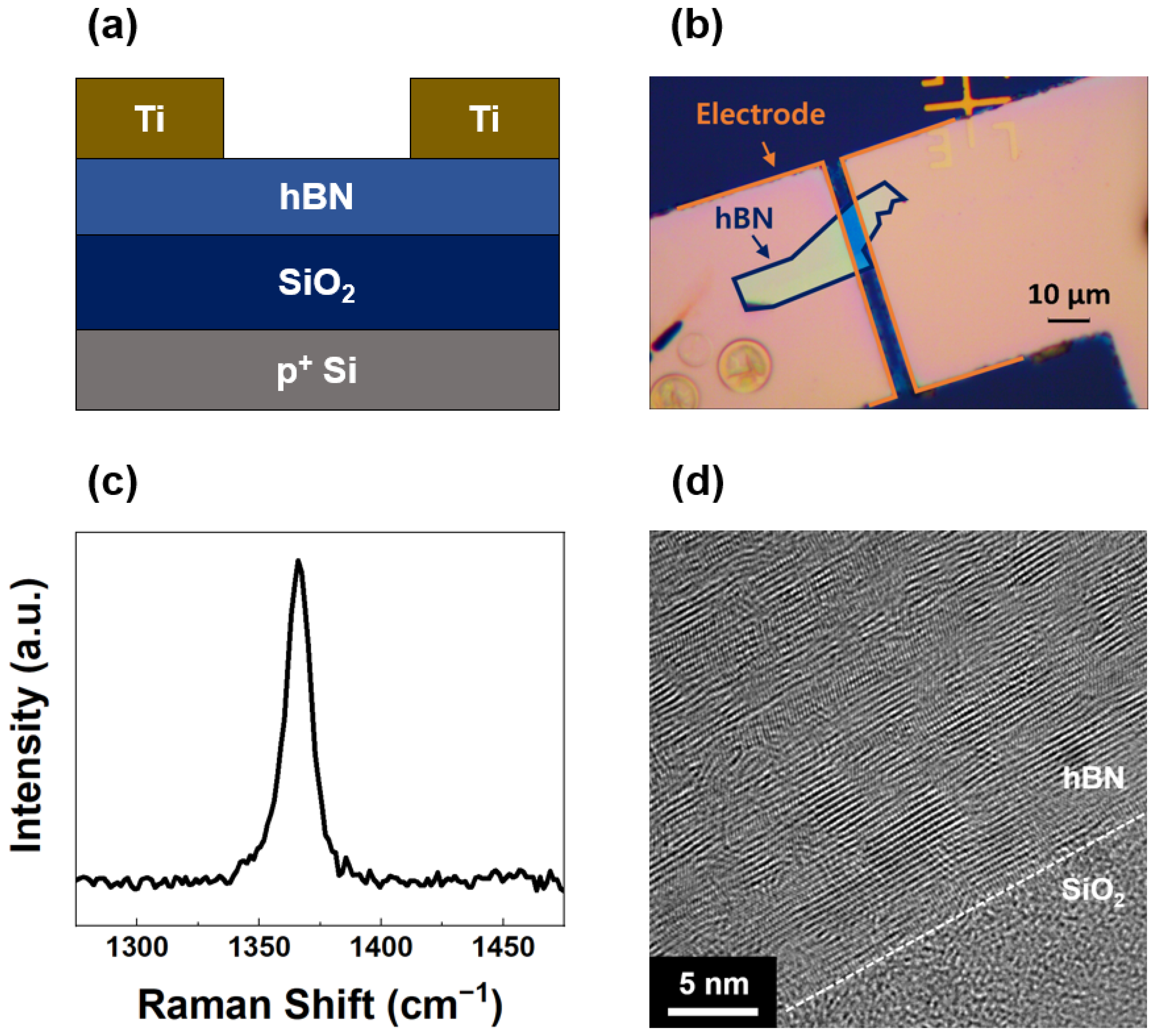
2. Materials and Methods

2.1. Materials

The fabrication of the UV photodetector device began with the mechanical exfoliation of the hBN flake (33 nm) from bulk hBN crystals supplied by 2D Semiconductors Corporation. Exfoliation was performed on a 90 nm silicon dioxide (SiO2) substrate. Prior to the exfoliation, the SiO2 substrate was cleaned to ensure optimal surface conditions. The cleaning process involved immersion in a piranha solution (H2SO4:H2O2 = 2:1) to remove organic contaminants, followed by an oxygen plasma treatment to enhance surface hydrophilicity. After exfoliation, the hBN flakes were annealed at 400 °C in a diluted H2 atmosphere to eliminate residual scotch tape adhesive, thereby ensuring a pristine surface quality. Titanium (Ti) electrodes with a 100 nm thickness were thermally evaporated and patterned with channel dimensions of 12 µm in width and 5 µm in length. The patterning process was carried out using a negative photoresist (NR9, Futurrex, Franklin, NJ, USA) in conjunction with a mask aligner (MA6, Karl Suss, Garching, Germany). Raman spectra were recorded using a high-resolution dispersive Raman microscope (514 nm Ar ion laser, ARAMIS, Horiba Jobin Yvon, Kyoto, Japan) with a laser spot size of 2 µm to ensure accurate measurements of the flakes. Transmission electron microscopy (TEM) images were captured using a Cs-corrected Titan TEM instrument (FEI) to provide high-resolution insights into the atomic structure of the hBN layers. AuNPs were formed on the hBN surface using a thermal evaporator (KVE-T2010, KVT, Gimpo, Republic of Korea). A thin gold film with a thickness of 3 nm was deposited at a rate of 0.1 nm/s under high vacuum conditions (~5 × 10⁻⁶ Torr). Due to the agglomeration of gold at atomically thin thicknesses, this process led to the formation of AuNPs. This thermal process was employed to produce high-quality AuNPs in a clean environment without the use of any solvents [30,31]. The formation of AuNPs was analyzed using scanning electron microscopy (SEM) (Magellan 400, FEI, Hillsboro, OR, USA) after coating the surface with a 5 nm thick layer of osmium (Os) to mitigate electron charging.

2.2. Methods

The performance of the photodetector under UV illumination was evaluated using a UV lamp (Dongseo Science, Dangjin, Republic of Korea) that emitted at two specific wavelengths: UV-B (306 nm) with an intensity of 1107 µW/cm2 and UV-C (254 nm) with an intensity of 1422 µW/cm2. Electrical measurements were performed using a chamber probe station (MST-1000B, MS TECH, Hwaseong, Republic of Korea) integrated with a parameter analyzer (4200 SCS, Tektronix, Beaverton, OR, USA) to ensure precise control and data acquisition during the experiments. During the evaluation of the optoelectronic performance, the UV lamp was fixed 5 cm directly above the device placed on the probe station stage.

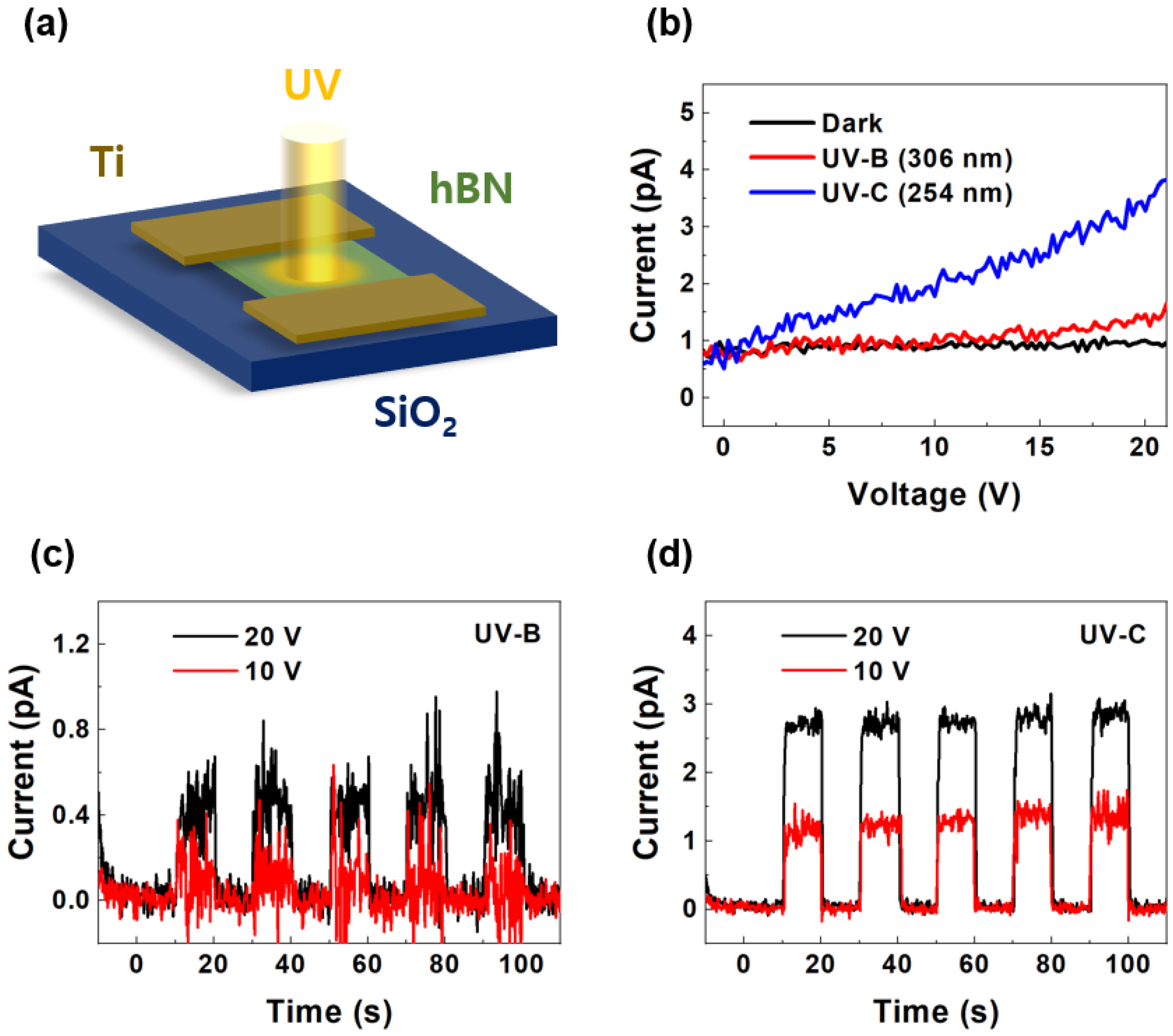
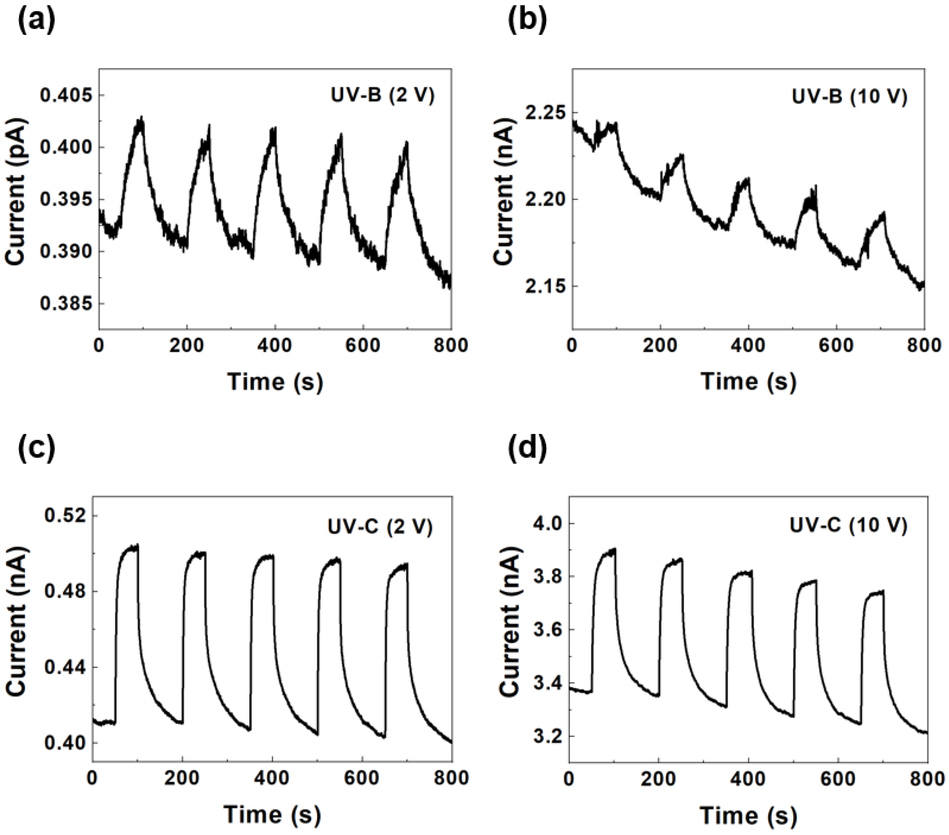
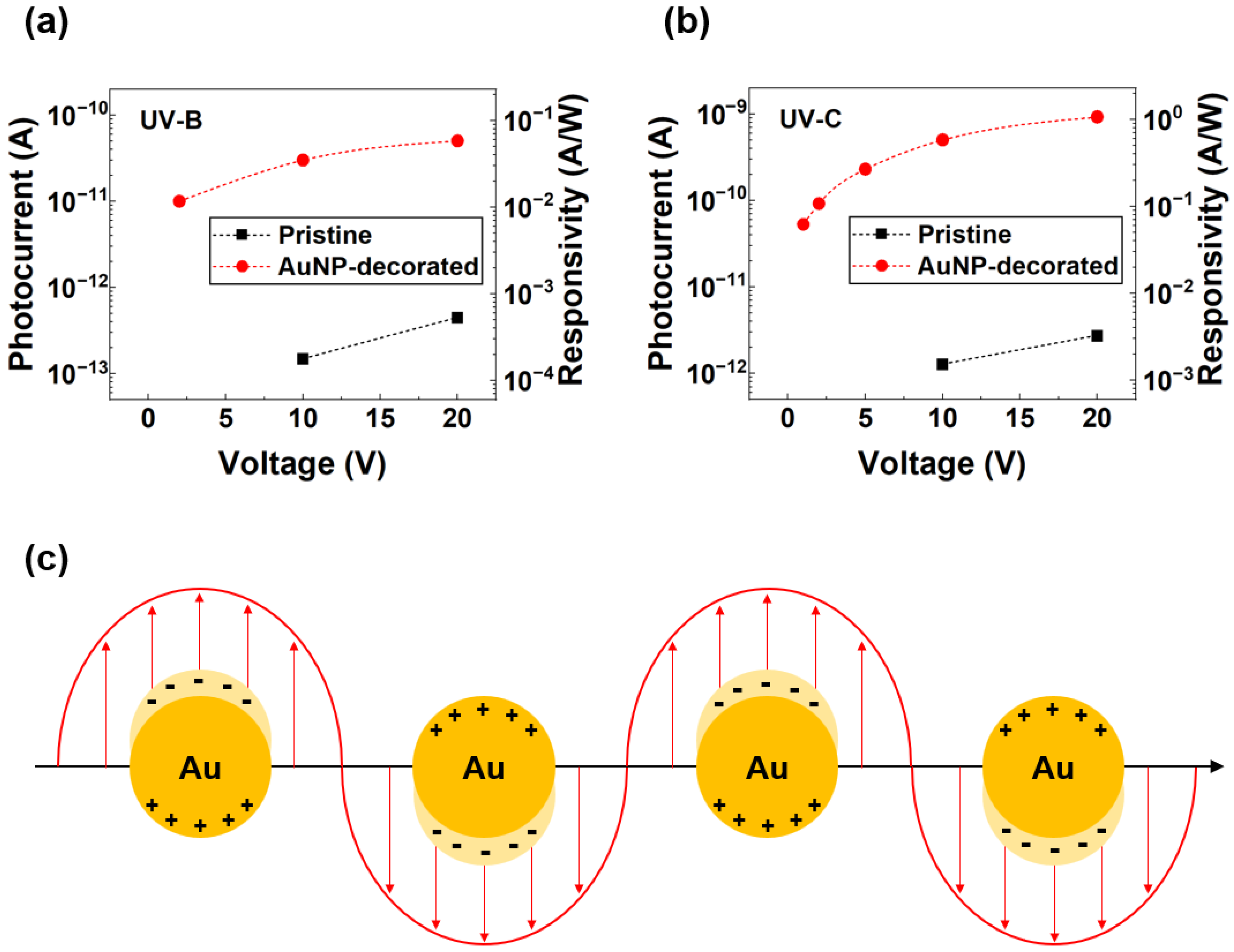
3. Results and Discussion

Figure 1a,b show a cross-sectional schematic and an optical microscopic image of the photodetector device, respectively, which consists of the hBN flake and the top electrodes on a SiO2/Si substrate. The blue line outlines the shape of the hBN flake, while the orange line represents the shape of the electrodes. Figure 1c shows the Raman spectrum of the hBN flake, exhibiting the peak at 1366 cm−1, corresponding to the in-plane E2g phonon mode. In contrast to graphene, which exhibits G, D, and 2D Raman bands, hBN exhibits only a G band corresponding to the E2g vibrational mode. The absence of a D band in hBN is attributed to the lack of a Kohn anomaly [32]. The Raman peak observed at 1366 cm−1 confirms that the hBN layer consists of multiple layers, more than four. Additionally, the sharp peak without additional peaks demonstrated the crystalline quality with minimal contamination from the exfoliation and annealing processes. Figure 1d shows a cross-sectional TEM image of the hBN-SiO2 interface. The interlayer van der Waals distances of hBN observed in the image are 0.33 nm along the c-axis of the crystal. The layered van der Waals structure of hBN and the well-defined interface between hBN and SiO2 are clearly visible. The results presented in Figure 1a–d collectively show the high quality of the hBN flake and its compatibility with the architecture of the device. The exfoliation and annealing processes successfully produced defect-free, uniform flakes with strong adhesion to the substrate. The Raman and TEM analyses validated the crystalline property and structural integrity of hBN which are critical for achieving high performance in UV photodetection.
Figure 2 shows the performance of the pristine hBN-based UV photodetector under UV-B (306 nm) and UV-C (254 nm) illumination. Figure 2a depicts the device and measurement configuration, in which UV light irradiates the hBN surface with an applied voltage through the Ti electrodes. Figure 2b shows the current–voltage (I-V) characteristics of the device under dark conditions (black), UV-B illumination (red), and UV-C illumination (blue). The photocurrent increased more under UV illumination than under dark conditions, with a higher response observed for UV-C because of its higher photon energy. The results confirm the sensitivity of the device to UV wavelengths and the effective charge carrier generation under UV exposure. Furthermore, the current and voltage exhibited a linear relationship, confirming the drift of photocarriers under the applied electric field. Figure 2c,d show the time-dependent photocurrent responses of the device under UV-B and UV-C illumination, respectively, at bias voltages of 10 V (red) and 20 V (black). The UV lamp was turned on and off at 10 s intervals, and the current flowing through the UV photodetector was continuously measured during this process. The bandgap of hBN was ~6 eV, whereas the UV-B and UV-C energy values were 4.1 and 4.9 eV, respectively. These energy values are insufficient to excite electrons from the valence band to the conduction band in hBN, as they do not overcome the bandgap of hBN. Consequently, direct band-to-band photo-excitation does not occur under UB-B and UV-C illumination, resulting in a low photocurrent and responsivity. Nevertheless, all cases demonstrated consistent and repeatable photocurrent responses, with higher photocurrent values observed at 20 V than at 10 V. The measured current exhibited a flickering noise, which was noticeable because the current level was extremely low in the pA range. The periodic on–off behavior in both cases indicates the reversibility and stability of the device during repeated UV exposure cycles. However, the photodetector with the pristine hBN flake exhibited low photocurrent levels and responsivities.
Noble metal nanoparticles exhibit significantly enhanced optical properties due to LSPR. When exposed to light, the collective oscillation of the electrons generates a highly concentrated electromagnetic field within nanoscale gaps of the metallic nanostructure. Achieving a strong electromagnetic field near noble metals requires nanoscale separation, as the field intensity is inversely proportional to the size of the metallic gap [33]. Therefore, we deposited AuNPs on the hBN surface via thermal evaporation of gold to improve the photocurrent and responsivity based on LSPR. Figure 3a–c are SEM images of the AuNPs formed on the SiO2 substrate at different resolutions. These images were obtained after depositing a 3 nm thick gold film, which subsequently agglomerated to form nanoparticles. The AuNPs were uniformly dispersed, having a nanoscale size (~15 nm) and distribution with a number density of 8.5 × 1010 cm−2 across the substrate. This uniformity is crucial to ensure consistent plasmonic enhancements throughout the device. Furthermore, the SEM images confirmed the spherical shape of the AuNPs and highlighted their nanoscale dimensions. These characteristics are essential for achieving the desired LSPR properties that amplify the UV light absorption and improve the charge carrier dynamics.
Figure 4 shows the photocurrent of the AuNP-decorated hBN photodetector under repeated exposure to UV-B and UV-C illumination at different bias voltages. The UV lamp cycled on and off every 10 s, while the current through the UV photodetector was continuously monitored throughout the process. Figure 4a,b show the time-dependent photocurrent responses under UV-B illumination at bias voltages of 2 V and 10 V, respectively. The device exhibited stable and repeatable photocurrent behavior over multiple on–off cycles. The higher photocurrent observed at 10 V rather than at 2 V reflects the enhanced charge carrier separation and transport efficiency at a higher electric field across the hBN channel. Figure 4c,d show the time-dependent photocurrent responses under UV-C illumination at bias voltages of 2 V and 10 V, respectively. Similarly to the UV-B response, the device exhibited consistent on–off switching behavior with higher photocurrent values under UV-C illumination, attributable to the higher photon energy from the shorter wavelength of UV-C light. The well-defined transitions between the on and off states highlight the rapid response and recovery times of the photodetectors. For the 10 V bias cases, the current decreased over the cycles owing to the Joule heating effect. Higher temperatures induce lower carrier mobilities in 2D materials, such as graphene, MoS2, MoSe2, WS2, and WSe2 [34,35,36,37,38]. Therefore, the mobility of the UV-generated carriers decreased as the number of on–off cycles increased at a high bias voltage.
Figure 5a,b show the photocurrent and responsivity as functions of the applied voltage for the pristine (black squares) and AuNP-decorated (red circles) devices under UV-B and UV-C radiation, respectively. The responsivity values were calculated as R = IP/P, where R, IP, and P are the responsivity, photocurrent, and power, respectively. The AuNP-decorated device exhibited a significantly higher photocurrent and responsivity across the voltage range, underscoring the plasmonic enhancement effect introduced by the AuNPs. In particular, the responsivity reached an increase of nearly two orders of magnitude compared to that of the pristine device at a bias of 10 V. Specifically, under UV-B illumination, the responsivity improved by 2.0 × 10² times, and, under UV-C illumination, it improved by 3.9 × 10² times at the same bias. The plasmonic enhancement by AuNPs significantly improves the ability of the device to detect UV light, making it a promising candidate for practical applications of high-performance UV photodetection. The stability further reinforces the potential of the device for long-term practical use. Figure 5b shows the LSPR between AuNPs on the hBN surface under UV illumination. LSPR promotes optical absorption, which increases the generation of charge carriers that contribute to the photocurrent. Furthermore, LSPR is directly influenced by the amplification of the local electric field (red arrows in Figure 5c), which promotes the separation of the generated electron-hole pairs, thereby further improving the photocurrent [26,27].

4. Conclusions

In this study, we fabricated and characterized a high-performance UV photodetector using AuNP-decorated hBN. The results highlight the significant enhancements in photocurrent and responsivity achieved through the integration of AuNPs, which are attributed to their LSPR effects. The hybrid hBN-AuNP device exhibited stable, repeatable, and sensitive operations under UV-B and UV-C illumination, making it a promising candidate for advanced UV detection applications. Comprehensive analyses, including structural characterization via Raman spectroscopy, TEM, and SEM, confirmed the high quality of the materials and their effective integration. Electrical measurements further demonstrated the robustness and reliability of the device under prolonged UV exposure, demonstrating its potential for practical applications. A key achievement of this study is the nearly two orders of magnitude improvement in responsivity compared to pristine hBN devices, making the first demonstration of an hBN photodetector enhanced with AuNPs. This enhancement highlights the effectiveness of plasmonic nanostructures in overcoming the inherent limitations of hBN, such as weak light absorption and inefficient charge carrier transport. The integration of hBN with the plasmonic properties of AuNPs provides a versatile platform for the development of advanced photodetectors. Future work should include other plasmonic materials and 2D substrates to open new pathways for designing next-generation UV photodetectors with improved efficiency and versatility.

Author Contributions

Conceptualization, D.C.K. and H.P.; methodology, D.C.K. and H.P.; validation, D.C.K. and H.P.; formal analysis, D.C.K.; investigation, D.C.K. and H.P.; writing—original draft preparation, D.C.K.; writing—review and editing, H.P.; project administration, H.P.; funding acquisition, H.P. All authors have read and agreed to the published version of the manuscript.

Funding

The present research was funded by the excellent researcher support project of Kwangwoon University in 2024.

Institutional Review Board Statement

Not applicable.

Informed Consent Statement

Not applicable.

Data Availability Statement

The data presented in this study are available upon request from the corresponding author.

Conflicts of Interest

The authors declare no conflicts of interest.

References

  1. Zheng, Y.L.; Li, Y.; Tang, X.; Wang, W.L.; Li, G.Q. A Self-Powered High-Performance UV Photodetector Based on Core-Shell GaN/MoO3-x Nanorod Array Heterojunction. Adv. Opt. Mater. 2020, 8, 2000197. [Google Scholar] [CrossRef]
  2. Xi, Z.Y.; Liu, Z.; Yang, L.L.; Tang, K.; Li, L.; Shen, G.H.; Zhang, M.L.; Li, S.; Guo, Y.F.; Tang, W.H. Comprehensive Study on Ultra-Wide Band Gap La2O3/ε-Ga2O3 p-n Heterojunction Self-Powered Deep-UV Photodiodes for Flame Sensing. ACS Appl. Mater. Interfaces 2023, 15, 40744–40752. [Google Scholar] [CrossRef]
  3. Zhang, W.X.; Xu, A.Q.; Zhou, X.; Zhang, D.; Li, H.L. A Facile Synthesis of TiO2-α-Ga2O3-Based Self-Powered Broad-Band UVC/UVA Photodetector and Optical Communication Study. Materials 2024, 17, 4103. [Google Scholar] [CrossRef] [PubMed]
  4. Zhang, Z.; Geng, Y.; Cao, S.Q.; Chen, Z.; Gao, H.F.; Zhu, X.B.; Zhang, X.S.; Wu, Y.C. Ultraviolet Photodetectors Based on Polymer Microwire Arrays toward Wearable Medical Devices. ACS Appl. Mater. Interfaces 2022, 14, 41257–41263. [Google Scholar] [CrossRef]
  5. Rogalski, A.; Bielecki, Z.; Mikolajczyk, J.; Wojtas, J. Ultraviolet Photodetectors: From Photocathodes to Low-Dimensional Solids. Sensors 2023, 23, 4452. [Google Scholar] [CrossRef] [PubMed]
  6. Wu, W.L.; Liu, C.K.; Han, L.X.; Wang, X.Z.; Li, J.B. Wafer-scale high sensitive UV photodetectors based on novel AlGaN/n-GaN/p-GaN heterostructure HEMT. Appl. Surf. Sci. 2023, 618, 156618. [Google Scholar] [CrossRef]
  7. Wang, N.; Liu, Y.; Li, M.Y.; Zhao, J.T.; Zhang, X.Q.; Jiang, D.Y. Self-Powered p-NiO/n-ZnO Heterojunction Ultraviolet Photodetector Based on Honeycomb Nano-Mesh Structure. Sensors 2024, 24, 7733. [Google Scholar] [CrossRef]
  8. Jehad, A.K.; Fidan, M.; Ünverdi, Ö.; Çelebi, C. CVD graphene/SiC UV photodetector with enhanced spectral responsivity and response speed. Sens. Actuat. A-Phys. 2023, 355, 114309. [Google Scholar] [CrossRef]
  9. Alhalaili, B.; Al-Duweesh, A.; Popescu, I.N.; Vidu, R.; Vladareanu, L.; Islam, M.S. Improvement of Schottky Contacts of Gallium Oxide (Ga2O3) Nanowires for UV Applications. Sensors 2022, 22, 2048. [Google Scholar] [CrossRef]
  10. Abdullah, M.; Younis, M.; Sohail, M.T.; Wu, S.F.; Zhang, X.; Khan, K.; Asif, M.; Yan, P.G. Recent Progress of 2D Materials-Based Photodetectors from UV to THz Waves: Principles, Materials, and Applications. Small 2024, 20, 2402668. [Google Scholar] [CrossRef]
  11. Chaves, A.; Azadani, J.G.; Alsalman, H.; da Costa, D.R.; Frisenda, R.; Chaves, A.J.; Song, S.H.; Kim, Y.D.; He, D.W.; Zhou, J.D.; et al. Bandgap engineering of two-dimensional semiconductor materials. NPJ 2D Mater. Appl. 2020, 4, 29. [Google Scholar] [CrossRef]
  12. Bachmann, M.D.; Sharpe, A.L.; Baker, G.; Barnard, A.W.; Putzke, C.; Scaffidi, T.; Nandi, N.; McGuinness, P.H.; Zhakina, E.; Moravec, M.; et al. Directional ballistic transport in the two-dimensional metal PdCoO. Nat. Phys. 2022, 18, 819–824. [Google Scholar] [CrossRef]
  13. Wang, Y.N.; Gao, X.; Yang, K.N.; Gu, P.F.; Lu, X.; Zhang, S.H.; Gao, Y.C.; Ren, N.J.; Dong, B.J.; Jiang, Y.H.; et al. Quantum Hall phase in graphene engineered by interfacial charge coupling. Nat. Nanotechnol. 2022, 17, 1272–1279. [Google Scholar] [CrossRef] [PubMed]
  14. Ahn, E.C. 2D materials for spintronic devices. NPJ 2D Mater. Appl. 2020, 4, 17. [Google Scholar] [CrossRef]
  15. Cassabois, G.; Valvin, P.; Gil, B. Hexagonal boron nitride is an indirect bandgap semiconductor. Nat. Photonics 2016, 10, 262–266. [Google Scholar] [CrossRef]
  16. Naclerio, A.E.; Kidambi, P.R. A Review of Scalable Hexagonal Boron Nitride (h-BN) Synthesis for Present and Future Applications. Adv. Mater. 2023, 35, 2207374. [Google Scholar] [CrossRef]
  17. Aldalbahi, A.; Feng, P. Development of 2-D Boron Nitride Nanosheets UV Photoconductive Detectors. IEEE Trans. Electron Devices 2015, 62, 1885–1890. [Google Scholar] [CrossRef]
  18. Liu, H.; Meng, J.H.; Zhang, X.W.; Chen, Y.A.; Yin, Z.G.; Wang, D.G.; Wang, Y.; You, J.B.; Gao, M.L.; Jin, P. High-performance deep ultraviolet photodetectors based on few-layer hexagonal boron nitride. Nanoscale 2018, 10, 5559–5565. [Google Scholar] [CrossRef] [PubMed]
  19. Zhang, Q.F.; Li, Q.; Chen, R.S.; Zhang, M.Y.; Fang, W.N.; Li, J.X.; Wang, M.D.; Yun, F.; Wang, T.; Hao, Y. Large-Area Self-Assembled Hexagonal Boron Nitride Nanosheet Films for Ultralow Dark Current Vacuum-Ultraviolet Photodetectors. Adv. Funct. Mater. 2024, 34, 2315149. [Google Scholar] [CrossRef]
  20. Zhu, X.R.; Chen, L.; Tang, X.M.; Wang, H.Y.; Xiao, Y.H.; Gao, W.; Yin, H. Plasmonic enhancement in deep ultraviolet photoresponse of hexagonal boron nitride thin films. Appl. Phys. Lett. 2022, 120, 091109. [Google Scholar] [CrossRef]
  21. Ismail, M.N.S.M.; Fahri, M.A.S.A.; Tan, C.L.; Zakaria, R. Plasmon-enhanced visible photodetectors based on hexagonal boron nitride (hBN) with gold (Au), silver (Ag), and non-alloyed bimetallic (Au/Ag) nanoparticles. Sci. Rep. 2025, 15, 6. [Google Scholar] [CrossRef] [PubMed]
  22. Goswami, L.; Aggarwal, N.; Krishna, S.; Singh, M.; Vashishtha, P.; Singh, S.P.; Husale, S.; Pandey, R.; Gupta, G. Au-Nanoplasmonics-Mediated Surface Plasmon-Enhanced GaN Nanostructured UV Photodetectors. ACS Omega 2020, 5, 14535–14542. [Google Scholar] [CrossRef] [PubMed]
  23. Kumar, A.; Ahuja, J.; Mondal, A.; Bag, A. Ga-In Nanoparticle Induced UV Plasmonic Impact on Heterojunction Based Deep UV Photodetector. IEEE Trans. Nanotechnol. 2022, 21, 196–203. [Google Scholar] [CrossRef]
  24. Zheng, L.X.; Yang, Y.; Bowen, C.R.; Jiang, L.; Shu, Z.; He, Y.; Yang, H.L.; Xie, Z.Z.; Lu, T.X.; Hu, F.; et al. A high-performance UV photodetector with superior responsivity enabled by a synergistic photo/thermal enhancement of localized surface plasmon resonance. J. Mater. Chem. C 2023, 11, 6227–6238. [Google Scholar] [CrossRef]
  25. Hosseini, Z.S.; Bafrani, H.A.; Naseri, A.; Moshfegh, A.Z. High-performance UV-Vis-NIR photodetectors based on plasmonic effect in Au nanoparticles/ZnO nanofibers. Appl. Surf. Sci. 2019, 483, 1110–1117. [Google Scholar] [CrossRef]
  26. Kaushik, S.; Karmakar, S.; Varshney, R.K.; Sheoran, H.; Chugh, D.; Jagadish, C.; Tan, H.H.; Singh, R. Deep-Ultraviolet Photodetectors Based on Hexagonal Boron Nitride Nanosheets Enhanced by Localized Surface Plasmon Resonance in Al Nanoparticles. ACS Appl. Nano Mater. 2022, 5, 7481–7491. [Google Scholar] [CrossRef]
  27. Huang, J.A.; Luo, L.B. Low-Dimensional Plasmonic Photodetectors: Recent Progress and Future Opportunities. Adv. Opt. Mater. 2018, 6, 1701282. [Google Scholar] [CrossRef]
  28. Rahmati, B.; Hajzadeh, I.; Taheri, M.; Karimzadeh, R.; Mohajerzadeh, S.; Mohseni, S.M. Plasmonic improvement photoresponse of vertical-MoS2 nanostructure photodetector by Au nanoparticles. Appl. Surf. Sci. 2019, 490, 165–171. [Google Scholar] [CrossRef]
  29. Waitkus, J.; Chang, Y.; Liu, L.; Puttaswamy, S.; Chung, T.; Vargas, A.M.M.; Dollery, S.J.; O’Connell, M.R.; Cai, H.G.; Tobin, G.J.; et al. Gold Nanoparticle Enabled Localized Surface Plasmon Resonance on Unique Gold Nanomushroom Structures for On-Chip CRISPR-Cas13a Sensing. Adv. Mater. Interfaces 2023, 10, 2201261. [Google Scholar] [CrossRef] [PubMed]
  30. Gaspar, D.; Pimentel, A.C.; Mateus, T.; Leitao, J.P.; Soares, J.; Falcao, B.P.; Araújo, A.; Vicente, A.; Filonovich, S.A.; Aguas, H.; et al. Influence of the layer thickness in plasmonic gold nanoparticles produced by thermal evaporation. Sci. Rep. 2013, 3, 1469. [Google Scholar] [CrossRef]
  31. Gromov, D.G.; Dubkov, S.V.; Savitskiy, A.I.; Shaman, Y.P.; Polokhin, A.A.; Belogorokhov, I.A.; Trifonov, A.Y. Optimization of nanostructures based on Au, Ag, Au-Ag nanoparticles formed by thermal evaporation in vacuum for SERS applications. Appl. Surf. Sci. 2019, 489, 701–707. [Google Scholar] [CrossRef]
  32. Li, L.H.; Chen, Y. Atomically Thin Boron Nitride: Unique Properties and Applications. Adv. Funct. Mater. 2016, 26, 2594–2608. [Google Scholar] [CrossRef]
  33. Zhu, Y.Z.; Zhou, R.Y.; Hu, S.; Li, J.F.; Tian, Z.Q. Shell-Isolated Nanoparticle-Enhanced Raman Spectroscopy: Toward High Sensitivity and Broad Applicability. ACS Nano 2024, 18, 32287–32298. [Google Scholar] [CrossRef] [PubMed]
  34. Okigawa, Y.; Masuzawa, T.; Watanabe, K.; Taniguchi, T.; Yamada, T. Temperature dependence of carrier mobility in chemical vapor deposited graphene on high-pressure, high-temperature hexagonal boron nitride. Appl. Surf. Sci. 2021, 562, 150146. [Google Scholar] [CrossRef]
  35. Kovalenko, K.L.; Kozlovskiy, S.I.; Sharan, N.N. Electron Mobility in Molybdenum Disulfide: From Bulk to Monolayer. Phys. Status Solidi B 2020, 257, 1900635. [Google Scholar] [CrossRef]
  36. Chamlagain, B.; Li, Q.; Ghimire, N.J.; Chuang, H.J.; Perera, M.M.; Tu, H.G.; Xu, Y.; Pan, M.; Xaio, D.; Yan, J.Q.; et al. Mobility Improvement and Temperature Dependence in MoSe2 Field-Effect Transistors on Parylene-C Substrate. ACS Nano 2014, 8, 5079–5088. [Google Scholar] [CrossRef] [PubMed]
  37. Ovchinnikov, D.; Allain, A.; Huang, Y.S.; Dumcenco, D.; Kis, A. Electrical Transport Properties of Single-Layer WS. ACS Nano 2014, 8, 8174–8181. [Google Scholar] [CrossRef] [PubMed]
  38. Movva, H.C.P.; Rai, A.; Kang, S.; Kim, K.; Fallahazad, B.; Taniguchi, T.; Watanabe, K.; Tutuc, E.; Banerjee, S.K. High-Mobility Holes in Dual-Gated WSe2 Field-Effect Transistors. ACS Nano 2015, 9, 10402–10410. [Google Scholar] [CrossRef] [PubMed]
Figure 1. (a) Cross-sectional schematic and (b) optical microscopic image of the hBN photodetector. (c) Raman spectrum of the hBN flake. (d) Cross-sectional TEM image of the hBN-SiO2 interface.
Sensors 25 00759 g001
Figure 2. (a) Schematic of the device and measurement configuration of the pristine hBN device under UV illumination. (b) Current–voltage characteristics measured under dark conditions (black), UV-B illumination (red), and UV-C illumination (blue). Time-dependent photocurrent responses under (c) UV-B and (d) UV-C illumination at bias voltages of 10 V (red) and 20 V (black).
Sensors 25 00759 g002
Figure 3. SEM images of AuNPs on the hBN surface with scale bars of (a) 500 nm, (b) 200 nm, and (c) 100 nm.
Sensors 25 00759 g003
Figure 4. Time-dependent photocurrent responses of AuNP-decorated device under UV-B illumination at bias voltages of (a) 2 V and (b) 10 V, and under UV-C illumination at bias voltages of (c) 2 V and (d) 10 V.
Sensors 25 00759 g004
Figure 5. Photocurrent and responsivity under (a) UV-B and (b) UV-C radiation as functions of the applied voltage for pristine devices (black squares) and AuNP-decorated devices (red circles). (c) Schematic of localized surface plasmon resonance (LSPR) between AuNPs under UV illumination.
Sensors 25 00759 g005
Disclaimer/Publisher’s Note: The statements, opinions and data contained in all publications are solely those of the individual author(s) and contributor(s) and not of MDPI and/or the editor(s). MDPI and/or the editor(s) disclaim responsibility for any injury to people or property resulting from any ideas, methods, instructions or products referred to in the content.

