Article

Three-Component Synthesis of 2-Amino-3-cyano-4H-chromenes, In Silico Analysis of Their Pharmacological Profile, and In Vitro Anticancer and Antifungal Testing

by
Alberto Feliciano
1,2,
Omar Gómez-García
1,*,
Carlos H. Escalante
1,
Mario A. Rodríguez-Hernández
1,
Mariana Vargas-Fuentes
1,
Dulce Andrade-Pavón
3,4,
Lourdes Villa-Tanaca
3,
Cecilio Álvarez-Toledano
5,
María Teresa Ramírez-Apan
5,
Miguel A. Vázquez
2,
Joaquín Tamariz
1 and
Francisco Delgado
1,*
1
Departamento de Química Orgánica, Escuela Nacional de Ciencias Biológicas, Instituto Politécnico Nacional, Prolongación de Carpio y Plan de Ayala S/N, Mexico City 11340, Mexico
2
Departamento de Química, Universidad de Guanajuato, Noria Alta S/N, Guanajuato 36050, Mexico
3
Laboratorio de Biología Molecular de Bacterias y Levaduras, Departamento de Microbiología, Escuela Nacional de Ciencias Biológicas, Instituto Politécnico Nacional, Prolongación de Carpio y Plan de Ayala S/N, Mexico City 11340, Mexico
4
Departamento de Fisiología, Escuela Nacional de Ciencias Biológicas, Instituto Politécnico Nacional, Av. Wilfrido Massieu S/N, Mexico City 07738, Mexico
5
Instituto de Química-UNAM, Circuito Exterior, Ciudad Universitaria, Coyoacán, C.P., Mexico City 04510, Mexico
*
Authors to whom correspondence should be addressed.
Pharmaceuticals 2021, 14(11), 1110; https://doi.org/10.3390/ph14111110
Submission received: 4 September 2021 / Revised: 27 October 2021 / Accepted: 27 October 2021 / Published: 30 October 2021
(This article belongs to the Special Issue Privileged Structures as Leads in Medicinal Chemistry)

Abstract

:
Chromenes are compounds that may be useful for inhibiting topoisomerase and cytochrome, enzymes involved in the growth of cancer and fungal cells, respectively. The aim of this study was to synthesize a series of some novel 2-amino-3-cyano-4-aryl-6,7-methylendioxy-4H-chromenes 4a–o and 2-amino-3-cyano-5,7-dimethoxy-4-aryl-4H-chromenes 6a–h by a three-component reaction, and test these derivatives for anticancer and antifungal activity. Compounds 4a and 4b were more active than cisplatin (9) and topotecan (7) in SK-LU-1 cells, and more active than 9 in PC-3 cells. An evaluation was also made of the series of compounds 4 and 6 as potential antifungal agents against six Candida strains, finding their MIC50 to be less than or equal to that of fluconazole (8). Molecular docking studies are herein reported, for the interaction of 4 and 6 with topoisomerase IB and the active site of CYP51 of Candida spp. Compounds 4a–o and 6a–h interacted in a similar way as 7 with key amino acids of the active site of topoisomerase IB and showed better binding energy than 8 at the active site of CYP51. Hence, 4a–o and 6a–h are good candidates for further research, having demonstrated their dual inhibition of enzymes that participate in the growth of cancer and fungal cells.

1. Introduction

Heterocyclic systems, in particular oxygen-containing structures, constitute a privileged group of compounds based on their wide range of biological activity. Derivatives of 4H-chromenes have attracted attention because of containing a pharmacophore group that is found in many molecules with pharmacological effects, including anticancer [1], antifungal [2], antibacterial, antioxidant [3], and antiprotozoal [4] agents. They show promise as inhibitors of topoisomerase and lanosterol 14α-demethylase, enzymes linked to the growth of cancer and fungal cells, respectively.
Cancer is a serious health problem, placed as the second leading cause of death worldwide [5,6]. It is characterized by the presence of a group of cells that multiply uncontrollably and autonomously, invading neighboring and remote organs. This disease is engendered by external factors (chemicals, tobacco, infectious organisms, and radiation) and internal conditions (immune problems, inherited mutations, hormones, and mutations generated by metabolism). It is most commonly manifested in the prostate, lungs, breasts, colon, liver, bronchi, uterine corpus, stomach, and blood (leukemia), according to a study in the United States in 2016 [7].
The most commonly used cancer treatments are surgery, radiation, chemotherapy, hormonal therapy, and targeted therapy. Several enzymes involved in the pathology of different carcinomas are considered important therapeutic targets, including topoisomerases I and II [8,9], aurora kinase A [10], CDKs [11], tubulin [12], telomerase [13], and caspase [14]. For instance, topoisomerases break and reattach the DNA strands needed by cells for growth. Consequently, their inhibition can destroy cancer cells [8]. Topotecan (7), a selective topoisomerase I inhibitor, is the first line drug in the treatment of some types of cancer (e.g., ovarian and lung). Since this drug is costly and generates many side effects, it is necessary to develop new specific topoisomerase I inhibitors with a better pharmacological profile and lower toxicity for healthy cells.
Mycoses, a group of infectious diseases produced by pathogenic mycelia and unicellular fungi (yeasts), have diverse clinical manifestations, and represent an increasingly difficult medical challenge for two main reasons. Firstly, fungi are ubiquitous microorganisms in nature, widely distributed in the environment, and impossible to eradicate. Secondly, drug-resistant pathogenic strains are the cause of a growing number of hospital infections in immunocompromised patients.
One target for treating fungal infections is lanosterol 14α-demethylase, an enzyme of the cytochrome P450 family (CYP51) [15]. It participates in the biosynthesis of sterols, including the main sterol, ergosterol, in the yeast membrane. Indeed, the inhibition of CYP51 in yeasts is the mechanism of action of azoles (fluconazole (8) and itraconazole), the most widely used drugs in antifungal therapy. Unfortunately, the number of Candida yeast strains developing resistance mechanisms against these and other antifungal drugs is on the rise [16], creating the need for the discovery of new antifungal agents [17].
Chromene derivatives have demonstrated antifungal activity in vitro against several kind of pathogenic fungi, such as Cryptococcus neoformans, Torulopsis glabrata, Trichosporon cutaneum, Candida albicans, C. parapsilosis and C. tropicalis [18]. Moreover, there are descriptions in the literature of 4H-chromenes as specific inhibitors of enzymes involved in tumor growth, including topoisomerases I and II [19,20], tubulin [21], caspase [22], COT kinase [23], 17β-dehydrogenase-4 (HSD17B4) [24], telomerase [25], and tyrosinase [26]. However, few reports exist on topoisomerase IB, and no molecular docking studies have yet been carried out, to our knowledge, for 2-amino-3-cyano-4H-chromenes at the active site of this enzyme.
Among the different approaches for synthesizing 4H-chromenes are multicomponent reactions [27,28] assisted by microwave irradiation [29], sonication [30], electrocatalysis [31], mechanochemistry [32], and infrared radiation (IR) [33]. They can also be obtained by cyclocondensation reactions [34] with heterogeneous phase catalysts [35], through the oxidation of 2H-chromenes and dihydro-1-benzoxepines with hypervalent iodine [36]. Additionally, the formation of these compounds is promoted by potassium carbonate [4], triethylamine [4], silica-supported catalysts [37], cyclodextrins [38], magnetic nanoparticles [39], including the use of ionic liquids [40].
Here, two homologous series of 2-amino-3-cyano-4H-chromenes, 4a–o and 6a–h, were synthesized by a three-component reaction under mild conditions. They were analyzed in silico to determine their pharmacological profile and toxicity, and to observe molecular docking on the active site of two enzymes: CYP51 of Candida spp. and topoisomerase IBJK. The compounds were tested in vitro for possible cytotoxicity against human prostate (PC-3) and lung (SK-LU-1) cancer cell lines, with topotecan (7) as the reference drug, and for antifungal activity against six species of Candida spp., with fluconazole (8) as the reference. The results showed that 2-amino-3-cyano-4H-chromenes, 4a–o and 6a–h exhibited a high anticancer activity in human prostate (PC-3) and lung (SK-LU-1) cancer cell lines, as well as a better antifungal activity in the six species of Candida genus with respect to the reference drugs (Topotecan and Fluconazole). Likewise, the molecular coupling studies revealed that both series of 2-amino-3-cyano-4H-chromenes, 4a–o and 6a–h, recognize the active site of the enzymes: DNA Topoisomerase IBJK and CYP51 of Candida spp. Such findings suggest that the compounds proposed here could be considered in the future as alternatives in anticancer and antifungal therapy.

2. Materials and Methods

2.1. Chemicals and Instruments

The synthesized compounds were purified by flash column chromatography with silica gel (Natland 230–400 mesh). TLC was performed with silica plates and visualized by using a UV lamp at 254 nm or iodine. The aldehydes, malononitrile and phenols were purchased from Sigma-Aldrich (St. Louis, MO, USA). Infrared spectra were recorded on a Perkin Elmer 2000 FT-IR instrument. Melting points were determined with a Fisher-Johns melting point apparatus. High resolution mass spectra (HRMS) were captured with electron ionization (70 eV) on a JEOL JSM-GC Mate II spectrometer. NMR spectra were obtained on Varian Mercury (300 MHz), Varian VNMR System (500 MHz), and Bruker 600 AVANCE III (600 MHz) instruments, utilizing acetone-d6 and CDCl3 as solvents. Most of the NMR assignments are based on extensive 2D homo- and hetero-nuclear experiments. The purity of compounds was determined by high performance liquid chromatography (HPLC) with UV-visible detector Agilent Technologies 1260 model with Eclipse XDB-C18 column of 5 µm and 4.6 × 250 mm. All samples were run with a flow of 100% MeCN (1 mL/min).

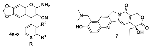
2.2. General Procedure for the Preparation of 2-Amino-3-cyano-4H-chromenes 4a–o and 6a–h

A mixture of aldehyde (1a–i), malononitrile (2) and 3,4-methylenedioxyphenol (3) or 3,5-dimethoxyphenol (5) (1:1:1 mol equiv) were combined in ethanol (15 mL), and then piperidine (0.2 mol equiv) was added and stirred at room temperature for 20 h. The solvent was removed under vacuum and the residue purified by flash chromatography on silica gel using n-hexane/EtOAc (8:2) as eluent, resulting in the corresponding 2-amino-3-cyano-4H-chromenes 4a–o and 6a–h. To our knowledge, there are no previous reports of 4d, 4e, 4i–o, 6b, 6d or 6e. Although 4a, 4b, 4c, 4f, 4g, 4h, 6a, 6c and 6f–h are not new compounds, some of them have not been fully characterized in the literature. Consequently, their reported melting points are indicated, and the missing spectroscopic data are herein provided for their proper structural characterization.

2.2.1. 2-Amino-3-cyano-4-phenyl-6,7-methylendioxy-4H-chromene (4a)

According to the general method, aldehyde 1a (0.23 g, 2.17 mmol), malononitrile (2) (0.143 g, 2.17 mmol), phenol 3 (0.3 g, 2.17 mmol), and piperidine (0.037 g, 0.434 mmol) were mixed in ethanol. The crude was separated by flash column chromatography, affording 0.47 g (74%) of 4a as a white solid; mp 203–205 °C (Lit. [41] 207–210 °C). HRMS (EI): m/z [M]+ calcd for C17H12N2O3: 292.0848; found: 292.0838.

2.2.2. 2-Amino-3-cyano-4-(4-fluorophenyl)-6,7-methylendioxy-4H-chromene (4b)

Following the general method, 1b (0.27 g, 2.17 mmol), 2 (0.143 g, 2.17 mmol), 3 (0.3 g, 2.17 mmol), and piperidine (0.037 g, 0.434 mmol) were combined in ethanol, furnishing 0.43 g (64%) of 4b as an orange solid; mp 200–203 °C. FT-IR (CH2Cl2) vmax 3451, 3333, 3223, 2194, 1672 cm−1. HRMS (EI): m/z [M]+ calcd for C17H11N2O3F: 310.0754; found: 310.0754 (Lit. [42] 311.0826).

2.2.3. 2-Amino-3-cyano-4-(4-chlorophenyl)-6,7-methylendioxy-4H-chromene (4c)

According to the general method, 1c (0.305 g, 2.17 mmol), 2 (0.143 g, 2.17 mmol), 3 (0.3 g, 2.17 mmol), and piperidine (0.037 g, 0.434 mmol) were mixed in ethanol, delivering 0.433 g (61%) of 4c as a beige solid; mp 202–204 °C (Lit. [41], 205–208 °C). HRMS (EI): m/z [M]+ calcd for C17H11N2O3CI: 326.0458; found: 326.0459.

2.2.4. 2-Amino-4-(4-bromophenyl)-3-cyano-6,7-methylendioxy-4H-chromene (4d)

Following the general method, 1d (0.402 g, 2.17 mmol), 2 (0.143 g, 2.17 mmol), 3 (0.3 g, 2.17 mmol), and piperidine (0.037 g, 0.434 mmol) were combined in ethanol, obtaining 0.572 g (71%) of 4d as a pale orange solid; mp 220–223 °C. FT-IR (CH2Cl2) vmax 3450, 3338, 3222, 2193, 1671 cm−1. 1H NMR (600 MHz, acetone-d6) δ 4.68 (s, 1H, H-4), 5.96 (d, J = 22.2 Hz, 2H, H-13), 6.21 (bs, 2H, NH2), 6.49 (s, 1H, H-5), 6.58 (s, 1H, H-8), 7.21 (d, J = 8.4, 2H, H-10), 7.50 (d, J = 8.4, 2H, H-11). 13C NMR (150 MHz, acetone-d6) δ 41.7 (C-4), 58.3 (C-2), 98.5 (C-5), 102.8 (C-13), 108.2 (C-8), 115.8 (C-5a), 120.0 (CN), 121.1 (C-9), 130.5 (C-10), 132.5 (C-11), 144.0 (C-8a), 145.6 (C-7), 146.2 (C-12), 148.3 (C-6), 161.0 (C-3). HRMS (EI): m/z [M]+ calcd for C17H11N2O3Br: 369.9953; found: 369.9968.

2.2.5. 2-Amino-3-cyano-4-(4-cyanophenyl)-6,7-methylendioxy-4H-chromene (4e)

According to the general method, 1e (0.285 g, 2.17 mmol), 2 (0.143 g, 2.17 mmol), 3 (0.3 g, 2.17 mmol), and piperidine (0.037 g, 0.434 mmol) were mixed in ethanol, giving 0.47 g (68%) of 4e as a beige solid; mp 250–252 °C. FT-IR (CH2Cl2) vmax 3422, 3297, 3178, 3178, 2155, 1655 cm−1. 1H NMR (600 MHz, acetone-d6) δ 4.81 (s, 1H, H-4), 5.97 (d, J = 21 Hz, 2H, H-13), 6.24 (bs, 2H, NH2), 6.51 (s, 1H, H-8), 6.60 (s, 1H, H-5), 7.46 (d, J = 7.8 Hz, 2H, H-10), 7.74 (d, J = 8.4 Hz, 2H, H-11). 13C NMR (150 MHz, acetone-d6) δ 42.3 (C-4), 58.0 (C-2), 98.7 (C-5), 102.9 (C-13), 108.1 (C-8), 111.6 (C-9), 115.1 (C-5a), 119.2 (Ar-CN), 119.8 (CN), 129.5 (C-10), 133.5 (C-13), 144.2 (C-8a), 145.8 (C-7), 148.5 (C-6), 152.0 (C-12), 161.3 (C-3). HRMS (EI): m/z [M]+ calcd for C18H11N3O3: 317.0800; found: 317.0801.

2.2.6. 2-Amino-3-cyano-6,7-methylendioxy-4-(4-nitrophenyl)-4H-chromene (4f)

Following the general method, 1f (0.328 g, 2.17 mmol), 2 (0.143 g, 2.17 mmol), 3 (0.3 g, 2.17 mmol), and piperidine (0.37 g, 0.434 mmol) were combined in ethanol, generating 0.47 g (64%) of 4f as a pale orange solid; mp 258–260 °C (Lit. [43] 162–165 °C). 13C NMR (75 MHz, acetone-d6) δ 41.2 (C-4), 59.260.5 (C-2), 98.3 (C-5), 102.0 (C-13), 104.8 (C-8), 113.2 (C-5a), 119.3 (CN), 124.3 (C-11), 128.8 (C-10), 143.2 (C-8a), 145.2 (C-6), 147.3 (C-12), 147.9 (C-7), 151.7 (C-9), 159.5 (C-3).

2.2.7. 2-Amino-3-cyano-6,7-methylendioxy-4-(p-tolyl)-4H-chromene (4g)

According to the general method, 1g (0.261 g, 2.17 mmol) 2 (0.143 g, 2.17 mmol), 3 (0.3 g, 2.17 mmol), and piperidine (0.037 g, 0.434 mmol) were mixed in ethanol, producing 0.56 g (84%) of 4g as a cream-colored solid; mp 208–210 °C (Lit. [41,42] 208–211 °C). HRMS (EI): m/z [M]+ calcd for C18H14N2O3: 306.1005; found: 306.1004.

2.2.8. 2-Amino-3-cyano-6,7-methylendioxy-4-(4-methoxyphenyl)-4H-chromene (4h)

Following the general method, 1h (0.296 g, 2.17 mmol), 2 (0.143 g, 2.17 mmol), 3 (0.3 g, 2.17 mmol), and piperidine (0.037 g, 0.434 mmol) were combined in ethanol, yielding 0.336 g (48%) of 4h as a pale orange solid; mp 190–192 °C (Lit. [41,42] 197–201 °C). HRMS (EI): m/z [M]+ calcd for C18H14N2O4: 322.0954; found: 322.0951.

2.2.9. 2-Amino-3-cyano-6,7-methylendioxy-4-(pyridin-4-yl)-4H-chromene (4i)

According to the general method, 1i (0.232 g, 2.17 mmol), 2 (0.143 g, 2.17 mmol), 3 (0.3 g, 2.17 mmol), and piperidine (0.037 g, 0.434 mmol) were mixed in ethanol, leading to 0.35 g (55%) of 4i as a yellow solid; mp 225–227 °C. FT-IR (CH2Cl2) vmax 3309, 3081, 2159, 1660, 1597 cm−1. 1H NMR (600 MHz, acetone-d6) δ 4.71 (s, 1H, H-4), 5.98 (d, J = 22.2 Hz, 2H, H-12), 6.27 (bs, 2H, NH2), 6.54 (s, 1H, H-8), 6.61 (s, 1H, H-5), 7.22–7.24 (m, 2H, H-10), 8.50–8.53 (m, 2H, H-11). 13C NMR (150 MHz, acetone-d6) δ 41.7 (C-4), 57.5 (C-2), 98.7 (C-5), 102.8 (C-12), 108.0 (C-8), 114.9 (C-5a), 119.8 (CN), 123.4 (C-10), 144.3 (C-8a), 145.7 (C-7), 148.5 (C-6), 151.1 (C-11), 154.8 (C-9), 161.5 (C-3). HRMS (EI): m/z [M]+ calcd for C16H11N3O3: 293.0800; found: 293.0805.

2.2.10. 2-Amino-3-cyano-4-(3-fluorophenyl)-6,7-methylendioxy-4H-chromene (4j)

According to general method, 1a (0.192 g, 1.81 mmol), 2 (0.119 g, 1.81 mmol), 3 (0.25 g, 1.81 mmol) and piperidine (0.031 g, 0.362 mmol) were mixed in ethanol, leading to 0.32 g (57%) of 4j as a pale-yellow solid; mp 194–196 °C. FT-IR (CH2Cl2) vmax 3452, 3330, 3213, 2903, 2192, 1660 cm–1. NMR 1H (600 MHz, DMSO-d6) δ 4.69 (s, 1H, H-4), 6.00 (d, J = 32.4 Hz, 2H, H-15), 6.60 (s, 1H, H-8), 6.69 (s, 1H, H-5), 6.90–7.20 (m, 5H, H-10, H-12, H-14, NH2), 7.33–7.46 (m, 1H, H-13). NMR 13C (150 MHz, DMSO-d6) δ 40.8 (C-4), 55.6 (C-2), 98.3 (C-5), 102.2 (C-15), 107.7 (C-8), 114.1 (C-10, J = 20.8 Hz), 114.4 (C-12, J = 21.3 Hz), 115.3 (C-5a), 120.9 (CN), 123.8 (C-14), 131.2 (C-13, J = 7.95 Hz), 143.2 (C-8a), 144.6 (C-7), 147.4 (C-6), 149.3 (C-9, J = 6 Hz), 161.0 (C-3), 162.7 (C-11, J = 242.8 Hz). HRMS (EI): m/z calcd for C17H11N2O3F [M]+ 310.0754 found; 310.0756.

2.2.11. 2-Amino-3-cyano-4-(3-cyanophenyl)-6,7-methylendioxy-4H-chromene (4k)

According to general method, 1a (0.237 g, 1.81 mmol), 2 (0.119 g, 1.81 mmol), 3 (0.25 g, 1.81 mmol) and piperidine (0.031 g, 0.362 mmol) were mixed in ethanol, leading to 0.4 g (70%) of 4k as a pale-yellow solid; mp 232–234 °C. FT-IR (CH2Cl2) vmax 3441, 3342, 2905, 2228, 2188, 1656 cm–1. NMR 1H (600 MHz, DMSO-d6) δ 4.77 (s, 1H, H-4), 6.00 (d, J = 33 Hz, 2H, H-15), 6.58 (s, 1H, H-8), 6.70 (s, 1H, H-5), 7.00 (br s, 2H, NH2), 7.50–7.60 (m, 2H, H-13, H-14), 7.68 (s, 1H, H-10), 7.69–7.75 (m, 1H, H-12). NMR 13C (150 MHz, DMSO-d6) δ 40.6 (C-4), 55.2 (C-2), 98.3 (C-5), 102.2 (C-15), 107.7 (C-8), 112.0 (C-11), 114.7 (C-5a), 119.2 (CN’), 120.7 (CN), 130.6 (C-13), 131.3 (C-10), 131.3 (C-12), 132.8 (C-14), 143.2 (C-8a), 144.7 (C-7), 147.5 (C-6), 147.8 (C-9), 161.0 (C-3). HRMS (EI): m/z calcd for C18H11N3O3 [M]+ 317.0800 found; 317.0801.

2.2.12. 2-Amino-3-cyano-4-(2-fluorophenyl)-6,7-methylendioxy-4H-chromene (4l)

According to general method, 1a (0.225 g, 1.81 mmol), 2 (0.119 g, 1.81 mmol), 3 (0.25 g, 1.81 mmol) and piperidine (0.031 g, 0.363 mmol) were mixed in ethanol, leading to 0.47 g (84%) of 4l as a beige solid; mp 198–200 °C. FT-IR (CH2Cl2) vmax 3459, 3328, 3197, 2887, 2197, 1663 cm–1. NMR 1H (600 MHz, DMSO-d6) δ 4.91 (s, 1H, H-4), 5.98 (d, J = 30 Hz, 2H, H-15), 6.48 (s, 1H, H-8), 6.69 (s, 1H, H-5), 6.91 (br s, 2H, NH2), 7.13–7.24 (m, 3H, H-11, H-12, H-13), 7.26–7.35 (m, 1H, H-14). NMR 13C (150 MHz, DMSO-d6) δ 35.6 (C-4), 54.6 (C-2), 98.2 (C-5), 102.2 (C-15), 107.4 (C-8), 114.5 (C-5a), 116.2 (C-11, J = 21.45 Hz), 120.8 (CN), 125.2 (C-13), 129.5 (C-14, J = 8.1 Hz), 130.2 (C-12), 132.7 (C-9, J = 12.45 Hz), 143.5 (C-8a), 144.6 (C-7), 147.4 (C-6), 160.4 (C-10, J = 244.2 Hz), 161.2 (C-3). HRMS (EI): m/z calcd for C17H11N2O3F [M]+ 310.0754 found; 310.0753.

2.2.13. 2-Amino-3-cyano-4-(2-chlorophenyl)-6,7-methylendioxy-4H-chromene (4m)

According to the general method, 1a (0.254 g, 1.81 mmol), 2 (0.119 g, 1.81 mmol), 3 (0.25 g, 1.81 mmol) and piperidine (0.031 g, 0.363 mmol) were mixed in ethanol, leading to 0.3 g (51%) of 4m as a pale yellow solid; mp 218–220 °C. FT-IR (CH2Cl2) vmax 3442, 3327, 3201, 2899, 2204, 1664 cm–1. NMR 1H (600 MHz, DMSO-d6) δ 5.14 (s, 1H, H-4), 5.98 (d, J = 28.8 Hz, 2H, H-15), 6.39 (s, 1H, H-8), 6.69 (s, 1H, H-5), 6.92 (s, 2H, NH2), 7.19–7.23 (m, 1H, H-14), 7.25–7.35 (m, 2H, H-13, H-12), 7.42–7.46 (m, 1H, H-11). NMR 13C (150 MHz, DMSO-d6) δ 38.5 (C-4), 54.8 (C-2), 98.2 (C-5), 102.2 (C-15), 107.0 (C-8), 114.5 (C-5a), 120.6 (CN), 128.3 (C-14), 129.2 (C-12), 130.3 (C-11), 131.2 (C-13), 132.2 (C-10), 142.8 (C-9), 143.4 (C-8a), 144.6 (C-7), 147.5 (C-6) 161.0 (C-3). HRMS (EI): m/z calcd for C17H11N2O3Cl [M]+ 326.0458 found; 326.0460.

2.2.14. 2-Amino-3-cyano-4-(2-bromophenyl)-6,7-methylendioxy-4H-chromene (4n)

According to the general method, 1a (0.335 g, 1.81 mmol), 2 (0.119 g, 1.81 mmol), 3 (0.25 g, 1.81 mmol) and piperidine (0.031 g, 0.363 mmol) were mixed in ethanol, leading to 0.46 g (68%) of 4n as a dark yellow solid; mp 223–225 °C. FT-IR (CH2Cl2) vmax 3442, 3327, 3201, 2899, 2204, 1664 cm–1. FT-IR (CH2Cl2) vmax 3444, 3326, 3199, 2204, 1664 cm–1. NMR 1H (600 MHz, DMSO-d6) δ 5.16 (s, 1H, H-4), 5.98 (d, J = 28.2 Hz, 2H, H-15), 6.37 (s, 1H, H-8), 6.70 (s, 1H, H-5), 6.93 (br s, 2H, NH2), 7.16–7.25 (m, 2H, H-13, H-14), 7.33–7.40 (m, 1H, H-12), 7.58–7.64 (m, 1H, H-11). NMR 13C (150 MHz, DMSO-d6) δ 40.6 (C-4), 55.1 (C-2), 98.3 (C-5), 102.2 (C-15), 106.8 (C-8), 114.6 (C-5a), 120.5 (CN), 122.6 (C-10), 129.0 (C-12), 129.5 (C-13), 131.4 (C-14), 133.4 (C-11), 143.3 (C-7), 144.6 (C-9, C-8a), 147.5 (C-6), 160.9 (C-3). HRMS (EI): m/z calcd for C17H11N2O3Br [M]+ 369.9953 found; 369.9954.

2.2.15. 2-Amino-3-cyano-4-(2-nitrophenyl)-6,7-methylendioxy-4H-chromene (4o)

According to the general method, 1a (0.273 g, 1.81 mmol), 2 (0.119g, 1.81 mmol), 3 (0.25 g, 1.81 mmol) and piperidine (0.031 g, 0.363 mmol) were mixed in ethanol, leading to 0.43 g (70%) of 4n as a pale yellow solid; mp 208–210 °C. FT-IR (CH2Cl2) vmax 3442, 3350, 2185, 1656 cm–1. NMR 1H (600 MHz, DMSO-d6) δ 4.67 (s, 1H, H-4), 5.98 (d, J = 31.8 Hz, 2H, H-15), 6.58 (s, 1H, H-8), 6.69 (s, 1H, H-5), 6.96 (br s, 2H, NH2), 7.15–7.20 (m, 1H, H-14), 7.24 (s, 1H, H-13), 7.27–7.32 (m, 1H, H-12), 7.33–7.40 (m, 1H, H-11). NMR 13C (150 MHz, DMSO-d6) δ 40.7 (C-4), 55.6 (C-2), 98.3 (C-5), 102.2 (C-15), 107.7 (C-8), 115.2 (C-5a), 120.8 (CN), 126.5 (C-14), 127.3 (C-12), 127.5 (C-13), 131.1 (C-11), 133.7 (C-9), 143.2 (C-8a), 144.6 (C-7), 147.4 (C-6), 148.9 (C-10), 161.0 (C-3). HRMS (EI): m/z calcd for C17H11N3O5 [M]+ 337.0699 found; 337.0694.

2.2.16. 2-Amino-3-cyano-5,7-dimethoxy-4-phenyl-4H-chromene (6a)

Following the general method, 1a (0.138 g, 1.3 mmol), 2 (0.086 g, 1.3 mmol), 5 (0.2 g, 1.3 mmol), and piperidine (0.022 g, 0.259 mmol) were combined in ethanol, resulting in 0.23 g (57%) of 6a as a beige solid; mp 176–178 °C (Lit. [44], 212–214 °C). FT-IR (KBr): vmax 3427, 3344, 3002, 2190, 1655, 1619, 1585, 1500, 1457, 1403, 1202, 1147, 1105, 827, 809, 753, 705 cm−1. HRMS (EI): m/z [M]+ calcd for C18H16N2O3: 308.1161; found: 308.1159.

2.2.17. 2-Amino-3-cyano-5,7-dimethoxy-4-(4-fluorophenyl)-4H-chromene (6b)

According to the general method, 1b (0.161 g, 1.3 mmol), 2 (0.086 g, 1.3 mmol), 5 (0.2 g, 1.3 mmol), and piperidine (0.022g, 0.259 mmol) were mixed in ethanol, providing 0.192 g (45%) of 6b as a beige solid; mp 188–190 °C. FT-IR (CH2Cl2): vmax 3475, 3329, 2962, 2196, 1658, 1582, 1505, 1399, 1223, 1204, 1147, 1105, 1049, 822, 798 cm−1. 1H NMR (600 MHz, acetone-d6) δ 3.67 (s, 3H, 5-OCH3), 3.78 (s, 3H, 7-OCH3), 4.62 (s, 1H, H-4), 6.15 (br s, 2H, NH2), 6.24 (d, J = 2.3 Hz, 1H, H-8), 6.30 (d, J = 2.2 Hz, 1H, H-6), 6.98–7.02 (m, 2H, H-11), 7.22–7.17 (m, 2H, H-10). 13C NMR (150 MHz, acetone-d6) δ 36.3 (C-4), 55.1 (7-OCH3), 55.4 (5-OCH3), 59.6 (C-3), 93.1 (C-8), 95.1 (C-6), 105.1 (C-4a), 114.8, 114.9 (C-11, J = 21.25 Hz), 119.5 (CN), 129.1, 129.2 (C-10, J = 21.25 Hz), 142.2 (C-9, J = 2.55 Hz), 150.4 (C-8a), 157.7 (C-5), 160.3 (C-2), 160.7 (C-7), 162.3 (C-12, J = 241.05 Hz). HRMS (EI): m/z [M]+ calcd for C18H15N2O3F: 326.1067; found: 326.1070.

2.2.18. 2-Amino-3-cyano-5,7-dimethoxy-4-(4-chlorophenyl)-4H-chromene (6c)

Following the general method, 1c (0.182 g, 1.3 mmol), 2 (0.86 g, 1.3 mmol), 5 (0.2 g, 1.3 mmol), and piperidine (0.022 g, 0.259 mmol) were combined in ethanol, furnishing 0.191 g (43%) of 6c as a white solid; mp 193–195 °C (Lit. [44], 234–236 °C). FT-IR (CH2Cl2): vmax 3334, 2193, 1652, 1621, 1582, 1471, 1397, 1204, 1146, 1103, 818, 740 cm−1. HRMS (EI): m/z [M]+ calcd for C18H15N2O3Cl: 342.0771; found: 342.0768.

2.2.19. 2-Amino-4-(4-bromophenyl)-3-cyano-5,7-dimethoxy-4H-chromene (6d)

According to the general method, 1d (0.24 g, 1.3 mmol), 2 (0.086 g, 1.3 mmol), 5 (0.2 g, 1.3 mmol), and piperidine (0.022 g, 0.259 mmol) were mixed in ethanol, affording 0.238 g (47%) of 6d as a cream-colored solid; mp 192–194 °C. FT-IR (KBr): vmax 3472, 3328, 3207, 2196, 1660, 1587, 1400, 1205, 1147, 1101, 1058, 825, 806, 790 cm−1. 1H NMR (600 MHz, acetone-d6) δ 3.67 (s, 3H, 5-OCH3), 3.78 (s, 3H, 7-OCH3), 4.60 (s, 1H, H-4), 6.17 (br s, 2H, NH2), 6.24 (d, J = 2.0 Hz, 1H, H-8), 6.30 (d, J = 2.0 Hz, 1H, H-6), 7.12 (d, J = 8.3 Hz, 2H, H-10), 7.43 (d, J = 8.3 Hz, 2H, H-11). 13C NMR (150 MHz, acetone-d6) δ 37.2 (C-4), 55.8 (7-OCH3), 56.1 (5-OCH3), 59.9 (C-3), 93.8 (C-8), 95.8 (C-6), 105.35 (C-4a), 120.1 (CN), 120.4 (C-12), 130.2 (C-10), 132.0 (C-11), 146.2 (C-9), 151.2 (C-8a), 158.4 (C-5), 161.0 (C-2), 161.5 (C-7). HRMS (EI): m/z [M]+ calcd for C18H15N2O3Br: 386.0266; found: 386.0262.

2.2.20. 2-Amino-3-cyano-4-(4-cyanophenyl)-5,7-dimethoxy-4H-chromene (6e)

Following the general method, 1e (0.17 g, 1.3 mmol), 2 (0.086 g, 1.3 mmol), 5 (0.2 g, 1.3 mmol), and piperidine (0.022 g, 0.259 mmol) were combined in ethanol, delivering 0.2 g (46%) of 6e as a white solid; mp 243–245 °C. FT-IR (KBr): vmax 3400, 3331, 3217, 2232, 2190, 1662, 1633, 1585, 1500, 1405, 1204, 1149, 1103, 1055, 820, 795 cm−1. 1H NMR (500 MHz, acetone-d6) δ 3.67 (s, 3H, 5-OCH3), 3.79 (s, 3H, 7-OCH3), 4.71 (s, 1H, H-4), 6.20 (br s, 1H, NH2), 6.26 (d, J = 2.3 Hz, 1H, H-8), 6.32 (d, J = 2.3 Hz, 1H, H-6), 7.36 (d, J = 8.5 Hz, 2H, H-10), 7.67 (d, J = 8 Hz, 2H, H-11). 13C NMR (125 MHz, acetone-d6) δ 37.9 (C-4), 55.9 (7-OCH3), 56.2 (5-OCH3), 59.3 (C-3), 94.0 (C-8), 95.9 (C-6), 104.7 (C-4a), 111.0 (C-12), 119.3 (Ar-CN), 119.9 (CN), 129.2 (C-10), 133.0 (C-11), 151.2 (C-8a), 152.2 (C-9), 158.5 (C-5), 161.1 (C-2), 161.7 (C-7). HRMS (EI): m/z [M]+ calcd for C19H15N3O3: 333.1113; found: 333.1108.

2.2.21. 2-Amino-3-cyano-5,7-dimethoxy-4-(4-nitrophenyl)-4H-chromene (6f)

According to the general method, 1f (0.196 g, 1.3 mmol), 2 (0.086 g, 1.3 mmol), 5 (0.2 g, 1.3 mmol), and piperidine (0.022 g, 0.259 mmol) were mixed in ethanol, obtaining 0.207 g (45%) of 6f as a cream-colored solid; mp 230–232 °C (Lit. [44], 245–247 °C). FT-IR(KBr): vmax 3434, 3316, 3198, 2980, 2200, 1655, 1579, 1515, 1500, 1405, 1349, 1208, 1148, 1053, 824, 711 cm−1. HRMS (EI): m/z [M]+ calcd for C18H15N3O5: 353.1012; found: 353.1014.

2.2.22. 2-Amino-3-cyano-5,7-dimethoxy-4-(p-tolyl)-4H-chromene (6g)

Following the general method, 1g (0.156 g, 1.3 mmol), 2 (0.086 g, 1.3 mmol), 5 (0.2 g, 1.3 mmol), and piperidine (0.022 g, 0.259 mmol) were combined in ethanol, giving 0.184 g (44%) of 6g as a pale-yellow solid; mp 172–174 °C (Lit. [44], 217–219 °C). FT-IR(KBr): vmax 3475, 3329, 2962, 2196, 1658 cm−1. HRMS (EI): m/z [M]+ calcd for C19H18N2O3: 322.1318; found: 322.1318.

2.2.23. 2-Amino-3-cyano-5,7-dimethoxy-4-(4-methoxyphenyl)-4H-chromene (6h)

According to the general method, 1h (0.176 g, 1.3 mmol), 2 (0.086 g, 1.3 mmol), 5 (0.2 g, 1.3 mmol), and piperidine (0.022 g, 0.259 mmol) were mixed in ethanol, generating 0.132 g (30%) of 6h as a cream-colored solid; mp 186–188 °C (Lit. [44], 220–222 °C). FT-IR(KBr): vmax 3331, 2942, 2197, 1660, 1622, 1397, 1204, 1146, 1102, 1057, 798 cm−1. HRMS (EI): m/z [M]+ calcd for C19H18N2O4: 338.1267; found: 338.1268.

2.3. In-Silico Analysis of 2-Amino-3-cyano-4-aryl-6,7-methylendioxy-4H-chromenes 4a–o and 2-Amino-3-cyano-5,7-dimethoxy-4-aryl-4H-chromenes 6a–h

The physicochemical and toxicological properties of 2-amino-3-cyano-4H-chromenes 4a–o and 6a–h were determined on the OSIRIS DataWarrior V4.7.2 program (https://openmolecules.org/datawarrior/, accessed on 26 October 2021) [45]. The drug similarity and pharmacokinetic properties, including molar refractivity and the number of rotatable bonds, were calculated with the SwissADME server platform [46]. The physicochemical properties were evaluated based on Lipinski’s rules, considering Log P, molecular weight, hydrogen bond donors, and hydrogen bond acceptors [47].

2.3.1. Multiple Sequence Alignment and Generation of 3D CYP51 from Candida spp. through Homology Modeling

The sequences of the lanosterol 14α-demethylase enzymes (CYP51) were downloaded from the NCBI database (https://www.ncbi.nlm.nih.gov, accessed on 26 October 2021) [48] for C. albicans AATCC 10231 (CYP51CA), C. dubliniensis CD36 (CYP51CD), C. glabrata CBS138 (CYP51CG), C. kefyr (CYP51CKE), C. krusei ATCC 6358 (CYP51CK) and C. parapsilosis ATCC 22019 (CYP51CP). The percentage of identity of each of the CYP51 sequences of Candida spp. with the target protein CYP51 of Saccharomyces cerevisiae (CYP51SC), complexed with fluconazole (8) in the active site (PDB: 4WMZ), was determined with the BLASTp (protein query-protein database) server. The multiple alignment of sequences was performed with CLUSTAL X [49]. The sequences of CYP51 Candida spp. were used for the elaboration of 3D models, based on the homology modeling technique with the Modeller 9.23 program [50]. For these models, the crystallized structure of S. cerevisiae CYP51 (PDB: 4wmz), deposited in the protein data bank (https://www.rcsb.org/structure/4WMZ, accessed on 26 October 2021) [51], served as a template. The overlapping of the six selected CYP51 models of Candida spp. was carried out on the Discovery Studio Visualizer [52].

2.3.2. Evaluation and Validation of the 3D Model of CYP51 from Candida spp.

Of the fifteen models obtained for each CYP51 of the different Candida spp., the one with the minimum score was chosen. The 3D models of CYP51 were examined with the DOPE method [53] and validated with the VERIFY3D [54] and PROCHECK [55] programs. The former was utilized to assess the compatibility of an atomic model (3D) with its own amino acid sequence (1D), and the latter to check the stereochemical quality of the Ramachandran plots that show the amino acid residues in the allowed regions.

2.3.3. Molecular Docking on CYP51

The 4H-chromenes 4a–o and 6a–h were docked on the active site of the CYP51 enzymes of C. albicans, C. dubliniensis, C. glabrata, C. kefyr, C. krusei, and C. parapsilosis by using the AutoDock4 program [56]. The CYP51 enzymes of the Candida spp. were previously modeled with the crystallized proteins of CYP51 from Saccharomyces cerevisiae (CYP51SC) (PDB: 4WMZ), deposited in the protein data bank [57], as a template. The proteins were prepared by adding hydrogen atoms and removing water molecules around them, and then were optimized on the Nanoscale Molecular Dynamics (NAMD) program [58].
Fluconazole (8) and the 2-amino-3-cyano-4H-chromenes 4a–o and 6a–h were drawn with ChemBioDraw Ultra 12.0 software [59], converted to 3D on the Open Babel GUI program [60], and optimized with GaussView 6.0 software [61], which involves the addition of hydrogen atoms and the verification of the bond lengths and angles.
The docking parameters were established with AutoDock Tools, with grid dimensions of 44 × 44 × 44 Å3 and points separated by 0.375 Å. The grid center used for the CYP51 Candida spp. was (X = 27.7, Y = 10.1 and Z = 18.95). The Lamarckian Genetic algorithm was used with 100 docking runs. The docked model with the lowest binding energy was selected. The docking results were edited in Discovery Studio [52].

2.3.4. Molecular Docking on Topoisomerase I

The 3D structure of topoisomerase I was obtained from the Protein Data Bank (PDB entry no. 1SC7, https://www.rcsb.org/structure/1SC7, accessed on 26 October 2021) [62]. The water molecules were removed from the structure to delimit the grid, and the active site was identified with AutoGrid. Hydrogen atoms were added to the polar atoms (considering a pH of 7.4), and the Kollman charges were assigned. The topotecan 3D structure was downloaded from Zincdatabase [63]. The 2D structures of the 2-amino-3-cyano-4H-chromenes 4a–o and 6a–h ligands were drawn with ChemSketch (https://www.acdlabs.com/resources/freeware/chemsketch/index.php, accessed on 26 October 2021), and subsequently, the optimization of the structure to an AM1 level was performed with Gaussian 98 [61], in order to find the lowest energy conformation for each ligand.
Molecular docking simulations were carried out with AutoDock version 4.0 [56]. For topoisomerase I, the grid dimensions were 90 × 90 × 90 Å3 with the points separated by 0.375 Å. The grid center was X = 85.385, Y = −10.629 and Z = 10945. Default values of translation, quaternation, torsion steps, atomic partial charges, and non-polar hydrogens were utilized for the simulation. The hybrid Lamarckian Genetic Algorithm (at default settings) was applied for minimization. The number of docking runs was 100, with a population size of 150. The maximum number of energy evaluation ranges from 27,000 to 250,000, the latter being the maximum number of generations. The mutation rate was 0.02 and the cross-over rate was 0.8. The docked model with the lowest binding energy was considered for all further simulations. Docking results were analyzed on AutoDockTools and edited with Discovery 4.0 Client [52].

2.4. Antifungal Activity Assays

To determine the in vitro minimum inhibitory concentrations (MIC) for fluconazole (8) and the 2-amino-3-cyano-4H-chromenes 4a–i and 6a–h on six Candida species (C. albicans, C. dubliniensis, C. glabrata, C. kefyr, C. krusei, and C parapsilosis), the standard guidelines of the National Committee for Clinical Laboratory Standards (NCCLS) were employed, with an RPMI of 1640 (Sigma) buffered with 0.165 MMOPS (Sigma) as the test medium [64]. The MIC value was defined as the lowest concentration of a compound that generated a culture with turbidity less than or equal to 100% inhibition when compared with the growth of the control. The 4H-chromene derivatives 4a–i and 6a–h were dissolved in DMSO solvent and serially dripped into the growth medium. The concentration gradient of 8 was from 64–0.033 µg/mL and for the derivatives 160–0.078 µg/mL. The inoculum was prepared by resuspending colonies (from a yeast culture of 24 h growth) in a tube with saline solution (0.85% NaCl), and then adjusting them to an optical density 0.5 McFarland. Subsequently, they were incubated at 37 °C in an incubator. The MIC results were quantified in a Multiskan™ GO microplate spectrophotometer by agitation of the plates, followed by a spectrophotometric reading at 450 nm. The value is expressed as the average of three replicates.

2.5. Cytotoxicity Assay

The in vitro cytotoxicity of the compounds was evaluated on PC-3 and SK-LU-1 cell lines (human prostatic and lung adenocarcinoma, respectively), using the protein binding dye sulforhodamine B (SRB) in a microculture assay to measure cell growth [65]. The cell lines were grown in RPMI-1640 medium (Sigma Chemical Co., Ltd., St. Louis, MO, USA) supplemented with 10% fetal bovine serum (Invitrogen Corporation, Waltham, MA, USA), 2 mM L-glutamine, 10,000 units/mL of penicillin G, 10,000 µg/mL streptomycin, and 0.25 µg/mL of fungizone (Gibco, Amarillo, TX, USA). The cells were maintained at 37 °C in a 5% CO2 atmosphere with 95% humidity.
Preparations were made with 7.5104 cell/mL of the PC-3 cell line and 10104 cell/mL of the SK-LU-1 cell line, adding 100 mL of each of these suspensions to the wells of 96-well plates. Incubation was carried out for 24 h to promote cell attachment, and then 100 µL per well of a test compound, topotecan (7) or cisplatin (9) (as controls) were added. After 48 h, the adhered cell cultures were fixed in situ with a cold 50% (w/v) solution of trichloroacetic acid, followed by incubation at 4 °C for 1 h. Upon completion of this time, the supernatant was discarded and the plates were washed and air dried. Cells cultured with TCA were stained with 100 µL of 0.4% SRB solution for 30 min. The protein-bound dye was extracted with a 10 nm unbuffered tris base and the optical density was read at 515 nm on a Synergy HT Microplate Reader (Elx 808, BIO-TEK Instruments, Inc, Winooski, VT, USA). IC50 values were determined by the Monks protocol. Accordingly, a concentration–response curve was constructed for each of the 2-amino-3-cyano-4H-chromenes 4a–o and 6a–h, and the values corresponding to an inhibition of 50% (IC50) were calculated from non-linear regression equations. IC50 values are expressed as the mean ± standard error (SE) [66].

3. Results and Discussion

3.1. Chemistry

In view of the relevance of 4H-chromene derivatives as potential therapeutic agents, a synthetic access to 2-amino-3-cyano-4H-chromenes 4a–o and 6a–h was explored. Of these compounds, 4d, e, i–o as well as 6b, d, and e are reported for the first time. The synthesis took place via a three-component reaction, evaluating the effect of the base, solvent, and reaction temperature. Multicomponent reactions (MCRs) represent a powerful tool that can be utilized to form novel and complex molecules by reacting to three or more different starting materials in a one-pot strategy, resulting in great efficiency and atom economy [33]. The multicomponent approach included the cooperation of benzaldehydes (1a–o), malononitrile (2) and 3,4-methylenedioxyphenol (3) or 3,5-dimethoxyphenol (5), considering their reactivity and selectivity on the efficiency of the process.
Initially, the method focused on the efficient one-pot reaction of p-nitrobenzaldehyde (1f), 2, and 3 (1:1:1 mol equiv), with Et3N (0.2 mol equiv) and ethanol as the solvent, at room temperature for 20 h. Moreover, 2-amino-3-cyano-6,7-methylendioxy-4-(4-nitrophenyl)-4H-chromene (4f) was provided in low yield (36%) (Table 1, entry 1). Besides 4f, only the starting material and the Knoevenagel product were identified in the crude reaction mixture, without evidence of any other 4H-chromene regioisomer.
In order to improve the yield, bases such as piperidine and potassium carbonate were tested under identical reaction conditions. A better yield (64%) was obtained with piperidine (Table 1, entry 2), but an increase in its concentration and the reaction temperature did not lead to further improvement (Table 1, entries 4 and 8). Finally, to analyze the possible effect of the solvent on the reactivity and selectivity of the process, protic and aprotic solvents were used (ethanol, water, dioxane and acetonitrile) under the same reaction conditions. A protic polar solvent (ethanol) generated the best yields (Table 1, entry 2).
Under the optimized reaction conditions, a variety of aromatic aldehydes with electron-donating and electron-withdrawing groups were employed to determine the scope of this methodology. The reactions of aryl aldehydes 1ao, 2 and 3 afforded 2-amino-3-cyano-4H-chromenes 4ao in 48–84% yields (Table 2, entries 1–15). By utilizing 3,5-dimethoxyphenol (5) rather than 3 under similar conditions, the respective 2-amino-3-cyano-4H-chromenes 6ah were produced in 30–57% yields (Table 2, entries 16–23). The yields were higher when the aryl aldehyde contained no substituent or electron-withdrawing group (Table 2, entries 1–6, 10–15 and 16–21).
The structures of 2-amino-3-cyano-4H-chromenes 4ao and 6ah were elucidated on the basis of their spectrometric data (1D and 2D experiment, IR and HRMS). For example, the 1H NMR spectrum of 4d showed the presence of four singlets observed at 4.68, 6.21, 6.49 and 6.58 ppm, attributed to H-4, NH2 (as a broad singlet integrating for two protons), H-5 and H-8, respectively. Three doublets at 5.96, 7.21 and 7.50 ppm were assigned to the H-13, H-10 and H-11 protons, respectively. The 13C NMR spectrum displayed signals for the cyano group at 120.0 ppm, ten signals for aromatic carbons in the region of 98.5–148.3 ppm, two signals for sp2 carbon atoms at 58.3 (C-2) and 161.0 ppm (C-3), and two signals for sp3 carbons at 41.7 (C-4) and 102.8 (C-13) ppm. Furthermore, the HMBC spectrum of 4d revealed two- and three-bond C/H long-range correlations between the proton at 4.68 ppm and the quaternary carbon atoms at 161.0 (C-3), 115.8 (C-5a), 121.1 (C-9), 58.3 (C-2), 144.0 (C-8a) and 120.0 (CN) ppm. Likewise, the signal at 5.96 ppm (H-13) indicated a three-bond C/H long-range correlation with the aromatic quaternary carbon atoms at 145.6 (C-7) and 148.3 (C-6) ppm. Hence, all the NMR data coincide with the existence of a 4H-chromene core. Finally, the HRMS (EI) of 4d confirmed the expected mass (m/z 369.9968), while the infrared spectrum exhibited absorption bands at 3453 and 3327 cm−1, characteristic of NH2, and at 2193 cm−1, characteristic of CN [32,38].
A plausible mechanism of formation of 2-amino-3cyano-4H-chromenes has been proposed (Scheme 1). First, piperidine generates the deprotonation of the hydrogen acid in 2, and the resulting malononitrile carbanion reacts with aromatic benzaldehydes (1ao) to produce 2-arylidenemalononitrile intermediate A. Subsequently, piperidine reacts with the acidic protons of phenol (3 or 5) to form the corresponding phenoxide ion B, which undergoes a Michael addition with the arylidenemalononitrile to give rise to intermediate C. The latter cyclizes intramolecularly, evolving into D. Through a series of acid-base reactions, intermediate E is afforded and then converted by tautomerization to products 4ao or 6ah, respectively [30].

3.2. In Silico Analysis of Physicochemical, Pharmacokinetic, Drug-likeness and Toxicological Properties of 2-Amino-3-cyano-4H-chromenes 4a–o and 6a–h

Various pharmacological properties were evaluated with the OSIRIS DataWarrior program to predict the efficacy and behavior in the human body of the two series of 2-amino-3-cyano-4H-chromenes, 4ao and 6ah [45]. The first descriptor to be examined was lipophilicity (octanol/water partition coefficient, Log P), which indicates the concentration ratio of a neutral substance between two immiscible solvents in equilibrium, and reveals the differential solubility of this substance in the two solvents. Usually, one of these solvents is water and the other, a hydrophobic solvent, is octanol.
Lipophilicity is a very important physicochemical parameter because it influences the bioavailability, permeability and toxicity of a drug. Permeability is high, with Log P values close to 5 and low with negative values. The Log P values of 4a–o and 6a–h were acceptable, all under 5.0 and none of them negative. The highest values (3.21 and 3.26) correspond to 4d and 6d (R = Br). Compounds 4f and 6f, containing the polar substituent (R = NO2), had Log P values of 1.76 and 1.92, respectively (Table 3 and Table 4) [67].
Aqueous solubility (Log S) influences the absorption and distribution of a biologically active compound in an organism [68]. According to the calculated values, most of the present 2-amino-3-cyano-4H-chromenes are moderately soluble in water (−6 > Log S > −4), and only some of them (4a, 4e, 4i, 4k, 6a and 6e) have a slight tendency to solubilize (−4 > Log S > −2). Another effective descriptor for predicting the drug solubility and transport properties of a molecule is the polar surface area (PSA), which is defined as the sum of the surfaces of its polar (N and O) and slightly polar atoms (S and P), and the hydrogen atoms attached to them. PSA is an indicator of gastrointestinal absorption and penetration of the blood–brain barrier. PSA values of 77.50 to 123.32 Å2 were found in the two series, 4a–o and 6a–h, evidencing an acceptable permeability. The best values were generated by the derivatives bearing halogen substituents on the phenyl aromatic ring. Compounds 4f and 6f, with nitro substituents and thus a greater number of electronegative atoms, showed the highest values of PSA (123.32 Å2), and consequently, the lowest permeability of cell membranes [69].
A different and useful parameter, ligand efficiency (LE), evaluates the effectiveness of a drug in its interaction with the active site of a therapeutic target. It is obtained by dividing the free binding energy of each molecule by the number of heavy non-hydrogen atoms in the structure (LE = ΔGinteraction/[number of heavy non-hydrogen atoms]). Therefore, it considers the affinity and size of the ligand molecule, but not the size and topological properties of the molecular target [70]. Compounds 4a, 4i and 4j exhibited the highest ligand efficiency value in the series (LE = 0.407 and 0.512), which was above that of 7 (LE = 0.377; Table 3). This coincides with the cytotoxicity assays with human lung adenocarcinoma (SKLU), finding a better IC50 for 4a than 7 (IC50 = 0.52 ± 0.03 vs. 2.0 ± 0.1; Table S11).
The ligand-lipophilicity efficiency (LLE) is also a relevant parameter for designing new drugs. It correlates the in vitro potency of a drug with the partition coefficient (log P) or the distribution coefficient (log D). A molecule with an LLE value of 0 (based on cLog P, or target affinity) has the same affinity for a therapeutic target as does n-octanol. In contrast, a compound with an LLE value of 6 has a million times more affinity towards the therapeutic target than n-octanol. Poor affinity for the therapeutic target is indicated by a negative LLE value, which was not the case for any of the derivatives in the two series, 4a–o and 6a–h (Table 3 and Table 4). These compounds all have a greater affinity for the active site of the target enzyme than n-octanol (due to specific interactions between the compounds and the amino acid side chain of the enzyme) [71].
Ligand efficiency-dependent lipophilicity (LELP) is a global descriptor involving lipophilicity, molecular size and potency. The optimal range of LELP values is −10 to 10 and corresponds to a range of Log P values of −3 to 3. The values of the test compounds are within the optimal range. Taking all the parameters evaluated into account, the two series of 2-amino-3-cyano-4H-chromenes can be considered as drug candidates [72].
The analysis of the data (Tables S1 and S2) demonstrates that all the 2-amino-3-cyano-4H-chromenes in the two series, 4a–o and 6a–h, comply with Lipinski’s rule of five (RO5) by meeting the following requirements: (a) molecular weight <500 g/mol; (b) <5 hydrogen bond donors (NH, OH or SH); (c) <10 hydrogen bond acceptors; and (d) Log P < 4.15 [47]. Hence, the compounds should have efficient absorption and good oral bioavailability [73].
The drug-like properties were also determined [74,75], predicting good lipid solubility for the present 2-amino-3-cyano-4H-chromenes based on WLog P in the range of −0.4 < WLog P < 5.6 and PSA < 131. The steric descriptors of the test compounds, like those of topotecan (7), are within the desirable values: (a) 120 < molecular weight < 480, (b) 40 < molar refractivity < 130, and (c) the number of rings <7 [74,75]. According to the results, 2-amino-3-cyano-4H-chromenes 4a–o and 6a–h should have good absorption and an appropriate steric volume. Because of complying with the acceptable range of values of key parameters established for screening compounds in the drug development process, 4a–o and 6a–h can be considered drug candidates.
Regarding the pharmacokinetic properties (Tables S3 and S4), all the test compounds are characterized by good gastrointestinal absorption, like 7, thus allowing for oral administration. Most should be able to cross the blood–brain barrier and reach targets in the central nervous system. However, this is not the case for the compounds with a greater number of polar functional groups, such as 4e, 4k and 6e (R = CN), 4f, 4o and 6f (R = NO2), and 4h, 6h (R = OMe), due to their strong inductive effect.
Log Kp (Tables S3 and S4) is a parameter that indicates the ability of a compound to pass through the skin, with a more negative value translating into greater permeability. In both series, the derivatives with a substituent R = CN (4e, 4k and 6e) and R = NO2 (4f, 4o and 6f) gave more negative values, while those with a substituent R = Cl (4c, 4m and 6c) and R = Me (4g and 6g) had less negative values and therefore a lower permeability [76]. Additionally, the capacity of 4a–o and 6a–h to cross membranes was evaluated in relation to two types of transporters: the P450 cytochrome enzyme superfamily (CYP) and the ATP binding transporters (P-gp substrate). Almost all the compounds in the 4a–o series are substrates of CYP transporters, allowing them to be metabolized and excreted by these transporters (Tables S3 and S4). Similarly, most compounds in the 6a–h series bind to all CYP transporters. No affinity was shown for CYP2D6 by 6c–e (with substituents R = Cl, R = Br and R = CN). Nevertheless, their excretion can be favored because they have affinity for the other four CYP transporters (CYP1A2, CYP2C19, CYP2C9 and CYP3A4). For its part, topotecan (7) is a substrate of the ATP binding transporters (P-gp substrate), leading to its elimination through the membrane.
The possible risk of toxicity was assessed for both series of compounds, 4a–o and 6a–h (Tables S5 and S6). There was no evidence of tumorigenicity, mutagenicity, irritation, or reproductive effects, with the exception of the reproductive effects found for 4h and 6h. The results for the rest of the compounds demonstrate an ample margin of safety between the effective dose and any serious risk to human health.

3.3. Multiple Sequence Alignment of CYP51 Enzymes of Candida spp.

Multiple sequence alignment was performed for CYP51 enzymes of Candida spp. with the crystallized CYP51SC enzyme sequence [57] (Figure S55). A large proportion of the amino acid residues in these enzymes proved to be highly conserved, especially the FGGGRHRCIG sequence motif. Within this sequence, a cysteine residue involved in binding the heme iron is conserved in the C-terminal of CYP51s [77]. Additionally, two conserved arginine residues are in the fifth and seventh positions of the motif and surround a histidine residue [78]. Other amino acids that are part of the active site of the CYP51 enzymes are Tyr118 and Thr311, which have been identified in C. albicans. Due to the aforementioned characteristics, the CYP51 protein family of yeast enzymes represents an excellent model for the study of new inhibitors with possible antifungal activity [79].

3.4. Generating 3D Models of CYP51 Enzymes of Candida spp.

Fifteen models of the CYP51 enzyme of each Candida species were provided by homology modeling, selecting the best of the fifteen based on its discrete optimized protein energy (DOPE) score. The overlap of the CYP51 models with the S. cerevisiae template demonstrated an identity of over 60% (Figure S56). CYP51CG displayed the highest value of identity. All the models obtained were evaluated, graphing their DOPE potential compared to the template for S. cerevisiae (CYP51SC, PDB 4WMZ) [57]. The graphs reveal a high degree of similarity between these structures (Figures S57–S62). The validation of the three-dimensional (3D) models was determined by compatibility with their own 1D amino acid sequence. In all the models analyzed, over 80% of the residues have an average 3D-1D score ≥0.2 (Figures S63–S68). Ramachandran plots were also created for each model (Figures S69–S74), showing over 90% of the amino acid residues in the most favorable regions. Moreover, the models proved to be of good quality, as in previous studies by our group [80].

3.5. Molecular Docking Studies

3.5.1. Molecular Docking of 2-Amino-3-cyano-4H-chromenes 4a–o and 6a–h and Fluconazole (8) with CYP51 from Candida spp.

Docking simulations were carried out for the two series of 2-amino-3-cyano-4H-chromenes, 4a–o and 6a–h, at the active site of the CYP51 of Candida spp. Some of these compounds with high binding energy values and the reference drug 8 are illustrated in (Figure 1 and Figure S139–S150), bound to the active site of the CYP51 enzyme (the heme group). As has been previously reported with azole derivatives [81,82], series 4a–o and 6a–h all display the same binding mode. This suggests that the CYP51 of yeasts could be a molecular target of these derivatives.
The orientation in relation to each of the CYP51 enzymes herein tested was determined for the 2-amino-3-cyano-4H-chromenes 4d and 6a, as well as fluconazole (8) (Figures S151–S156). Many of the derivatives exhibited a similar orientation within the active site.
With molecular docking, the binding energy was determined for each of the derivatives (Table S7), finding better values (ranging from −9.09 to −10.88 Kcal/mol) for the 2-amino-3-cyano-4H-chromenes than the reference drug 8. Among all the test compounds, the best one (in bold type, Table S7) bound in a way notably more akin to the species of Candida, which suggests that the nature of the substituent influences the binding mode. For example, the presence of the electron withdrawing halogen substituents, R = Cl and R = Br in 4m and 4n, as well as R = CN in 4k, favors interaction with the active site of the enzymes CYP51CD and CYP51CKE, respectively.
In the series 4a–o, derivative 4m (with R = Cl on the phenyl ring) exhibits the best binding energy with the CYP51 of three Candida species (C. albicans, C. glabrata and C. kefyr). In the series 6a–h, a similar result is found for 6d (with R = Br on the phenyl ring). Accordingly, the substituents R = Me and R = Br could be involved in the inhibitory effect on the yeasts (vide infra). The highest binding energy shown by most of the 2-amino-3-cyano-4H-chromenes 4a–o (−10.83 kcal/mol) and 6a–h (−10.25 kcal/mol) was in relation to the CYP51 enzyme of C. kefyr.
Regarding the binding of the two series of 2-amino-3-cyano-4H-chromenes (4ao and 6ah) and fluconazole (8) with the CYP51 enzymes of Candida spp., the amino acid residues involved, the binding mode, and the protein–ligand interactions are shown (Table 5 and Table S8). The predominant amino acids in the interactions between the compounds and CYP51 are hydrophobic in nature (Ile, Phe, Gly, Leu, Val and Met). They frequently establish π–alkyl, π–π T-shaped and π–sigma interactions, which were found presently and have been previously reported in relation to the reference drug 8 [83].
Most ligand–protein interactions include Tyr and Thr, hydrophilic amino acid residues that form carbon–hydrogen and conventional hydrogen bonds. Tyr118 and Tyr132 are observed in three of the Candida species (C. albicans, C. dubliniensis and C. parapsilosis), and they have influence in the interaction of the CYP51 of C. albicans with other azole derivatives [60]. As additional evidence of the importance of these amino acid residues in the activity of CYP51 of Candida spp., their mutation is known to cause yeast resistance mechanisms to the inhibitory effects of azoles [84,85].
Three amino acids with basic side chains, His, Arg and Lys, also greatly influence the mechanism of action of CYP51. The most frequent positions for such interactions are 310, 312, 372, 462, 463 and 468 for His, 379 and 381 for Arg, and 143 for Lys. In this research, as well as in previous studies, we corroborated that the following amino acids: Tyr118, Phe145, Ile231, Met306, His310, Thr311, Ser378, Val509 and Val511 are involved in the active site of CYP51 of C. albicans [80,83,86].
According to the present findings, the 2-amino-3-cyano-4H-chromenes 4a–o and 6a–h could possibly be administered to target the CYP51 enzymes of many Candida species, given that they bind to the active site. Hence, further research is warranted on their antifungal activity.

3.5.2. Molecular Docking of 2-Amino-3-cyano-4H-chromenes 4a–o, 6a–h and Topotecan (7) on Topoisomerase I

A molecular docking study was carried out to determine the affinity of 4a–o and 6a–h with the active site of the topoisomerase I enzyme. Some key amino acids of the active site of the enzyme that are known to interact with 7 (the most common topoisomerase I inhibitor) also interact with 4a–o and 6a–h. Among such residues are Arg364, Arg488, Lys532, Asp533, Ile535, His632 and Thr718. Additionally, both series of 2-amino-3-cyano-4H-chromenes interact with certain DNA fragments, including TGP11, DG12 and DT10 [87,88]. Better binding energies were found for 4a–o (Table 6) than 6a–h (Table S9), a finding in agreement with the results of the biological activity tests, in which 4a–o were more active than 6a–h (see Section 3.7).
Compounds 4ad and 4g showed the greatest affinity to the active site of the protein (−8.42 to −10.05 kcal/mol). Indeed, the binding energy was higher for 4c, 4d and 4g than 7 (−9.86, −10.05 and −9.75 kcal/mol, respectively, vs. −9.56 kcal/mol). Like 7, these three compounds interact with most of amino acid side chain of the active site of topoisomerase I.
For 4g and 7, there was a similar π–alkyl hydrophobic coupling with the TGP11 nucleotide of the complex. With a binding energy of −9.27 kcal/mol, 4b displayed most of the same interactions as 4c, 4d and 4g. Although the binding energy was lower (−8.42 Kcal/mol) for 4a, this compound also exhibited similarities with the binding interactions of 7, such as hydrophilic interactions with the TGP11 DNA fragment and π–anion electrostatic interactions with the residue Asp533.
In addition, we carried out the docking study with the series of enantiomers of the compounds that showed greater cytotoxic activity 4a–c, 4g, as well as of the compounds 4j–o, where we can observe a greater affinity shown by the S series (−7.66 to −10.0 kcal/mol) for the active site of the enzyme topoisomerase I with respect to the R series (−7.37 to −8.37 kcal/mol) (Table S10). Moreover, we can observe that both series of enantiomers share key amino acids of the active site of the enzyme and DNA fragments just like topotecan.
In the series of 2-amino-3-cyano-5,7-dimethoxy-4-aryl-4H-chromenes 6a–h, the best binding energies were found for 6a, 6c and 6d (−8.42 to −9.10 kcal/mol; Table S9). Moreover, 6a–f, 6h and 7 each have conventional hydrogen bond and carbon–hydrogen bond interactions with DNA fragments, including TGP11 and/or DG12, and these compounds share interactions with Arg364, Asp533 and Thr718 in the active site of the enzyme.
The structures of 4a, 4b, 4c, 4d, 4g and 7 display π–anion electrostatic interactions with the amino acid side chain residue Asp533 in the active site of the enzyme (Figure 2). For 4b, 4c, 4d and 4g, there are additional linkages not shared by 7, such as a π–sigma interaction with the side chain amino acid Thr718. For 4b, 4c and 4d, π–π stacked interactions were observed with the DNA residues DT10 and TGP11, and for 4g with TGP11 and DA113. Furthermore, conventional hydrogen bonds were found between the Arg364 residue and the nitrogen atom of the cyano group of 4b, 4c, 4d and 4g. These same four chromenes and 7 share important hydrophilic interactions (involving carbon–hydrogen bonds) with the DNA fragment DG12. Overall, the 4H-chromene derivatives and 7 act in a very similar manner on the active site of topoisomerase I, revealing that they may all have the same mechanism of action. The interactions of the 2-amino-3-cyano-4H-chromenes 4e–f, 4h–i and 6a–h are illustrated in Figures S93 and S94.
The binding mode was analyzed for 7 and 4ao at the active site of topoisomerase I (Figures S95 and S96). There were three binding orientations adopted by the 2-amino-3-cyano-4H-chromenes within the active site. One orientation is shared by 4a, 4b, 4c and 4g (Figure S95B), and a distinct one by 4e, 4f and 4h (Figure S96). Finally, the conformations of 4a and 4i were similar to one another, but slightly different from the aforementioned groups.
The binding mode of the structures of 2-amino-3-cyano-5,7-dimethoxy-4H-chromenes 6a–h and reference drug 7 at the active site of the topoisomerase I enzyme (3D representation, Figure S97A) indicates that 6d and 6f are oriented in an identical manner, and that 6c, 6b and 6h adopt a conformation distinct from the former group, but similar among themselves (Figure S97B).
Overall, the two chromene series 4a–o and 6a–h have interactions very similar to 7 at the binding site of the topoisomerase I enzyme, and therefore are likely to have the same mechanism of action.

3.6. Antifungal Activity

Given the good binding energy found for the 2-amino-3-cyano-4H-chromenes 4ai and 6ah with the CYP51 enzymes, the antifungal activity of the compounds was explored experimentally. When tested against Candida spp., both series of compounds exhibited a MIC50 less than or equal to that of the reference drug 8 (0.063–0.5 µg/mL vs. 0.50–4.0 µg/mL; Table 7). Thus, the experimental and docking data are in agreement, considering the better binding energy of the derivatives than 8.
For the 2-amino-3-cyano-4H-chromenes currently evaluated in relation to antifungal activity against six Candida species, the MIC values were similar to or lower than those described in previous reports for some 2-amino-3-cyano-4H-chromenes tested on C. albicans, Aspergillus niger, A. fumigatus, A. flavus and S. cerevisiae [15,89]. Since scant research has been carried out on Candida species, the inhibitors 4ai and 6ah were presently assayed against different species of this genus. The ability of these two series of chromenes to bind to the CYP51 enzyme and their strong inhibitory effect on the six Candida species herein tested suggest that both series, 4ai and 6ah, are potential antifungals.

3.7. Cytotoxicity Assay

In order to evaluate the cytotoxic activity of compounds, we carried out the determination at 25 μM for 4a–i and 6a–h in six tumor cells: U251 (human glioblastoma), PC-3 (human prostatic adenocarcinoma), K-562 (human chronic myelogenous leukemia), HCT-15 (human colorectal adenocarcinoma), MCF-7 (human mammary adenocarcinoma), and SKLU-1 (human lung adenocarcinoma). However, only four compounds (4a, 4b, 4c and 4g) showed an inhibition percentage greater than 50% in the SK-LU-1 and PC-3 cell lines. More detailed studies on the action of these compounds are shown in Supplementary material1.

4. Conclusions

Two series of 2-amino-3-cyano-4H-chromenes, 4ao and 6ah, were synthesized through an efficient and environmentally friendly methodology. This is the first report, to our knowledge, of 4d, e, i–o, as well as 6b, d and e. Regarding the evaluation of the physicochemical, pharmacokinetic, toxicological and drug-like properties of the test compounds, the values of key parameters fall within the acceptable range established for screening compounds in the drug development process. Moreover, the in silico data revealed an ample margin of safety between the effective dose of the series of compounds 4 and 6 and any possible toxicity.
The molecular docking studies provided evidence that 4ao, 6ah, and fluconazole (8) (the reference drug) interact with the active site of the CYP51 enzymes of C. albicans, C. dubliniensis, C. glabrata, C. kefyr, C. krusei and C. parapsilosis. CYP51 is vital for the production of ergosterol in the yeast cell membrane. The test compounds appear to have the same mechanism of action as the reference drug, since they interact with the same amino acids in the active site of the CYP51 enzyme. Better in silico binding energy was found for 4ao and 6ah than 8 on this enzyme, and the in vitro MIC50 of the test compounds was less than or equal to that of 8 (0.063–0.50 vs. 0.5–4.0 µg/mL, respectively).
According to the results of molecular docking on topoisomerase I (involved in the growth of cancer cells), 4ao and 6ah interact with the same amino acids of the active site of the enzyme as one of the reference drugs 7. Compounds 4c, 4d and 4g showed better binding energy than 7. The experimental assays revealed that 4a–o were more cytotoxic than 6a–h. Furthermore, 4a and 4b were more active than topotecan (7) and cisplatin (9). This is the first report, to our knowledge, of molecular docking studies on 2-amino-3-cyano-4H-chromenes 4ao and 6ah with the active site of CYP51 of Candida spp., and topoisomerase I. Due to the encouraging results, in which some chromene compounds displayed better binding energy, inhibition and cytotoxicity than the reference compounds, the two series of derivatives are good candidates for future research on the design and development of new 2-amino-3-cyano-4H-chromenes, with dual activity against cancer and fungal infections.

Supplementary Materials

The supplementary materials (Figures S1–S161, Tables S1–S13) are available online at https://www.mdpi.com/article/10.3390/ph14111110/s1.

Author Contributions

F.D., O.G.-G. and A.F. conceived and designed the experiments. D.A.-P., O.G.-G., A.F., M.T.R.-A., C.H.E., M.A.R.-H. and M.V.-F. performed the experiments. F.D., J.T., M.A.V., C.Á.-T., L.V.-T. and O.G.-G. supervised the experiments. All authors discussed the results and commented on the manuscript. All authors have read and agreed to the published version of the manuscript.

Funding

This research received no external funding.

Institutional Review Board Statement

Not applicable.

Informed Consent Statement

Not applicable.

Data Availability Statement

Data is contained within the article and supplementary material1.

Acknowledgments

O.G.-G. thanks the SIP-IPN for financial support (grants 20196435, 20200950 and 20210835). D.A.-P. thanks the SIP-IPN for financing the projects (grants 20195615, 20200204 and 20210765). L.V.-T. is grateful to the SIP-IPN for supporting the projects (grants 20195606 and 20200775). FD greatly appreciates the CONACYT (grant 282033) and the SIP-IPN (grants 20195287, 20200977 and 20210851) for the funding provided. J.T. is beholden to the CONACYT (grants A1-S-17131 and 300520) and the SIP-IPN (grants 20195228, 20200227 and 20210700) for financial support. M.A.V. is grateful to the CONACYT (grant A1-S-27694) and the CIIC-UG (034/2021) for financing the project. A.F. greatly appreciates the postdoctoral grant from the CONACYT (CB-2016-01/282033 and the CVU 472610). C.H.E., M.A.R.-H. and M.V.-F. are thankful to the CONACYT for graduate scholarships and to the SIP-IPN (BEIFI) for scholarship complements. We thank Bruce A. Larsen for proofreading and Fortunata Santoyo for recording IR spectroscopy. O.G.-G., L.V.-T., J.T. and F.D. are fellows of the EDI-IPN and/or COFAA-IPN programs.

Conflicts of Interest

The authors declare that they have no known competing financial interest or personal relationships that could have appeared to influence the work reported in this paper.

References

  1. Patil, S.A.; Wang, J.; Li, X.S.; Chen, J.; Jones, T.S.; Hosni-Ahmed, A.; Patil, R.; Seibel, W.L.; Li, W.; Miller, D.D. New substituted 4H-chromenes as anticancer agents. Bioorg. Med. Chem. Lett. 2012, 22, 4458–4461. [Google Scholar] [CrossRef] [Green Version]
  2. Thanh, N.D.; Hai, D.S.; Bich, V.T.N.; Hien, P.T.T.; Duyen, N.T.K.; Mai, N.T.; Dung, T.T.; Toan, V.N.; Van, H.T.K.; Dang, L.H.; et al. Efficient click chemistry towards novel 1H-1,2,3-triazole-tethered 4H-chromene-d-glucose conjugates: Design, synthesis, and evaluation of in vitro antibacterial, MRSA and antifungal activities. Eur. J. Med. Chem. 2019, 167, 454–471. [Google Scholar] [CrossRef]
  3. Subbareddy, C.V.; Sundarrajan, S.; Mohanapriya, A.; Subashini, R.; Shanmugam, R. Synthesis, antioxidant, antibacterial, solvatochromism and molecular docking studies of indolyl-4H-chromene-phenylprop-2-en-1-one derivatives. J. Mol. Liq. 2018, 251, 296–307. [Google Scholar] [CrossRef]
  4. Martin, E.F.; Mbaveng, A.T.; de Moraes, M.H.; Kuete, V.; Biavatti, M.W.; Steindel, M.; Efferth, T.; Sandjo, L.P. Prospecting for cytotoxic and antiprotozoal 4-aryl-4H-chromenes and 10-aryldihydropyrano[2,3-f]chromenes. Arch. Pharm. (Weinh.) 2018, 351, e1800100. [Google Scholar] [CrossRef]
  5. Shestopalov, A.M.; Litvinov, Y.M.; Rodinovskaya, L.A.; Malyshev, O.R.; Semenova, M.N.; Semenov, V.V. Polyalkoxy substituted 4H-chromenes: Synthesis by domino reaction and anticancer activity. ACS Comb. Sci. 2012, 14, 484–490. [Google Scholar] [CrossRef] [PubMed]
  6. Thakur, A.; Singla, R.; Jaitak, V. Coumarins as anticancer agents: A review on synthetic strategies, mechanism of action and SAR studies. Eur. J. Med. Chem. 2015, 101, 476–495. [Google Scholar] [CrossRef]
  7. Hassanpour, S.H.; Dehghani, M. Review of cancer from perspective of molecular. J. Cancer Res. Pract. 2017, 4, 127–129. [Google Scholar] [CrossRef]
  8. Pommier, Y. DNA topoisomerase I inhibitors: Chemistry, biology, and interfacial inhibition. Chem. Rev. 2009, 109, 2894–2902. [Google Scholar] [CrossRef] [PubMed] [Green Version]
  9. Pommier, Y. Topoisomerase I inhibitors: Camptothecins and beyond. Nat. Rev. Cancer 2006, 6, 789–802. [Google Scholar] [CrossRef] [PubMed]
  10. Tang, A.; Gao, K.; Chu, L.; Zhang, R.; Yang, J.; Zheng, J. Aurora kinases: Novel therapy targets in cancers. Oncotarget 2017, 8, 23937–23954. [Google Scholar] [CrossRef] [PubMed] [Green Version]
  11. Vijayaraghavan, S.; Moulder, S.; Keyomarsi, K.; Layman, R.M. Inhibiting CDK in cancer therapy: Current evidence and future directions. Target. Oncol. 2018, 13, 21–38. [Google Scholar] [CrossRef]
  12. Katsetos, C.D.; Draber, P. Tubulins as therapeutic targets in cancer: From bench to bedside. Curr. Pharm. Des. 2012, 18, 2778–2792. [Google Scholar] [CrossRef]
  13. Jafri, M.A.; Ansari, S.A.; Alqahtani, M.H.; Shay, J.W. Roles of telomeres and telomerase in cancer, and advances in telomerase-targeted therapies. Genome Med. 2016, 8, 69. [Google Scholar] [CrossRef] [PubMed] [Green Version]
  14. Olsson, M.; Zhivotovsky, B. Caspases and cancer. Cell Death Differ. 2011, 18, 1441–1449. [Google Scholar] [CrossRef] [Green Version]
  15. Becher, R.; Wirsel, S.G.R. Fungal cytochrome P450 sterol 14α-demethylase (CYP51) and azole resistance in plant and human pathogens. Appl. Microbiol. Biotechnol. 2012, 95, 825–840. [Google Scholar] [CrossRef]
  16. Wiederhold, N.P. The antifungal arsenal: Alternative drugs and future targets. Int. J. Antimicrob. Agents 2018, 51, 333–339. [Google Scholar] [CrossRef] [PubMed]
  17. Zhang, M.R.; Zhao, F.; Wang, S.; Lv, S.; Mou, Y.; Yao, C.L.; Zhou, Y.; Li, F.Q. Molecular mechanism of azoles resistant Candida albicans in a patient with chronic mucocutaneous candidiasis. BMC. Infect. Dis. 2020, 20, 1–6. [Google Scholar] [CrossRef] [PubMed] [Green Version]
  18. Nakib, T.A.; Bezjak, V.; Rashid, S.; Fullam, B.; Meegan, M.J. The synthesis and antifungal activity of 2-amino-4-aryl-4H,5H-[1]benzothiopyrano[4,3-b]pyran-3-carbonitriles and 2-alkoxy-4-aryl-5H-[1]benzothiopyrano[4,3-b]pyridine-3-carbonitriles. Eur. J. Med. Chem. 1991, 26, 221–230. [Google Scholar] [CrossRef]
  19. Fouda, A.M.; Assiri, M.A.; Mora, A.; Ali, T.E.; Afifi, T.H.; El-Agrody, A.M. Microwave synthesis of novel halogenated β-enaminonitriles linked 9-bromo-1H-benzo[f]chromene moieties: Induces cell cycle arrest and apoptosis in human cancer cells via dual inhibition of topoisomerase I and II. Bioorg. Chem. 2019, 93, 103289. [Google Scholar] [CrossRef]
  20. Hermanson, D.L.; Das, S.G.; Li, Y.; Xing, C. Overexpression of Mcl-1 confers multidrug resistance, whereas topoisomerase IIβ downregulation introduces mitoxantrone-specific drug resistance in acute myeloid leukemia. Mol. Pharmacol. 2013, 84, 236–243. [Google Scholar] [CrossRef] [PubMed] [Green Version]
  21. Prakash Rao, H.S.; Adigopula, L.N.; Ramalingam, G.; Lone, J.A.; Ramadas, K. Design, synthesis, anticancer properties and in silico evaluation of C(4)-N-heteroaryl-4H-chromenes. Chem. Select. 2018, 3, 13161–13166. [Google Scholar] [CrossRef]
  22. Kemnitzer, W.; Drewe, J.; Jiang, S.; Zhang, H.; Wang, Y.; Zhao, J.; Jia, S.; Herich, J.; Labreque, D.; Storer, R.; et al. Discovery of 4-aryl-4H-chromenes as a new series of apoptosis inducers using a cell- and caspase-based high-throughput screening assay. 1. Structure-activity relationships of the 4-aryl group. J. Med. Chem. 2004, 47, 6299–6310. [Google Scholar] [CrossRef] [PubMed]
  23. Hussain, A.; Verma, C.K. A combination of pharmacophore modeling, molecular docking and virtual screening study reveals 3,5,7-trihydroxy-2-(3,4,5-trihydroxyphenyl)-4H-chromen-4-one as a potential anti-cancer agent of COT kinase. Indian J. Pharm. Educ. Res. 2018, 52, 699–706. [Google Scholar] [CrossRef] [Green Version]
  24. Frasinyuk, M.S.; Zhang, W.; Wyrebek, P.; Yu, T.; Xu, X.; Sviripa, V.M.; Bondarenko, S.P.; Xie, Y.; Ngo, H.X.; Morris, A.J.; et al. Developing antineoplastic agents that target peroxisomal enzymes: Cytisine-linked isoflavonoids as inhibitors of hydroxysteroid 17-beta-dehydrogenase-4 (HSD17B4). Org. Biomol. Chem. 2017, 36, 7623–7629. [Google Scholar] [CrossRef] [PubMed]
  25. Quan-Wang, J.Q.; Yang, M.D.; Chen, X.; Wang, Y.; Chen, L.Z.; Cheng, X.; Liu, X.H. Discovery of new chromen-4-one derivatives as telomerase inhibitors through regulating expression of dyskerin. J. Enzym. Inhib. Med. Chem. 2018, 33, 1199–1211. [Google Scholar] [CrossRef] [Green Version]
  26. Brasil, E.M.; Canavieira, L.M.; Cardoso, E.T.C.; Silva, E.O.; Lameira, J.O.; Nascimento, J.L.M.; Eifler-Lima, V.L.; Macchi, B.M.; Sriram, D.; Bernhardt, P.V.; et al. Inhibition of tyrosinase by 4H-chromene analogs: Synthesis, kinetic studies, and computational analysis. Chem. Biol. Drug Des. 2017, 90, 804–810. [Google Scholar] [CrossRef] [PubMed] [Green Version]
  27. Gao, M.; Wang, M.; Miller, K.D.; Hutchins, G.D.; Zheng, Q.H. Synthesis of carbon-11-labeled 4-aryl-4H-chromens as new PET agents for imaging of apoptosis in cancer. Appl. Radiat. Isot. 2010, 68, 110–116. [Google Scholar] [CrossRef]
  28. Kemnitzer, W.; Drewe, J.; Jiang, S.; Zhang, H.; Crogan-Grundy, C.; Labreque, D.; Bubenick, M.; Attardo, G.; Denis, R.; Lamothe, S.; et al. Discovery of 4-aryl-4H-chromenes as a new series of apoptosis inducers using a cell-and caspase-based HTS assay. Part 5: Modifications of the 2- and 3-positions. J. Med. Chem. 2008, 51, 417–423. [Google Scholar] [CrossRef]
  29. Kidwai, M.; Saxena, S.; Rahaman Khan, M.K.; Thukral, S.S. Aqua mediated synthesis of substituted 2-amino-4H-chromenes and in vitro study as antibacterial agents. Bioorg. Med. Chem. Lett. 2005, 15, 4295–4298. [Google Scholar] [CrossRef]
  30. Datta, B.; Pasha, M.A. Glycine catalyzed convenient synthesis of 2-amino-4H-chromenes in aqueous medium under sonic condition. Ultrason. Sonochem. 2012, 19, 725–728. [Google Scholar] [CrossRef] [Green Version]
  31. Elinson, M.N.; Dorofeev, A.S.; Feducovich, S.K.; Gorbunov, S.V.; Nasybullin, R.F.; Miloserdov, F.M.; Nikishin, G.I. The implication of electrocatalysis in MCR strategy: Electrocatalytic multicomponent transformation of cyclic 1,3-Diketones, Aldehydes and Malononitrile into substituted 5,6,7,8-tetrahydro-4H-chromenes. Eur. J. Org. Chem. 2006, 4335–4339. [Google Scholar] [CrossRef]
  32. Nikpassand, M.; Fekri, L.Z.; Ahmadi, P. Grinding synthesis of 2-amino-4H-chromenes using 3,3-(butane-1,4-diyl) bis (1,2-dimethyl-1H-imidazole-3-ium) Br-can as a novel reagent. J. Chil. Chem. Soc. 2017, 62, 3399–3402. [Google Scholar] [CrossRef] [Green Version]
  33. Sánchez, A.; Hernández, F.; Cruz, P.C.; Alcaraz, Y.; Tamariz, J.; Delgado, F.; Vázquez, M.A. Infrared irradiation-assisted multicomponent synthesis of 2-amino-3-cyano-4H-pyran derivatives. J. Mex. Chem. Soc. 2012, 56, 121–127. [Google Scholar] [CrossRef]
  34. El-Agrody, A.M.; Fouda, A.M.; Khattab, E.S.A.E.H. Synthesis, antitumor activity of 2-amino-4H-benzo[h]chromene derivatives, and structure–activity relationships of the 3- and 4-positions. Med. Chem. Res. 2013, 22, 6105–6120. [Google Scholar] [CrossRef]
  35. Saikia, M.; Saikia, L. Sulfonic acid-functionalized MIL-101(Cr) as a highly efficient heterogeneous catalyst for one-pot synthesis of 2-amino-4H-chromenes in aqueous medium. RSC Adv. 2016, 6, 15846–15853. [Google Scholar] [CrossRef]
  36. Ahmad, A.; Silva, L.F., Jr. Synthesis of chromanes and 4H-chromenes: Exploring the oxidation of 2H-chromenes and dihydro-1-benzoxepines by hypervalent iodine (III). Synthesis 2012, 44, 3671–3677. [Google Scholar] [CrossRef]
  37. Mondal, J.; Modak, A.; Nandi, M.; Uyama, H.; Bhaumik, A. Triazine functionalized ordered mesoporous organosilica as a novel organocatalyst for the facile one-pot synthesis of 2-amino-4H-chromenes under solvent-free conditions. RSC Adv. 2012, 2, 11306–11317. [Google Scholar] [CrossRef]
  38. Ren, Y.; Zhang, W.; Lu, J.; Gao, K.; Liao, X.; Chen, X. One-pot synthesis of tetrahydro-4H-chromenes by supramolecular catalysis in water. RSC Adv. 2015, 5, 79405–79412. [Google Scholar] [CrossRef]
  39. Azarifar, A.; Nejat-Yami, R.; Al Kobaisi, M.; Azarifar, D. Magnetic La0.7Sr0.3MnO3 nanoparticles: Recyclable and efficient catalyst for ultrasound-accelerated synthesis of 4H-chromenes, and 4H-pyrano[2,3-c]pyrazoles. J. Iran. Chem. Soc. 2013, 10, 439–446. [Google Scholar] [CrossRef]
  40. Funabiki, K.; Komeda, T.; Kubota, Y.; Matsui, M. Brönsted acid ionic liquid-catalyzed direct benzylation, allylation and propargylation of 1,3-dicarbonyl compounds with alcohols as well as one-pot synthesis of 4H-chromenes. Tetrahedron 2009, 65, 7457–7463. [Google Scholar] [CrossRef]
  41. Heravi, M.M.; Daraie, M. Heterogeneous catalytic three-component one-pot synthesis of novel 8H-[1,3]dioxolo[4,5-g]chromenes by basic alumina in water. Monatsh. Chem. 2014, 145, 1479–1482. [Google Scholar] [CrossRef]
  42. Wu, B.; Gao, X.; Yan, Z.; Chen, M.W.; Zhou, Y.G. C-H Oxidation/Michael addition/cyclization cascade for enantioselective synthesis of functionalized 2-amino-4H-chromenes. Org. Lett. 2015, 17, 6134–6137. [Google Scholar] [CrossRef]
  43. Ballini, R.; Bosica, G.; Conforti, M.L.; Maggi, R.; Mazzacani, A.; Righi, P.; Sartori, G. Three-component process for the synthesis of 2-amino-2-chromenes in aqueous media. Tetrahedron 2001, 57, 1395–1398. [Google Scholar] [CrossRef]
  44. Jabbarzare, S.; Ghashang, M. Preparation of 2-amino-5,7-dimethoxy-4-aryl/alkyl-4H-chromene-3-carbonitriles using Na2O-Al2O3-P2O5 glass-ceramic system. Chin. Chem. Lett. 2015, 26, 1385–1388. [Google Scholar] [CrossRef]
  45. Sander, T.; Freyss, J.; Von Korff, M.; Rufener, C. 2015 data warrior: An open-source program for chemistry aware data visualization and analysis. J. Chem. Inf. Model. 2015, 55, 460–473. [Google Scholar] [CrossRef]
  46. Daina, A.; Michielin, O.; Zoete, V. SwissADME: A free web tool to evaluate pharmacokinetics, drug likeness and medicinal chemistry friendliness of small molecules. Sci. Rep. 2017, 7, 42717. [Google Scholar] [CrossRef] [Green Version]
  47. Lipinski, C.A.; Lombardo, F.; Dominy, B.W.; Feeney, P.J. Experimental and computational approaches to estimate solubility and permeability in drug discovery and development settings. Adv. Drug. Deliv. Rev. 1997, 23, 3–25. [Google Scholar] [CrossRef]
  48. Sharma, S.; Ciufo, S.; Starchenko, E.; Darji, D.; Chlumsky, L.; Karsch-Mizrachi, I.; Schoch, C.L. The NCBI bioCollections database. Database 2018, 2018, bay006. [Google Scholar] [CrossRef] [PubMed] [Green Version]
  49. Aiyar, A. The use of CLUSTAL W and CLUSTAL X for multiple sequence alignment. Methods Mol. Biol. 2000, 132, 221–241. [Google Scholar] [CrossRef]
  50. Webb, B.; Sali, A. Comparative protein structure modeling using MODELLER. Curr. Protoc. Bioinform. 2014, 47, 5–6. [Google Scholar] [CrossRef] [PubMed] [Green Version]
  51. Berman, H.M.; Westbrook, J.; Feng, Z.; Gilliland, G.; Bhat, T.N.; Weissig, H.; Shindyalov, I.N.; Bourne, P.E. The protein data bank. Nucleic Acids Res. 2000, 28, 235–242. [Google Scholar] [CrossRef] [Green Version]
  52. Dassault Systèmes BIOVIA. Discovery Studio Modeling Environment; Release 2017; Dassault Systèmes: San Diego, CA, USA, 2016. [Google Scholar]
  53. Shen, M.Y.; Sali, A. Statistical potential for assessment and prediction of protein structures. Protein Sci. 2006, 15, 2507–2524. [Google Scholar] [CrossRef] [Green Version]
  54. Eisenberg, D.; Luthy, R.; Bowie, J.U. VERIFY3D: Assessment of protein models with three-dimensional profiles. Methods Enzymol. 1997, 277, 396–404. [Google Scholar] [CrossRef]
  55. Laskowski, R.A.; MacArthur, M.W.; Moss, D.S.; Thornton, J.M. PROCHECK—A program to check the stereochemical quality of protein structures. J. App. Cryst. 1993, 26, 283–291. [Google Scholar] [CrossRef]
  56. Morris, G.M.; Huey, R.; Lindstrom, W.; Sanner, M.F.; Belew, R.K.; Goodsell, D.S.; Olson, A.J. AutoDock4 and AutoDockTools4: Auto-mated docking with selective receptorflexiblity. J. Comput. Chem. 2009, 30, 2785–2791. [Google Scholar] [CrossRef] [PubMed] [Green Version]
  57. Sagatova, A.A.; Keniya, M.V.; Wilson, R.K.; Monk, B.C.; Tyndall, J.D. Structural insights into binding of the antifungal drug fluconazole to Saccharomyces cerevisiae lanosterol 14α-demethylase. Antimicrob. Agents Chemother. 2015, 59, 4982–4989. [Google Scholar] [CrossRef] [Green Version]
  58. Phillips, J.C.; Braun, R.; Wang, W.; Gumbart, J.; Tajkhorshid, E.; Villa, E.; Chipot, C.; Skeel, R.D.; Kalé, L.; Schulten, K. Scalable molecular dynamics with NAMD. J. Comput. Chem. 2005, 26, 1781–1802. [Google Scholar] [CrossRef] [Green Version]
  59. Cousins, K.R. Computer review of ChemDraw Ultra 12.0. J. Am. Chem. Soc. 2011, 133, 8388. [Google Scholar] [CrossRef]
  60. O’Boyle, N.M.; Banck, M.; James, C.A.; Morley, C.; Vandermeersch, T.; Hutchison, G.R. Open babel: An open chemical toolbox. J. Cheminform. 2011, 3, 33. [Google Scholar] [CrossRef] [Green Version]
  61. Frisch, M.J.; Trucks, G.W.; Schlegel, H.B. Gaussian 98 Inc., Version A.6, Wallingford CT. 2004. Available online: http://gaussian.com/g03citation/ (accessed on 26 October 2021).
  62. Staker, B.L.; Feese, M.D.; Cushman, M.; Pommier, Y.; Zembower, D.; Stewart, L.; Burgin, A.B. Structures of three classes of anticancer agents bound to the human topoisomerase I-DNA covalent complex. J. Med. Chem. 2005, 48, 2336–2345. [Google Scholar] [CrossRef]
  63. Irwin, J.J.; Shoichet, B.K. ZINC–A free database of commercially available compounds for virtual screening. J. Chem. Inf. Model. 2005, 45, 177–182. [Google Scholar] [CrossRef] [Green Version]
  64. Cuenca-Estrella, M.; Lee-Yang, W.; Ciblak, M.A.; Arthington-Skaggs, B.A.; Mellado, E.; Warnock, D.W.; Rodríguez-Tudela, J.L. Comparative evaluation of NCCLS M27-A and EUCAST broth microdilution procedures for antifungal susceptibility testing of Candida Species. Antimicrob. Agents Chemother. 2002, 46, 3644–4647. [Google Scholar] [CrossRef] [PubMed] [Green Version]
  65. Gómez, E.; Contreras-Ordoñez, G.; Ramírez-Apan, T. Synthesis, characterization and in vitro cytotoxicity of pentacoordinated Tin(IV) complexes derived from amino alcohols. Chem. Pharm. Bull. 2006, 54, 54–57. [Google Scholar] [CrossRef] [PubMed] [Green Version]
  66. Gómez-García, O.; Gómez, E.; Monzón-González, C.; Ramírez-Apan, T.; Álvarez-Toledano, C. An efficient strategy for the synthesis of 1-(trifluoromethylsulfonamido)propan-2-yl esters and the evaluation of their cytotoxic activity. Chem. Pharm. Bull. 2017, 65, 248–252. [Google Scholar] [CrossRef] [Green Version]
  67. Vraka, C.; Nics, L.; Wagner, K.H.; Hacker, M.; Wadsak, W.; Mitterhauser, M. 2017 Log P, a yesterday’s value? Nucl. Med. Biol. 2017, 50, 1–10. [Google Scholar] [CrossRef]
  68. Trapani, A.; Lopedota, A.; Denora, N.; Laquintana, V.; Franco, M.; Latrofa, A.; Trapani, G.; Liso, G. A rapid screening tool for estimating the potential of 2-hydroxypropyl-beta-cyclodextrin complexation for solubilization purposes. Int. J. Pharm. 2005, 295, 163–175. [Google Scholar] [CrossRef]
  69. Ertl, P.; Rohde, B.; Selzer, P. Calculation of molecular polar surface area as a sum of fragment-based contributions and its application to the prediction of drug transport properties. J. Med. Chem. 2000, 43, 3714–3717. [Google Scholar] [CrossRef]
  70. Schultes, S.; De Graaf, C.; Haaksma, E.E.J.; De Esch, I.J.P.; Leurs, R.; Krämer, O. Ligand efficiency as a guide in fragment hit selection and optimization. Drug Discov. Today Technol. 2010, 7, e157–e162. [Google Scholar] [CrossRef] [PubMed] [Green Version]
  71. Perola, E. An analysis of the binding efficiencies of drugs and their leads in successful drug discovery programs. J. Med. Chem. 2010, 53, 2986–2997. [Google Scholar] [CrossRef]
  72. Rackham, M.D.; Brannigan, J.A.; Rangachari, K.; Meister, S.; Wilkinson, A.J.; Holder, A.A.; Leatherbarrow, R.J.; Tate, E.W. Design, and synthesis of high affinity inhibitors of Plasmodium falciparum and plasmodium vivax N-myristoyltransferases directed by ligand efficiency dependent lipophilicity (LELP). J. Med. Chem. 2014, 57, 2773–2788. [Google Scholar] [CrossRef] [PubMed] [Green Version]
  73. Oprea, T.I. Property distribution of drug-related chemical databases. J. Comput.-Aided Mol. Des. 2000, 14, 251–264. [Google Scholar] [CrossRef]
  74. Ghose, A.K.; Viswanadhan, V.N.; Wendoloski, J.J. A Knowledge-based approach in designing combinatorial or medicinal chemistry libraries for drug discovery. 1. A qualitative and quantitative characterization of known drug databases. J. Comb. Chem. 1999, 1, 55–68. [Google Scholar] [CrossRef]
  75. Veber, D.F.; Johnson, S.R.; Cheng, H.Y.; Smith, B.R.; Ward, K.W.; Kopple, K.D. Molecular properties that influence the oral bioavailability of drug candidates. J. Med. Chem. 2002, 45, 2615–2623. [Google Scholar] [CrossRef] [PubMed]
  76. Potts, R.O.; Guy, R.H. Predicting skin permeability. Pharm. Res. 1992, 9, 663–669. [Google Scholar] [CrossRef] [PubMed]
  77. Li, L.; Chang, Z.; Pan, Z.; Fu, Z.Q.; Wang, X. Modes of heme binding and substrate access for cytochrome P450 CYP74A revealed by crystal structures of allene oxide synthase. Proc. Natl. Acad. Sci. USA 2008, 105, 13883–13888. [Google Scholar] [CrossRef] [PubMed] [Green Version]
  78. Leiva, K.; Werner, N.; Sepúlveda, D.; Barahona, S.; Baeza, M.; Cifuentes, V.; Alcaíno, J. Identification and functional characterization of the CYP51 gene from the yeast xanthophyllomyces dendrorhous that is involved in ergosterol biosynthesis. BMC Microbiol. 2015, 15, 1–12. [Google Scholar] [CrossRef] [Green Version]
  79. Zhang, J.; Li, L.; Lv, Q.; Yan, L.; Wang, Y.; Jiang, Y. The fungal CYP51s: Their functions, structures, related drug resistance, and inhibitors. Front. Microbiol. 2019, 10, 691. [Google Scholar] [CrossRef] [PubMed]
  80. Gómez-García, O.; Andrade-Pavón, D.; Campos-Aldrete, E.; Ballinas-Indilí, R.; Méndez-Tenorio, A.; Villa-Tanaca, L.; Álvarez-Toledano, C. Synthesis, molecular docking, and antimycotic evaluation of some 3-acylimidazo[1,2-a]pyrimidines. Molecules 2018, 23, 599. [Google Scholar] [CrossRef] [PubMed] [Green Version]
  81. Davood, A.; Rahimi, A.; Iman, M.; Azerang, P.; Sardar-I., S.; Mahboubi, A. Design and synthesis of new antifungals based on N-un-substituted azoles as 14α-demethylase inhibitor. Curr. Comput.-Aided Drug. Des. 2021, 17, 235–243. [Google Scholar] [CrossRef]
  82. Che, X.; Sheng, C.; Wang, W.; Cao, Y.; Xu, Y.; Ji, H.; Dong, G.; Miao, Z.; Yao, J.; Zhang, W. New azoles with potent antifungal activity: Design, synthesis and molecular docking. Eur. J. Med. Chem. 2009, 44, 4218–4226. [Google Scholar] [CrossRef] [PubMed]
  83. Ji, H.; Zhang, W.; Zhou, Y.; Zhang, M.; Zhu, J.; Song, Y.; Lü, J.; Zhu, J. A three-dimensional model of lanosterol 14-α-demethylase of Candida albicans and its interaction with azole antifungals. J. Med. Chem. 2000, 43, 2493–2505. [Google Scholar] [CrossRef]
  84. Chen, S.H.; Sheng, C.Q.; Xu, X.H.; Jiang, Y.Y.; Zhang, W.N.; He, C. Identification of Y118 amino acid residue in Candida albicans sterol 14-alpha-demethylase associated with the enzyme activity and selective antifungal activity of azole analogues. Biol. Pharm. Bull. 2007, 30, 1246–1253. [Google Scholar] [CrossRef] [PubMed] [Green Version]
  85. Kudo, M.; Ohi, M.; Aoyama, Y.; Nitahara, Y.; Chung, S.K.; Yoshida, Y. Effects of Y132H and F145L substitutions on the activity, azole resistance and spectral properties of Candida albicans sterol 14-demethylase P450 (CYP51): A live example showing the selection of altered P450 through interaction with environmental compounds. J. Biochem. 2005, 137, 625–632. [Google Scholar] [CrossRef] [PubMed]
  86. Sheng, C.; Wang, W.; Che, X.; Dong, G.; Wang, S.; Ji, H.; Miao, Z.; Yao, J.; Zhang, W. Improved model of lanosterol 14-alpha-demethylase by ligand-supported homology modeling: Validation by virtual screening and azole optimization. ChemMedChem 2010, 5, 390–397. [Google Scholar] [CrossRef]
  87. Rocha-Del Castillo, E.; Gómez-García, O.; Andrade-Pavón, D.; Villa-Tanaca, L.; Ramírez-Apan, T.; Nieto-Camacho, A.; Gómez, E. Dibutyltin(IV) complexes derived from L-DOPA: Synthesis, molecular docking, cytotoxic and antifungal activity. Chem. Pharm. Bull. 2018, 66, 1104–1113. [Google Scholar] [CrossRef] [PubMed] [Green Version]
  88. Andrade-Pavón, D.; Gómez-Garcia, O.; Álvarez-Toledano, C. Exploring the binding mode of triflamide derivatives at the active site of Topo I and Topo II enzymes: In silico analysis and precise molecular docking. J. Chem. Sci. 2020, 132, 50. [Google Scholar] [CrossRef]
  89. Okasha, R.M.; Albalawi, F.F.; Afifi, T.H.; Fouda, A.M.; Al-Dies, A.A.M.; El-Agrody, A.M. Structural characterization and antimicrobial activities of 7H-Benzo[h]chromeno[2,3-d]pyrimidine and 14H-benzo[h]chromeno[3,2-e][1,2,4]triazolo[1,5-c]pyrimidine Derivatives. Molecules 2016, 21, 1450. [Google Scholar] [CrossRef] [Green Version]
Scheme 1. Plausible mechanism for the synthesis of 2-amino-3-cyano-4H-chromenes 4ao and 6ah.
Pharmaceuticals 14 01110 sch001
Figure 1. Schematic representation of the following binding modes: (A) 4d, 6a, and 8 at the active site of CYP51CD (C. dubliniensis), and (B) 4b, 6a and 8 at the active site of CYP51CKE (C. kefyr). Here, 3D and 2D models are shown. The 3D model depicts the hydrophilic bonds and amino acid residues that are part of the active site of CYP51. In the 2D model, the interactions denoted with dotted lines are conventional hydrogen bonds (green), carbon–hydrogen (yellow), π–cation (orange), π–sigma (purple), π–alkyl (pink), π–π T-shaped (fuchsia), and halogen (cyan). The heme group is in red. The solvent accessible surface is illustrated for the amino acid residues and ligands. The amino acids are highlighted in pink (basic), yellow (hydrophobic), and blue (hydrophilic).
Pharmaceuticals 14 01110 g001aPharmaceuticals 14 01110 g001b
Figure 2. Schematic representation of the binding mode of 2-amino-3-cyano-6,7-methylendioxy-4H-chromenes 4a, 4b, 4c, 4d and 4g and topotecan (7) at the active site of topoisomerase I. The 3D models portray the hydrophilic bonds and amino acid residues found in the active site of the enzyme. In the 2D models, the following types of interactions are denoted with dotted lines: conventional hydrogen bond (green), carbon–hydrogen (yellow), π–cation (orange), π–anion (blue), π–sigma (purple), π–alkyl (pink), π–π-stacked (red), π–π T-shaped (fuchsia), and halogen (cyan). The solvent accessible surface is illustrated for the amino acid residues and ligands. The amino acids are depicted in pink (basic), green (acidic), yellow (hydrophobic) and blue (hydrophilic). The DNA residues appear in gray.
Pharmaceuticals 14 01110 g002aPharmaceuticals 14 01110 g002b
Table 1. Optimization of the reaction conditions for the synthesis of 2-amino-3-cyano-6,7-methylendioxy-4-(4-nitrophenyl)-4H-chromene 4f a.
Pharmaceuticals 14 01110 i001
EntrySolventBase (Mol Equiv)Temperature (°C)Time (h)Yield (%) b
1EtOHEt3N (0.2)rt2036
2EtOHpiperidine (0.2)rt2064
3EtOHK2CO3 (0.2)rt2012
4EtOHpiperidine (2.0)rt2055
5H2Opiperidine (0.2)rt2034
6dioxanepiperidine (0.2)rt2016
7MeCNpiperidine (0.2)rt2050
8EtOHpiperidine (0.2)78442
a All reactions were carried out with an equimolecular mixture of 1f, 2 and 3. b Isolated yield after purification by column chromatography.
Table 2. Synthesis of 2-amino-3-cyano-4H-chromenes 4a–o and 6a–h a.
Pharmaceuticals 14 01110 i002
EntryAldehydeRR1R2XPhenolProductYield (%) b
11aHHHC34a74
21bFHHC34b64
31cClHHC34c61
41dBrHHC34d c71
51eCNHHC34e c68
61fNO2HHC34f64
71gMeHHC34g84
81hOMeHHC34h48
91iHHHN34i c55
101jHFHC34j c57
111kHCNHC34k c70
121lHHFC34l c84
131mHHClC34m c51
141nHHBrC34n c68
151oHHNO2C34o c70
161aHHHC56a57
171bFHHC56b c45
181cClHHC56c43
191dBrHHC56d c47
201eCNHHC56e c46
211fNO2HHC56f45
221gMeHHC56g44
231hOMeHHC56h30
a All reactions were carried out at room temperature for 20 h with an equimolecular mixture of aryl aldehyde, 2 and phenol in ethanol, using piperidine (0.2 mol equiv) as the base. b Isolated yield after purification by column chromatography. c Previously unreported compounds.
Table 3. Calculated physicochemical properties of 2-amino-3-cyano-4H-chromenes 4a–o and topotecan 7.
Pharmaceuticals 14 01110 i003
CompoundXRR1R2MWLog PLog SPSALELLELELPH-AH-D
(g/mol)
4aCHHH292.2932.5−3.9577.50.4074.0416.11851
4bCFHH310.2832.82−4.1177.50.3883.9146.68261
4cCClHH326.7383.09−4.5477.50.3863.3868.01151
4dCBrHH371.1893.21−4.8677.50.3833.2128.39151
4eCCNHH317.3032.83−3.89101.290.3714.1696.26961
4fCNO2HH337.291.76−4.01123.320.3556.627−0.43781
4gCMeHH306.322.837−4.2577.50.3883.6767.30251
4hCOMeHH322.3192.5−4.0286.730.3714.0686.5361
4iN-HH293.2761.77−3.2890.390.4075.043.66361
4jCFHH310.2832.81−4.1177.50.5125.9925.06551
4kCHCNH317.3032.27−3.89101.290.3714.1696.26961
4lCHHF310.2832.81−4.1177.50.3883.9146.68251
4mCHHCl326.7383.03−4.5477.50.3863.3868.01151
4nCHHBr371.1893.11−4.8677.50.3833.2128.39151
4oCHHNO2337.291.77−4.01123.320.3556.627−0.43781
7----421.4520.461−1.959103.20.3778.0611.22382
MW, molecular weight; Log P, partition coefficient; Log S, water solubility; PSA, polar surface area; LE, ligand efficiency; LLE, ligand-lipophilicity efficiency; LELP, ligand efficiency-dependent lipophilicity; H-A, hydrogen-bond acceptors; H-D, hydrogen-bond donors.
Table 4. Calculated physicochemical properties of 2-amino-3-cyano-4H-chromenes 6a–h and topotecan 7 as inhibitors of topoisomerase I.
Pharmaceuticals 14 01110 i004
CompoundRMWLog PLog SPSALELLELELPH-AH-D
(g/mol)
6aH308.3362.65−3.9877.50.3884.2695.77251
6bF326.3262.96−4.1477.50.374.1436.31851
6cCl342.7813.19−4.5777.50.3693.6177.70651
6dBr387.2323.26−4.8977.50.3663.4458.09551
6eCN333.3462.43−3.92101.290.3554.3995.84461
6fNO2353.1011.92−4.04123.320.346.858−1.19481
6gMe322.3632.99−4.28103.20.3713.9066.96851
6hOMe338.3622.65−4.0586.730.3554.2986.11661
7-421.4520.461−1.959103.20.3778.0611.22382
MW, molecular weight; Log P, partition coefficient; Log S, water solubility; PSA, polar surface area; LE, ligand efficiency; LLE, ligand-lipophilicity efficiency; LELP, ligand efficiency-dependent lipophilicity; H-A, hydrogen-bond acceptors; H-D, hydrogen-bond donors.
Table 5. Of the two series, the compounds with the best binding energy were chosen, along with 8 to analyze their main interactions with CYP51 enzymes of Candida spp.
CompoundInteracting ResiduesInteractions
PolarHydrophobic
CD aTyr118, Phe126, Ile131, Phe228, Gly303, Ile304, Gly307, His310, Thr311, Leu376, Ile379, Met508, Val509N…..H-O (Gly307)halogen (Gly303)
π-alkyl (Leu376)
π-alkyl (Val509)
8
4dTyr118, Ile131, Tyr132, Phe228, Gly303, Ile304, Gly307, His310, Thr311, Leu376, Ile379, Arg381, Met508, Val509N-H…..O (Tyr132)π-alkyl (Ile131)
π-alkyl (Phe228)
π-alkyl (Gly307)
π-alkyl (His310)
π-alkyl (Leu376)
π-alkyl (Met508)
π-alkyl (Val509)
6aTyr118, Thr122, Phe126, Ile131, Tyr132, Phe228, Gly303, Ile304, Gly307, Gly308, His310, Thr311, Leu376, Ile379, Val509C-H…..O (Thr122)
N-H…..O (Gly307)
N…..H-O (Thr311)
π-π T-shaped (Tyr118)
π-sigma (Gly307)
π-alkyl (Leu376)
CKE aTyr126, Thr130, Phe134, Ile139, Tyr140, Phe236, Gly310, Val311, Gly314, Thr318, Leu380C-H…..O (Tyr140)
N…..C-H (Val311)
π-alkyl (Ile139)
π-alkyl (Val311)
8
4bLeu129, Thr130, Phe134, Ile139, Tyr140, Phe236, Gly310, Val311, Gly314, Gly315, Thr318, Pro379, Leu380, Leu383N-H…..O (Val311)π-π T-shaped (Phe134)
π-alkyl (Leu380)
6aLeu129, Thr130, Phe134, Ile139, Tyr140, Phe236, Gly310, Val311, Gly314, Gly315, Thr318, Pro379, Leu380, Leu383N-H…..O (Val311)-
6dTyr126, Ile139, Tyr140, Phe231, Gly305, Val306, Gly309, Gly310, His312, Thr313, Leu376, Ile379, His473, Met510, Val511N-H…..O (Tyr140)
C-H…..O (Gly309)
π-alkyl (Ile139)
π-alkyl (Val306)
π-sigma (Leu376)
CP aThr122, Phe126, Ile131, Tyr132, Gln142, Lys143, Phe228, Leu300, Gly303, Val304, Gly307, Gly308, Thr311C-H…..O (Gly303)
O…..H-C (Gly304)
π-sigma (Ile131)
halogen (Gly303)
halogen (Val304)
halogen (Gly307)
8
4aLeu121, Thr122, Phe126, Ile131, Tyr132, Leu139, Gln142, Lys143, Phe228, Leu300, Gly303, Val304, Met306, Gly307, Gly308, Gln309, Thr311N-H…..O (Val304)
O…..H-C (Val304)
π-alkyl (Ile131)
π-sigma (Ile131)
4eTyr118, Ile131, Tyr132, Phe228, Leu300, Gly303, Val304, Gly307, Gly308, His310, Thr311, Leu376, His462, Met502, Val503N-H…..O (Tyr132)
C-H…..O (Thr311)
C-H…..N (Met502)
π-alkyl (Ile131)
a CD (C. dubliniensis), CKE (C. kefyr), and CP (C. parapsilosis).
Table 6. Binding energy and interactions of the 2-amino-3-cyano-6,7-methylendioxy-4H-chromenes 4a–d and 4g and topotecan (7) at the active site of topoisomerase I (PDB entry no. 1SC7).
CompoundBinding Energy
ΔG
(Kcal/mol)
Interacting ResiduesInteractions
PolarHydrophobic
7−9.56Arg364, Arg488, Lys532, Asp533, His632, Gly717, Thr718, Ser719, Ptr723.
DT10, TGP11, DG12, DC112, DA113.
C-H......O (DT10)
O...H-C (TGP11)
C-H....N (DG12)
C-H..O (DC112)
O-H..O (Asp533)
π-alkyl (TGP11).
π-anion (Asp533)
4a−8.42Arg364, Arg488, Asp533, Ile535, Asn631, His632, Thr718.
DT10, TGP11, DG12, DA113.
N......H-N (TGP11)
C-H......O (Asn631)
π-anion (Asp533)
π-π stacked (TGP11, DA113)
4b−9.27Arg364, Arg488, Lys532, Asp533, His632, Thr718, Asn722, Ptr723.
DT10, TGP11, DG12, DA113.
N-H......O (DG12)
O......H-C (DG12)
N...H-N (Arg364)
π-alkyl (Lys532).
π-anion (Asp533)
π-sigma (Thr718)
π-π stacked (DT10, TGP11)
4c−9.86Arg364, Arg488, Lys532, Asp533, His632, Thr718, Asn722, Ptr723.
DT10, TGP11, DG12, DA113.
N-H......O (DG12)
O......H-C (DG12)
N......H-N (Arg364)
π-alkyl (Lys532).
π-anion (Asp533)
π-sigma (Thr718)
π-π stacked (DT10, TGP11)
halogen (DT10, TGP11)
4d−10.05Arg364, Arg488, Lys532, Asp533, His632, Thr718, Asn722, Ptr723.
DT10, TGP11, DG12, DA113.
N-H......O (DG12)
O......H-C (DG12)
N......H-N (Arg364)
π-alkyl (Lys532).
π-anion (Asp533)
π-sigma (Thr718)
π-π stacked (DT10, TGP11)
halogen (DT10, TGP11)
4g−9.75Arg364, Arg488, Lys532, Asp533, His632, Thr718, Asn722, Ptr723, DT10, TGP11, DG12, DA113.N-H......O (DG12)
O......H-C (DG12)
N......H-N (Arg364)
π-alkyl (Lys532, DT10, TGP11).
π-anion (Asp533)
π-sigma (Thr718)
π-π stacked (TGP11, DA113)
Table 7. Antifungal activity against Candida spp. by 2-amino-3-cyano-4H-chromenes 4ai and 6ah and fluconazole (8).
CompoundMIC50
(µg/mL)
CA aCD aCG aCKE aCK aCP a
80.5000.5004.0000.5004.0002.000
4a0.1250.2500.0630.1250.1250.500
4b0.1250.1250.0630.0630.1250.500
4c0.2500.0630.1250.0630.1250.125
4d0.2500.0630.0630.0630.251.000
4e0.1250.0630.1250.1250.1250.250
4f0.0630.2500.1250.1250.1250.063
4g0.250.1250.0630.1250.2500.250
4h0.0630.0630.0630.0630.2500.063
4i0.1250.2500.1250.0630.2500.063
6a0.1250.1250.2500.0630.1250.125
6b0.1250.0630.2500.0630.5000.063
6c0.1250.0630.0630.0630.5000.125
6d0.0630.0630.0630.0630.1250.063
6e0.0630.0630.2500.0630.2500.063
6f0.1250.0630.2500.1250.2500.250
6g0.1250.0630.1250.1250.250.25
6h0.1250.0630.1250.1250.1250.125
a CA (C. albicans), CD (C. dubliniensis), CG (C. glabrata), CKE (C. kefyr), CK (C. krusei), and CP (C. parapsilosis).
Publisher’s Note: MDPI stays neutral with regard to jurisdictional claims in published maps and institutional affiliations.

