Article

Development of Non-Ethoxypropanoic Acid Type Cryptochrome Inhibitors with Circadian Molecular Clock-Enhancing Activity by Bioisosteric Replacement

1
Research Institute of Pharmaceutical Sciences, College of Pharmacy, Kyungpook National University, Daegu 41566, Korea
2
Vessel-Organ Interaction Research Center, Kyungpook National University, Daegu 41566, Korea
3
College of Pharmacy, Ajou University, Suwon 16499, Korea
4
Department of Biomedical Sciences, College of Medicine, Korea University, Seoul 02841, Korea
5
College of Pharmacy, Gachon University, Incheon 21936, Korea
6
Department of Brain and Cognitive Sciences, Scranton College, Ewha Womans University, Seoul 03760, Korea
*
Authors to whom correspondence should be addressed.
These authors equally contributed to the work.
Pharmaceuticals 2021, 14(6), 496; https://doi.org/10.3390/ph14060496
Submission received: 14 April 2021 / Revised: 17 May 2021 / Accepted: 18 May 2021 / Published: 24 May 2021
(This article belongs to the Section Medicinal Chemistry)

Abstract

:
Circadian dysfunction is closely associated with an increased risk of various diseases. Considering that molecular clock machinery serves as an intrinsic time-keeping system underlying the circadian rhythm of biological processes, the modulation of the molecular clock machinery is an attractive therapeutic target with novel mechanisms of action. Based on the previous structure–activity relationship study of small molecule cryptochrome (CRY) inhibitors possessing an ethoxypropanoic acid moiety, non-ethoxypropanoic acid-type inhibitors have been developed by bioisosteric replacement. They were evaluated as potent and effective enhancers of E-box-mediated transcription, and, in particular, ester 5d and its hydrolysis product 2d exhibited desirable metabolic and pharmacokinetic profiles as promising drug candidates. Compound 2d directly bound to both CRY1 and 2 in surface plasmon resonance analyses, suggesting that the molecular target is CRY. Effects of compound 5d and 2d on suppressive action of CRY1 on CLOCK:BMAL1-activated E-box-LUC reporter activity revealed that both compounds inhibited the negative feedback actions of CRY on CLOCK:BMAL1. Most importantly, compounds 5d and 2d exhibited significant effects on molecular circadian rhythmicity to be considered circadian clock-enhancers, distinct from the previously developed CRY inhibitors possessing an ethoxypropanoic acid moiety.

Graphical Abstract

1. Introduction

Circadian rhythms of biological processes are evolutionarily well-conserved and ubiquitous biological oscillations with an approximate 24 h daily cycle. The rhythm produced by the intrinsic time-keeping system coordinates internal biological processes to anticipate periodic environmental changes. The intrinsic time-keeping system, also known as the circadian clock, is closely related to behavioral, physiological, and metabolic cyclicities. Numerous studies have revealed that the increased risk of various common diseases is associated with circadian dysfunctions, which are attributed to environmental disruptions and intrinsic misalignment [1]. Thus, the circadian clock is attracting attention as a potential therapeutic target with an improved understanding of the circadian basis of health and disease. Behavioral intervention [2], chronotherapeutic drug administration and delivery, and the modulation of the molecular clock components improved existing therapies and developed novel ones [3]. Moreover, infections are also closely related to circadian rhythms [4]. It was recently reported that COVID-19 management is significantly affected by the circadian clock, which controls the immune system, virus replication, and infection severity [5].
Therefore, attempts for chemical regulation of the circadian clock system have emerged; small molecule modulators of core clock components have also been recently developed. For example, KL001 is the first-in-class activator of cryptochrome (CRY) [6]. Derivatives of KL001 have also been extensively explored; for example, CRY1/2 isoform-selective activators, KL101 and TH301, along with their derivatives were recently reported [7], and improved CRY1-selective stabilizers such as TH303 and TH129 were also developed [8]. More importantly, several azobenzene derivatives of TH129 were developed as CRY1-selective and light-controllable stabilizers, enabling the spatiotemporal manipulation of the circadian clock [8]. Besides, small molecules and natural products for the other core clock components have been identified and developed. GSK4112 is the first synthetic NR1D1/2 agonist [9], and the optimization of GSK4112 provided various agonists and antagonists for NR1D1/2. Cholesterol and its analogs are natural ligands of ROR or ROR. Stearic and retinoic acids are natural ligands of ROR. Natural products, such as neoruscogenin [10] and nobiletin [11] were also reported as ROR ligands. T0901317 [12] and its derivatives are representative synthetic modulators targeting RORs. Reviews are available on the development of small molecule modulators targeting molecular clock components [13,14,15].
KS15 (1, Figure 1), an ethoxypropanoic acid derivative, and its analogs, are small molecules that target CRY in the mammalian clock machinery [16]. To the best of our knowledge, KS15 is the first synthetic CRY inhibitor. KS15 augmented E-box-mediated transcription. KS15 directly binds to both CRY1 and CRY2, and attenuated rhythmic transcription of clock-controlled genes in cultured fibroblasts in a CRY1/2-dependent fashion. It has also been suggested that inhibition of CRY by KS15 exerts antiproliferative effects on human breast cancer cell cultures [17]. A recent mode-of-action study revealed that KS15 inhibited the interaction between CRYs and brain and muscle Arnt-like Protein 1 (BMAL1), thereby impairing CRY-evoked negative feedback actions on E-box-dependent transcription by CLOCK:BMAL1 heterodimer [18].
During the structure–activity relationship study of KS15 analogs, bioisosteric replacement of the ethoxypropanoic acid moiety provided a non-ethoxypropanoic acid-type KS15 analog, SJ019 (2a, Figure 1) [18]. SJ019 exhibited a comparable effect with KS15 on E-box-mediated transcription and circadian oscillations of the Bmal1 promoter. Interested in the non-ethoxypropanoic acid-type KS15 analog, we undertook further optimization by the structural tuning and evaluation of pharmacological and pharmacokinetic profiles, expecting the possibility of achieving optimal drug-like profiles. Here, we report the pharmacological and pharmacokinetic assessments of the non-ethoxypropanoic acid-type KS15 analogs for cryptochrome inhibitor development.

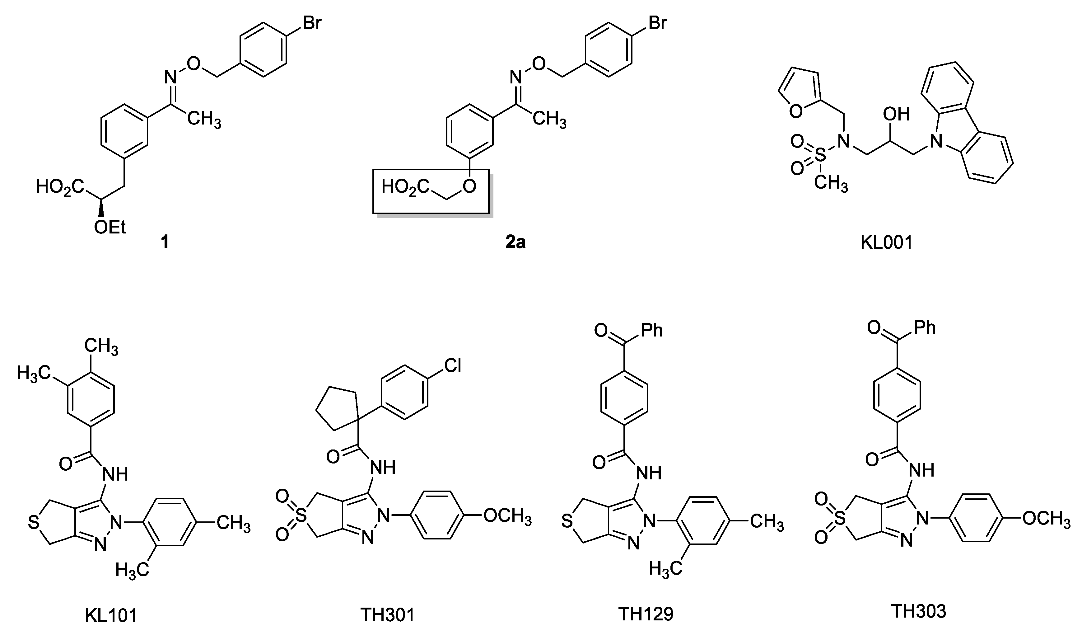
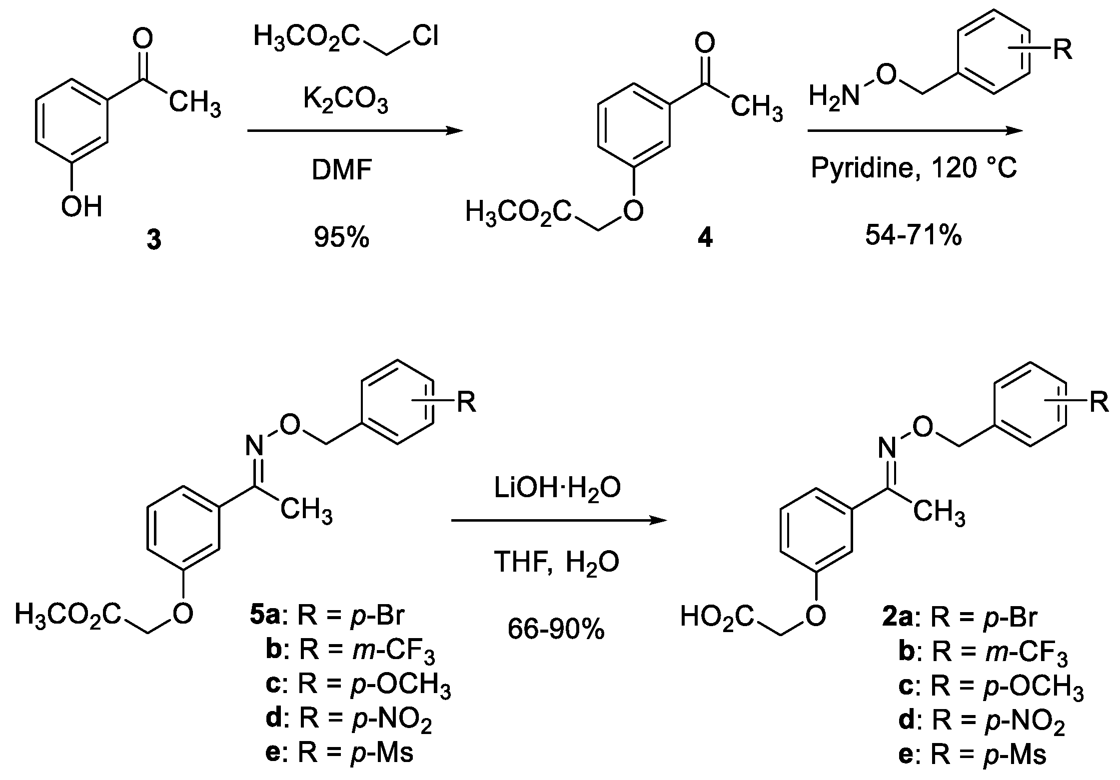
2. Results and Discussion

Our work commenced with the synthesis of non-ethoxypropanoic acid-type SJ019 analogs. As shown in Scheme 1, the analogs were synthesized in a similar way to SJ019 [18]. Briefly, commercially available 3-hydroxyacetophenone 3 was alkylated to possess an oxypropanoic acid moiety as a bioisostere of the 2-ethoxypropanoic acid moiety of KS15. Then, the resulting acetophenone 4 was condensed with variously substituted benzyloxyamines to afford oximes 5ae. Hydrolysis of 5ae possessing terminal ester provided carboxylic acid 2ae.
With the available selected compounds, their effects on the molecular circadian rhythms were examined. As shown in Table 1, carboxylic acids 2ae and their methyl ester 5ae were examined with an expectation of developing prodrugs, but they showed similar potency and efficacy. Following the previous structure–activity relationship study, most of the analogs were potent transcriptional enhancers, except for 2e and 5e with the para-methylsulfonyl group. Among the analogs, those with the para-methoxy (2c and 5c) and the para-nitro groups (2d and 5d) were more effective transcriptional enhancers compared to SJ019 (2a). Notably, KS15 itself with the para-bromo group was the most potent and effective transcriptional enhancer among the KS15 derivatives possessing the 2-ethoxypropanoic acid moiety. Thus, a combination of functional groups and chemical moieties in the analogs tune the transcriptional activities. Finally, compound 5d was chosen for further development in terms of efficacy and potency.
To further develop the selected compound 5d, inhibitory activities of 5d against five cytochrome P450 (P450) isozymes were examined using a cocktail method with human liver microsomes (HLMs) and P450 probe drugs. KS15 exhibited moderate inhibitory activities against CYP1A2 (IC50 = 14.9 μM), CYP2C9 (IC50 = 9.1 μM), CYP2C19 (IC50 = 20.2 μM), CYP2D6 (IC50 = 23.1 μM), and CYP3A (IC50 = 24.0 μM) (Table 2). Resultantly, KS15 has raised the potential of drug–drug interaction through metabolism. In contrast, the selected analog 5d did not significantly interfere with the P450 isozymes at the tested concentrations, and all the IC50 values were more than 50 μM. Moreover, metabolites from the incubation of 5d with HLMs during the experiments did not significantly interfere with the P450 isozymes.
Next, we examined the metabolic stability of 5d in HLMs. Rapid hydrolysis of 5d was anticipated because 5d possesses a methyl ester group in its chemical structure. As shown in Figure 2, KS15 was relatively stable (t1/2 > 60 min), while 5d was time-dependently decreased in HLMs (t1/2 = 2.4 min). The metabolite was determined as the hydrolysis product, i.e., 2d, by mass spectrometry. We also examined the metabolic stability of 2d, which was found to be stable in HLMs (t1/2 > 60 min).
We also conducted pharmacokinetic studies in vivo in rats after intravenous (IV, 2 mg/kg) or an oral (PO, 5 mg/kg) administration of 5d and 2d (Figure 3). For the pharmacokinetic study of 5d, the concentrations of 2d (formed metabolite) in plasma were measured after the IV or PO administration of 5d, due to its metabolic instability. Pharmacokinetic parameters following IV or PO administration of each compound were summarized for the 2d form in Table 3.
When we compared the pharmacokinetic characteristics of 5d with that of 2d (Figure 3), pharmacokinetic profiles after IV administrations of each compound were similar to what was expected. Table 3 shows some pharmacokinetic differences: AUCs after IV administration of 5d were smaller than those of 2d, while Vss was slightly larger. We speculated that those differences might be attributed to that the fact that the hydrolysis of 5d to 2d is fast but not prompt in vivo, and which is supported by slightly bigger values of 5d in half-life (t1/2), clearance (CL), and mean retention time (MRT). Interestingly, the total clearance was slow as the 2d form and Vss was small, indicating the limited tissue distribution of the 2d form in the body. For PO administrations, the absorptions were almost complete (i.e., approximately 100%) in both cases, but a significant delay in tmax was observed for 5d. Both 5d and 2d might be super-permeable from the gastrointestinal tract to the systemic blood circulation with a negligible first-pass effect on the 2d. Otherwise, 5d might be rapidly converted to a super-permeable 2d, absorbed in the gastrointestinal tract. Indeed, the Cmax values were high (i.e., 13.5 and 14.6 μg/mL for 5d and 2d administration). The Tmax for PO administration of 5d was prolonged with a shift of the curve compared with the PO administration of 2d, possibly due to a delay in absorption or due to a metabolic conversion.
In the next set of experiments, we examined whether compound 2d acts on mammalian CRYs. Therefore, direct binding between compound 2d and CRY1/2 was analyzed by surface plasmon resonance. Previously, biotin-tagged KS15 was reported to directly bind to the C-termini of both CRY1 and 2 in a pull-down assay [15]. Co-immunoprecipitation experiments also suggested that the binding of KS15 to CRY inhibits the interaction between CRY and BMAL1, thereby disrupting the feedback action of a circadian transcription-repressor complex [17]. As shown in Figure 4, compound 2d is directly bound to CRY1 and CRY2 to a similar extent, indicating that the molecular target of 2d is CRY1/2 for the modulation of the circadian clock.
In good agreement with the binding assay, compounds 5d and 2d functionally affected the negative feedback actions of CRY on the CLOCK:BMAL1 heterodimer. As shown in Figure 5a, enhanced E-box-mediated transcription by the CLOCK:BMAL1 heterodimer is strongly suppressed by co-expression of human CRY1. However, cell treatment with the compounds 5d or 2d dose-dependently restored E-box-mediated transcriptional activities, demonstrating their inhibitory effects on CRY-mediated negative feedback actions.
Most importantly, 5d and 2d are distinct from ethoxypropanoic acid-type KS15 regarding their effects on molecular circadian rhythmicity measured by periodic PER2 protein accumulation. Following our previous results [16], treatment with 20 μM KS15 attenuates amplitude but did not significantly alter the period of the rhythm in dexamethasone (DEX)-synchronized Per2-luciferase knock-in (Per2-Luc-KI) fibroblast cultures (Figure 5b). However, 5d and 2d (20 μM each) significantly augmented the amplitude of cyclic Per2 expression. Furthermore, they barely affect the robustness of the rhythm as examined by the Cosinor regression, implying that our new compounds do not have destabilizing effects on the molecular rhythm in contrast to KS15. Altogether, the results strongly suggest that 5d and 2d can be considered “circadian clock-enhancers” that strengthen the molecular circadian rhythmicity as well as E-box-mediated transcription evoked by the CLOCK:BMAL1 heterodimer through inhibition of CRY actions.
Nevertheless, it remains unclear how 5d and 2d reinforced cyclic PER2 protein accumulation in contrast to KS15. Both 5d and 2d maintained key structures required for binding to CRY proteins, which we previously identified in our structure–activity relationship study [18]. Indeed, 2d and KS15 exhibited similar binding properties in SPR analyses with recombinant CRY proteins. Furthermore, 2d showed no apparent isoform selectivity between CRY1 and CRY2 (Figure 4). It is plausible that these compounds may have distinct effects on a certain region/domain of CRY proteins residing outside their main binding site, thereby affecting post-translational regulation. In this context, it should be noted that the C-terminal domain, but not the binding pocket was responsible for the selectivity of isoform-specific CRY stabilizers, including KL101 and TH301 [7]. Therefore, 5d and 2d may have affected post-translational regulatory steps such as oligomerization with PER proteins, nuclear translocation, association with the CLOCK:BMAL1 heterodimer and/or inactivation/degradation, particularly, in a different way than KS15. The effects of these non-ethoxypropanoic acid-type inhibitors on the post-translational regulation of CRY proteins should be further examined to clarify the exact molecular mechanism(s) underlying their clock-enhancing activities.

3. Materials and Methods

3.1. Chemistry

Starting materials and reagents for chemical reactions were obtained from commercial suppliers unless otherwise stated. Organic solvents were dried over appropriate drying agents or distilled prior to use. We used Merck silica gel 60 F254 plates for thin-layer chromatography. Flash chromatography was performed with Biotage Isolera, and manual chromatography was conducted using Merck silica gel 60 (230–400 mesh). Nuclear magnetic resonance (NMR) spectra were acquired on a Bruker Avance-500 in deuterated chloroform (chloroform-d, CDCl3) or deuterated methanol (methanol-d, CD3OD); chemical shifts δ in parts per million (ppm) were measured relative to TMS with the residual solvent peak used as an internal reference, and J values were measured in Hertz. High-resolution mass spectrometry was conducted using Jeol JMS-700 at the Korea Basic Science Institute Daegu branch.
The analogs 5be and 2be were synthesized using previously reported procedures for 5a and 2a [18], or a slight modification of those procedures. NMR spectra for all new compounds 5be and 2be are available in the Supplementary Materials.

3.1.1. Methyl 2-(3-acetylphenoxy)acetate (4)

Methyl chloroacetate (1.54 mL, 17.6 mmol) was added to a solution of 3-hydroxyacetophenone (3, 2.00 g, 1.47 mmol) and K2CO3 (6.10 g, 44.1 mmol) in DMF (50 mL). The reaction mixture was stirred overnight, and then filtered through a short pad of celite. Volatiles in the filtrate were removed under the reduced pressure, and the remaining residue was diluted with EtOAc. The diluted solution was washed with water and brine. The organic solution was dried over MgSO4, filtered, and concentrated. The residue was purified by SiO2 chromatography to afford 4 as a white solid (270 mg, 95% yield). Spectral data were well matched with previously reported data [18].

3.1.2. Representative Procedure: Methyl 2-(3-(1-(((4-nitrobenzyl)oxy)imino)ethyl)phenoxy)acetate (5d)

Methyl 2-(3-acetylphenoxy)acetate (4, 1.08 g, 5.56 mmol) and O-(4-nitrobenzyl)hydroxylamine hydrochloride (1.37 g, 6.67 mmol) were dissolved in pyridine (24 mL). The reaction mixture was refluxed with stirring for 2 h at 120 °C. After cooled to rt, volatiles were removed under the reduced pressure. The residue was diluted with EtOAc, and then treated with 1N-HCl. The mixture was extracted with EtOAc, washed with water and brine. The organic solution was dried over MgSO4, filtered, and concentrated in vacuo. The residue was purified by SiO2 chromatography to afford the compound 5d as a white solid (1.41 g, 71%).

3.1.3. Characterization Data for 5be

Methyl 2-(3-(1-(((3-trifluoromethylbenzyl)oxy)imino)ethyl)phenoxy)acetate (5b): colorless oil, 54% yield. Rf = 0.5 (EtOAc:n-hexane = 1:4). 1H NMR (500 MHz, Chloroform-d) δ 7.70 (m, 1H, Ar), 7.70–7.54 (m, 2H, Ar), 7.51 (t, J = 7.7 Hz, 1H, Ar), 7.33–7.25 (m, 3H, Ar), 6.94 (m, 1H, Ar), 5.30 (s, 2H, O-CH2-Ar), 4.68 (s, 2H, O-CH2-CO2Me), 3.71 (s, 3H, OCH3) 2.29 (s, 3H, CH3). 13C NMR (125 MHz, Chloroform-d) δ 169.4 (C=O), 157.9 (OAr), 155.2 (C=N), 139.3 (Ar), 138.1 (Ar), 138.1 (Ar), 131.4 (Ar), 130.8 (q, J = 32.3 Hz, C-CF3), 129.7 (Ar), 129.0 (Ar), 124.9 (q, J = 3.9 Hz, CH-C-CF3), 124.7 (q, J = 3.8 Hz, CH-C-CF3), 124.3 (q, J = 272.1 Hz, CF3), 119.9 (Ar), 115.5 (Ar), 112.6 (Ar), 75.5 (O-CH2-Ar), 65.5 (O-CH2-CO2Me), 52.4 (OCH3), 13.1 (CH3). HR-EI-MS m/z 381.1190 [M]+ (calculated for C19H18F3NO4 381.1188). ATR-IR νmax 2918, 2372, 2323, 1761 cm−1.
Methyl 2-(3-(1-(((4-methoxybenzyl)oxy)imino)ethyl)phenoxy)acetate (5c): White solid, 67% yield. Rf = 0.3 (EtOAc:n-hexane = 1:4). 1H NMR (500 MHz, Chloroform-d) δ 7.36 (d, J = 8.7 Hz, 2H, Ar), 7.29–7.24 (m, 3H, Ar), 6.92–6.87 (m, 3H, Ar), 5.16 (s, 2H, O-CH2-Ar), 4.66 (s, 2H, O-CH2-CO2Me), 3.81 (s, 3H, OCH3), 3.80 (s, 3H, OCH3), 2.21 (s, 3H, CH3). 13C NMR (125 MHz, Chloroform-d) δ 169.4 (C=O), 159.5 (OAr), 157.9 (OAr), 154.4 (C=N), 138.4 (Ar), 130.2 (Ar), 130.1 (2C, Ar), 129.6 (Ar), 119.9 (Ar), 115.3 (Ar), 113.9 (2C, Ar), 112.5 (Ar), 76.1 (O-CH2-Ar), 65.5 (O-CH2-CO2Me), 55.4 (OCH3), 52.4 (OCH3), 13.0 (CH3). HR-EI-MS m/z 343.1420 [M]+ (calculated for C19H21NO5 343.1420). ATR-IR νmax 2867, 2372, 2326, 1735 cm−1.
Methyl 2-(3-(1-(((4-nitrobenzyl)oxy)imino)ethyl)phenoxy)acetate (5d): White solid, 71% yield. Rf = 0.3 (EtOAc:n-hexane = 1:3). 1H NMR (500 MHz, Chloroform-d) δ 8.12 (d, 2H, J = 8.7 Hz, Ar), 7.45 (d, 2H, J = 8.8 Hz, Ar), 7.24–7.10 (m, 3H, Ar), 6.81 (m, 1H, Ar), 5.23 (s, 2H, O-CH2-Ar), 4.56 (s, 2H, O-CH2-CO2Me), 3.71 (s, 3H, OCH3), 2.18 (s, 3H, CH3). 13C NMR (125 MHz, Chloroform-d) δ 169.2 (C=O), 157.8 (OAr), 155.4 (C=N), 147.4 (Ar), 145.9 (Ar), 137.7 (Ar), 129.6 (Ar), 128.3 (2C, Ar), 123.6 (2C, Ar), 119.7 (Ar), 115.3 (Ar), 112.6 (Ar), 74.7 (O-CH2-Ar), 65.3 (O-CH2-CO2Me), 52.3 (OCH3), 12.9 (CH3). HR-EI-MS m/z 358.1165 [M]+ (calculated for C18H18N2O6 358.1164). ATR-IR νmax 2855, 2371, 2324, 1736 cm−1.
Methyl 2-(3-(1-(((4-methylsulfonylbenzyl)oxy)imino)ethyl)phenoxy)acetate (5e): White solid, 36% yield. Rf = 0.4 (EtOAc:n-hexane = 1:1). 1H NMR (500 MHz, Chloroform-d) δ 7.96 (d, J = 8.4 Hz, 2H, Ar), 7.61 (d, J = 8.4 Hz, 2H, Ar), 7.34–7.22 (m, 3H, Ar), 6.92 (m, 1H, Ar), 5.33 (s, 2H, O-CH2-Ar), 4.67 (s, 2H, O-CH2-CO2Me), 3.82 (s, 3H, OCH3), 3.08 (s, 3H, SO2CH3), 2.30 (s, 3H, CH3). 13C NMR (125 MHz, Chloroform-d) δ 169.2 (C=O), 157.8 (OAr), 155.4 (C=N), 144.8 (Ar), 139.7 (Ar), 137.8 (Ar), 129.6 (Ar), 128.4 (2C, Ar), 127.5 (2C, Ar), 119.8 (Ar), 115.3 (Ar), 112.7 (Ar), 74.9 (O-CH2-Ar), 65.3 (O-CH2-CO2Me), 52.3 (OCH3), 44.6 (SO2CH3), 13.0 (CH3). HR-EI-MS m/z 391.1090 [M]+ (calculated for C19H21NO6S 391.1092). ATR-IR νmax 2926, 2371, 2324, 1755 cm−1.

3.1.4. Representative Procedure: 2-(3-(1-(((4-nitrobenzyl)oxy)imino)ethyl)phenoxy)acetic acid (2d)

LiOH·H2O (281 mg, 6.70 mmol) was added to a solution of the compound 5d (800 mg, 2.23 mmol) in THF (6 mL) and H2O (6 mL). The reaction mixture was stirred for 2 h, then acidified with 1N-HCl. The resulting mixture was extracted with EtOAc. The organic solution was dried over MgSO4, filtered, and concentrated in vacuo. The residue was purified by SiO2 chromatography to afford the compound 5d as a white solid (543 mg, 71%).

3.1.5. Characterization Data for 2be

2-(3-(1-(((3-trifluoromethylbenzyl)oxy)imino)ethyl)phenoxy)acetic acid (2b): Colorless oil, 90% yield. Rf = 0.5 (MeOH:CH2Cl2 = 1:19). 1H NMR (500 MHz, Chloroform-d) δ 9.82 (br, 1H, CO2H), 7.57 (m, 1H, Ar), 7.51–7.43 (m, 2H, Ar), 7.37 (t, J = 7.7 Hz, 1H, Ar), 7.20–7.11 (m, 3H, Ar), 6.80 (m, 1H, Ar), 5.17 (s, 2H, O-CH2-Ar), 4.56 (s, 2H, O-CH2-CO2H), 2.15 (s, 3H, CH3). 13C NMR (125 MHz, Chloroform-d) δ 172.9 (C=O), 156.1 (OAr), 153.8 (C=N), 137.7 (Ar), 136.6 (Ar), 130.0 (Ar), 129.4 (q, J = 31.3 Hz, C-CF3), 128.3 (Ar), 127.5 (Ar), 123.4 (q, J = 3.8 Hz, CH-C-CF3), 123.2 (q, J = 3.8 Hz, CH-C-CF3), 122.8 (q, J = 271.3 Hz, CF3), 118.6 (Ar), 114.1 (Ar), 111.1 (Ar), 74.0 (O-CH2-Ar), 63.6 (OCH3), 11.6 (CH3). HR-EI-MS m/z 367.1031 [M]+ (calculated for C18H16F3NO4 367.1029). ATR-IR νmax 2936, 2372, 2324, 1735 cm−1.
2-(3-(1-(((4-methoxybenzyl)oxy)imino)ethyl)phenoxy)acetic acid (2c): White solid, 90% yield. Rf = 0.4 (EtOAc:n-hexane = 2:1). 1H NMR (500 MHz, Chloroform-d) δ 9.76 (br, 1H, CO2H), 7.37 (d, J = 8.6 Hz, 2H, Ar), 7.32–7.24 (m, 3H, Ar), 6.94–6.88 (m, 3H, Ar), 5.17 (s, 2H, O-CH2-Ar), 4.70 (s, 2H, O-CH2-CO2H), 3.81 (s, 3H, OCH3), 2.22 (s, 3H, CH3). 13C NMR (125 MHz, Chloroform-d) δ 174.1 (C=O), 159.4 (OAr), 157.4 (OAr), 154.4 (C=N), 138.4 (Ar), 130.0 (2C, Ar), 129.6 (Ar), 120.0 (Ar), 115.3 (Ar), 113.8 (2C, Ar), 112.3 (Ar), 76.1 (O-CH2-Ar), 64.8 (O-CH2-CO2H), 55.3 (OCH3), 13.0 (CH3). HR-EI-MS m/z 329.1263 [M]+ (calculated for C18H19NO5 329.1267). ATR-IR νmax 2906, 2372, 2326, 1733 cm1.
2-(3-(1-(((4-nitrobenzyl)oxy)imino)ethyl)phenoxy)acetic acid (2d): White solid, 71% yield. Rf = 0.4 (MeOH:CH2Cl2 = 1:19). 1H NMR (500 MHz, Chloroform-d) δ 8.14 (d, J = 8.9 Hz, 2H, Ar), 7.47 (d, J = 8.7 Hz, 2H, Ar), 7.24–7.15 (m, 3H, Ar), 6.85 (m, 1H, Ar), 5.24 (s, 2H, O-CH2-Ar), 4.62 (s, 2H, O-CH2-CO2H), 2.21 (s, 3H, CH3). 13C NMR (125 MHz, Chloroform-d) δ 173.5 (C=O), 157.4 (OAr), 155.4 (C=N), 147.5 (Ar), 145.8 (Ar), 137.8 (Ar), 129.7 (Ar), 128.3 (2C, Ar), 123.7 (2C, Ar), 120.1 (Ar), 115.4 (Ar), 112.5 (Ar), 74.8(O-CH2-Ar), 64.8 (O-CH2-CO2H), 13.0 (CH3). HR-EI-MS m/z 344.1008 [M]+ (calculated for C17H16N2O6 344.1005). ATR-IR νmax 2910, 2372, 2324, 1739 cm1.
2-(3-(1-(((4-methylsulfonylbenzyl)oxy)imino)ethyl)phenoxy)acetic acid (2e): White solid, 66% yield. Rf = 0.1 (EtOAc:n-hexane = 2:1). 1H NMR (500 MHz, Methanol-d4) δ 7.94 (d, J = 8.4 Hz, 2H, Ar), 7.65 (d, J = 8.1 Hz, 2H, Ar), 7.29–7.21 (m, 3H, Ar), 6.95 (m, 1H, Ar), 5.31 (s, 2H, O-CH2-Ar), 4.66 (s, 2H, O-CH2-CO2H), 3.10 (s, 3H, SO2CH3), 2.27 (s, 3H, CH3).13C NMR (125 MHz, Methanol-d4) δ 171.2 (C=O), 158.0 (OAr), 155.4 (C=N), 145.0 (Ar), 139.8 (Ar), 137.6 (Ar), 129.2 (Ar), 128.3 (2C, Ar), 127.1 (2C, Ar), 119.1 (Ar), 115.2 (Ar), 112.0 (Ar), 74.5 (O-CH2-Ar), 64.5 (O-CH2-CO2H), 43.0 (SO2CH3), 11.6 (CH3). HR-EI-MS m/z 377.0933 [M]+ (calculated for C18H19NO6S 377.0934). ATR-IR νmax 2921, 2372, 2324, 1728 cm1.

3.2. Cell Cultures and Luciferase Reporter Assay

Materials for cell culture were purchased from Thermo Fisher Scientific, unless otherwise stated. NIH3T3 and HEK293T cells were maintained in Dulbecco’s modified Eagle medium supplemented with 5% fetal bovine serum and 100 U/mL penicillin/streptomycin in a humidified atmosphere containing 5% CO2 at 37 °C. Plasmids and transfection experiments for luciferase reporter assay were described in our previous report [18]. For dose-response experiments, mouse NIH3T3 cells stably transfected with an E-box-LUC reporter [16,18] were plated in 12-well culture plates, treated as indicated for 36 h, and subjected to luciferase assay. For transient transfections, we seeded HEK293T cells in 12-well culture plates and used Lipofectamine® 3000 reagent to transfect cells with the E-box-LUC reporter and the indicated clock gene expression plasmids together with a thymidine kinase promoter-driven Renilla luciferase (pRL-TK) plasmids. We kept total DNA levels constant by adding an empty pcDNA3.1 plasmid. After 24 h of transfection, cells were treated with vehicle (0.1% DMSO) or chemical compounds at various concentrations for 36 h. Cell extracts were prepared by incubating cells in 0.3 mL reporter lysis buffer for 15 min at room temperature. Luciferase activity was measured with a commercial dual luciferase assay kit (Promega, Madison, WI, USA).

3.3. Cytochrome P450 Inhibition Assay

The inhibitory effects of KS15 and compound 5d on the metabolism of five cytochrome P450 (P450) probe substrates were evaluated using previous methods with slight modifications [19,20]. The following P450 probe substrates were used: phenacetin (100 μM) for CYP1A2, tolbutamide (100 μM) for CYP2C9, omeprazole (20 μM) for CYP2C19, dextromethorphan (5 μM) for CYP2D6, and midazolam (5 μM) for CYP3A. Incubation mixtures containing pooled human liver microsomes (HLMs, XTreme 200, XenoTech, Lenexa, KS, USA), P450 probe substrates, and KS15 or compound 5d (final concentrations: 0, 0.5, 2, 5, 20, and 50 μM) were preincubated for 5 min at 37 °C. After preincubation, the NADPH-generating system (including 3.3 mM G6P, 1.3 mM β-NADP+, 3.3 mM MgCl2, and 1.0 U/mL G6PDH) was added to the incubation mixtures, and then incubated for 10 min at 37 °C. The reaction was stopped by adding 50 μL of 7 nM trimipramine (internal standard, IS) in ice-cold acetonitrile. After centrifugation at 18,000× g (5 min, 4 °C), supernatant aliquots were analyzed by liquid chromatography-tandem mass spectrometry (LC-MS/MS). All microsomal incubations were conducted in triplicate.
All metabolites and IS were separated on a Kinetex C18 column (100 mm × 2.1 mm, 2.6 μm ID., Phenomenex, Torrance, CA, USA) and analyzed using an LC-MS/MS system (LCMS 8060, Shimadzu, Tokyo, Japan) equipped with an electrospray ionization (ESI) interface. The mobile phase solvent A was 0.1% formic acid in water, and solvent B was 0.1% formic acid in acetonitrile. Gradient separation was performed as follows: 0–7 min (10–90% B) and 7.1–10 min (10% B). The flow rate was 0.2 mL/min, and the run time was 10 min for each sample. Electrospray ionization was conducted in the positive ion mode at 4 kV. Quantitation was performed in the selected reaction monitoring (SRM) of the [M+H]+ ion and the related product ion for each metabolite and IS: acetaminophen (m/z 152 > 110, collision energy (CE) 25 eV), 4-hydroxytolbutamide (m/z 287 > 89, CE 60 eV), 5-hydroxyomeprazole (m/z 362 > 214, CE 10 eV), dextromethorphan (m/z 258 > 157, CE 35 eV), 1′-hydroxymidazolam (m/z 342 > 203, CE 25 eV), and IS (m/z 295 > 100, CE 17 eV).

3.4. Metabolic Stability Test

The metabolic stability of KS15, 5d, or 2d was evaluated using pooled HLMs. Pooled HLMs (1.0 mg/mL), a potassium phosphate buffer, and a test compound (1 μM) were pre-incubated (37 °C, 5 min). To initiate the reaction, the NADPH generating system was added to the samples and incubated for specific time points (0, 5, 15, 20, 45, and 60 min). Then, aliquots were taken and reactions terminated in acetonitrile containing IS. After centrifugation, supernatants were analyzed using LC-MS/MS to determine KS15, 5d, or 2d concentrations in samples. Analyte separation was performed using a Kinetex C18 column (100 mm × 2.1 mm, 2.6 μm ID., Phenomenex). The mobile phase solvent A was 0.1% formic acid in water, and solvent B was 0.1% formic acid in acetonitrile. Gradient separation was performed as follows: 0–3 min (10–90% B) and 3.1–8 min (10% B). The flow rate was 0.2 mL/min, and the run time was 8 min for each sample. Electrospray ionization was conducted in positive ion mode at 4 kV. Optimum operating conditions were as follows: vaporizer temperature, 300 °C; capillary temperature, 250 °C; and collision gas (argon) pressure 17 kPa. Quantitation was performed in the SRM of the [M+H]+ ion and the related product ion for each test compound and IS: KS-15 (m/z 420 > 169, CE 30 eV), 5d (m/z 359 > 206, CE 20 eV), 2d (m/z 345 > 192, CE 22 eV), and IS (m/z 295 > 100, CE 17 eV).

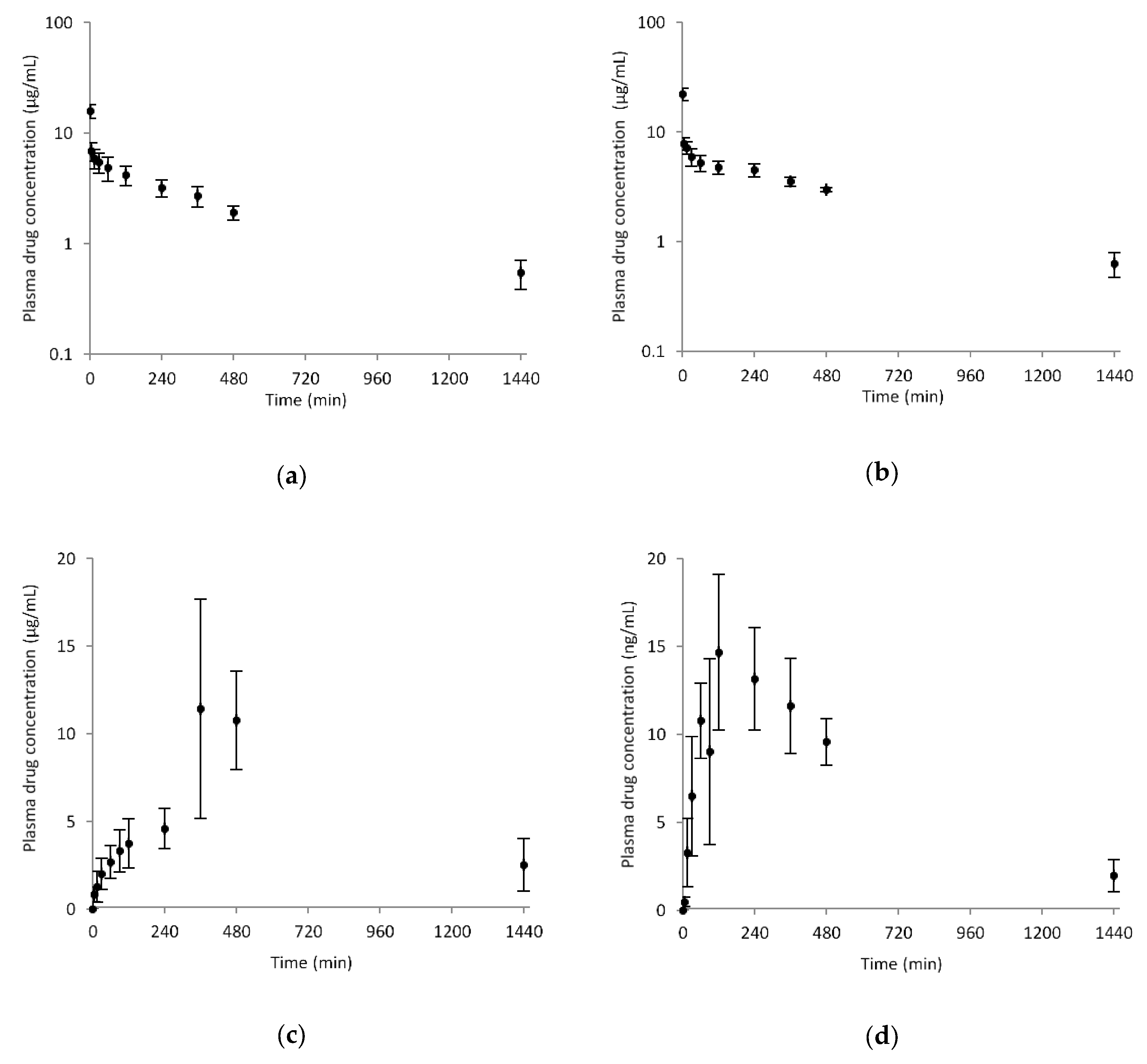
3.5. Pharmacokinetics

For in vivo pharmacokinetic studies, Sprague Dawley (SD) male rats (weighing 270–280 g) were purchased from Orient Bio Inc. (Seongnam, Korea). Experimental protocols involving animal use were carefully reviewed and approved by the Institutional Animal Care and Use Committee at Gachon University. Prior to studies, animals fasted for 18 h, but with free access to water. Compound 5d or 2d was dissolved in a dosing vehicle consisting of PEG400, normal saline, and dimethylsulfoxide in a ratio of 6:3:1 (v/v/v%) to make a 2 mg/mL dosing solution. This was intravenously bolus-injected into the right femoral vein at a volume of 1 mL/kg (2 mg/kg) for intravenous administration or intragastrically injected using an oral zone at a volume of 2.5 mL/kg (5 mg/kg) for p.o. administration. Blood samples (approximately 100 μL each) were collected in heparinized tubes via the right femoral artery at 0, 1, 5, 15, 30, 60, 120, 240, 360, 480, 1440 min after intravenous administration, and 0, 5, 15, 30, 60, 90, 120, 240, 360, 480, 1440 min after p.o. administration.
To determine drug concentration levels in plasma, the Agilent 1290 UHPLC-DAD system with Polar RP column (3 μm, 150 mm × 2 mm ID., Phenomenex) was used for chromatographic separations. The mobile phase was composed of 0.1% formic acid in acetonitrile and 0.1% formic acid in DDW in a ratio of 40:60 and was isocratically delivered at a flow rate of 0.2 mL/min. The total run time was 4.4 min for samples which were monitored at 260 nm. Working standard solutions of compound 2d were serially prepared in methanol in the 50–10,000 ng/mL concentration range, and 10 μL aliquots were added to 90 μL blank plasma to prepare calibration standards. For sample preparation, 100 μL aliquots of plasma samples or calibration standards were transferred to microcentrifuge tubes and mixed by vortexing vigorously for 5 min. They were then centrifuged at 14,000 rpm for 10 min at 4 °C. Supernatants were transferred to fresh analysis vials and a 2 μL aliquot injected onto the UHPLC-DAD system.

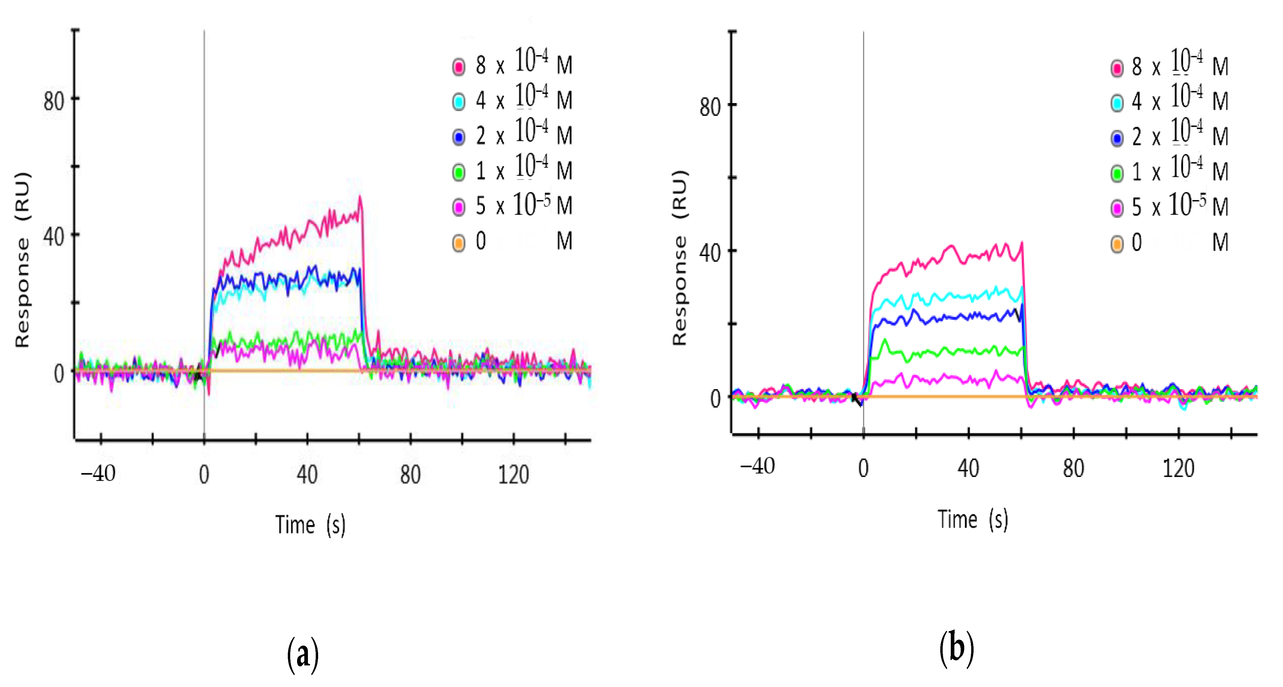
3.6. Surface Plasmon Resonance

We used a ProteOn XPR36 system (Bio-Rad, Hercules, CA, USA) equipped with a HTE sensor chip (high capacity tris-NTA for histidine-tagged small molecule capture, Bio-Rad) for surface plasmon resonance analyses. Purified target proteins (6xHis- and GST-tagged hCRY1 and hCRY2) were prepared by a baculovirus expression system (MyBioSource, San Diego, CA, USA). Phosphate buffered saline (PBS) plus 0.005% Tween 20 was used as the running buffer. The 6xHis-hCRY1 and 6xHis-hCRY2 proteins were immobilized onto separate channels of a HTE chip by injecting 210 μL protein sample (2.0 μg/mL) into flow cells (30 μL/min) to allow saturation. Serially diluted compound 2d samples (50–800 μM) were prepared in 4% DMSO in TBS-T buffer. Real-time binding kinetics analysis was performed by injecting 100 μL sample aliquots into the channels of the HTE chip for 1 min at 100 μL/min. Then PBS-T buffer (100 μL/min) was flushed over the chip for 200 s to dissociate bound analytes. Recorded resonance units (RU) were normalized and plotted against chemical concentration. We used ProteOn Manager v.3.1.0.6 software for recording, plotting, and analyzing binding curves.

3.7. Real-Time Bioluminescence Monitoring

For real-time bioluminescence monitoring, we used mouse embryonic fibroblast (MEF) cultures established from Per2-LUC-KI mice [16,21]. To examine the possible effects of CRYs inhibitors on cyclic PER2 accumulation, Per2-LUC-KI MEFs were plated in 35 mm dishes, grown for 24 h, and synchronized with 200 nM DEX for 2 h. The medium was then replaced with a recording medium containing 0.1 mM D-luciferin (Promega) along with vehicle (0.1% DMSO), KS15 (20 μM), 5d (20 μM), or 2d (20 μM). Light emission for 2 min at intervals of 20 min was integrated using a dish-type wheeled luminometer (Kronos-Dio, ATTO Cooperation, Tokyo, Japan). Background-subtracted bioluminescence profiles were analyzed using Cosinor analysis software (available at http://www.circadian.org, last accessed on 19 May 2021).

3.8. Statistical Evaluation

The EC50 values for luciferase reporter assay were calculated based on a logistic four-parametric equation using the mean values of intrinsic activation (SigmaPlot 10.0; Systat Software Inc., San Jose, CA, USA). The IC50 and t1/2 values for the cytochrome P450 inhibition assay and metabolic stability test were calculated by WinNonlin software (Pharsight, Mountain View, CA, USA). Pharmacokinetic parameters were calculated using non-compartmental analysis (WinNonlin v.8.3; Pharsight, Mountain View, CA, USA). Kinetic and equilibrium constants for SPR analysis were obtained by global fitting of the Langmuir 1:1 bimolecular kinetic model (ProteOn Manager v.3.1.0.6, Bio-Rad, Hercules, CA, USA). Circadian molecular clock-enhancing activities were expressed as means ± SE and statistically evaluated by Student’s t-test or one-way analysis of variance (ANOVA) followed by Newman–Keuls multiple comparison test for post hoc comparisons. The significance level for the analyses was set at p < 0.05.

4. Conclusions

Bioisosteric replacement is a useful and prevalent strategy for drug design and molecular modification in medicinal chemistry. Years of cumulative data proved that rational design with such a strategy for modifying lead compounds provided better candidates with improved potency, selectivity, and drug-likeness [22,23,24]. In this context, we undertook synthesis and evaluation of non-ethoxypropanoic acid analogs of KS15 by bioisosteric replacement. Among the synthesized, compound 5d and its hydrolyzed product 2d were potent and effective enhancers on the E-box mediated transcription. Compounds 5d and 2d also exhibited desirable metabolic and pharmacokinetic profiles as promising drug candidates. Furthermore, compound 2d directly binds to CRY1 and 2 in surface plasmon resonance analyses, suggesting that the pharmacological target of 2d is CRYs. Compounds 5d and 2d inhibited the negative feedback actions of CRY on the CLOCK:BMAL1 heterodimer, which also suggested that their molecular target is CRYs. Most importantly, compounds 5d and 2d exhibited significant enough effects on molecular circadian rhythmicity to be considered circadian clock-enhancers, which are distinct from the KS15 possessing ethoxypropanoic acid moiety. Taken altogether, novel cryptochrome inhibitors with circadian clock-enhancing activity have been developed by bioisosteric replacement.

Supplementary Materials

The Supplementary Material for this article is available online at https://www.mdpi.com/article/10.3390/ph14060496/s1, including 1H and 13C NMR spectra.

Author Contributions

Conceptualization, H.-E.J., J.-W.J., S.C. and G.H.S.; structural design and synthesis, Y.U.J., H.J. and B.K.; biological assay, H.Y.L., S.C. and G.H.S.; metabolism, G.-H.L. and K.-H.L.; pharmacokinetics, H.-J.M.; SPR analysis, G.C.; data curation, Y.U.J. and G.H.S.; writing—original draft preparation, Y.U.J. and J.-W.J.; writing—review and editing, H.-E.J., J.-W.J., S.C. and G.H.S.; visualization, Y.U.J.; supervision, J.-W.J. and G.H.S.; funding acquisition, J.-W.J. and G.H.S. All authors have read and agreed to the published version of the manuscript.

Funding

This work was supported by the National Research Foundation of Korea (NRF) grants funded by the Korean government (MSIT) (Grant No. 2015M3A9E7029176, 2019M3C7A1032764, 2020R1C1C1013670 and 2020R1A5A2017323) and the 4th BK21 project funded by the Korean Ministry of Education (5199990614732). G.H.S was supported by the Korea University Research Grant.

Institutional Review Board Statement

The study was conducted according to the Guide for the Care and Use of Laboratory Animals issued by the National Institute of Health, and approved by the Animal Care and Use Committee of Gachon University (GIACUC-R2020025).

Data Availability Statement

The data presented in this study are available in the article and Supplementary Material.

Acknowledgments

Aziz Sancar (University of North Carolina, Chapel Hill, NC, USA) kindly provided the Flag/Myc/His-tagged human CRY1 expression construct (pfmh-hCry1; Addgene plasmid #25843).

Conflicts of Interest

The authors declare no conflict of interest.

References

  1. Patke, A.; Young, M.W.; Axelrod, S. Molecular mechanisms and physiological importance of circadian rhythms. Nat. Rev. Mol. Cell Biol. 2020, 21, 67–84. [Google Scholar] [CrossRef]
  2. Walker, W.H.; Walton, J.C.; DeVries, A.C.; Nelson, R.J. Circadian rhythm disruption and mental health. Transl. Psychiatry 2020, 10, 1–13. [Google Scholar] [CrossRef] [PubMed] [Green Version]
  3. Chen, Z.; Yoo, S.-H.; Takahashi, J.S. Development and therapeutic potential of small-molecule modulators of circadian systems. Annu. Rev. Pharmacol. Toxicol. 2018, 58, 231–252. [Google Scholar] [CrossRef] [PubMed]
  4. Diallo, A.B.; Coiffard, B.; Leone, M.; Mezouar, S.; Mege, J.-L. For whom the clock ticks: Clinical chronobiology for infectious diseases. Front. Immunol. 2020, 11, 1457. [Google Scholar] [CrossRef]
  5. Ray, S.; Reddy, A.B. COVID-19 management in light of the circadian clock. Nat. Rev. Mol. Cell Biol. 2020, 21, 494–495. [Google Scholar] [CrossRef]
  6. Hirota, T.; Lee, J.W.; John, P.C.S.; Sawa, M.; Iwaisako, K.; Noguchi, T.; Pongsawakul, P.Y.; Sonntag, T.; Welsh, D.K.; Brenner, D.A.; et al. Identification of small molecule activators of cryptochrome. Science 2012, 337, 1094–1097. [Google Scholar] [CrossRef] [PubMed] [Green Version]
  7. Miller, S.; Son, Y.L.; Aikawa, Y.; Makino, E.; Nagai, Y.; Srivastava, A.; Oshima, T.; Sugiyama, A.; Hara, A.; Abe, K.; et al. Isoform-selective regulation of mammalian cryptochromes. Nat. Chem. Biol. 2020, 16, 676–685. [Google Scholar] [CrossRef] [PubMed]
  8. Kolarski, D.; Miller, M.; Oshima, T.; Nagai, Y.; Aoki, Y.; Kobauri, P.; Srivastava, A.; Sugiyama, A.; Amaike, K.; Sato, A.; et al. Photopharmacological manipulation of mammalian CRY1 for regulation of the circadian clock. J. Am. Chem. Soc. 2021, 143, 2078–2087. [Google Scholar] [CrossRef] [PubMed]
  9. Grant, D.; Yin, L.; Collins, J.L.; Parks, D.J.; Orband-Miller, L.A.; Wisely, G.B.; Joshi, S.; Lazar, M.A.; Willson, T.M.; Zuercher, W.J. GSK4112, a small molecule chemical probe for the cell biology of the nuclear heme receptor Rev-erbα. ACS Chem. Biol. 2010, 5, 925–932. [Google Scholar] [CrossRef] [PubMed]
  10. Helleboid, S.; Haug, C.; Lamottke, K.; Zhou, Y.; Wei, J.; Daix, S.; Cambula, L.; Rigou, G.; Hum, D.W.; Walczak, R. The identification of naturally occurring neoruscogenin as a bioavailable, potent, and high-affinity agonist of the nuclear receptor RORα (NR1F1). J. Biomol. Screen. 2014, 19, 399–406. [Google Scholar] [CrossRef] [PubMed] [Green Version]
  11. He, B.; Nohara, K.; Park, N.; Park, Y.-S.; Guillory, B.; Zhao, Z.; Garcia, J.M.; Koike, N.; Lee, C.C.; Takahashi, J.S.; et al. The small molecule nobiletin targets the molecular oscillator to enhance circadian rhythms and protect against metabolic syndrome. Cell Metab. 2016, 23, 610–621. [Google Scholar] [CrossRef] [Green Version]
  12. Kumar, N.; Solt, L.A.; Conkright, J.J.; Wang, Y.; Istrate, M.A.; Busby, S.A.; Garcia-Ordonez, R.D.; Burris, T.P.; Griffin, P.R. The benzenesulfoamide T0901317 [N-(2, 2, 2-trifluoroethyl)-N-[4-[2, 2, 2-trifluoro-1-hydroxy-1-(trifluoromethyl) ethyl] phenyl]-benzenesulfonamide] is a novel retinoic acid receptor-related orphan receptor-α/γ inverse agonist. Mol. Pharm. 2010, 77, 228–236. [Google Scholar] [CrossRef] [PubMed] [Green Version]
  13. Rahman, S.; Wittine, K.; Sedić, M.; Markova-Car, E.P. Small molecules targeting biological clock; A novel prospective for anti-cancer drugs. Molecules 2020, 25, 4937. [Google Scholar] [CrossRef] [PubMed]
  14. Amaike, K.; Oshima, T.; Skoulding, N.S.; Toyama, Y.; Hirota, T.; Itami, K. Small molecules modulating mammalian biological clocks: Exciting new opportunities for synthetic chemistry. Chem 2020, 6, 2186–2198. [Google Scholar] [CrossRef]
  15. Cha, H.K.; Chung, S.; Lim, H.Y.; Jung, J.-W.; Son, G.H. Small molecule modulators of the circadian molecular clock with implications for neuropsychiatric diseases. Front. Mol. Neurosci. 2019, 11, 496. [Google Scholar] [CrossRef] [PubMed] [Green Version]
  16. Chun, S.K.; Jang, J.; Chung, S.; Yun, H.; Kim, N.-J.; Jung, J.-W.; Son, G.H.; Suh, Y.-G.; Kim, K. Identification and validation of cryptochrome inhibitors that modulate the molecular circadian clock. ACS Chem. Biol. 2014, 9, 703–710. [Google Scholar] [CrossRef] [PubMed]
  17. Chun, S.K.; Chung, S.; Kim, H.-D.; Lee, J.H.; Jang, J.; Kim, J.; Kim, D.; Son, G.H.; Oh, Y.J.; Suh, Y.-G.; et al. A synthetic cryptochrome inhibitor induces anti-proliferative effects and increases chemosensitivity in human breast cancer cells. Biochem. Biophys. Res. Commun. 2015, 467, 441–446. [Google Scholar] [CrossRef]
  18. Jang, J.; Chung, S.; Choi, Y.; Lim, H.Y.; Son, Y.; Chun, S.K.; Son, G.H.; Kim, K.; Suh, Y.-G.; Jung, J.-W. The cryptochrome inhibitor KS15 enhances E-box-mediated transcription by disrupting the feedback action of a circadian transcription-repressor complex. Life Sci. 2018, 200, 49–55. [Google Scholar] [CrossRef] [PubMed]
  19. Park, S.-Y.; Nguyen, P.-H.; Kim, G.; Jang, S.-N.; Lee, G.-H.; Phuc, N.M.; Wu, Z.; Liu, K.-H. Strong and selective inhibitory effects of the biflavonoid selamariscina a against CYP2C8 and CYP2C9 enzyme activities in human liver microsomes. Pharmaceutics 2020, 12, 343. [Google Scholar] [CrossRef]
  20. Song, Y.; Park, S.Y.; Wu, Z.; Liu, K.-H.; Seo, Y.H. Hybrid inhibitors of DNA and HDACs remarkably enhance cytotoxicity in leukaemia cells. J. Enzym. Inhib. Med. Chem. 2020, 35, 1069–1079. [Google Scholar] [CrossRef] [Green Version]
  21. Yoo, S.-H.; Yamazaki, S.; Lowrey, P.L.; Shimomura, K.; Ko, C.H.; Buhr, E.D.; Siepka, S.M.; Hong, H.-K.; Oh, W.J.; Yoo, O.J.; et al. PERIOD2: LUCIFERASE real-time reporting of circadian dynamics reveals persistent circadian oscillations in mouse peripheral tissues. Proc. Natl. Acad. Sci. USA 2004, 101, 5339–5346. [Google Scholar] [CrossRef] [PubMed] [Green Version]
  22. Kumari, S.; Carmona, A.V.; Tiwari, A.K.; Trippier, P.C. Amide bond bioisosteres: Strategies, synthesis, and successes. J. Med. Chem. 2020, 63, 12290–12358. [Google Scholar] [CrossRef] [PubMed]
  23. Lima, L.M.; Barreiro, E.J. Bioisosterism: A useful strategy for molecular modification and drug design. Curr. Med. Chem. 2005, 12, 23–49. [Google Scholar] [CrossRef] [PubMed]
  24. Dick, A.; Cocklin, S. Bioisosteric replacement as a tool in anti-HIV drug design. Pharmaceuticals 2020, 13, 36. [Google Scholar] [CrossRef] [Green Version]
Figure 1. The chemical structure of KS15 (1), SJ019 (2a), and selected CRYs modulators.
Pharmaceuticals 14 00496 g001
Scheme 1. Synthesis of non-ethoxypropanoic acid type KS15 analogs.
Pharmaceuticals 14 00496 sch001
Figure 2. Metabolic stability of KS15 (a), 5d (b), and 2d (c) in human liver microsomes in the presence of an NADPH generating system. Data are the means ± SD of triplicate experiments.
Pharmaceuticals 14 00496 g002
Figure 3. Pharmacokinetic profiles of 5d and 2d: Plasma concentration vs. time profiles after IV administration of 2 mg/kg of (a) 5d and (b) 2d to rats (mean ± SD, n = 3–4). Mean plasma concentration profiles after single PO administration of 5 mg/kg of (c) 5d and (d) 2d in rats (mean ± SD, n = 3–4).
Pharmaceuticals 14 00496 g003
Figure 4. Surface plasmon resonance (SPR) analysis for the interaction between 2d and purified CRY1 or CRY2 proteins: His-tagged CRY1 and CRY2 proteins were purified and used for SPR analysis. RU = resonance units. (a) Interaction between 2d and CRY1. KD = 3.89 × 10⁻⁴ M. (b) Interaction between 2d and CRY2. KD = 3.80 × 10⁻⁴ M.
Pharmaceuticals 14 00496 g004
Figure 5. The circadian molecular clock-enhancing activity of 5d and 2d. (a) Effects of 5d and 2d on suppressive action of CRY1 on the CLOCK:BMAL1-activated E-box-LUC reporter activity. (b) Bioluminescence recordings of dexamethasone (DEX)-synchronized Per2-luciferase knock-in (Per2-Luc-KI) fibroblast cultures. Representative traces for rhythmic Per2-Luc activity (upper) and Cosinor analysis results (lower panels; mean ± SE, n = 4). * p < 0.05; one-way ANOVA with Newman-Keuls multiple comparison test for post-hoc comparisons; ** p < 0.01; one-way ANOVA with Newman-Keuls multiple comparison test for post-hoc comparisons.
Pharmaceuticals 14 00496 g005
Table 1. Effect of the synthesized derivatives on the E-box-LUC reporter activities.
EntryCompoundMax (%) 1EC50 (μM)
12a164.81.54
25a149.1n/d 2
32b130.7n/d
45b171.5n/d
52c182.19.88
65c151.86.32
72d161.310.17
85d193.06.44
92e98.7n/d
105e104.7n/d
1 Maximum responses expressed as a percentage of the vehicle-treated group. 2 EC50 values were not determined within the tested dosages.
Table 2. Inhibitory effects of KS15 and 5d on specific cytochrome P450 (P450) activities in human liver microsomes.
P450Enzyme ActivityIC50 (μM)
KS155d
1A2Phenacetin O-deethylation14.9>50
2C9Tolbutamide 4-methylhydroxylation9.1>50
2C19Omeprazole hydroxylation20.2>50
2D6Dextromethorphan O-demethylation23.1>50
3AMidazolam 1’-hydroxylation24.0>50
Table 3. Pharmacokinetic parameters of 5d and 2d following PO and IV administrations.
ParameterIntravenous (2 mg/kg)Oral (5 mg/kg)
5d2d5d2d
AUClast (μg/mL·min)2891.2 ± 465.03885.9 ± 278.59471.5 ± 1281.710,204.7 ± 1308.5
AUCinfinite (μg/mL·min)3268.2 ± 600.34290.6 ± 398.7n/a11,069.2 ± 1130.5
t1/2 (min)467.1 ± 61.3433.0 ± 58.0n/a391.2 ± 47.6
Cmax (μg/mL)--13.5 ± 4.714.6 ± 5.1
tmax (min)--420.0 ± 69.3160.0 ± 69.2
CL (mL/min/kg)0.6 ± 0.10.5 ± 0.0--
MRT (min)588.5 ± 75.6557.1 ± 73.7--
Vss (mL/kg)349.8 ± 34.9259.7 ± 25.8--
F (%)--> 99> 99
Publisher’s Note: MDPI stays neutral with regard to jurisdictional claims in published maps and institutional affiliations.

