Article

Rational Design, Synthesis and Biological Evaluation of Novel Pyrazoline-Based Antiproliferative Agents in MCF-7 Cancer Cells

1
Department of Pharmaceutical Medicinal Chemistry, Faculty of Pharmacy, Suez Canal University, Ismailia 41522, Egypt
2
Department of Pharmaceutical Chemistry, Faculty of Pharmacy, Egyptian Russian University, Badr 11829, Egypt
3
Department of Chemistry, Faculty of Science, Suez Canal University, Ismailia 41522, Egypt
4
Department of Pharmacy Practice, Faculty of Pharmacy, King Abdulaziz University, Jeddah 21589, Saudi Arabia
5
Department of Pharmacology and Toxicology, Faculty of Pharmacy, King Abdulaziz University, Jeddah 21589, Saudi Arabia
*
Author to whom correspondence should be addressed.
Pharmaceuticals 2022, 15(10), 1245; https://doi.org/10.3390/ph15101245
Submission received: 21 September 2022 / Revised: 29 September 2022 / Accepted: 6 October 2022 / Published: 10 October 2022
(This article belongs to the Section Medicinal Chemistry)

Abstract

:
Breast cancer is a disease in which cells in the breast divide continuously without control. There are great limitations in cancer chemotherapy. Hence, it is essential to search for new cancer therapeutics. Herein, a novel series of EGFR/HER2 dual inhibitors has been designed based on the hybridization of thiazole and pyrazoline fragments. The synthesized compounds were screened for their anti-proliferative activity against MCF-7 breast cancer cell line and MCF-10 normal breast cell line. Interestingly, synthesized compounds 6e and 6k showed very potent antiproliferative activity towards MCF-7 with IC50 values of 7.21 and 8.02 µM, respectively. Furthermore, enzymatic assay was performed against EGFR and HER2 to prove the dual inhibitory action. Compounds 6e and 6k showed potent inhibitory activity for EGFR with IC50 of 0.009 and 0.051 µM, respectively, and for HER2 with IC50 of 0.013 and 0.027 µM, respectively. Additionally, compounds 6e and 6k significantly stimulated apoptotic breast cancer cell death. Compound 6e was further explored for its anticancer activity in vivo using a Xenograft model. Moreover, computational modeling studies, ADMET studies and toxicity prediction were performed to investigate their potential drug candidates.

1. Introduction

Cancer is considered one of the most challenging causes of death in the world, as it is estimated to have caused nearly 10 million deaths in 2020 according to WHO [1,2]. Cancer is not a single disease, but is rather a collective term that describes the distress of the fundamental regulatory genes that are responsible for controlling cell proliferation, differentiation, and survival [3]. The traditional treatment strategies were based on the unspecific death induction targeting the DNA synthesis process without discriminating between tumor cells and rapidly dividing normal cells, which lead to many side effects [4,5,6]. Currently, it is an essential demand to develop more safe and selective molecular targeted chemotherapeutic agents [7]. In this regard, one of the powerful approaches is to target transduction-related macromolecules, especially protein kinases [8,9,10].
Protein kinases represent the fifth largest human protein family that exert their functions as key regulators of signal transduction mechanisms such as proliferation, metabolism, vascularization, and apoptosis [10,11,12,13]. According to the amino acid that they phosphorylate, protein kinases can be classified into three main classes: tyrosine kinases, Serine–threonine kinases, and Histidine kinases [14]. The epidermal growth factor (EGF) receptor family belongs to tyrosine kinases and is highly expressed or mutated in several cancers such as lung cancer, myeloid leukemia, hormone-dependent and hormone-independent breast cancer. Breast cancer is the leading cause of death among women, with nearly 2.26 million reported new cases worldwide in 2020 as stated in WHO [1]. The EGF receptor family embraces four members: EGFR (HER1/ErbB-1), HER2 (ErbB-2), HER3 (ErbB-3) and HER4 (ErbB-4). These proteins serve as cell surface receptors and consist of an extracellular module and an intracellular kinase domain, connected by a single-helix transmembrane segment and a juxta-membrane segment. EGF receptor family activation is dependent on dimerization, where ligand binding provokes homo- and/or heterodimerization and autophosphorylation on key cytoplasmic residues. The phosphorylated receptor enrolls adapter proteins, which activate complex downstream signaling cascades [15,16] (Figure 1). EGFR and HER2 overexpression in breast cancer is associated with resistance to chemotherapy and poor prognosis [17,18,19,20,21]. Therefore, the usage of dual EGFR/HER2 inhibitors/modulators in breast cancer treatment is an approach worth considering [22,23,24].
Pyrazolines are reported to display various pharmacological activities including anticancer activity [25,26,27,28,29,30,31], in addition to possessing cancer chemo-preventive properties for many pyrazoline-based cytotoxic agents [32,33,34]. For example, compounds I, II and III showed potent inhibitory activity in breast cancer cells MCF-7 with GI50 = 0.13 µM, IC50 = 0.42 µM and IC50 = 34.10 µM, respectively [35,36,37]. Moreover, using methoxy aryl derivatives is reported as increasing anti-proliferative activity, particularly against MCF-7 [38,39].
Besides, thiazole-based compounds are intensively studied for their anticancer effects [40,41,42] through the inhibition of different molecular targets such as receptor tyrosine kinases (RTKs), non-receptor tyrosine kinases (nRTKs), phosphatidylinositol-3-kinases (PI3Ks), serine/threonine kinases (STKs), Bcl-2 family, histone deacetylases (HDACs), and tumor necrosis factor-alpha (TNF-α) [43,44,45]. Dasatinib (IV) was confirmed to possess potential tyrosine kinase inhibitory activity [46]. The study of hydrazinyl thiazole derivative (V) revealed high activity against MCF-7 (IC50 = 9.06 µM), with selective inhibition to EGFR [47]. In another study, a phenylthiazole derivative (VI) had an IC50 = 0.19 µM against EGFR [48]. EAI045 (VII) is a fourth-generation allosteric inhibitor of mutant EGFR (L858R) with high potency (IC50 = 0.019 µM) [49] (Figure 2).

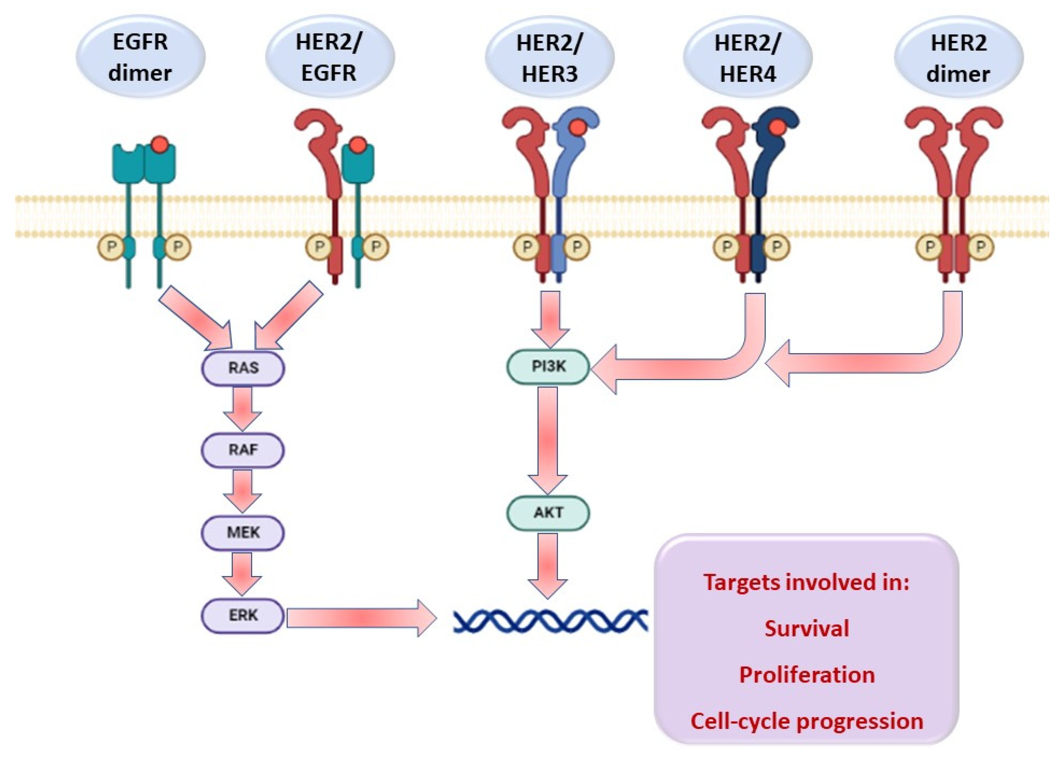
Rationale of the Work

Molecular hybridization of both thiazole and pyrazoline scaffolds has been used in multi-targeting kinase inhibitors [50], topoisomerases, apoptosis, with significant antitumor activity against MCF-7 and A595 cell lines [27]. Based on the aforementioned protein kinase inhibitory activity of thiazole and pyrazoline nuclei, especially on EGFR and HER2, a strategy of hybridizing these two luminous moieties was designed to synthesize dual EGFR/HER2 hybrid modulators/inhibitors targeting breast cancer [51]. The new scaffold aimed to find novel lead structures with better activity and lower toxicity [52] (Figure 3).
In this study, a new series of thiazolyl–pyrazoline derivatives was designed, synthesized, and screened for their activity against MCF-7 and MCF-10, and their inhibitory activity was further explored as dual-EGFR/HER2 inhibitors. In addition, the promising compounds were investigated for apoptosis activity and in vivo studies. The molecular modelling studies were performed for the designed compounds to confirm their binding modes to EGFR and HER2 at the ATP binding site.

2. Results and Discussion

2.1. Chemistry

The synthetic routes of the thiazolyl–pyrazoline derivatives 3ac, 6al and 9af are considered in Chart 1, Chart 2 and Chart 3. First, the 3-(4-aryl)-1-(3,4-dimethoxyphenyl)prop-2-en-1-ones 2ac were obtained through the Claisen–Schmidt reaction [53] by direct condensation of aromatic aldehydes 1a–c with dimethoxy acetophenone in the presence of sodium hydroxide [54,55,56]. This step was followed by the reaction of compounds 2ac with thiosemicarbazide under a basic condition to give 3-(3,4-dimethoxyphenyl)-5-(4-aryl)-4,5-dihydro-1H-pyrazole-1-carbothioamides 3ac, respectively (Chart 1) [57,58]. This reaction proceeded through a condensation reaction followed by subsequent cyclization via Michael addition reaction. The IR spectra for compounds 3ac showed the characteristic NH2 band in the range 3422–3260 cm−1, in addition to a band at 1265–1248 cm−1 referring to C=S group. Additionally, 1H NMR of compounds 3ac displayed three doublets of doublet (dd) of pyrazoline ring due to ABX pattern. These three (dd) signals of HA, HB, and HX appeared at δ 3.12–3.14, 3.8 and 5.8–5.9 ppm, respectively. Moreover, NH2 exchangeable proton signal of compounds 3ac appeared around δ 8.0 ppm. 13C NMR of compounds 3ac revealed the signals for CHAHB and CHX carbons of pyrazoline ring around δ 42.9 and 62.8 ppm, respectively, as well as C=S signals at δ 176.2 ppm.
Next, the reaction of carbothioamide compounds 3ac with an equimolar amount of phenacyl bromide derivatives 5a–d [59] in absolute ethanol yielded the corresponding thiazolyl–pyrazoline derivatives 6al in good yields (Chart 2) [60]. Compounds 5a–d were prepared by bromination of corresponding acetophenone using bromine and acetic acid in diethyl ether as solvent. The IR spectra of compounds 6al showed the disappearance of the NH2 characteristic band in the region 3422–3260 cm−1. 1H NMR spectra also revealed the absence of NH2 proton around δ 8.0 ppm. 1H NMR spectra of compounds 6al showed the signals of aromatic protons at δ 7.8–6.9 ppm to be assignable to thiazolyl and phenyl protons. In addition, an extra signal at δ 2.3 ppm appeared due to the presence of CH3 in compounds 6b, 6f, and 6j. The ABX pattern was confirmed by COSY spectra for compound 6k to prove the formation of the pyrazoline ring of two non-equivalent protons at C4 (HA and HB) and one proton at C5 (HX). 13C NMR spectra of compounds 6al showed the signals of the aromatic carbons at 103.5–165 ppm. 13C NMR spectra of compounds 6g, 6c, and 6k (4-FC6H4) showed the splitting of the three carbon atoms at δ 115.9–115.7, 128–127.8, 131.7–131.54 ppm due to their coupling with the fluorine atom. Also, 13C NMR spectra revealed the appearance of singlet signal at δ 21.2–21.3 ppm which belongs to CH3 (tolyl carbon) of compounds 6b, 6f, and 6j. Mass spectra of compounds 6a–l showed the molecular ions peak in accord with their molecular weight.
Furthermore, compounds 8ab were synthesized by treating pentane-2,4-dione or methyl 3-oxobutanoate with sulfuryl chloride (SO2Cl2) [61]. Treatment of 3-(3,4-dimethoxyphenyl)-5-aryl-4,5-dihydro-1H-pyrazole-1-carbothioamide derivatives 3ac with 3-chloropentane-2,4-dione 8a or ethyl 2-chloro-3-oxobutanoate 8b in refluxing ethanol produced the intended thiazolyl–pyrazoline derivatives 9af (Chart 3) [62]. IR spectra of compounds 9a–c showed a band at 1617–1613 cm−1 representing the C=O group. 1H NMR spectra of compounds 9a–c elicited two singlet signals at δ 2.39 and 2.37 ppm belonging to two CH3 of methyl and acetyl group, respectively, whereas their 13C NMR displayed signals at δ 19.1, 30.1, and 189 were attributed to SP3 carbons of two CH3 and C=O carbons, respectively.
IR spectra of compounds 9df displayed a band at 1708–1695 cm−1 representing C=O. 1H NMR spectrum also showed a typical quartet–triplet pattern of the ethyl protons at δ 4.2 and 1.25 ppm, respectively. However, signals of thiazole CH3 appeared at δ 2.36–2.35 ppm. Furthermore, 13C NMR spectrum revealed the signals of ethyl carbons at δ 14.8 and 60.6 ppm, and signal at 165.1–165.2 ppm attributed C=O of ester.

2.2. Biological Evaluation
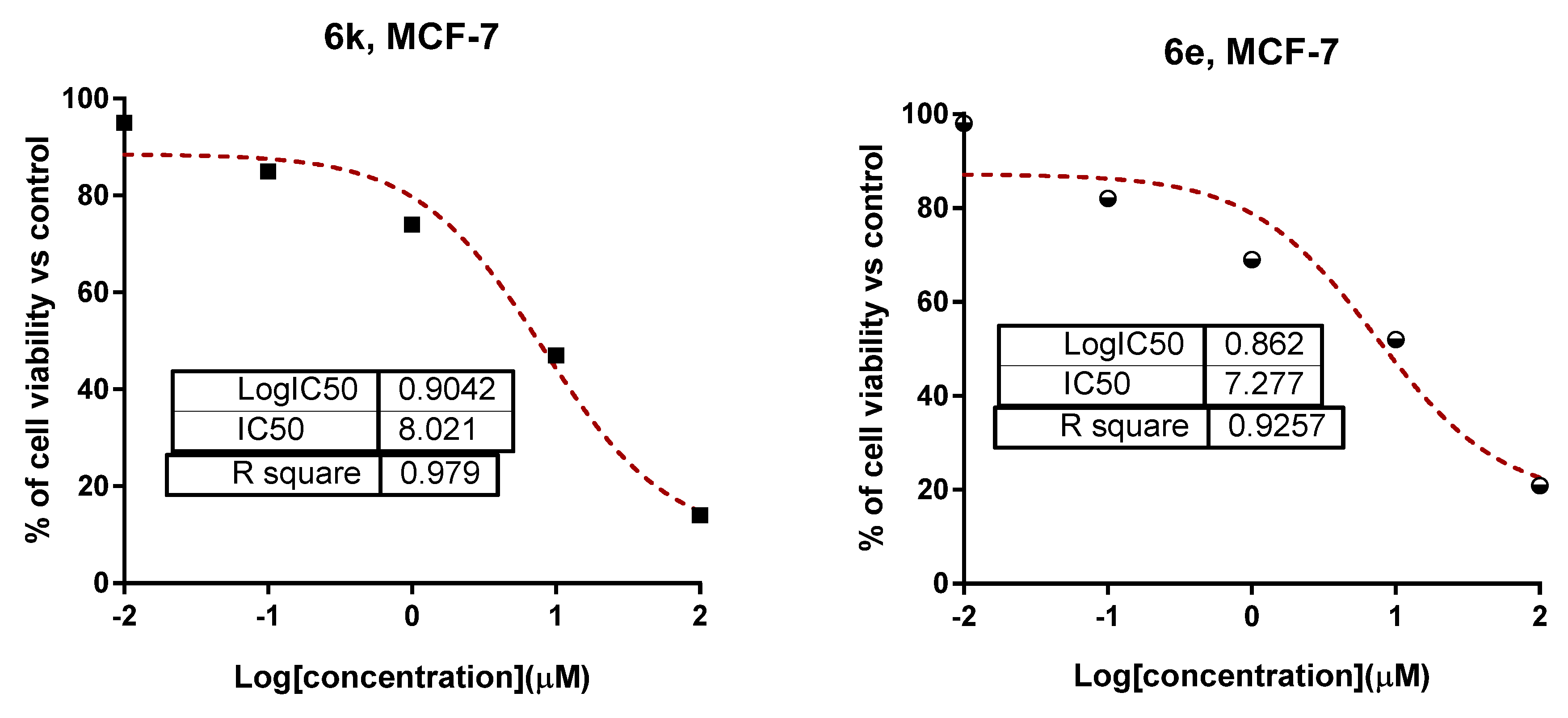
2.2.1. Cytotoxicity

The MTT assay was used to test the activity and selectivity of all synthesized compounds for cytotoxic activity against MCF-7 cancer cells and MCF-10A normal breast cells. As seen in Table 1, compounds 3c, 6b and 6f were found to be more cytotoxic against MCF-7, with IC50 ranges of 11.09, 13.36 and 11.05 µM, respectively. Interestingly, compounds 6e, 9f and 6k exhibited remarkable cytotoxic activities, with IC50 values of 7.21, 8.35 and 8.02 µM, respectively, compared to Lapatinib (IC50 = 7.45 µM) with non-cytotoxic activity against the MCF-10A. Other compounds exhibited relatively moderate-to-weak cytotoxic activities. Hence, three compounds; 6e, 9f, and 6k, were chosen to be studied for their potential molecular targets and ability to induce apoptosis in MCF-7 cells (Figure 4).

2.2.2. Potential EGFR/HER2 Kinase Inhibitory Assay

To further elucidate the mechanisms behind the cytotoxic effects on MCF-7 cells, compounds 3c, 6e, 6k, 6f and 9f were tested for their ability to inhibit EGFR and HER2. As seen in Table 2, the tested compounds exhibited promising dual EGFR/HER2 inhibition activities. Remarkably, compounds 6k and 6e had IC50 values of 0.014 and 0.009 µM against EGFR and IC50 values of 0.027 and 0.013 µM against HER2, respectively, in a comparable way to Lapatinib. Due to their potent cytotoxic and EGFR/HER2 inhibitory effects, compounds 6k and 6e merited investigation for their ability to induce apoptosis.

2.2.3. Apoptotic Investigation

Annexin V/PI Staining with Cell Cycle Analysis

Flow cytometric analysis of Annexin V/PI staining was used to examine apoptotic cell death in untreated and treated MCF-7 cells to determine the apoptotic activity of the two promising compounds, 6k and 6e (IC50 = 8.02, 7.21 µM, 48 h). As seen in Figure 5A, compounds 6k, and 6e significantly stimulated apoptotic breast cancer cell death with 22.44% (14.38% for late apoptosis, 8.06% for early apoptosis) and 27.99% (18.52% for late apoptosis, 9.47% early apoptosis), respectively, compared to 0.68% in the control.
Cytotoxic substances were tested using DNA flow cytometry to determine what percentage of cells were in each cell cycle following treatment. Figure 5B, the percentage of cells in the S phase increased from 34.18 to 44.52 % after treatment with compound 6e, indicating that compound 6e induced cell cycle arrest in the S phase, while the percentage of cells in the G2/M phase was reduced by 2.01% after treatment with compound 6k, compared to the control (9.7%). This result indicates that compound 6k induced cell cycle arrest in the G2/M-phase. By comparison, other phases were not significantly changed in either treatment.

RT-PCR

Further validation of the apoptosis-inducing activities of both tested compounds 6k and 6e in MCF-7 cells, the gene expression levels of apoptosis-related genes in both untreated and treated MCF-7 cells through the RT-PCR. As seen in Figure 6, compounds 6k and 6e treatments increased P53 level by 3.91- and 4.8-fold, Bax level by 5.24- and 6.39-fold, caspase 3 level by 5.79- and 7.12-fold, caspase 8 level by 3.91- and 1.71-fold, caspase 9 level by 6.07- and 8.19-fold, while both compound treatments decreased Bcl-2 level “as the anti-apoptotic gene” by 0.41- and 0.32-fold compared to untreated control.
P53 is a tumor suppressor gene that is essential for apoptosis. The activation of caspases 3 and 9 and the decreased expression of the Bcl-2 gene may cause p53 apoptosis. The results showed the intrinsic apoptotic pathway through activation of P53, Bax, and caspases 3 and 9 levels. Further, the treatment-induced upregulation of caspase 8 gene promotes an extrinsic apoptotic pathway in MCF-7 cells. As a result, our RT-PCR findings corroborated those of previous studies, showing that the apoptotic pathway is highlighted by the upregulation of proapoptotic genes and the downregulation of anti-apoptotic genes (Table 3).

2.2.4. In Vivo Studies

For evaluation of the anticancer activity of compound 6e, mice, MCF-7 cells (Xenograft model) were treated with compounds 6e and lapatinib at a dose (10 mg/ kg BW, IP). As seen in Figure 7A, an increase in the mice weight of the MCF-7 control mice reached 32.28 g, compared to 24.5 g in normal mice, while treatment with compound 6e and lapatinib reduced the mice weight to 28.23 g and 27.35 g, respectively. The reduction in the mice’s weight was consequently related to a decrease in the tumor mass; tumor volume decreased from 89.8 mg to 28.4 mm3, respectively, upon treatment with compound 6e. Hence, compound 6e treatment enhanced tumor inhibition ratio by 52.46% compared to 50.53% in Lapatinib treatment Figure 7B.
As seen in Figure 7C, the hemoglobin and red blood cell concentrations dropped dropped dramatically to 5.7 (g/dL) and 2.9 (106/L), respectively. Conversely, white blood cell count was elevated to 7.0 (103/L) from normal control levels. Routine consequences of tumor proliferation are reduced levels of RBC, hemoglobin, and increased WBC counts [63,64]. CBC was nearly maintained at normal levels following treatment with compound 6e, where Hb (7.0 g/dL) and RBC’s count (4.8 106/μL) were raised, and WBCs count was lowered (4.12 103/μL). While Lapatinib-treatment increased Hb to 7.43 g/dL, RBC’s count to 4.99 106/μL and decreased WBC’s count to 4.21 103/μL compared to MCF-7 group.
As seen in Figure 7D, liver enzymes of ALT, AST were significantly elevated to 70.9, 70.65 (U/L), respectively in MCF-7 control mice compared to normal mice at 38.9 and 48.9 (U/L), respectively, caused by tumor inoculation-related hepatocellular damage, while liver enzymes were markedly mitigated by treatment with compound 6e. Liver enzymes were reduced to 41.8, 59.9 U/L, respectively, and this significantly reduced the toxicity to the liver caused by the cancer. While Lapatinib treatment decreased ALT to 40.8 U/L, AST fell to 57.7 U/L compared to the MCF-7 group
In agreement with the amelioration of biochemical parameters, histopathological examinations Figure 7E in liver sections in 6e-treated mice showed normal nuclei morphology in most of the cells with slight hydrophobic degeneration compared to MCF-7 control mice, which showed chronic inflammation (red arrow), and hepatocytes showing hydropic degeneration (arrowheads).
Taken together, amelioration in hematological, and biochemical results of compound 6e treatment agreed with the in vitro ones of cytotoxicity, EGFR/HER2 targeting, and apoptosis induction.

2.3. Computational Studies

2.3.1. Molecular Docking

To discover the most appropriate drug target for the proposed compounds, SwissTargetPrediction [65] was used. The studied compounds revealed that kinase proteins were the first class to be targeted, with a probability of 40% (Figure 8).
Molecular docking was carried out to estimate the interaction of the designed hybrid compounds with EGFR/HER2 kinase domains to rationalize their biological activity, and to reveal their probable binding pattern. Crystal structures were downloaded (PDB codes: 1XKK [66] and 3RCD [67]) for EGFR and HER2, respectively. Initially, autodocking of the co-crystallized ligands in EGFR and HER2 active sites was performed to validate the docking method. In the EGFR active site, the docking poses of lapatinib formed the key interactions with the active site through H-bond formation with amino acid residues Met793, Asp800, and Leu788 at distance of 3.17, 3.3, and 3.19 Å, respectively. In addition, arene H interactions with Leu844 and Leu718 were formed as illustrated in Figure 9A. As for HER2, the docking pose of TAK-285 produced the key interactions with the active site; it interacts through an H bond with amino acid residues Asp863, Gly727, and 2 H bonds with Met801 at distance of 3.22, 3.18, 3.02, 3.82 Å, respectively. Besides, π–H interactions with Phe864, Lys753, Leu852, and Leu800 were formed as seen in Figure 9B. The method validation was demonstrated by the resulted small RMSD values between the docked and the co-crystallized ligand poses in EGFR (0.917 Å, Figure S1) and HER2 (0.625 Å, Figure S2).
The newly synthesized thiazole–pyrazoline hybrids showed comparable binding patterns in both tyrosine kinases. Binding features of compounds 6e, 6k, 3c, and 9f were studied in more detail and compared to the docked co-crystallized inhibitors of either EGFR (Figure 10) and HER2 (Figure 11). From binding modes analyses, we can conclude that:
(1)
Compound 6e inside the binding site of the EGFR receptor (binding free energy = −13.05 kcal/mol) formed H bonds with Met793, Pro749, Arg841, Cys797, Gly796, and Leu792 at distances of 4.08, 2.69, 2.45, 2.03, 3.36 and 4.23 Å, respectively. Besides, π–H bonds with Lys745 and Val726 were formed. For HER2, compound 6e (binding free energy = −11.92 kcal/mol) showed H bonds with Cys805, Lys753, Thr862, Val734 and Asp863 at distance of 2.09, 2.31, 4.3, 3.99 and 3.32 Å, respectively. In addition, a π–H bond with Val734 was formed.
(2)
Compound 6k inside the binding site of EGFR receptor (binding free energy = −13.04 kcal/mol) achieved an H bond with Met793, Asp855, and Lys745 at a distance of 2.85, 3.05, 2.45 Å, respectively. Π–H bond with Val726, Leu718, Arg841, and Gly719 were formed. Furthermore, inside the binding site of HER2 receptor, compound 6k (binding free energy = −13.11 kcal/mol) established H bonds with Met801, Arg849, Phe864, Val734, and Asp863 at distance of 2.76, 2.13, 3.54, 4.26 and 3.1 Å, respectively. Besides π–H bonds with Cys805, Lys753 and Leu852 were noticed.
(3)
Compound 3c inside the binding site of EGFR receptor (binding free energy = −13.36 kcal/mol) achieved H bonds with Met793, Asp855, Lys745, and Gly796 at distance of 3.04, 2.99, 3.36, and 3.56 Å, in addition to the formation of π–H bonds with Leu718 and Phe856. Moreover, regarding the binding pocket of Her2 receptor (binding free energy = −11.3 kcal/mol), compound 3c formed H bonds with Lys753, Asp863, and Thr862 at a distance of 3.06, 2.77, and 4.23 Å, respectively and one π–H bond with Leu785.
(4)
For compound 9f inside the EGFR receptor binding site (binding free energy = −13.93 kcal/mol), H bonds with Met793, Asp855, Thr854, Lys745, Val726, and Met1002 were formed at distance of 3.05, 3.2, 2.18, 2.61, 3.55, and 3.8 Å, respectively, in addition to π–H bonds with Lys745 and Val726. On the other hand, compound 9f binding within the binding pocket of HER2 receptor (binding free energy = −11.85 kcal/mol) showed H bonds with Met801, Lys753, and Leu852 at distance of 3.01, 2.69, and 4.09 Å, respectively, besides one π–H bond with Val734.

2.3.2. In Silico Physicochemical Descriptors, Pharmacokinetic Properties and Bioactivity Prediction

Different parameters were measured to explore drug-likeness properties (Table S1) of the target thiazolyl–pyrazoline compounds, all of which obey Veber’s rule and Lipinski’s rule and may meet the criteria for orally active drugs, except for 6l which has 2 violations of the Lipinski rule. All compounds were predicted to have a clogP value in the range of 2.00–6.00, TPSA ≤ 110.72 Å2, rotatable bonds (RB) ≤ 9, H-bond acceptor moieties (HBA) ≤ 7, H-bond donor moieties (HBD) ≤ 1 and molar refractivity (MR) ≤ 147.86. Compound solubility was in the range of poorly soluble to moderately soluble, except for compounds 3a and 3b which were soluble. Bioavailability radar (Figures S3 and S4) indicated that molecules 3ac and 9ae are expected to be orally bioavailable.
In silico ADME prediction of pharmacokinetic properties was investigated for the target compounds (Table S2). All compounds showed very high HIA values, ranging from 97.5–99.6%, indicating very high GI absorption. All compounds exerted a strong bound effect on plasma protein (binding values of 87.45–93.09%). All compounds are not blood-brain barrier permeant and have skin p values from −2.35 to −3.43. On inspecting the boiled-egg model (Figure S5), all compounds were found to be in the white region indicating their proper GI absorption.
An in silico phase I metabolism study indicated that all compounds could inhibit CYP2C9, and that all compounds have CYP3A4-inhibiting activity except 6l. None of the thiazolyl–pyrazoline hybrids could inhibit CYP1A2.
Toxic properties such as irritant, mutagenic, and reproductive effects using were predicted Osiris server (Table S3). All compounds have no mutagenic, irritant, or reproductive fragments except for 3a, 3b, and 3c, which have reproductive effects due to the 1-ethyl hydrazine-1-carbothioamide fragment.

2.3.3. Structure–Activity Relationship (SAR) Study

SAR of the newly synthesized compounds 3ac (Chart 1), 6al (Chart 2) and 9af (Chart 3) was determined depending on the biological results as well as the docking binding free energy values. By superimposition of the most active compounds; 3c (IC50 MCF-7 = 11.19 ± 0.54 µM, IC50 EGFR = 0.067 ± 0.009 µM, IC50 HER2 = 0.07 ± 0.018 µM), 6e (IC50 MCF-7 = 7.21 ± 0.48 µM, IC50 EGFR = 7.21 ± 0.48 µM, IC50 HER2 = 0.013 ± 0.001 µM), and 9f (IC50 MCF-7 = 8.35 ± 0.29 µM, IC50 EGFR = 0.051 ± 0.003 µM, IC50 HER2 = 0.14 ± 0.006 µM), we conclude that all of them formed the necessary interactions in the Hinge region of the kinase domain due to the presence of the pyrazoline moiety in their structures. 3,4-dimethoxyphenyl moiety increases the activity due to the interactions formed in the Hydrophobic II pocket. Different substituents, oriented towards the Back pocket, showed different activities depending on the other moieties in the structure that can embed in the Hydrophobic I pocket (Figure 12).

3. Materials and Methods

3.1. Chemistry

NMR spectra were recorded by a Bruker spectrometer. 1H NMR spectra were run at 400 MHz, and 13C NMR spectra were run at 100 MHz in deuterated dimethylsulfoxide (DMSO-d6). Chemical shifts (δH and δC) were reported relative to the solvent (DMSO-d6). Infrared spectra were recorded on a Shimadzu FT-IR 8400S spectrophotometer as KBr disks at the faculty of science, Sohag University. Elemental analysis was performed using FLASH 2000 CHNS/O analyzer, at the Regional Center for Microbiology and Biotechnology, Al-Azhar University. The reaction progress was monitored using thin-layer chromatography (TLC)-precoated silica gel G plates, and the formation of products was visualized by irradiation with UV light (254 nm). Melting points were determined using Stuart Scientific apparatus and were uncorrected. All the chemicals were purchased from Alpha Aesar and Sigma-Aldrich (Figures S6–S79).

3.1.1. Synthesis of 3-(4-Aryl)-1-(3,4-dimethoxyphenyl)prop-2-en-1-one (2ac)

A solution of dimethoxy acetophenone (1.08 g, 6.0 mmol) in absolute ethanol (30 mL) was stirred with an equimolar weight of the appropriate aromatic aldehyde 1a-c (6.0 mmol), NaOH (2 g, 50 mmol) in water (15 mL) was added within 30 min. The reaction mixture was stirred for 5 h at room temperature. The reaction progress was monitored using TLC and methylene chloride/methanol (9:1) system until the completion of the reaction. Then, the resulting precipitate was filtered and washed with ethanol, and then recrystallized from absolute ethanol to give a yield of 95–98%.

3.1.2. Synthesis of 3-(3,4-Dimethoxyphenyl)-5-(4-aryl)-4,5-dihydro-1H-pyrazole-1-carbothioamide (3ac)

A mixture of 3-(4-aryl)-1-(3,4-dimethoxyphenyl)prop-2-en-1-one (2a-c) (20 mmol), thiosemicarbazide (1.8 g, 20 mmol) and NaOH (36 mmol) in ethanol (25 mL) was refluxed for 12 h. The formed precipitate was filtered on hot, washed with ethanol and then recrystallized from EtOH/DMF to furnish carbothioamides 3ac, respectively.

Synthesis of 3-(3,4-Dimethoxyphenyl)-5-phenyl-4,5-dihydro-1H-pyrazole-1-carbothioamide (3a)

White powder, 79% yield; m.p. 188–190 °C; IR (KBr) vmax cm−1 3422, 32874 (NH2), 3070 (C–H Str. of alkene), 2958 (C–H Str. of alkane), 1607 (C=N), 1596 (C=C); 1H NMR (DMSO-d6) δ 8.01 (br. s, 2H, NH2), 7.64 (s, 1H, Ar), 7.33–7.22 (m, 4H, Ar), 7.14 (d, J = 7.2 Hz, 2H, Ar), 6.98 (d, J = 8.4 Hz, 1H, Ar), 5.93 (dd, J = 11.2, 2.4 Hz, 1H, H5 of pyrazoline), 3.83 (dd, J = 18, 11.2 Hz, 1H, Hb4 of pyrazoline), 3.84 (s, 3H, 3-OCH3), 3.79 (s, 3H, 4-OCH3), 3.13 (dd, J = 18, 2.4 Hz, 1H, Ha4 of pyrazoline); 13C NMR (DMSO-d6) δ 42.94 (C4 pyrazoline), 56.07 (4-OCH3), 56.14 (3-OCH3), 63.26 (C5 pyrazoline), 109.91 (Ar-C), 111.67 (Ar-C), 121.61 (Ar-C), 123.92 (Ar-C), 125.75 (2x C Ar), 127.35 (Ar-C), 128.95 (2x C Ar), 143.53 (Ar-C), 149.34 (Ar-C), 151.58 (Ar-C), 155.42 (Ar-C), 176.2 (CS). MS (m/z) (%) 341.27 (M+); Anal. Calcd. For: C18H19N3O2S (341.43): C, 63.32; H, 5.61; N, 12.31; S, 9.39; Found: C, 63.59; H, 5.78; N, 12.52, S, 9.48.

Synthesis of 3-(3,4-Dimethoxyphenyl)-5-(4-methoxyphenyl)-4,5-dihydro-1H-pyrazole-1-carbothioamide (3b)

White powder, 80% yield; m.p. 221–223 °C; IR (KBr) νmax/cm−1 3395, 3259 (NH2), 2961 (C–H Str. of alkane), 1603 (C=N), 1569 (C=C); 1H NMR (DMSO-d6) δ 7.98 (br. s, 2H, NH2 exchangeable D2O), 7.65 (s, 1H, Ar), 7.25 (d, J = 8.0 Hz, 1H, Ar), 7.07 (d, J = 8.3 Hz, 2H, Ar), 6.98 (d, J = 8.3 Hz, 1H, Ar), 6.87 (d, J = 8.1 Hz, 2H, Ar), 5.88 (dd, J = 11.2, 3.3 Hz, 1H, H5 pyrazoline), 3.78 (dd, J = 17.9, 11.2 Hz, 1H, Hb4 pyrazoline), 3.85 (s, 3H, 3-OCH3), 3.80 (s, 3H, 4-OCH3), 3.67 (s, 3H, 4-OCH3), 3.12 (dd, J = 17.9, 3.3 Hz, 1H, Ha4 pyrazoline); 13C NMR (DMSO-d6) δ 42.91 (C4 pyrazoline), 55.49 (4-OCH3), 56.05 (4-OCH3), 56.13 (3-OCH3), 62.76 (C5 pyrazoline), 109.87 (Ar-C), 111.65 (Ar-C), 114.26 (2x C Ar), 121.59 (Ar-C), 123.98 (Ar-C), 127.10 (2x C Ar), 135.57 (Ar-C), 149.35 (Ar-C), 151.57 (Ar-C), 155.47 (Ar-C), 158.65 (Ar-C), 176.2 (CS). MS (m/z) (%) 370.3 [M − 1]; Anal. Calcd. For: C19H21N3O3S (371.5): C, 61.44; H, 5.7; N, 11.31; S, 8.63; Found: C, 61.72; H, 5.86; N, 11.58; S, 8.75.

Synthesis of 5-(4-Chlorophenyl)-3-(3,4-dimethoxyphenyl)-4,5-dihydro-1H-pyrazole-1-carbothioamide (3c)

Yellowish white powder, 60% yield; m.p. 230–232 °C; IR (KBr) νmax/cm−1 3393, 3265 (NH2), 3010 (C–H Str. of alkene), 2936 (C–H Str. of alkane), 1604 ( C=N), 1572 (C=C); 1H NMR (DMSO-d6) δ 8.04 (br. s, 2H, NH2), 7.64 (d, J = 2 Hz,1H, Ar), 7.37 (d, J = 8.4 Hz, 2H, Ar), 7.25 (dd, J = 8.4, 2 Hz, 1H, Ar), 7.16 (d, J = 8.4 Hz, 2H, Ar) 6.98 (d, J = 8.4 Hz, 1H, Ar), 5.92 (dd, J = 11.2, 3.5 Hz, 1H, H5 pyrazoline), 3.75 (dd, J = 18, 11.2 Hz, 1H, Hb4 pyrazoline), 3.84 (s, 3H, 3-OCH3), 3.79 (s, 3H, 4-OCH3), 3.14 (dd, J = 18, 3.5 Hz, 1H, Ha4 pyrazoline); 13C NMR (DMSO-d6) δ 42.73 (C4 pyrazoline), 56.07 (4-OCH3), 56.14 (3-OCH3), 62.72 (C5 pyrazoline), 109.93 (Ar-C), 111.65 (Ar-C), 121.65 (Ar-C), 123.81 (Ar-C), 127.79 (2x C Ar), 128.92 (2x C Ar), 131.86 (Ar-C), 142.51 (Ar-C), 149.33 (Ar-C), 151.62 (Ar-C), 155.37 (Ar-C), 176.17 (CS). MS (APCI+) m/z (%) 376.5 [M + 1]+; Anal. Calcd. For: C18H18N3O2SCl (375.87): C, 57.52; H, 4.83; N, 11.18; S, 8.53; Found: C, 57.65; H, 4.97; N, 11.45; S, 8.70.

3.1.3. Synthesis of 2-(3-(3,4-Dimethoxyphenyl)-5-(aryl)-4,5-dihydro-1H-pyrazol-1-yl)-4-(aryl1) Thiazole

The appropriate amount of phenacyl bromide 5ad (0.6 mmol) was added to a solution of pyrazoline derivatives 3ac (0.6 mmol) in absolute ethanol (15 mL), before it was refluxed at 90 °C for 5 h. After cooling, the obtained solid was filtered off, washed with ethanol, and recrystallized from EtOH/DMF to give compounds 6al.

Synthesis of 2-(3-(3,4-Dimethoxyphenyl)-5-phenyl-4,5-dihydro-1H-pyrazol-1-yl)-4-phenylthiazole (6a)

Yellowish white powder, 55% yield; m.p. 202–205 °C; IR (KBr) νmax/cm−1 3108 (C–H Str. of alkene), 2959 (C–H Str. of alkane), 1600 (C=N), 1543, 1543 (Ar C=C); 1H NMR (DMSO-d6) δ 7.71 (d, J = 7.2 Hz, 2H, Ar), 7.43–7.23 (m, 11H, Ar), 7.05 (d, J = 8.5 Hz, 1H, Ar), 5.67 (dd, J = 12, 6.4 Hz, 1H, H5 pyrazoline), 4.05 (dd, J = 18, 12 Hz, 1H, Hb4 pyrazoline), 3.84 (s, 3H, Hs of 3-OCH3), 3.82 (s, 3H, 4-OCH3), 3.36 (dd, J = 18, 6.4 Hz, 1H, Ha4 pyrazoline); 13C NMR (DMSO-d6) δ 43.87 (C4 pyrazoline), 56.02 (4-OCH3), 56.1 (3-OCH3), 64.56 (C5 pyrazoline), 104.53 (Ar-C), 109.51 (Ar-C), 112.10 (Ar-C), 120.63 (Ar-C), 124.06 (Ar-C), 125.93 (2x C Ar), 127 (2x C Ar), 127.95 (2x C Ar), 128.95 (2x C Ar), 129.03 (2x C Ar), 134.96 (Ar-C), 142.49 (Ar-C), 149.29 (Ar-C), 150.87 (Ar-C), 151.09 (Ar-C), 153.39 (Ar-C), 164.90 (Ar-C). MS (ESI+) m/z (%) 442.5[M + H]+; Anal. Calcd. For: C26H23N3O2S (441.55): C, 70.73; H, 5.25; N, 9.52; S, 7.26; Found: C, 70.95; H, 5.41; N, 9.80; S, 7.43.

Synthesis of 2-(3-(3,4-Dimethoxyphenyl)-5-phenyl-4,5-dihydro-1H-pyrazol-1-yl)-4-(p-tolyl)thiazole (6b)

White powder, 77.6% yield; m.p. 210–215 °C; IR (KBr) νmax/cm−1 2923 (C–H Str. of alkane), 1599 (C=N), 1549, 1500 (C=C). 1H NMR (DMSO-d6) δ 7.61 (d, J = 8 Hz, 2H, Ar), 7.43–7.25 (m, 7H, Ar), 7.21 (s, 1H, H of thiazole), 7.14 (d, J = 7.8 Hz, 2H, Ar), 7.03 (d, J = 8.4 Hz, 1H, Ar), 5.64 (dd, J = 14.8, 6.4 Hz, 1H, H5 pyrazoline), 4.03 (dd, J = 18.6, 14.8 Hz, 1H, Hb4 pyrazoline), 3.84 (s, 3H, 3-OCH3), 3.81 (s, 3H, 4-OCH3), 3.30 (dd, J = 18.6, 6.4 Hz, 1H, Ha4 pyrazoline), 2.28 (s, 3H, 4-CH3); 13C NMR (DMSO-d6) δ 21.24 (4-CH3), 43.81 (C4 pyrazoline), 55.99 (4-OCH3), 56.07 (3-OCH3), 64.57 (C5 pyrazoline), 103.59 (Ar-C), 109.47 (Ar-C), 112.04 (Ar-C), 120.59 (Ar-C), 124.08 (Ar-C), 125.90 (2 x C Ar), 127.02 (2 x C Ar), 127.91 (Ar-C), 128.99 (2 x C Ar), 129.50 (2 x C Ar), 132.36 (Ar-C), 137.21 (Ar-C), 142.50 (Ar-C), 149.29 (Ar-C), 150.98 (Ar-C), 151.08 (Ar-C), 153.24 (Ar-C), 164.84 (Ar-C); MS m/z (%) 455.27 (M+); Anal. Calcd. For: C27H25N3O2S (455.58): C, 71.18; H, 5.53; N, 9.22; S, 7.04; Found: C, 71.42; H, 5.67; N, 9.49; S, 7.18.

Synthesis of 2-(3-(3,4-Dimethoxyphenyl)-5-phenyl-4,5-dihydro-1H-pyrazol-1-yl)-4-(4-fluorophenyl)thiazole (6c)

White powder, 76% yield; m.p. 135–140 °C; IR (KBr) νmax/cm−1 1601(C=N), 1551, 1501(C=C); 1H NMR (DMSO-d6) δ 7.75 (td, J = 6, 2.8 Hz, 2H, 4-FC6H4), 7.41–7.25 (m, 8H, Ar), 7.18 (t, J = 8.9 Hz, 2H, 4- FC6H4), 7.04 (d, J = 8.8 Hz, 1H, Ar), 5.65 (dd, J = 11.8, 6.6 Hz, 1H, H5 pyrazoline), 4.04 (dd, J = 17.8, 11.8 Hz, 1H, Hb4 pyrazoline), 3.84 (s, 3H, 3-OCH3), 3.82 (s, 3H, 4-OCH3), 3.30 (dd, J = 17.8, 6.6 Hz, 1H, Ha4 pyrazoline); 13C NMR (DMSO-d6) δ 43.89 (C4 pyrazoline), 56.02 (4-OCH3), 56.10 (3-OCH3), 64.54 (C5 pyrazoline), 104.26 (Ar-C), 109.52 (Ar-C), 112.08 (Ar-C), 115.68 (Ar-C), 115.89 (Ar-C), 120.64 (Ar-C), 124.03 (Ar-C), 126.98 (2 x C Ar), 127.85 (Ar-C), 127.94 (Ar-C), 129.04 (2 x C Ar), 131.61 (Ar-C), 142.45 (Ar-C), 149.29 (Ar-C), 149.83 (Ar-C), 151.11 (Ar-C), 153.47 (Ar-C), 160.80 (Ar-C), 163.23 (Ar-C), 164.99 (Ar-C). MS (ESI+) m/z (%) 460.2 [M + 1]+; Anal. Calcd. For: C26H22N3O2SF (459.54): C, 67.96; H, 4.83; N, 9.14; S, 6.98; Found: C, 67.78; H, 5.01; N, 9.42; S, 6.85.

Synthesis of 4-(4-Chlorophenyl)-2-(3-(3,4-dimethoxyphenyl)-5-phenyl-4,5-dihydro-1H-pyrazol-1-yl)thiazole (6d)

White powder, 81.3% yield; m.p. 195–198 °C; IR (KBr) νmax/cm−1 2935 (C–H Str. of alkane), 1600 (C=N), 1549 (C=C); 1H NMR (DMSO-d6) δ 7.72 (d, J = 8.4 Hz, 2H, Ar), 7.41–7.25 (m, 10H, Ar), 7.04 (d, J = 8.4 Hz, 1H, Ar), 5.66 (dd, J = 12, 6.4 Hz, 1H, H5 pyrazoline), 4.04 (dd, J = 17.6, 12 Hz, 1H, Hb4 pyrazoline), 3.84 (s, 3H, 4-OCH3), 3.82 (s, 3H, 3-OCH3), 3.35 (dd, J = 17.6, 6.4 Hz, 1H, Ha4 pyrazoline); 13C NMR (DMSO-d6) δ 43.43 (C4 pyrazoline), 55.55 (-OCH3), 55.64 (-OCH3), 64.03 (C5 pyrazoline), 104.88 (Ar-C), 109.07 (Ar-C), 111.61 (Ar-C), 120.19 (Ar-C), 123.54 (Ar-C), 126.52(2 x C Ar), 127.15(2 x C Ar), 127.49 (Ar-C), 128.51(2 x C Ar), 128.59 (2 x C Ar), 131.88 (Ar-C), 133.37 (Ar-C), 141.93 (Ar-C), 148.83 (Ar-C), 149.17 (Ar-C), 150.67 (Ar-C), 153.11 (Ar-C), 164.53 (Ar-C). MS (APCI+) m/z (%) 476.6 (M + 1)+; Anal. Calcd. For: C26H22N3O2SCl (476): C, 65.61; H, 4.66; N, 8.83; S, 6.74; Found: C, 65.87; H, 4.80; N, 9.09; S, 6.85.

Synthesis of 2-(3-(3,4-Dimethoxyphenyl)-5-(4-methoxyphenyl)-4,5-dihydro-1H-pyrazol-1-yl)-4-phenylthiazole (6e)

Off-white powder, 36.6% yield; m.p. 168–170 °C; IR (KBr) νmax/cm−1 1603, 1535; 1H NMR (DMSO-d6) δ 7.75 (d, J = 7.6 Hz, 2H, Ar), 7.39–7.24 (m, 8H, Ar), 7.05 (d, J = 8 Hz, 1H, Ar), 6.93 (d, J = 8.4 Hz, 2H, Ar), 5.62 (dd, J = 12, 6 Hz, 1H, H5 pyrazoline), 4.00 (dd, J = 17.6, 12 Hz, 1H, Hb4 pyrazoline), 3.85 (s, 3H, 3-OCH3), 3.83 (s, 3H, 4-OCH3), 3.72 (s, 3H, 4-OCH3), 3.34 (dd, J = 17.6, 6 Hz, 1H, Ha4 pyrazoline); 13C NMR (DMSO-d6) δ 43.70 (C4 pyrazoline), 55.51 (4-OCH3), 56.01 (4-OCH3), 56.09 (3-OCH3), 64.03 (C5 pyrazoline), 104.41 (Ar-C), 109.48 (Ar-C), 112.07 (Ar-C), 114.34 (2 x C Ar), 120.59 (Ar-C), 124.16 (Ar-C), 125.97 (2 x C Ar), 127.94 (Ar-C), 128.37 (2 x C Ar), 128.96 (2 x C Ar), 134.38 (Ar-C), 135.03 (Ar-C), 149.31 (Ar-C), 150.91 (Ar-C), 151.08 (Ar-C), 153.32 (Ar-C), 159.06 (Ar-C), 164.87 (Ar-C). MS m/z (%) 471.45 (M+); Anal. Calcd. For: C27H25N3O3S (471.58): C, 68.77; H, 5.34; N, 8.91; S, 6.80; Found: C, 69.04; H, 5.60; N, 9.17; S, 6.93.

Synthesis of 2-(3-(3,4-Dimethoxyphenyl)-5-(4-methoxyphenyl)-4,5-dihydro-1H-pyrazol-1-yl)-4-(p-tolyl)thiazole (6f)

Brown powder, 45% yield; m.p. 150–152 °C; IR (KBr) νmax/cm−1 2932 (C–H Str. of alkane), 1605, 1549; 1H NMR (DMSO-d6) δ 7.64 (d, J = 8.4 Hz, 2H, Ar), 7.37–7.28 (m, 4H, Ar), 7.2 (s, 1H, thiazole), 7.16 (d, J = 8.2 Hz, 3H, Ar), 7.03 (d, J = 8.4 Hz, 1H, Ar), 6.93 (d, J = 8.8 Hz, 2H, Ar), 5.6 (dd, J = 12, 6.4 Hz, 1H, H5 pyrazoline), 3.96 (dd, J = 18, 12 Hz, 1H, Hb4 pyrazoline), 3.84 (s, 3H, 3-OCH3), 3.82 (s, 3H, 4-OCH3), 3.71 (s, 3H, 4-OCH3), 3.34 (dd, J = 18, 6.4 Hz, 1H, Ha4 pyrazoline), 2.296 (s, 3H, CH3); 13C NMR (DMSO-d6) δ 21.25 (CH3), 43.65 (C4 pyrazoline), 55.51 (4-OCH3), 56.00 (4-OCH3), 56.08 (3-OCH3), 64.02 (C5 pyrazoline), 103.47 (Ar-C), 109.45 (Ar-C), 112.06 (Ar-C), 114.31 (2 x C Ar), 120.57 (Ar-C), 124.17 (Ar-C), 125.92 (2 x C Ar), 128.39 (2 x C Ar), 129.52 (2 x C Ar), 132.39 (Ar-C), 134.38 (Ar-C), 137.21 (Ar-C), 149.30 (Ar-C), 150.98 (Ar-C), 151.06 (Ar-C), 153.23 (Ar-C), 159.05 (Ar-C), 164.79 (Ar-C). MS (ESI+) m/z (%) 486.7 [M + H]+; Anal. Calcd. For: C28H27N3O3S (485.6): C, 69.26; H, 5.6; N, 8.65; S, 6.6; Found: C, 69.47; H, 5.78; N, 8.81; S, 6.72.

Synthesis of 2-(3-(3,4-Dimethoxyphenyl)-5-(4-methoxyphenyl)-4,5-dihydro-1H-pyrazol-1-yl)-4-(4-fluorophenyl)thiazole (6g)

Brown powder, 58% yield; m.p. 140–143 °C; IR (KBr) νmax/cm−1 1602, 1550; 1H NMR (DMSO-d6) δ 7.79 (td, J = 5.6, 3.2 Hz, 2H, 4-FC6H4), 7.38 (d, J = 1.6 Hz, 1H, Ar), 7.33 (d, J = 8,8 Hz, 2H, Ar), 7.29 (dd, J = 8, 2 Hz, 1H, Ar), 7.25 (s, 1H, thiazole), 7.19 (t, J = 9.2 Hz, 2H, 4-FC6H4), 7.02 (d, J = 8.4 Hz, 1H, Ar), 6.91 (d, J = 8.4 Hz, 2H, Ar), 5.6 (dd, J = 11.6, 6.4 Hz, 1H, H5 pyrazoline), 3.96 (dd, J = 17.6, 11.6 Hz, 1H, Hb4 pyrazoline), 3.84 (s, 3H, 3-OCH3), 3.81 (s, 3H, 4-OCH3), 3.71 (s, 3H, 4-OCH3), 3.34 (dd, J = 17.6, 6.4 Hz, 1H, Ha4 pyrazoline); 13C NMR (DMSO-d6) δ 43.69 (C4 pyrazoline), 55.47 (4-OCH3), 55.97 (3-OCH3), 56.04 (4-OCH3), 64.04 (C5 pyrazoline), 104.11 (Ar-C), 109.46 (Ar-C), 112 (Ar-C), 114.33 (2 x C Ar), 115.67 (Ar-C), 115.89 (Ar-C), 120.59 (Ar-C), 124.15 (Ar-C), 127.88 (Ar-C), 127.96 (Ar-C), 128.36 (2 x C Ar), 131.66 (Ar-C), 131.69 (Ar-C), 134.32 (Ar-C), 149.32 (Ar-C), 149.9 (Ar-C), 151.11 (Ar-C), 153.36 (Ar-C), 159.07 (Ar-C), 160.83 (Ar-C), 163.26 (Ar-C), 164.97 (Ar-C). MS (APCI+) m/z (%) 490.63 [M + H]+; Anal. Calcd. For: C27H24N3O3SF (489.57): C, 66.24; H, 4.94; N, 8.58; S, 6.55; Found: C, 66.51; H, 5.11; N, 8.75; S, 6.68.

Synthesis of 4-(4-Chlorophenyl)-2-(3-(3,4-dimethoxyphenyl)-5-(4-methoxyphenyl)-4,5-dihydro-1H-pyrazol-1-yl)thiazole (6h)

White powder, 55% yield; m.p. 149–151 °C; IR (KBr) νmax/cm−1 1608 (C=N), 1552 (C=C); 1H NMR (DMSO-d6) δ 7.76 (d, J = 8.5 Hz, 2H, Ar), 7.42 (d, J = 8.7 Hz, 2H, Ar) 7.37–7.28 (m, 5H, Ar), 7.05 (d, J = 8.5 Hz, 1H, Ar), 6.92 (d, J = 8.7 Hz, 2H, Ar), 5.62 (dd, J = 11.8, 6.4 Hz, 1H, H5 pyrazoline), 4.00 (dd, J = 18, 11.8 Hz, 1H, Hb4 pyrazoline), 3.85 (s, 3H, 3-OCH3), 3.83 (s, 3H, 4-OCH3), 3.72 (s, 3H, 4-OCH3), 3.35 (dd, J = 18, 6.4 Hz, 1H, Ha4 pyrazoline); 13C NMR (DMSO-d6) δ 43.70 (C4 pyrazoline), 55.52 (-OCH3), 56.01 (-OCH3), 56.09 (-OCH3), 63.98 (C5 pyrazoline), 105.21 (Ar-C), 109.49 (Ar-C), 112.07 (Ar-C), 114.35 (2 x C Ar), 120.63 (Ar-C), 124.09 (Ar-C), 127.64 (2 x C Ar), 128.38 (2 x C Ar), 128.99 (2 x C Ar), 132.34 (Ar-C), 133.87 (Ar-C), 134.24 (Ar-C), 149.30 (Ar-C), 149.66 (Ar-C), 151.11 (Ar-C), 153.53 (Ar-C), 159.07 (Ar-C), 164.95 (Ar-C). MS (ESI-) m/z (%) 505.2 [M-H]-, 508.5 [M + 2]+; Anal. Calcd. For: C27H24N3O3SCl (506): C, 64.09; H, 4.78; N, 8.3; S, 6.34; Found: C, 64.23; H, 4.93; N, 8.47; S, 6.50.

Synthesis of 2-(5-(4-Chlorophenyl)-3-(3,4-dimethoxyphenyl)-4,5-dihydro-1H-pyrazol-1-yl)-4-phenylthiazole (6i)

Brown powder, 66% yield; m.p. 208–210 °C; IR (KBr) νmax/cm−1 1599 (C = N), 1544 (C = C); 1H NMR (DMSO-d6) δ 7.72 (d, J = 7.3 Hz, 2H, Ar), 7.44–7.46 (m, 4H, Ar), 7.38–7.24 (m, 6H, Ar), 7.06 (d, J = 7.3 Hz, 1H, Ar), 5.67 (dd, J = 11.9, 6.5 Hz, 1H, H5 pyrazoline), 4.04 (dd, J = 18, 11.9 Hz, 1H, Hb4 pyrazoline), 3.85 (s, 3H, 3-OCH3), 3.83 (s, 3H, 4-OCH3), 3.37 (dd, J = 18, 6.5 Hz, 1H, Ha4 pyrazoline); 13C NMR (DMSO-d6) δ 43.67 (C4 pyrazoline), 56.01 (4-OCH3), 56.09 (3-OCH3), 63.97 (C5 pyrazoline), 104.68 (Ar-C), 109.52 (Ar-C), 112.06 (Ar-C), 120.68 (Ar-C), 123.95 (Ar-C), 125.93 (Ar-C), 128 (3 x C Ar), 129.(4 x C Ar), 129.03 (Ar-C), 132.45 (Ar-C), 134.91 (Ar-C), 141.42 (Ar-C), 149.29 (Ar-C), 150.88 (Ar-C), 151.15 (Ar-C), 153.45 (Ar-C), 164.90 (Ar-C); MS (APCI+) m/z (%) 476.4 [M+], 478.4 [M + 2]+; Anal. Calcd. For: C26H22N3O2S CL (476): C, 65.61; H, 4.66; N, 8.83; S, 6.74; Found: C, 65.43; H, 4.85; N, 9.12; S, 6.78.

Synthesis of 2-(5-(4-Chlorophenyl)-3-(3,4-dimethoxyphenyl)-4,5-dihydro-1H-pyrazol-1-yl)-4-(p-tolyl)thiazole (6j)

Brown powder, 60% yield; m.p. 189–191 °C; IR (KBr) νmax/cm−1 1601 (C=N), 1547 (C=C); 1H NMR (DMSO-d6) δ 7.60 (d, J = 8.1 Hz, 2H, Ar), 7.44–7.46 (m, 4H, Ar) 7.36 (d, J = 1.9 Hz, 1H, Ar), 7.31 (dd, J = 8.4, 1.9 Hz, 1H, Ar), 7.23 (s, 1H, thiazole), 7.16 (d, J = 8.1 Hz, 2H, Ar), 7.05 (d, J = 8.4 Hz, 1H, Ar), 5.67 (dd, J = 11.8, 6.4 Hz, 1H, H5 pyrazoline), 4.03 (dd, J = 18, 11.8 Hz, 1H, Hb4 pyrazoline), 3.84 (s, 3H, 3-OCH3), 3.82 (s, 3H, Hs of 4-OCH3), 3.34 (dd, J = 18, 6.4 Hz, 1H, Ha4 pyrazoline), 2.29 (s, 3H, CH3); 13C NMR (DMSO-d6) δ 21.26 (CH3), 43.65 (C4 pyrazoline), 56.02 (4-OCH3), 56.1 (3-OCH3), 63.97 (C5 pyrazoline), 103.75 (Ar-C), 109.54 (Ar-C), 112.08 (Ar-C), 120.65 (Ar-C), 123.99 (Ar-C), 125.88 (2 x C Ar), 128.98 (2 x C Ar), 129.05 (2 x C Ar), 129.54 (2 x C Ar), 132.29 (Ar-C), 132.42 (Ar-C), 137.25 (Ar-C), 141.46 (Ar-C), 149.30 (Ar-C), 150.93 (Ar-C), 151.14 (Ar-C), 153.37 (Ar-C), 164.80 (Ar-C). MS m/z (%) 491.4 [M + H]+; Anal. Calcd. For: C27H24N3O2S Cl (490): C,66.18; H, 4.94; N, 8.58; S, 6.54; Found: C, 66.46; H, 5.11; N, 8.79; S, 6.61.

Synthesis of 2-(5-(4-Chlorophenyl)-3-(3,4-dimethoxyphenyl)-4,5-dihydro-1H-pyrazol-1-yl)-4-(4-fluorophenyl)thiazole (6k)

Yellow powder, 68% yield; m.p. 203–205 °C; IR (KBr) νmax/cm−1 3107(C–H Str. of alkene), 1638.46, 1601, 1543.18; 1H NMR (DMSO-d6) δ 7.74 (dt, J = 6, 2.8 Hz, 2H, 4-FC6H4), 7.44 (s, 4H, 4- ClC6H4), 7.36–7.32 (m, 2H, Ar), 7.30 (s, 1H, thiazole), 7.19 (t, J = 8.9 Hz, 2H, 4- FC6H4), 7.05 (d, J = 8.4 Hz, 1H, Ar), 5.67 (dd, J = 12, 6.4 Hz, 1H, H5 pyrazoline), 4.04 (dd, J = 18, 12 Hz, 1H, Hb4 pyrazoline), 3.84 (s, 3H, 3-OCH3), 3.82 (s, 3H, 4-OCH3), 3.36 (dd, J = 18, 6.4 Hz, 1H, Ha4 pyrazoline); 13C NMR (DMSO-d6) δ 43.70 (C4 pyrazoline), 56.03 (-OCH3), 56.11 (-OCH3), 63.94 (C5 pyrazoline), 104.41 (Ar-C), 109.59 (Ar-C), 112.09 (Ar-C), 115.72 (Ar-C), 115.94 (Ar-C), 120.69 (Ar-C), 123.94 (Ar-C), 127.85 (Ar-C), 127.93 (Ar-C), 129.02 (4 x C Ar), 131.54 (Ar-C), 131.57 (Ar-C), 132.46 (Ar-C), 141.40 (Ar-C), 149.31 (Ar-C), 149.84 (Ar-C), 151.19 (Ar-C), 153.55 (Ar-C), 160.83 (Ar-C), 163.26 (Ar-C), 164.99 (Ar-C). MS (APCI-) m/z (%) 494.2 [M+], 495.6 [M + 2]+; Anal. Calcd. For: C26H21N3O2SFCl (494): C, 63.22; H, 4.29; N, 8.51; S, 6.49; Found: C, 63.51; H, 4.45; N, 8.75; S, 6.42.

Synthesis of 4-(4-Chlorophenyl)-2-(5-(4-chlorophenyl)-3-(3,4-dimethoxyphenyl)-4,5-dihydro-1H-pyrazol-1-yl)thiazole (6l)

White powder, 74% yield; m.p. 195–197 °C; IR (KBr) νmax/cm−1 2966 (C–H Str. of alkane), 1600 (C=N), 1545 (C=C); 1HNMR (DMSO-d6) δ 7.73 (d, J = 8.5 Hz, 2H, Ar), 7.36–7.43 (m, 8H, Ar), 7.31 (d, J = 11.9, 6.4 Hz, 1H, Ar), 7.04 (d, 1H, Ar), 5.67 (dd, J = 11.9, 6.4 Hz, 1H, H5 pyrazoline), 4.04 (dd, J = 18, 11.9 Hz, 1H, Hb4 pyrazoline), 3.84 (s, 3H, 3-OCH3), 3.82 (s, 3H, 4-OCH3), 3.36 (dd, J = 18, 6.4 Hz, 1H, Ha4 pyrazoline); 13C NMR (DMSO-d6) δ 43.67 (C4 pyrazoline), 56.01 (4-OCH3), 56.09 (3-OCH3), 63.90 (C5 pyrazoline), 105.46 (Ar-C), 109.54 (Ar-C), 112.05 (Ar-C), 120.71 (Ar-C), 123.89 (Ar-C), 127.61 (2 x C Ar), 129.02 (6 x C Ar), 132.41 (Ar-C), 132.49 (Ar-C),133.76 (Ar-C), 141.30 (Ar-C), 149.29 (Ar-C), 149.64 (Ar-C), 151.19 (Ar-C), 153.63 (Ar-C),164.99 (Ar-C). MS (ESI-) m/z (%) 512.2 (M+ + 2); Anal. Calcd. For: C26H21N3O2S Cl2 (510.43): C, 61.18; H, 4.15; N, 8.23; S, 6.28; Found: C, 61.40; H, 4.29; N, 8.51; S, 6.37.

Synthesis of 1-(2-(3-(3,4-Dimethoxyphenyl)-5-aryl-4,5-dihydro-1H-pyrazol-1-yl)-4-methylthiazol-5-yl)ethan-1-one (9af)

To a solution of 3-(3,4-dimethoxyphenyl)-5-aryl-4,5-dihydro-1H-pyrazole-1-carbothioamide derivatives 3a, c (0.6 mmol) in absolute ethanol (10 mL), 3-chloropentane-2,4-dione (0.7 mmol) was added then refluxed at 80 °C for 4 h. Direct hot filtration proceeded and the obtained solid was washed with hot ethanol and recrystallized from EtOH/DMF to give the aimed compounds 9af.

Synthesis of 1-(2-(3-(3,4-Dimethoxyphenyl)-5-phenyl-4,5-dihydro-1H-pyrazol-1-yl)-4-methylthiazol-5-yl)ethan-1-one (9a)

Greenish white powder, 63% yield; m.p 235–238 °C; IR (KBr) νmax/cm−1 3003 (C–H Str. of alkene), 2928 (C–H Str. of alkane), 1613 (C=O), 1542(C = C); 1H NMR (DMSO-d6) δ 7.32–7.24 (m, 7H, Ar), 7.05 (d, J = 8.4 Hz,1H, Ar), 5.75 (dd, J = 12, 4.8 Hz, 1H, H5 pyrazoline), 4.04 (dd, J = 18, 12 Hz, 1H, Hb4 pyrazoline), 3.838 (s, 3H, 3-OCH3), 3.82 (s, 3H, 4-OCH3), 3.36 (dd, J = 18, 6.4 Hz, 1H, Ha4 pyrazoline), 2.39 (s, 3H, CH3), 2.37 (s, 3H, COCH3); 13C NMR (DMSO-d6) δ 19.11 (CH3), 30.02 (CH3), 44.15 (C4 pyrazoline), 56.04 (4-OCH3), 56.14 (3-OCH3), 63.32 (C5 pyrazoline), 109.61 (Ar-C), 112.04 (Ar-C), 121.23 (Ar-C), 123.41 (Ar-C), 123.97 (Ar-C), 126.20 (2 x C Ar), 128.06 (Ar-C), 129.28 (2 x C Ar), 141.79 (Ar-C), 149.31 (Ar-C), 151.6 (Ar-C), 156.23 (Ar-C), 157.78 (Ar-C), 165.11 (Ar-C), 189.48.(C= O); MS (ESI-) m/z (%) 420.2 [M-1]-; Anal. Calcd. For: C23H23N3O3S (421.52): C, 65.54; H, 5.5; N, 9.97; S, 7.61; Found: C, 65.78; H, 5.63; N, 10.15; S, 7.68.

Synthesis of 1-(2-(3-(3,4-Dimethoxyphenyl)-5-(4-methoxyphenyl)-4,5-dihydro-1H-pyrazol-1-yl)-4-methylthiazol-5-yl)ethan-1-one (9b)

Greenish yellow powder, 75% yield; m.p. 213–215 °C; IR (KBr) νmax/cm−1 1613 (C=O), 1535 (C=C), 1510; 1H NMR (DMSO-d6) δ 7.368 (d, J = 2 Hz, 1H, Ar), 7.31 (dd, J = 8.4, 1.6 Hz, 1H, Ar), 7.17 (dd, J = 6.8, 2 Hz, 2H, Ar), 7.05 (dd, J = 8.4, 2 Hz, 1H, Ar), 6.8 (dd, J = 6.8, 2 Hz, 2H, Ar), 5.68 (dd, J = 11.6, 4.4 Hz, 1H, H5 pyrazoline), 3.99 (dd, J = 18, 11.6 Hz, 1H, Hb4 pyrazoline), 3.839 (s, 3H, 3-OCH3), 3.82 (s, 3H, 4-OCH3), 3.72 (s, 3H, 4-OCH3), 3.33 (dd, J = 18, 4.4 Hz, 1H, Ha4 pyrazoline), 2.39 (s, 3H, 4-CH3), 2.38 (s, 3H, COCH3); 13C NMR (DMSO-d6) δ 19.14 (CH3), 30.01 (CH3), 44.02 (C4 pyrazoline), 55.55 (OCH3), 56.03 (OCH3), 56.13 (OCH3), 62.90 (C5 pyrazoline), 109.56 (Ar-C), 112.02 (Ar-C), 114.57 (2 x C Ar), 121.20 (Ar-C), 123.50 (Ar-C), 123.86 (Ar-C), 127.62 (2 x C Ar), 133.72 (Ar-C), 149.32 (Ar-C), 151.58 (Ar-C), 156.20 (Ar-C), 157.78 (Ar-C), 159.12 (Ar-C), 165.08 (Ar-C), 189.37 (C=O); MS m/z (%) 451.39 [M+]; Anal. Calcd. For: C24H25N3O4S (451.54): C, 63.84; H, 5.58; N, 9.31; S, 7.1; Found: C, 64.09; H, 5.67; N, 9.48; S, 7.28.

Synthesis of 1-(2-(5-(4-Chlorophenyl)-3-(3,4-dimethoxyphenyl)-4,5-dihydro-1H-pyrazol-1-yl)-4-methylthiazol-5-yl)ethan-1-one (9c)

Greenish yellow powder, 65% yield; m.p. 215–218 °C; IR (KBr) νmax/cm−1 3003 (C–H Str. of alkene), 2960 (C–H Str. of alkane), 1617 (C=O), 1536, 1509; 1H NMR (DMSO-d6) δ 7.45–7.40 (m, 2H, Ar), 7.36 (d, J = 2.0 Hz, 1H, Ar), 7.32 (dd, J = 8.3, 2.0 Hz, 1H, Ar), 7.30–7.25 (m, 2H, Ar), 7.05 (d, J = 8.4 Hz, 1H, Ar), 5.75 (dd, J = 12, 4.8 Hz, 1H, H5 pyrazoline), 4.04 (dd, J = 18, 12 Hz, 1H, Hb4 pyrazoline), 3.835 (s, 3H, 3-OCH3), 3.82 (s, 3H, 4-OCH3), 3.3 (dd, J = 18, 4.8 Hz, 1H, Ha4 pyrazoline), 2.39 (s, 3H, CH3), 2.38 (s, 3H, COCH3); 13C NMR (DMSO-d6) δ 19.14 (CH3), 29.98 (CH3), 43.93 (C4 pyrazoline), 56.00 (OCH3), 56.11 (OCH3), 62.74 (C5 pyrazoline), 109.61 (Ar-C), 112.00 (Ar-C), 121.26 (Ar-C), 123.31 (Ar-C), 124.12 (Ar-C), 128.31 (2 x C Ar), 129.23 (2 x C Ar), 132.59 (Ar-C), 140.72 (Ar-C), 149.29 (Ar-C), 151.62 (Ar-C), 156.14 (Ar-C), 157.68 (Ar-C), 165.04 (Ar-C), 189.46 (C=O); MS (APCI+) m/z (%) 456.5 [M+], 458.7 [M + 2]+; Anal. Calcd. For: C23H22N3O3SCL (456): C, 60.59; H, 4.86; N, 9.22; S, 7.03; Found: C, 60.81; H, 5.08; N, 9.41; S, 7.14.

Synthesis of Ethyl 2-(3-(3,4-Dimethoxyphenyl)-5-phenyl-4,5-dihydro-1H-pyrazol-1-yl)-4-methylthiazole-5-carboxylate (9d)

Greenish yellow powder, 65% yield; m.p. 215–218 °C; IR (KBr) νmax/cm−1 1695 (C=O), 1637, 1507; 1H NMR (DMSO-d6) δ 7.38–7.24 (m, 7H, Ar), 7.04 (d, J = 8.4 Hz, 1H, Ar), 5.72 (dd, J = 11.6, 4.8 Hz, 1H, H5 pyrazoline), 4.19 (q, J = 7.0 Hz, 2H, CH2), 4.05 (dd, J = 18, 11.6 Hz, 1H, Hb4 pyrazoline), 3.84 (s, 3H, 3-OCH3), 3.82 (s, 3H, 4- OCH3), 3.3 (dd, J = 18, 4.8 Hz, 1H, Ha4 of pyrazoline), 2.35 (s, 3H, CH3), 1.25 (t, J = 7.1 Hz, 3H, -CH2-CH3); 13C NMR (DMSO-d6) δ 14.79 (-CH2-CH3), 17.94 (CH3), 44.2 (C4 pyrazoline), 56.05 (OCH3), 56.13 (OCH3), 60.60 (CH2CH3), 63.40 (C5 pyrazoline), 109.61 (Ar-C), 110.61 (Ar-C), 112.03 (Ar-C), 121.20 (Ar-C), 123.43 (Ar-C), 126.21 (2 x C Ar), 128.04 (Ar-C), 129.27 (2 x C Ar), 141.81 (Ar-C), 149.31 (Ar-C), 151.56 (Ar-C), 155.94 (Ar-C), 159.73 (Ar-C), 162.34 (C=O), 165.21(Ar-C); MS (ESI) m/z (%) 450.4 [M-H]; Anal. Calcd. For: C24H25N3O4S (451.54): C, 63.84; H, 5.58; N, 9.3; S, 7.1; Found: C, 64.07; H, 5.69; N, 9.53; S, 7.23.

Synthesis of Ethyl 2-(3-(3,4-Dimethoxyphenyl)-5-(4-methoxyphenyl)-4,5-dihydro-1H-pyrazol-1-yl)-4-methylthiazole-5-carboxylate (9e)

Yellow powder, 77.5% yield; m.p. 169–171 °C; IR (KBr) νmax/cm−1 1698 (C=O), 1601 (C=N), 1545; 1H NMR (DMSO-d6) δ 7.37 (d, J = 2 Hz, 1H, Ar), 7.32 (dd, J = 8, 2 Hz, 1H, Ar), 7.18 (dd, J = 6.8, 2 Hz, 2H, Ar), 7.04 (d, J = 8.5 Hz, 1H, Ar), 6.90 (d, J = 6.8, 2 Hz, 2H, Ar) 5.66 (dd, J = 11.6, 4.8 Hz, 1H, H5 pyrazoline), 4.19 (q, J = 7.0 Hz, 2H, CH2), 4.01 (dd, J = 18, 11.6 Hz, 1H, Hb4 pyrazoline), 3.84 (s, 3H, 3-OCH3), 3.82 (s, 3H, 4-OCH3), 3.72 (s, 3H, 4- OCH3), 3.32 (dd, J = 18, 4.8 Hz, 1H, Ha4 pyrazoline), 2.36 (s, 3H, 4-CH3), 1.25 (t, J = 7.1 Hz, 3H, CH2-CH3); 13CNMR (DMSO-d6) δ 14.78 (CH2-CH3), 17.92 (CH3), 44.10 (C4 pyrazoline), 55.55 (OCH3), 56.05(OCH3), 56.12 (OCH3), 60.59 (CH2CH3), 63.01 (C5 pyrazoline), 109.58 (Ar-C), 110.39 (Ar-C), 112.02 (Ar-C), 114.57 (2 x C Ar), 121.20 (Ar-C), 123.50 (Ar-C), 127.64 (2 x C Ar), 133.67 (Ar-C), 149.31 (Ar-C), 151.55 (Ar-C), 156.02 (Ar-C), 159.11 (Ar-C), 159.61 (Ar-C), 162.33 (C=O), 165.11 (Ar-C); MS (ESI+) m/z (%) 482.6 [M + 1]+; Anal. Calcd. For: C25H27N3O5S (481.57): C, 62.35; H, 5.65; N, 8.73; S, 6.66; Found: C, 62.51; H, 5.83; N, 8.97; S, 6.81.

Synthesis of Ethyl 2-(5-(4-Chlorophenyl)-3-(3,4-dimethoxyphenyl)-4,5-dihydro-1H-pyrazol-1-yl)-4-methylthiazole-5-carboxylate (9f)

Yellow powder, 61% yield; m.p. 197–200 °C; IR (KBr) νmax/cm−1 2992 (C–H Str. of alkane), 1708 (C=O), 1602, 1571; 1H NMR (DMSO-d6) δ 7.42 (d, J = 8.4 Hz, 2H, Ar), 7.35 (d, J = 1.6 Hz, 1H, Ar), 7.27–7.32 (m, 3H, Ar), 7.04 (d, J = 8.4 Hz, 1H, Ar), 5.72 (dd, J = 11.6, 4.8 Hz, 1H, H5 pyrazoline), 4.19 (q, J = 7.0 Hz, 2H, CH2), 4.04 (dd, J = 18, 11.6 Hz, 1H, Hb4 pyrazoline), 3.832 (s, 3H, 3-OCH3), 3.817 (s, 3H, 4-OCH3), 3.34 (dd, J = 18, 4.8 Hz, 1H, Ha4 pyrazoline), 2.35 (s, 3H, 4-CH3), 1.25 (t, J = 7.1 Hz, 3H, -CH2CH3); 13C NMR (DMSO-d6) δ 14.78 (CH2CH3), 17.93 (CH3), 43.99 (C4 pyrazoline), 56.05 (OCH3), 56.13 (OCH3), 60.63 (CH2CH3), 62.82 (C5 pyrazoline), 109.62 (Ar-C), 110.69 (Ar-C), 112.03 (Ar-C), 121.24 (Ar-C), 123.34 (Ar-C), 128.31 (2 x C Ar), 129.24 (2 x C Ar), 132.50 (Ar-C), 140.74 (Ar-C), 149.30 (Ar-C), 151.60 (Ar-C), 155.95 (Ar-C), 159.67 (Ar-C), 162.31 (C= O), 165.15 (Ar-C). MS m/z (%) 486.26 [M+], 487.91 [M + 1]; Anal. Calcd. For: C24H24N3O4SCl (486): C, 59.32; H, 4.98; N, 8.65; S, 6.6; Found: C, 59.58; H, 5.06; N, 8.89; S, 6.72.

3.2. Biological Evaluations

3.2.1. Cytotoxicity

Both breast cancer (MCF-7) and normal breast (MCF-10A) cells were purchased from National Research Institute, Egypt, and maintained in complete media of RPMI-1640 medium L-Glutamine (Lonza Verviers SPRL, Belgium, cat#12-604F). Cells were incubated following standard tissue culture work. Cell viability was measured 48 h later, utilizing MTT colorimetric assay (Promega, Madison, WI, USA) [63]. Absorbance was consequently assessed (at 570 nm) using an ELISA microplate reader (BIO-RAD, model iMark, Tokyo, Japan). Using GraphPad Prism 7, the IC50 values were determined, and the viability at each concentration was determined in comparison to the control [68,69].

3.2.2. EGFR/HER2 Kinase Inhibitory Assay

The EGFR Kinase Assay Kit (BPS Bioscience kit, Cat#40321) and the HER2 Kinase Assay Kit (BPS Bioscience kit, Cat#40721) were used to evaluate the potential inhibitory activities against EGFR and HER2, respectively. The inhibitory potency of compounds 3c, 6b, 6e, 6k, and 9f against EGFR and HER2 kinases was determined using kinase inhibitory assays. Compounds’ autophosphorylation percentage inhibition was computed according to the following equation: 100 [ ( A   c o n t r o l A   t r e a t e d C o n t r o l ) ] , then IC50 values were calculated using GraphPad prism software [70].

3.2.3. Investigation of Apoptosis

Annexin V/PI Staining and Cell Cycle Analysis

MCF-7 breast cancer cells were seeded into 6-well culture plates (3–5 × 10⁵ cells/well) and incubated for 24 h. Subsequently, compounds 6k and 6e (at their IC50 values) were used to treat the seeded cells for 48 h. After that, the collection of the media supernatants and the cells was performed before undergoing rinsing with ice-cold PBS. The cells were suspended in 100 µL of annexin-binding buffer solution “25 mM CaCl2, 1.4 M NaCl, and 0.1 M Hepes/NaOH, pH 7.4” and incubated with “Annexin V-FITC solution (1:100) and propidium iodide (PI)” at a concentration equal 10 µg/mL in the dark for 30 min. Stained cells were then obtained by a Cytoflex FACS machine, and data analysis was performed using cytExpert software [71,72,73].

Real-Time Polymerase Chain Reaction for the Selected Genes

Furthermore, to explore the apoptotic pathway, gene expression of proapoptotic genes (P53, Bax, Caspapses-3,8,9) and antiapoptotic gene (Bcl-2) was evaluated using routine RT-PCR in the untreated and treated MCF-7 cells. Ct values were calculated to determine the relative genes expression in all samples by normalization to β-actin (housekeeping gene) [71,74].

3.2.4. In Vivo Study

Ethics Statement

The experimental protocol was approved by the Research Ethics Committee at Suez Canal University (Approval number REC-10-2021; October 2021), Chemistry Department, Faculty of Science, Suez Canal University.

Animal, Tumor Inoculation Experiment Design

In vivo experiments for lethal dose (LD50), experimental design, tumor mass, blood parameters, histopathological examinations were accomplished as previously described [71]. After inoculating the tumor cells, masses of the solid tumor started to appear ten days later. Seven administrations of 6e and Lapatinib were completed, each at a dose of 10 mg/Kg BW/IP. Solid tumor masses’ weight and volume were measured at the end of the experiment. Animals from each group were sacrificed, and their blood was drawn to measure hemoglobin (Hb), red blood cell (RBC), white blood cell (WBC), and liver enzymes (ALT, AST, albumin, and total protein) levels.

3.3. Computational Studies

3.3.1. Molecular Docking

Two PDB codes for the molecular targets in the proposed antiproliferative mechanism were selected. Both 1XKK and 3RCD were chosen as crystal structures for EGFR and HER2 receptors, respectively. Docking program used was AutoDock Vina [75,76], following multiple ligands’ docking protocol regarding water molecule deletion, as well as both ligand and receptor preparation [77]. Grid box size was prepared based on the active residues of the kinase domains of the receptors. After docking simulation, poses with the highest negative binding free energy (kcal/mol) were selected as the best poses for corresponding ligand binding. Visualization of the 3D receptor binding site, disposition of the original (co-crystallized) ligand, and the main ligand receptor interaction in terms of hydrogen bonding with the key amino acid residues was performed using Chimera software [78].

3.3.2. In Silico Physicochemical Descriptors, Pharmacokinetic Properties, and Bioactivity Prediction

In silico estimation of physicochemical properties, pharmacokinetics and drug-likeness was performed using SwissADME web tool [65] and PreADMET server [79]. An osiris server [80] was used for toxicity prediction by measuring irritant, mutagenic, tumorigenic, and reproductive effects.

4. Conclusions

Herein, a novel series of pyrazoline derivatives 3ac, and thiazolyl-pyrazolines 6al and 9af were designed and synthesized as dual EGFR/HER2 inhibitors. All compounds were assessed for their anti-antiproliferative activities towards MCF-7 and MCF-10 (cancer and normal breast cell line). The results revealed that compounds 3c, 6b, 6e, 6f, 6k, and 9f displayed potent and selective anticancer activity against MCF-7 cell lines compared to lapatinib and were safe for the normal breast cell. Moreover, the highly potent compounds were investigated for their inhibitory properties against EGFR and HER2 compared to lapatinib. Interestingly, compound 6e showed potent EGFR and HER2 inhibitory effects (IC50 = 0.009 and 0.013 µM, respectively), superior to the reference drug lapatinib (IC50 = 0.006 and 0.017 µM, respectively). Moreover, compounds 6e and 6k also significantly induced apoptosis in the targeted breast cell line with 27.99% and 22.44%, respectively, compared to 0.68% for the control. Furthermore, the in vivo study for compound 6e showed a tumor inhibition ratio by 52.46% compared to 50.53% in lapatinib treatment. Molecular docking results revealed that compounds 6e and 6k had good binding affinity to the ATP binding site of EGFR with H bonds formed with the crucial amino acid residue Met793. Besides, compound 6e also anchored well in the HER2 binding site and participating in the crucial interaction with Thr862. According to the in vitro, in vivo, and in silico studies, compound 6e stands out as a potential drug candidate for the development of future anticancer agents, having a dual inhibitory activity against EGFR and HER2 receptors.

Supplementary Materials

The following supporting information can be downloaded at: https://www.mdpi.com/article/10.3390/ph15101245/s1, Figure S1: Redocking validation for EGFR; RMSD = 0.6 and S-score = −11.83, Figure S2: Redocking validation for HER2; RMSD = 0.6 and S-score = −10.11, Figure S3: bioavalibility radar for Lapatinib, Figure S4: (A) bioavailibility radar for 6e; (B) bioavailibility radar for 6k, Figure S5: Boiled egg model as indication of bioavalibility, Figure S6: 1H NMR of compound 3a, Figure S7: 13C NMR of compound 3a, Figure S8: Mass of compound 3a, Figure S9: IR of compound 3a, Figure S10: 1H NMR of compound 3b, Figure S6: 1H NMR (D2O) of compound 3b, Figure S7: 13C NMR of compound 3b, Figure S8: Mass of compound 3b, Figure S9: IR of compound 3b, Figure S10: 1H NMR of compound 3c, Figure S11: 13C NMR of compound 3C, Figure S12: Mass of compound 3C, Figure S13: IR of compound 3C, Figure S14: 1H NMR of compound 6a, Figure S15: 13C NMR of compound 6a, Figure S16: Mass of compound 6a, Figure S17: IR of compound 6a, Figure S18: 1H NMR of compound 6b, Figure S19: 13C NMR of compound 6b, Figure S20: Mass of compound 6b, Figure S21: IR of compound 6b, Figure S22: 1H NMR of compound 6C, Figure S23: 13C NMR of compound 6C, Figure S24: Mass of compound 6C, Figure S25: 1H NMR of compound 6d, Figure S26: 13C NMR of compound 6d, Figure S27: Mass of compound 6d, Figure S28: IR of compound 6d, Figure S29: 1H NMR of compound 6e, Figure S30: 13C NMR of compound 6e, Figure S31: Mass of compound 6e, Figure S32: IR of compound 6e, Figure S33: 1H NMR of compound 6f, Figure S34: 13C NMR of compound 6f, Figure S35: Mass of compound 6f, Figure S36: 1H NMR of compound 6g, Figure S37: 13CNMR of compound 6g, Figure S38: Mass of compound 6g, Figure S39: 1H NMR of compound 6h, Figure S40: 13C NMR of compound 6h, Figure S41: Mass of compound 6h, Figure S42: IR of compound 6h, Figure S43: 1H NMR of compound 6i, Figure S44: 13C NMR of compound 6i, Figure S45: Mass of compound 6i, Figure S46: IR of compound 6i, Figure S47: 1H NMR of compound 6j, Figure S48: 13C NMR of compound 6j, Figure S49: Mass of compound 6j, Figure S50: 1H NMR of compound 6k, Figure S51: 13C NMR of compound 6k, Figure S52: 1H−1H Cosy of compound 6k, Figure S53: Mass of compound 6K, Figure S54: 1H NMR of compound 6l, Figure S55: 13C NMR of compound 6l, Figure S56: Mass of compound 6l, Figure S57: IR of compound 6l, Figure S58: 1H NMR of compound 9a, Figure S59: 13C NMR of compound 9a, Figure S60: Mass of compound 9a, Figure S61: 1H NMR of compound 9b, Figure S62: 13C NMR of compound 9b, Figure S63: Mass of compound 9b, Figure S64: 1H NMR of compound 9c, Figure S65: 13C NMR of compound 9c, Figure S66: Mass of compound 9c, Figure S67: IR of compound 9c, Figure S68: 1H NMR of compound 9d, Figure S69: 13C NHR of compound 9d, Figure S70: Mass of compound 9d, Figure S71: 1H NMR of compound 9e, Figure S72: 13C NMR of compound 9e, Figure S73: Mass of compound 9e, Figure S74: IR of compound 9e, Figure S75: 1H NMR of compound 9f, Figure S76: 13C NMR of compound 9f, Figure S77: Mass of compound 9f, Figure S78: IR of compound 9f, Figure S79: Elemental analysis (CHN) for 3ac, 6al, 9af.

Author Contributions

Conceptualization, M.M.F., S.M.K., I.S. and K.M.; methodology, M.M.F., K.M., M.S.N., I.S. and S.M.K.; software, M.M.F. and S.M.K.; validation, M.M.F., K.M., S.M.K., M.S.N. and I.S.; formal analysis, M.M.F., K.M., S.M.K., M.S.N. and I.S.; investigation, M.M.F., K.M., S.M.K., M.S.N. and I.S.; resources, R.H.H. and A.O.N.; data curation, R.H.H. and A.O.N.; writing—original draft preparation, M.M.F., K.M., S.M.K., M.S.N. and I.S.; writing—review and editing, M.M.F., K.M., S.M.K., I.S., R.H.H. and A.O.N.; visualization, M.M.F. and S.M.K.; supervision, I.S., S.M.K., K.M.; project administration, R.H.H. and A.O.N.; funding acquisition, R.H.H. and A.O.N. All authors have read and agreed to the published version of the manuscript.

Funding

This research was funded by the Deanship of Scientific Research (DSR) at King Abdulaziz University (KAU), Jeddah, Saudi Arabia, under grant number (RG-27-166-43).

Institutional Review Board Statement

The animal study protocol was approved by the Ethics Committee of Faculty of Science, Suez Canal University (protocol code REC-10-2021, approved on 3 October 2021.

Informed Consent Statement

Not applicable.

Data Availability Statement

Data is contained within the article and supplementary material.

Acknowledgments

The Deanship of Scientific Research (DSR) at King Abdulaziz University (KAU), Jeddah, Saudi Arabia, has funded this project under grant no. (RG-27-166-43). Therefore, all the authors acknowledge, with thanks, DSR for technical and financial support.

Conflicts of Interest

The authors declare no conflict of interest.

References

  1. Sung, H.; Ferlay, J.; Siegel, R.L.; Laversanne, M.; Soerjomataram, I.; Jemal, A.; Bray, F. Global cancer statistics 2020: GLOBOCAN estimates of incidence and mortality worldwide for 36 cancers in 185 countries. Cancer J. Clin. 2021, 71, 209–249. [Google Scholar] [CrossRef] [PubMed]
  2. Sidney, S.; Go, A.S.; Rana, J.S. Transition From Heart Disease to Cancer as the Leading Cause of Death in the United States. Ann. Intern. Med. 2019, 171, 225. [Google Scholar] [CrossRef] [PubMed]
  3. Cheung-Ong, K.; Giaever, G.; Nislow, C. Biology, DNA-damaging agents in cancer chemotherapy: Serendipity and chemical biology. Chem. Biol. 2013, 20, 648–659. [Google Scholar] [CrossRef] [PubMed] [Green Version]
  4. DeVita, V.T.; Chu, E. A history of cancer chemotherapy. Cancer Res. 2008, 68, 8643–8653. [Google Scholar] [CrossRef] [Green Version]
  5. Farber, S.; Diamond, L.K.; Mercer, R.D.; Sylvester, R.F., Jr.; Wolff, J.A. Temporary remissions in acute leukemia in children produced by folic acid antagonist, 4-aminopteroyl-glutamic acid (aminopterin). N. Engl. J. Med. 1948, 238, 787–793. [Google Scholar] [CrossRef] [PubMed]
  6. Szakács, G.; Paterson, J.K.; Ludwig, J.A.; Booth-Genthe, C.; Gottesman, M.M. Targeting multidrug resistance in cancer. Nat. Rev. Drug Discov. 2006, 5, 219–234. [Google Scholar] [CrossRef] [PubMed]
  7. Hurley, L.H. DNA and its associated processes as targets for cancer therapy. Nat. Rev. Cancer 2002, 2, 188–200. [Google Scholar] [CrossRef]
  8. Bennasroune, A.; Gardin, A.; Aunis, D.; Crémel, G.; Hubert, P. Tyrosine kinase receptors as attractive targets of cancer therapy. Crit. Rev. Oncol. Hematol. 2004, 50, 23–38. [Google Scholar] [CrossRef]
  9. Kannaiyan, R.; Mahadevan, D. A comprehensive review of protein kinase inhibitors for cancer therapy. Exp. Rev. Anticancer Ther. 2018, 18, 1249–1270. [Google Scholar] [CrossRef]
  10. Bononi, A.; Agnoletto, C.; De Marchi, E.; Marchi, S.; Patergnani, S.; Bonora, M.; Giorgi, C.; Missiroli, S.; Poletti, F.; Rimessi, A. Protein kinases and phosphatases in the control of cell fate. Enzyme Res. 2011, 2011, 329098. [Google Scholar] [CrossRef]
  11. Gschwind, A.; Fischer, O.M.; Ullrich, A. The discovery of receptor tyrosine kinases: Targets for cancer therapy. Nat. Rev. Cancer 2004, 4, 361–370. [Google Scholar] [CrossRef] [PubMed]
  12. Parang, K.; Sun, G. Protein kinase inhibitors in drug discovery. Pharm. Sci. Encycl. Drug Discov. Dev. Manuf. 2010, 1–67. [Google Scholar]
  13. Manning, G.; Whyte, D.B.; Martinez, R.; Hunter, T.; Sudarsanam, S. The protein kinase complement of the human genome. Science 2002, 298, 1912–1934. [Google Scholar] [CrossRef] [Green Version]
  14. Roskoski, R., Jr. Classification of small molecule protein kinase inhibitors based upon the structures of their drug-enzyme complexes. Pharmacol. Res. 2016, 103, 26–48. [Google Scholar] [CrossRef]
  15. Hu, S.; Sun, Y.; Meng, Y.; Wang, X.; Yang, W.; Fu, W.; Guo, H.; Qian, W.; Hou, S.; Li, B.; et al. Molecular architecture of the ErbB2 extracellular domain homodimer. Oncotarget 2015, 6, 1695–1706. [Google Scholar] [CrossRef] [PubMed] [Green Version]
  16. Arkhipov, A.; Shan, Y.; Das, R.; Endres, N.F.; Eastwood, M.P.; Wemmer, D.E.; Kuriyan, J.; Shaw, D.E. Architecture and Membrane Interactions of the EGF Receptor. Cell 2013, 152, 557–569. [Google Scholar] [CrossRef] [Green Version]
  17. Rait, A.S.; Pirollo, K.F.; Rait, V.; Krygier, J.E.; Xiang, L.; Chang, E.H. Inhibitory effects of the combination of HER-2 antisense oligonucleotide and chemotherapeutic agents used for the treatment of human breast cancer. Cancer Gene Ther. 2001, 8, 728–739. [Google Scholar] [CrossRef] [PubMed] [Green Version]
  18. Othman, I.M.; Alamshany, Z.M.; Tashkandi, N.Y.; Gad-Elkareem, M.A.; Abd El-Karim, S.S.; Nossier, E.S.J.R.A. Synthesis and biological evaluation of new derivatives of thieno-thiazole and dihydrothiazolo-thiazole scaffolds integrated with a pyrazoline nucleus as anticancer and multi-targeting kinase inhibitors. RSC Adv. 2022, 12, 561–577. [Google Scholar] [CrossRef]
  19. Hennipman, A.; Van Oirschot, B.; Smits, J.; Rijksen, G.; Staal, G. Tyrosine kinase activity in breast cancer, benign breast disease, and normal breast tissue. Cancer Res. 1989, 49, 516–521. [Google Scholar]
  20. Tai, W.; Mahato, R.; Cheng, K.J. The role of HER2 in cancer therapy and targeted drug delivery. J. Control. Release 2010, 146, 264–275. [Google Scholar] [CrossRef] [Green Version]
  21. Mahmoud, K.A.; Wersig, T.; Slynko, I.; Totzke, F.; Schächtele, C.; Oelze, M.; Sippl, W.; Ritter, C.; Hilgeroth, A. Novel inhibitors of breast cancer relevant kinases Brk and HER2. MedChemComm 2014, 5, 659–664. [Google Scholar] [CrossRef]
  22. Zhang, J.; Yang, P.L.; Gray, N.S. Targeting cancer with small molecule kinase inhibitors. Nat. Rev. Cancer 2009, 9, 28–39. [Google Scholar] [CrossRef]
  23. Li, J.Y.; Perry, S.R.; Muniz-Medina, V.; Wang, X.; Wetzel, L.K.; Rebelatto, M.C.; Hinrichs, M.J.M.; Bezabeh, B.Z.; Fleming, R.L.; Dimasi, N. A biparatopic HER2-targeting antibody-drug conjugate induces tumor regression in primary models refractory to or ineligible for HER2-targeted therapy. Cancer Cell 2016, 29, 117–129. [Google Scholar] [CrossRef] [Green Version]
  24. Roskoski, R., Jr. The ErbB/HER family of protein-tyrosine kinases and cancer. Pharmacol. Res. 2014, 79, 34–74. [Google Scholar] [CrossRef]
  25. Awadallah, F.M.; Piazza, G.A.; Gary, B.D.; Keeton, A.B.; Canzoneri, J.C. Synthesis of some dihydropyrimidine-based compounds bearing pyrazoline moiety and evaluation of their antiproliferative activity. Eur. J. Med. Chem. 2013, 70, 273–279. [Google Scholar] [CrossRef] [Green Version]
  26. George, R.F.; Fouad, M.A.; Gomaa, I.E. Synthesis and cytotoxic activities of some pyrazoline derivatives bearing phenyl pyridazine core as new apoptosis inducers. Eur. J. Med. Chem. 2016, 112, 48–59. [Google Scholar] [CrossRef] [PubMed]
  27. Matiadis, D.; Sagnou, M.J. Pyrazoline hybrids as promising anticancer agents: An up-to-date overview. Int. J. Mol. Sci. 2020, 21, 5507. [Google Scholar] [CrossRef] [PubMed]
  28. Varghese, B.; Al-Busafi, S.N.; Suliman, F.O.; Al-Kindy, S.M. Unveiling a versatile heterocycle: Pyrazoline—A review. RSC Adv. 2017, 7, 46999–47016. [Google Scholar] [CrossRef] [Green Version]
  29. Park, H.-J.; Lee, K.; Park, S.-J.; Ahn, B.; Lee, J.-C.; Cho, H.; Lee, K.-I.; Letters, M.C. Identification of antitumor activity of pyrazole oxime ethers. Bioorg. Med. Chem. Lett. 2005, 15, 3307–3312. [Google Scholar] [CrossRef] [PubMed]
  30. Hayat, F.; Salahuddin, A.; Umar, S.; Azam, A. Synthesis, characterization, antiamoebic activity and cytotoxicity of novel series of pyrazoline derivatives bearing quinoline tail. Eur. J. Med. Chem. 2010, 45, 4669–4675. [Google Scholar] [CrossRef] [PubMed]
  31. Rana, M.; Arif, R.; Khan, F.I.; Maurya, V.; Singh, R.; Faizan, M.I.; Yasmeen, S.; Dar, S.H.; Alam, R.; Sahu, A. Pyrazoline analogs as potential anticancer agents and their apoptosis, molecular docking, MD simulation, DNA binding and antioxidant studies. Bioorg. Chem. 2021, 108, 104665. [Google Scholar] [CrossRef]
  32. Singh, P.; Singh, J.; Pant, G.J.; Rawat, M.S. 2-Pyrazolines as biologically active and fluorescent agents, an overview. Anti-Cancer Agents Med. Chem. 2018, 18, 1366–1385. [Google Scholar] [CrossRef] [PubMed]
  33. Alex, J.M.; Kumar, R.J. Chemistry, m. 4, 5-Dihydro-1 H-pyrazole: An indispensable scaffold. J. Enzyme Inhibit. Med. Chem. 2014, 29, 427–442. [Google Scholar] [CrossRef] [PubMed]
  34. Shaaban, M.R.; Mayhoub, A.S.; Farag, A.M. Recent advances in the therapeutic applications of pyrazolines. Exp. Opin. Ther. Pat. 2012, 22, 253–291. [Google Scholar] [CrossRef]
  35. Luan, S.; Zhong, H.; Zhao, X.; Yang, J.; Jing, Y.; Liu, D.; Zhao, L. Synthesis, anticancer evaluation and pharmacokinetic study of novel 10-O-phenyl ethers of dihydroartemisinin. Eur. J. Med. Chem. 2017, 141, 584–595. [Google Scholar] [CrossRef]
  36. Tilekar, K.; Upadhyay, N.; Meyer-Almes, F.J.; Loiodice, F.; Anisimova, N.Y.; Spirina, T.S.; Sokolova, D.V.; Smirnova, G.B.; Choe, J.Y.; Pokrovsky, V.S. Synthesis and Biological Evaluation of Pyrazoline and Pyrrolidine-2, 5-dione Hybrids as Potential Antitumor Agents. Chem. Med. Chem. 2020, 15, 1813–1825. [Google Scholar] [CrossRef]
  37. Amin, K.M.; Eissa, A.A.; Abou-Seri, S.M.; Awadallah, F.M.; Hassan, G.S. Synthesis and biological evaluation of novel coumarin–pyrazoline hybrids endowed with phenylsulfonyl moiety as antitumor agents. Eur. J. Med. Chem. 2013, 60, 187–198. [Google Scholar] [CrossRef] [PubMed]
  38. Nepali, K.; Sharma, S.; Sharma, M.; Bedi, P.; Dhar, K.J.E. Rational approaches, design strategies, structure activity relationship and mechanistic insights for anticancer hybrids. Eur. J. Med. Chem. 2014, 77, 422–487. [Google Scholar] [CrossRef] [PubMed]
  39. Stephen, H.; Weizmann, C. Transactions, CI.—Derivatives of 3: 4-dimethoxyacetophenone and 4: 5-dimethoxy-o-tolyl methyl ketone, and the synthesis of phenylglyoxalines containing substituents in the benzene ring. J. Chem. Soc. Trans. 1914, 105, 1046–1057. [Google Scholar]
  40. Hay, M.P.; Turcotte, S.; Flanagan, J.U.; Bonnet, M.; Chan, D.A.; Sutphin, P.D.; Nguyen, P.; Giaccia, A.J.; Denny, W.A. 4-Pyridylanilinothiazoles that selectively target von Hippel− Lindau deficient renal cell carcinoma cells by inducing autophagic cell death. J. Med. Chem. 2010, 53, 787–797. [Google Scholar] [CrossRef] [Green Version]
  41. Ramos-Inza, S.; Aydillo, C.; Sanmartín, C.; Plano, D. Thiazole moiety: An interesting scaffold for developing new antitumoral compounds. In Heterocycles-Synthesis and Biological Activities; IntechOpen: London, UK, 2019. [Google Scholar]
  42. Sharma, P.C.; Bansal, K.K.; Sharma, A.; Sharma, D.; Deep, A. Thiazole-containing compounds as therapeutic targets for cancer therapy. Eur. J. Med. Chem. 2020, 188, 112016. [Google Scholar] [CrossRef] [PubMed]
  43. Guerrero-Pepinosa, N.Y.; Cardona-Trujillo, M.C.; Garzon-Castano, S.C.; Veloza, L.A.; Sepúlveda-Arias, J.C.J.B. Pharmacotherapy, Antiproliferative activity of thiazole and oxazole derivatives: A systematic review of in vitro and in vivo studies. Biomed. Pharmacother. 2021, 138, 111495. [Google Scholar] [CrossRef] [PubMed]
  44. Merino, P.; Tejero, T.; Unzurrunzaga, F.J.; Franco, S.; Chiacchio, U.; Saita, M.G.; Iannazzo, D.; Piperno, A.; Romeo, G.J.T.A. An efficient approach to enantiomeric isoxazolidinyl analogues of tiazofurin based on nitrone cycloadditions. Tetrahedron Asymm. 2005, 16, 3865–3876. [Google Scholar] [CrossRef]
  45. Jain, S.; Pattnaik, S.; Pathak, K.; Kumar, S.; Pathak, D.; Jain, S.; Vaidya, A.J.M. Anticancer potential of thiazole derivatives: A retrospective review. Mini Rev. Med. Chem. 2018, 18, 640–655. [Google Scholar] [CrossRef] [PubMed]
  46. Olivieri, A.; Manzione, L.J.A.o.O. Dasatinib: A new step in molecular target therapy. Ann. Oncol. 2007, 18, vi42–vi46. [Google Scholar] [CrossRef]
  47. Donarska, B.; Świtalska, M.; Płaziński, W.; Wietrzyk, J.; Łączkowski, K.Z.J.B.C. Effect of the dichloro-substitution on antiproliferative activity of phthalimide-thiazole derivatives. Rational design, synthesis, elastase, caspase 3/7, and EGFR tyrosine kinase activity and molecular modeling study. Bioorg. Chem. 2021, 110, 104819. [Google Scholar] [CrossRef] [PubMed]
  48. Abdel-Aziz, S.A.; Taher, E.S.; Lan, P.; Asaad, G.F.; Gomaa, H.A.; El-Koussi, N.A.; Youssif, B.G.J.B.C. Design, synthesis, and biological evaluation of new pyrimidine-5-carbonitrile derivatives bearing 1, 3-thiazole moiety as novel anti-inflammatory EGFR inhibitors with cardiac safety profile. Bioorg. Chem. 2021, 111, 104890. [Google Scholar] [CrossRef] [PubMed]
  49. Levantini, E.; Maroni, G.; Del Re, M.; Tenen, D.G. EGFR signaling pathway as therapeutic target in human cancers. In Seminars in Cancer Biology; Elsevier: Amsterdam, The Netherlands, 2022. [Google Scholar]
  50. Qiu, K.-M.; Wang, H.-H.; Wang, L.-M.; Luo, Y.; Yang, X.-H.; Wang, X.-M.; Zhu, H.-L. Design, synthesis and biological evaluation of pyrazolyl-thiazolinone derivatives as potential EGFR and HER-2 kinase inhibitors. Bioorg. Med. Chem. 2012, 20, 2010–2018. [Google Scholar] [CrossRef] [PubMed]
  51. Gediya, L.K.; Njar, V.C. Promise and challenges in drug discovery and development of hybrid anticancer drugs. Exp. Opin. Drug Discov. 2009, 4, 1099–1111. [Google Scholar] [CrossRef] [PubMed]
  52. Upadhyay, N.; Tilekar, K.; Loiodice, F.; Anisimova, N.Y.; Spirina, T.S.; Sokolova, D.V.; Smirnova, G.B.; Choe, J.-Y.; Meyer-Almes, F.-J.; Pokrovsky, V.S.J.B.C. Pharmacophore hybridization approach to discover novel pyrazoline-based hydantoin analogs with anti-tumor efficacy. Bioorg. Chem. 2021, 107, 104527. [Google Scholar] [CrossRef]
  53. Dhar, D.N. The Chemistry of Chalcones and Related Compounds; John Wiley & Sons: Hoboken, NJ, USA, 1981. [Google Scholar]
  54. Batt, D.G.; Goodman, R.; Jones, D.G.; Kerr, J.S.; Mantegna, L.R.; McAllister, C.; Newton, R.C.; Nurnberg, S.; Welch, P.K.; Covington, M.B. 2′-Substituted chalcone derivatives as inhibitors of interleukin-1 biosynthesis. J. Med. Chem. 1993, 36, 1434–1442. [Google Scholar] [CrossRef] [PubMed]
  55. Abdel-Halim, M.; Sigler, S.; Racheed, N.A.; Hefnawy, A.; Fathalla, R.K.; Hammam, M.A.; Maher, A.; Maxuitenko, Y.; Keeton, A.B.; Hartmann, R.W.; et al. From Celecoxib to a Novel Class of Phosphodiesterase 5 Inhibitors: Trisubstituted Pyrazolines as Novel Phosphodiesterase 5 Inhibitors with Extremely High Potency and Phosphodiesterase Isozyme Selectivity. J. Med. Chem. 2021, 64, 4462–4477. [Google Scholar] [CrossRef] [PubMed]
  56. Wang, B.; Wang, L.-R.; Liu, L.-L.; Wang, W.; Man, R.-J.; Zheng, D.-J.; Deng, Y.-S.; Yang, Y.-S.; Xu, C.; Zhu, H.-L.; et al. A novel series of benzothiazepine derivatives as tubulin polymerization inhibitors with anti-tumor potency. Bioorg. Chem. 2021, 108, 104585. [Google Scholar] [CrossRef] [PubMed]
  57. Singh, K.D.; Kulshreshtha, M.; Kumar, Y.; Chawla, A.P.; Ved, A.; Shukla, S.K. Design, Synthesis, Characterization and In Silico Molecular Docking Studies and In Vivo Anti-inflammatory Activity of Pyrazoline Clubbed Thiazolinone Derivatives. Lett. Org. Chem. 2021, 18, 735–748. [Google Scholar] [CrossRef]
  58. Singh, A.; Singour, P.K. QSAR studies, synthesis and biological evaluation of pyrazole derivatives containing thiourea as tyrosine kinase inhibitors: An approach to design anticancer agents. Int. J. Pharm. Sci. Res. 2020, 11, 4388–4394. [Google Scholar]
  59. Doğan, İ.S.; Sarac, S.; Sari, S.; Kart, D.; Gökhan, Ş.E.; Vural, I.; Dalkara, S.J. New azole derivatives showing antimicrobial effects and their mechanism of antifungal activity by molecular modeling studies. Eur. J. Med. Chem. 2017, 130, 124–138. [Google Scholar] [CrossRef]
  60. Abdel-Wahab, B.F.; Abdel-Aziz, H.A.; Ahmed, E.M. Synthesis and antimicrobial evaluation of 1-(benzofuran-2-yl)-4-nitro-3-arylbutan-1-ones and 3-(benzofuran-2-yl)-4,5-dihydro-5-aryl-1-[4-(aryl)-1,3-thiazol-2-yl]-1H-pyrazoles. Eur. J. Med. Chem. 2009, 44, 2632–2635. [Google Scholar] [CrossRef]
  61. Perrone, M.G.; Santandrea, E.; Dell’Uomo, N.; Giannessi, F.; Milazzo, F.M.; Sciarroni, A.F.; Scilimati, A.; Tortorella, V. Synthesis and biological evaluation of new clofibrate analogues as potential PPARα agonists. Eur. J. Med. Chem. 2005, 40, 143–154. [Google Scholar] [CrossRef]
  62. Ibrahim, S.A.; Rizk, H.F. Synthesis and biological evaluation of thiazole derivatives. In Azoles-Synthesis, Properties, Applications and Perspectives; IntechOpen: London, UK, 2020. [Google Scholar]
  63. Nagy, M.I.; Darwish, K.M.; Kishk, S.M.; Tantawy, M.A.; Nasr, A.M.; Qushawy, M.; Swidan, S.A.; Mostafa, S.M.; Salama, I. Design, synthesis, anticancer activity, and solid lipid nanoparticle formulation of indole-and benzimidazole-based compounds as pro-apoptotic agents targeting bcl-2 protein. Pharmaceuticals 2021, 14, 113. [Google Scholar] [CrossRef]
  64. Boraei, A.T.; Eltamany, E.H.; Ali, I.A.; Gebriel, S.M.; Nafie, M.S.J. Synthesis of new substituted pyridine derivatives as potent anti-liver cancer agents through apoptosis induction: In vitro, in vivo, and in silico integrated approaches. Bioorg. Chem. 2021, 111, 104877. [Google Scholar] [CrossRef]
  65. SwissADME. Available online: http://www.swissadme.ch/ (accessed on 2 July 2022).
  66. Wood, E.R.; Truesdale, A.T.; McDonald, O.B.; Yuan, D.; Hassell, A.; Dickerson, S.H.; Ellis, B.; Pennisi, C.; Horne, E.; Lackey, K.J. A unique structure for epidermal growth factor receptor bound to GW572016 (Lapatinib) relationships among protein conformation, inhibitor off-rate, and receptor activity in tumor cells. Cancer Res. 2004, 64, 6652–6659. [Google Scholar] [CrossRef] [PubMed] [Green Version]
  67. Ishikawa, T.; Seto, M.; Banno, H.; Kawakita, Y.; Oorui, M.; Taniguchi, T.; Ohta, Y.; Tamura, T.; Nakayama, A.; Miki, H. Design and synthesis of novel human epidermal growth factor receptor 2 (HER2)/epidermal growth factor receptor (EGFR) dual inhibitors bearing a pyrrolo [3, 2-d] pyrimidine scaffold. J. Med. Chem. 2011, 54, 8030–8050. [Google Scholar] [CrossRef] [PubMed]
  68. Mosdam, T.J. Rapid colorimetric assay for cellular growth and survival: Application to proliferation and cytotoxic assay. J. Immunol. Methods 1983, 65, 55–63. [Google Scholar]
  69. Tantawy, E.S.; Amer, A.M.; Mohamed, E.K.; Abd Alla, M.M.; Nafie, M.S. Synthesis, characterization of some pyrazine derivatives as anti-cancer agents: In vitro and in Silico approaches. J. Mol. Struct. 2020, 1210, 128013. [Google Scholar] [CrossRef]
  70. Hisham, M.; Youssif, B.G.; Osman, E.E.A.; Hayallah, A.M.; Abdel-Aziz, M. Synthesis and biological evaluation of novel xanthine derivatives as potential apoptotic antitumor agents. Eur. J. Med. Chem. 2019, 176, 117–128. [Google Scholar] [CrossRef]
  71. Nafie, M.S.; Arafa, K.; Sedky, N.K.; Alakhdar, A.A.; Arafa, R. Triaryl dicationic DNA minor-groove binders with antioxidant activity display cytotoxicity and induce apoptosis in breast cancer. Chem. Biol. Interact. 2020, 324, 109087. [Google Scholar] [CrossRef]
  72. Nafie, M.S.; Kishk, S.M.; Mahgoub, S.; Amer, A.M. Quinoline-based thiazolidinone derivatives as potent cytotoxic and apoptosis-inducing agents through EGFR inhibition. Chem. Biol. Drug Des. 2022, 99, 547–560. [Google Scholar] [CrossRef]
  73. Gad, E.M.; Nafie, M.S.; Eltamany, E.H.; Hammad, M.S.; Barakat, A.; Boraei, A. Discovery of new apoptosis-inducing agents for breast cancer based on ethyl 2-amino-4,5,6,7-tetra hydrobenzo [b] thiophene-3-carboxylate: Synthesis, in vitro, and in vivo activity evaluation. Molecules 2020, 25, 2523. [Google Scholar] [CrossRef]
  74. Nafie, M.S.; Mahgoub, S.; Amer, A. Antimicrobial and antiproliferative activities of novel synthesized 6-(quinolin-2-ylthio) pyridine derivatives with molecular docking study as multi-targeted JAK2/STAT3 inhibitors. Chem. Biol. Drug Des. 2021, 97, 553–564. [Google Scholar] [CrossRef]
  75. Eberhardt, J.; Santos-Martins, D.; Tillack, A.F.; Forli, S. AutoDock Vina 1.2.0: New Docking Methods, Expanded Force Field, and Python Bindings. J. Chem. Inform. Model. 2021, 61, 3891–3898. [Google Scholar] [CrossRef]
  76. Trott, O.; Olson, A.J. AutoDock Vina: Improving the speed and accuracy of docking with a new scoring function, efficient optimization, and multithreading. J. Comput. Chem. 2010, 31, 455–461. [Google Scholar] [CrossRef] [PubMed] [Green Version]
  77. AutoDock Vina Documentation. Release 1.2.0, Center of Computational Structural Biology (CCSB). Available online: https://autodock-vina.readthedocs.io/en/latest/introduction.html (accessed on 10 April 2022).
  78. Pettersen, E.F.; Goddard, T.D.; Huang, C.C.; Couch, G.S.; Greenblatt, D.M.; Meng, E.C.; Ferrrint, T.E. UCSF Chimera—A visualization system for exploratory research and analysis. J. Comput. Chem. 2004, 25, 1605–1612. [Google Scholar] [CrossRef] [PubMed]
  79. PreADMET. Available online: https://preadmet.qsarhub.com/ (accessed on 4 July 2022).
  80. Openmolecules. Available online: https://openmolecules.org/propertyexplorer/applet.html (accessed on 5 July 2022).
Figure 1. EGF receptors family dimerization mechanism in cell proliferation.
Pharmaceuticals 15 01245 g001
Figure 2. Some reported pyrazoline-based and thiazole-based anticancer agents.
Pharmaceuticals 15 01245 g002
Figure 3. Designed strategy of the thiazolyl–pyrazoline hybrids as dual EGFR/HER2 inhibitors.
Pharmaceuticals 15 01245 g003
Figure 4. Dose–response nonlinear regression curve fitting the percentage of cell viability vs. log [conc. µM], R square = 1 using the GraphPad prism.
Pharmaceuticals 15 01245 g004
Figure 5. (A) Cryptographs of annexin-V/Propidium Iodide staining of untreated and 6k and 6e-treated MCf-7 cells with the IC50 values, 48 h, Q1-UL (necrosis, AV−/PI+), Q1-UR (late apoptotic cells, AV+/PI+), Q1-LL (normal cells, AV−/PI−), Q1-LR (early apoptotic cells, AV+/PI−), (B) Bar representation of the percentage of cell population at each cell cycle G0-G1, S, G2/M, and Pre-G1 using DNA content–flow cytometry-aided cell cycle analysis. The data demonstrated is the mean of 3-independent experimental runs (mean ± SD). ** p < 0.001 and * p < 0.05 compared to control using an unpaired t test by GraphPad prism.
Pharmaceuticals 15 01245 g005
Figure 6. Quantitative RT-PCR results analysis of the apoptosis-related genes; P53, Bax, Caspases 3, 8, 9, and Bcl-2, respectively in MCF-7 cells treated with 6e and 6k with the IC50 values, 48 h. The data illustrated are the average of 3 independent experimental runs (mean ± SD). Data were normalized using β-actin as a housekeeping gene. Red dashed line represents the gene expression for untreated control.
Pharmaceuticals 15 01245 g006
Figure 7. Assessment of in vivo results (Xenograft model) (A) bodyweight increase during experiment duration, (B) antitumor potentiality of solid mass volume, mass, and tumor inhibition ratio (TIR%), (C) Hematological assays of Hemoglobin, Red blood cells, and white blood cells, (D) biochemical assays of ALT, AST activities, and (E) histopathological examinations of liver tissues in different treated groups; Normal control, MCF-7 control, MCF-7 + 6e, and MCF-7 + Lapatinib. (Arrowhead) inflammatory infiltration, (KL) karyolysis, (KR) Karyorrhexis, (PK) Pyknosis. Results are expressed as mean ± SD of three independent trials. * (p ≤ 0.05) significantly different between MCF-7 control and normal control, while # (p ≤ 0.05) is significantly different between treated groups compared to MCF-7 control.
Pharmaceuticals 15 01245 g007
Figure 8. Target prediction for the proposed compounds using Swiss Target Prediction online tool.
Pharmaceuticals 15 01245 g008
Figure 9. Co-crystallized ligands interactions: (A) Lapatinib in EGFR active binding site, (B) TAK285 interactions in HER2 active binding site.
Pharmaceuticals 15 01245 g009
Figure 10. Binding interactions of the promising derivatives (6e, 6k, 3c, and 9f) in the EGFR receptor (1XKK) showing ligands (magenta) and H bonds (white-dashed).
Pharmaceuticals 15 01245 g010
Figure 11. Binding interactions of the promising derivatives (6e, 6k, 3c, and 9f) in the HER2 receptor (3RCD) showing ligands (magenta) and H bonds (white-dashed).
Pharmaceuticals 15 01245 g011
Figure 12. SAR of the newly synthesized compounds 3ac (Scheme 1), 6al (Scheme 2) and 9af (Scheme 3).
Pharmaceuticals 15 01245 g012
Chart 1. Synthetic pathway for obtaining compounds 3ac. Reagents and conditions: i. Ethanol, 30% NaOH, Stirring, 5 h; ii. Abs. EtOH, 1.8 eq NaOH, reflux, 8 h.
Pharmaceuticals 15 01245 ch001
Chart 2. Synthetic pathway for obtaining compounds 6al. Reagents and conditions: i. Br2/AcOH/stirring 4–5 h; ii. EtOH/DMF, reflux, 5 h.
Pharmaceuticals 15 01245 ch002
Chart 3. Synthetic pathway for obtaining compounds 9af. Reagents and conditions: i. Sulphuryl chloride, stirring, 5 h; ii. EtOH, DMF, reflux, 4 h.
Pharmaceuticals 15 01245 ch003
Table 1. Cytotoxic IC50 values of the tested compounds against MCF-7 and MCF-10A lines using the MTT assay.
CompoundsIC50 ± SD * (µM)CompoundsIC50 ± SD * (µM)
MCF-7MCF-10AMCF-7MCF-10A
3aNA36.5 ± 1.156i45.36 ± 1.8715.82 ± 1.08
3b23.6 ± 0.7234.8 ± 1.356jNA≥50
3c11.19 ± 0.54≥506k8.02 ± 0.98≥50
6aNA≥506l17.58 ± 0.9735.7 ± 1.85
6b13.36 ± 0.98≥509a34.25 ± 1.6818.9 ± 1.09
6c18.69 ± 0.84≥509b31.69 ± 1.9626.34 ± 1.07
6dNA45.2 ± 1.249cNA≥50
6e7.21 ± 0.48≥509dNA24.3 ± 1.39
6f11.05 ± 0.8535.4 ± 1.849e18.95 ± 1.0343.6 ± 1.85
6g15.36 ± 0.7924.5 ± 1.019f8.35 ± 0.29≥50
6h35.25 ± 1.2726.5 ± 1.59Lapatinib7.45≥50
* Values are expressed as mean ± SD of three independent triplets (n = 3). NA: Non-Active.
Table 2. IC50 values of EGFR and HER2 kinase activities of the tested compounds.
CompoundIC50 [µM] *
EGFR Kinase HER2 Kinase
3c0.067± 0.0090.07 ± 0.018
6f0.23 ± 0.0240.12 ± 0.025
6e0.009 ± 0.0010.013 ± 0.001
6k0.014 ± 0.0010.027 ± 0.002
9f0.051 ± 0.0030.14 ± 0.006
Lapatinib0.006 ± 0.0010.017 ± 0.002
* Values are expressed as the average of three independent replicates (mean ± SD). IC50 values were calculated using a sigmoidal non-linear regression curve fit of percentage inhibition against five concentrations of each compound.
Table 3. Fold change of apoptosis-related genes in untreated and treated MCF-7 cells.
SampleFold Change = 2−ΔΔCT
Proapoptotic GeneAnti-Apoptotic Gene
P53BaxCasp-3Casp-8Casp-9Bcl-2
6k- treated 3.91 ± 0.505.24 ± 0.45.79 ± 0.43.91 ± 0.426.07 ± 0.230.41 ± 0.09
6e- treated4.85 ± 0.46.39 ± 0.87.12 ± 0.531.71 ± 0.188.19 ± 0.320.22 ± 0.06
Untreated 1
Values are expressed as mean ± SD of three independent replicates. Data were normalized using β-actin as housekeeping gene.
Publisher’s Note: MDPI stays neutral with regard to jurisdictional claims in published maps and institutional affiliations.

