1. Introduction
Medicinal plants have been a therapeutic alternative for thousands of years, mainly in Middle Eastern countries and Asia. Historically, several species were used in the treatment of several health disorders, the prevention of epidemics, and in microbial and antifungal control [
1,
2,
3]. Brazil has an excellent tradition in the use of different natural products, due mainly to the large diversity in plant species in their natural biomes [
4]. In general, traditional preparations from medicinal plants concentrate many metabolites [
5], which, despite their effectiveness, can cause significant adverse effects [
6,
7,
8].
Among the Brazilian species with medicinal properties,
Eugenia uniflora L. (Myrtaceae) stands out. In Brazil, the fruits of
E. uniflora are consumed in natura, but their main use is in the industrial and domestic preparation of pulps and juices. The species can also be used in the manufacture of ice cream, refreshments, jellies, liqueurs, and wine [
4]. In the 15th century, the species was included in traditional medicine by the Guarani Indians. Popularly, it is used in the form of decoction or infusion in the treatment of hypertension, as well as gastric and digestive disorders. Moreover, the consumption of its leaves and fruits showed anti-inflammatory, antioxidant, analgesic, antibacterial, and antifungal effects [
4,
5,
9,
10,
11]. Several secondary metabolites were described for
E. uniflora. Recently, Souza et al. [
12] characterized the presence of several flavonoids, tannins, and terpenes in crude extracts and volatile oil from
E. uniflora leaves, including selina-1,3,7(11)-trien-8-one, oxidoselina-1,3,7(11)-trien-8-one, germacrene B, curzerene, β-caryophyllene, germacrone, and β-elemene, which were related to their pharmacological activities.
Diseases of the genital tract, such as bacterial vaginosis, candidiasis, vulvovaginitis, and chlamydia, affect women of all ages by altering the local microbiota, raising the pH, and causing discharge, edema, and vaginal odor [
11,
13]. In Brazil, plants such as
Schinus terebinthifolius Raddi (“Aroeira”),
Stryphnodendron barbatimam Mart. (“Barbatimão”), and
Anacardium occidentale L. (“Caju”) showed beneficial properties for the treatment of diseases of the gynecological tracts, being even recommended by the Ministry of Health [
14,
15]. It is worth noting that many studies with different preparations obtained from medicinal plants showed significant effects against several pathogens including
Staphylococcus aureus,
Listeria monocytogenes,
Bacillus subtilis,
Streptococcus faecalis,
Staphylococcus epidermidis, as well as resistant strains of
Candida albicans [
11,
16]. Thus, in this study we investigated the pre-clinical toxicological of two vaginal formulations obtained from the leaves of
E. uniflora, aiming to provide safety for its use in the treatment of vulvovaginitis.
3. Discussion
Previous studies have shown several beneficial activities of different formulations of
Eugenia uniflora, including bactericidal, anti-inflammatory, antifungal, and antioxidant effects [
4,
5,
9,
10,
11,
17,
18]. Thus, due to the perspective of its topical application in the vaginal region, safety studies are necessary to prove that the extract does not induce deleterious effects in the application site, as well as changes in clinical parameters and in the structures of hormone-responsive organs. In our study, different formulations obtained from
E. uniflora leaves did not present any systemic clinical signs of toxicity, nor did they induce local edema, pruritus, pain, secretion, bleeding, or mucous plug in the vaginal region after 14 days of treatment.
The female reproductive system has excellent blood irrigation due to high local vascularization, which can significantly facilitate the systemic absorption of numerous locally applied drugs [
19,
20]. Therefore, safety studies of vaginal formulations comprise, in addition to the direct assessment of the site of administration, a set of parameters that may indicate signs of systemic toxicity. Among the data to be evaluated is the relative weight of the organs, which may increase under conditions of chemical or metabolic stress [
21,
22]. In addition, serum markers of the hepatic, pancreatic, renal, and endocrine functions may indicate biochemical changes resulting from aggression or work overload for the liver, pancreas, kidneys, as well as the hypothalamic–pituitary–ovarian axis [
23,
24]. In addition, the evaluation of blood cell production is also worth mentioning, as highly toxic compounds can affect bone marrow and hematopoiesis [
21,
22]. Both the crude extract and the vaginal gel obtained from
E. uniflora did not cause any significant systemic changes, as all physical, hematological, and biochemical markers as well as the female sex hormones remained unchanged.
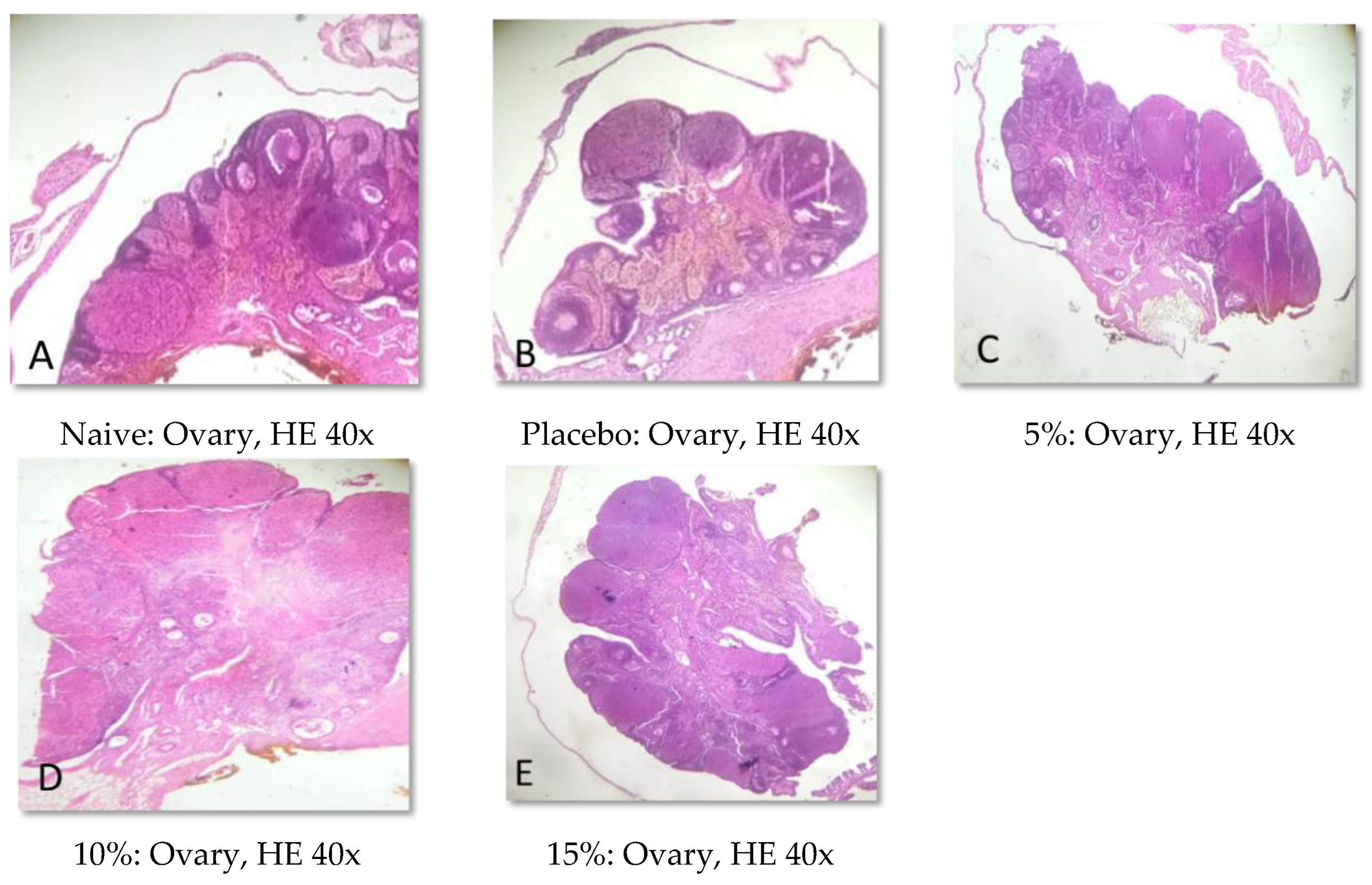
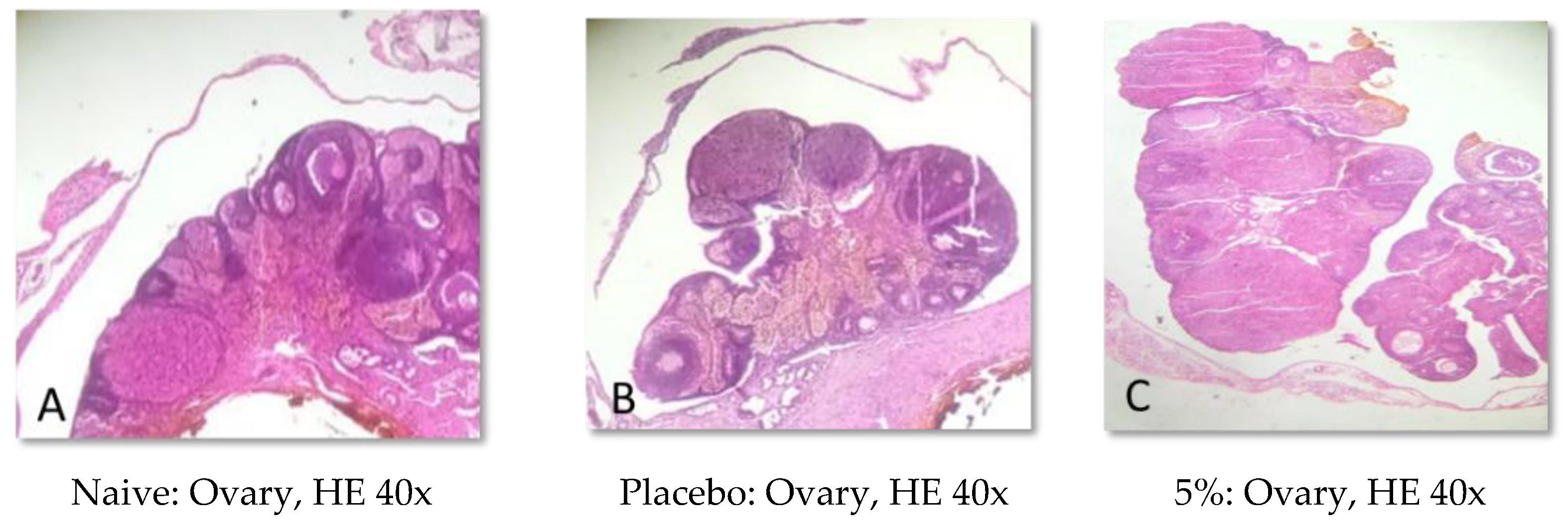
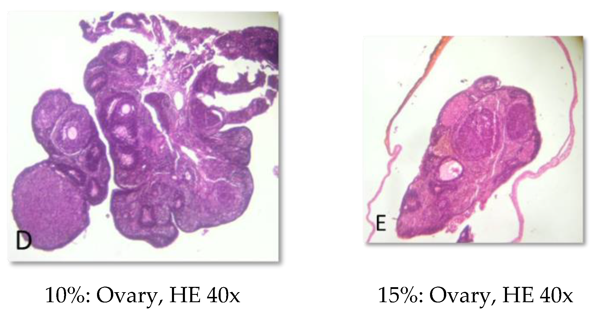
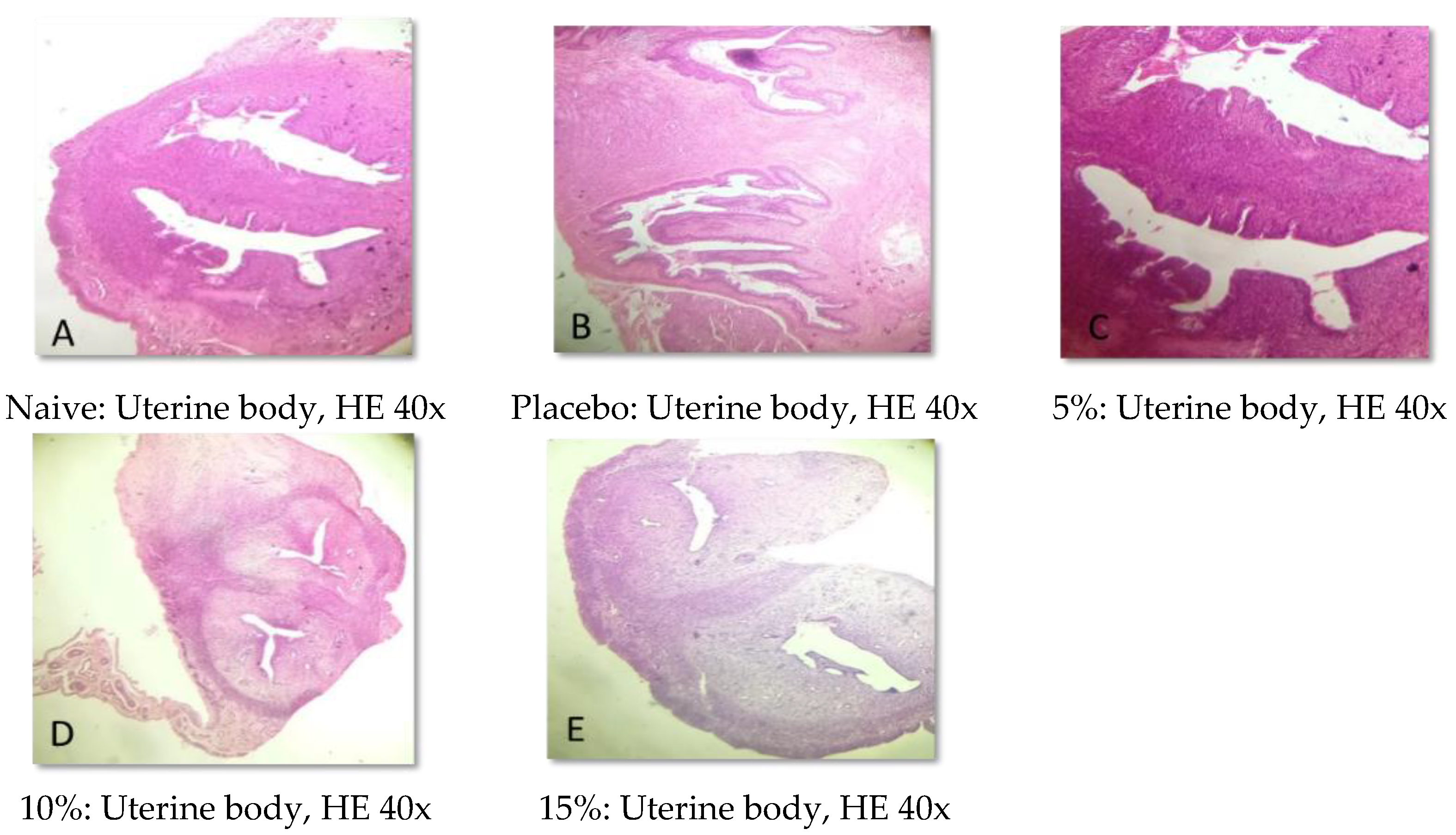
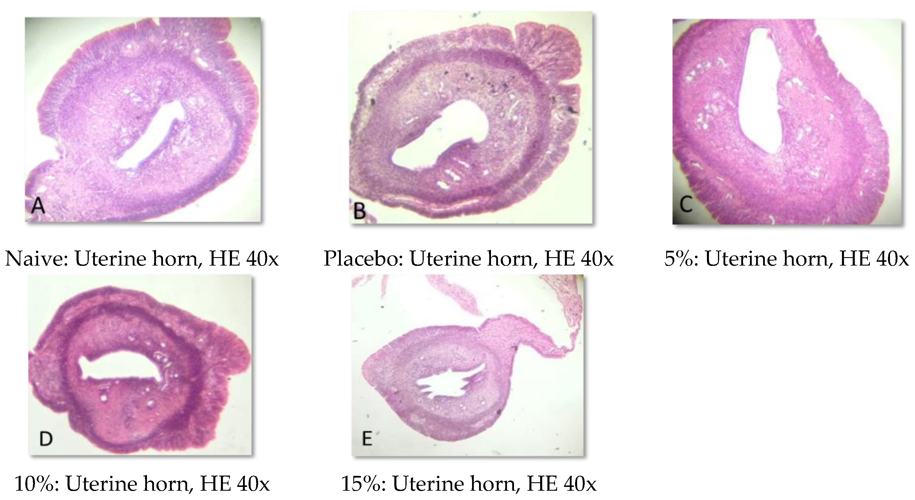
In addition to evaluating the visible effects on the vaginal mucosa, there was a concern about a possible change in vaginal, ovarian, and uterine morphology. In all reproductive organs evaluated, we did not identify any significant morphological changes that could indicate signs of inflammation, degeneration, apoptosis, necrosis, or any adaptation disorders, suggesting a maintained anatomical structure when compared to animals that did not receive any treatment or that were treated only with the vehicle.
In Brazil, several medicinal species are popularly used for the treatment of vulvovaginitis. One of the most relevant is
Stryphnodendron spp., popularly known as “barbatimão” [
25]. In fact, several vaginal pathogenic yeasts can be treated with the stem bark of this species [
26]. Despite the benefits, seed extracts of
S. adstringens and
S. polyphyllum showed an abortive effect on female rats [
27]. In this sense, it is imperative that species popularly used for the treatment of vulvovaginitis undergo a rigorous safety assessment.
Data are available on the toxicological evaluation of different preparations of
E. uniflora. Assunção Ferreira et al. [
22] performed acute and repeated dose toxicity studies and a genotoxicity evaluation with the aqueous extract obtained from
E. uniflora leaves. This preparation, rich in phenolic compounds such as gallic acid, ellagic acid, and myricetin, showed non-toxic and non-genotoxic effects in rodents. In addition, the hepatoprotective effects of the ethyl acetate extract from
E. uniflora leaves have already been described, which may indicate additional clinical aspects of the low toxicity of the species [
23]. Thus, our study complements the scientific data on the safety of
E. uniflora preparations and helps to form a knowledge base for the development of new herbal medicine.
4. Materials and Methods
4.1. Plant Material and Crude Extract Preparation
Leaves of E. uniflora were collected and identified by the Instituto Agronômico de Pernambuco (IPA). The crude extract was obtained by the Herbarium Pharmaceutical Laboratory in Phytomedicine (Colombo, Paraná, Brazil). Initially, 10 g of the leaf powder was turbo-extracted with 100 mL of acetone: water (7:3, v/v) in four five-minute rounds. The resulting extract was filtered and concentrated by rotary evaporation at 40 °C and 150 rpm (Laborota 4000, Heidolph). The concentrate was subjected to lyophilization at −64 °C and 0.006 mBar (ALPHA 1-2 LDplus, Fisher Scientific, Illkirch-Graffenstaden, France). The hydroacetonic extract was stored at 4 °C until the experiments.
4.2. Vaginal Gel Obtention
The vaginal gel was obtained by the Herbarium Pharmaceutical Laboratory in Phytomedicine (Colombo, Paraná, Brazil) at concentrations of 5%, 10%, and 15%. The excipients used in the formulation are in accordance with the Brazilian Pharmacopeia and include polyacrylamide, C13-14 isoalkanes, lauryl alcohol ethoxylate 7 OE, paraben, imidazolidinyl urea at 50%, and purified water. These concentrations were determined according to the pharmaceutical preparations found in the vaginal gels based on Schinus terebinthifolia Raddi (“aroeira”), Stryphnodendron adstringens (Mart.) (“barbatimão”), and Melaleuca alternifolia Cheel (“melaleuca”), which are already available on the Brazilian market.
4.3. Pharmacological Assays
4.3.1. Animals
Fifty-six female Wistar rats (90 to 100 days old) were acquired from the vivarium of the Federal University of Grande Dourados (UFGD). The animals were kept in the vivarium at Paranaense University (UNIPAR) in rooms with a constant temperature (22 ± 2 °C) and a light/dark phase of 12 h (lights on from 7 a.m. to 7 p.m.). The animals were divided into eight groups of seven animals and received water and food ad libitum. All procedures were in accordance with the guidelines of the National Council for the Control of Animal Experimentation (CONCEA) and were approved by the Ethics Committee in Research Involving Animal Experimentation of UNIPAR (AUTH number 38108—25 May 2020).
4.3.2. Experimental Design
Initially, the animals were divided into eight experimental groups and received the following treatments:
Group 1: Naive: Animals that were not exposed to treatment.
Group 2: Placebo: Animals that were exposed to the vehicle used in the formulation of the vaginal gel.
Group 3: Vaginal gel 5%: Animals that were exposed to the vaginal gel of E. uniflora at 5%.
Group 4: Vaginal gel 10%: Animals that were exposed to the vaginal gel of E. uniflora at 10%.
Group 5: Vaginal gel 15%: Animals that were exposed to the vaginal gel of E. uniflora at 15%.
Group 6: Crude extract 5%: Animals that were exposed to the crude extract of E. uniflora at 5%.
Group 7: Crude extract 10%: Animals that were exposed to the crude extract of E. uniflora at 10%.
Group 8: Crude extract 15%: Animals that were exposed to the crude extract of E. uniflora at 15%.
The vaginal gel and crude extract of E. uniflora were applied over the vaginal cavity, once a day (with the aid of a swab), for 14 days. The crude extract was resuspended in distilled water at the time of application.
4.3.3. Clinical and Behavioral Observations
Daily clinical evaluations of the vaginal region (pruritus, swelling, irritation, burning, pain, and vaginal secretion) were performed. Body weight was determined prior to the beginning of the experiments and on the day of euthanasia. Vaginal smears were performed before the beginning of the treatments and every five days for the entire experimental period. Vaginal lavage was obtained with the aid of a micropipette through vaginal washes with 50 µL of distilled water. The evaluation was performed under optical microscopy (200×). On the morning of the 15th day, the animals were euthanized by deepening anesthetic with Isoflurane at 5% followed by decapitation.
4.3.4. Biochemical Analysis
Approximately 5 mL of blood was collected (by the decapitation method) into tubules with a clotting agent and a polymer gel for serum separation The biochemical parameters analyzed were: urea, creatinine, total protein, albumin, globulins, aspartate amino transferase (AST), alanine amino transferase (ALT), gamma glutamyl transpeptidase (GGT), alkaline phosphatase (AP), total bilirubin (TB), thyroid stimulating hormone (TSH), free T3, lipid profile (HDL-cholesterol, LDL-cholesterol, VLDL-cholesterol, total cholesterol (TC), and triglycerides (TG)), progesterone, and estradiol. Analyses were performed on an automated biochemical analyzer (Selectra E, Vital Scientific, The Netherlands) [
28].
4.3.5. Hematological Investigation
Another sample of 3 mL of blood was collected (by the decapitation method) into tubes containing K2EDTA. The samples were properly homogenized and stored under refrigeration (about 4 °C) until processing. The total number of red blood cells (mm
3), hemoglobin (g/dL), hematocrit (%), mean corpuscular volume (MCV, fL), mean corpuscular hemoglobin (MCM, pG), mean corpuscular hemoglobin concentration (HCCM, g/dL), platelets (mm
3), monocytes (mm
3), segmented (mm
3), lymphocytes (mm
3), and eosinophils (mm
3) was measured in an automated hematology analyzer (Abbott Cell Dyn 3500, Abbott Diagnostics, Irving, TX, USA) [
28].
4.3.6. Macroscopic Evaluation and Relative Organ Weight
After the euthanasia, the liver, spleen, kidneys, and reproductive system (ovaries, uterus, and vaginal cavity) were removed and submitted to necropsy. After we had determined the absolute weight of each organ, the relative weight was calculated by dividing each animal’s organ weight by their body weight × 100.
4.3.7. Histopathological Analysis
Samples of the uterus and ovaries were collected, fixed in 10% formalin solution, dehydrated, diaphanized, paraffinized, and sectioned to a thickness of 4 µm in a Leica® semi-automatic microtome. The sections were stained with hematoxylin and eosin staining and evaluated by light microscopy.
4.4. Statistical Analysis
The results are shown as mean ± standard error of the mean (SEM) of seven rats per group. Parametric data were analyzed by one-way analysis of variance (ANOVA) and differences between groups were evaluated following the Bonferroni correction. Statistical analysis was performed using a GraphPad Prism version 9.4.1 for macOS (GraphPad Prism, San Diego, CA, USA). A p-value less than 0.05 was considered statistically significant.


 ,
, 


