Article

Uracil as a Zn-Binding Bioisostere of the Allergic Benzenesulfonamide in the Design of Quinoline–Uracil Hybrids as Anticancer Carbonic Anhydrase Inhibitors

by
Samar A. El-Kalyoubi
1,
Ehab S. Taher
2,
Tarek S. Ibrahim
3,4,*,
Mohammed Farrag El-Behairy
5 and
Amany M. M. Al-Mahmoudy
4
1
Department of Pharmaceutical Organic Chemistry, Faculty of Pharmacy (Girls), Al-Azhar University, Nasr City, Cairo 11651, Egypt
2
Department of Pharmaceutical Organic Chemistry, Faculty of Pharmacy, Al-Azhar University, Assiut 71524, Egypt
3
Department of Pharmaceutical Chemistry, Faculty of Pharmacy, King Abdulaziz University, Jeddah 21589, Saudi Arabia
4
Department of Pharmaceutical Organic Chemistry, Faculty of Pharmacy, Zagazig University, Zagazig 44519, Egypt
5
Department of Organic and Medicinal Chemistry, Faculty of Pharmacy, University of Sadat City, Menoufiya 32897, Egypt
*
Author to whom correspondence should be addressed.
Pharmaceuticals 2022, 15(5), 494; https://doi.org/10.3390/ph15050494
Submission received: 23 March 2022 / Revised: 10 April 2022 / Accepted: 15 April 2022 / Published: 19 April 2022
(This article belongs to the Special Issue Design of Enzyme Inhibitors as Potential Drugs 2022)

Abstract

:
A series of quinoline–uracil hybrids (10a–l) has been rationalized and synthesized. The inhibitory activity against hCA isoforms I, II, IX, and XII was explored. Compounds 10a–l demonstrated powerful inhibitory activity against all tested hCA isoforms. Compound 10h displayed the best selectivity profile with good activity. Compound 10d displayed the best activity profile with minimal selectivity. Compound 10l emerged as the best congener considering both activity (IC50 = 140 and 190 nM for hCA IX and hCA XII, respectively) and selectivity (S.I. = 13.20 and 9.75 for II/IX, and II/XII, respectively). The most active hybrids were assayed for antiproliferative and pro-apoptotic activities against MCF-7 and A549. In silico studies, molecular docking, physicochemical parameters, and ADMET analysis were performed to explain the acquired CA inhibitory action of all hybrids. A study of the structure–activity relationship revealed that bulky substituents at uracil N-1 were unfavored for activity while substituted quinoline and thiouracil were effective for selectivity.

Graphical Abstract

1. Introduction

Carbonic anhydrases (CAs, EC 4.2.1.1) are a superfamily of metalloenzymes that are distributed extensively throughout living organisms [1]. CAs are subclassified to eight gene families (α, β, γ, δ, ζ, η, θ, and ι) [2]. As these enzymes are ubiquitously represented in the body, they are major participants in reaction catalysis and physiological processes. In addition to their physiological impacts, the modulation of the natural function or upregulation of these enzymes demonstrate immense benefits for controlling many pathological conditions [3]. The α-CA enzymes found in mammals are divided into four subgroups consisting of several isoforms. The CA I and CA II isoforms belong to the cytosolic subgroup of α-CA, while CA IX and CA XII isoforms are part of the membrane-associated α-CAs subgroup [4]. For instance, sulfonamides were determined as potent inhibitors of α-CAs and can be used to exploit many clinical applications such as diuresis, antiglaucoma, anticonvulsant, antiobesity, analgesic, and anticancer activities [5].
Cancer one of the leading mortality-causing diseases, competing with cardiovascular diseases for first place. It is also one of the overwhelming barriers to high life expectancy owing to its social and economic consequences [6]. In 2020, 19.3 million people were diagnosed with cancer, with 10.0 million deaths reported. This number is expected to reach 28.4 million in 2040 [7]. Many molecular mechanisms and biological targets are connected to the underlying mechanisms of cancer. Of these, human carbonic anhydrases (hCA) IX and XII have been recognized as tumor-associated proteins in hypoxic and other solid tumors where they actively contribute to the permanence and metastasis of the necrotic cell [8]. Notably, a great number of scientists have emphasized that both are biomarkers and therapeutic targets for various cancer types. Accordingly, dual targeting, i.e., inhibition, of the two latter isozymes represent a remarkable challenge for the development of novel anticancer drugs [9,10,11,12].
Following the successful impact of its modulation, the search for further pharmacophores—other than sulfonamides—that can fit/block the active site of CAs has become an interesting topic in medicinal chemistry research [13,14,15]. Afterwards, many chemical moieties have been revealed as isosteres for sulfonamides, such as hydroxybenzene, fullerenes, benzopyrone, thiobenzopyrone, and boronic acids [3]. These new effective chemical pharmacophores have fostered the ability to design promising pharmacologically active candidates that circumvent sulfonamide side effects. In addition, they support further understanding of the underlying mechanisms of CAs and can be utilized for the rational drug design of novel candidates as CAs modulators.
It is important to indicate that CAs are not the sole Zn-containing enzymes that extensively affect cancer physiology [16]. Histone deacetylases (HDACs) are Zn-containing enzymes that play a crucial role in anticancer therapy [17]. Fortunately, the zinc-binding groups (ZBGs) of HDAC inhibitors are different from those reported for CAs, as per the recent reviews [18,19]. The ZBGs of HDACIs are classified into classical and nonclassical groups. The classical ZBGs, such as hydroxamic acid [20] and benzamide [21], are characterized by potent activity, selectivity, toxicity, and in vivo instability [22]. Carboxylic acid [23] and thiol [24] groups were also used as ZBGs in HDACI design. The nonclassical type include imidazole-thione [25], tropolone derivatives [26], 3-hydroxypyridin-2-thione (3-HPT) [27], chelidamic derivatives [28], benzoylhydrazide [29], trifluoromethyloxadiazolyl (TFMO) [30], 2-(oxazol-2-yl)phenol [31], hydroxypyrimidines [32], and β-hydroxymethyl chalcone [33] (Figure 1).
Quinoline is an abundant pharmacophore in medicinal chemistry and exhibits remarkable pharmacological efficacies such as antituberculosis [34], antimalarial [35], antiviral [36,37], anticancer [35,38], antibacterial [37], antifungal [37,39], antileishmanial [40], and anti-inflammatory [41] activity. Moreover, several quinoline-based candidates showed very promising anticancer activities, including neratinib [42], bosutinib [43], foretinib [44], and topotecan [45], which are currently in clinical trials. Additionally, kinase inhibitors [35,46,47], apoptotic agents [35,48], microtubule-targeting agents [35,49], topoisomerase inhibitors [35,50], epigenetic enzyme inhibitors [35,51], transcription factor inhibitors [35,52], and carbonic anhydrase inhibitors [53] are represented (Figure 2).
Similarly, the pyrimidine moiety is a well-documented pharmacophore in medicinal chemistry [53,54,55,56]. Anticancer pyrimidine-containing marketed drugs exhibit their biological activity via assorted mechanisms, such as tyrosine kinase inhibition (imatinib, dasatinib) [57], the inhibition of DNA synthesis (uracil mustard) [58], thymidylate synthase (fluorouracil) [59], nucleoside metabolic inhibition (gemcitabine, trifluridine) [60], antimetabolite (floxuridine) [61], DNA polymerase inhibition (cytarabine) [62], DNA methyltransferase inhibitor (azacitidine) [63], inducing DNA hypomethylation and corresponding alterations in gene expression (decitabine) [64], and transition state analog inhibition of cytidine deaminase (zebularine) [65] (Figure 3).
Primary benzenesulfonamide derivatives are well known for their CA inhibition, which confers several biological activities, especially anticancer activity. In this regard, quinoline-based sulfonamides have been recently reported with impressive CA inhibition, where compound I displayed inhibitory activity against hCA I and II isoforms with Ki ranges of 0.96–9.09 µM and 83.3–3.59 µM for hCA I and II, respectively [53]. Compound II displayed potent inhibitory activity against the well-known cancer-related isozymes hCA IX and XII with Ki values of 0.019 µM and 0.009 µM, respectively [66].
In all the investigated compounds, the quinoline backbone was important for the biological activity; however, the zinc-binding group (primary benzenesulfonamide) is the essential moiety [53,67,68,69,70]. Despite its key impact, the allergic response to sulfonamide drugs is a very unfavorable reaction [71,72]. Thus, the search for an alternative bioisostere is of much interest in the field of medicinal chemistry. Notably, uracil compounds III and IV are non-sulfonamide derivatives that show promising CA inhibitory activity and were confirmed as leads for generating potent CAIs, triggering further isoforms [73]. From the perspective of the structure–activity relationship, the uracil moiety with C=O, N, and NH2 represents an active pharmacophore, rich with binding groups that can substitute the sulfonamide interactions in the receptor (Figure 4).
In light of the abovementioned findings, and by applying the tactic of pharmacophoric hybridization, a novel series of quinoline-based uracil synthetic molecules has been rationalized, synthesized, and tested for their CA inhibitory and anticancer activities. The utilized uracils with diverse functionality were similar to the primary benzenesulfonamide moiety regarding the two sites for both H-bond donors and H-bond acceptors. This rationale has promoted the exploration of such hybrids. These hybrid structures have been lined chemically via an imine bond (Schiff’s base), which has been carefully selected as an anchor based on the reported CA inhibitory activity of its derivatives (compound V) [74] (Figure 4). An additional notable feature of our novel hybrids is the synthesis of quinoline-based thiouracil to realize the power of sulfur-based compounds as a zinc-binding group [75]. Docking and in silico studies have been performed to determine the CA inhibitory profile for the targeted quinolones.

2. Results and Discussion

2.1. Chemistry

The utilized protocol of our designed targets 10a–l consists of two key structural features; first, the quinoline-3-carbaldehyde moiety (Scheme 1), and second, the 5,6-diaminouracil/thiouracil pharmacophores (Scheme 2). The straightforward synthesis leading to 2-quinolone-3-carbaldehyde derivatives (4ac), which has benefit of conciseness, is shown in Scheme 1. The acetylation of aniline derivatives 1ac through glacial acetic acid/acetic anhydride at a temperature of 0 °C furnished the corresponding anilides (2ac) [76,77]. These compounds underwent the Vilsmeier–Haack reaction to give the 2-chloroquinoline aldehyde derivatives (3ac) [78,79]. The oxidation of the chlorine group at position 2 into a ketone has been successfully performed under reflux in acidic conditions (acetic acid) to deliver the desired synthons (4ac). This reaction was initiated by oxidation with acetic acid, then completed by the departure of chlorine as a good leaving group to produce the favored product. The reaction appeared to be facilitated through the electron withdrawing effect of nitrogenous heteroatom [78,79].
As shown in Scheme 2, the variable 5,6-diaminouracil/thiouracil derivatives 9a–e were prepared via consecutive cyclization of N-alkylurea/thiourea with ethyl cyanoacetate in the presence of sodium ethoxide, which initially gave 6-aminouracil/thiouracils 7ae. This was readily followed by conversion via a nitrosation process using nitrous acid and then reduction of nitroso uracil/thiouracils 8a–e via the reducing agent ammonium sulfide [80,81].
Condensation of diamino-uracil/thiouracils 9a–e with the appropriate quinoline aldehydes 4a–c under refluxing condition in ethanol for 1 h furnished the anticipated quinoline-based uracils/thiouracils 10a–l (Scheme 3). The structures of these novel compounds were also characterized by using thin-layer chromatography and melting point methods. Our novel later hybrids were determined using 1H NMR, 13C NMR, elemental, and mass spectroscopic techniques. The 1H NMR spectra showed characteristic imino protons (N=CH) of Schiff bases appearing in the range from 8.45–9.91 ppm as sharp singlets. The 13C NMR spectra further supported the assigned structures. The 13C NMR shift of N=CH carbon atoms appeared in the range from 161.32–164.41 ppm as a singlet signal.

2.2. Biology

2.2.1. Carbonic Anhydrase Inhibition

Activities of all CA isoenzymes were estimated following the previously described colorimetric method of Verpoorte et al. (1967) [82,83] using BioVision Carbonic Anhydrase (CA) Inhibitor Screening Kit (Catalog # K473-100). As per Table 1, all candidates 10al showed variable inhibitory activities against the tested CA isoforms. For hCAI, all the synthesized candidates 10al demonstrated inhibitory activities. Compounds 10a, 10d, 10f, 10j, and 10l exhibited inhibitory concentrations in the nanomolar range (230–970 nM), whereas the other compounds showed activity in the micromolar range (Table 1 and Figure 5). Compounds 10d, 10f, and 10l displayed higher inhibitory activity (IC50 = 230, 330, and 250 nM, respectively) than the reference standard acetazolamide (AAZ, IC50 = 760 nM). It is shown that the placement of small lipophilic methyl groups at N-1 uracil (where X = O), i.e., compound 10d, was preferred to X = S and the bulkier ethyl and benzyl groups.
In the case of the physiologically dominant hCA II, candidates 10a, 10c, 10d, and 10f showed potent inhibitory activity in the nanomolar range (IC50 = 520, 660, 260, and 790 nM, respectively, Table 1 and Figure 5). Similarly, compound 10d was found to be the most potent hCA II inhibitor with a lower inhibitory concentration (IC50 = 260 nM) than the reference standard acetazolamide (AAZ, IC50 390 nM). This highlights the effect of bulkiness on the interaction with the hCA II binding site. Fortunately, tumor-linked hCA IX was efficiently inhibited by most of our candidates in the nanomolar range, while three candidates (10e, 10g, and 10k) showed inhibition in the low micromolar range (IC50 = 1.37, 1.08, and 1.28 µM, respectively). Moreover, compounds 10a, 10d, and 10f displayed superior inhibitory activity against the hCA IX isozyme in the nanomolar range, with IC50 values of 250, 220, and 270 nM, respectively. Thiouracil hybrid 10l represented the best hCA IX inhibitor with a half-inhibitory concentration (IC50 = 140 nM) lower than the standard acetazolamide (AAZ, IC50 = 150 nM). In addition to its potent activity, hybrid 10l showed higher selectivity (13.25-fold) towards the tumor-linked hCA IX enzyme than the physiologically dominant hCA II and 1.75-fold higher selectivity to hCA I (Table 2 and Figure 6). Similarly, compound 10h displayed very good selectivity to transmembrane tumor-associated isoform hCA IX. A further tumor-related CA isoform (hCAXII) was potentially inhibited by our quinoline–uracil hybrids. All our synthesized targets 10al inhibited hCA XII in the nanomolar range. Compounds 10d, 10f, and 10l (where IC50 = 190, 170, and 190 nM, respectively) were more potent than acetazolamide (IC50 = 230 nM) while compound 10i (IC50 = 230 nM) was equipotent to the standard acetazolamide.
Regarding selectivity profile, compounds 10g, 10h, 10i, 10k, and 10l were the most selective for hCA XII over both hCAI and hCAII (Table 2 and Figure 7). Compound 10h displayed the best selectivity profile (S.I. = 5.39, 7.52, 8.00, and 11.16 for I/IX, II/IX, I/XII, and II/XII, respectively) with good activity (600 and 400 nM for hCA IX and hCA XII, respectively). However, compound 10d displayed the best activity profile against both hCA IX and hCA XII (IC50 = 220 and 190 nM, respectively) with a minimal selectivity profile (S.I. = 1.09, 1.21, 1.24, and 1.37 for I/IX, II/IX, I/XII, and II/XII, respectively). Herein, compound 10l emerged as the best congener considering both activity and selectivity. Compound 10l (IC50 = 190 nM) showed better activity than 10h (IC50 = 400 nM) and the same activity as 10d (IC50 = 190 nM) against hCA XII. Further, 10l (IC50 = 140 nM) showed better activity than both 10h and 10d against hCAIX (IC50 = 600 and 220 nM, respectively). In addition, 10l is the only candidate with superior activity (IC50 = 140 and 190 nM for hCA IX and hCA XII, respectively) in comparison to acetazolamide (AAZ, IC50 = 150 and 230 nM for hCA IX and hCA XII, respectively). In addition, 10l displayed better selectivity than acetazolamide (S.I. = 13.20 and 9.75 for II/IX, and II/XII, respectively) toward the tumor-linked isoforms hCAs IX and XII against physiologically dominant hCA II (Table 2).

2.2.2. Antiproliferative Activity

Based on their activities and selectivity against tumor-linked CA isoforms, compounds 10d and 10l were selected for further screening against the breast cancer cell line (MCF-7) (HTB-22 from ATCC, Manassas, VA, USA) and lung cancer cell line (A549) (CCL-185 from ATCC, Manassas, VA, USA) under hypoxic conditions to evaluate their in vitro antiproliferative activity using the MTT assay protocol [84] (Table 3). Both compounds showed activity against the tested cell lines. However, compound 10d had more potent activity than 10l against the MCF7 breast cancer cell line with IC50 = 2.87 ± 0.05 compared with compound 10l IC50 value of 4.08 ± 0.08 µM. For the reference standard staurosporine, the IC50 was 6.92 ± 0.18 µM. Despite their superior activity over staurosporine against breast cancer cell line, compounds 10d and 10l had lower activity than staurosporine towards the lung cancer cell line (Figure 7).

2.2.3. Assessment of Apoptotic Marker Levels

The two cytoplasmic proteins, B-cell lymphoma protein 2 (Bcl-2) and its associated X protein (Bax), are essential for apoptosis in normal cells. Bax is a promoter and Bcl-2 is an inhibitor of apoptosis [85]. Thus, the effect of 10d and 10l on apoptotic markers Bax and Bcl-2 in a breast cancer cell line (MCF-7) and a lung cancer cell line (A549) has been estimated. Treatment of MCF-7 and A549 cell lines with compounds 10d and 10l resulted in the upregulation of Bax levels by nearly six-fold relative to the control while the expression of Bcl-2 levels was downregulated in comparison with the control (Table 3).

2.3. In Silico Study

2.3.1. Physicochemical and Pharmacokinetic Parameters

The SWISSADME server [86] was utilized to assess the ability of the investigated compounds (10a–l) to act as drugs via estimating the physicochemical and pharmacokinetic properties. No compounds were expected to cross the BBB, inhibit cytochrome enzymes, violate the Lipinski’s rule, or give PAINS alerts. In addition, all compounds (except 10g, 10h, and 10l) demonstrated high GIT absorption. The investigated compounds showed good synthetic accessibility and bioavailability scores (Table 4). Moreover, no compounds inhibited cytochrome P450 enzymes, indicating there was no expected pharmacokinetic-related drug–drug interactions.

2.3.2. Molecular Docking Study

Molecular docking studies were performed to investigate the interactions of the target compounds 10a10l with the binding site of the human carbonic anhydrases IX using Discovery Studio. hCA IX is considered promising targets for cancer treatment and their inhibition can reduce the growth of primary tumors and metastases. For the CA IX isoform, the PDB file 5FL4 containing hCA IX co-crystallized with 5-(1-naphthalen-1-yl-1,2,3-triazol-4-yl)thiophene-2 sulfonamide was obtained from the Protein Data Bank [87].
The protein structure was prepared by 3D protonation and the water molecule and ligand that were not implicated in the active site were removed. The active site then generated with the default protocol [88]. The 2D and 3D interaction diagrams for the ligand and compounds 10d, 10f, and 10l are shown in Figure 8. Analyzing the ligand–protein interactions can help better understand the selectivity of the compound for hCA IX with respect to the other hCA isoforms.
The co-crystallized ligand forms two hydrogen bonds with Thr 200 and Leu199; it was found to chelate the zinc ion through the sulfonamide group. It formed pi–sulfur interaction with His 94 and Trp 210. Additionally, it formed pi–alkyl interaction with Val 130 and Val 121 and pi–pi T-shaped interaction with His 94. It exhibited van der Waals interactions with Gln 92, Gln 71, Val 142, Glu 106, His 96, and Thr 201.
Compound 10d binds to hCA IX through Zn (II) attractive charge interaction along with hydrogen bonds with Thr 200, His 96, and His 86 (Figure 8), as well as pi–alkyl interaction with Leu 199 and alkyl interaction with His 94. This showed van der Waals interaction with Val 121, Thr 200, Trp 210, Glu 106, and His 119.
Compound 10l binds to hCA IX through hydrogen bonds with His 68, Gln 71, and Gln 92. Additionally, it exhibited pi–alkyl interaction with Val 130. It showed van der Waals interactions with crucial amino acids such as Leu 91, His 96, Ser 69, His 94, Thr 200, Leu 199, and Val 121.
Compound 10f binds to hCA IX through Zn ion and forms hydrogen bonds with Thr 201, Trp 9, His 68, and His 96 (Figure 8). Moreover, it exhibited pi–alkyl interaction with His 94, Leu 199, Val 121, and Trp 210, and van der Waals interaction with Ser 69, Pro 202, Gln 71, Val 121, Asp 131, and Gln 92.

2.4. SAR Study

Uracil is well documented as a metal-binding pharmacophore [89,90,91] with particular emphasis on its Zn+2-binding abilities [92,93]. In the present work, uracil has been established as a bioisostere of the zinc-binding moiety benzenesulfonamide in our synthesized carbonic anhydrase inhibitors 10a–l. The uracil Zn-binding ability has been proven using modeling studies and realized by the biological screening against CA isoforms (I, II, IX, XII) and cancer cell lines.
The SAR of the synthesized candidates can be summarized as follows (Figure 9):
-
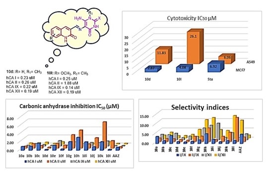
Both uracil and thiouracil had CA inhibitory activity.
-
Substitution on uracil N-1 with a bulky group (benzyl 10b and 10i) decreases activity.
-
Substitution on the quinoline ring has tolerable activity, but greatly improves the selectivity, particularly when in combination with thiouracil (10l).

3. Materials and Methods

3.1. Chemistry

Melting points were determined with a Gallenkamp (London, UK) melting point apparatus and were uncorrected. 1H NMR and 13C NMR spectra were recorded on a Varian Gemini-400 (400MHz, Foster City, Calif., USA) spectrometer using chloroform (CDCl3), dimethylsulphoxide and/or (DMSO/D2O) as solvents and tetramethylsilane (TMS) as an internal standard (chemical shift in δ, ppm). 1H NMR data were recorded as follows: chemical shift (δ) [multiplicity, coupling constant(s) J (Hz), relative integral], where multiplicity is defined as s = singlet, d = doublet, t = triplet, q = quartet, and m = multiplet, or combinations of the above. High-resolution measurements were conducted on a time-of-flight instrument. All the results of elemental analyses corresponded to the calculated values within experimental error. The reaction progress was observed by thin-layer chromatography (TLC) using TLC sheets precoated with ultraviolet (UV) fluorescent silica gel (Merck 60F254) and spots were visualized by irradiation with UV light (254 nm) or iodine vapors. All starting materials and reagents were generally commercially available and purchased from Sigma-Aldrich or Lancaster Synthesis Corporation (Lancaster, UK). Compounds 4a–c and 9a–e were prepared according to the reported method [80,81,94,95,96].

3.1.1. General Procedures for the Preparation of 10a–l

A mixture of 5,6-diamino-uracils/thiouracils (1.28 mmol) and quinolone carbaldehydes (1.28 mmol) in ethanol (50 mL) was heated under reflux for 1 h. Cool the reaction mixture, the formed precipitate was filtered, washed with ethanol and crystallized from DMF/ethanol (1:1).

(E)-6-Amino-5-(((2-oxo-1,2-dihydroquinolin-3-yl)methylene)amino)pyrimidine-2,4(1H,3H)-dione (10a)

Yellowish white solid, Yield: 90%; m.p.: 270–272 °C; 1H NMR (DMSO-d6, 400 MHz) δ: 7.17–7.30 (m, 3H, 1Ar-H+NH2), 7.36 (d, J = 8.3 Hz, 1H), 7.44–7.47 (s, 1H, 1ArH), 7.64–7.70 (m, 1H, ArH), 7.91 (s, 1H, CH quinoline), 8.51 (s, 1H, CH=N), 8.74 (s, 1H, NH uracil), 9.75 (s, 1H, NH quinoline), 10.24 (s, 1H, NH uracil) ppm. 13C NMR (DMSO-d6, 100 MHz) δ: 189.78, 161.75, 159.39, 142.44, 138.66, 133.69, 130.92, 129.29, 128.42, 125.60, 122.07, 119.63, 115.41, 99.46 ppm (Figure S1). MS: m/z (rel. int.) = 297 (M+, 10), 145 (49.00), 111 (35.00), 44 (100.00) (Table S2). Anal. Calcd for C14H11N5O3: C, 56.57; H, 3.73; N, 23.56; Found: C, 56.79; H, 3.89; N, 23.34%.

(E)-6-Amino-1-benzyl-5-(((2-oxo-1,2-dihydroquinolin-3-yl)methylene)amino) pyrimidine-2,4(1H,3H)-dione (10b)

Yellowish white solid, Yield: 80%; m.p.: 280–282 °C; 1H NMR (DMSO-d6, 400 MHz) δ: 5.24 (s, 2H, CH2-benzyl), 7.16–7.20 (m, 1H, Ar-H), 7.26–7.31 (m, 4H, Ar-H), 7.35–7.39 (m, 2H, Ar-H), 7.45–7.49 (m, 3H, 1ArH+NH2), 7.70 (d, J = 7.3 Hz, 1H, ArH), 8.80 (s, 1H, CH quinoline), 9.84 (s, 1H, CH=N), 10.93 (s, 1H, NH quinoline), 11.88 (s, 1H, NH uracil) ppm. 13C NMR (DMSO-d6, 100 MHz) δ: 161.78, 157.91, 154.40, 149.38, 144.34, 138.73, 136.20, 134.02, 129.14, 128.56, 127.27, 126.39, 122.08, 119.64, 114.95, 100.09, 44.57 ppm (Figure S2). MS: m/z (rel. int.) = 387 (M+, 6.00), 77 (83.00), 43 (99.00), 91 (100.00) (Table S2). Anal. Calcd for C21H17N5O3: C, 56.11; H, 4.42; N, 18.08; Found: C, 65.29; H, 4.57; N, 18.26%.

(E)-6-Amino-1-ethyl-5-(((2-oxo-1,2-dihydroquinolin-3-yl)methylene)amino)pyrimidine-2,4(1H,3H)-dione (10c)

Yellowish solid, Yield: 87%; m.p.: 290–292 °C; 1H NMR (DMSO-d6, 400 MHz) δ: 1.17 (t, J = 6.9 Hz, 3H, CH3), 3.97 (q, J = 6.7 Hz, 2H, CH2), 7.18–7.48 (m, 3H, Ar-H), 7.49 (s, 2H, NH2), 7.73 (d, J = 7.7 Hz, 1H, ArH), 8.81 (s, 1H, CH quinoline), 9.81 (s, 1H, CH=N), 10.47 (s, 1H, NH quinoline), 11.87 (s, 1H, NH uracil) ppm. 13C NMR (DMSO-d6, 100 MHz) δ: 161.83, 157.87, 154.15, 148.98, 143.96, 138.70, 133.90, 130.31, 129.24, 128.64, 122.12, 119.70, 114.98, 99.99, 37.17, 13.08 ppm (Figure S3). MS: m/z (rel. int.) = 325 (M+, 18), 278 (40.00), 131 (70.00), 100 (84.00), 40 (100.00) (Table S2). Anal. Calcd for C16H15N5O3: C, 59.07; H, 4.65; N, 21.53; Found: C, 59.31; H, 4.79; N, 21.69%.

(E)-6-Amino-1-methyl-5-(((2-oxo-1,2-dihydroquinolin-3-yl)methylene)amino) pyrimidine-2,4(1H,3H)-dione (10d)

Yellow solid, Yield: 90%; m.p.: 298–300 °C; 1H NMR (DMSO-d6, 400 MHz) δ: 3.35 (s, 3H, CH3), 7.18–7.22 (m, 1H, Ar-H), 7.30 (d, J = 8.2 Hz, 1H, ArH), 7.45–7.50 (s, 3H, 1ArH+NH2), 7.73 (d, J = 7.0 Hz, 1H, ArH), 8.82 (s, 1H, CH quinoline), 9.82 (s, 1H, CH=N), 10.75 (s, 1H, NH quinoline), 11.87 (s, 1H, NH uracil) ppm. 13C NMR (DMSO-d6, 100 MHz) δ: 161.76, 157.80, 155.07, 149.21, 143.76, 138.66, 133.74, 130.19, 129.24, 128.54, 122.02, 119.65, 114.91, 100.03, 29.45 ppm (Figure S4). MS: m/z (rel. int.) = 311 (M+, 30), 233 (100.00), 163 (61.00), 138 (43.00), 54 (50.00) (Table S2). Anal. Calcd for C15H13N5O3: C, 57.87; H, 4.21; N, 22.50; Found: C, 58.04; H, 4.37; N, 22.32%.

(E)-3-(((6-Amino-1-methyl-4-Oxo-2-thioxo-1,2,3,4-tetrahydropyrimidin-5-yl)imino)methyl) quinolin-2(1H)-one (10e)

Yellowish solid, Yield: 85%; m.p.: 284–286 °C; 1H NMR (DMSO-d6, 400 MHz) δ: 3.85 (s, 3H, N-CH3), 7.19–7.32 (m, 2H, Ar-H), 7.48–7.52 (m, 1H, ArH), 7.60 (s, 2H, NH2), 7.74 (d, J = 7.1 Hz, 1H, ArH), 8.90 (s, 1H, CH quinoline), 9.90 (s, 1H, CH=N), 11.92 (s, 1H, NH quinoline), 12.22 (s, 1H, NH uracil) ppm. 13C NMR (DMSO-d6, 100 MHz) δ: 173.72, 161.67, 155.12, 154.31, 146.77, 138.95, 134.90, 130.61, 128.77, 128.75, 122.12, 119.53, 115.00, 103.79, 36.44 ppm (Figure S5). MS: m/z (rel. int.) = 327 (M+, 21), 297 (53.00), 242 (80.00), 228 (100.00), 197 (134.00) (Table S2). Anal. Calcd for C15H13N5O2S: C, 55.04; H, 4.00; N, 21.39; Found: C, 55.27; H, 4.18; N, 21.53%.

(E)-6-Amino-1-ethyl-5-(((8-methyl-2-oxo-1,2-dihydroquinolin-3-yl)methylene) amino)pyrimidine-2,4(1H,3H)-dione (10f)

Yellow solid, Yield: 82%; m.p.: 278–280 °C; 1H NMR (DMSO-d6, 400 MHz) δ: 1.17 (t, J = 7.0 Hz, 3H, CH3), 2.44 (s, 3H, CH3), 3.97 (q, J = 6.7 Hz, 2H, CH2), 7.10–7.14 (m, 1H, Ar-H), 7.33 (d, J = 7.2 Hz, 1H, ArH), 7.50 (s, 2H, NH2), 7.60 (d, J = 7.7 Hz, 1H, ArH), 8.81 (s, 1H, CH quinoline), 9.84 (s, 1H, CH=N), 10.75 (s, 1H, NH quinoline), 11.01 (s, 1H, NH uracil) ppm. 13C NMR (DMSO-d6, 100 MHz) δ: 162.23, 157.84, 154.10, 148.92, 143.79, 137.06, 134.48, 131.55, 128.83, 126.78, 123.28, 121.85, 119.70, 99.92, 36.47, 17.19, 13.04 ppm (Figure S6). MS: m/z (rel. int.) = 339 (M+, 10), 294 (38.00), 239 (37.00), 105 (62.00), 77 (100.00), 44 (88.00) (Table S2). Anal. Calcd for C17H17N5O3: C, 60.17; H, 5.05; N, 20.64; Found: C, 60.38; H, 5.27; N, 20.89%.

(E)-3-(((6-Amino-1-methyl-4-oxo-2-thioxo-1,2,3,4-tetrahydropyrimidin-5-yl) imino)methyl)-8-methylquinolin-2(1H)-one (10g)

Straw yellow solid, Yield: 78%; m.p.: 265–267 °C; 1H NMR (DMSO-d6, 400 MHz) δ: 2.44 (s, 3H, CH3), 3.85 (s, 3H, N-CH3), 7.11–7.15 (m, 1H, Ar-H), 7.35 (d, J = 7.2 Hz, 1H, ArH), 7.60–7.62 (m, 3H, 1ArH+NH2), 8.90 (s, 1H, CH quinoline), 9.91 (s, 1H, CH=N), 11.07 (s, 1H, NH quinoline), 12.22 (s, 1H, NH uracil) ppm. 13C NMR (DMSO-d6, 100 MHz) δ: 174.18, 162.62, 155.61, 154.80, 147.18, 137.81, 136.04, 132.42, 128.85, 127.44, 123.85, 122.42, 120.07, 104.25, 36.93, 17.65 ppm (Figure S7). MS: m/z (rel. int.) = 341 (M+, 14), 225 (24.00), 172 (100.00), 161 (83.00) (Table S2). Anal. Calcd for C16H15N5O2S: C, 56.29; H, 4.43; N, 20.51; Found: C, 56.43; H, 4.60; N, 20.46%.

(E)-6-Amino-5-(((6-methoxy-2-oxo-1,2-dihydroquinolin-3-yl)methylene)amino) pyrimidine-2,4(1H,3H)-dione (10h)

Faint yellow solid, Yield: 85%; m.p.: 275–277 °C; 1H NMR (DMSO-d6, 400 MHz) δ: 3.80 (s, 3H, OCH3), 7.11–7.32 (m, 5H, 3Ar-H+NH2), 7.47 (s, 1H, CH quinoline), 8.45 (s, 1H, CH=N), 8.70 (s, 1H, NH uracil), 9.74 (s, 1H, NH quinoline), 10.24 (s, 1H, NH uracil) ppm. 13C NMR (DMSO-d6, 100 MHz) δ: 164.41, 161.35, 159.39, 154.20, 133.20, 132.87, 129.79, 120.25, 119.48, 116.18, 109.28, 99.70, 55.34 ppm (Figure S8). MS: m/z (rel. int.) = 327 (M+, 5), 160 (50.00), 104 (62.00), 43 (100.00) (Table S2). Anal. Calcd for C15H13N5O4: C, 55.05; H, 4.00; N, 21.40; Found: C, 55.32; H, 4.21; N, 21.79%.

(E)-6-Amino-1-benzyl-5-(((6-methoxy-2-oxo-1,2-dihydroquinolin-3-yl)methylene) amino) pyrimidine-2,4(1H,3H)-dione (10i)

Yellowish solid, Yield: 88%; m.p.: 291–293 °C; 1H NMR (DMSO-d6, 400 MHz) δ: 3.80 (s, 3H, OCH3), 5.25 (s, 2H, CH2-benzyl), 7.13–7.21 (m, 2H, Ar-H), 7.24–7.31 (m, 4H, Ar-H), 7.36–7.40 (m, 2H, Ar-H), 7.46 (s, 2H, NH2), 8.76 (s, 1H, CH quinoline), 9.84 (s, 1H, CH=N), 10.94 (s, 1H, NH quinoline), 11.80 (s, 1H, NH uracil) ppm. 13C NMR (DMSO-d6, 100 MHz) δ: 161.32, 157.88, 154.39, 154.23, 149.35, 144.50, 136.21, 133.60, 133.38, 129.42, 128.54, 127.25, 126.39, 120.16, 119.73, 116.25, 109.51, 100.13, 55.40, 44.58 ppm (Figure S9). MS: m/z (rel. int.) = 417 (M+, 15), 361 (32.00), 193 (42.00), 161 (48.00), 109 (100.00) (Table S2). Anal. Calcd for C22H19N5O4: C, 63.30; H, 4.59; N, 16.78; Found: C, 63.57; H, 4.70; N, 16.94%.

(E)-6-Amino-1-ethyl-5-(((6-methoxy-2-oxo-1,2-dihydroquinolin-3-yl)methylene)amino)pyrimidine-2,4(1H,3H)-dione (10j)

Yellowish solid, Yield: 79%; m.p.: 287–289 °C; 1H NMR (DMSO-d6, 400 MHz) δ: 1.17 (t, J = 6.9 Hz, 3H, CH3), 3.80 (s, 3H, OCH3), 3.98 (q, J = 6.7 Hz, 2H, CH2), 7.12–7.25 (m, 3H, Ar-H), 7.47 (s, 2H, NH2), 8.76 (s, 1H, CH quinoline), 9.82 (s, 1H, CH=N), 10.75 (s, 1H, NH quinoline), 11.78 (s, 1H, NH uracil) ppm. 13C NMR (DMSO-d6, 100 MHz) δ: 161.46, 157.92, 154.37, 154.21, 149.04, 144.23, 133.57, 133.41, 129.55, 120.30, 119.82, 116.37, 109.65, 100.10, 55.53, 37.25, 13.12 ppm (Figure S10). MS: m/z (rel. int.) = 355 (M+, x11), 298 (86.00), 282 (86.00), 246 (29.00), 196 (100.00) (Table S2). Anal. Calcd for C17H17N5O4: C, 57.46; H, 4.82; N, 19.71; Found: C, 57.70; H, 4.95; N, 19.97%.

(E)-6-Amino-5-(((6-methoxy-2-oxo-1,2-dihydroquinolin-3-yl)methylene)amino)-1-methyl pyrimidine-2,4(1H,3H)-dione (10k)

Whitish solid, Yield: 89%; m.p.: 293–295 °C; 1H NMR (DMSO-d6, 400 MHz) δ: 3.35 (s, 3H, N-CH3), 3.81 (s, 3H, OCH3), 7.12–7.25 (m, 3H, Ar-H), 7.42 (s, 2H, NH2), 8.77 (s, 1H, CH quinoline), 9.82 (s, 1H, CH=N), 10.75 (s, 1H, NH quinoline), 11.78 (s, 1H, NH uracil) ppm. 13C NMR (DMSO-d6, 100 MHz) δ: 161.34, 159.42, 157.81, 155.08, 154.23, 149.22, 143.98, 133.33, 129.53, 120.19, 119.67, 116.24, 109.47, 100.10, 55.42, 29.49 ppm (Figure S11). MS: m/z (rel. int.) = 341 (M+, 53), 288 (55.00), 238 (82.00), 177 (48.00), 56 (100.00) (Table S2). Anal. Calcd for C16H15N5O4: C, 56.30; H, 4.43; N, 20.52; Found: C, 56.48; H, 4.50; N, 20.81%.

(E)-3-(((6-Amino-1-methyl-4-oxo-2-thioxo-1,2,3,4-tetrahydropyrimidin-5-yl)imino)methyl)-6-methoxyquinolin-2(1H)-one (10l)

Yellowish solid, Yield: 84%; m.p.: 295–297 °C; 1H NMR (DMSO-d6, 400 MHz) δ: 3.81 (s, 3H, N-CH3), 3.85 (s, 3H, OCH3), 7.17–7.26 (m, 3H, Ar-H), 7.58 (s, 2H, NH2), 8.85 (s, 1H, CH quinoline), 9.89 (s, 1H, CH=N), 11.83 (s, 1H, NH quinoline), 12.22 (s, 1H, NH uracil) ppm. 13C NMR (DMSO-d6, 100 MHz) δ: 173.71, 161.24, 155.11, 154.31, 154.26, 146.98, 134.47, 133.64, 129.05, 120.15, 120.06, 116.34, 109.57, 103.86, 55.42, 36.48 ppm (Figure S12). MS: m/z (rel. int.) = 357 (M+, 20), 77 (94.00), 58 (100.0), 43 (85.00) (Table S2). Anal. Calcd for C16H15N5O3S: C, 53.77; H, 4.23; N, 19.60; Found: C, 53.89; H, 4.41; N, 19.87%.

3.2. Biology

All adopted procedures for the conducted in vitro biological assays were performed as described earlier; CA (stopped-flow [4,30,36]), cytotoxicity (MTT [37,38]), and assessment of apoptotic markers [39,40] assays, as well as the induction of hypoxia with cobalt chloride [41,42]. Cancer cell line 250 (MCF-7) (HTB-22 from ATCC) and lung cancer cell line (A549) (CCL-185 from ATCC) used in this study were obtained from the VACSERA (Giza, Egypt) cell culture unit that was originally acquired from ATCC (Manassas, VA, USA) https://www.vacsera.com/ (accessed on 20 November 2021).

3.3. Computational Studies

3.3.1. Molecular Modeling Study

The molecular modeling studies were fulfilled by the Molecular Operating Environment software (Discovery Studio). The crystal structure for hCA IX co-crystallized with 5-(1-naphthalen-1-yl-1,2,3-triazol-4-yl) thiophene-2-sulfonamide was downloaded from the Protein Data Bank (PDB ID: 5FL4) [87]. The protein was prepared for docking as follows: water molecules were ignored; hydrogen atoms were added; and the co-crystallized ligand was used to determine the binding pocket. The compounds were drawn on ChemDraw and transferred to Discovery Studio (Table S1).

3.3.2. Prediction of Pharmacokinetics Properties and Drug Likeliness

SwissADME server—a free web tool (http://www.swissadme.ch/index.php (accessed on 10 November 2021) developed by Swiss Institute of Bioinformatics—was utilized to compute physicochemical descriptors as well as to predict ADME parameters, pharmacokinetic properties, druglike nature, and the medicinal chemistry friendliness of compounds 6a–m to support drug discovery [86].

4. Conclusions

A novel set of quinoline-based uracil hybrids has been tailored and synthesized. The ability of the later hybrids 10a–l toward inhibition of hCA I, II (cytosolic) and hCA IX, XII (transmembrane, tumor-associated isoforms) using colorimetric assay was evaluated. The results revealed that our novel hybrids 10a–l had selective inhibition of hCA IX and XII comparable with hCA I and II in the micromolar range. Hybrids 10d and 10l have been carefully selected, as optimal compounds for higher hCA IX inhibitory activity and selectivity, for further investigation of their antiproliferative activity against the breast cancerous cell line MCF-7 and the lung cancer cell line A549 using the MTT protocol, which was comparable to the reference standard staurosporine. Compound 10d had a superior inhibition of MCF-7 cells than A549 cells with IC50 = 2.87 ± 0.05 and 11.83 ± 0.22, respectively. Similarly, the hybrid 10l displayed stronger inhibition of MCF-7 than of A549 with IC50 = 4.08 ± 0.08 and 26.10 ± 0.56, respectively. Further, both hybrids induced apoptosis in MCF-7 and A549 cells, together with worthy and desirable changes in Bax/Bcl expression ratio. The modeling study displayed high docking scores and good binding interactions of the most active compounds, 10d and 10l, within the hCA-IX active pocket, with adoption of orientation similar to that of co-crystalized ligand. This highlights our proposition of the impact of the electron-rich environment of the sulfur atom on the uracil backbone, as well as the insertion of small lipophilic and non-sterically hindered groups on both quinoline and uracil pharmacophores as crucial features for accessing highly selective hCA IX and XII inhibitors. Overall, our novel hybrids have opened the door to a new authentic approach for the engaging the quinoline scaffold with the uracil pharmacophore, which is a tactic that has rarely been discussed to date. Indeed, compounds 10d and 10l are likely to be potential lead candidates for further investigation and optimization, i.e., the rational development of novel, potent tumor-associated hCAs IX and XII selective inhibitors as agents for cancer treatment.

Supplementary Materials

The following supporting information can be downloaded at: https://www.mdpi.com/article/10.3390/ph15050494/s1, 1H NMR and 13C NMR spectra of (10a), Figure S1, (10b), Figure S2, 1H NMR and 13C NMR spectra of (10c), Figure S3, 1H NMR and 13C NMR spectra of (10d), Figure S4, 1H NMR and 13C NMR spectra of (10e), Figure S5, 1H NMR and 13C NMR spectra of (10f), Figure S6, 1H NMR and 13C NMR spectra of (10g), Figure S7, 1H NMR and 13C NMR spectra of (10h), Figure S8, 1H NMR and 13C NMR spectra of (10i), Figure S9, 1H NMR and 13C NMR spectra of (10j), Figure S10, 1H NMR and 13C NMR spectra of (10k), Figure S11, 1H NMR and 13C NMR spectra of (10l), Figure S12, 2D and 3D interactions of new compounds with the binding site of the human carbonic anhydrases IX (PDB file 5FL4), Table S1, Mass spectra for compounds 10a–l, Table S2.

Author Contributions

Conceptualization, S.A.E.-K., E.S.T. and T.S.I.; methodology, S.A.E.-K. and A.M.M.A.-M.; software, M.F.E.-B.; validation, S.A.E.-K., M.F.E.-B. and A.M.M.A.-M.; formal analysis, S.A.E.-K. and A.M.M.A.-M.; investigation, S.A.E.-K. and E.S.T.; resources, S.A.E.-K., and T.S.I.; data curation, E.S.T. and M.F.E.-B.; writing—original draft preparation, E.S.T. and M.F.E.-B.; writing—review and editing, E.S.T. and T.S.I.; visualization, A.M.M.A.-M.; supervision, E.S.T.; project administration, T.S.I.; funding acquisition, T.S.I. and A.M.M.A.-M. All authors have read and agreed to the published version of the manuscript.

Funding

The Deanship of Scientific Research (DSR) at King Abdulaziz University (KAU), Jeddah, Saudi Arabia, has funded this project under grant No. (RG-15-166-42). The authors therefore gratefully acknowledge DSR technical and financial support.

Institutional Review Board Statement

Cancer cell line 250 (MCF-7) (HTB-22 from ATCC) and lung cancer cell line (A549) (CCL-185 from ATCC) used in this study were obtained from the VACSERA (Giza, Egypt) cell culture unit that was originally acquired from ATCC (Manassas, VA, USA). https://www.vacsera.com/ (accessed on 20 November 2021).

Informed Consent Statement

Not applicable.

Data Availability Statement

The data is contained within the article and supplementary files.

Acknowledgments

The authors gratefully acknowledge DSR at King Abdulaziz University for technical and financial support.

Conflicts of Interest

The authors declare no conflict of interest.

References

  1. Supuran, C. Carbonic Anhydrases An Overview. Curr. Pharm. Des. 2008, 14, 603–614. [Google Scholar] [CrossRef] [PubMed]
  2. Nocentini, A.; Supuran, C.T.; Capasso, C. An overview on the recently discovered iota-carbonic anhydrases. J. Enzym. Inhib. Med. Chem. 2021, 36, 1988–1995. [Google Scholar] [CrossRef] [PubMed]
  3. Supuran, C.T. Carbonic Anhydrase Inhibition/Activation: Trip of a Scientist Around the World in the Search of Novel Chemotypes and Drug Targets. Curr. Pharm. Des. 2010, 16, 3233–3245. [Google Scholar] [CrossRef] [PubMed]
  4. Strapcova, S.; Takacova, M.; Csaderova, L.; Martinelli, P.; Lukacikova, L.; Gal, V.; Kopacek, J.; Svastova, E. Clinical and Pre-Clinical Evidence of Carbonic Anhydrase IX in Pancreatic Cancer and Its High Expression in Pre-Cancerous Lesions. Cancers 2020, 12, 2005. [Google Scholar] [CrossRef]
  5. Supuran, C.T. Carbonic anhydrases: Novel therapeutic applications for inhibitors and activators. Nat. Rev. Drug Discov. 2008, 7, 168–181. [Google Scholar] [CrossRef]
  6. Bray, F.; Laversanne, M.; Weiderpass, E.; Soerjomataram, I. The ever-increasing importance of cancer as a leading cause of premature death worldwide. Cancer 2021, 127, 3029–3030. [Google Scholar] [CrossRef]
  7. Sung, H.; Ferlay, J.; Siegel, R.L.; Laversanne, M.; Soerjomataram, I.; Jemal, A.; Bray, F. Global Cancer Statistics 2020: GLOBOCAN Estimates of Incidence and Mortality Worldwide for 36 Cancers in 185 Countries. CA Cancer J. Clin. 2021, 71, 209–249. [Google Scholar] [CrossRef]
  8. Nocentini, A.; Supuran, C.T. Carbonic anhydrase inhibitors as antitumor/antimetastatic agents: A patent review (2008–2018). Expert Opin. Ther. Pat. 2018, 28, 729–740. [Google Scholar] [CrossRef]
  9. Krasavin, M.; Kalinin, S.; Sharonova, T.; Supuran, C.T. Inhibitory activity against carbonic anhydrase IX and XII as a candidate selection criterion in the development of new anticancer agents. J. Enzym. Inhib. Med. Chem. 2020, 35, 1555–1561. [Google Scholar] [CrossRef]
  10. Supuran, C.T. Inhibition of carbonic anhydrase IX as a novel anticancer mechanism. World J. Clin. Oncol. 2012, 3, 98. [Google Scholar] [CrossRef] [Green Version]
  11. Supuran, C.T. Experimental Carbonic Anhydrase Inhibitors for the Treatment of Hypoxic Tumors. J. Exp. Pharmacol. 2020, 12, 603–617. [Google Scholar] [CrossRef] [PubMed]
  12. Temiz, E.; Koyuncu, I.; Durgun, M.; Caglayan, M.; Gonel, A.; Güler, E.M.; Kocyigit, A.; Supuran, C.T. Inhibition of Carbonic Anhydrase IX Promotes Apoptosis through Intracellular pH Level Alterations in Cervical Cancer Cells. Int. J. Mol. Sci. 2021, 22, 6098. [Google Scholar] [CrossRef] [PubMed]
  13. Temperini, C.; Scozzafava, A.; Vullo, D.; Supuran, C.T. Carbonic anhydrase activators. Activation of isoforms I, II, IV, VA, VII, and XIV with L-and D-phenylalanine and crystallographic analysis of their adducts with isozyme II: Stereospecific recognition within the active site of an enzyme and its consequences for the drug design. J. Med. Chem. 2006, 49, 3019–3027. [Google Scholar] [PubMed]
  14. Supuran, C. Carbonic Anhydrases as Drug Targets—An Overview. Curr. Top. Med. Chem. 2007, 7, 825–833. [Google Scholar] [CrossRef]
  15. Supuran, C.T.; Scozzafava, A. Carbonic anhydrases as targets for medicinal chemistry. Bioorg. Med. Chem. 2007, 15, 4336–4350. [Google Scholar] [CrossRef]
  16. Ye, R.; Tan, C.; Chen, B.; Li, R.; Mao, Z. Zinc-Containing Metalloenzymes: Inhibition by Metal-Based Anticancer Agents. Front. Chem. 2020, 8, 402. [Google Scholar] [CrossRef]
  17. Glozak, M.A.; Seto, E. Histone deacetylases and cancer. Oncogene 2007, 26, 5420–5432. [Google Scholar] [CrossRef] [Green Version]
  18. Frühauf, A.; Meyer-Almes, F.-J. Non-Hydroxamate Zinc-Binding Groups as Warheads for Histone Deacetylases. Molecules 2021, 26, 5151. [Google Scholar] [CrossRef]
  19. Zhang, L.; Zhang, J.; Jiang, Q.; Zhang, L.; Song, W. Zinc binding groups for histone deacetylase inhibitors. J. Enzym. Inhib. Med. Chem. 2018, 33, 714–721. [Google Scholar] [CrossRef]
  20. Yoshida, M.; Kijima, M.; Akita, M.; Beppu, T. Potent and specific inhibition of mammalian histone deacetylase both in vivo and in vitro by trichostatin A. J. Biol. Chem. 1990, 265, 17174–17179. [Google Scholar] [CrossRef]
  21. Dong, M.; Ning, Z.; Newman, M.J.; Xu, J.; Dou, G.; Cao, H.; Shi, Y.; Gingras, M.A.; Lu, X.; Feng, F. Phase I study of chidamide (CS055/HBI-8000), a novel histone deacetylase inhibitor, in patients with advanced solid tumors and lymphomas. J. Clin. Oncol. 2009, 27, 3529. [Google Scholar] [CrossRef]
  22. Clawson, G.A. Histone deacetylase inhibitors as cancer therapeutics. Ann. Transl. Med. 2016, 4, 14. [Google Scholar] [CrossRef] [PubMed] [Green Version]
  23. Krauze, A.V.; Myrehaug, S.D.; Chang, M.G.; Holdford, D.J.; Smith, S.; Shih, J.; Tofilon, P.J.; Fine, H.A.; Camphausen, K. A Phase 2 Study of Concurrent Radiation Therapy, Temozolomide, and the Histone Deacetylase Inhibitor Valproic Acid for Patients With Glioblastoma. Int. J. Radiat. Oncol. Biol. Phys. 2015, 92, 986–992. [Google Scholar] [CrossRef] [PubMed] [Green Version]
  24. Suzuki, T.; Kouketsu, A.; Matsuura, A.; Kohara, A.; Ninomiya, S.-I.; Kohda, K.; Miyata, N. Thiol-based SAHA analogues as potent histone deacetylase inhibitors. Bioorg. Med. Chem. Lett. 2004, 14, 3313–3317. [Google Scholar] [CrossRef] [PubMed]
  25. Hu, E.; Dul, E.; Sung, C.-M.; Chen, Z.; Kirkpatrick, R.; Zhang, G.-F.; Johanson, K.; Liu, R.; Lago, A.; Hofmann, G.; et al. Identification of Novel Isoform-Selective Inhibitors within Class I Histone Deacetylases. J. Pharmacol. Exp. Ther. 2003, 307, 720–728. [Google Scholar] [CrossRef] [PubMed] [Green Version]
  26. Ononye, S.N.; VanHeyst, M.D.; Oblak, E.Z.; Zhou, W.; Ammar, M.; Anderson, A.C.; Wright, D.L. Tropolones As Lead-Like Natural Products: The Development of Potent and Selective Histone Deacetylase Inhibitors. ACS Med. Chem. Lett. 2013, 4, 757–761. [Google Scholar] [CrossRef] [Green Version]
  27. Patil, V.; Sodji, Q.H.; Kornacki, J.R.; Mrksich, M.; Oyelere, A.K. 3-Hydroxypyridin-2-thione as Novel Zinc Binding Group for Selective Histone Deacetylase Inhibition. J. Med. Chem. 2013, 56, 3492–3506. [Google Scholar] [CrossRef] [Green Version]
  28. Valente, S.; Conte, M.; Tardugno, M.; Nebbioso, A.; Tinari, G.; Altucci, L.; Mai, A. Developing novel non-hydroxamate histone deacetylase inhibitors: The chelidamic warhead. MedChemComm 2012, 3, 298–304. [Google Scholar] [CrossRef] [Green Version]
  29. Wang, Y.; Stowe, R.L.; Pinello, C.E.; Tian, G.; Madoux, F.; Li, D.; Zhao, L.Y.; Li, J.L.; Wang, Y.; Wang, Y.; et al. Identification of Histone Deacetylase Inhibitors with Benzoylhydrazide Scaffold that Selectively Inhibit Class I Histone Deacetylases. Chem. Biol. 2015, 22, 273–284. [Google Scholar] [CrossRef] [Green Version]
  30. Lobera, M.; Madauss, K.P.; Pohlhaus, D.T.; Wright, Q.G.; Trocha, M.; Schmidt, D.R.; Baloglu, E.; Trump, R.P.; Head, M.S.; Hofmann, G.A.; et al. Selective class IIa histone deacetylase inhibition via a nonchelating zinc-binding group. Nat. Chem. Biol. 2013, 9, 319–325. [Google Scholar] [CrossRef]
  31. Li, Y.; Woster, P.M. Discovery of a new class of histone deacetylase inhibitors with a novel zinc binding group. MedChemComm 2015, 6, 613–618. [Google Scholar] [CrossRef] [PubMed] [Green Version]
  32. Kemp, M.M.; Wang, Q.; Fuller, J.H.; West, N.; Martinez, N.M.; Morse, E.M.; Weïwer, M.; Schreiber, S.L.; Bradner, J.E.; Koehler, A.N. A novel HDAC inhibitor with a hydroxy-pyrimidine scaffold. Bioorg. Med. Chem. Lett. 2011, 21, 4164–4169. [Google Scholar] [CrossRef] [PubMed] [Green Version]
  33. Zhou, J.; Li, M.; Chen, N.; Wang, S.; Luo, H.-B.; Zhang, Y.; Wu, R. Computational Design of a Time-Dependent Histone Deacetylase 2 Selective Inhibitor. ACS Chem. Biol. 2015, 10, 687–692. [Google Scholar] [CrossRef]
  34. Beena; Rawat, D.S. Antituberculosis Drug Research: A Critical Overview. Med. Res. Rev. 2013, 33, 693–764. [Google Scholar] [CrossRef] [PubMed]
  35. Van de Walle, T.; Cools, L.; Mangelinckx, S.; D’Hooghe, M. Recent contributions of quinolines to antimalarial and anticancer drug discovery research. Eur. J. Med. Chem. 2021, 226, 113865. [Google Scholar] [CrossRef] [PubMed]
  36. Kaur, R.; Kumar, K. Synthetic and medicinal perspective of quinolines as antiviral agents. Eur. J. Med. Chem. 2021, 215, 113220. [Google Scholar] [CrossRef] [PubMed]
  37. Senerovic, L.; Opsenica, D.; Moric, I.; Aleksic, I.; Spasić, M.; Vasiljevic, B. Quinolines and Quinolones as Antibacterial, Antifungal, Anti-virulence, Antiviral and Anti-parasitic Agents. Adv. Microbiol. Infect. Dis. Public Health 2019, 1282, 37–69. [Google Scholar] [CrossRef] [Green Version]
  38. Jain, S.; Chandra, V.; Kumar Jain, P.; Pathak, K.; Pathak, D.; Vaidya, A. Comprehensive review on current developments of quinoline-based anticancer agents. Arab. J. Chem. 2019, 12, 4920–4946. [Google Scholar] [CrossRef] [Green Version]
  39. Musiol, R.; Serda, M.; Hensel-Bielowka, S.; Polanski, J. Quinoline-Based Antifungals. Curr. Med. Chem. 2010, 17, 1960–1973. [Google Scholar] [CrossRef]
  40. Razzaghi-Asl, N.; Sepehri, S.; Ebadi, A.; Karami, P.; Nejatkhah, N.; Johari-Ahar, M. Insights into the current status of privileged N-heterocycles as antileishmanial agents. Mol. Divers. 2019, 24, 525–569. [Google Scholar] [CrossRef]
  41. Mukherjee, S.; Pal, M. Quinolines: A new hope against inflammation. Drug Discov. Today 2013, 18, 389–398. [Google Scholar] [CrossRef] [PubMed]
  42. Patel, H.M.; Pawara, R.; Surana, S.J. Chapter 1—Introduction. In Third Generation EGFR Inhibitors; Patel, H.M., Pawara, R., Surana, S.J., Eds.; Elsevier: Amsterdam, The Netherlands, 2019; pp. 1–24. [Google Scholar] [CrossRef]
  43. Isakoff, S.J.; Wang, D.; Campone, M.; Calles, A.; Leip, E.; Turnbull, K.; Bardy-Bouxin, N.; Duvillié, L.; Calvo, E. Bosutinib plus capecitabine for selected advanced solid tumours: Results of a phase 1 dose-escalation study. Br. J. Cancer 2014, 111, 2058–2066. [Google Scholar] [CrossRef] [PubMed] [Green Version]
  44. Bronte, E.; Galvano, A.; Novo, G.; Russo, A. Chapter 5—Cardiotoxic Effects of Anti-VEGFR Tyrosine Kinase Inhibitors. In Cardio-Oncology; Gottlieb, R.A., Mehta, P.K., Eds.; Academic Press: Boston, MA, USA, 2017; pp. 69–89. [Google Scholar] [CrossRef]
  45. Kollmannsberger, C.; Mross, K.; Jakob, A.; Kanz, L.; Bokemeyer, C. Topotecan—A Novel Topoisomerase I Inhibitor: Pharmacology and Clinical Experience. Oncology 1999, 56, 1–12. [Google Scholar] [CrossRef]
  46. El-Sayed, M.A.A.; El-Husseiny, W.M.; Abdel-Aziz, N.I.; El-Azab, A.S.; Abuelizz, H.A.; Abdel-Aziz, A.A.M. Synthesis and biological evaluation of 2-styrylquinolines as antitumour agents and EGFR kinase inhibitors: Molecular docking study. J. Enzym. Inhib. Med. Chem. 2017, 33, 199–209. [Google Scholar] [CrossRef] [PubMed]
  47. George, R.F.; Samir, E.M.; Abdelhamed, M.N.; Abdel-Aziz, H.A.; Abbas, S.E.S. Synthesis and anti-proliferative activity of some new quinoline based 4,5-dihydropyrazoles and their thiazole hybrids as EGFR inhibitors. Bioorg. Chem. 2019, 83, 186–197. [Google Scholar] [CrossRef] [PubMed]
  48. Hamdy, R.; Elseginy, S.; Ziedan, N.; Jones, A.; Westwell, A. New Quinoline-Based Heterocycles as Anticancer Agents Targeting Bcl-2. Molecules 2019, 24, 1274. [Google Scholar] [CrossRef] [PubMed] [Green Version]
  49. Abdelbaset, M.S.; Abuo-Rahma, G.E.-D.A.; Abdelrahman, M.H.; Ramadan, M.; Youssif, B.G.M.; Bukhari, S.N.A.; Mohamed, M.F.A.; Abdel-Aziz, M. Novel pyrrol-2(3H)-ones and pyridazin-3(2H)-ones carrying quinoline scaffold as anti-proliferative tubulin polymerization inhibitors. Bioorg. Chem. 2018, 80, 151–163. [Google Scholar] [CrossRef]
  50. Kundu, B.; Das, S.K.; Paul Chowdhuri, S.; Pal, S.; Sarkar, D.; Ghosh, A.; Mukherjee, A.; Bhattacharya, D.; Das, B.B.; Talukdar, A. Discovery and Mechanistic Study of Tailor-Made Quinoline Derivatives as Topoisomerase 1 Poison with Potent Anticancer Activity. J. Med. Chem. 2019, 62, 3428–3446. [Google Scholar] [CrossRef]
  51. Rabal, O.; Sánchez-Arias, J.A.; San José-Enériz, E.; Agirre, X.; de Miguel, I.; Garate, L.; Miranda, E.; Sáez, E.; Roa, S.; Martínez-Climent, J.A.; et al. Detailed Exploration around 4-Aminoquinolines Chemical Space to Navigate the Lysine Methyltransferase G9a and DNA Methyltransferase Biological Spaces. J. Med. Chem. 2018, 61, 6546–6573. [Google Scholar] [CrossRef]
  52. Chung, P.; Lam, P.; Zhou, Y.; Gasparello, J.; Finotti, A.; Chilin, A.; Marzaro, G.; Gambari, R.; Bian, Z.; Kwok, W.; et al. Targeting DNA Binding for NF-κB as an Anticancer Approach in Hepatocellular Carcinoma. Cells 2018, 7, 177. [Google Scholar] [CrossRef] [Green Version]
  53. Al-Sanea, M.M.; Elkamhawy, A.; Paik, S.; Bua, S.; Ha Lee, S.; Abdelgawad, M.A.; Roh, E.J.; Eldehna, W.M.; Supuran, C.T. Synthesis and biological evaluation of novel 3-(quinolin-4-ylamino)benzenesulfonamides as carbonic anhydrase isoforms I and II inhibitors. J. Enzym. Inhib. Med. Chem. 2019, 34, 1457–1464. [Google Scholar] [CrossRef] [PubMed] [Green Version]
  54. Sharma, V.; Chitranshi, N.; Agarwal, A.K. Significance and Biological Importance of Pyrimidine in the Microbial World. Int. J. Med. Chem. 2014, 2014, 202784. [Google Scholar] [CrossRef] [PubMed] [Green Version]
  55. Sahu, M.; Siddiqui, N. A review on biological importance of pyrimidines in the new era. Int. J. Pharm. Pharm. Sci. 2016, 8, 8–21. [Google Scholar]
  56. Nerkar, A.U. Use of Pyrimidine and Its Derivative in Pharmaceuticals: A Review. J. Adv. Chem. Sci. 2021, 7, 729–732. [Google Scholar] [CrossRef]
  57. Haouala, A.; Widmer, N.; Duchosal, M.A.; Montemurro, M.; Buclin, T.; Decosterd, L.A. Drug interactions with the tyrosine kinase inhibitors imatinib, dasatinib, and nilotinib. Blood J. Am. Soc. Hematol. 2011, 117, e75–e87. [Google Scholar] [CrossRef] [PubMed] [Green Version]
  58. Bartzatt, R. Potential antineoplastic structural variations of uracil mustard (uramustine) retaining cytotoxic activity and drug-likeness suitable for oral administration. J. Cancer Tumor Int. 2015, 2, 50. [Google Scholar] [CrossRef] [Green Version]
  59. Pullarkat, S.T.; Stoehlmacher, J.; Ghaderi, V.; Xiong, Y.P.; Ingles, S.A.; Sherrod, A.; Warren, R.; Tsao-Wei, D.; Groshen, S.; Lenz, H.J. Thymidylate synthase gene polymorphism determines response and toxicity of 5-FU chemotherapy. Pharm. J. 2001, 1, 65–70. [Google Scholar] [CrossRef] [Green Version]
  60. Ariav, Y.; Ch’ng, J.H.; Christofk, H.R.; Ron-Harel, N.; Erez, A. Targeting nucleotide metabolism as the nexus of viral infections, cancer, and the immune response. Sci. Adv. 2021, 7, eabg6165. [Google Scholar] [CrossRef]
  61. Grem, J.L.; Chabner, B.A.; Chu, E.; Johnson, P.; Yeh, G.C.; Allegra, C.J. Antimetabolites. Cancer Chemother. Biol. Response Modif. 1991, 12, 1–25. [Google Scholar]
  62. Rider, B.J. Cytarabine. In xPharm: The Comprehensive Pharmacology Reference; Enna, S.J., Bylund, D.B., Eds.; Elsevier: New York, NY, USA, 2007; pp. 1–5. [Google Scholar] [CrossRef]
  63. Flotho, C.; Claus, R.; Batz, C.; Schneider, M.; Sandrock, I.; Ihde, S.; Plass, C.; Niemeyer, C.M.; Lübbert, M. The DNA methyltransferase inhibitors azacitidine, decitabine and zebularine exert differential effects on cancer gene expression in acute myeloid leukemia cells. Leukemia 2009, 23, 1019–1028. [Google Scholar] [CrossRef] [Green Version]
  64. Si, J.; Boumber, Y.A.; Shu, J.; Qin, T.; Ahmed, S.; He, R.; Jelinek, J.; Issa, J.-P.J. Chromatin remodeling is required for gene reactivation after decitabine-mediated DNA hypomethylation. Cancer Res. 2010, 70, 6968–6977. [Google Scholar] [CrossRef] [PubMed] [Green Version]
  65. Lemaire, M.; Momparler, L.F.; Raynal, N.J.M.; Bernstein, M.L.; Momparler, R.L. Inhibition of cytidine deaminase by zebularine enhances the antineoplastic action of 5-aza-2′-deoxycytidine. Cancer Chemother. Pharmacol. 2008, 63, 411–416. [Google Scholar] [CrossRef] [PubMed]
  66. Shaldam, M.; Nocentini, A.; Elsayed, Z.M.; Ibrahim, T.M.; Salem, R.; El-Domany, R.A.; Capasso, C.; Supuran, C.T.; Eldehna, W.M. Development of Novel Quinoline-Based Sulfonamides as Selective Cancer-Associated Carbonic Anhydrase Isoform IX Inhibitors. Int. J. Mol. Sci. 2021, 22, 11119. [Google Scholar] [CrossRef]
  67. Nemr, M.T.M.; AboulMagd, A.M.; Hassan, H.M.; Hamed, A.A.; Hamed, M.I.A.; Elsaadi, M.T. Design, synthesis and mechanistic study of new benzenesulfonamide derivatives as anticancer and antimicrobial agents via carbonic anhydrase IX inhibition. RSC Adv. 2021, 11, 26241–26257. [Google Scholar] [CrossRef]
  68. Supuran, C.T.; Scozzafava, A. Carbonic Anhydrase Inhibitors: Aromatic Sulfonamides and Disulfonamides Act as Efficient Tumor Growth Inhibitors. J. Enzym. Inhib. 2008, 15, 597–610. [Google Scholar] [CrossRef] [PubMed] [Green Version]
  69. Supuran, C.T.; Briganti, F.; Tilli, S.; Chegwidden, W.R.; Scozzafava, A. Carbonic anhydrase inhibitors: Sulfonamides as antitumor agents? Bioorg. Med. Chem. 2001, 9, 703–714. [Google Scholar] [CrossRef]
  70. Zhao, C.; Rakesh, K.P.; Ravidar, L.; Fang, W.-Y.; Qin, H.-L. Pharmaceutical and medicinal significance of sulfur (SVI)-Containing motifs for drug discovery: A critical review. Eur. J. Med. Chem. 2019, 162, 679–734. [Google Scholar] [CrossRef]
  71. Dorn, J.M.; Alpern, M.; McNulty, C.; Volcheck, G.W. Sulfonamide Drug Allergy. Curr. Allergy Asthma Rep. 2018, 18, 38. [Google Scholar] [CrossRef]
  72. Giles, A.; Foushee, J.; Lantz, E.; Gumina, G. Sulfonamide Allergies. Pharmacy 2019, 7, 132. [Google Scholar] [CrossRef] [Green Version]
  73. Alper Türkoğlu, E.; Şentürk, M.; Supuran, C.T.; Ekinci, D. Carbonic anhydrase inhibitory properties of some uracil derivatives. J. Enzym. Inhib. Med. Chem. 2017, 32, 74–77. [Google Scholar] [CrossRef] [Green Version]
  74. Yiğit, B.; Yiğit, M.; Taslimi, P.; Gök, Y.; Gülçin, İ. Schiff bases and their amines: Synthesis and discovery of carbonic anhydrase and acetylcholinesterase enzymes inhibitors. Arch. Pharm. 2018, 351, 1800146. [Google Scholar] [CrossRef] [PubMed]
  75. Supuran, C.T. Carbon- versus sulphur-based zinc binding groups for carbonic anhydrase inhibitors? J. Enzym. Inhib. Med. Chem. 2018, 33, 485–495. [Google Scholar] [CrossRef] [PubMed] [Green Version]
  76. Trah, S.; Lamberth, C. Synthesis of novel 3, 4, 6-trisubstituted quinolines enabled by a Gould-Jacobs cyclization. Tetrahedron Lett. 2017, 58, 794–796. [Google Scholar] [CrossRef]
  77. Abdelrahman, M.H.; Youssif, B.G.M.; Abd El Ghany, M.A.; Abdelazeem, A.H.; Ibrahim, H.M.; Moustafa, A.E.G.A.; Treamblu, L.; Bukhari, S.N.A. Synthesis, biological evaluation, docking study and ulcerogenicity profiling of some novel quinoline-2-carboxamides as dual COXs/LOX inhibitors endowed with anti-inflammatory activity. Eur. J. Med. Chem. 2017, 127, 972–985. [Google Scholar] [CrossRef]
  78. Wang, G.-W.; Jia, C.-S.; Dong, Y.-W. Benign and highly efficient synthesis of quinolines from 2-aminoarylketone or 2-aminoarylaldehyde and carbonyl compounds mediated by hydrochloric acid in water. Tetrahedron Lett. 2006, 47, 1059–1063. [Google Scholar] [CrossRef]
  79. Mizuno, M.; Yamashita, M.; Sawai, Y.; Nakamoto, K.; Goto, M. Syntheses of metabolites of ethyl 4-(3, 4-dimethoxyphenyl)-6, 7-dimethoxy-2-(1, 2, 4-triazol-1-ylmethyl) quinoline-3-carboxylate (TAK-603). Tetrahedron 2006, 62, 8707–8714. [Google Scholar] [CrossRef]
  80. El-Kalyoubi, S.; Agili, F. Synthesis, In Silico Prediction and In Vitro Evaluation of Antitumor Activities of Novel Pyrido [2,3-d]pyrimidine, Xanthine and Lumazine Derivatives. Molecules 2020, 25, 5205. [Google Scholar] [CrossRef]
  81. El-Kalyoubi, S.; Agili, F.; Zordok, W.A.; El-Sayed, A.S.A. Synthesis, In Silico Prediction and In Vitro Evaluation of Antimicrobial Activity, DFT Calculation and Theoretical Investigation of Novel Xanthines and Uracil Containing Imidazolone Derivatives. Int. J. Mol. Sci. 2021, 22, 10979. [Google Scholar] [CrossRef]
  82. Verpoorte, J.A.; Mehta, S.; Edsall, J.T. Esterase activities of human carbonic anhydrases B and C. J. Biol. Chem. 1967, 242, 4221–4229. [Google Scholar] [CrossRef]
  83. Boztaş, M.; Çetinkaya, Y.; Topal, M.; Gülçin, İ.; Menzek, A.; Şahin, E.; Tanc, M.; Supuran, C.T. Synthesis and Carbonic Anhydrase Isoenzymes I, II, IX, and XII Inhibitory Effects of Dimethoxybromophenol Derivatives Incorporating Cyclopropane Moieties. J. Med. Chem. 2014, 58, 640–650. [Google Scholar] [CrossRef]
  84. van Meerloo, J.; Kaspers, G.J.L.; Cloos, J. Cell Sensitivity Assays: The MTT Assay. Cancer Cell Cult. 2011, 731, 237–245. [Google Scholar] [CrossRef]
  85. Kulsoom, B.; Shamsi, T.S.; Afsar, N.; Memon, Z.; Ahmed, N.; Hasnain, S.N. Bax, Bcl-2, and Bax/Bcl-2 as prognostic markers in acute myeloid leukemia: Are we ready for Bcl-2-directed therapy? Cancer Manag. Res. 2018, 10, 403–416. [Google Scholar] [CrossRef] [Green Version]
  86. Daina, A.; Michielin, O.; Zoete, V. SwissADME: A free web tool to evaluate pharmacokinetics, drug-likeness and medicinal chemistry friendliness of small molecules. Sci. Rep. 2017, 7, 42717. [Google Scholar] [CrossRef] [PubMed] [Green Version]
  87. Soliman, A.M.; Ghorab, M.M.; Bua, S.; Supuran, C.T. Iodoquinazolinones bearing benzenesulfonamide as human carbonic anhydrase I, II, IX and XII inhibitors: Synthesis, biological evaluation and radiosensitizing activity. Eur. J. Med. Chem. 2020, 200, 112449. [Google Scholar] [CrossRef]
  88. Wassel, M.M.S.; Ragab, A.; Elhag Ali, G.A.M.; Mehany, A.B.M.; Ammar, Y.A. Novel adamantane-pyrazole and hydrazone hybridized: Design, synthesis, cytotoxic evaluation, SAR study and molecular docking simulation as carbonic anhydrase inhibitors. J. Mol. Struct. 2021, 1223, 128966. [Google Scholar] [CrossRef]
  89. Höpp, M.; Erxleben, A.; Rombeck, I.; Lippert, B. The Uracil C(5) Position as a Metal Binding Site:  Solution and X-ray Crystal Structure Studies of PtII and HgII Compounds. Inorg. Chem. 1996, 35, 397–403. [Google Scholar] [CrossRef] [PubMed]
  90. Zamora, F.; Amo-Ochoa, P.; Lippert, B. Pyrimidine Nucleobases as Versatile and Multidentate Ligands for Heavy Metal Ions. Significance of Metal Binding to the C(5) Sites of Uracil and Cytosine. In Cytotoxic, Mutagenic and Carcinogenic Potential of Heavy Metals Related to Human Environment; Springer: Dordrecht, The Netherlands, 1997; Volume 26, pp. 511–520. [Google Scholar] [CrossRef]
  91. Patil, Y.P.; Nethaji, M. Synthesis and crystal structure of copper (II) uracil ternary polymeric complex with 1,10-phenanthroline along with the Hirshfeld surface analysis of the metal binding sites for the uracil ligand. J. Mol. Struct. 2015, 1081, 14–21. [Google Scholar] [CrossRef]
  92. Siters, K.E.; Sander, S.A.; Morrow, J.R. Selective Binding of Zn2+ Complexes to Non-Canonical Thymine or Uracil in DNA or RNA. Prog. Inorg. Chem. 2014, 59, 245–298. [Google Scholar] [CrossRef]
  93. Xia, C.-Q.; Jiang, N.; Zhang, J.; Chen, S.-Y.; Lin, H.-H.; Tan, X.-Y.; Yue, Y.; Yu, X.-Q. The conjugates of uracil–cyclen Zn(II) complexes: Synthesis, characterization, and their interaction with plasmid DNA. Bioorg. Med. Chem. 2006, 14, 5756–5764. [Google Scholar] [CrossRef]
  94. El-kalyoubi, S.; Agili, F.; Youssif, S. Novel 2-Thioxanthine and Dipyrimidopyridine Derivatives: Synthesis and Antimicrobial Activity. Molecules 2015, 20, 19263–19276. [Google Scholar] [CrossRef] [Green Version]
  95. Singh, M.K.; Chandra, A.; Singh, B.; Singh, R.M. Synthesis of diastereomeric 2,4-disubstituted pyrano [2,3-b]quinolines from 3-formyl-2-quinolones through O–C bond formation via intramolecular electrophilic cyclization. Tetrahedron Lett. 2007, 48, 5987–5990. [Google Scholar] [CrossRef]
  96. Vettorazzi, M.; Insuasty, D.; Lima, S.; Gutiérrez, L.; Nogueras, M.; Marchal, A.; Abonia, R.; Andújar, S.; Spiegel, S.; Cobo, J.; et al. Design of new quinolin-2-one-pyrimidine hybrids as sphingosine kinases inhibitors. Bioorg. Chem. 2020, 94, 103414. [Google Scholar] [CrossRef] [PubMed]
Figure 1. Zn-binding groups (ZBGs) of HDACIs.
Pharmaceuticals 15 00494 g001
Figure 2. Marketed quinoline-based anticancer drugs.
Pharmaceuticals 15 00494 g002
Figure 3. Marketed pyrimidine-based anticancer drugs.
Pharmaceuticals 15 00494 g003
Figure 4. Rational design of novel hybrids 10a–l.
Pharmaceuticals 15 00494 g004
Scheme 1. Synthesis of compounds 4a–c. Reagents and conditions: (i) Ac2O, AcOH, 0–5 °C, 1 h; (ii) DMF, POCl3, 70–90 °C, 18 h; (iii) AcOH, reflux, 6 h.
Pharmaceuticals 15 00494 sch001
Scheme 2. Synthesis of compounds 9a–e. Reagents and conditions: (i) EtONa/EtOH, reflux, 8 h; (ii) HNO2, r.t., 30 min.; (iii) (NH4)2S, 60 °C, 15 min.
Pharmaceuticals 15 00494 sch002
Scheme 3. Synthesis of targets 10a–l. Reagents and conditions: (i) EtOH, r.t., 1 h.
Pharmaceuticals 15 00494 sch003
Figure 5. hCA I, II, IX, and XII inhibition profile for hybrids 10a–l.
Pharmaceuticals 15 00494 g005
Figure 6. Selectivity indices inhibition profile of hCA I, II, IX and XII isozymes for hybrids 10a-l.
Pharmaceuticals 15 00494 g006
Figure 7. Cytotoxicity of 10d and 10l to breast cancer cell line (MCF-7) and lung cancer cell line (A549).
Pharmaceuticals 15 00494 g007
Figure 8. 2D and 3D representation for (A) ligand; (B) compound 10d; (C) compound 10l; (D) compound 10f in the active site for hCA IX (PDB ID: 5FL4).
Pharmaceuticals 15 00494 g008
Figure 9. Structure–activity relationship (SAR) of the target hybrids 10–l.
Pharmaceuticals 15 00494 g009
Table 1. The inhibition values of novel quinoline–uracil hybrids 10al against human carbonic anhydrase isoenzymes I, II, IX, and XII.
CompoundIC50 (µM)Ki (µM)
hCA IhCA IIhCA IXhCA XIIhCA IhCA IIhCA IXhCA XII
10a0.970.520.250.320.530.290.140.17
10b1.011.140.630.350.560.630.350.19
10c1.760.660.400.490.970.370.220.27
10d0.230.260.220.190.130.140.120.11
10e1.251.211.370.590.690.670.760.33
10f0.330.790.270.170.180.440.140.09
10g2.163.361.080.331.201.860.600.18
10h3.234.500.600.401.782.490.330.22
10i2.111.070.910.231.170.590.510.13
10j0.892.600.870.620.491.440.480.34
10k2.126.571.280.541.173.630.710.30
10l0.251.880.140.190.141.040.080.11
AAZ0.760.390.150.230.420.220.080.13
Table 2. Selectivity indices for the inhibition of hCA IX and XII over hCA I and II of novel quinoline-uracil hybrids 10al.
CompoundI/IXII/IXI/XIIII/XII
10a3.882.093.051.65
10b1.601.812.883.24
10c4.371.653.621.37
10d1.091.211.241.37
10e0.910.882.122.06
10f1.232.991.954.72
10g1.993.106.4710.05
10h5.397.528.0011.16
10i2.311.179.074.60
10j1.022.981.434.18
10k1.665.143.9612.26
10l1.7513.201.299.75
AAZ5.122.613.311.69
Table 3. Cytotoxicity and effect of 10d and 10l on apoptotic markers Bax and Bcl-2 in breast cancer cell line (MCF-7) and lung cancer cell line (A549).
CompoundMCF-7A549Cytotoxicity IC50 µM
Bcl2 nm/mLBax pg/mLBcl2 nm/mLBax pg/mLMCF7A549
10d2.587 ± 0.03403.1 ± 4.692.841 ± 0.01278.6 ± 9.142.87 ± 0.0511.83 ± 0.22
10l3.296 ± 0.09337.2 ± 7.554.605 ± 0.28208.4 ± 4.074.08 ± 0.0826.10 ± 0.56
Staurosporine2.829 ± 0.07381.8 ± 11.43.78 ± 0.14310.5 ± 9.76.92 ± 0.186.06 ± 0.17
control7.727 ± 0.262.86 ± 4.78.63 ± 0.1647.72 ± 2.31--
Table 4. Predicted parameters of compounds 10al using SWISSADME server.
Compound MRTPSALog PGI AbsorptionBBB PermeantCYP1A2 InhibitorLipinski #ViolationsBioavailability ScorePAINS #AlertsSynthetic Accessibility
10a83.91136.960.72HighNoNo00.5502.84
10b113.3126.12.15HighNoNo00.5503.27
10c93.62126.11.26HighNoNo00.5503.01
10d88.81126.10.92HighNoNo00.5502.89
10e93.38141.121.65HighNoNo00.5502.93
10f98.58126.11.61HighNoNo00.5503.13
10g98.34141.121.99LowNoNo00.5503.05
10h90.4146.190.84LowNoNo00.5502.86
10i119.79135.332.14HighNoNo00.5503.34
10j100.11135.331.25HighNoNo00.5503.07
10k95.3135.330.95HighNoNo00.5502.95
10l99.87150.351.67LowNoNo00.5502.99
Publisher’s Note: MDPI stays neutral with regard to jurisdictional claims in published maps and institutional affiliations.

