Article

QSAR, ADMET In Silico Pharmacokinetics, Molecular Docking and Molecular Dynamics Studies of Novel Bicyclo (Aryl Methyl) Benzamides as Potent GlyT1 Inhibitors for the Treatment of Schizophrenia

1
Engineering Materials, Modeling and Environmental Laboratory, Faculty of Sciences Dhar El Mehraz, Sidi Mohammed Ben Abdellah University, Fez 30000, Morocco
2
Laboratory of Biotechnology, Conservation and Valorisation of Naturals Resources, Faculty of Sciences Dhar El Mehraz, Sidi Mohamed Ben Abdellah University, Fez 30000, Morocco
3
Laboratory of Functional Ecology and Environment, Faculty of Sciences and Technology, Sidi Mohamed Ben Abdellah University, Imouzzer Street, Fez 30000, Morocco
4
Molecular Chemistry and Natural Substances Laboratory, Department of Chemistry, Faculty of Sciences, University Moulay Ismail, Meknes 50000, Morocco
5
Department of Basic Science, College of Medicine, Princess Nourah Bint Abdulrahman University, Riyadh 11671, Saudi Arabia
6
Department of Tourism and Culinary Management, Faculty of Economics, University of Food Technologies, 4000 Plovdiv, Bulgaria
7
Department of Pharmacognosy (MAPPRC), College of Pharmacy, King Saud University, Riyadh 11451, Saudi Arabia
8
Department of Horticulture, Agricultural Faculty, Ataturk University, Erzurum TR-25240, Turkey
*
Authors to whom correspondence should be addressed.
Pharmaceuticals 2022, 15(6), 670; https://doi.org/10.3390/ph15060670
Submission received: 5 May 2022 / Revised: 20 May 2022 / Accepted: 21 May 2022 / Published: 27 May 2022
(This article belongs to the Special Issue In Silico Approaches in Drug Design)

Abstract

:
Forty-four bicyclo ((aryl) methyl) benzamides, acting as glycine transporter type 1 (GlyT1) inhibitors, are developed using molecular modeling techniques. QSAR models generated by multiple linear and non-linear regressions affirm that the biological inhibitory activity against the schizophrenia disease is strongly and significantly correlated with physicochemical, geometrical and topological descriptors, in particular: Hydrogen bond donor, polarizability, surface tension, stretch and torsion energies and topological diameter. According to in silico ADMET properties, the most active ligands (L6, L9, L30, L31 and L37) are the molecules having the highest probability of penetrating the central nervous system (CNS), but the molecule 32 has the highest probability of being absorbed by the gastrointestinal tract. Molecular docking results indicate that Tyr124, Phe43, Phe325, Asp46, Phe319 and Val120 amino acids are the active sites of the dopamine transporter (DAT) membrane protein, in which the most active ligands can inhibit the glycine transporter type 1 (GlyT1). The results of molecular dynamics (MD) simulation revealed that all five inhibitors remained stable in the active sites of the DAT protein during 100 ns, demonstrating their promising role as candidate drugs for the treatment of schizophrenia.

1. Introduction

About 1% of the worldwide population is affected by schizophrenia as a serious neuropsychiatric disease [1]. Despite the current regimens with favorable levels of efficacy and the great advancement in the treatment of schizophrenia, no antipsychotic medication can completely treat the cognitive dysfunction associated with this disorder, because its present treatments are accompanied by undesirable secondary effects. Therefore, the discovery of more clinically effective antipsychotic drugs are still necessary [2]. For this goal, the glycine transporter type 1 (GlyT1) inhibitors approved by the Food and Drug Administration (FDA) are a key therapeutic development strategy to treat a variety of central nervous system (CNS) disorders, in particular schizophrenia and cognitive disorders [3,4]. In this regard, type 1 glycine transporters regulate N-methyl-D-Aspartate (NMDA) receptor function via modulation of glycine concentration at the glutamatergic synapses, but their deficiency may affect the higher central nervous system functions [5,6]. In this paper, a systematic in silico study was performed on 44 GlyT1 inhibitors, which were tested in a locomotor activity assay (LMA) of the MK801 mouse to model the treatment of positive and negative symptoms of schizophrenia [4], by means of the following molecular modeling techniques: first of all, the quantitative structure activity relationships (QSAR) as a technology widely used in drug discovery, indicating ligands with a high affinity for a given macromolecular target and optimizing the quantitative linear and non-linear relationship established between structure and inhibitory activity [7,8]; secondly, in silico ADMET prediction of newly engineered drugs [9]; and third, the molecular docking study as an approach designed in computational chemistry to accelerate drug discovery at the early stages through the detection of typical intermolecular interactions, established between the potent ligands and the responsible protein target [10]. The last step concerns the molecular dynamics (MD) simulation as an efficient technique to investigate the dynamic conformational changes of the selected complexes (active ligands-protein target) [11,12]. In this context, we started our study with a molecular descriptors calculation for each GlyT1 inhibitor, using a quantum chemistry computation with the assistance of the molecular modeling method of MM2 type and the density functional theory (DFT) based on B3LYP/6-31 + G(d,p) level, in order to optimize the molecular configurations of all inhibitors [13]. Then, we reduced the dimension of the molecular descriptors using a principal component analysis (PCA) based on the correlation matrix. Next, two QSAR models were developed using multiple linear regression (MLR) and multiple nonlinear regression (MNLR). The robustness and reliability of the established QSAR models were examined using the external validation technique, followed by Y-randomization test, an applicability domain and a cross-validation technique with the Leave-One-Out process, as one of the decisive steps to assess the confidence of the developed model’s predictions for a new data set [14,15]. Moreover, we predicted the molecules having the highest inhibitory activity, based on their adsorption, distribution, metabolism, excretion and toxicity (ADMET) properties and the conditions mentioned in the rules of Lipinski, Ghose, Veber and Egan [16]. Additionally, we studied the intermolecular interactions established between the more active ligands and the dopamine transporter (DAT) membrane protein, encoded 4M48 as a crucial target for schizophrenia, with the assistance of the molecular docking approach [2,17], which was validated using docking validation protocol [18]. Lastly, we performed the molecular dynamics simulations to analyze and elaborate the details of interaction and stability of the potent ligands in the protein targets [19].

2. Results and Discussion

2.1. Pricipal Component Analysis

Principal component analysis (PCA) is one of the most widely applied multivariate techniques. It is used to reduce the size of the variables into a limited number of principal components (linear combinations of the original variables) [20]. In this paper, we calculated 40 different descriptors, which were later reduced to 27 descriptors based on the correlation matrix, since descriptors that are strongly correlated with each other (r pearson > 0.9) were removed. From this reduced number of variables, we were able to visualize the projection of the new database on the first two principal components (factorial axes), as shown in Figure 1, which clearly indicates that molecules 1 and 32 are poorly explained. Consequently, they are considered as outliers.

2.2. Statistical Database

Although observations 1 and 32 are considered as outliers, the new database will be represented by a matrix of 27 descriptors and 42 molecules. Using the k-means method, we randomly divided the database into training and test sets. The first one includes 80% of the total data (35 molecules) and was taken to develop the QSAR models, while the second one contains 20% of the total data (7 molecules) and was used to assess the validity of the developed models [21].

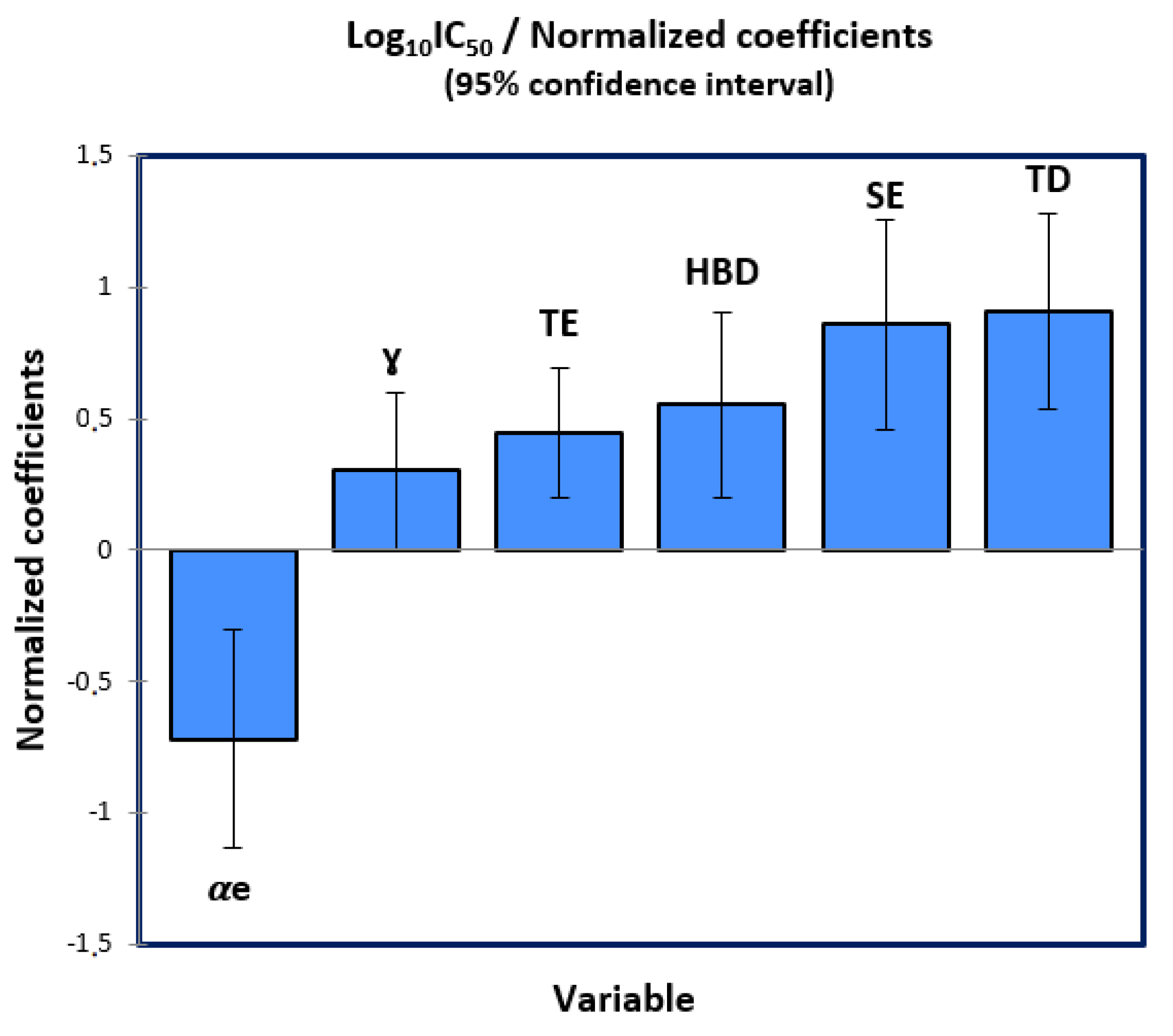
2.3. Multiple Linear Regression

The Quantitative structure-activity relationships (QSAR) have the potential to reduce the time and effort of molecular screening using mathematical predictive models [22]. One of these models is obtained by the multiple linear regression (MLR) technique, as a statistical tool for estimating the linear relationship between more than two variables which have cause-effect relations [23]. Thus, the first QSAR model was applied using the MLR technique with stepwise selection, on a training set of thirty-five molecules (N = 35), where the process was repeated more than a thousand times based on statistical criteria: in particular, the determination and correlation coefficients, provided that they will be validated in the next stage. Accordingly, the best QSAR model is given by the following equation:
Log10IC50 = −10,407−0.279 × αe + 0.069 × ɣ + 0.156 × TE + 1.83 × HBD + 1.716 × SE + 1.029 × TD.
This constructed model shows that the biological activity at the log scale is a quantitative variable affected by the following six descriptors: polarizability (αe), surface tension (ɣ), torsion energy (TE), Hydrogen bond donor (HBD), stretch energy (SE) and topological diameter (TD), which have been calculated and presented in Table 1. Moreover, the significance test demonstrates that the slope of each variable has a probability inferior to 5% as shown in Table 2, and so the selected descriptors have a significant weight on the biological inhibitory activity at a 95% confidence interval. Except the polarizability, all five molecular descriptors affect positively the biological activity as shown in Figure 2, where a molecule can be more active if it is less polar and has higher values of surface tension, torsion and stretch energies, hydrogen bond donor and topological diameter.
Additionally, the null hypothesis (H0) postulated by the Fisher statistical test is rejected, because the calculated Fisher value (F = 10.325) is so much higher than its critical value: [F (35,6) = 2.37, p < 0.0001], as presented in the one Anova test (Table 3). Therefore, the variance between the response (Log10IC50) and the six predictor variables is homogeneous. Moreover, the correlation and determination coefficients of R = 0.83 and R2 = 0.69, respectively, confirm that there is a strong relationship between the descriptors and the inhibitory activity. Thus, the first QSAR model generated via MLR technique has a good predictive performance, with a low standard error (RMSE = 0.66).

2.4. Multiple Non-Linear Regression

The multiple non-linear regression (MNLR) technique is applied using a set of adapted algorithms to generate the quantitative predictive models [24]. In the present study, we relied on the programmed function of the type:
Y = a 0 + i = 1 n ( ai × Xi + bi × Xi 2 )  
As:
Y: is the predicted biological activity (Log10IC50)
Xi: is the explicative variable
a0: is the constant of the QSAR model
ai and bi: are the slopes of each descriptor to one and two degrees, respectively.
Finally, we arrived at the second QSAR model given by the following equation:
Log10IC50 = −19.699 – 0.009 × αe – 0.056 × ɣ − 0.161 × TE + 1.466 × HBD + 0.5 × SE + 2.693 × TD
   −0.002 × αe^2 + 0.001 × ɣ^2 + 0.013 × TE^2 + 0.148 × SE^2 – 0.07 × TD^2.
This mathematical model has a good predictive capacity, justified by a strong non-linear relationship between the biological activity and the six descriptors, as it is defined by a good correlation coefficient (R = 0.84) and a good coefficient of determination (R2 = 0.71), in addition to its minimal mean square error (RMSE = 0.72).

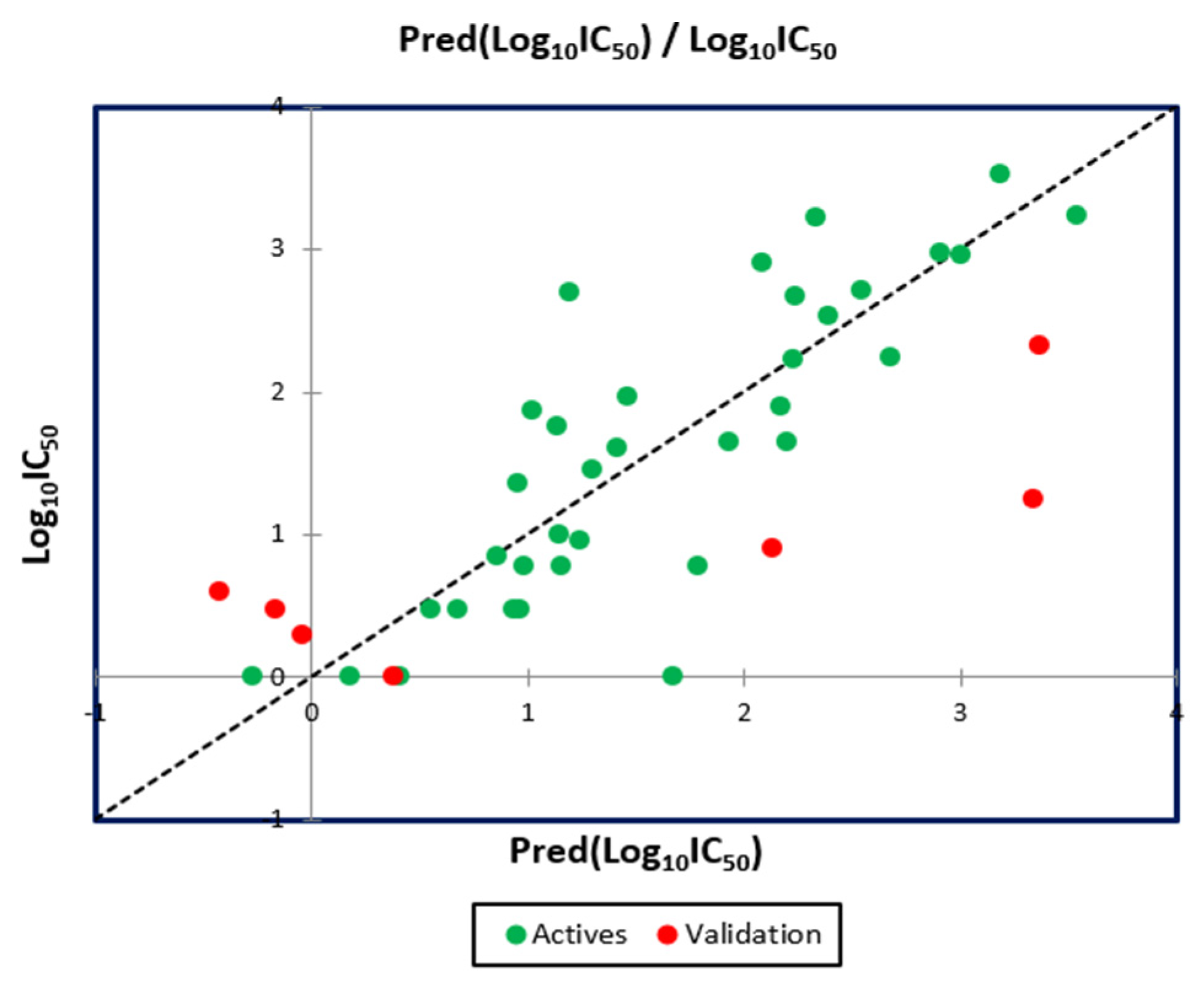
2.5. QSAR Model Validation

2.5.1. Applicability Domain

The applicability domain (AD) of a quantitative structure-activity relationship (QSAR) model is necessary to verify its reliability on new compounds (test set) that were not considered during its development [25]. This technique has been evaluated by an analysis expressed as a Williams diagram (Figure 3), which confirms that the molecules (1 and 32) belonging to the test set are really outliers, because they exceed the warning leverage (h* = 0.6), where: h* = 3 × K/n and K = p + 1, (p = 6, K = 7, n = 35) as, n: is the number of training set, and p: is the number of predictor descriptors [26,27]. Next, we noted that the compound 33 from training test is not an outlier because it does not exceed the critical leverage (h*). Therefore, except for molecules (1 and 32), all the others are well explained because they have in addition a normalized residual included in the 3 times standard deviation interval. Consequently, the 42 remaining molecules are tested in the applicability domain and the QSAR model was predicted correctly.

2.5.2. External Validation

To assess the accuracy of the QSAR predictive model and guarantee its generalizability, it is absolutely needed to validate it on new molecules included in the test set, before its application in clinical practice [28]. Based on a training test (35 molecules), we tested the seven new molecules from the test set and got the results presented in Table 4.
The results mentioned in Figure 4 indicate that the MLR QSAR model is given by an external validation correlation coefficient (R2ext = 0.63), and the results noted in Figure 5 indicate that the MNLR QSAR model is characterized by an external validation correlation coefficient of R2ext = 0.68. According to the Alexander Golbraikh and Alexander Tropsha theory, a QSAR model is externally validated if the correlation coefficient of its external validation is greater than 0.6. Therefore, the mathematical models developed with the help of MLR and MNLR techniques are externally validated.

2.5.3. Internal Validation

To validate internally the QSAR model, we applied the cross-validation technique with the leave-one-out procedure (CVLOO), so that each observation is tested exactly once, by executing a new model each run on thirty-four compounds (N-1 = 34) and predicting the biological activity of the removed sample, as shown in Table 5. This technique is based on the calculation of the quadratic coefficient of cross validation (Q2cv), which is expressed in the following equation [29,30]: Q 2 c v = 1 i n   ( Y p r e d Y o b s )   2 i n   ( Y o b s Y m e a n )   2   (4) AS: Ypred: is the predicted activity value, Yobs: is the observed activity value, Ymean: is the mean of the observed activity values. A high value of Q2cv = 0.57 (superior than 0.5) signifies that the established model is reliable, robust and has better internal predictivity.

2.5.4. Validation Using Y-Randomisation Test

The statistical study of Alexander Golbraikh and Alexander Tropsha confirms that the cross-validation technique is necessary but not sufficient, as the internal predictive accuracy of the cross-validation procedure tends to be overestimated and the high value of the quadratic coefficient may be the result of chance correlation. For this reason, the Y-randomisation test is necessary [31]. Using java Platform SE binary, we tested the QSAR model quality by running one hundred randomizations, as presented in Table 6. The results of the Y-randomisation test demonstrate that the (cR2p = 0.602) criteria is superior than 0.5; moreover, the R, R2 and R2cv values of the original model are much better than the values obtained by 100 randomizations. Consequently, the biological activity values predicted by the original model are not due to chance.

2.5.5. Golbreikh and Tropsha Criteria

The quantitative structure-activity relationship (QSAR) model, defined by the first Equation (1), satisfies the threshold criteria postulated by Golbraikh and Tropsha theory, as shown in Table 7.

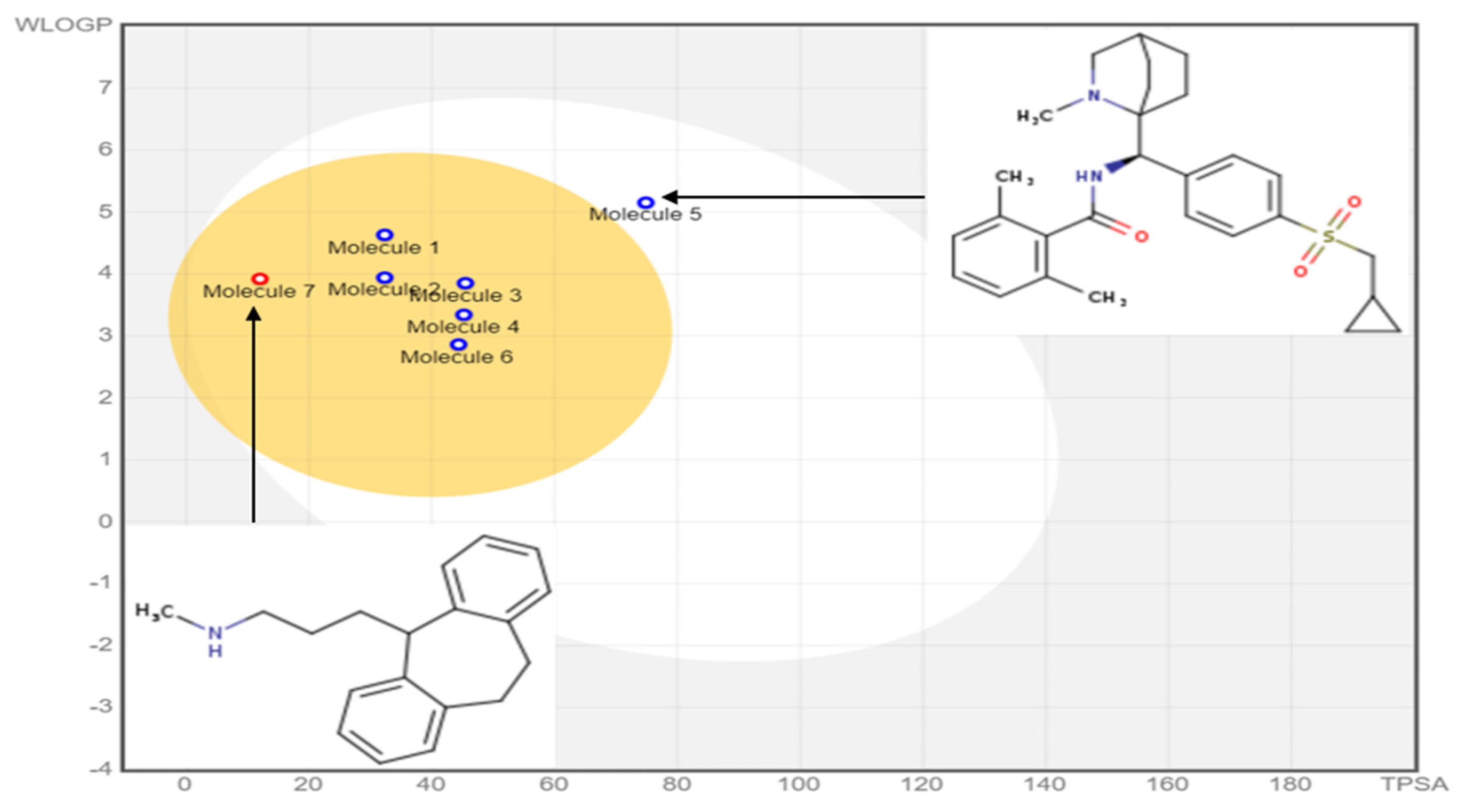
2.6. In Silico Pharmacokinetics ADMET Prediction

The most active ligands (L6, L9, L30, L31, L32 and L37), acting as inhibitors of type 1 glycine transporters, were tested based on the rules of Lipinski, Veber, Egan, and Ghose, and the pharmacokinetic properties (ADMET) [32], which were compared to the obtained results for nortriptyline as a co-crystallized ligand bound to the dopamine transporter (DAT) membrane protein encoded 4M48. The results presented in Table 8 indicate that all molecules respect the rules of Lipinski, Veber, Egan and Ghose except the ligand 32, because its molar refractivity index exceeds 130 and its Ghose violation number is equal to 2 (exceed 1). Additionally, the exact predictive model (BOILED-Egg), highly practical in the context of drug discovery and medicinal chemistry, and based on the calculation of lipophilicity given by the logarithm of the partition coefficient between n-octanol and water (Log PO/W) and polarity signaled by the topological polar surface area (TPSA) of small molecules, clearly shows that the molecule (L32) is the only one that does not belong to the yellow Egan-egg, as presented in Figure 6. Therefore, the five ligands (L6, L9, L30, L31 and L37) are the molecules having the highest probability to penetrate the brain. In comparison, the molecule 32 belonging to the white region of the egg has the highest probability of being absorbed by the gastrointestinal tract [33], which is why it was an outlier in the previous QSAR study.
The pharmacokinetic parameters of adsorption, distribution, metabolism, excretion and toxicity (ADMET) of the most active ligands as presented in Table 9 indicate that the ligands have a good absorption in the human intestine (IAH so higher than 70%), and a good distribution, since their human distribution volumes are estimated to be greater than −0.44 Log L/kg. Their permeability to the blood-brain barrier (BBB) is greater than −1 Log BB, and their permeability to the central nervous system (CNS) outside the interval (of −2 to −3) Log PS. Thus, they all penetrate the central nervous system (CNS) with the exception of ligands L32 and L30. In addition, the molecules are all predicted as inhibitors of cytochrome 2D6 except ligand 32. Consequently, the ligands (L6, L9, L30, L31 and L37) are designed to be agents of the central nervous system due to the highest probability of penetrating the blood-brain barrier (BBB).

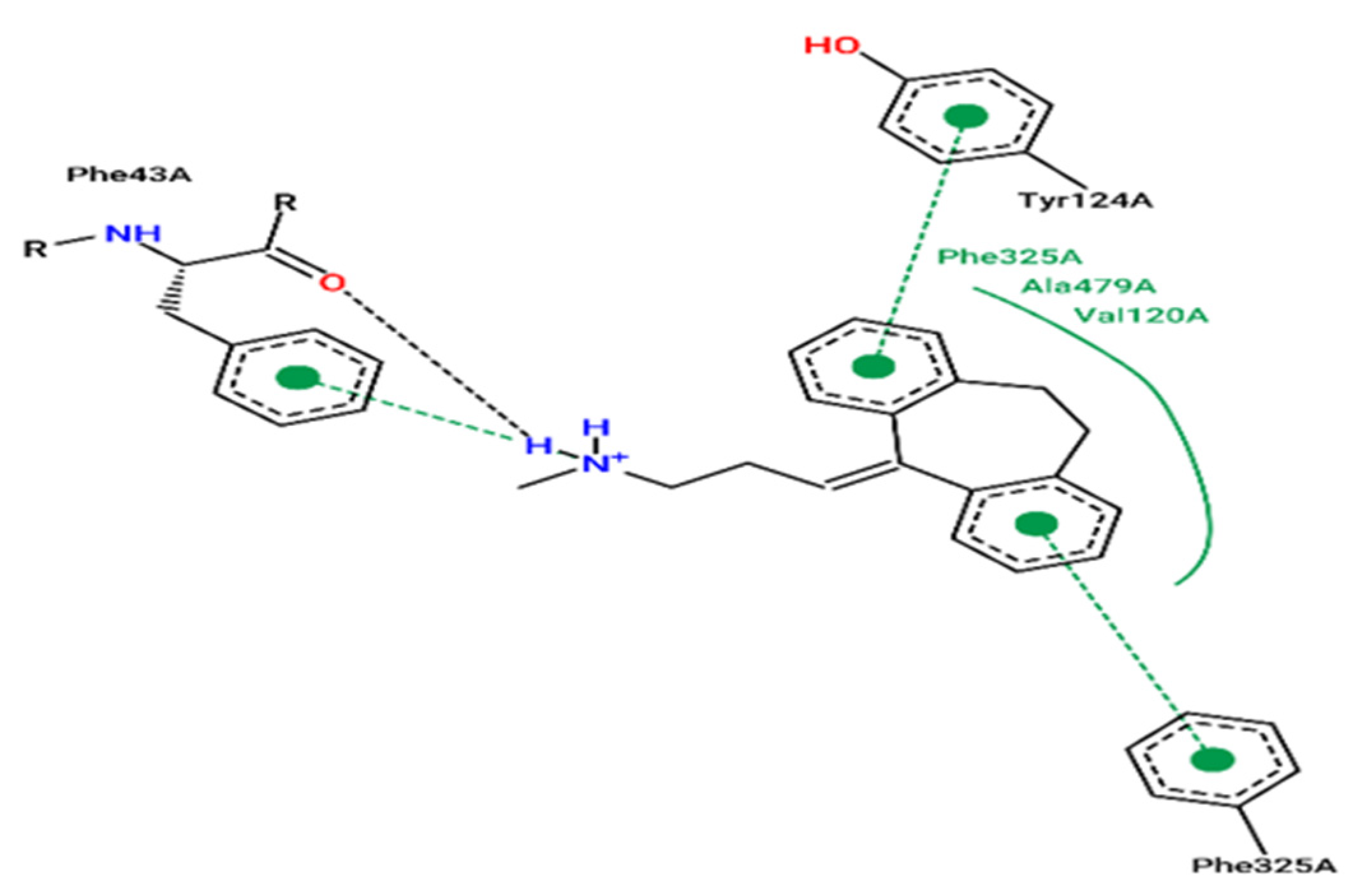
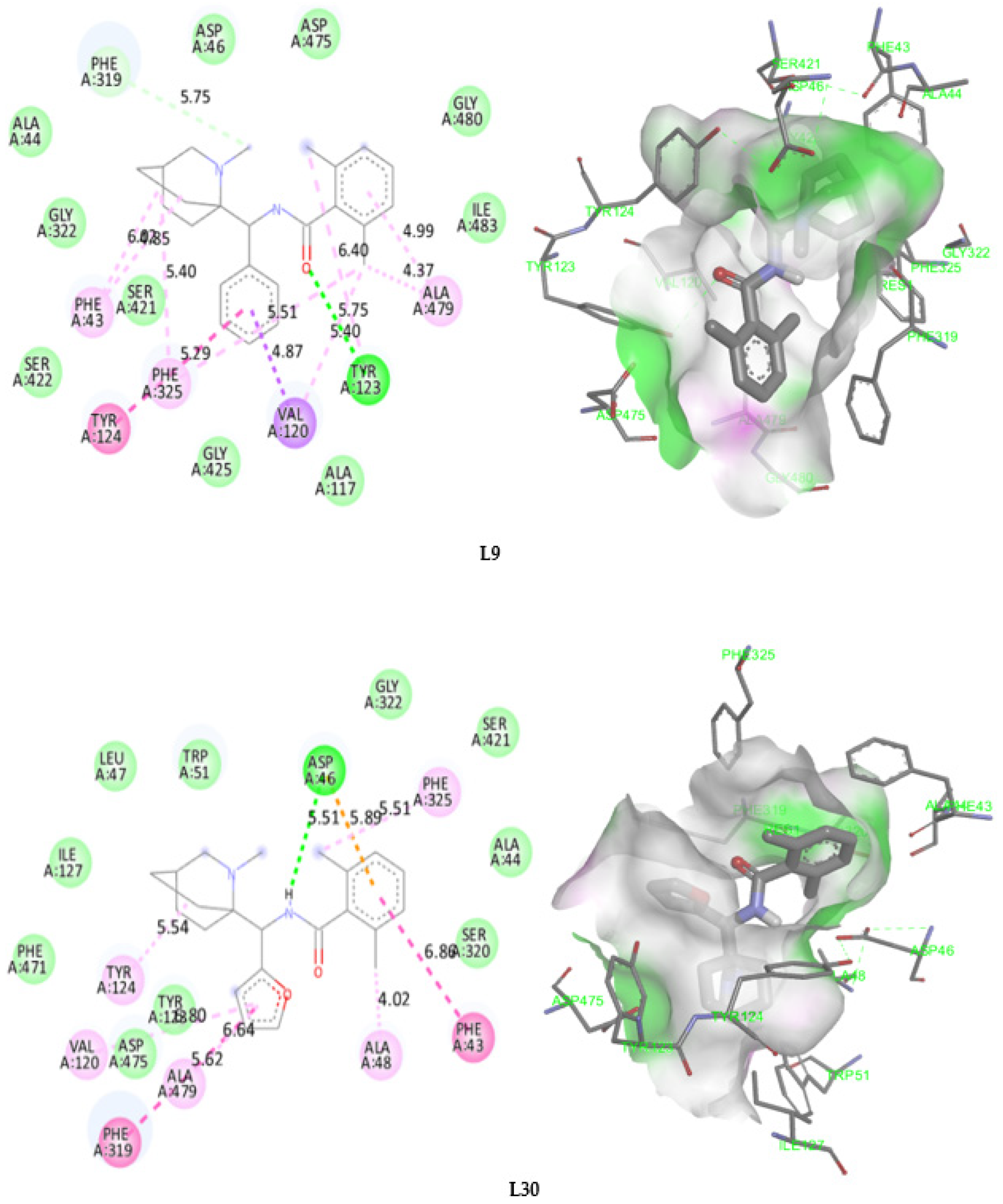
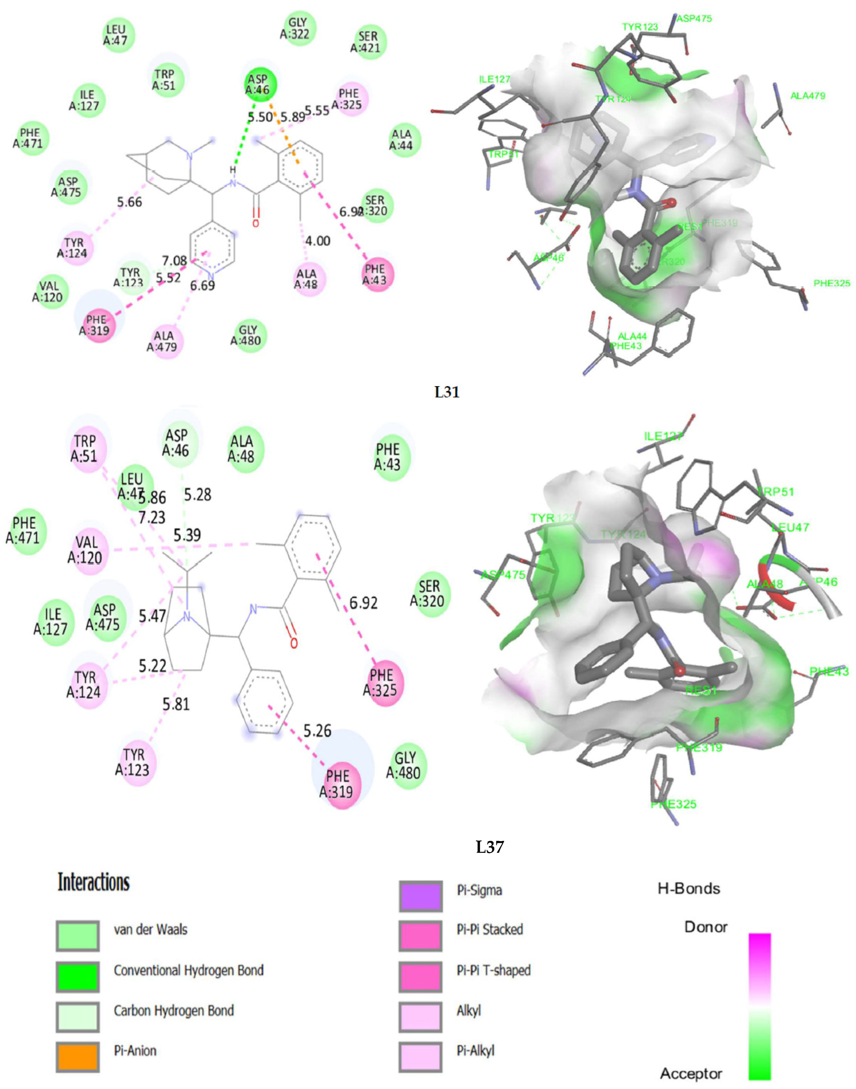
2.7. Molecular Docking

Molecular docking results are focused on the dopamine transporter (DAT) bound to the tricyclic antidepressant nortriptyline, as a transmembrane protein that removes the neurotransmitter dopamine from the synaptic cleft and transports it into the cytosol of surrounding cells. The crystal structure of this receptor is extracted using the X-ray diffraction method at a resolution of 2.96 Å taken from the protein data base (PDB) [34,35,36]. In this part of the research, the molecular docking process is started for the following most active molecules (L6, L9, L30, L31 and L37) to predict the type of Intermolecular interactions established with the protein encoded 4M48, compared to the established interactions with the co-crystallized ligand (nortriptyline) pictured in Figure 7, which indicate that Phe43A, Phe325A and Tyr124A amino acids, are the active sites of the target protein, as sourced using the ProteinsPlus online server [37].
The results of molecular docking applied on the more active ligands, presented in Figure 8, show that the ligands L6 and L9 share common molecular interactions as the chemical bonds of type pi-sigma and Pi-Pi T-shaped established between the benzenic cycle and (Val120 and Tyr124) amino acids respectively, in addition to two bonds of alkyl type with Phe325 and Phe43 amino acids. L30 and L31 ligands also form common bonds, like the hydrogen bond linked to the nitrogen atom, with the amino acid Asp46 at the same nuclear distance (5.5 Å), in addition to the alkyl bond with bicyclo group and Tyr124 amino acid. The same type of bond was established between the methyl group and the amino acid Phe325 at a nuclear distance of 5.5 Å, more than Pi-Pi bonds with Phe43 and Phe319 amino acids. Even the ligand L37 formed an alkyl bond with Tyr124 amino acid, and two Pi-Pi chemical bonds with Phe325 and Phe319 amino acids. Therefore, we can conclude that Tyr124, Phe43, Phe325, Asp46, Phe319 and Val120 amino acids are the active sites of the dopamine transporter (DAT) membrane protein, in which the most active ligands can inhibit the glycine transporter type 1 (GlyT1).

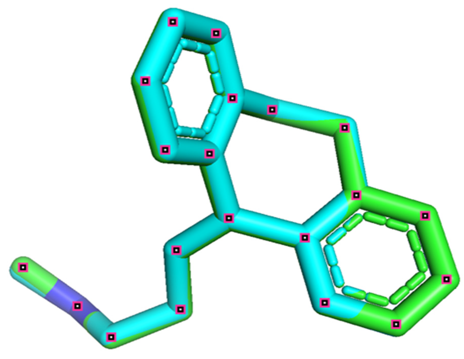
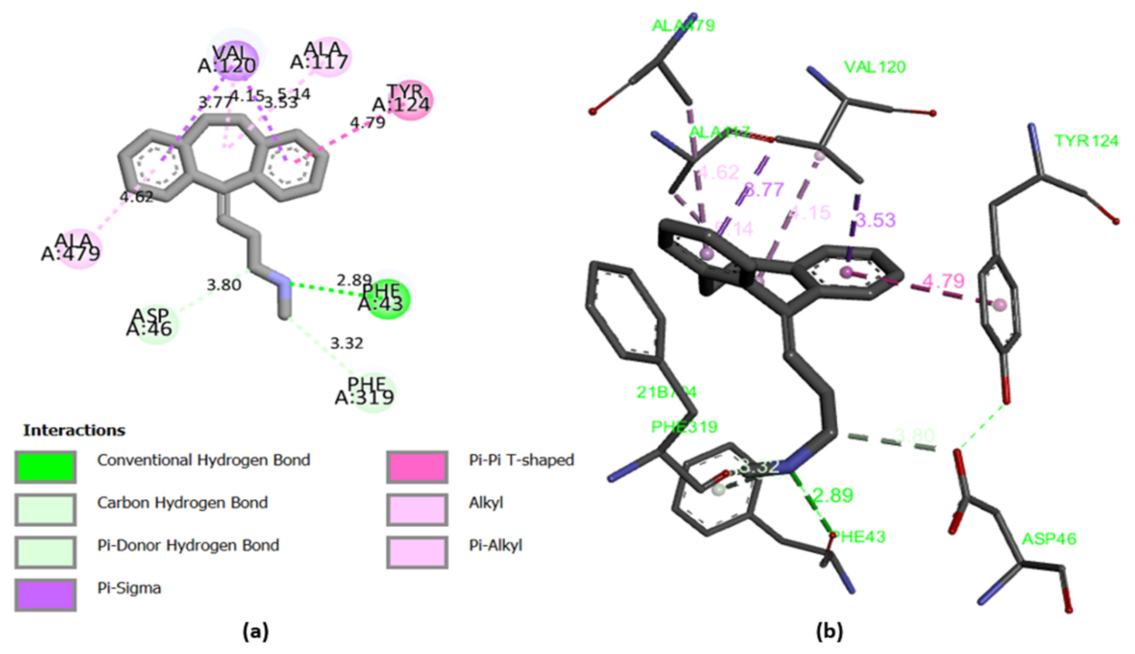
2.8. Docking Validation Protocol

The efficiency of the molecular docking algorithms was tested using the re-docking methodology, which is based on the superposition of the docked ligand on the protein-bound ligand, as shown in Figure 9. The superposition result indicates a root mean square deviation smaller than 2 (RMSD = 0.022 Å), which explains an exact pose prediction. Additionally, 2D and 3D visualization (Figure 10) of the intermolecular interaction between the docked nortriptyline and the protein target indicates that the chemical bonds formed with Phe43A and Tyr124A amino acids are the same as those observed experimentally. Thus, the molecular docking protocol is successfully validated [18].

2.9. Molecular Dynamics Simulations

The most active ligands (L6, L9, L30, L31 and L37) were chosen for the molecular dynamic’s simulation during 100 ns, to examine their stability toward DAT protein, where the conformational changes of one of these ligands are presented in Figure 11, and the others were presented in Figure S1.
The dynamic changes of conformation for (L9-protein) complex shown in Figure 11, indicate that the simulation is well-equilibrated, as the fluctuations of the root mean square deviation (RMSD) of the protein (left Y-axis) are around the thermal mean structure throughout the simulation time (100 ns), because the changes of the order of 1–3 Å are perfectly acceptable for small globular proteins. Moreover, the RMSD evolution of the heavy atoms of the ligand (right Y-axis) shows its stability with respect to the protein, when the protein-ligand complex is first aligned on the protein backbone of the reference, because the observed values are significantly smaller than the RMSD of the protein, and so the ligand did not diffuse away from its initial binding site.
The root mean square fluctuation (RMSF) values are also computed to examine the impact of the ligand binding on the internal dynamics of the target protein during 100 ns, where the tails (N- and C-terminal) fluctuate more than any other part of the protein and the secondary structure elements like alpha helices and beta strands are usually more rigid than the unstructured part of the protein; for this reason, they fluctuate less than the loop regions. Except for a single fluctuation of 3.2 Å, detected in the loop region of residue 390, all fluctuations were less than 3 Å, indicating the binding strength between the ligand 9 and the DAT protein, and no significant change in the protein conformation resulting from ligand binding.
Additionally, the radius of gyration (r Gyr) values fluctuated in a small interval from 3.45 to 3.76 Å until the end of the simulation, as shown in Figure 12, indicating that there are just some changes in the compactness of the ligand; thus, the protein has a good flexibility after its binding with the ligand 9. Moreover, the solvent accessibility of the protein-ligand 9 complex was evaluated by the solvent accessible surface area (SASA) analysis, which fluctuated between 0 and 15 Å2 for 100 ns; this graph revealed that the structure of compound 9 was relatively stable during the simulation time. The polar surface area (PSA) is a solvent accessible surface area in a molecule contributed only by oxygen and nitrogen atoms; this parameter varies between 8 and 32 Å2, accompanied by some maximal and minimal fluctuations during the simulation time. The contributions of this type of atoms make the ligand relatively unstable. However, the molecular surface area (MolSA) illustrates the molecular surface calculation with a probe radius of 1.4 Å, equivalent to a Van der Waals surface, showing only minimal fluctuations.
Lastly, the graph of the total energy presented in Figure 12 shows a minimal variation about the average −53.8682 kcal.mol−1, which means that the energy of the L9- DAT protein complex remained in equilibrium throughout the MD simulation.
The dynamic changes of conformation for other complexes are available in the supplementary material (Figure S1). The protein-ligands interactions fluctuate with a root mean square deviation (RMSD) of 1 to 3 Å along the simulation time (100 ns), except for the L31-protein complex, which oscillates for the first 20 nanoseconds from 1 to 3 Å, then destabilizes until 50 ns, and stabilizes again with a deviation 4 Å of about until the end of the simulation time.
Minimal fluctuations have been observed during the established interactions between the protein and the ligand 6, such as the observed root mean square fluctuations (RMSF) of 3.4 Å, 4.4 Å and 3.9 Å detected in the loop region of 10, 240 and 480 residues, respectively, in addition to a maximal fluctuation of 5.5 Å recorded in the loop area of residue 380. No fluctuation was noticed greater than 3 Å for L30-protein and L37-protein complexes. Three fluctuations have been recorded for L31-protein complex of 3.6 Å, 3.5 Å and 7 Å reported in the loop zone of 10, 480 and 520 residues, respectively.
Overall, we note that the protein has a good flexibility of binding with L6, L30, L31 and L37 inhibitors, as there are just a few changes in the compactness of the ligand, since the r Gyr, SASA, MolSA and PSA parameters are varied with minimal fluctuations about the mean along 100 ns.
Finally, the total energy plots presented in Figure S2 show a minimal variance around the average energy of the total system, which was in Kcal/mol of −47.7003, −48.8821 −49.5236 and −62.5749 for L30, L31, L37 and L6 inhibitors, respectively, indicating that the energies of the ligands-protein complexes have remained in equilibrium over the course of the MD simulation.
We conclude that the molecular dynamics simulations reinforce the previous results obtained by QSAR and docking studies, since the ligands L6, L9, L30, L31 and L37 are the most active inhibitors, forming typical static interactions with some amino acids of the target protein. These interactions form dynamically stable complexes during the 100 ns of the simulation time, as there is no change in their properties, except for the minimal fluctuations that were observed.

3. Materials and Methods

3.1. Database

The present study is performed on 44 bicyclo ((aryl) methyl) benzamides as glycine transporter type 1 (GlyT1) inhibitors, whose biological activities are expressed on a logarithmic decimal scale (log10IC50), as illustrated in Table S1.

3.2. Molecular Descriptors Calculation

To build the quantitative structure-activity relationship (QSAR) models that provide information on the correlation between activities and structure-based molecular descriptors, we calculated various types of molecular descriptors [38], as shown in Table S2. Initially, the constitutional descriptors were calculated using the ACD/chemsketch software [39]. Subsequently, the thermodynamic and physicochemical descriptors were extracted using the MM2 technique via the ChemBio3D software [40]. Lastly, the quantum descriptors are calculated through Gaussian 09 software [41], using the density function theory (DFT)/B3LYP [42], combined with the 6-31 + G(d,p) basis set, in order to ensure the molecules stability and optimize their three-dimensional geometries.

3.3. Statistical Methods

The Quantitative structure-activity relationships (QSARs) are developed with the help of XLSTAT 2014 software [43], using different statistical methods such as: the principal component analysis (PCA), multiple linear regression (MLR) and multiple non-linear regression (MNLR). The principal component analysis method is a very important step that serves to minimize the molecular descriptor dimension so as to identify the most predictive variables [44]. This limited number of descriptors is mathematically modeled by the multiple linear regression (MLR) and multiple non-linear regression (MNLR) techniques. Therefore, the two obtained QSAR models were generated to predict the linear and non-linear relationships established between the biological activity of glycine transporter type 1 (GlyT1) inhibitors and their relevant descriptors. For their applicability, these two models have been evaluated by external and internal validation, as well as the molecules, which have been tested in the applicability domain [45]. Additionally, the Golbreikh and Tropsha criteria and the Y-randomization test were used to verify the robustness and predictive potential of the established QSAR model [31,46].

3.4. Drug Likeness and In Silico Pharmacokinetics ADMET Prediction

To make the drugs applicable in clinical trials, it is necessary to study their absorption, distribution, metabolism, excretion and toxicity (ADMET) in the human body before starting the investigation protocols [21,47], respecting some important rules such as those of Lipinski [48], Veber [49], Ghose [50] and Egan [51,52]. This technique is also applied to eliminate the compounds with potentially undesirable physiological qualities, taking into account toxicity and pharmacokinetic properties [53]. For this task, we estimated the drug similarity and in silico pharmacokinetic properties of the newly selected molecules as GluT1 inhibitory agents, using the online SwissADMET [54] and pkCSM [55] servers, respectively.

3.5. Molecular Docking Modeling

The computational technique of molecular docking is an efficient, fast and powerful tool for drug discovery [56]. For this project, we uploaded the three-dimensional coordinates of the target protein from the protein data bank (pdb) using the Discovery Studio 2021 (BIOVIA) software package [57]. To improve the performance of the cavity method, water molecules and suspended ligands bound to the protein were removed and polar hydrogens were added. Accordingly, the prepared protein was docked with the most active ligands, previously optimized by the density functional theory (DFT), with the assistance of AutoDock 4.2 [58]. Moreover, the grid box was centralized on (−42.562 Å, −0.46 Å, −55.066 Å) with the help of AUTOGRID algorithm, by putting the sizes (80, 80, 80) in their three-dimensional structure, and running 10 genetic algorithms with a total of 25 million trials. Finally, the molecular interactions of the protein-ligand were visualized using discovery studio 2021 [59].

3.6. Molecular Dynamics

Based on QSAR and molecular docking results, the five best-docked ligands, having the highest activity, were chosen for the molecular dynamics simulations in order to identify the molecular recognition between the ligand and the dopamine transporter (DAT) membrane protein. The MD simulations were performed for 100 nanoseconds using Desmond software, a package of Schrödinger LLC [60]. The first stage in the molecular dynamic’s simulation of protein-ligand complexes was obtained by docking studies, and preprocessed using Protein Preparation Maestro, which performs optimization and minimization of complexes that have been prepared by the System Builder tool using a solvent model with an orthorhombic box that was chosen as TIP3P (transferable intermolecular interaction potential 3 points), using the OPLS force field [61]. At 300 K temperature and 1 atm pressure, the models were made neutral with the addition of water molecules, and counter ions, such as 0.15 M salt (Na+, Cl) were added to mimic the physiological conditions. Finally, the trajectories were saved after every 10 ps for analysis, and the stability of simulations was evaluated by calculating the root mean square deviation (RMSD) of the protein and ligand over time. In addition, the root-mean-square fluctuation (RMSF), gyration radius (Rg), solvent accessible surface area (SASA), molecular surface area (MolSA) and polar surface area (PSA) were recorded for 100 ns, and the free energies of the inhibitors-protein interactions were evaluated using MM-GBSA approach [62].

4. Conclusions

A systematic in silico study was applied on 44 bicyclo((aryl)methyl)benzamide derivatives as glycine transporter type 1 (GlyT1) inhibitors to discover effective antipsychotic candidates for the treatment of schizophrenia. Initially, two QSAR models were developed using MLR and MNLR techniques and were examined through external and internal validation, applicability domain (AD), Y-randomization test and Golbreikh and tropsha criteria, indicating a significant effect of hydrogen bond donor, polarizability, surface tension, stretch and torsion energies and topological diameter on the locomotor activity (LMA). Subsequently, ADMET in silico pharmacokinetics prediction revealed a favorable profile of the most active ligands, where L6, L9, L30, L31 and L37 were predicted as non-toxic inhibitors for 2D6 cytochrome, which respect the rules of Lipinski, Veber, Egan and Ghose, with an excellent absorption exceeded 91% and highest probability to penetrate the central nervous system (CNS). In contrast, the ligand L32 as an outlier in the QSAR study has an unfavorable ADMET profile with the highest probability of being absorbed by the gastrointestinal tract. Lastly, the obtained results were further strengthened and qualified using molecular docking and molecular dynamics studies, which confirm that L6, L9, L30, L31 and L37 react specifically with Tyr124, Phe43, Phe325, Asp46, Phe319 and Val120 amino acids of the dopamine transporter (DAT) membrane protein in a way that blocks glycine transporter type 1 (GlyT1) forming dynamically stable complexes during 100 ns of MD simulation time. Therefore, they could be used as therapeutics in medicine to treat schizophrenia. However, they must be subjected to in vitro and in vivo investigations to evaluate their efficacy and safety as anti-schizophrenia drugs.

Supplementary Materials

The following are available online at https://www.mdpi.com/article/10.3390/ph15060670/s1, Table S1: The 44 molecules and their biological activities. Figure S1: RMSD and RMSF graphs for L6, L30, L31, L37 ligands complexed with the dopamine transporter membrane protein during 100 ns. Figure S2: Rg, MolSA, SASA and PSA during 100 ns of MD simulation, and the variation of total free energy for L6, L30, L31 and L37 ligands complexed with DAT protein. Table S2: The calculated molecular descriptors.

Author Contributions

Conceptualization, M.E.f., M.E. and M.K.; methodology, M.E.f., A.A. (Amine Assouguem) and M.E.-R.; software, M.E.f. and A.A. (Amal Alotaibi); validation, A.B., N.N.M. and H.F. data curation, R.U., S.E. and S.Z.; writing—original draft preparation, M.E.f. and M.E.-R.; writing—review and editing, M.K. and M.E.; supervision, M.E. All authors have read and agreed to the published version of the manuscript.

Funding

This research was funded by Princess Nourah bint Abdulrahman University Researchers Supporting Project number (PNURSP2022R33), Princess Nourah bint Abdulrahman University, Riyadh, Saudi Arabia.

Institutional Review Board Statement

Not applicable.

Informed Consent Statement

Not applicable.

Data Availability Statement

Data is contained within the article and Supplementary Materials.

Acknowledgments

Authors wish to thank Princess Nourah bint Abdulrahman University Researchers Supporting Project number (PNURSP2022R33), Princess Nourah bint Abdulrahman University, Riyadh, Saudi Arabia, for financial support.

Conflicts of Interest

The authors declare no conflict of interest.

References

  1. Zheng, X.; Wang, C.; Zhai, N.; Luo, X.; Liu, G.; Ju, X. In Silico Screening of Novel A1-GABAA Receptor PAMs towards Schizophrenia Based on Combined Modeling Studies of Imidazo [1,2-a]-Pyridines. Int. J. Mol. Sci. 2021, 22, 9645. [Google Scholar] [CrossRef] [PubMed]
  2. Gao, L.; Hao, C.; Chen, J.; Ma, R.; Zheng, L.; Wu, Q.; Liu, X.; Liu, B.-F.; Zhang, G.; Chen, Y.; et al. Discovery of a New Class of Multi-Target Heterocycle Piperidine Derivatives as Potential Antipsychotics with pro-Cognitive Effect. Bioorg. Med. Chem. Lett. 2021, 40, 127909. [Google Scholar] [CrossRef] [PubMed]
  3. Shahsavar, A.; Stohler, P.; Bourenkov, G.; Zimmermann, I.; Siegrist, M.; Guba, W.; Pinard, E.; Seeger, M.A.; Schneider, T.R.; Dawson, R.J.P.; et al. Structural Insights into Glycine Reuptake Inhibition; 2020; BioRxiv. Available online: https://www.biorxiv.org/content/10.1101/2020.12.20.110478v1 (accessed on 14 April 2022).
  4. Varnes, J.G.; Xiong, H.; Forst, J.M.; Holmquist, C.R.; Ernst, G.E.; Frietze, W.; Dembofsky, B.; Andisik, D.W.; Palmer, W.E.; Hinkley, L.; et al. Bicyclo((Aryl)Methyl)Benzamides as Inhibitors of GlyT1. Bioorg. Med. Chem. Lett. 2018, 28, 1043–1049. [Google Scholar] [CrossRef] [PubMed]
  5. Cubelos, B.; Giménez, C.; Zafra, F. The Glycine Transporter GLYT1 Interacts with Sec3, a Component of the Exocyst Complex. Neuropharmacology 2005, 49, 935–944. [Google Scholar] [CrossRef] [PubMed]
  6. Marques, B.L.; Oliveira-Lima, O.C.; Carvalho, G.A.; de Almeida Chiarelli, R.; Ribeiro, R.I.; Parreira, R.C.; da Madeira Freitas, E.M.; Resende, R.R.; Klempin, F.; Ulrich, H.; et al. Neurobiology of Glycine Transporters: From Molecules to Behavior. Neurosci. Biobehav. Rev. 2020, 118, 97–110. [Google Scholar] [CrossRef] [PubMed]
  7. Lill, M.A. Multi-Dimensional QSAR in Drug Discovery. Drug Discov. Today 2007, 12, 1013–1017. [Google Scholar] [CrossRef]
  8. Vilar, S.; Cozza, G.; Moro, S. Medicinal Chemistry and the Molecular Operating Environment (MOE): Application of QSAR and Molecular Docking to Drug Discovery. Curr. Top. Med. Chem. 2008, 8, 1555–1572. [Google Scholar] [CrossRef]
  9. Tian, S.; Wang, J.; Li, Y.; Li, D.; Xu, L.; Hou, T. The Application of in Silico Drug-Likeness Predictions in Pharmaceutical Research. Adv. Drug Deliv. Rev. 2015, 86, 2–10. [Google Scholar] [CrossRef]
  10. Serrano, A.; Imbernón, B.; Pérez-Sánchez, H.; Cecilia, J.M.; Bueno-Crespo, A.; Abellán, J.L. QN-Docking: An Innovative Molecular Docking Methodology Based on Q-Networks. Appl. Soft Comput. 2020, 96, 106678. [Google Scholar] [CrossRef]
  11. El Khatabi, K.; El-mernissi, R.; Aanouz, I.; Ajana, M.A.; Lakhlifi, T.; Shahinozzaman, M.; Bouachrine, M. Benzimidazole Derivatives in Identifying Novel Acetylcholinesterase Inhibitors: A Combination of 3D-QSAR, Docking and Molecular Dynamics Simulation. Phys. Chem. Res. 2022, 10, 237–249. [Google Scholar] [CrossRef]
  12. de Oliveira, T.A.; Medaglia, L.R.; Maia, E.H.B.; Assis, L.C.; de Carvalho, P.B.; da Silva, A.M.; Taranto, A.G. Evaluation of Docking Machine Learning and Molecular Dynamics Methodologies for DNA-Ligand Systems. Pharmaceuticals 2022, 15, 132. [Google Scholar] [CrossRef] [PubMed]
  13. Wang, H.; Jia, Y.; Jing, G.; Wu, X. A Novel Toxicity Prediction Model for Hydrazine Compounds Based on 1D–3D Molecular Descriptors. Comput. Toxicol. 2021, 18, 100169. [Google Scholar] [CrossRef]
  14. Halder, A.K.; Jha, T. Validated Predictive QSAR Modeling of N-Aryl-Oxazolidinone-5-Carboxamides for Anti-HIV Protease Activity. Bioorg. Med. Chem. Lett. 2010, 20, 6082–6087. [Google Scholar] [CrossRef] [PubMed]
  15. Roy, K.; Kar, S.; Das, R.N. Validation of QSAR Models. In Understanding the Basics of QSAR for Applications in Pharmaceutical Sciences and Risk Assessment; Elsevier: Amsterdam, The Netherlands, 2015; pp. 231–289. ISSN 978-0-12-801505-6. [Google Scholar]
  16. El Mchichi, L.; El Aissouq, A.; Kasmi, R.; Belhassan, A.; El-Mernissi, R.; Ouammou, A.; Lakhlifi, T.; Bouachrine, M. In Silico Design of Novel Pyrazole Derivatives Containing Thiourea Skeleton as Anti-Cancer Agents Using: 3D QSAR, Drug-Likeness Studies, ADMET Prediction and Molecular Docking. Mater. Today Proc. 2021, 45, 7661–7674. [Google Scholar] [CrossRef]
  17. Mazigh, M.; El’mbarki, C.; Hadni, H.; Elhallaoui, M. QSAR Studies Combined with DFT-Calculations and Molecular Docking of Polyamine-Sensitive Inhibitors of the NMDA Receptor. Mediterr. J. Chem. 2019, 9, 164–174. [Google Scholar] [CrossRef] [Green Version]
  18. Abdullahi, M.; Shallangwa, G.A.; Uzairu, A. In Silico QSAR and Molecular Docking Simulation of Some Novel Aryl Sulfonamide Derivatives as Inhibitors of H5N1 Influenza A Virus Subtype. Beni-Suef Univ. J. Basic Appl. Sci. 2020, 9, 2. [Google Scholar] [CrossRef] [Green Version]
  19. Krieger, E.; Darden, T.; Nabuurs, S.B.; Finkelstein, A.; Vriend, G. Making Optimal Use of Empirical Energy Functions: Force-Field Parameterization in Crystal Space. Proteins Struct. Funct. Bioinforma. 2004, 57, 678–683. [Google Scholar] [CrossRef]
  20. Bastianoni, A.; Guastaldi, E.; Barbagli, A.; Bernardinetti, S.; Zirulia, A.; Brancale, M.; Colonna, T. Multivariate Analysis Applied to Aquifer Hydrogeochemical Evaluation: A Case Study in the Coastal Significant Subterranean Water Body between “Cecina River and San Vincenzo”, Tuscany (Italy). Appl. Sci. 2021, 11, 7595. [Google Scholar] [CrossRef]
  21. Daoui, O.; Elkhattabi, S.; Chtita, S.; Elkhalabi, R.; Zgou, H.; Benjelloun, A.T. QSAR, Molecular Docking and ADMET Properties in Silico Studies of Novel 4,5,6,7-Tetrahydrobenzo[D]-Thiazol-2-Yl Derivatives Derived from Dimedone as Potent Anti-Tumor Agents through Inhibition of C-Met Receptor Tyrosine Kinase. Heliyon 2021, 7, e07463. [Google Scholar] [CrossRef]
  22. Matsuzaka, Y.; Uesawa, Y. A Deep Learning-Based Quantitative Structure–Activity Relationship System Construct Prediction Model of Agonist and Antagonist with High Performance. Int. J. Mol. Sci. 2022, 23, 2141. [Google Scholar] [CrossRef]
  23. Uyanık, G.K.; Güler, N. A Study on Multiple Linear Regression Analysis. Procedia Soc. Behav. Sci. 2013, 106, 234–240. [Google Scholar] [CrossRef] [Green Version]
  24. Kravić, N.; Savosina, J.; Agafonova-Moroz, M.; Babain, V.; Legin, A.; Kirsanov, D. Nonlinear Multivariate Regression Algorithms for Improving Precision of Multisensor Potentiometry in Analysis of Spent Nuclear Fuel Reprocessing Solutions. Chemosensors 2022, 10, 90. [Google Scholar] [CrossRef]
  25. Ruiz, I.L.; Gómez-Nieto, M.Á. Study of the Applicability Domain of the QSAR Classification Models by Means of the Rivality and Modelability Indexes. Molecules 2018, 23, 2756. [Google Scholar] [CrossRef] [PubMed] [Green Version]
  26. Pourbasheer, E.; Riahi, S.; Ganjali, M.R.; Norouzi, P. Quantitative Structure–Activity Relationship (QSAR) Study of Interleukin-1 Receptor Associated Kinase 4 (IRAK-4) Inhibitor Activity by the Genetic Algorithm and Multiple Linear Regression (GA-MLR) Method. J. Enzyme Inhib. Med. Chem. 2010, 25, 844–853. [Google Scholar] [CrossRef]
  27. Chtita, S.; Belhassan, A.; Bakhouch, M.; Taourati, A.I.; Aouidate, A.; Belaidi, S.; Moutaabbid, M.; Belaaouad, S.; Bouachrine, M.; Lakhlifi, T. QSAR Study of Unsymmetrical Aromatic Disulfides as Potent Avian SARS-CoV Main Protease Inhibitors Using Quantum Chemical Descriptors and Statistical Methods. Chemom. Intell. Lab. Syst. 2021, 210, 104266. [Google Scholar] [CrossRef]
  28. de Castro Rezende, K.B.; da Cunha, A.J.L.; Amim Junior, J.; Bornia, R.G. External Validation of the Fetal Medicine Foundation Algorithm for the Prediction of Preeclampsia in a Brazilian Population. Pregnancy Hypertens. 2019, 17, 64–68. [Google Scholar] [CrossRef]
  29. Rafało, M. Cross Validation Methods: Analysis Based on Diagnostics of Thyroid Cancer Metastasis. ICT Express 2021, S2405959521000552. [Google Scholar] [CrossRef]
  30. Chtita, S.; Ghamali, M.; Ousaa, A.; Aouidate, A.; Belhassan, A.; Taourati, A.I.; Masand, V.H.; Bouachrine, M.; Lakhlifi, T. QSAR Study of Anti-Human African Trypanosomiasis Activity for 2-Phenylimidazopyridines Derivatives Using DFT and Lipinski’s Descriptors. Heliyon 2019, 5, e01304. [Google Scholar] [CrossRef] [Green Version]
  31. Golbraikh, A.; Tropsha, A. Beware of Q2! J. Mol. Graph. Model. 2002, 20, 269–276. [Google Scholar] [CrossRef]
  32. Daina, A.; Michielin, O.; Zoete, V. ILOGP: A Simple, Robust, and Efficient Description of n -Octanol/Water Partition Coefficient for Drug Design Using the GB/SA Approach. J. Chem. Inf. Model. 2014, 54, 3284–3301. [Google Scholar] [CrossRef]
  33. Daina, A.; Zoete, V. A BOILED-Egg To Predict Gastrointestinal Absorption and Brain Penetration of Small Molecules. ChemMedChem 2016, 11, 1117–1121. [Google Scholar] [CrossRef] [PubMed] [Green Version]
  34. Belhassan, A.; Zaki, H.; Benlyas, M.; Lakhlifi, T.; Bouachrine, M. Study of Novel Triazolo-Benzodiazepine Analogues as Antidepressants Targeting by Molecular Docking and ADMET Properties Prediction. Heliyon 2019, 5, e02446. [Google Scholar] [CrossRef] [PubMed]
  35. Adamski, A.; Kruszka, D.; Dutkiewicz, Z.; Kubicki, M.; Gorczyński, A.; Patroniak, V. Novel Family of Fused Tricyclic [1,4]Diazepines: Design, Synthesis, Crystal Structures and Molecular Docking Studies. Tetrahedron 2017, 73, 3377–3386. [Google Scholar] [CrossRef]
  36. Penmatsa, A.; Wang, K.H.; Gouaux, E. X-Ray Structure of Dopamine Transporter Elucidates Antidepressant Mechanism. Nature 2013, 503, 85–90. [Google Scholar] [CrossRef] [PubMed] [Green Version]
  37. Zentrum Für Bioinformatik: Universität Hamburg-Proteins Plus Server. Available online: https://proteins.plus/ (accessed on 8 March 2022).
  38. Spiegel, J.; Senderowitz, H. Evaluation of QSAR Equations for Virtual Screening. Int. J. Mol. Sci. 2020, 21, 7828. [Google Scholar] [CrossRef] [PubMed]
  39. Österberg, T.; Norinder, U. Prediction of Drug Transport Processes Using Simple Parameters and PLS Statistics The Use of ACD/LogP and ACD/ChemSketch Descriptors. Eur. J. Pharm. Sci. 2001, 12, 327–337. [Google Scholar] [CrossRef]
  40. Milne, G.W.A. Software Review of ChemBioDraw 12.0. J. Chem. Inf. Model. 2010, 50, 2053. [Google Scholar] [CrossRef]
  41. Citation Gaussienne 09 | Gaussian.Com. Available online: https://gaussian.com/g09citation/ (accessed on 8 March 2022).
  42. Parr, R.G.; Weitao, Y. Density-Functional Theory of Atoms and Molecules; International Series of Monographs on Chemistry; Oxford University Press: New York, NY, USA, 1995; ISBN 978-0-19-509276-9. [Google Scholar]
  43. Introducing the XLSTAT Free Edition. Available online: https://www.xlstat.com/en/news/introducing-the-xlstat-free-edition (accessed on 8 March 2022).
  44. Guerra Tort, C.; Aguiar Pulido, V.; Suárez Ulloa, V.; Docampo Boedo, F.; López Gestal, J.M.; Pereira Loureiro, J. Electronic Health Records Exploitation Using Artificial Intelligence Techniques. Proceedings 2020, 54, 60. [Google Scholar] [CrossRef]
  45. Hadni, H.; Elhallaoui, M. 3D-QSAR, Docking and ADMET Properties of Aurone Analogues as Antimalarial Agents. Heliyon 2020, 6, e03580. [Google Scholar] [CrossRef]
  46. van der Voet, H. Comparing the Predictive Accuracy of Models Using a Simple Randomization Test. Chemom. Intell. Lab. Syst. 1994, 25, 313–323. [Google Scholar] [CrossRef]
  47. Ebenezer, O.; Damoyi, N.; Jordaan, M.A.; Shapi, M. Unveiling of Pyrimidindinones as Potential Anti-Norovirus Agents—A Pharmacoinformatic-Based Approach. Molecules 2022, 27, 380. [Google Scholar] [CrossRef] [PubMed]
  48. Lipinski, C.A.; Lombardo, F.; Dominy, B.W.; Feeney, P.J. Experimental and Computational Approaches to Estimate Solubility and Permeability in Drug Discovery and Development Settings. Adv. Drug Deliv. Rev. 1997, 23, 3–25. [Google Scholar] [CrossRef]
  49. Veber, D.F.; Johnson, S.R.; Cheng, H.-Y.; Smith, B.R.; Ward, K.W.; Kopple, K.D. Molecular Properties That Influence the Oral Bioavailability of Drug Candidates. J. Med. Chem. 2002, 45, 2615–2623. [Google Scholar] [CrossRef] [PubMed]
  50. Yalcin, S. Molecular Docking, Drug Likeness, and ADMET Analyses of Passiflora Compounds as P-Glycoprotein (P-Gp) Inhibitor for the Treatment of Cancer. Curr. Pharmacol. Rep. 2020, 6, 429–440. [Google Scholar] [CrossRef]
  51. Egan, W.J.; Merz, K.M.; Baldwin, J.J. Prediction of Drug Absorption Using Multivariate Statistics. J. Med. Chem. 2000, 43, 3867–3877. [Google Scholar] [CrossRef]
  52. Egan, W.J.; Lauri, G. Prediction of Intestinal Permeability. Adv. Drug Deliv. Rev. 2002, 54, 273–289. [Google Scholar] [CrossRef]
  53. Gomes, D.; Silvestre, S.; Duarte, A.P.; Venuti, A.; Soares, C.P.; Passarinha, L.; Sousa, Â. In Silico Approaches: A Way to Unveil Novel Therapeutic Drugs for Cervical Cancer Management. Pharmaceuticals 2021, 14, 741. [Google Scholar] [CrossRef]
  54. Daina, A.; Michielin, O.; Zoete, V. SwissADME: A Free Web Tool to Evaluate Pharmacokinetics, Drug-Likeness and Medicinal Chemistry Friendliness of Small Molecules. Sci. Rep. 2017, 7, 42717. [Google Scholar] [CrossRef] [Green Version]
  55. PkCSM. Available online: http://biosig.unimelb.edu.au/pkcsm/run_example? (accessed on 8 March 2022).
  56. Bassani, D.; Pavan, M.; Bolcato, G.; Sturlese, M.; Moro, S. Re-Exploring the Ability of Common Docking Programs to Correctly Reproduce the Binding Modes of Non-Covalent Inhibitors of SARS-CoV-2 Protease Mpro. Pharmaceuticals 2022, 15, 180. [Google Scholar] [CrossRef]
  57. Iqbal, D.; Rehman, M.T.; Bin Dukhyil, A.; Rizvi, S.M.D.; Al Ajmi, M.F.; Alshehri, B.M.; Banawas, S.; Khan, M.S.; Alturaiki, W.; Alsaweed, M. High-Throughput Screening and Molecular Dynamics Simulation of Natural Product-like Compounds against Alzheimer’s Disease through Multitarget Approach. Pharmaceuticals 2021, 14, 937. [Google Scholar] [CrossRef]
  58. Norgan, A.P.; Coffman, P.K.; Kocher, J.-P.A.; Katzmann, D.J.; Sosa, C.P. Multilevel Parallelization of AutoDock 4.2. J. Cheminformatics 2011, 3, 12. [Google Scholar] [CrossRef] [PubMed] [Green Version]
  59. BIOVIA Discovery Studio - BIOVIA - Dassault Systèmes®. Available online: https://www.3ds.com/products-services/biovia/products/molecular-modeling-simulation/biovia-discovery-studio/ (accessed on 8 March 2022).
  60. Desmond | Schrodinger. Available online: https://www.schrodinger.com/products/desmond (accessed on 28 April 2022).
  61. Kaminski, G.A.; Friesner, R.A.; Tirado-Rives, J.; Jorgensen, W.L. Evaluation and Reparametrization of the OPLS-AA Force Field for Proteins via Comparison with Accurate Quantum Chemical Calculations on Peptides. J. Phys. Chem. B 2001, 105, 6474–6487. [Google Scholar] [CrossRef]
  62. Wang, E.; Sun, H.; Wang, J.; Wang, Z.; Liu, H.; Zhang, J.Z.H.; Hou, T. End-Point Binding Free Energy Calculation with MM/PBSA and MM/GBSA: Strategies and Applications in Drug Design. Chem. Rev. 2019, 119, 9478–9508. [Google Scholar] [CrossRef] [PubMed]
Figure 1. Data visualization on the first two principal components.
Pharmaceuticals 15 00670 g001
Figure 2. Influence degree of the descriptors on the biological activity.
Pharmaceuticals 15 00670 g002
Figure 3. William’s diagram of the MLR model established by Equation (1). 1 and 32 are outliers in the test set and 33 is a non-aberrant molecule in the training test.
Pharmaceuticals 15 00670 g003
Figure 4. Correlation between the observed and predicted activities using MLR technique.
Pharmaceuticals 15 00670 g004
Figure 5. Correlation between the observed and predicted activities using MNLR technique.
Pharmaceuticals 15 00670 g005
Figure 6. BOILED-egg predictive model of the most active ligands.
Pharmaceuticals 15 00670 g006
Figure 7. Experimental pose view of Nortriptyline with the protein’s active sites.
Pharmaceuticals 15 00670 g007
Figure 8. 2D and 3D visualization of intermolecular interactions between DAT (PDB code: 4M48) and the more active ligands (L6, L9, L30, L31 and L37), with binding energies of −10.74 kcal/mol, −10.22 kcal/mol, −8.46 kcal/mol, −8.78 kcal/mol and −8.59 kcal/mol, respectively.
Pharmaceuticals 15 00670 g008aPharmaceuticals 15 00670 g008bPharmaceuticals 15 00670 g008c
Figure 9. Re-docking pose with RMSD equal to 0.022 Å (original nortriptyline of cyan color, and docked nortriptyline of green color).
Pharmaceuticals 15 00670 g009
Figure 10. 2D (a) and 3D (b) visualization of intermolecular interaction between the docked nortriptyline and the protein target (binding energy of −9.11 kcal/mol).
Pharmaceuticals 15 00670 g010
Figure 11. RMSD and RMSF graphs for the ligand 9 complexed with the dopamine transporter membrane protein during 100 ns.
Pharmaceuticals 15 00670 g011
Figure 12. Rg, MolSA, SASA and PSA during 100 ns of MD simulation, and the variation of total free energy for the ligand 9 complexed with DAT protein.
Pharmaceuticals 15 00670 g012
Table 1. The values of selected descriptors for 44 molecules.
aeɣTEHBDSETDLog10IC50
241.0540.312.913612.609110.47712126
343.943.912.83313.43111.61278386
443.943.912.283713.7251110.77815125
541.9644.610.504113.3398111.96378783
643.8750.95.856213.5148100
743.1154.310.967813.5012100.77815125
844.2957.710.682713.31100.47712126
1043.0549.213.609213.581102.69372695
1143.8251.616.836613.7574102.67577834
1242.2853.712.013513.2349101.87506126
1343.1154.315.986413.5181102.90794852
1443.8750.912.076513.6081101.36172784
1543.1154.315.21113.5393101.65321251
1745.2846.311.424214.1245100.77815125
1846.5543.311.129514.5556112.53147892
1945.8143.211.509814.5198112.71096312
2046.9545.712.335413.7527110.95424251
2145.8143.211.576713.7965111.462398
2246.5543.312.364914.0183122.24797327
2345.8143.211.564713.7752123.23121465
2446.5543.312.327213.9781133.24526584
2541.538.418.537813.5883113.52659771
2640.8543.814.106813.3294111.8920946
2943.1446.617.535213.5318112.95616843
3042.6745.311.649613.2701100
3143.0549.215.969513.5677100
3352.924513.487913.9397142.97589114
3441.8648.57.328522.3774100.47712126
3745.114610.982413.3677100
3844.3548.916.640113.3293101
4039.5351.17.677422.2337101.76342799
4138.7754.513.349122.2124101.65321251
4240.1549.38.320913.3316100.84509804
4341.9847.710.820813.622100.47712126
4438.252.25.211422.8062102.22530928
1 *45.2541.339.234712.6809131.56820172
9 *43.8146.210.308413.6309100
16 *43.655.28.80223.3159100.90308999
27 *43.1446.618.007213.6639111.25527251
28 *43.1446.618.010513.682112.32428246
32 *53.8450.158.089315.1759140
35 *41.4847.39.052612.9116100.47712126
36 *43.3145.99.497813.0768100.60205999
39 *44.3254.89.978312.9100.30103
* indicates test set molecules.
Table 2. Significance test of the slopes.
SourceValueStandard DeviationtPr > |t|Lower Terminal (95%)Higher Terminal (95%)
Constante−10.4083.172−3.2810.003−16.905−3.910
αe−0.2790.079−3.5500.001−0.441−0.118
ɣ0.0700.0332.1010.0450.0020.138
TE0.1560.0423.7310.0010.0710.242
HBD1.8300.5713.2080.0030.6612.999
SE1.7160.3874.4290.0000.9222.510
TD1.0300.2084.956<0.00010.6041.455
Table 3. Variance analysis.
SourceDDLTotal SquareMean SquareFPr > F
Model626.7534.45910.325<0.0001
Error2812.0920.432
Adjusted total3438.846
Table 4. External validation results of the MLR and MNLR models.
Molecule NumberObserved Log10IC50Predicted Log10IC50(MLR)Predicted Log10IC50(MNLR)
9 *0.0000.5450.380
16 *0.9032.2852.134
27 *1.2553.0513.342
28 *2.3243.0833.372
35 *0.477−0.158−0.164
36 *0.602−0.414−0.429
39 *0.301−0.304−0.046
* Indicates test set molecules.
Table 5. Observed and predicted activity values from the QSAR models.
Molecules NumberObserved Log10IC50Predicted Log10IC50 (MLR)Predicted Log10IC50 (MNLR)Predicted Log10IC50 (CV)
20.477121260.5880.6790.664
31.612783861.4391.4151.428
40.778151251.8601.7901.922
51.963787831.5111.4641.441
60−0.0400.405−0.053
70.778151251.1861.1571.245
80.477121260.7210.9330.801
102.693726951.3971.1951.321
112.675778342.1572.2402.057
121.875061261.0831.0180.970
132.907948522.0002.0811.827
141.361727841.0930.9541.075
151.653212511.9151.9331.959
170.778151251.1630.9821.247
182.531478922.3222.3922.250
192.710963122.5202.5482.458
200.954242511.1891.2451.217
211.4623981.2891.3021.272
222.247973272.6232.6802.667
233.231214652.2812.3382.174
243.245265843.5783.5383.710
253.526597712.8913.1832.562
261.89209462.3112.1712.396
292.956168432.7513.0002.717
3000.3910.1760.443
3101.7441.6751.934
332.975891143.0622.9073.204
340.477121260.4630.5550.454
370−0.178−0.276−0.228
3811.0561.1511.068
401.763427991.1041.1360.867
411.653212512.4042.1992.852
420.845098040.9590.8550.993
430.477121261.2260.9611.294
Table 6. Y-randomization test results.
ModelRR^2Q^2ModelRR^2Q^2
Original0.8298840.6887070.572045Random 510.2069830.042842−0.46272
Random 10.2523310.063671−0.50878Random 520.5373960.288794−0.15592
Random 20.4576150.209411−0.17702Random 530.3798610.144294−0.43774
Random 30.477950.228436−0.41001Random 540.3675380.135084−0.29876
Random 40.3755180.141014−0.34708Random 550.1792510.032131−0.54121
Random 50.4224470.178462−0.39625Random 560.6631410.4397560.029755
Random 60.4806020.230979−0.17775Random 570.361460.130653−0.41471
Random 70.3067910.09412−0.47744Random 580.4459430.198865−0.22915
Random 80.3549550.125993−0.40713Random 590.4179560.174687−0.19669
Random 90.2098470.044036−0.71484Random 600.2043690.041767−0.88175
Random 100.3952670.156236−0.36218Random 610.5578040.3111450.016035
Random 110.5209280.271366−0.1806Random 620.506390.256431−0.3063
Random 120.5104120.260521−0.21009Random 630.372930.139077−0.46899
Random 130.4276340.182871−0.23082Random 640.3836430.147182−0.41262
Random 140.4451480.198156−0.41414Random 650.4144280.171751−0.30301
Random 150.212780.045275−0.4451Random 660.2927630.08571−0.36258
Random 160.5168920.267178−0.45198Random 670.5261410.276824−0.1287
Random 170.376860.142024−0.55449Random 680.2846570.08103−0.54548
Random 180.1546920.023929−0.85659Random 690.4560420.207974−0.2171
Random 190.4910840.241163−0.24676Random 700.4511390.203526−0.15451
Random 200.4247950.180451−0.30099Random 710.4021630.161735−0.15347
Random 210.5136990.263886−0.1961Random 720.4801220.230517−0.17729
Random 220.3162510.100015−0.30938Random 730.4262940.181727−0.22948
Random 230.3019490.091173−0.63655Random 740.4758590.226442−0.23411
Random 240.3326280.110641−0.8224Random 750.4626080.214006−0.12839
Random 250.6337270.4016090.166923Random 760.538160.289616−0.33075
Random 260.3287040.108046−0.48201Random 770.3837090.147233−0.30145
Random 270.465850.217016−0.16011Random 780.388220.150715−0.41903
Random 280.4417310.195126−0.25279Random 790.5287820.279611−0.29561
Random 290.3550190.126039−0.31878Random 800.3300010.1089−0.41611
Random 300.3299820.108888−0.42698Random 810.4136540.171109−0.22613
Random 310.3784350.143213−0.25482Random 820.4934910.243533−0.12853
Random 320.4623260.213746−0.13151Random 830.3812020.145315−0.49761
Random 330.3434880.117984−0.53921Random 840.3235930.104712−0.30559
Random 340.4626730.214066−0.27221Random 850.321060.103079−0.33856
Random 350.350630.122941−0.3394Random 860.300710.090427−0.55488
Random 360.5229640.273491−0.09258Random 870.5183340.26867−0.1494
Random 370.2226310.049564−0.75169Random 880.3876950.150307−0.45639
Random 380.2417840.058459−0.47485Random 890.366520.134337−0.30196
Random 390.3395370.115286−0.4132Random 900.2795620.078155−0.47573
Random 400.4483160.200987−0.47037Random 910.5758060.331552−0.03852
Random 410.4875610.237716−0.34662Random 920.57060.3255850.021398
Random 420.3690030.136164−0.33599Random 930.3818370.1458−0.44739
Random 430.4007560.160605−0.30621Random 940.3852360.148406−0.6547
Random 440.3435950.118058−0.42487Random 950.2517730.06339−0.49809
Random 450.3902890.152325−0.27962Random 960.4465480.199405−0.47359
Random 460.3501850.12263−0.22911Random 970.3167430.100326−0.95316
Random 470.4639470.215247−0.27397Random 980.3673660.134958−0.3352
Random 480.374350.140138−0.27999Random 990.6313420.3985920.090509
Random 490.4521680.204456−0.40118Random 1000.561620.3154170.016006
Random 500.2668810.071225−0.47945
Table 7. Golbraikh and Tropsha statistical criteria to validate the designed QSAR model.
ParameterEquationModel ScoreThresholdComment
R 2 R 2 = 1   ( Y o b s Y   c a l   )   2   ( Y o b s Y o b s ¯ )   2 0.69>0.6Accepted
R 2 a d j R 2 a d j = ( N 1 ) R   2 p N p 1 0.62>0.6Accepted
R 2 t e s t R 2 t e s t = 1   ( Y c a l ( t e s t ) Y   o b s ( t e s t )   )   2   ( Y o b s ( t e s t ) Y o b s ¯ ( t r a i n ) )   2 0.63>0.6Accepted
Q 2 c v Q 2 c v = 1   ( Y c a l Y   o b s   )   2   ( Y o b s Y o b s ¯ )   2 0.57>0.5Accepted
R2 randAverage of the 100 R2 rand (i)0.17<R2Accepted
Q 2 c v ‘LOO’ randAverage of the 100 Q 2 c v ‘LOO‘ rand (i)−0.34<Q2cvAccepted
cR2pcR2p = R* ( R 2 (   A v e r a g e   R r a n d   )   2 ) 2 0.60>0.5Accepted
Yobs and Ycalc: refer to the observed and calculated/predicted response values. Y o b s ¯ and Y c a l ¯ refer to the mean of the observed and calculated/predicted response values. N and p refer to the number of data points (compounds) and descriptors.
Table 8. Prediction of the physicochemical properties of nortriptyline and more active ligands, based on Lipinski, Veber, Egan and Ghose violations.
Ligands
Number
Physico-Chemical PropitiesLipinski ViolationsVeber ViolationsEgan ViolationsGhose ViolationsSynthetic Accessiblity
Molecular Weight (g/mol)Molar Refractive IndexRotatable BondsLog p (Octanol/Water)H-BAH-BD
Rule≤50040 ≤ MR ≤ 130<10<5≤10<5≤1Yes/NoYes/NoYes/No0 < S.A < 10
(1) L6403.34115.0253.60211YesYesYes4.24
(2) L9362.51114.9353.58210YesYesYes4.21
(3) L30366.50112.1753.75310YesYesYes4.97
(4) L31363.50112.7353.15310YesYesYes4.28
(5) L32480.66140.3483.61410YesYesNo5.19
(6) L37377.52121.6563.23320YesYesYes4.47
(7) nortriptyline265.3985.7443.24111YesYesYes3.28
Table 9. Prediction of ADMET pharmacokinetic properties of nortriptyline and more active ligands.
Ligands NumberAbsorptionDistributionMetabolismExcretionToxicity
Intestinal Absorption
(Human)
VDss
(Human)
BBB PermeabilityCNS PermeabilitySubstrateInhibitorTotal
Clearance
AMES Toxicity
CYP
2D63A41A22C192C92D63A4
Numeric (%
Absorbed)
Numeric
(Log L/kg)
Numeric (Log BB)Numeric (Log PS)Categorical (Yes/No)Numeric
(Log ml/min/kg)
Categorical (Yes/No)
(1) L691.1941.4310.199−1.06YesYesYesYesNoYesYes1.058Not toxic
(2) L993.3731.4770.223−1.072YesYesYesNoNoYesNo0.978Not toxic
(3) L3093.3441.2420.176−2.055YesYesNoNoNoYesNo0.883Not toxic
(4) L3195.1051.1870.048−1.976YesYesNoNoNoYesYes0.948Not toxic
(5) L3294.3311.038−0.378−2.005NoYesYesNoNoNoYes0.85Not toxic
(6) L3792.7651.8140.044−0.657YesYesNoNoNoYesNo0.905Not toxic
(7) nortriptyline98.5191.6880.854−1.287NoYesYesNoNoYesNo1.077Not toxic
Publisher’s Note: MDPI stays neutral with regard to jurisdictional claims in published maps and institutional affiliations.

