Article

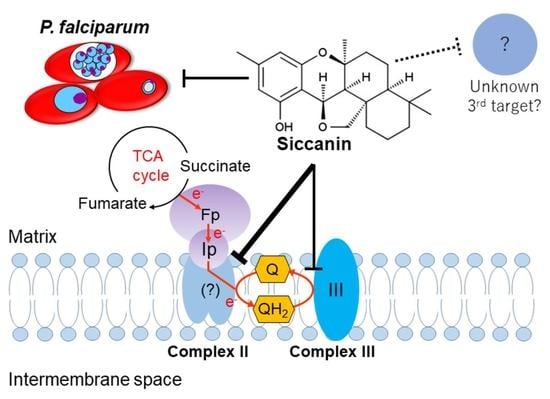
Siccanin Is a Dual-Target Inhibitor of Plasmodium falciparum Mitochondrial Complex II and Complex III

1
Department of Biomedical Chemistry, Graduate School of Medicine, The University of Tokyo, Tokyo 113-0033, Japan
2
Laboratory of Biomembrane, Tokyo Metropolitan Institute of Medical Science, Tokyo 156-8506, Japan
3
Department of Molecular Infection Dynamics, Institute of Tropical Medicine (NEKKEN), Nagasaki University, Nagasaki 852-8523, Japan
4
School of Tropical Medicine and Global Health, Nagasaki University, Sakamoto, Nagasaki 852-8523, Japan
5
Graduate School of Infection Control Sciences, Kitasato University, Tokyo 108-8641, Japan
6
Ōmura Satoshi Memorial Institute, Kitasato University, Minato-ku, Tokyo 108-8641, Japan
7
Department of Infection and Host Defense, Graduate School of Medicine, Chiba University, Chiba 260-8670, Japan
8
Department of Host-Defense Biochemistry, Institute of Tropical Medicine (NEKKEN), Nagasaki University, Nagasaki 852-8523, Japan
*
Authors to whom correspondence should be addressed.
These authors contributed equally to this work.
Pharmaceuticals 2022, 15(7), 903; https://doi.org/10.3390/ph15070903
Submission received: 7 June 2022 / Revised: 19 July 2022 / Accepted: 20 July 2022 / Published: 21 July 2022
(This article belongs to the Special Issue Drug Candidates for the Treatment of Infectious Diseases)

Abstract

:
Plasmodium falciparum contains several mitochondrial electron transport chain (ETC) dehydrogenases shuttling electrons from the respective substrates to the ubiquinone pool, from which electrons are consecutively transferred to complex III, complex IV, and finally to the molecular oxygen. The antimalarial drug atovaquone inhibits complex III and validates this parasite’s ETC as an attractive target for chemotherapy. Among the ETC dehydrogenases from P. falciparum, dihydroorotate dehydrogenase, an essential enzyme used in de novo pyrimidine biosynthesis, and complex III are the two enzymes that have been characterized and validated as drug targets in the blood-stage parasite, while complex II has been shown to be essential for parasite survival in the mosquito stage; therefore, these enzymes and complex II are considered candidate drug targets for blocking parasite transmission. In this study, we identified siccanin as the first (to our knowledge) nanomolar inhibitor of the P. falciparum complex II. Moreover, we demonstrated that siccanin also inhibits complex III in the low-micromolar range. Siccanin did not inhibit the corresponding complexes from mammalian mitochondria even at high concentrations. Siccanin inhibited the growth of P. falciparum with IC50 of 8.4 μM. However, the growth inhibition of the P. falciparum blood stage did not correlate with ETC inhibition, as demonstrated by lack of resistance to siccanin in the yDHODH-3D7 (EC50 = 10.26 μM) and Dd2-ELQ300 strains (EC50 = 18.70 μM), suggesting a third mechanism of action that is unrelated to mitochondrial ETC inhibition. Hence, siccanin has at least a dual mechanism of action, being the first potent and selective inhibitor of P. falciparum complexes II and III over mammalian enzymes and so is a potential candidate for the development of a new class of antimalarial drugs.

Graphical Abstract

1. Introduction

Human falciparum malaria, caused by Plasmodium falciparum, accounts for an estimated 241 million cases and 627,000 deaths annually. Most of these cases are children under the age of five in developing countries [1]. Unfortunately, the number of cases and deaths is presumed to have increased during the COVID-19 pandemic as a result of disruptions in the provision of malaria prevention, diagnosis, and treatment. Currently, the World Health Organization (WHO) recommends that malaria be treated with a combination therapy consisting of artemisinin and other antimalarial drugs (Artemisinin Combination Therapy; ACT). Moreover, WHO’s goal of malaria control appears to be far from being achieved because no effective vaccine is yet available, and P. falciparum, with artemisinin resistance, is emerging in southeastern Asia [2] and has recently independently emerged in Africa [3,4,5]. Hence, new drugs with different or multiple mechanisms of action are needed to control malaria. In that sense, drug targets with distinct biochemical features or even those that are absent from the host are desired. Currently, all approved antimalarial drugs were developed targeting the P. falciparum blood stage with some, such as atovaquone, proguanil (prodrug of cycloguanil), pyrimethamine and doxycycline, found to be very effective prophylactic agents killing liver schizonts. However, for the elimination of malaria, novel interventions targeting several stages (e. g., the mosquito and liver stages) as well as the blood stage of P. falciparum are required [6].
The electron transport chain (ETC) of the parasite’s mitochondrion is highly diverse. In P. falciparum, the ETC is composed of five dehydrogenases, including type-II NADH dehydrogenase (NDH-2) [7], succinate dehydrogenase (SDH or complex II) [8,9], malate:quinone oxidoreductase (MQO) [10], dihydroorotate dehydrogenase (DHODH) [11], and glycerol-3-phosphate dehydrogenase (G3PDH) [12]; collectively, these enzymes shuttle electrons to the ubiquinone pool (Q-pool) (Figure 1). Among plasmodial ETC dehydrogenases, NDH2 and MQO are not present in the mammalian cell [13], and SDH [14] and DHODH display biochemical features distinct from the mammalian homologues and do not share cross-sensitivity to inhibitors with the mammalian enzymes (Figure 1) [15,16,17,18,19,20]. Little is known about the role of the G3PDH in the plasmodial ETC; however, it has been suggested that this enzyme plays a role in lipid biosynthesis and redox homeostasis in the parasite [12]. The antimalarial drug atovaquone inhibits mitochondrial complex III (cytochrome bc1 complex or quinol: cytochrome c reductase), which transfers electrons from the Q-pool to cytochrome c [21]. We previously demonstrated that complex II is not essential for the survival of P. falciparum [9] and P. berghei [8] blood stages. However, it is important to note that a knockout in the gene encoding the Fp subunit of complex II (sdha) causes growth retardation in the blood stage; this defect is rescued by the addition of succinate but not by fumarate [9], suggesting that complex II may function as a quinol-fumarate reductase (QFR) rather than as a succinate-quinone reductase (SQR) [22]. Similarly, sdha knockout P. berghei shows a delayed onset in the initial parasitemia, which subsequently returns to wild-type levels within three days [8]. In the mosquito stage, sdha knockout P. berghei is not able to develop into oocysts within the vector [8], indicating that both a functional ETC and the tricarboxylic acid (TCA) cycle are essential for parasite development and survival in the insect stage. Thus, complex II is considered a novel target for development of transmission-blocking agents with activity against plasmodial infection. Consistent with this hypothesis, malaria parasites resistant to atovaquone have reduced complex III activity and failed to develop into oocysts, and, thus, are unable to spread the disease [21]. In addition, atovaquone potently inhibits the development of sporozoites into merozoites in the liver stage [23]. Thus, the ETC of P. falciparum includes enzymes that are essential at multiple stages and therefore may be considered a promising drug target for the development of new antimalarials.
Mitochondrial complex II participates in the TCA cycle, transferring electrons from succinate to ubiquinone, thereby producing fumarate and ubiquinol (Figure 1) [24,25]. Interestingly, the subunit composition of complex II is highly diverse. For instance, in mammals and nematodes, complex II consists of four subunits [25] (flavoprotein (Fp); iron-sulfur cluster protein (Ip); and two membrane anchor subunits, the cytochrome b large (CybL) and small (CybS) subunits), whereas in apicomplexan parasites Toxoplasma gondii and Eimeria tenella, complex II consists of 9 and 15 subunits, respectively [26,27,28]. A previous report suggested the presence of a four-subunit-type complex II in P. yoelii yoelii [14]. However, in P. falciparum, only the Fp and Ip subunits have been identified to date, but not the CybS and CybL subunits, indicating low sequence similarity among the CybL and CybS subunits, even among Plasmodium species. Previously, we identified atpenin A5 as the first potent and specific inhibitor of complex II from mammals and nematodes [29]. Surprisingly, atpenin A5 and other classical complex II inhibitors, such as 2-thenoyltrifluoroacetone thenoyltrifluoroacetone (TTFA) and carboxin, are not effective against complex II from Plasmodium [14,30] (Table 1) and E. tenella [27], indicating that the structure of the ubiquinone-binding site of complex II is significantly divergent between apicomplexan parasites and mammals.
The development of drugs targeting complex II has been reported for compounds with activity against many pathogens, including Ascaris suum [29], Trichophyton mentagrophytes [31], Mycobacterium tuberculosis [32], and Helicobacter pylori [33]. In the present study, we identified siccanin (Figure 2a) as the first (to our knowledge) nanomolar and selective inhibitor of the P. falciparum complex II. We also showed that siccanin inhibits complex III at micromolar concentrations. Moreover, we demonstrated that siccanin inhibits the growth of blood-stage P. falciparum, an effect that appears to be distinct from the ETC inhibitory activity. Therefore, siccanin is a promising lead compound for the development of new antimalarial drugs for the treatment of Plasmodium, including the potential for blocking parasite transmission.
Table 1. Selectivity of classical complex II inhibitor against Plasmodium spp. and mammal complex II.
InhibitorComplex II IC50 [μM]Selectivity a
Plasmodium spp.Mammal
Malonate13.2 ± 0.49 b3.4 d0.26
TTFA>50 b5.4 e<0.11
Atpenin A54.6 ± 0.2 c0.004 e 0.00087
Carboxin3.6 ± 1.0 c1.0 e0.28
a Selectivity was calculated as the ratio between mammalian cell line and P. falciparum 50% inhibitory concentrations (IC50s). Adapted from Suraveratum et al. [30] b, Kawahara et al. [14] c, Mogi et al. [34] d, and Miyadera et al. [29] e.

2. Results

2.1. Siccanin Strongly Inhibits P. falciparum SQR Activity

Siccanin (Figure 2a), an antibiotic produced by the plant pathogenic fungus Helminthosporium siccans Dreschsler [35,36], was previously reported to be a potent and selective inhibitor of fungal, trypanosomal, and nematode complex II [37,38,39,40] and has been used clinically for the treatment of tinea pedis (Tackle®, Sankyo-Pharma). Our previous study showed that siccanin is a species-selective complex II inhibitor, effective against complex II from trypanosomatid parasites [38], Pseudomonas aeruginosa, P. putida, rat, and mouse but ineffective against the enzymes from Escherichia coli, Corynebacterium glutamicum, and porcine [34]. Therefore, we examined whether siccanin inhibits P. falciparum SQR activity in a crude mitochondrial fraction from P. falciparum 3D7. We found that siccanin strongly inhibited the SQR activity of this fraction (Figure 2b). In contrast, classical ubiquinone-site inhibitors, such as TTFA [30], atpenin and carboxin, were ineffective against the Plasmodium mitochondrial complex II [14] (Table 1). The inhibition by siccanin exhibited a classical biphasic inhibition pattern with 50% inhibitory concentration (IC50) values (mean ± SD) of the first and second phases of 0.016 ± 0.006 µM and 8.93 ± 2.44 µM, respectively (Figure 2b and Table 2). As a next step, the effect of siccanin at a concentration of 10 µM against other mitochondrial dehydrogenases (Figure 3a,b) was evaluated, revealing that siccanin did not inhibit MQO, DHODH, and NDH2 activities but weakly inhibited G3PDH (Figure 3b). These results clearly showed that siccanin is a selective inhibitor of the P. falciparum complex II amongst the mitochondrial ETC dehydrogenases that shuttle electrons to the Q-pool (Figure 3b). Our results also indicated that the P. falciparum complex II has higher sensitivity to siccanin than does the enzyme from T. mentagrophytes (IC50~90 nM) [31].
Table 2. Selectivity of siccanin against complexes II and III, and also the growth of P. falciparum 3D7 and mammalian cells.
AssaySiccanin IC50 [μM]Selectivity a
P. falciparumMammal
Complex II0.016 and 8.93861 b57,400 and 96
Complex III8.39>500>60
Growth8.434.2 c
16.1 d
4.1
1.9
a Selectivity was calculated as the ratio between mammalian cell line and P. falciparum 50% inhibitory concentrations (IC50s). b Adapted from Mogi et al. [34]; c DLD-1 cells; d HDF cells.

2.2. Inhibition of P. falciparum Growth by Siccanin

In other organisms living in anaerobic or microaerophilic environments (e.g., E. coli [41], Mycobacterium tuberculosis [32,42,43,44], Ascaris suum (adult stage) [29], Echinococcus multilocularis (protoscoleces) [45], and even in several solid tumor cells [46]), the QFR activity of complex II is well documented and has been suggested to play an important role in environmental adaptation. Previously, we demonstrated that the disruption of the Fp subunit-encoding gene of P. falciparum (sdha) impairs the growth of blood-stage parasites; the growth of the Δsdha mutant was rescued by succinate but not by fumarate, suggesting that this plasmodial complex II might function as a QFR rather than as a SQR [9]. Since siccanin potently inhibited complex II from P. falciparum, we next tested whether siccanin exposure phenocopied the Δsdha mutant. As described above, complex II is not essential for the survival of blood-stage P. falciparum; however, we could not detect live parasites under the microscope following exposure to siccanin at a concentration of 50 µM (Figure 4a). Further experiments demonstrated that siccanin modestly inhibits the growth of P. falciparum with an IC50 of 8.40 ± 0.60 µM (Figure 4b, Table 2). This result suggested that siccanin may have a secondary target other than complex II in P. falciparum; presumably this second target is essential for survival in the blood stage.

2.3. Effect of Succinate or Fumarate, and Pfsdha Disruption, on Growth Inhibition by Siccanin

Next, we tested whether addition of succinate or fumarate [9] rescues the growth-inhibitory effect of siccanin (Figure 4a). Compared to the control’s IC50 of 8.4 μM, the IC50 of siccanin in the presence of succinate or fumarate was 10.8 or 11.5 μM, respectively (Figure 4b), suggesting that the 3D7-growth inhibition caused by siccanin was independent of the compound’s effect on complex II activity. Consistent with this hypothesis, the effect of siccanin on Δsdha-3D7 P. falciparum growth was similar to that on the parent 3D7, with IC50s of 6.13 and 6.10 μM, respectively (Figure 4c). Together, these results strongly indicated the existence of a secondary essential target of siccanin in the blood-stage parasite.

2.4. Siccanin Inhibits P. falciparum Complex III

DHODH, MQO, and complex III (Figure 1) are ETC enzymes that have been suggested to be essential for the survival of the blood-stage P. falciparum [11,47]. Since siccanin did not inhibit DHODH and MQO activities (Figure 3b), we tested whether complex III activity is inhibited by siccanin. The inhibition of complex III was evaluated using DHO-cytochrome c reductase. This assay measures electron transfer from DHODH to cytochrome c via complex III by recording the absorbance change of cytochrome c at 550 nm [14]. The results showed that, at 60 μM concentration, siccanin does not inhibit DHODH activity (Figure 5a) but completely inhibits DHO-cyt c activity with an IC50 value of 8.39 ± 2.92 μM (Figure 5b, Table 2), indicating that siccanin inhibits complex III and not DHODH. It has been reported that siccanin does not inhibit mammalian complex II [34] (Table 2). We therefore evaluated whether siccanin inhibits mammalian complex I and III activities (NADH-cytochrome c reductase). Notably, even at concentrations as high as 500 µM, siccanin did not inhibit the mammalian complex I and III (not shown), demonstrating that siccanin is a selective inhibitor of complexes II and III from P. falciparum, with maximum selectivities of 57,400-fold and of more than 60-fold, respectively (Table 1). Although siccanin did not inhibit mammalian complexes I, II, or III, the growth of DLD-1 cells and HDF cells were inhibited by siccanin, which exhibited 50% effective concentrations (EC50s) of 34.2 ± 2.73 µM and 16.1 ± 1.21 µM, respectively (selectivities of 1.9 and 4.1-fold, Table 2). These results indicated that, in human cells, siccanin might have target(s) other than ETC enzymes.

2.5. Interaction of Atovaquone with Siccanin against P. falciparum In Vitro

Compounds inhibiting complex III can bind either at Qo (quinol binding site facing the outer membrane) or Qi (quinone binding site facing the matrix side) sites [48,49,50]. Because the growth inhibition of blood-stage parasites by siccanin was likely due to the inhibition of complex III, we tested by modified isobologram [51] based on the parasite’s LDH assay as previously described [52], whether the binding site of siccanin overlaps with that of atovaquone, which is a well-known Qo site inhibitor. A pair of fractional IC50s for each combination of siccanin and atovaquone was plotted for the isobologram analysis. The fractional IC50 of siccanin was calculated by dividing the IC50 of siccanin combined with each atovaquone by the IC50 obtained for siccanin alone and plotted on the X-axis. Similarly, the corresponding atovaquone fractional IC50s were calculated and plotted on the Y-axis. In general, if two compounds bind at the same site, the isobologram will show an antagonistic pattern with a combination index exceeding 1. On the other hand, if the binding sites are distinct, the isobologram will show a synergistic or additive pattern, with a combination index of less than or equal to 1, respectively [53,54,55,56]. In the case of siccanin, the determined combination index was 1.0, indicating that siccanin has an additive effect when parasites are treated with the combination of siccanin and atovaquone (Figure 5c), thus, suggesting Qi site as the binding site of siccanin.

2.6. Effect of Siccanin on P. falciparum yDHODH-3D7 and Dd2 Drug-Resistant-Panel Strains

Previously, it was demonstrated that P. falciparum expressing the yeast DHODH (yDHODH) can oxidize DHO in a ubiquinone-independent manner, resulting in resistance to DHODH and complex III Qo/Qi site inhibitors [11]. Because siccanin inhibited complex III of this parasite (Table 2), we tested whether the P. falciparum 3D7-yDHODH strain was resistant to siccanin. Surprisingly, siccanin was equally active against both the parent (3D7) and 3D7-yDHODH strains, showing EC50s of 11.65 μM and 10.26 μM, respectively (Figure 6a). In contrast, atovaquone was a potent growth inhibitor of 3D7 (EC50 = 0.560 nM) but not of 3D7-yDHODH (EC50 > 100 nM) (Figure 6b).
Next, siccanin was tested against multidrug-resistant Dd2 and drug-resistant panel strains, Dd2_048 (PI4K-S743T mutation, MMV390048R) [57], Dd2_DDD (eEF2-Y186N mutation, DDD107498R) [58], Dd2_DHIQ (ATP4-G358S mutation; cipargaminR) [59], Dd2_GNF (Carl-I1139K mutation; GNF156R) [60], and Dd2_ELQ300 (cyt b-I22L mutation at Qi site; ELQ300R) [61] to investigate the cross-resistance and potential target protein(s). No siccanin resistance, however, was detected in any of these strains, for which siccanin exhibited EC50s for growth inhibition ranging from 12.4 μM to 18.7 μM (Figure 6c), indicating that siccanin targets other protein(s) except for the mutated gene products of these strains.

3. Discussion

In this study, we demonstrated that siccanin is a nanomolar-order inhibitor of complex II from an apicomplexan parasite. We also demonstrated that, while siccanin does not affect other ETC dehydrogenases, the compound shows (in addition to its activity against complex II) micromolar-order inhibition of complex III. Thus, siccanin represents a novel scaffold compared to all known complex-III inhibitors.
In mammals, mitochondrial ETC is essential for the maintenance of several processes, including energy production [62] and de novo biosynthesis of pyrimidine [63]. In contrast, the blood stage of P. falciparum does not depend on ETC for energy production, but instead on cytoplasmic glycolysis [64,65]. In blood-stage P. falciparum, ETC is essential for the de novo biosynthesis of pyrimidine, a process that is linked at the level of DHODH, Q-pool, and complex III [11] (Figure 1). Since the P. falciparum genome apparently does not encode a pyrimidine salvage pathway, the pathway for the de novo biosynthesis of pyrimidines and/or complex III are attractive targets for development of new antimalarial drugs [11]. Other P. falciparum ETC dehydrogenases, such as G3PDH and MQO, seem to be functional in the blood stage [66]. Notably, the MQO activity was higher than DHODH activity in our assay (Figure 3a). Recent studies showing that the MQO-encoding locus cannot be genetically ablated in P. falciparum suggests that MQO is essential for survival in the blood stage [47]. The essentiality of MQO was attributed to the key role played by MQO in linking the ETC to the fumarate cycle, given that the latter is essential for the purine salvage pathway [10]. However, other reports have suggested that PfMQO is not an appealing target [67]. Clearly, further studies will be required to determine the druggability of PfMQO.
The knockout of the sdha gene in P. falciparum has been shown to exhibit a growth defect that is rescued by the supplementation of the culture medium with succinate, though not with fumarate [9]. In P. berghei, mutant parasites devoid of complex II as well as NDH2 activities have been reported to be impaired for development in the insect stage [8,68]. Complex IIs from apicomplexan parasites are known to be insensitive to classical inhibitors, and, to date, no potent inhibitors of those enzymes have been reported [27,30]. Interestingly, as shown in the present study, siccanin is a potent inhibitor of the P. falciparum complex II, exhibiting a biphasic inhibition pattern against this enzyme (Figure 2b). A similar biphasic inhibition pattern has been reported for S. cerevisiae complex II and its dinitrophenol derivative inhibitor, suggesting the existence of two quinone-binding sites [69]. The existence of two binding sites for quinone species also has been described for the quinol-fumarate reductases from E. coli and Wolinella succinogenes based on several approaches [70,71,72,73], including crystallographic studies [74]. This inference for the P. falciparum complex II is supported by previous reports demonstrating that malonate-sensitive NADH-fumarate reductase activity is detectable in the mitochondria-rich fraction of P. falciparum and also E. tenella (malonate is a general complex II inhibitor binding at the Fp subunit) [22,27,75]. Although similar studies of the P. falciparum complex II will be needed to definitively confirm this hypothesis, it is tempting to speculate that two quinone-binding sites also may exist in the plasmodial complex II; presumably, one of the quinone-binding sites has a higher affinity for siccanin than the other, resulting in the biphasic inhibition observed in Figure 2b. The first phase would be inhibited by siccanin at nanomolar-order concentration (0.016 μM), while the second phase would be inhibited by the same compound at micromolar-order concentration (8.93 μM). Interestingly, in addition to its activity against complex II, siccanin also inhibits complex III with an IC50 of 8.39 μM. Moreover, siccanin inhibits the P. falciparum complex II with over 520-fold (first phase) and 0.93-fold (second phase) greater selectively than the inhibition of complex III but with no apparent effect against mammalian respiratory chain complexes (Table 2). These results suggest that the growth inhibition of blood-stage P. falciparum by siccanin may be the consequence of ETC inhibition, exerted primarily at the level of complex III (rather than by the direct inhibition of the TCA cycle via complex II). However, similar sensitivity between the 3D7 and 3D7-yDHODH, as well as Dd2 and Dd2-ELQ300 towards siccanin seems to exclude complex III as its primary target in P. falciparum.

4. Materials and Methods

4.1. Chemicals

Siccanin (1S,4R,12S,15S,20R)-8,16,16-trimethyl-3,11-dioxapentacyclo [10.7.1.01,15.04,20.05,10] icosa-5,7,9-trien-6-ol) was obtained from Daiichi-Sankyo Pharma (Tokyo, Japan) (Figure 2a). Atovaquone was purchased from US Pharmacopeia and ICN Biomedical, Inc. (Costa Mesa, CA, USA). Unless otherwise stated, all other chemicals were purchased from Sigma-Aldrich (St. Louis, MO, USA).

4.2. Malaria Parasite Strains and Cultivation

The P. falciparum 3D7 strain was cultured, as described previously, in 3% hematocrit type A human red blood cells (RBCs) in RPMI1640 medium (Invitrogen; San Diego, CA, USA) supplemented with 25 mM sodium hydrogen biocarbonate, 10 μg/mL hypoxanthine, 40 μg/mL gentamicin sulfate, and 0.5% (w/v) Albumax II (Invitrogen) (“complete medium”) [9,52]. Cultures were maintained in a MG-70M multi-gas incubator (Taitec) under the conditions of 5% O2, 5% CO2, and 90% N2 at 37 °C. The medium was replaced daily [76]. Parasitemia was measured by thin blood smears stained with Giemsa. In our previous study, we generated mutant P. falciparum strains expressing either disrupted or full-length versions of the flavoprotein (Fp) subunit (encoded by the Pfsdha gene) [9]. Both strains were cultured in complete medium at 3% hematocrit supplemented with 5 nM WR99210 (Jacobus Pharmaceuticals; Plainsboro, NJ, USA) [9]. The experiments using human RBCs were performed under the guidelines of Research Ethics Committee of the Faculty of Medicine of the University of Tokyo and Institutional Review Board (IRB) of Nagasaki University (Permission nos. 10050 and no. 19, respectively). Human RBCs were obtained from The Japanese Red Cross Society. The synchronization of parasite cultures was performed by treating with 5% (w/v) sorbitol for 10 min [77].
P. falciparum 3D7-yDHODH was prepared and maintained as described previously [11,78,79]. P. falciparum Dd2, and Dd2-derived mutant strains were kindly provided by David A. Fidock [80], and maintained at Nagasaki University essentially as described in reference, except without supplementation with WR99210.

4.3. Assessment of Parasite Survival

Synchronized ring-form parasites (2% parasitemia) were prepared in complete medium containing 50 µM siccanin (at a fixed final DMSO concentration of 0.1% (v/v)) and distributed to 24-well plates at 1 mL/well. After incubation for 16, 24, 32, and 48 h, parasitemia was assessed under the microscope using thin blood smears stained with Giemsa.

4.4. Drug sensitivity Assay

Synchronized ring-form parasites (0.3% parasitemia) were prepared in complete medium containing different concentration of various compounds (at a fixed final DMSO concentration of 0.1% (v/v)) and distributed to 96-well plates at 100 μL/well. After 72 h of incubation, parasite growth was monitored by the previously described lactate dehydrogenase (PfLDH)-based assay method [81]; plates were read using a SpectraMax® Paradigm spectrophotometer (Molecular Devices, Inc.; San Jose, CA, USA). EC50 values were calculated using Prism® ver.6.01 (GraphPad; San Diego, CA, USA). Values are presented as the means of three independent experiments.

4.5. Mammalian Cell Cytotoxicity Assay

Human colorectal adenocarcinoma cells (DLD-1) and Human Dermal Fibroblasts (HDF) cells were obtained from Taiho Pharmaceutical Company (Tokyo, Japan). DLD-1 cells were cultured in RPMI 1640 medium supplemented with 10% (v/v) heat-inactivated fetal bovine serum (FBS) (Gibco; San Diego, CA, USA); HDF cells were cultured in DMEM/F12 medium supplemented with 10% (v/v) heat-inactivated FBS. For both lines, cells were grown under 5% CO2 at 37 °C in an ACI-165D CO2 incubator (ASTEC; Fukuoka, Japan).
DLD-1 and HDF cells, (2.5 × 10⁴ cells/mL) in 96-well plates, were cultured for 24 h. The cells then were washed with phosphate-buffered saline (PBS) and the medium was replaced with fresh medium supplemented with 10% (v/v) FBS. Medium containing siccanin at various concentrations was added to the wells and the cells were cultured for another 48 h. Controls were treated with medium containing 1% (v/v) DMSO and 10% (v/v) FBS. Next, the cells were washed with PBS and the medium was replaced with fresh medium supplemented with 10% (v/v) FBS. An aliquot (10 µL) of CCK8 solution (Cell Counting Kit-8; Dojindo Laboratories; Kumamoto, Japan) was added to each well and the plates were incubated for another 2 h, at which point absorbance at 450 nm was monitored using a SpectraMax® Paradigm spectrophotometer. The EC50 values were calculated using Prism® ver.6.01. The selectivity was calculated as the ratio between DLD-1 or HDF versus P. falciparum IC50s.

4.6. Preparation of Crude P. falciparum Mitochondrial Fraction

A synchronized parasite culture was grown for 64 h and a crude mitochondrial fraction was prepared from trophozoite-stage cells (as confirmed by Giemsa staining). Infected red blood cells were collected by centrifugation at 800× g for 5 min at 4 °C and incubated for 5 min at room temperature with 40 mL of 0.075% (w/v) saponin in AIM buffer (120 mM KCl, 20 mM NaCl, 10 mM PIPES (1,4-piperazinediethanesulfonic acid) buffer [pH 6.7], 1 mM MgCl2, 5 mM glucose). Released parasites were collected by centrifugation at 2780× g for 7 min at 4 °C and washed three times with AIM buffer. P. falciparum was suspended in MSE buffer [225 mM mannitol, 75 mM sucrose, 0.1 mM ethylenediaminetetraacetic acid (EDTA), 3 mM Tris-HCl buffer, pH 7.4] containing 1 mM phenylmethylsulphonyl fluoride, and disrupted by nitrogen cavitation at 1200 psi using a 4639 Cell Disruption Bomb (Parr Instrument Company; Moline, IL, USA). The unbroken cells and cell debris were removed by centrifugation at 800× g for 5 min at 4 °C. The supernatant was centrifuged at 5000× g for 20 min at 4 °C. The resulting pellet was suspended in 200–400 μL MSE buffer and used as crude mitochondrial fraction for all enzymatic assays, as described previously [22].

4.7. Measurement of P. falciparum ETC Enzyme Activities

Complex II, DHODH, MQO, NDH2, G3PDH, and dihydroorotate-cytochrome c (DHO-cyt c) reductase activities were measured, as described previously, using a V-660 spectrophotometer (JASCO; Tokyo, Japan) [14,27]. Briefly, complex II activity was measured at 25 °C in a 1 mL reaction mixture containing 45 μM 2,6-dichlorophenolidophenol (DCIP), 100 μM ubiquinone-2 (UQ2), and 2 mM KCN in 50 mM potassium phosphate buffer, pH 8.0. DHODH, MQO, and G3PDH activities were measured at 25 °C in 1 mL of the reaction mixture containing 45 μM DCIP, 100 μM UQ2, and 2 mM KCN in 30 mM Tris-HCl buffer, pH 8.0. Complex II, DHODH, MQO, and G3PDH activities were measured by monitoring the change in absorbance at 600 nm (reflecting DCIP reduction) after the reaction was initiated by adding 10 mM succinate, 500 μM dihydroorotate, 10 mM malate, or 500 μM glycerol-3-phosphate, respectively. NDH2 activity was measured at 25 °C in 1 mL of the reaction mixture containing 100 µM UQ1 and 2 mM KCN in 50 mM potassium phosphate buffer, pH 8.0. NDH2 activity was determined by monitoring the change in absorbance at 340 nm (reflecting NADH reduction) after the reaction was initiated by the addition of 20 μM NADH.
DHO-cyt c activity was measured at 25 °C in 1 mL of the reaction mixture containing 20 μM cytochrome c and 2 mM KCN in 30 mM Tris-HCl buffer, pH 8.0. The reduction of cytochrome c was monitored at 550 nm after initiation of the reaction by addition of 500 μM dihydroorotate [14,27]. Various concentrations of siccanin (formulated in DMSO) were added to the reaction mixtures before the initiation of the reactions. In all assays, the final concentration of DMSO was fixed at 0.1% (v/v). IC50 values were calculated using Prism® ver.6.01.

4.8. Measurement of NADH-Cytochrome c Reductase Activity in Porcine Mitochondria

Porcine mitochondria were prepared as described previously [82]. NADH-cytochrome c (NADH-cyt c) activity was measured at 25 °C in 1 mL of the reaction mixture containing 1 mM MgCl2, 2 mM KCN, and 20 μM cytochrome c in 30 mM potassium phosphate buffer, pH 7.5. Activity was measured by recording the absorbance change of cytochrome c at 550 nm after the reaction was initiated by the addition of 20 μM NADH. Various concentrations of siccanin dissolved in DMSO were added to reaction mixtures 5 min before the initiation of the reactions. Selectivity was calculated as the ratio of mammalian to P. falciparum IC50s.

4.9. Isobologram Analysis with Atovaquone and Siccanin

The effect of the combination of atovaquone and siccanin was evaluated using isobologram analysis to evaluate the synergy, additivity, or antagonism between the two compounds [51,83]. Synchronized ring-form parasites (0.3% parasitemia) were prepared in complete medium containing different concentrations of siccanin and atovaquone (at a fixed final DMSO concentration of 0.2% (v/v)) and distributed to 96-well plates at 100 μL/well. After 72 h of incubation, parasite growth was monitored by the previously described PfLDH-based assay using a SpectraMax® Paradigm spectrophotometer. IC50 values were calculated using Prism® ver.6.01. Data represent the means of three independent experiments, each performed in triplicate. The isobologram analysis was performed as described previously [51,83]. For each combination assay, IC50s were calculated from each culture of P. falciparum grown in the presence of atovaquone or siccanin alone and in combination of various concentrations of the two compounds. The fractional inhibitory concentration (FIC) value was determined using the following equation:
FIC   = IC 50   of   compound   in   mixture IC 50   of   compound   alone
The combination index (CI) value was determined using the following equation:
CI   = IC 50   of   atovaquone   in   mixture IC 50   of   atovaquone   alone + IC 50   of   siccanin   in   mixture IC 50   of   siccanin   alone
Values of CI < 1 represent synergism, CI = 1 represent additivity, and CI > 1 represent antagonism.

5. Conclusions

In conclusion, we identified siccanin as a promising antimalarial drug candidate effective not only against blood-stage parasites but also with the potential to be active against the insect stage. Such a dual-stage-targeting drug might show utility for blocking transmission as well as for the treatment of malaria patients. Notably, siccanin has no effect on mammalian respiratory chain enzymes (complex I, II, and III). However, given that siccanin inhibits the growth of DLD-1 (cancer cell line) and HDF (a normal cell line), it will be critical to determine the mammalian target of siccanin in order to increase the compound’s selectivity against the parasite for potential future development as anti-parasitic drug.

Author Contributions

K.K. (Kiyoshi Kita) directed the work. K.K. (Kiyoshi Kita)., K.S., T.N., K.H. and S.Ō. designed the research. K.K. (Keisuke Komatsuya) and T.S. performed the research. K.K. (Keisuke Komatsuya), T.S. and D.K.I. analyzed the data. K.K. (Keisuke Komatsuya), T.S. and D.K.I. wrote the paper. All authors directly participated in this work, including preparation of the manuscript and approval of the final version. All authors have read and agreed to the published version of the manuscript.

Funding

This work was supported, in part, by funding as follows: grants for Infectious Disease Control from the Science and Technology Research Partnership for Sustainable Development (SATREPS; No. JP10000284 to K.K. (Kiyoshi Kita) and No. JP14425718 to D.K.I. and T.N.) from the Agency for Medical Research and Development (AMED); a Grant-in-aid for Scientific Research on Priority Areas (No. JP18073004 to K.K. (Kiyoshi Kita)); a Creative Scientific Research Grant (No. JP18GS0314 to K.K. (Kiyoshi Kita)) from the Japan Society for the Promotion of Science (JSPS); Grants-in-aid for Scientific Research (B) (Nos. JP16K19114 and JP19H03436 to K.K. (Kiyoshi Kita) and D.K.I.) and (C) (No. JP20239116 to D.K.I. and K.H.); a Grant-in-Aid for Young Scientists (No. JP22512047 to T.S.); a grant from The Leading Initiative for Excellent Young Researchers (LEADER; No. JP16811362 to D.K.I.) from the Japanese Ministry of Education, Science, Culture, Sports and Technology (MEXT); and by Grants-in-aid for Research on Emerging and Re-emerging Infectious Diseases from the Japanese Ministry of Health and Welfare (No. JP17929833 to K.K. (Kiyoshi Kita) and T.N.; No. JP20314363 to D.K.I. and T.N.). Ke.K. is a Research Fellow of the JSPS and is supported by Research Fellowships for Young Scientists of the JSPS (No. JP15J04920 to K.K. (Keisuke Komatsuya)).

Institutional Review Board Statement

Not applicable.

Informed Consent Statement

Not applicable.

Data Availability Statement

Data is contained within the article.

Acknowledgments

The authors thank the Japanese Red Cross Society for providing human red blood cells (RBCs; Registry No. R030038) and David A Fidock (Columbia University, USA) for providing Dd2 drug-resistant-panel strains.

Conflicts of Interest

The authors declare no conflict of interest.

References

  1. World Health Organization. World Malaria Report: 2020; World Health Organization: Geneva, Switzerland, 2020. [Google Scholar]
  2. Dondorp, A.M.; Nosten, F.; Yi, P.; Das, D.; Phyo, A.P.; Tarning, J.; Lwin, K.M.; Ariey, F.; Hanpithakpong, W.; Lee, S.J.; et al. Artemisinin resistance in Plasmodium falciparum malaria. N. Engl. J. Med. 2009, 361, 455–467. [Google Scholar] [CrossRef] [Green Version]
  3. Balikagala, B.; Fukuda, N.; Ikeda, M.; Katuro, O.T.; Tachibana, S.I.; Yamauchi, M.; Opio, W.; Emoto, S.; Anywar, D.A.; Kimura, E.; et al. Evidence of Artemisinin-Resistant Malaria in Africa. N. Engl. J. Med. 2021, 385, 1163–1171. [Google Scholar] [CrossRef] [PubMed]
  4. Ikeda, M.; Kaneko, M.; Tachibana, S.I.; Balikagala, B.; Sakurai-Yatsushiro, M.; Yatsushiro, S.; Takahashi, N.; Yamauchi, M.; Sekihara, M.; Hashimoto, M.; et al. Artemisinin-Resistant Plasmodium falciparum with High Survival Rates, Uganda, 2014–2016. Emerg. Infect. Dis. 2018, 24, 718–726. [Google Scholar] [CrossRef] [PubMed] [Green Version]
  5. Uwimana, A.; Umulisa, N.; Venkatesan, M.; Svigel, S.S.; Zhou, Z.; Munyaneza, T.; Habimana, R.M.; Rucogoza, A.; Moriarty, L.F.; Sandford, R.; et al. Association of Plasmodium falciparum kelch13 R561H genotypes with delayed parasite clearance in Rwanda: An open-label, single-arm, multicentre, therapeutic efficacy study. Lancet Infect. Dis. 2021, 21, 1120–1128. [Google Scholar] [CrossRef]
  6. Blagborough, A.M.; Churcher, T.S.; Upton, L.M.; Ghani, A.C.; Gething, P.W.; Sinden, R.E. Transmission-blocking interventions eliminate malaria from laboratory populations. Nat. Commun. 2013, 4, 1812. [Google Scholar] [CrossRef] [Green Version]
  7. Biagini, G.A.; Viriyavejakul, P.; O’Neill, P.M.; Bray, P.G.; Ward, S.A. Functional characterization and target validation of alternative complex I of Plasmodium falciparum mitochondria. Antimicrob. Agents Chemother. 2006, 50, 1841–1851. [Google Scholar] [CrossRef] [Green Version]
  8. Hino, A.; Hirai, M.; Tanaka, T.Q.; Watanabe, Y.; Matsuoka, H.; Kita, K. Critical roles of the mitochondrial complex II in oocyst formation of rodent malaria parasite Plasmodium berghei. J. Biochem. 2012, 152, 259–268. [Google Scholar] [CrossRef]
  9. Tanaka, T.Q.; Hirai, M.; Watanabe, Y.; Kita, K. Toward understanding the role of mitochondrial complex II in the intraerythrocytic stages of Plasmodium falciparum: Gene targeting of the Fp subunit. Parasitol. Int. 2012, 61, 726–728. [Google Scholar] [CrossRef]
  10. Bulusu, V.; Jayaraman, V.; Balaram, H. Metabolic fate of fumarate, a side product of the purine salvage pathway in the intraerythrocytic stages of Plasmodium falciparum. J. Biol. Chem. 2011, 286, 9236–9245. [Google Scholar] [CrossRef] [Green Version]
  11. Painter, H.J.; Morrisey, J.M.; Mather, M.W.; Vaidya, A.B. Specific role of mitochondrial electron transport in blood-stage Plasmodium falciparum. Nature 2007, 446, 88–91. [Google Scholar] [CrossRef]
  12. Shears, M.J.; Botte, C.Y.; McFadden, G.I. Fatty acid metabolism in the Plasmodium apicoplast: Drugs, doubts and knockouts. Mol. Biochem. Parasitol. 2015, 199, 34–50. [Google Scholar] [CrossRef]
  13. Stocks, P.A.; Barton, V.; Antoine, T.; Biagini, G.A.; Ward, S.A.; O’Neill, P.M. Novel inhibitors of the Plasmodium falciparum electron transport chain. Parasitology 2014, 141, 50–65. [Google Scholar] [CrossRef]
  14. Kawahara, K.; Mogi, T.; Tanaka, T.Q.; Hata, M.; Miyoshi, H.; Kita, K. Mitochondrial dehydrogenases in the aerobic respiratory chain of the rodent malaria parasite Plasmodium yoelii yoelii. J. Biochem. 2009, 145, 229–237. [Google Scholar] [CrossRef]
  15. Belard, S.; Ramharter, M. DSM265: A novel drug for single-dose cure of Plasmodium falciparum malaria. Lancet Infect. Dis. 2018, 18, 819–820. [Google Scholar] [CrossRef]
  16. Collins, K.A.; Ruckle, T.; Elliott, S.; Marquart, L.; Ballard, E.; Chalon, S.; Griffin, P.; Mohrle, J.J.; McCarthy, J.S. DSM265 at 400 Milligrams Clears Asexual Stage Parasites but Not Mature Gametocytes from the Blood of Healthy Subjects Experimentally Infected with Plasmodium falciparum. Antimicrob. Agents Chemother. 2019, 63, e01837-18. [Google Scholar] [CrossRef] [Green Version]
  17. Llanos-Cuentas, A.; Casapia, M.; Chuquiyauri, R.; Hinojosa, J.C.; Kerr, N.; Rosario, M.; Toovey, S.; Arch, R.H.; Phillips, M.A.; Rozenberg, F.D.; et al. Antimalarial activity of single-dose DSM265, a novel plasmodium dihydroorotate dehydrogenase inhibitor, in patients with uncomplicated Plasmodium falciparum or Plasmodium vivax malaria infection: A proof-of-concept, open-label, phase 2a study. Lancet Infect. Dis. 2018, 18, 874–883. [Google Scholar] [CrossRef]
  18. Murphy, S.C.; Duke, E.R.; Shipman, K.J.; Jensen, R.L.; Fong, Y.; Ferguson, S.; Janes, H.E.; Gillespie, K.; Seilie, A.M.; Hanron, A.E.; et al. A Randomized Trial Evaluating the Prophylactic Activity of DSM265 against Preerythrocytic Plasmodium falciparum Infection during Controlled Human Malarial Infection by Mosquito Bites and Direct Venous Inoculation. J. Infect. Dis. 2018, 217, 693–702. [Google Scholar] [CrossRef] [Green Version]
  19. Phillips, M.A.; Lotharius, J.; Marsh, K.; White, J.; Dayan, A.; White, K.L.; Njoroge, J.W.; El Mazouni, F.; Lao, Y.; Kokkonda, S.; et al. A long-duration dihydroorotate dehydrogenase inhibitor (DSM265) for prevention and treatment of malaria. Sci. Transl. Med. 2015, 7, 296ra111. [Google Scholar] [CrossRef] [Green Version]
  20. Sulyok, M.; Ruckle, T.; Roth, A.; Murbeth, R.E.; Chalon, S.; Kerr, N.; Samec, S.S.; Gobeau, N.; Calle, C.L.; Ibanez, J.; et al. DSM265 for Plasmodium falciparum chemoprophylaxis: A randomised, double blinded, phase 1 trial with controlled human malaria infection. Lancet Infect. Dis. 2017, 17, 636–644. [Google Scholar] [CrossRef] [Green Version]
  21. Goodman, C.D.; Siregar, J.E.; Mollard, V.; Vega-Rodríguez, J.; Syafruddin, D.; Matsuoka, H.; Matsuzaki, M.; Toyama, T.; Sturm, A.; Cozijnsen, A.; et al. Parasites resistant to the antimalarial atovaquone fail to transmit by mosquitoes. Science 2016, 352, 349–353. [Google Scholar] [CrossRef] [Green Version]
  22. Takashima, E.; Takamiya, S.; Takeo, S.; Mi-ichi, F.; Amino, H.; Kita, K. Isolation of mitochondria from Plasmodium falciparum showing dihydroorotate dependent respiration. Parasitol. Int. 2001, 50, 273–278. [Google Scholar] [CrossRef]
  23. Shapiro, T.A.; Ranasinha, C.D.; Kumar, N.; Barditch-Crovo, P. Prophylactic activity of atovaquone against Plasmodium falciparum in humans. Am. J. Trop. Med. Hyg. 1999, 60, 831–836. [Google Scholar] [CrossRef] [PubMed] [Green Version]
  24. Cecchini, G. Function and structure of complex II of the respiratory chain. Annu. Rev. Biochem. 2003, 72, 77–109. [Google Scholar] [CrossRef] [PubMed] [Green Version]
  25. Harada, S.; Inaoka, D.K.; Ohmori, J.; Kita, K. Diversity of parasite complex II. Biochim. Biophys. Acta 2013, 1827, 658–667. [Google Scholar] [CrossRef] [PubMed] [Green Version]
  26. Maclean, A.E.; Bridges, H.R.; Silva, M.F.; Ding, S.; Ovciarikova, J.; Hirst, J.; Sheiner, L. Complexome profile of Toxoplasma gondii mitochondria identifies divergent subunits of respiratory chain complexes including new subunits of cytochrome bc1 complex. PLoS Pathog. 2021, 17, e1009301. [Google Scholar] [CrossRef]
  27. Matsubayashi, M.; Inaoka, D.K.; Komatsuya, K.; Hatta, T.; Kawahara, F.; Sakamoto, K.; Hikosaka, K.; Yamagishi, J.; Sasai, K.; Shiba, T.; et al. Novel Characteristics of Mitochondrial Electron Transport Chain from Eimeria tenella. Genes 2019, 10, 29. [Google Scholar] [CrossRef] [Green Version]
  28. Millar, A.H.; Eubel, H.; Jänsch, L.; Kruft, V.; Heazlewood, J.L.; Braun, H.P. Mitochondrial cytochrome c oxidase and succinate dehydrogenase complexes contain plant specific subunits. Plant Mol. Biol. 2004, 56, 77–90. [Google Scholar] [CrossRef]
  29. Miyadera, H.; Shiomi, K.; Ui, H.; Yamaguchi, Y.; Masuma, R.; Tomoda, H.; Miyoshi, H.; Osanai, A.; Kita, K.; Omura, S. Atpenins, potent and specific inhibitors of mitochondrial complex II (succinate-ubiquinone oxidoreductase). Proc. Natl. Acad. Sci. USA 2003, 100, 473–477. [Google Scholar] [CrossRef] [Green Version]
  30. Suraveratum, N.; Krungkrai, S.R.; Leangaramgul, P.; Prapunwattana, P.; Krungkrai, J. Purification and characterization of Plasmodium falciparum succinate dehydrogenase. Mol. Biochem. Parasitol. 2000, 105, 215–222. [Google Scholar] [CrossRef]
  31. Nose, K.; Endo, A. Mode of action of the antibiotic siccanin on intact cells and mitochondria of Trichophyton mentagrophytes. J. Bacteriol. 1971, 105, 176–184. [Google Scholar] [CrossRef] [Green Version]
  32. Hartman, T.; Weinrick, B.; Vilchèze, C.; Berney, M.; Tufariello, J.; Cook, G.M.; Jacobs, W.R. Succinate dehydrogenase is the regulator of respiration in Mycobacterium tuberculosis. PLoS Pathog. 2014, 10, e1004510. [Google Scholar] [CrossRef] [PubMed]
  33. Mendz, G.L.; Hazell, S.L.; Srinivasan, S. Fumarate reductase: A target for therapeutic intervention against Helicobacter pylori. Arch. Biochem. Biophys. 1995, 321, 153–159. [Google Scholar] [CrossRef]
  34. Mogi, T.; Kawakami, T.; Arai, H.; Igarashi, Y.; Matsushita, K.; Mori, M.; Shiomi, K.; Omura, S.; Harada, S.; Kita, K. Siccanin rediscovered as a species-selective succinate dehydrogenase inhibitor. J. Biochem. 2009, 146, 383–387. [Google Scholar] [CrossRef] [PubMed]
  35. Hirai, K.; Nozoe, S.; Tsuda, K.; Iitaka, Y.; Ishibashi, K.; Shirasaka, M. The structure of siccainin. Tetrahedron. Lett. 1967, 23, 2177–2179. [Google Scholar] [CrossRef]
  36. Ishibashi, K. Studies on Antibiotics from Helminthosporium sp. Fungi. VII Siccanin, a New Antifungal Antibiotic Produced by Helminthosporium Siccans. J. Antibiot. Ser. A 1962, 15, 161–167. [Google Scholar] [CrossRef]
  37. Arai, M.; Ishibashi, K.; Okazaki, H. Siccanin, a new antifungal antibiotic. I. In vitro studies. Antimicrob. Agents Chemother. (Bethesda) 1969, 9, 247–252. [Google Scholar]
  38. Nihashi, N.; Inaoka, D.K.; Tsuge, C.; Balogun, E.O.; Osada, Y.; Goto, Y.; Matsumoto, Y.; Nara, T.; Mogi, T.; Harada, S.; et al. Siccanin is a novel selective inhibitor of trypanosomatid complex II (succinate-ubiquinone reductase) and a potent broad-spectrum anti-trypanosomatid drug candidate. In Kala Azar in South Asia: Current Status and Sustainable Challenges; Noiri, E., Jha, T.K., Eds.; Springer International Publishing: Cham, Switzerland, 2016; pp. 101–122. [Google Scholar] [CrossRef]
  39. Sakuda, S.; Prabowo, D.F.; Takagi, K.; Shiomi, K.; Mori, M.; Omura, S.; Nagasawa, H. Inhibitory effects of respiration inhibitors on aflatoxin production. Toxins 2014, 6, 1193–1200. [Google Scholar] [CrossRef] [Green Version]
  40. Yamamuro, D.; Uchida, R.; Takahashi, Y.; Masuma, R.; Tomoda, H. Screening for microbial metabolites affecting phenotype of Caenorhabditis elegans. Biol. Pharm. Bull. 2011, 34, 1619–1623. [Google Scholar] [CrossRef] [Green Version]
  41. Kröger, A.; Geisler, V.; Lemma, E.; Theis, F.; Lenger, R. Bacterial fumarate respiration. Arch. Microbiol. 1992, 158, 311–314. [Google Scholar] [CrossRef]
  42. Cook, G.M.; Greening, C.; Hards, K.; Berney, M. Energetics of pathogenic bacteria and opportunities for drug development. Adv. Microb. Physiol. 2014, 65, 1–62. [Google Scholar] [CrossRef]
  43. Eoh, H.; Rhee, K.Y. Multifunctional essentiality of succinate metabolism in adaptation to hypoxia in Mycobacterium tuberculosis. Proc. Natl. Acad. Sci. USA 2013, 110, 6554–6559. [Google Scholar] [CrossRef] [Green Version]
  44. Watanabe, S.; Zimmermann, M.; Goodwin, M.B.; Sauer, U.; Barry, C.E., 3rd; Boshoff, H.I. Fumarate reductase activity maintains an energized membrane in anaerobic Mycobacterium tuberculosis. PLoS Pathog. 2011, 7, e1002287. [Google Scholar] [CrossRef] [PubMed] [Green Version]
  45. Enkai, S.; Inaoka, D.K.; Kouguchi, H.; Irie, T.; Yagi, K.; Kita, K. Mitochondrial complex III in larval stage of Echinococcus multilocularis as a potential chemotherapeutic target and in vivo efficacy of atovaquone against primary hydatid cysts. Parasitol. Int. 2020, 75, 102004. [Google Scholar] [CrossRef] [PubMed]
  46. Kumar, R.; Landry, A.P.; Guha, A.; Vitvitsky, V.; Lee, H.J.; Seike, K.; Reddy, P.; Lyssiotis, C.A.; Banerjee, R. A redox cycle with complex II prioritizes sulfide quinone oxidoreductase-dependent H2S oxidation. J. Biol. Chem. 2022, 298, 101435. [Google Scholar] [CrossRef] [PubMed]
  47. Ke, H.; Lewis, I.A.; Morrisey, J.M.; McLean, K.J.; Ganesan, S.M.; Painter, H.J.; Mather, M.W.; Jacobs-Lorena, M.; Llinás, M.; Vaidya, A.B. Genetic investigation of tricarboxylic acid metabolism during the Plasmodium falciparum life cycle. Cell Rep. 2015, 11, 164–174. [Google Scholar] [CrossRef] [Green Version]
  48. McConnell, E.V.; Bruzual, I.; Pou, S.; Winter, R.; Dodean, R.A.; Smilkstein, M.J.; Krollenbrock, A.; Nilsen, A.; Zakharov, L.N.; Riscoe, M.K.; et al. Targeted Structure-Activity Analysis of Endochin-like Quinolones Reveals Potent Qi and Qo Site Inhibitors of Toxoplasma gondii and Plasmodium falciparum Cytochrome bc1 and Identifies ELQ-400 as a Remarkably Effective Compound against Acute Experimental Toxoplasmosis. ACS Infect. Dis. 2018, 4, 1574–1584. [Google Scholar] [CrossRef]
  49. Song, Z.; Iorga, B.I.; Mounkoro, P.; Fisher, N.; Meunier, B. The antimalarial compound ELQ-400 is an unusual inhibitor of the bc1 complex, targeting both Qo and Qi sites. FEBS Lett. 2018, 592, 1346–1356. [Google Scholar] [CrossRef] [Green Version]
  50. Stickles Allison, M.; Smilkstein Martin, J.; Morrisey Joanne, M.; Li, Y.; Forquer Isaac, P.; Kelly Jane, X.; Pou, S.; Winter Rolf, W.; Nilsen, A.; Vaidya Akhil, B.; et al. Atovaquone and ELQ-300 Combination Therapy as a Novel Dual-Site Cytochrome bc1 Inhibition Strategy for Malaria. Antimicrob. Agents Chemother. 2016, 60, 4853–4859. [Google Scholar] [CrossRef] [Green Version]
  51. Fivelman, Q.L.; Adagu, I.S.; Warhurst, D.C. Modified fixed-ratio isobologram method for studying in vitro interactions between atovaquone and proguanil or dihydroartemisinin against drug-resistant strains of Plasmodium falciparum. Antimicrob. Agents Chemother. 2004, 48, 4097–4102. [Google Scholar] [CrossRef] [Green Version]
  52. Hartuti, E.D.; Inaoka, D.K.; Komatsuya, K.; Miyazaki, Y.; Miller, R.J.; Xinying, W.; Sadikin, M.; Prabandari, E.E.; Waluyo, D.; Kuroda, M.; et al. Biochemical studies of membrane bound Plasmodium falciparum mitochondrial L-malate:quinone oxidoreductase, a potential drug target. Biochim. Biophys. Acta. Bioenerg. 2018, 1859, 191–200. [Google Scholar] [CrossRef]
  53. Ameyaw, E.O.; Asmah, K.B.; Biney, R.P.; Henneh, I.T.; Owusu-Agyei, P.; Prah, J.; Forkuo, A.D. Isobolographic analysis of co-administration of two plant-derived antiplasmodial drug candidates, cryptolepine and xylopic acid, in Plasmodium berghei. Malar. J. 2018, 17, 153. [Google Scholar] [CrossRef] [PubMed]
  54. Co, E.M.; Dennull, R.A.; Reinbold, D.D.; Waters, N.C.; Johnson, J.D. Assessment of malaria in vitro drug combination screening and mixed-strain infections using the malaria Sybr green I-based fluorescence assay. Antimicrob. Agents Chemother. 2009, 53, 2557–2563. [Google Scholar] [CrossRef] [PubMed] [Green Version]
  55. Gorka, A.P.; Jacobs, L.M.; Roepe, P.D. Cytostatic versus cytocidal profiling of quinoline drug combinations via modified fixed-ratio isobologram analysis. Malar. J. 2013, 12, 332. [Google Scholar] [CrossRef] [PubMed] [Green Version]
  56. Kemirembe, K.; Cabrera, M.; Cui, L. Interactions between tafenoquine and artemisinin-combination therapy partner drug in asexual and sexual stage Plasmodium falciparum. Int. J. Parasitol. Drugs Drug Resist. 2017, 7, 131–137. [Google Scholar] [CrossRef] [PubMed]
  57. Paquet, T.; Le Manach, C.; Cabrera Diego, G.; Younis, Y.; Henrich Philipp, P.; Abraham Tara, S.; Lee Marcus, C.S.; Basak, R.; Ghidelli-Disse, S.; Lafuente-Monasterio María, J.; et al. Antimalarial efficacy of MMV390048, an inhibitor of Plasmodium phosphatidylinositol 4-kinase. Sci. Transl. Med. 2017, 9, eaad9735. [Google Scholar] [CrossRef] [Green Version]
  58. Baragaña, B.; Hallyburton, I.; Lee, M.C.S.; Norcross, N.R.; Grimaldi, R.; Otto, T.D.; Proto, W.R.; Blagborough, A.M.; Meister, S.; Wirjanata, G.; et al. A novel multiple-stage antimalarial agent that inhibits protein synthesis. Nature 2015, 522, 315–320. [Google Scholar] [CrossRef]
  59. Jiménez-Díaz María, B.; Ebert, D.; Salinas, Y.; Pradhan, A.; Lehane Adele, M.; Myrand-Lapierre, M.-E.; O’Loughlin Kathleen, G.; Shackleford David, M.; Justino de Almeida, M.; Carrillo Angela, K.; et al. (+)-SJ733, a clinical candidate for malaria that acts through ATP4 to induce rapid host-mediated clearance of Plasmodium. Proc. Natl. Acad. Sci. USA 2014, 111, E5455–E5462. [Google Scholar] [CrossRef] [Green Version]
  60. Meister, S.; Plouffe, D.M.; Kuhen, K.L.; Bonamy, G.M.; Wu, T.; Barnes, S.W.; Bopp, S.E.; Borboa, R.; Bright, A.T.; Che, J.; et al. Imaging of Plasmodium liver stages to drive next-generation antimalarial drug discovery. Science 2011, 334, 1372–1377. [Google Scholar] [CrossRef] [Green Version]
  61. Stickles Allison, M.; de Almeida Mariana, J.; Morrisey Joanne, M.; Sheridan Kayla, A.; Forquer Isaac, P.; Nilsen, A.; Winter Rolf, W.; Burrows Jeremy, N.; Fidock David, A.; Vaidya Akhil, B.; et al. Subtle Changes in Endochin-Like Quinolone Structure Alter the Site of Inhibition within the Cytochrome bc1 Complex of Plasmodium falciparum. Antimicrob. Agents Chemother. 2015, 59, 1977–1982. [Google Scholar] [CrossRef] [Green Version]
  62. Hatefi, Y. The mitochondrial electron transport and oxidative phosphorylation system. Annu. Rev. Biochem. 1985, 54, 1015–1069. [Google Scholar] [CrossRef]
  63. Hines, V.; Keys, L.D.; Johnston, M. Purification and properties of the bovine liver mitochondrial dihydroorotate dehydrogenase. J. Biol. Chem. 1986, 261, 11386–11392. [Google Scholar] [CrossRef]
  64. Bryant, C.; Voller, A.; Smith, M.J. The incorporation of radioactivity from (14c)glucose into the soluble metabolic intermediates of malaria parasites. Am. J. Trop. Med. Hyg. 1964, 13, 515–519. [Google Scholar] [CrossRef] [PubMed]
  65. Scheibel, L.W.; Pflaum, W.K. Cytochrome oxidase activity in platelet-free preparations of Plasmodium falciparum. J. Parasitol. 1970, 56, 1054. [Google Scholar] [CrossRef]
  66. Sakata-Kato, T.; Wirth, D.F. A Novel Methodology for Bioenergetic Analysis of Plasmodium falciparum Reveals a Glucose-Regulated Metabolic Shift and Enables Mode of Action Analyses of Mitochondrial Inhibitors. ACS Infect. Dis. 2016, 2, 903–916. [Google Scholar] [CrossRef] [PubMed] [Green Version]
  67. Zhang, M.; Wang, C.; Otto, T.D.; Oberstaller, J.; Liao, X.; Adapa, S.R.; Udenze, K.; Bronner, I.F.; Casandra, D.; Mayho, M.; et al. Uncovering the essential genes of the human malaria parasite Plasmodium falciparum by saturation mutagenesis. Science 2018, 360, eaap7847. [Google Scholar] [CrossRef] [Green Version]
  68. Boysen, K.E.; Matuschewski, K. Arrested oocyst maturation in Plasmodium parasites lacking type II NADH:ubiquinone dehydrogenase. J. Biol. Chem. 2011, 286, 32661–32671. [Google Scholar] [CrossRef] [PubMed] [Green Version]
  69. Oyedotun, K.S.; Lemire, B.D. The Quinone-binding sites of the Saccharomyces cerevisiae succinate-ubiquinone oxidoreductase. J. Biol. Chem. 2001, 276, 16936–16943. [Google Scholar] [CrossRef] [Green Version]
  70. Lancaster, C.R.; Kröger, A.; Auer, M.; Michel, H. Structure of fumarate reductase from Wolinella succinogenes at 2.2 A resolution. Nature 1999, 402, 377–385. [Google Scholar] [CrossRef]
  71. Salerno, J.C.; Harmon, H.J.; Blum, H.; Leigh, J.S.; Ohnishi, T. A transmembrane quinone pair in the succinate dehydrogenase--Cytochrome b region. FEBS Lett. 1977, 82, 179–182. [Google Scholar] [CrossRef] [Green Version]
  72. Hägerhäll, C.; Magnitsky, S.; Sled, V.D.; Schröder, I.; Gunsalus, R.P.; Cecchini, G.; Ohnishi, T. An Escherichia coli mutant quinol:fumarate reductase contains an EPR-detectable semiquinone stabilized at the proximal quinone-binding site. J. Biol. Chem. 1999, 274, 26157–26164. [Google Scholar] [CrossRef] [Green Version]
  73. Westenberg, D.J.; Gunsalus, R.P.; Ackrell, B.A.; Cecchini, G. Electron transfer from menaquinol to fumarate. Fumarate reductase anchor polypeptide mutants of Escherichia coli. J. Biol. Chem. 1990, 265, 19560–19567. [Google Scholar] [CrossRef]
  74. Iverson, T.M.; Luna-Chavez, C.; Croal, L.R.; Cecchini, G.; Rees, D.C. Crystallographic studies of the Escherichia coli quinol-fumarate reductase with inhibitors bound to the quinol-binding site. J. Biol. Chem. 2002, 277, 16124–16130. [Google Scholar] [CrossRef] [PubMed] [Green Version]
  75. Fry, M.; Beesley, J.E. Mitochondria of mammalian Plasmodium spp. Parasitology 1991, 102 Pt 1, 17–26. [Google Scholar] [CrossRef]
  76. Trager, W.; Jensen, J.B. Human malaria parasites in continuous culture. Science 1976, 193, 673–675. [Google Scholar] [CrossRef] [PubMed]
  77. Lambros, C.; Vanderberg, J.P. Synchronization of Plasmodium falciparum erythrocytic stages in culture. J. Parasitol. 1979, 65, 418–420. [Google Scholar] [CrossRef]
  78. Ganesan, S.M.; Morrisey, J.M.; Ke, H.; Painter, H.J.; Laroiya, K.; Phillips, M.A.; Rathod, P.K.; Mather, M.W.; Vaidya, A.B. Yeast dihydroorotate dehydrogenase as a new selectable marker for Plasmodium falciparum transfection. Mol. Biochem. Parasitol. 2011, 177, 29–34. [Google Scholar] [CrossRef] [Green Version]
  79. Hartuti, E.D.; Sakura, T.; Tagod, M.S.O.; Yoshida, E.; Wang, X.; Mochizuki, K.; Acharjee, R.; Matsuo, Y.; Tokumasu, F.; Mori, M.; et al. Identification of 3,4-Dihydro-2H,6H-pyrimido[1,2-c][1,3]benzothiazin-6-imine Derivatives as Novel Selective Inhibitors of Plasmodium falciparum Dihydroorotate Dehydrogenase. Int. J. Mol. Sci. 2021, 22, 7236. [Google Scholar] [CrossRef]
  80. Duffey, M.; Blasco, B.; Burrows, J.N.; Wells, T.N.C.; Fidock, D.A.; Leroy, D. Assessing risks of Plasmodium falciparum resistance to select next-generation antimalarials. Trends Parasitol. 2021, 37, 709–721. [Google Scholar] [CrossRef]
  81. Gamo, F.J.; Sanz, L.M.; Vidal, J.; de Cozar, C.; Alvarez, E.; Lavandera, J.L.; Vanderwall, D.E.; Green, D.V.; Kumar, V.; Hasan, S.; et al. Thousands of chemical starting points for antimalarial lead identification. Nature 2010, 465, 305–310. [Google Scholar] [CrossRef]
  82. Smith, A.L.; Hansen, M. Evidence for P/O ratios approaching 6 in mitochondrial oxidative phosphorylation. Biochem. Biophys. Res. Commun. 1964, 15, 431–435. [Google Scholar] [CrossRef]
  83. Canfield, C.J.; Pudney, M.; Gutteridge, W.E. Interactions of atovaquone with other antimalarial drugs against Plasmodium falciparum in vitro. Exp. Parasitol. 1995, 80, 373–381. [Google Scholar] [CrossRef] [PubMed]
Figure 1. Schematic representation of the mitochondrial electron transport chain in human (top) and P. falciparum (bottom). Orange and green arrows indicate the flow of electrons and protons, respectively. The anchor subunits of complex II (CybL and CybS) from P. falciparum have yet to be identified. The reactions mediated by complex II are known to be reversible, such that complex II can act as a succinate:quinone reductase (SQR, forward reaction) or a quinol:fumarate reductase (QFR, reverse reaction). Genes encoding the plasmodial homologues of human SQOR, MDH, PRODH, ETF, and complex I are missing from the P. falciparum genome, as are the genes encoding human homologues of P. falciparum NDH2, and MQO from the human genome. NADH, reduced nicotinamide adenine dinucleotide; NAD+, oxidized nicotinamide adenine dinucleotide; DHO, dihydroorotate; DHODH, DHO dehydrogenase; P5C, (S)-1-pyrroline-5-carboxylate; PRODH; proline dehydrogenase; SQOR, sulfide:quinone oxidoreductase; G3P, glycerol-3-phosphate; DHAP, dihydroxyacetone phosphate; G3PDH, G3P dehydrogenase; ETF, electron transfer flavoprotein; ETFDH, ETF dehydrogenase; Q, ubiquinone; QH2, ubiquinol; Cyt c, cytochrome c; MDH, soluble malate dehydrogenase; MQO, malate:quinone oxidoreductase; NDH2, type-II NADH dehydrogenase; ADP, adenosine diphosphate; Pi, inorganic phosphate; and ATP, adenosine triphosphate.
Pharmaceuticals 15 00903 g001
Figure 2. Siccanin is a potent inhibitor of Plasmodium falciparum complex II. (a) Chemical structure of siccanin. (b) Biphasic inhibition of succinate:ubiquinone reductase (SQR) activity by siccanin. Using a crude mitochondrial fraction isolated from parasite culture, the SQR activity was determined by monitoring 2,6-dichlorophenolindophenol (DCIP) reduction (see Section 4 for detailed information). The 50% inhibitory concentrations (IC50s) of 0.016 ± 0.006 μM and 8.93 ± 2.44 μM were calculated using the biphasic dose-response equation in GraphPad Prism® ver.6.01. Data are presented as mean ± SD (n = 3).
Pharmaceuticals 15 00903 g002
Figure 3. Siccanin is a specific inhibitor of P. falciparum complex II amongst P. falciparum electron transport chain (ETC) dehydrogenases. (a) Specific activities of five ETC dehydrogenases from a P. falciparum mitochondria-rich fraction. Specific activities of dihydroorotate dehydrogenase (DHODH), malate:quinone oxidoreductase (MQO), glycerol-3-phosphate dehydrogenase (G3PDH), and succinate:quinone oxidoreductase (SQR) were determined by monitoring the absorbance change associated with reduction of 2,6-dichlorophenolindophenol (DCIP). The type-II NADH dehydrogenase (NDH2) activity was determined by monitoring the reduction of NADH (see Section 4). DHODH-, MQO-, G3PDH-, SQR-, and NDH2-specific activities are 13.4 ± 1.2, 17.5 ± 1.1, 7.25 ± 0.1, 7.63 ± 1.3, and 32.3 ± 1.6 nmol/min/mg protein, respectively. (b) The inhibitory effect of siccanin at a concentration of 10 μM against the five dehydrogenases. Relative residual activities (%) of DHODH, MQO, G3PDH, SQR, and NDH2 were 101.2 ± 8.1, 109.2 ± 2.0, 72.5 ± 4.0, 2.7 ± 6.5, and 101.8 ± 8.1 nmol/min/mg protein, respectively. Data are presented as mean ± SD (n = 3).
Pharmaceuticals 15 00903 g003
Figure 4. P. falciparum complex II is not the primary target of siccanin at the blood stage. (a) Synchronized parasites were incubated for 16, 24, 32, and 48 h with vehicle (dimethyl sulfoxide; DMSO) (blue) or 50 µM siccanin (red) and parasitemia was measured by Giemsa staining. (b) P. falciparum 3D7 was incubated for 72 h with different concentrations of siccanin in the absence (black) and presence of either 5 mM succinate (blue) or 5 mM fumarate (red). The IC50s were calculated as 8.40 ± 0.60, 10.8 ± 2.16, and 11.5 ± 1.09 μM, respectively. (c) Effect of siccanin on the growth of Δsdha-3D7 (red) and the parent (wild-type) 3D7 (blue). The IC50s were calculated as 6.13 ± 0.91 μM and 6.10 ± 1.00 μM for Δsdha-3D7 and wild-type parasites, respectively, using the four-parameter logistic equation in GraphPad Prism® ver.6.01. Data are presented as mean ± SD (n = 3).
Pharmaceuticals 15 00903 g004
Figure 5. Inhibition of complex III by siccanin and its isobologram versus atovaquone. (a) Siccanin does not inhibit dihydroorotate dehydrogenase activity (red) but does exhibit dose-dependent inhibition of dihydroorotate–cytochrome c activity (corresponding to the coupled activity of DHODH and complex III) (blue). (b) The 50% inhibitory concentration (IC50) of siccanin against complex III was determined as 8.39 ± 2.92 μM using the four-parameter logistic method in GraphPad Prism® ver.6.01. Data are presented as mean ± SD (n = 3). (c) Isobologram analysis showing the combinatory effect of atovaquone and siccanin against the P. falciparum 3D7 strain (n = 3). Normalized fractional inhibitory concentration (FIC) index values were calculated as described in Section 4. The line connecting the FIC = 1.0 points on the two axes represents the line of additivity. The plots of FIC calculated for siccanin in the presence of varying concentrations of atovaquone are shown as black circles, which coincide with the line of additivity. This result clearly indicates an additive effect when parasites are treated with a combination of siccanin and atovaquone.
Pharmaceuticals 15 00903 g005
Figure 6. P. falciparum 3D7 (wild-type parent) and 3D7-yDHODH, as well as Dd2 and Dd2-derived mutant strains, are not resistant to siccanin. Growth inhibition of P. falciparum 3D7 and 3D7-yDHODH strains by (a) siccanin and (b) atovaquone. Data represent the means of three biological replicates, each tested in triplicate. (c) Growth inhibition of P. falciparum Dd2 (parent) and Dd2-derived mutant strains resistant to MMV390048 (048), DDD107498 (DDD), cipargamin (DHIQ), GNF156 (GNF), and ELQ300 (ELQ300). The calculated 50% effective concentration (EC50) values are shown next to the corresponding symbols in each panel. Data represent the means of two biological replicates, each tested in triplicate.
Pharmaceuticals 15 00903 g006
Publisher’s Note: MDPI stays neutral with regard to jurisdictional claims in published maps and institutional affiliations.

