Abstract
Opophytum forsskalii (O. forsskalii) is a desert plant that belongs to the Aizoaceae family. Although it is a natural food source for Bedouin tribes in northern Saudi Arabia, there is little information on its active metabolites. Therefore, the secondary metabolites of the hydroalcoholic extract from the leaves of this species were analyzed by liquid chromatography-mass chromatography (LC-MS). LC-MS identified a total of 30 secondary metabolites. These compounds represented two main categories among sixteen classes. Among them, flavonoids represented the largest proportion with eleven metabolites while fatty acids provided seven compounds. In addition, the extract was evaluated for its gastroprotective effect against gastric lesions induced by different models, such as indomethacin, stress, and necrotizing agents (80% ethanol, 0.2 mol/L NaOH, and 25% NaCl), in rats. For each method, group 1 was used as the control group while groups 2 and 3 received the leaf extract at doses of 200 and 400 mg/kg, respectively. The ulcer index (UI) and intraluminal bleeding score (IBS) were measured for each method. In addition, gastric tissue from the ethanol method was used for the analysis of nonprotein sulfhydrates (NP-SH), malondialdehyde (MDA), total protein (TP), and histopathologic evaluation. Pretreatment with O. forsskalii significantly decreased UI (p < 0.01) and IBS (p < 0.01) at 400 mg/kg. Pretreatment with O. forsskalii significantly improved total protein levels (p < 0.01) and NP-SH (p < 0.001) compared to the ethanol ulcer groups. MDA levels increased from 0.5 to 5.8 nmol/g in the normal groups compared to the ethanol groups and decreased to 2.34 nmol/g in the O. forsskalii pretreatment. In addition to the gastroprotective markers, histopathological examination of gastric tissue confirmed the gastroprotective potential of O. forsskalii extract against ethanol.
1. Introduction
A peptic ulcer is a lesion in the gastrointestinal tract (GI) caused by the destruction of digestive enzymes, such as gastric acid and pepsin. This condition usually affects the upper part of the small intestine and the inner lining of the stomach and commonly occurs in the stomach, duodenum, and esophagus. Peptic ulcers affect approximately 4% of the world’s population [1]. The use of nonsteroidal anti-inflammatory drugs (NSAIDs), infection with Helicobacter pylori (H. pylori), and cigarette smoking cause between 89% and 95% of severe upper gastrointestinal ulcers [2]. NSAID use is associated with duodenal and gastric ulcers and can lead to life-threatening complications of ulcers. Helicobacter pylori is a spiral-shaped, Gram-negative bacterium that is the most common cause of gastric ulcers in the first part of the small intestine, i.e., the stomach or duodenum [3].
Several synthetic over-the-counter peptic ulcers are available for treatment [4,5], but their efficacy is often exceeded by various side effects [6,7]. Therefore, an extensive search for peptic ulcer drugs from natural sources has been initiated to replace the drugs with uncertain safety and efficacy. Medicinal plants are safe, cheap, effective, and available sources of biologically active molecules, and their activity is mainly related to the presence of active secondary metabolites [8,9]. Various secondary metabolites have been investigated in numerous studies focusing on gastroprotective and antiulcerogenic properties [9,10]. Among the secondary metabolites, alkaloids [11], terpenes and terpenoids [12], flavonoids, phenolic acids, and tannins [13,14] are noteworthy. The antiulcerogenic effects of various medicinal plants have been demonstrated in animal studies using ethanol-, stress-, and indomethacin-induced ulcer models [15,16].
The Aizoaceae family is considered one of the most diverse and abundant families in the Arabian region. However, although several plants of this family are mentioned in the literature, they are still poorly studied for their medicinal value [17]. Some plant extracts (range 250–500 mg/mL) from the Azocea family were found to have antiulcer activity [18,19].
Bedouin tribes in northern Saudi Arabia mainly use seeds of Opophytum forsskalii (Hochst. ex Boiss.) N.E.Br (O. forsskalii; Synonym: Mesembryanthemum cryptanthum Hook.f., family: Aizoaceae) because they are rich in carbohydrates, proteins, and lipids. Moreover, these people mix the seed powder with dates and butter to prepare a traditional delicacy called pakilla [20]. In addition to the seeds, the aerial parts of the same or related species or other plants in the Aizoaceae family are also used as agents for hyperlipidemia, diuretics, antioxidants, cancer, rheumatism, laxatives, anti-inflammatories, and antimicrobials [21,22]. Abdel-Farid et al. reported that O. forsskalii has high antioxidant potential, followed by the seeds and fruits [20]. In Saudi Arabia, O. forsskalii grows abundantly in the northeastern part of the Aljouf region, especially in the Al-Adare and Bassita regions. The plant also occurs in other Arab regions such as Egypt, Qatar, Bahrain, and Palestine [23]. In addition, O. forsskalii has various medicinal properties such as antioxidants, cancer prevention, improvement of cholesterol and creatinine, antimicrobial activity, stomach pain, cytotoxic activities, etc., and has been widely used by Arabs [20,23,24].
Various analytical methods have been used to study secondary metabolite profiles [25,26]. Liquid chromatography-mass spectrometry (LC-MS) has high selectivity and sensitivity and allows the analysis of non-volatile, unstable, and high-molecular-weight compounds without derivatization. LC-MS is the most commonly used method for secondary metabolite profiling of plants [27]. Hamed et al. studied LC-MS analysis of 60% MeOH extract of Samh flour (Mesembryanthemum forsskalei) and identified 43 secondary metabolites, including several flavonoids and their glycosides [28]. However, according to our literature search, there are no data on the profile of secondary metabolites of the aerial parts of O. forsskalii.
This study analyzed the profiles of active secondary metabolites in O. forsskalii. We performed LC-MS to investigate this phenomenon. The results can be used to understand the role of this plant in pharmacological activities. Currently, there is limited information on the antiulcerative activity of this plant. This study investigated the in vivo antiulcerative activity of an 80% ethanol extract of the aerial parts of O. forsskalii using indomethacin, stress, and ethanol or other necrotizing agents that can be used for appropriate applications in the treatment of ulcers.
2. Results and Discussion
2.1. LC-MS Analysis of the Extracts
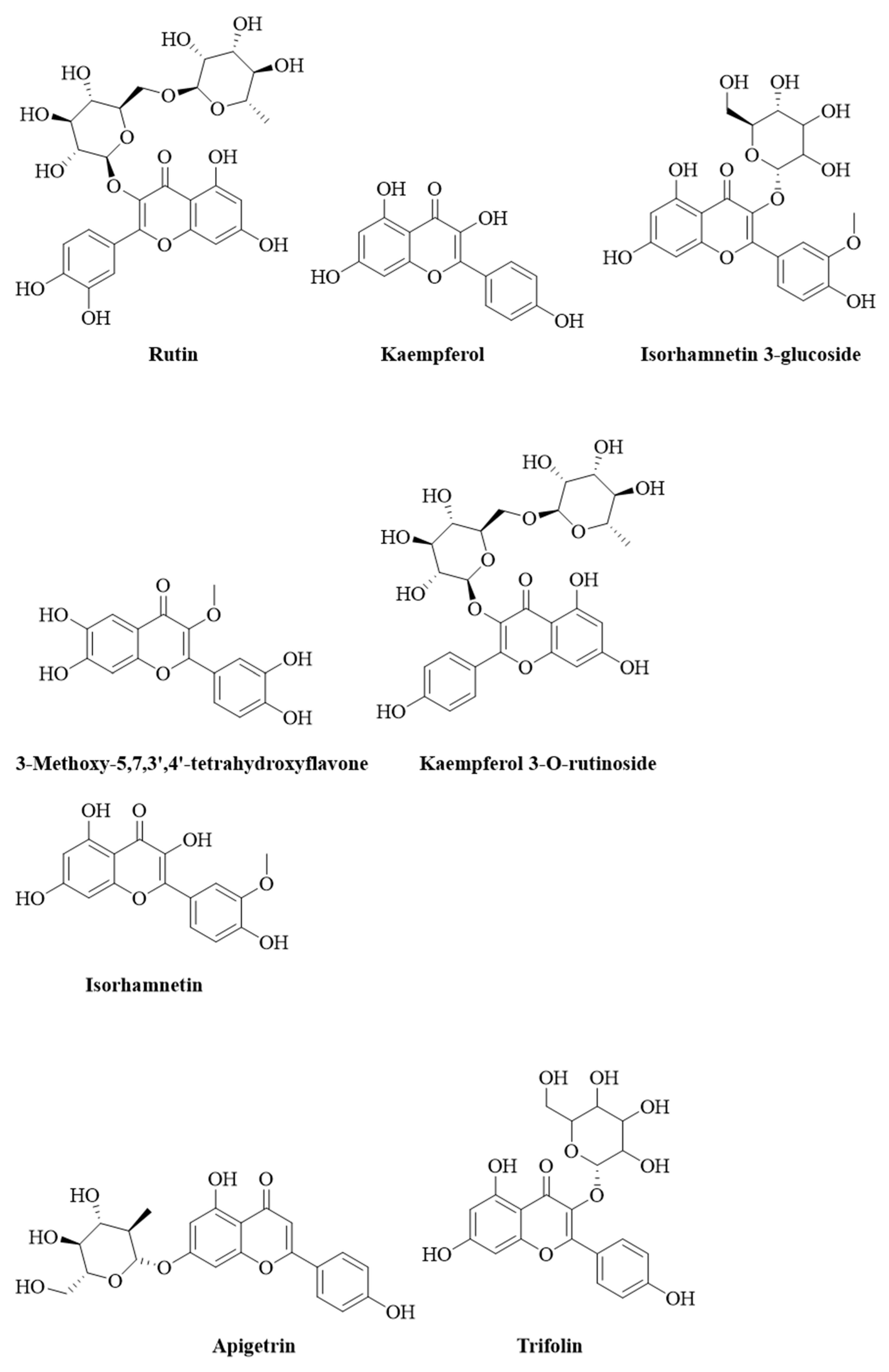
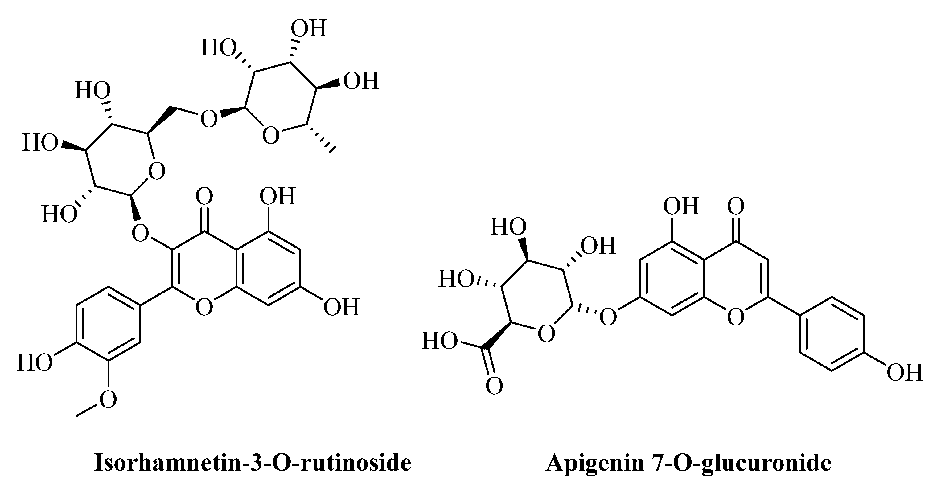
Table 1 presents the secondary metabolites in the aerial parts of O. forsskalii and Figure 1 shows the structures of the flavonoids and its glycosides compounds.

Table 1.
Active metabolites were obtained from O. forsskalii using LC-MS analysis.
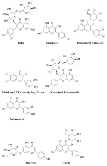

Figure 1.
Chemical structure (drawn using PubChem Sketcher V2.4) of flavonoids and their glycosides.
Some phytoconstituents such as apigenin, camphorol, isorhamnetin, and rutin have been described in previous studies of Mesembryanthemum forsskaolii (syn. O. forsskalii) from Egypt [29,30]. Therefore, the metabolites reported in this species were compared in terms of molecular weight with the peaks reported in the same genus [17]. However, some of the metabolites of the extract could not be recognized because in-depth phytochemical studies involving column chromatography of pure compounds and structure elucidation by NMR analysis or other instrumental analyses are required.
It is well-documented that flavonoids and their glycosides are responsible for antioxidant, anticancer, and antiulcerogenic activity (Sharifi-Rad et al., 2018). Phytochemical screening of the alcoholic extract using LC-MS chromatogram analysis revealed the presence of about 30 compounds, most of which are flavonoids, glycosides, and fatty acids esters. The present study identified the most abundant class of secondary metabolites as flavonoids, their glycosides, and fatty acid ester. The flavonoids include rutin, apigetrin, kaempferol, tetrahydroxyflavone, trifolin, isorhamnetin, genistein, and their glycosides as kaempferol 3-O-rutinoside, isorhamnetin 3-glucoside, isorhamnetin-3-o-rutinoside, and apigenin 7-o-glucuronide were the most recorded secondary metabolites. The fatty acids esters such as (2Z)-3,7-dimethyl-2,6-octadien-1-yl 3-oxobutanoate, (8E)-2-amino-8-octadecene-1,3,4-triol, 2-(14,15-epoxyeicosatrienoyl) glycerol, 2-arachidonoyl glycerol, 27-norcholestane-3,7,12,24,25,26-hexol, ethyl linoleate, and 1-α-linolenoyl-2-arachidonoyl-sn-glycerol were identified in the alcoholic extract of O. forsskalii. Compounds such as coumarin, furoic acid esters, alkali metals and phthalate derivatives, sesquiterpene, steroids phenols, etc. were also found in the aerial parts of O. forsskalii. Ahmed et al. (2021) found that the active ingredients of antiulcer drugs can be attributed primarily to the presence of flavonoids [31]. Rutin, apigetrin, kaempferol, trifolin, isorhamnetin, apigenin, and genistein and its glycosides were the main flavonoid compounds used as active antiulcer agents [32,33]. The glycosides in addition to flavonoid compounds, fat acids [34], alkaloids [35], sesquiterpenes [36], steroids [37], and phenols [38] exhibit potent gastric ulcer or hepatoprotective properties. This study showed that the phytoconstituents were present in the aerial parts of O. forsskalii. These secondary metabolites could contribute to the antiulcer activity. However, the active metabolites in O. forsskalii did not show detailed pharmacological activities in the experimental animal studies, especially antiulcer activity. Therefore, the data indicated the presence of active metabolites that could support the use of O. forsskalii as an antiulcer agent. In the present study, several secondary metabolites were identified in the alcoholic extract of the leaves of O. forsskalii (Figure 1). Several studies reported that hymecromone, apigetrin, kaempferol, rutin, trifolin, isorhamnetin, apigenin, hexylresorcinol, genistein, etc. are nontoxic and have antiulcerogenic effects [39,40,41,42], which were also found in O. forsskalii [30]. O. forsskalii is used in traditional medicine, and in previous studies, the seeds of this plant [43] and several other plants of this family were found to be nontoxic [23,43,44]. In several studies, the major identified compounds flavonoids, their glyosides, and fatty acid esters have been reported to have gastroprotective potential against ethanol-induced ulcers, supported by the present study [45,46].
2.2. Antiulcerogenic Effect of O. forsskalii
The oral dose of indomethacin significantly damaged the glandular stomach of rats. The 400 mg/kg dose of O. forsskalii significantly reduced the development of gastric lesions in rat stomachs (28.33%); however, the 200 mg/kg dose of O. forsskalii had a limited preventive effect compared with the 400 mg/kg dose (Table 2).
Table 2.
Effect of O. forsskalii on indomethacin-induced gastric mucosal lesions.
Indomethacin, a non-selective cyclooxygenase inhibitor, caused gastric damage in rats by numerous methods, including reducing prostaglandin production, oversupplying leukotrienes, acting as a topical irritant, and reducing local blood flow [47]. In this model, rats pretreated with O. forsskalii provided significant protection. The gastroprotective effect of O. forsskalii extract could be attributed to increased gastric mucus, which produces prostaglandins and inhibits leukotrienes.
Table 3 shows that a dose of 400 mg/kg body weight of O. forsskalii extracts significantly low intraluminal hemorrhage value (1.66%) and ulcer development (18.33%) caused by hypothermic stress. At a 200 mg/kg dose, body weight, intraluminal bleeding, and UI were reduced, but the difference was not significant. Because of the repeatability of the data, ulcers caused by hypothermic stress were used as an experimental paradigm for evaluating antiulcer activity in rats [48]. Disruption of gastric mucosal microcirculation, acid secretion enhancement, and mucus production reduction is mediated by histamine release and abnormal gastric motility [49].
Table 3.
Effect of the O. forsskalii extract on hypothermic-restrained stress-induced intraluminal bleeding and gastric lesions in rats.
Free radicals may play an important role in stress-induced gastric injury [50]. Stress deactivates prostaglandin synthesis in the mucosa by accumulating hydrogen peroxide, an inhibitor of prostaglandin biosynthesis that generates reactive oxygen species (ROS) [51]. Moreover, a positive relationship between the amount of lipid peroxidation products in the gastric mucosa, a marker of oxidative stress, and gastric injury were demonstrated in rats subjected to cold stress [52]. The antioxidant activity of the extract of O. forsskalii, previously documented in O. forsskalii [20], and its anti-secretagogue effect could explain the protective effect of the animal against cold stress.
The effect of O. forsskalii extract on necrotizing agent-induced gastric lesions is presented in Table 4. Control rats were treated with 80% ethanol, 0.2 mol/L NaOH, and 25% NaCl, which resulted in severe gastric lesions in the glandular mucosa of the stomach. The UI was 6.83 ± 0.30, 5.83 ± 0.30, and 6.16 ± 0.30, respectively, in 80% ethanol, 0.2 mol/L NaOH, and 25% NaCl control rats 1 h after the necrotizing drugs were administered. Pre-treatment of rats with O. forsskalii extract at doses of 200 mg/kg of 80% ethanol, 0.2 mol/L NaOH, and 25% NaCl resulted in an UI of 5.33 ± 0.33 (p < 0.01), 5.33 ± 0.33, (not significant), and 5.00 ± 0.36 (p < 0.05), respectively. Similarly, pre-treatment of rats with O. forsskalii extract at doses of 400 mg/kg of 80% ethanol, 0.2 mol/L NaOH, and 25% NaCl revealed an UI 4.00 ± 0.36 (p < 0.001), 4.16 ± 0.30 (p < 0.01), and 4.33 ± 0.42 (p < 0.01), respectively, which inhibited the formation of gastric lesions. The transition from an erosive mucus layer to a gastric lesion is aided by gastric acid output. By contrast, proton pump inhibitors and histamine H2-receptor antagonists speed up the healing of gastric lesions or prevent mucosal injury [53].
Table 4.
Effect of O. forsskalii on gastric lesions induced by necrotizing agents.
O. forsskalii protected the gastric mucosa from ulcers caused by various necrotizing agents, such as ethanol and strong alkalis, in a statistically significant and dose-dependent manner. The use of ethanol-induced gastric ulcers to evaluate the gas-protective function is standard. When ethanol is metabolized in the body, free superoxide anions and hydroperoxyl radicals are produced. Free radicals generated by oxygen are involved in acute and chronic gastric ulcers [54]. Khazaei and Salehi pointed out that the development of ethanol-induced gastric lesions is multifactorial, with a decrease in gastric mucus and significant formation of free radicals leading to increased lipid peroxidation, which damages cells and cell membranes [55].
2.3. Assessment of the Oxidative Damage in Ethanol-Induced Ulcer
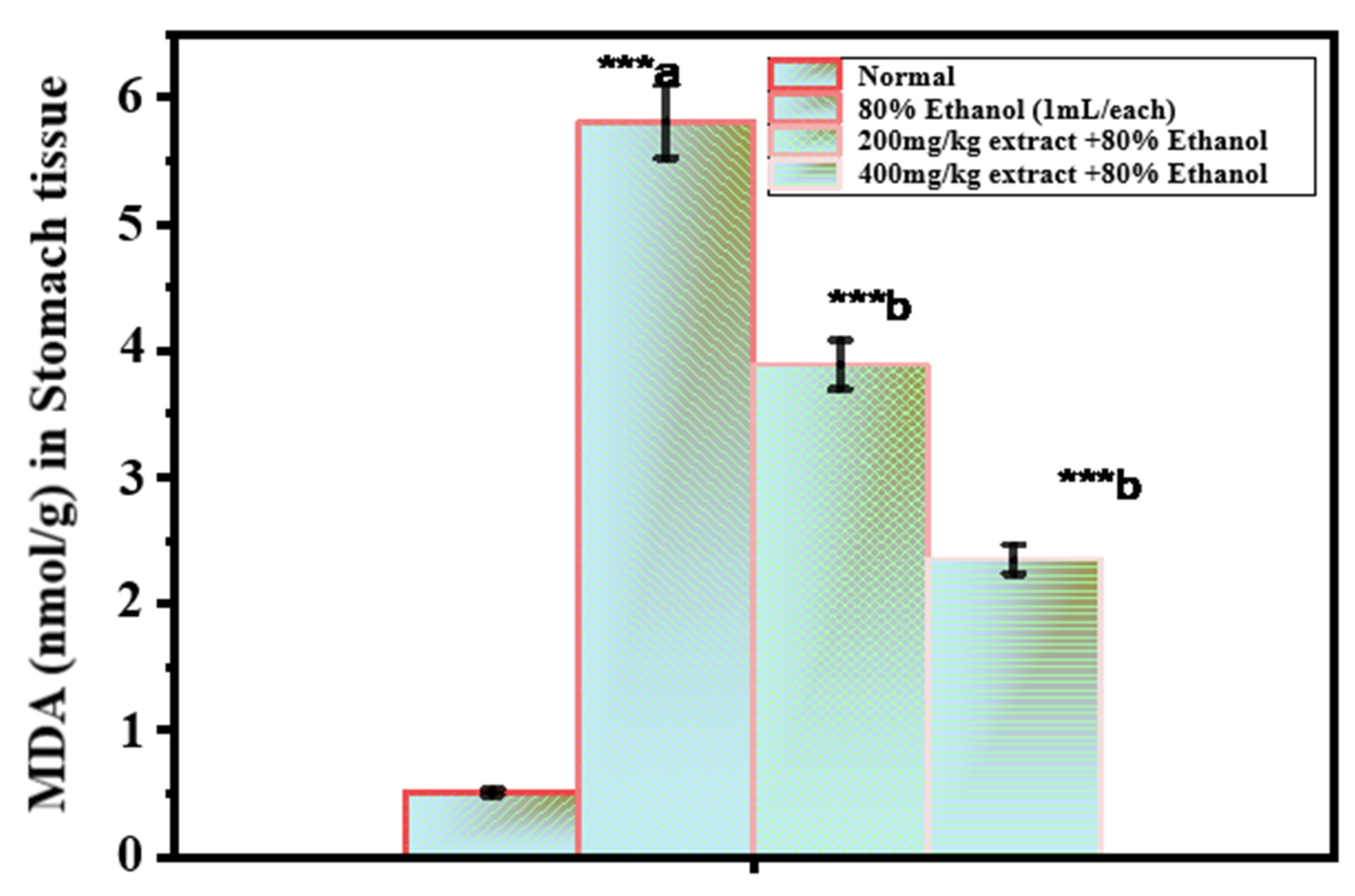
Figure 2 shows the MDA values in the gastric mucosa. The results used as the lipid peroxidation index were significantly higher in the ethanol-treated group than in the untreated control group (5.80 ± 0.34 μmol/g tissue; 0.54 ± 0.02 μmol/g tissue). At both doses (200 and 400 mg/kg), O. forsskalii (OF) significantly reduced the MDA content (3.88 ± 0.17 μmol/g and 2.34 ± 0.07 μmol/g, respectively). When the MDA content increases, free radicals, such as superoxide anion, hydrogen peroxide, and hydroxyl radicals, are formed. Cell degranulation is caused by the fact that these radicals increase the peroxidation of cell membrane lipids, leading to a loss of structural and functional integrity of cell membranes. When CAT does not scavenge hydrogen peroxide, it accumulates in the mitochondria and cytosol, increasing lipid peroxidation [56]. In addition, a clear relationship between the concentrations of lipid peroxidation end products in the gastric mucosa (a marker of oxidative stress) and gastric ulcers was found in stress-induced ulcers [49]. Since malondialdehyde (MDA) is the end product of lipid peroxidation, a decrease in the MDA concentration indicates a decrease in lipid peroxidation [57].
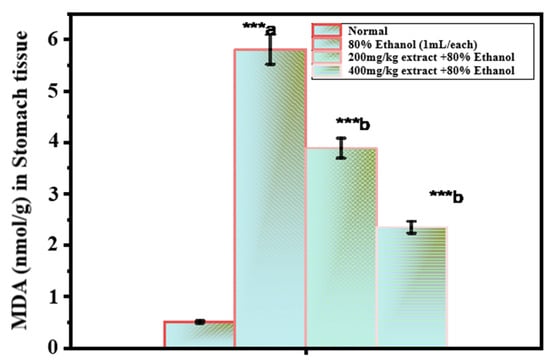
Figure 2.
Effect of O. forsskalii extracts on the MDA concentration in gastric ulcer induced by 80% ethanol. Six rats were used in each group. p values (*** p < 0.001). Where, a: 80% ethanol treated group was statistically compared to the normal group; b: O. forsskalii treated groups were compared to the ethanol-treated group.
Lipid peroxidation was significantly inhibited by O. forsskalii. Moreover, O. forsskalii significantly suppressed MDA production from lipids reacting with thiobarbituric acid. Thus, the antioxidant property of O. forsskalii prevents the oxidative damage caused by alcohol intoxication. Moreover, O. forsskalii strengthens the mucosal barrier, the first line of defense against endogenous and foreign ulcerogenic chemicals, as evidenced by its antiulcerogenic activity [16].
Figure 3 shows the concentration of NP-SH in the gastric mucosa. The concentration of NP-SH in the gastric mucosa of control rats was 5.80 ± 0.14 mmol/g tissue, which was significantly reduced to 2.53 ± 0.34 mmol/g (p < 0.001) after the administration of 80% ethanol. Ethanol-induced depletion of NP-SH was significantly increased in rats pretreated with an extract of O. forsskalii at both doses (200 and 400 mg/kg, respectively) (4.44 ± 0.44, p < 0.01; 5.23 ± 0.11, p < 0.001). Sulfhydryl compounds are critical for maintaining gastrointestinal integrity in living animals, especially when reactive oxygen species (ROS) play a role in causing tissue damage [58]. After ethanol administration, a sharp decrease in gastric NP-SH suggests a tremendous production of oxygen-generated free radicals.
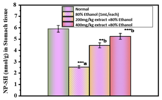
Figure 3.
Effect of O. forsskalii extracts on the NP-SH concentration in gastric ulcer induced by 80% ethanol. Six rats were used in each group. p values (** p < 0.01, *** p < 0.001). Where, a: 80% ethanol treated group was statistically compared to the normal group; b: O. forsskalii treated groups were compared to the ethanol-treated group.
The results of our studies support the findings of a previous study that again demonstrated sulfhydryl depletion in ethanol-induced gastric ulcers [59,60]. The reduction of glutathione exacerbates ulcerogenic-induced gastric mucosal injury in rats [61], whereas an increase in mucosal NP-SH leads to a gastroprotective effect. Our observations suggest that the extract of O. forsskalii protects the gastric mucosa.
Figure 4 shows the total protein (TP) level in gastric juice. The level of TP in the gastric juice of control rats was 108.57 ± 2.36 g/L gastric juice, which decreased significantly to 51.49 ± 1.82 g/L (p < 0.001) after administration of 80% ethanol. Pretreatment of rats with an extract of O. forsskalii at both doses (200 and 400 mg/kg) significantly increased the ethanol-induced decrease in TP (57.48 ± 1.38, p < 0.05; 78.63 ± 2.08 g/L, p < 0.001). Proteins play a key role in maintaining gastric integrity [62]. A significant decrease was observed in TP after ethanol administration, indicating malnutrition and disorders affecting the gastrointestinal system and interfering with the normal absorption of nutrients [63]. Our results are consistent with previous reports showing TP in ethanol-induced gastric lesions [64]. Treatment of rats with an extract of O. forsskalii resulted in a significant increase in TP. Our observations suggest that TP plays a role in protecting gastric mucosa by extracting O. forsskalii.
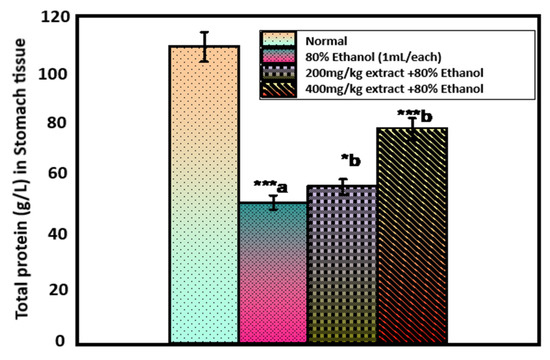
Figure 4.
Effect of O. forsskalii extracts on the total phenol concentration in gastric ulcer induced by 80% ethanol. Six rats were used in each group. p values (* p < 0.05, *** p < 0.001). Where, a: 80% ethanol treated group was statistically compared to the normal group; b: O. forsskalii treated groups were compared to the ethanol-treated group.
2.4. Histopathological Studies
Pretreatment with the extract of O. forsskalii reduced ethanol-induced necrosis in the superficial layers of the gastric mucosa with congestion, as shown by the histopathological findings (Figure 5A–I). Specimens were prepared in 10% neutral buffered formalin and stained with hematoxylin and eosin (H&E), Masson’s trichrome, and periodic acid Schiff’s (PAS) for light microscopic studies [65]. Previous studies identified inflammation, hemorrhage, edema, and loss of epithelial cells in gastric tissue as features of ethanol-induced injury [66]. According to the results of this study, the extract of O. forsskalii significantly decreased the amount of infiltration of banded leukocytes caused by ethanol-induced gastric injury. Ethanol reduced the mucosal density at the mucosal margins, resulting in necrosis and ulceration, as suggested by Mahmood et al. [67]. PAS staining showed features of gastric regions where mucopolysaccharides are released, according to Tarnawski et al. [68].
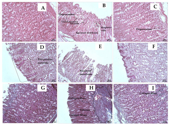
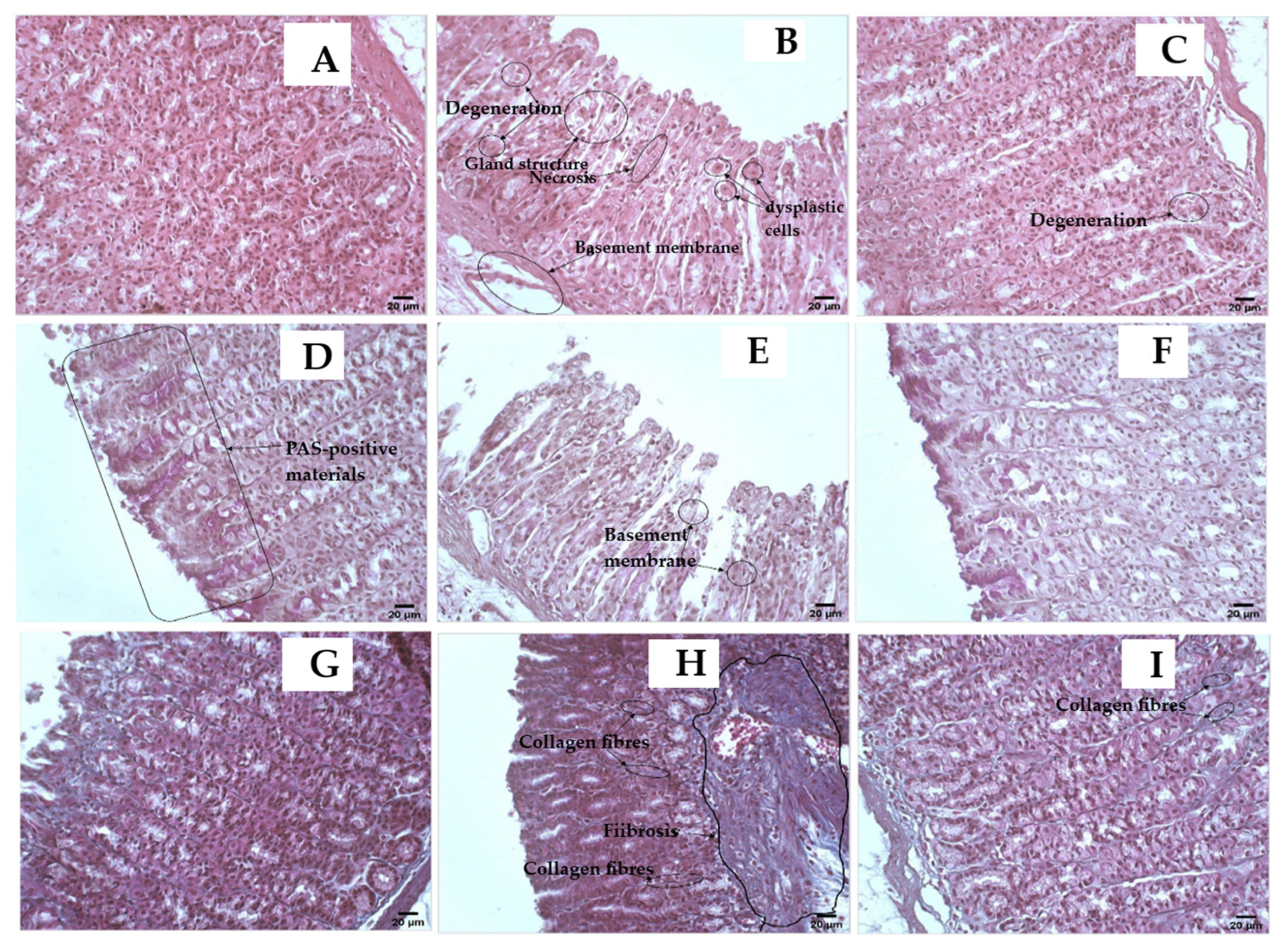
Figure 5.
(A–C), Hematoxylin and eosin (H&E, magnification is ×400, scale bar is 20 µ) stain, where (A) is the standard stomach sample that displays a typical appearance of stomach tissue; (B) is an 80% ethanol stomach sample showing the toxic effects, which are indicated by loss of the ability for secretion and presence of vacuolation in the cytoplasm, necrosis, degeneration, dysplastic cells, basement membrane and atrophic mucosa detachment, and reduction in gland size or even absence of gland structure; (C) is 400 mg/kg O. forsskalii extract and 80% ethanol stomach sample showing considerable improvement and restoration of the secreting stomach microanatomical architecture. However, some glands suffer from the remaining effects of degeneration and atrophy of the stomach’s mucosa. (D–F) Periodic acid Schiff (PAS, magnification is ×400, scale bar is 20 µ) stain, where (D) is a normal stomach sample showing the high activity of mucosa in production. The presence of PAS-positive materials that exhibit a dark magenta color, especially towards the lumen, which is in the left half of this photomicrograph and basement membranes are intact for each gland; (E) is the 80% ethanol stomach sample showing a severe reduction and the absence of the mucosal activity of production of PAS-positive materials. Basement membranes also exhibit damage and weakness in multiple areas; (F) is 400 mg/kg O. forsskalii extract, and the 80% ethanol stomach sample exhibits considerable improvement in the inability of stomach gland cells to secrete PAS-positive materials, and the basement membrane also improved. (G–I) Mason trichrome (magnification is ×400, scale bar is 20 µ) stain, where (G) is a normal stomach sample showing a normal distribution of connective tissue; (H) is the 80% ethanol stomach sample: this group shows massive fibrosis due to the accumulation of collagen fibers in the submucosa and infiltration of collagen fibers into mucosa; (I) is 400 mg/kg O. forsskalii extract and 80% ethanol stomach sample, exhibiting considerable improvement and the presence of a few areas of collagen fiber infiltrates into the mucosa.
Hematoxylin and eosin staining with ethanol showed cytoplasmic necrosis, degenerated cells, dysplastic cells, basement membrane, and a reduction in gland size [69]. The restriction of the microanatomical architecture using the alcoholic extract of O. forsskalii indicated the antiulcerogenic effect. PAS staining is used to detect mucopolysaccharides in gastric mucosa [68]. Due to the low PAS reactivity, the glycoprotein content of the gastric mucosa was lower in the II group. In contrast, the increased reactivity of PAS in the III group indicated an increase in glycoprotein. The Mason trichrome staining technique also revealed features of gastric sites where mucopolysaccharides are released. In individuals with ulcerative lesions in the stomach, gastrointestinal protective agents often increase PAS reactivity [70].
3. Materials and Methods
3.1. Materials
Sigma Aldrich (St.Louis, MO, USA) supplied the chemicals used in biochemical studies. The aerial parts of O. forsskalii were collected from Al-Kharj (the central region of Saudi Arabia). The sample was authenticated, and voucher number PSAU/PHARM/PF-101 was deposited at the Herbarium of the Pharmacognosy Department, Prince Sattam Bin Abdul-Aziz University (Al-Kharj, Saudi Arabia). The sample was dried in air and ground using a grinding machine (Geepas Countertop Mixer Grinder GSB5081, Dubai, United Arab Emirates) and passed to a mesh size of 40–60. The ethanol (70%) extracts were prepared by continuous shaking on an orbital shaker for 48 h each. A rotary evaporator (Buchi, Switzerland) was used to evaporate ethanol from the sample, and lyophilization using the freeze-drying (Freezone® 2.5 model 76530, Labconco Corp., Kansas, MO, USA) technique was used to remove the water content. The final yield obtained was 14.8%. LC-MS was performed on a 1 mg/mL concentration of the extract. The extract was separately reconstituted in distilled water to prepare doses of 200 and 400 mg/kg body weight (b.w.) for the in vivo antiulcer activity.
3.2. Analysis of Secondary Metabolites Using LC-MS
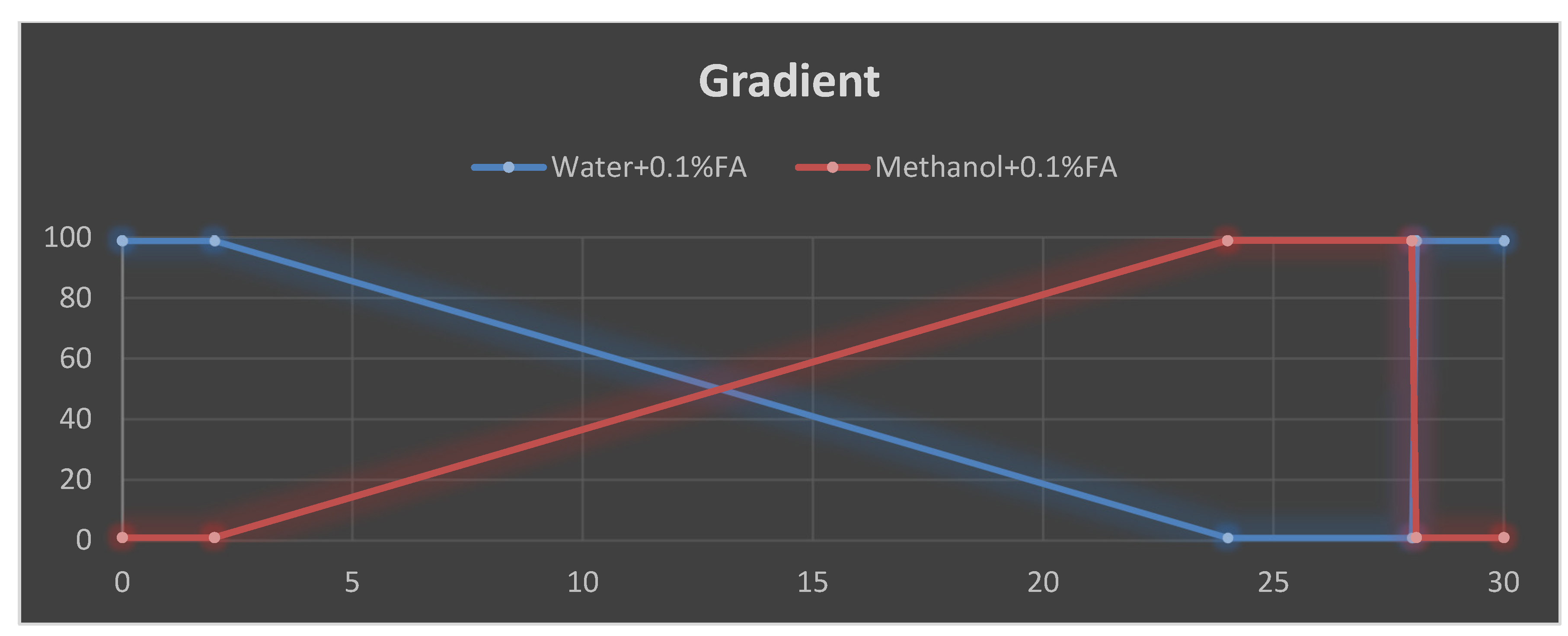
According to a previous method, the secondary metabolites were investigated using LC-MS (Thermo Scientific, Waltham, MA, USA) instrumentation analyses [71]. The Orbitrap ID-X is a mass spectrometer that contains three mass analyzers and was used to analyze the m/z of the studied molecules. The Orbitrap IDX spectrometer could reach a high resolution (>120,000) and reliable mass accuracy (<3 ppm mass error). The mass spectrometer was calibrated using a purchasable ’Calibration Mix ESI (Thermo Scientific)’ by following the manufacturer’s guidelines Electrospray ionization in the positive mode (ESI+) was applied for the studied compounds. The following parameters were applied: mobile phase (Figure 6), vaporized temperature = 100 °C, voltage = 3500 V, sheath gas = 30, auxiliary gas: 15, ion source fragmentation = 35 V, capillary temperature = 300 °C. Here, 10 µL of the total extract was injected through a loop injection into a C18 column using an independent UHPLC pump.

Figure 6.
Gradient elution profile used for the LC-MS separation, decreasing order of mobile phase (water and 0.1% formic acid), increasing order of mobile phase (methanol and 0.1% formic acid).
UHPLC: The extract was automatically infused (10 µL) through the UHPLC system using a C18 column (Acquity CSH 100 × 2.1 mm, 1.7 µm) for the separation. The flow rate was set to 0.5 mL/min, and a gradient was applied for the separation as follows:
Data Processing: Compound discoverer 3.1 was used to treat and process the data using mzCloud Mass Spectral Library and local compound databases (exact mass or formula).
3.3. In Vivo Antiulcer Assay
3.3.1. Animal Care
The instructions for the care and use of laboratory animals published by the College of Pharmacy (PSAU) were followed. Ethical clearance was obtained from the Department of Pharmacology, Prince Sattam bin Abdul-Aziz University (PSAU) and the standing committee of bioethics research (SCBR-009-2022) before the commencement of the research. Rats were procured, housed in plastic cages, and acclimatized for a week at the Animal Holdings of the Department of Pharmacology, PSAU, KSA, before research. The rats were kept at room temperature, fed a standard animal diet, and allowed free access to clean water. Sixty (60) albino Wistar rats (200 g) were obtained from the Animal House of the College of Pharmacy, PSAU. These animals were kept in clean, gauzed cages and acclimatized for two weeks at the animal house under standard temperature (25 ± 3 °C) and a 12:12 h light/dark periodicity. The animals were allowed free access to standard pellets and fresh water ad libitum. All the animals were handled in this study according to institutional guidelines describing the use of rats for studies.
3.3.2. Indomethacin-Induced Gastric Lesion
An indomethacin-induced gastric lesion assay was used on the animals according to the procedure described by Alqasoumi et al., 2009 [16]. The rats were fasted for 36 h and categorized into 3 groups (n = 6), and orally given 30 mg/kg body weight of indomethacin suspended in 1.0% carboxymethylcellulose (CMC) in water (6 mg/mL); an equivalent amount of vehicle was used to treat control rats. The sample was given at 200 and 400 mg/kg doses, 30 min before indomethacin administration. Six hours following treatment, their stomachs were removed and checked for ulcers after cleaning with normal saline.
3.3.3. Hypothermic-Restrained Stress-Induced Ulcers
A hypothermic-restrained stress-induced ulcer assay was used on the animals according to the method described by Alqasoumi et al., 2009 [16], with slight modifications. The animals were fasted for 36 h but given unlimited water. The rats were categorized into three groups (n = 6), restrained in restraint cages, and placed inside a vented refrigerator kept at 31 °C for 3 h, 30 min after oral administration of the extract (200 and 400 mg/kg). The stomachs of the animals were removed and assessed for ulceration and the severity of intraluminal hemorrhage using the arbitrary scale proposed by Chiu et al., in which 0 indicates no blood; 1 indicates that thin blood follows rugae; 2 indicates that thick blood follows the rugae; 3 indicates that thick blood follows the rugae with blood clots in specific regions; and 4 indicates that thick blood covers the whole gastric mucosal surface.
3.3.4. Gastric Lesions Induced by Necrotizing Agents
A necrotizing agent, such as 80% ethanol, 0.2 mol/L NaOH, and 25% NaCl induced ulcer assays, was performed on the animals according to the method described by Al Mofleh et al. with slight modifications [63]. The rats were fasted for 36 h and then categorized into 9 groups (n = 6). Animals were categorized as follows: in groups 1, 2, and 3, the ulcer control was given saline only for the assessment of 80% ethanol, 0.2 mol/L NaOH, and 25% NaCl necrotizing agents, respectively; groups 4, 5, and 6 were treated with 200 mg/kg BW for the assessment of 0.2 mol/L NaOH and 25% NaCl necrotizing agents; whereas groups 7, 8, and 9 were treated with 400 mg/kg BW for the assessment of 0.2 mol/L NaOH and 25% NaCl necrotizing agents. All treatments were administered intragastrically for 8 days, and the gastric ulcers were created using a solution of 1 mL/animal of necrotizing agents 80% ethanol, 0.2 mol/L NaOH, and 2% NaCl on the last day. An hour after the administration of necrotizing agents, the rats were killed. Their stomachs were excised, filled with 2.5 mL of a 4% formaldehyde solution, and placed in a formaldehyde beaker. Their stomachs were opened over the more significant curvature and cleaned with a 0.9% saline solution to eliminate the blood clots after 10 min. Each stomach sample was subsequently placed on a slide. The UI of each rate was computed using the following formula:
UI = (total area of mucosal lesion (mm2) × 100)/(total mucosal area (mm2)
3.3.5. Assessment of the Oxidative Damage in Ethanol-Induced Ulcer
After measuring the UI, the stomachs were washed with 0.9% (w/v) NaCl and used to determine various biochemical parameters.
NP-SH estimation was performed according to the previous method described by Sedlak and Lindsay [72]. Ice-cold 0.02 mmol/L ethylenediaminetetraacetic acid (EDTA) and the glandular portion of the stomach were homogenized in a Potter-Elvehjem type C homogenizer. Here, 5 mL of homogenate was combined with distilled water (4 mL) and 50% trichloroacetic acid (TCA, 1 mL). The tubes were centrifuged at 3000 r/min after being shaken intermittently for 10 min. Next, 2 mL of supernatant and 4 mL of 0.4 mol/L Tris buffer (pH 8.9) were mixed and agitated before the addition of 1 mL of 5,5’-dithio-bis (2-nitrobenzoic acid) (DTNB). The absorbance was measured at 412 nm against a reagent blank 5 min after DTNB was added.
Malondialdehyde (MDA) estimation was performed using the method described by Dursun et al. [73]. The stomach was removed and homogenized in 0.15 mol/L KCl (at 4 °C) to produce a 10% w/v homogenate. In a metabolic shaker, 1-mL aliquots of homogenate were incubated for 3 h at 37 °C. Next, 1 mL of 10% aqueous TCA was added and stirred. The mixture was then centrifuged for 10 min at 800 r/min and 1 mL of the supernatant was extracted and combined with 1 mL of water containing 0.67% thiobarbituric acid (TBA) for 10 min in a boiling water bath. After cooling, the liquid was diluted with 1 mL of distilled water. The absorbance was measured at 535 nm against a reagent blank. The MDA (nmol/g wet tissue) amount was estimated by referencing a standard curve of the MDA solution.
TP estimation was performed using the method described by Lowry et al. [74]. The alcoholic precipitate generated by mixing 90% alcohol with gastric juice in a 9:1 ratio was used to measure the dissolved proteins in gastric juice. The mixture of 1 mL of 0.1 N NaOH and 1 mL of alcoholic gastric juice was placed in a test tube. Subsequently, 0.05 mL was sampled, and 4 mL of the alkaline combination was added and allowed to stand. After 10 min, 0.4 mL of phenol reagent was added and allowed to rest for another 10 min for color development. The absorbance was measured at 610 nm against a reagent blank. The protein content was estimated by plotting a standard curve constructed with bovine albumin and expressed in g/L of gastric tissues.
3.3.6. Histopathological Evaluation
Stomach tissue samples with a 3–5-cm thickness were obtained from the 3 animal groups and labeled carefully. Subsequently, these samples were immersed in a sufficient amount of 10% formalin solution. Tissues were prepared by the automatic tissue processing machine (ASP300s, Leica Biosystems, Buffalo Grove, IL, USA). The samples were then fixed in paraffin wax blocks using a rotary microtome. Following, 5-µm-thick sections were prepared (SHUR/Cut 4500, TBS, Sanford, NC, USA). The slide was stained by the hematoxylin and eosin technique described by Bancroft and Layton [75].
3.4. Statistical Analysis
The table and figure values are expressed as a mean and standard deviation. A one-way analysis of variance (ANOVA) was used to assess the data, followed by a student’s t-test. In the analysis, the 80% ethanol group p-values were compared with the normal, and test group p-values were compared with the 80% ethanol group, and * p < 0.05 was referred to as statically significant; ** p < 0.01 referred to very significant; and *** p < 0.001 as highly significant.
4. Conclusions
Extracts of O. forsskalii contain pharmacologically active metabolites such as flavonoids, alkaloids, steroids, coumarin, furoic acid, sesquiterpenes, and phenolic compounds. In animal models, these compounds exhibit potential pharmacological properties for preventing ulcers induced by indomethacin, stress, and the necrotizing agent ethanol. The extract is not toxic in normal cells and is not toxic in animal studies. Groups pretreated with extracts of O. forsskalii could inhibit MDA production and stimulate the secretion of NP-SH and TP. Moreover, this plant extract did not show any damage in the histopathological study, confirming that this plant contains potential active ingredients against gastric ulcers and can be used to discover and develop new drugs.
Author Contributions
Conceptualization, A.I.F. and H.S.Y.; methodology, F.K.A., M.A.S. and A.A.; investigation, H.M.A., F.K.A. and M.A.S.; resources, M.H.A., A.I.F., H.S.Y. and M.A.S.; writing—original draft preparation, A.A. and M.A.S.; editing, A.A., M.H.A. and M.A.S.; supervision, A.I.F., H.S.Y. and H.M.A.; funding acquisition, A.I.F. and M.H.A. All authors have read and agreed to the published version of the manuscript.
Funding
This research has been supported by Prince Sattam bin Abdul-Aziz University Al-Kharj, a Master’s degree student.
Institutional Review Board Statement
Not applicable.
Informed Consent Statement
Not applicable.
Data Availability Statement
The data presented in this study are available in this article.
Acknowledgments
We thank Saud Malik, King Saud University, Riyadh Kingdom of Saudi Arabia for the anti-ulcerogenic study.
Conflicts of Interest
The author declares no conflict of interest.
Sample Availability
Samples of the plants are available from the authors.
References
- Albaqawi, A.; El-Fetoh, N.; Alanazi, R.; Alanazi, N.; Alrayya, S.E.; Alanazi, A.; Alenezi, S.; Alanazi, R.; Alshalan, A.M.; Alenezi, O.T.; et al. Profile of peptic ulcer disease and its risk factors in Arar, Northern Saudi Arabia. Electron. Phys. 2017, 9, 5740–5745. [Google Scholar] [CrossRef]
- Huang, Q.; Jia, X.; Chu, Y.; Zhang, X.; Ye, H. Helicobacter pylori Infection in Geriatric Patients: Current Situation and Treatment Regimens. Front. Med. 2021, 8, 713908. [Google Scholar] [CrossRef]
- Alzahrani, S.; Lina, T.T.; Gonzalez, J.; Pinchuk, I.V.; Beswick, E.J.; Reyes, V.E. Effect of Helicobacter pylori on gastric epithelial cells. World J. Gastroenterol. 2014, 20, 12767–12780. [Google Scholar] [CrossRef]
- Paguigan, N.D.; Castillo, D.H.; Chichioco-Hernandez, C.L. Anti-ulcer activity of leguminosae plants. Arq. Gastroenterol. 2014, 51, 64–67. [Google Scholar] [CrossRef]
- Jarosz, M.; Szkaradek, N.; Marona, H.; Nowak, G.; Młyniec, K.; Librowski, T. Evaluation of anti-inflammatory and ulcerogenic potential of zinc–ibuprofen and zinc–naproxen complexes in rats. Inflammopharmacology 2017, 25, 653–663. [Google Scholar] [CrossRef]
- McEvoy, L.; Carr, D.F.; Pirmohamed, M. Pharmacogenomics of NSAID Induced Upper Gastrointestinal Toxicity. Front. Pharmacol. 2021, 12, 684162. [Google Scholar] [CrossRef]
- Kuna, L.; Jakab, J.; Smolic, R.; Raguz-Lucic, N.; Vcev, A.; Smolic, M. Peptic Ulcer Disease: A Brief Review of Conventional Therapy and Herbal Treatment Options. J. Clin. Med. 2019, 8, 179. [Google Scholar] [CrossRef]
- Stojanović-Radić, Z.; Pejčić, M.; Stojanović, N.; Sharifi-Rad, J.; Stanković, N. Potential of Ocimum basilicum L. and Salvia officinalis L. Essential oils against biofilms of P. aeruginosa clinical isolates. Cell. Mol. Biol. 2016, 62, 27–32. [Google Scholar]
- Salehi, B.; Kumar, N.; Şener, B.; Sharifi-Rad, M.; Kılıç, M.; Mahady, G.; Vlaisavljevic, S.; Iriti, M.; Kobarfard, F.; Setzer, W.N.; et al. Medicinal plants used in the treatment of human immunodeficiency virus. Int. J. Mol. Sci. 2018, 19, 1459. [Google Scholar] [CrossRef]
- Setzer, M.S.; Sharifi-Rad, J.; Setzer, W.N. The search for herbal antibiotics: An in-silico investigation of antibacterial phytochemicals. Antibiotics 2016, 5, 30. [Google Scholar] [CrossRef]
- Kumadoh, D.; Archer, M.A.; Yeboah, G.N.; Kyene, M.O.; Boakye-Yiadom, M.; Adi-Dako, O.; Osei-Asare, C.; Adase, E.; Appiah, A.A.; Mintah, S.O. A review on anti-peptic ulcer activities of medicinal plants used in the formulation of Enterica Dyspepsia and NPK 500 capsules. Heliyon. 2021, 7, e08465. [Google Scholar] [CrossRef]
- Alam, A.; Rehman, N.U.; Ansari, M.N.; Palla, A.H. Effects of Essential Oils of Elettaria cardamomum Grown in India and Guatemala on Gram-Negative Bacteria and Gastrointestinal Disorders. Molecules 2021, 26, 2546. [Google Scholar] [CrossRef]
- Junior, I.F.S.; Balogun, S.O.; Oliveira, R.G.; Damazo, A.S.; Martins, D.T.O. Piper umbellatum l.: A medical plant with gastric-ulcer protective and ulcer healing effects in experimental rodent models. J. Ethnopharmacol. 2016, 192, 123–131. [Google Scholar] [CrossRef]
- de Lacerda Neto, L.J.; Ramos, A.G.; Santos Sales, V.; de Souza, S.D.; Dos Santos, A.T.; de Oliveira, L.R.; Kerntopf, M.R.; de Albuquerque, T.R.; Coutinho, H.D.; Quintans-Júnior, L.J.; et al. Gastroprotective and ulcer healing effects of hydroethanolic extract of leaves of Caryocar coriaceum: Mechanisms involved in the gastroprotective activity. Chem. Biol. Interact. 2017, 261, 56–62. [Google Scholar] [CrossRef]
- Nguelefack, T.B.; Feumebo, C.B.; Ateufack, G.; Watcho, P.; Tatsimo, S.; Atsamo, A.D.; Tane, P.; Kamanyi, A. Anti-ulcerogenic properties of the aqueous and methanol extracts from the leaves of Solanum torvum Swartz (Solanaceae) in rats. J. Ethnopharmacol. 2008, 119, 135–140. [Google Scholar] [CrossRef]
- Alqasoumi, S.; Al-Sohaibani, M.; Al-Howiriny, T.; Al-Yahya, M.; Rafatullah, S. Rocket “Eruca sativa”: A salad herb with potential gastric anti-ulcer activity. World J. Gastroenterol. 2009, 15, 1958–1965. [Google Scholar] [CrossRef]
- Youssif, K.; Elshamy, A.; Rabeh, M.; Gabr, N.; Haggag, E.A. Phytochemical and Biological Review on Plants of the family Aizoaceae. J. Adv. Pharm. Res. 2019, 3, 158–181. [Google Scholar] [CrossRef]
- Dhasan, P.B.; Jegadeesan, M.; Kavimani, S. Antiulcer activity of aqueous extract of fruits of Momordica cymbalaria Hook f. in Wistar rats. Pharmacogn. Res. 2010, 2, 58–61. [Google Scholar] [CrossRef]
- Prakash, R. Gastroprotective and antisecretory properties of methanolic extract of Trianthema portulacastrum. L in aspirin & pyloric ligature induced gastric ulcer in rats. PTB Rep. 2015, 1, 87–91. [Google Scholar]
- Abdel-Farid, I.B.; Mahalel, U.A.; Jahangir, M.J.; Elgebaly, H.A.; El-Naggar, S.A. Metabolomic profiling and antioxidant activity of Opophytum forsskalii. JUSEJ 2016, 3, 19–24. [Google Scholar] [CrossRef]
- Adedamola, A.K.; Eko, E.O.; Omoniyi, O.O. Ethnopharmacology, Therapeutic Properties and Nutritional Potentials of Carpobrotus edulis: A Comprehensive Review. Sci. Pharm. 2020, 88, 39. [Google Scholar] [CrossRef]
- El-Amier, Y.A.; Alghanem, S.M.; Al-hadithy, O.N.; Fahmy, A.A.; El-Zayat, M.M. Phytochemical analysis and biological activities of three wild Mesembryanthemum species growing in heterogeneous habitats. J. Phytol. 2021, 13, 01–08. [Google Scholar] [CrossRef]
- Aabed, K.; Mohammed, A.E. Phytoproduct, Arabic Gum and Opophytum forsskalii Seeds for Bio-Fabrication of Silver Nanoparticles: Antimicrobial and Cytotoxic Capabilities. Nanomaterials 2021, 11, 2573. [Google Scholar] [CrossRef]
- El-Amier, Y.A.; Haroun, S.; El-Shehaby, O.A.; Al-Hadith, O.N. Antioxidant and Antimicrobial Properties of Some Wild Aizoaceae species Growing in Egyptian Desert. J. Environ. Sci. 2016, 45, 1–10. [Google Scholar]
- Munyai, R.; Raletsena, M.V.; Modise, D.M. LC-MS Based Metabolomics Analysis of Potato (Solanum tuberosum L.) Cultivars Irrigated with Quicklime Treated Acid Mine Drainage Water. Metabolites 2022, 12, 221. [Google Scholar] [CrossRef]
- Shirahata, T.; Ishikawa, H.; Kudo, T.; Takada, Y.; Hoshino, A.; Taga, Y.; Minakuchi, Y.; Hasegawa, T.; Horiguchi, R.; Hirayama, T.; et al. Metabolic fingerprinting for discrimination of DNA-authenticated Atractylodes plants using 1H NMR spectroscopy. J. Nat. Med. 2021, 75, 475–488. [Google Scholar] [CrossRef] [PubMed]
- Kim, S.; Kim, J.; Kim, N.; Lee, D.; Lee, H.; Lee, D.Y.; Kim, K.H. Metabolomic Elucidation of the Effect of Sucrose on the Secondary Metabolite Profiles in Melissa officinalis by Ultraperformance Liquid Chromatography-Mass Spectrometry. ACS Omega 2020, 5, 33186–33195. [Google Scholar] [CrossRef]
- Hamed, A.I.; Said, R.B.; Kontek, B.; Al-Ayed, A.S.; Kowalczyk, M.; Moldoch, J.; Stochmal, A.; Olas, B. LC-ESI-MS/MS profile of phenolic and glucosinolate compounds in samh flour (Mesembryanthemum forsskalei Hochst. ex Boiss) and the inhibition of oxidative stress by these compounds in human plasma. Food Res. Int. 2016, 85, 282–290. [Google Scholar] [CrossRef]
- Moawad, A.; Mohamed, R. Secondary metabolites from Mesembryanthemum forsskaolii Hochst. ex. Boiss. Planta Medica 2014, 80, 1382558142. [Google Scholar] [CrossRef]
- Moawad, A.; Amin, E.; Mohammed, R. Diffusion-ordered Spectroscopy of Flavonol Mixture from Mesembryanthemum forsskaolii (Aizoaceae). Eur. J. Med. Plants 2016, 16, 1–8. [Google Scholar] [CrossRef]
- Ahmed, S.R.; Rabbee, M.F.; Roy, A.; Chowdhury, R.; Banik, A.; Kubra, K.; Hassan Chowdhury, M.M.; Baek, K.H. Therapeutic Promises of Medicinal Plants in Bangladesh and Their Bioactive Compounds against Ulcers and Inflammatory Diseases. Plants 2021, 10, 1348. [Google Scholar] [CrossRef]
- Shimada, H.; Eto, M.; Ohtaguro, M.; Ohtsubo, M.; Mizukami, Y.; Ide, T.; Imamura, Y. Differential mechanisms for the inhibition of human cytochrome P450 1A2 by apigenin and genistein. J. Biochem. Mol. Toxicol. 2010, 24, 230–234. [Google Scholar] [CrossRef] [PubMed]
- Huang, J.; Wang, S.; Zhu, M.; Chen, J.; Zhu, X. Effects of genistein, apigenin, quercetin, rutin and astilbin on serum uric acid levels and xanthine oxidase activities in normal and hyperuricemic mice. Food Chem. Toxicol. 2011, 49, 1943–1947. [Google Scholar] [CrossRef]
- Narkhede, K.P.; Satapathy, T.; Bibhas, P. Protective effect of Cod Liver Oil in Experimentally Induced Gastric Ulceration in Rats. Res. J. Pharm. Technol. 2019, 12, 5–10. [Google Scholar] [CrossRef]
- Salomone, F.; Galvano, F.; Li Volti, G. Molecular Bases Underlying the Hepatoprotective Effects of Coffee. Nutrients 2017, 9, 85. [Google Scholar] [CrossRef]
- Huang, C.C.; Lin, K.J.; Cheng, Y.W.; Hsu, C.A.; Yang, S.S.; Shyur, L.F. Hepatoprotective effect and mechanistic insights of deoxyelephantopin, a phyto-sesquiterpene lactone, against fulminant hepatitis. J. Nutr. Biochem. 2013, 24, 516–530. [Google Scholar] [CrossRef]
- Dembitsky, V.M. Antitumor and hepatoprotective activity of natural and synthetic neo steroids. Prog. Lipid Res. 2020, 79, 101048. [Google Scholar] [CrossRef]
- Pal, L.C.; Agrawal, S.; Gautam, A.; Chauhan, J.K.; Rao, C.V. Hepatoprotective and Antioxidant Potential of Phenolics-Enriched Fraction of Anogeissus acuminata Leaf against Alcohol-Induced Hepatotoxicity in Rats. Med. Sci. 2022, 10, 17. [Google Scholar] [CrossRef]
- Swanson, H.I.; Choi, E.Y.; Helton, W.B.; Gairola, C.G.; Valentino, J. Impact of apigenin and kaempferol on human head and neck squamous cell carcinoma. Oral Surg. Oral Med. Oral Pathol. Oral Radiol. 2014, 117, 214–220. [Google Scholar] [CrossRef]
- Zhang, Y.; Wang, D.; Yang, L.; Zhou, D.; Zhang, J. Purification and Characterization of Flavonoids from the Leaves of Zanthoxylum bungeanum and Correlation between Their Structure and Antioxidant Activity. PLoS ONE 2014, 9, e105725. [Google Scholar] [CrossRef]
- Nagy, N.; Kuipers, H.F.; Frymoyer, A.R.; Ishak, H.D.; Bollyky, J.B.; Wight, T.N.; Bollyky, P.L. 4-methylumbelliferone treatment and hyaluronan inhibition as a therapeutic strategy in inflammation, autoimmunity, and cancer. Front. Immunol. 2015, 6, 123. [Google Scholar] [CrossRef]
- Rashid, M.I.; Fareed, M.I.; Rashid, H.; Aziz, H.; Ehsan, N.; Khalid, S.; Ghaffar, I.; Ali, R.; Gul, A.; Hakeem, K.R. Flavonoids and Their Biological Secrets. Plant Hum. Health 2019, 2, 579–605. [Google Scholar] [CrossRef]
- Al-Faris, N.A.; Al-Sawadi, A.D.; Alokail, M.S. Effect of samh seeds supplementation (Mesembryanthemum forsskalei Hochst) on liver enzymes and lipid profiles of streptozotocin (STZ)-induced diabetic Wistar rats. Saudi J. Biol. Sci. 2010, 17, 23–28. [Google Scholar] [CrossRef] [PubMed] [Green Version]
- Rosaria, A.; Manuguerra, S.; Collins, E.; Mahdhi, A.; Renda, G.; Messina, C.M.; Santulli, A. Antioxidant Properties of a Supercritical Fluid Extract of the Halophyte Mesembryanthemum nodiflorum L. from Sicilian Coasts: Nutraceutical and Cosmeceutical Applications. Appl. Sci. 2020, 10, 2374. [Google Scholar] [CrossRef]
- Serafim, C.; Araruna, M.E.; Júnior, E.A.; Diniz, M.; Hiruma-Lima, C.; Batista, L. A Review of the Role of Flavonoids in Peptic Ulcer (2010-2020). Molecules 2020, 25, 5431. [Google Scholar] [CrossRef]
- Hamdi, A.; Majouli, K.; Abdelhamid, A.; Marzouk, B.; Belghith, H.; Chraief, I.; Bouraoui, A.; Marzouk, Z.; Heyden, Y.V. Pharmacological activities of the organic extracts and fatty acid composition of the petroleum ether extract from Haplophyllum tuberculatum leaves. J. Ethnopharmacol. 2018, 216, 97–103. [Google Scholar] [CrossRef]
- Shahin, N.N.; Abdelkader, N.F.; Safar, M.M. A Novel Role of Irbesartan in Gastroprotection against Indomethacin-Induced Gastric Injury in Rats: Targeting DDAH/ADMA and EGFR/ERK Signaling. Sci. Rep. 2018, 8, 4280. [Google Scholar] [CrossRef]
- Simões, S.; Lopes, R.; Campos, M.C.D.; Marruz, M.J.; da Cruz, M.E.M.; Corvo, L. Animal models of acute gastric mucosal injury: Macroscopic and microscopic evaluation. Anim. Model Exp. Med. 2019, 2, 121–126. [Google Scholar] [CrossRef]
- Di Cerbo, A.; Carnevale, G.; Avallone, R.; Zavatti, M.; Corsi, L. Protective Effects of Borago officinalis(Borago) on Cold Restraint Stress-Induced Gastric Ulcers in Rats: A Pilot Study. Front. Vet. Sci. 2020, 7, 427. [Google Scholar] [CrossRef]
- Azlina, M.F.N.; Qodriyah, H.M.S.; Akmal, M.N.; Ibrahim, I.A.A.; Kamisah, Y. In vivo effect of Piper sarmentosum methanolic extract on stress-induced gastric ulcers in rats. Arch. Med. Sci. 2019, 15, 223–231. [Google Scholar] [CrossRef]
- Sanpinit, S.; Chonsut, P.; Punsawad, C.; Wetchakul, P. Gastroprotective and Antioxidative Effects of the Traditional Thai Polyherbal Formula Phy-Blica-D against Ethanol-Induced Gastric Ulcers in Rats. Nutrients 2021, 14, 172. [Google Scholar] [CrossRef]
- Song, S.H.; Kim, J.E.; Sung, J.E.; Lee, H.A.; Yun, W.B.; Lee, Y.H.; Song, H.; Hwang, D. Anti-ulcer effect of Gallarhois extract with anti-oxidant activity in an ICR model of ethanol/hydrochloride acid-induced gastric injury. J. Tradit. Compl. Med. 2019, 4, 372–382. [Google Scholar] [CrossRef] [PubMed]
- Sharifi-Rad, M.; Fokou, P.V.T.; Sharopov, F.; Martorell, M.; Ademiluyi, A.O.; Rajkovic, J.; Salehi, B.; Martins, N.; Iriti, M.; Sharifi-Rad, J. Antiulcer Agents: From Plant Extracts to Phytochemicals in Healing Promotion. Molecules 2018, 23, 1751. [Google Scholar] [CrossRef] [Green Version]
- Umamaheswari, M.; Asokkumar, K.; Rathidevi, R.; Sivashanmugam, A.T.; Subhadradevi, V.; Ravi, T.K. Antiulcer and in vitro antioxidant activities of Jasminum grandiflorum L. J. Ethnopharmacol. 2007, 110, 464–470. [Google Scholar] [CrossRef] [PubMed]
- Khazaei, M.; Salehi, H. Protective effect of falcaria vulgaris extract on ethanol induced gastric ulcer in rat. Iran J. Pharmacol. Ther. 2006, 5, 43–46. [Google Scholar]
- Michiels, C.; Raes, M.; Toussaint, O.; Remacle, J. Importance of se-glutathione peroxidase, catalase, and Cu/Zn-SOD for cell survival against oxidative stress. Free Radic. Biol. Med. 1994, 17, 235–248. [Google Scholar] [CrossRef]
- Buege, J.A.; Aust, S.D. Microsomal lipid peroxidation. Methods Enzymol. 1978, 52, 302–310. [Google Scholar]
- Kimura, M.; Goto, S.; Ihara, Y.; Wada, A.; Yahiro, K.; Niidome, T.; Aoyagi, H.; Hirayama, T.; Kondo, T. Impairment of glutathione metabolism in human gastric epithelial cells treated with vacuolating cytotoxin from Helicobacter pylori. Microb. Pathog. 2001, 31, 29–36. [Google Scholar] [CrossRef]
- Miller, T.A.; Li, D.; Kuo, Y.J.; Schmidt, K.L.; Shanbour, L.L. Nonprotein sulfhydryl compounds in canine gastric mucosa: Effects of PGE2 and ethanol. Am. J. Physiol. 1985, 249, G137–G144. [Google Scholar] [CrossRef]
- Al Mofleh, I.A.; Alhaider, A.A.; Mossa, J.S.; Al-Soohaibani, M.O.; Rafatullah, S. Aqueous suspension of anise “Pimpinella anisum” protects rats against chemically induced gastric ulcers. World J. Gastroenterol. 2007, 13, 1112–1118. [Google Scholar] [CrossRef]
- Hiraishi, H.; Terano, A.; Ota, S.; Mutoh, H.; Sugimoto, T.; Harada, T.; Razandi, M.; Ivey, K.J. Protection of cultured rat gastric cells against oxidant-induced damage by exogenous glutathione. Gastroenterology 1994, 106, 1199–1207. [Google Scholar] [CrossRef]
- Chiou, S.K.; Moon, W.S.; Jones, M.K.; Tarnawski, A.S. Survivin expression in the stomach: Implications for mucosal integrity and protection. Biochem Biophys Res Commun. 2003, 305, 374–379. [Google Scholar] [CrossRef]
- Zuvarox, T.; Belletieri, C. Malabsorption Syndromes. [Updated 2021 Jul 30]. In StatPearls [Internet]; StatPearls Publishing: Treasure Island, Finland, 2022. Available online: https://www.ncbi.nlm.nih.gov/books/NBK553106 (accessed on 12 July 2022).
- Raish, M.; Shahid, M.; Bin Jardan, Y.A.; Ansari, M.A.; Alkharfy, K.M.; Ahad, A.; Abdelrahman, I.A.; Ahmad, A.; Al-Jenoobi, F.I. Gastroprotective Effect of Sinapic Acid on Ethanol-Induced Gastric Ulcers in Rats: Involvement of Nrf2/HO-1 and NF-κB Signaling and Antiapoptotic Role. Front. Pharmacol. 2021, 12, 622815. [Google Scholar] [CrossRef]
- Leong, A.S.; Milios, J. Rapid immunoperoxidase staining of lymphocyte antigens using microwave irradiation. J. Pathol. 1986, 148, 183–187. [Google Scholar] [CrossRef] [PubMed]
- Rios, E.; Rocha, N.; Venâncio, E.; Mour, B.; Feitosa, M.; Cerqueira, G.; Soares, P.M.; Woods, D.J.; de Sousa, F.C.; Leal, L.K.; et al. Mechanisms involved in the gastroprotective activity of esculin on acute gastric lesions in mice. Chem. Biol. Interact. 2010, 188, 246–254. [Google Scholar] [CrossRef] [PubMed]
- Mahmood, A.; Fard, A.; Harita, H.; Amin, Z.; Salmah, I. Evaluation of gastroprotective effects of Strobianthes crispus leaf extract on ethanol-induced gastric mucosal injury in rats. Sci. Res. Essays 2011, 6, 2306–2314. [Google Scholar]
- Tarnawski, A.; Tarnawski, A.; Szabo, I.L.; Husain, S.S.; Soreghan, B. Regeneration of gastric mucosa during ulcer healing is triggered by growth factors and signal transduction pathways. J. Physiol. Paris 2001, 95, 337–344. [Google Scholar] [CrossRef]
- Ansari, S.F.; Khan, A.U.; Qazi, N.G.; Shah, F.A.; Naeem, K. In Vivo, Proteomic, and In Silico Investigation of Sapodilla for Therapeutic Potential in Gastrointestinal Disorders. Biomed. Res. Int. 2019, 2019, 4921086. [Google Scholar] [CrossRef]
- Hajrezaie, M.; Salehen, N.; Karimian, H.; Zahedifard, M.; Shams, K.; Al, B.R. Biochanin a gastroprotective effects in ethanol-induced gastric mucosal ulceration in rats. PLoS ONE 2015, 10, e0121529. [Google Scholar]
- Al-Nemi, R.; Makki, A.A.; Sawalha, K.; Hajjar, D.; Jaremko, M. Untargeted Metabolomic Profiling and Antioxidant Capacities of Different Solvent Crude Extracts of Ephedra foeminea. Metabolites 2022, 12, 451. [Google Scholar] [CrossRef]
- Sedlak, J.; Lindsay, R.H. Estimation of total, protein-bound, and nonprotein sulfhydryl groups in tissue with Ellman’s reagent. Anal. Biochem. 1968, 25, 192–205. [Google Scholar] [CrossRef]
- Dursun, H.; Bilici, M.; Albayrak, F.; Ozturk, C.; Saglam, M.B.; Alp, H.H.; Suleyman, H. Antiulcer activity of fluvoxamine in rats and its effect on oxidant and antioxidant parameters in stomach tissue. BMC Gastroenterol. 2009, 9, 36. [Google Scholar] [CrossRef] [PubMed]
- Lowry, O.H.; Rosebrough, N.J.; Farr, A.L.; Randall, R.J. Protein measurement with the Folin phenol reagent. J. Biol. Chem. 1951, 193, 265–275. [Google Scholar] [CrossRef]
- Bancroft, J.D.; Layton, C. The Hematoxylins and Eosin in Bancroft’s Theory and Practice of Histological Techniques, 8th ed.; Suvarna, S.K., Layton, C., Bancroft, J.D., Eds.; Elsevier: Amsterdam, The Netherlands, 2018; p. 131. [Google Scholar]


Publisher’s Note: MDPI stays neutral with regard to jurisdictional claims in published maps and institutional affiliations. |
© 2022 by the authors. Licensee MDPI, Basel, Switzerland. This article is an open access article distributed under the terms and conditions of the Creative Commons Attribution (CC BY) license (https://creativecommons.org/licenses/by/4.0/).