Article

Virtual Screening of Benzimidazole Derivatives as Potential Triose Phosphate Isomerase Inhibitors with Biological Activity against Leishmania mexicana

by
Lenci K. Vázquez-Jiménez
1,
Alfredo Juárez-Saldivar
1,
Manuel J. Chan-Bacab
2,
Timoteo Delgado-Maldonado
1,
Luis D. González-Morales
1,
Isidro Palos
3,
Eyra Ortiz-Pérez
1,
Edgar E. Lara-Ramírez
1,
Esther Ramírez-Moreno
4 and
Gildardo Rivera
1,*
1
Laboratorio de Biotecnología Farmacéutica, Centro de Biotecnología Genómica, Instituto Politécnico Nacional, Reynosa 88710, Mexico
2
Centro de Investigación en Microbiología Ambiental y Biotecnología, Universidad Autónoma de Campeche, Campeche 24039, Mexico
3
Unidad Académica Multidisciplinaria Reynosa-Rodhe, Universidad Autónoma de Tamaulipas, Reynosa 88779, Mexico
4
Escuela Nacional de Medicina y Homeopatía, Instituto Politécnico Nacional, Ciudad de Mexico 07320, Mexico
*
Author to whom correspondence should be addressed.
Pharmaceuticals 2023, 16(3), 390; https://doi.org/10.3390/ph16030390
Submission received: 29 January 2023 / Revised: 24 February 2023 / Accepted: 28 February 2023 / Published: 3 March 2023
(This article belongs to the Special Issue Drug Discovery of Antiprotozoal Agents)

Abstract

:
Leishmania mexicana (L. mexicana) is a causal agent of cutaneous leishmaniasis (CL), a “Neglected disease”, for which the search for new drugs is a priority. Benzimidazole is a scaffold used to develop antiparasitic drugs; therefore, it is interesting molecule against L. mexicana. In this work, a ligand-based virtual screening (LBVS) of the ZINC15 database was performed. Subsequently, molecular docking was used to predict the compounds with potential binding at the dimer interface of triosephosphate isomerase (TIM) of L. mexicana (LmTIM). Compounds were selected on binding patterns, cost, and commercial availability for in vitro assays against L. mexicana blood promastigotes. The compounds were analyzed by molecular dynamics simulation on LmTIM and its homologous human TIM. Finally, the physicochemical and pharmacokinetic properties were determined in silico. A total of 175 molecules with docking scores between −10.8 and −9.0 Kcal/mol were obtained. Compound E2 showed the best leishmanicidal activity (IC50 = 4.04 µM) with a value similar to the reference drug pentamidine (IC50 = 2.23 µM). Molecular dynamics analysis predicted low affinity for human TIM. Furthermore, the pharmacokinetic and toxicological properties of the compounds were suitable for developing new leishmanicidal agents.

1. Introduction

Cutaneous leishmaniasis (CL) is a disease characterized by solitary or multiple ulcerated skin lesions [1]. The causative agents are Leishmania parasites transmitted by phlebotomid sandflies. In America, Leishmania mexicana (L. mexicana) and other subgenera are predominant agents [2,3]. Although CL is not a fatal disease, it does cause disability and permanent scars [4]. The World Health Organization (WHO) recommends pentavalent antimonials as first-line drug treatment [5]. Alternatives include the use of liposomal amphotericin B. However, these compounds are toxic, have low efficacy in the chronic phase of the disease, and cannot be widely used due to their high cost and administration requirements [6]. These factors potentiate the search for new and more effective drugs against CL.
In the last decades, different targets have been considered to develop new leishmanicidal agents, such as trypanothione reductase [7], cysteine proteinases [8], glyceraldehyde-3-phosphate dehydrogenase [9], and triosephosphate isomerase (TIM) [10]. The latter is involved in the fifth step of the glycolysis pathway and is essential in energy production [11,12]. TIM has structural differences with its human homologue, making the obtention of selective inhibitors possible. It has been widely studied in various protozoa as a pharmacological target [13,14,15,16], and, in this sense, the TIM of L. mexicana (LmTIM) can be considered a drug target for developing new leishmanicidal agents.
Benzimidazole is an aromatic heterocyclic compound with a wide range of biological activities [17] as an antiviral [18], an anthelmintic [19], an antimicrobial [20], an antiparasitic [21], and others [22,23]. Hybrids of benzimidazole and pentamidine derivatives have been tested against L. mexicana [24]. For example, Torres-Gómez et al., showed that the compounds C1 and C2 (Table 1) were more active (IC50 = 0.712 and 0.368 μM, respectively) than pentamidine (IC50 = 9.568 μM). Subsequently, Nieto-Meneses et al. [25] obtained compounds C3 and C4 (Table 1) with better leishmanicidal activity (IC50 = 2.62 and 3.21 μM, respectively) than miltefosine (IC50 = 15.34 μM) and lower cytotoxicity (SI = 91.76 and 317.75, respectively) than miltefosine and amphotericin B (SI = 10.23 and 6.5, respectively). These studies showed the effectiveness of benzimidazole-containing compounds as leishmanicidal agents.
In this work using the benzimidazole scaffold, a ligand-based virtual screening (LBVS) was performed of the ZINC15 database. Then, a molecular docking analysis was performed to identify potential binding at the interface of LmTIM. The selected compounds were further evaluated against the promastigote form of L. mexicana. In addition, molecular dynamics simulations were carried out to predict the stability of the compounds evaluated in complex with LmTIM and their affinity for human TIM (HsTIM). Finally, the pharmacokinetic and physicochemical properties were predicted.

2. Results and Discussion

2.1. Binding Site Prediction and Molecular Docking of Control Ligands

Six compounds with leishmanicidal and/or inhibitory activity against LmTIM were selected as control compounds [24,25,26] and analyzed by molecular docking at the interface of the LmTIM protein. Compounds C1C4 are benzimidazole derivatives, C5 is a benzothiazole derivative, and C6 is a quinoline derivative. Compound C2 has the best leishmanicidal activity (IC50 = 0.36 μM), and C5 shows a 70% inhibition on the LmTIM (Table 1) [10].
The binding site of the control compounds on the LmTIM protein (ID: 1AMK) for molecular docking analysis was established with the DoGSiteScorer tool. This pocket detection and analysis tool identifies potential binding pockets in protein structures. By default, it provides a simple drugability score for each pocket, based on a linear combination of three descriptors (volume, hydrophobicity, and enclosure) [27]. The results show the dimer interface as the best binding site with a drug score greater than 0.5.
Additionally, a blind molecular docking was performed to assess the potential binding of each control compound at the site predicted by the DoGSiteScorer tool (assigning a docking score) [28]. In this study, blind molecular docking showed docking scores of −9.2 to −7.1 Kcal/mol of the control compounds at the LmTIM dimer interface (Table 1). Compound C6 had the lowest docking score of −9.2 Kcal/mol. The other five control compounds showed docking scores from −7.8 to −7.1 Kcal/mol (Figure 1). The interface, contrary to the active site, is a non-conserved site. It has been studied because it causes a selective allosteric enzymatic inhibition in protozoa by promoting the destabilization of the proteins quaternary structure, leading to death [29,30].
The interaction profile of the six control compounds (Table 1) shows hydrophobic interactions, hydrogen bonding, and π-stacking interactions. Hydrophobic interactions with the Ile69, Glu105, and Ile109 residues of the B monomer predominated in the six control compounds, except Ile69 with the compound C5. These interactions have been reported to play a key role in inhibitory activity [16]. Additionally, the compounds C1, C2, and C3 present hydrogen bonds with Lys113 and Gln112 in monomer B, among others; compounds C4 and C5 were hydrogen-bonded only with Tyr103 in monomer A. The compound C6 with the best binding energy showed no hydrogen-bonding or π-stacking interactions. Only compounds C1 through C4 exhibited hydrogen-bonding interactions and π-stacking with at least some of the amino acid residues, Tyr102 in monomer A, and Phe75B or Tyr103 in monomer B. Interestingly, the interactions with amino acid residues at the TIM interface of other protozoa, such as Tyr102, Tyr103, Ile69, Asn67, Phe75, Thr70, Glu105, and Lys113, were also observed in other studies [31,32]. These results support the dimer interface as the best binding site for further virtual screenings. Additionally, in vitro enzymatic studies report that benzimidazole derivatives have up to 69% inhibition percentages on the TIM enzyme [30,33], supporting continuing efforts to search for this type of inhibitor.

2.2. LBSV in ZINC15

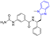
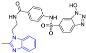
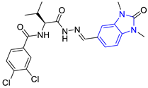
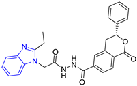
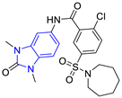
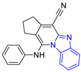
The LBVS in the ZINC15 database (750 million compounds) obtained 67,141 compounds by a substructure search using the benzimidazole scaffold. Applying Lipinskis rule as inclusion criteria, 53,410 compounds were filtered and selected for the molecular docking analysis. The results showed that 175 compounds had a docking score between −10.8 and −9.0 Kcal/mol, which was near and higher than the docking score of the control compound C6 ligand (−9.2 Kcal/mol). The compounds were grouped using two criteria: (a) based on the protein-ligand interaction profile (PLIP) and the scikit-learn library, and (b) on structure similarity descriptors using the algorithms of DataWarrior software. Table 2 shows the lead compounds of each group, arranged by their interaction profile, the number of compounds obtained, and the docking score. Group two had the highest number of compounds (n = 24), while group eight had the lowest number of compounds (n = 9) (full groups are shown in Supplementary Material S1).
In protozoa, the interface site is hydrophobic [33]; on the other hand, benzimidazole is a nitrogenous heterocycle with two equivalent tautomeric forms, in which the hydrogen atom can be located in any of the two nitrogen atoms, facilitating the formation of hydrogen bonds [34]. This block can also act as a proton acceptor or donor, binding to the protein through various interactions such as hydrogen bonds, van der Waals forces, and π–π stacking, among others [35]. In this sense, the lead compounds showed hydrophobic interactions, hydrogen bonding, and π-stacking. The hydrophobic interactions occurred with the amino acids Tyr102 or Tyr103 in the A monomer and Ile69 and Phe75 in the B monomer in most of the compounds. Hydrogen bonding interactions with the amino acids Gln112, Lys113, and Glu105 in the B monomer were the most common in the compounds. π-stacking interactions were also present with the amino acids Tyr102 and Tyr103 in monomer A and Phe75 in monomer B. Compound P4 with the lowest docking score (−10.3 Kcal/mol) did not show π-stacking interactions; however, it showed four additional hydrophobic interactions (Tyr103 in monomer A, Ile109, Gln112 and Glu116 monomer B) and two hydrogen bond interactions with the amino acids Ala70 and Arg99 in monomer B, which have been described as promoting the formation of more stable ligand-protein complexes [10]. In addition, they coincide with interactions presented by the control compounds (Table 1).
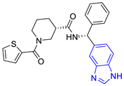
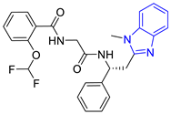
Table 3 shows the 10 groups obtained by structure, the number of compounds in each group, and the lead compound with the best docking score (−10.8 and −9.0 Kcal/mol). This grouping was based on the type of structure and how similar they were to each other. Group three presented the largest number of compounds (n = 102), while the rest presented 1 and up to 26 compounds per group (the entire groups are shown in Supplementary Material S1).
The interactions that predominated were hydrophobic with the amino acid residues Ile69, Ile109, and Phe75 in the B monomer in five compounds (E1, E2, E6, E7, and E8). The hydrogen bonding interaction with the Gln112 amino acid residue in the B monomer was the most common in most compounds and presented π-stacking interactions with the amino acid residues Tyr102 and Tyr103 in the A monomer and Phe75 in the B monomer. Only compound E9 presented a salt-bridge type interaction with the Lys113 amino acid residue in monomer B. In contrast, compound E10 showed an interaction of the halogen with the amino acid residue Lys71 in monomer B. Considering the described binding patterns, and the commercial availability and price, compounds P9 (Table 2) and E2 (Table 3) were purchased to determine their leishmanicidal activity.

2.3. Leishmanicidal Activity

The half-maximal inhibitory concentration (IC50) of compounds P9 and E2 against L. mexicana promastigotes is shown in Table 4. Compound E2 had better leishmanicidal activity against the promastigotes form of L. mexicana (IC50 = 4.04 µM) than P9 (IC50 > 148.63), and similar to the reference drug pentamidine (IC50 = 2.23 µM).

2.4. Molecular Dynamics Analysis

Molecular dynamics simulation was performed to predict the stability of the ligand–protein complex due to the leishmanicidal effect of compounds P9 and E2, their high binding affinity, and an interaction profile similar to the control compounds at the LmTIM interface. The apo form protein (LmTIM) and the control compound C6 in complex with LmTIM were also analyzed, considering previous studies [36,37].
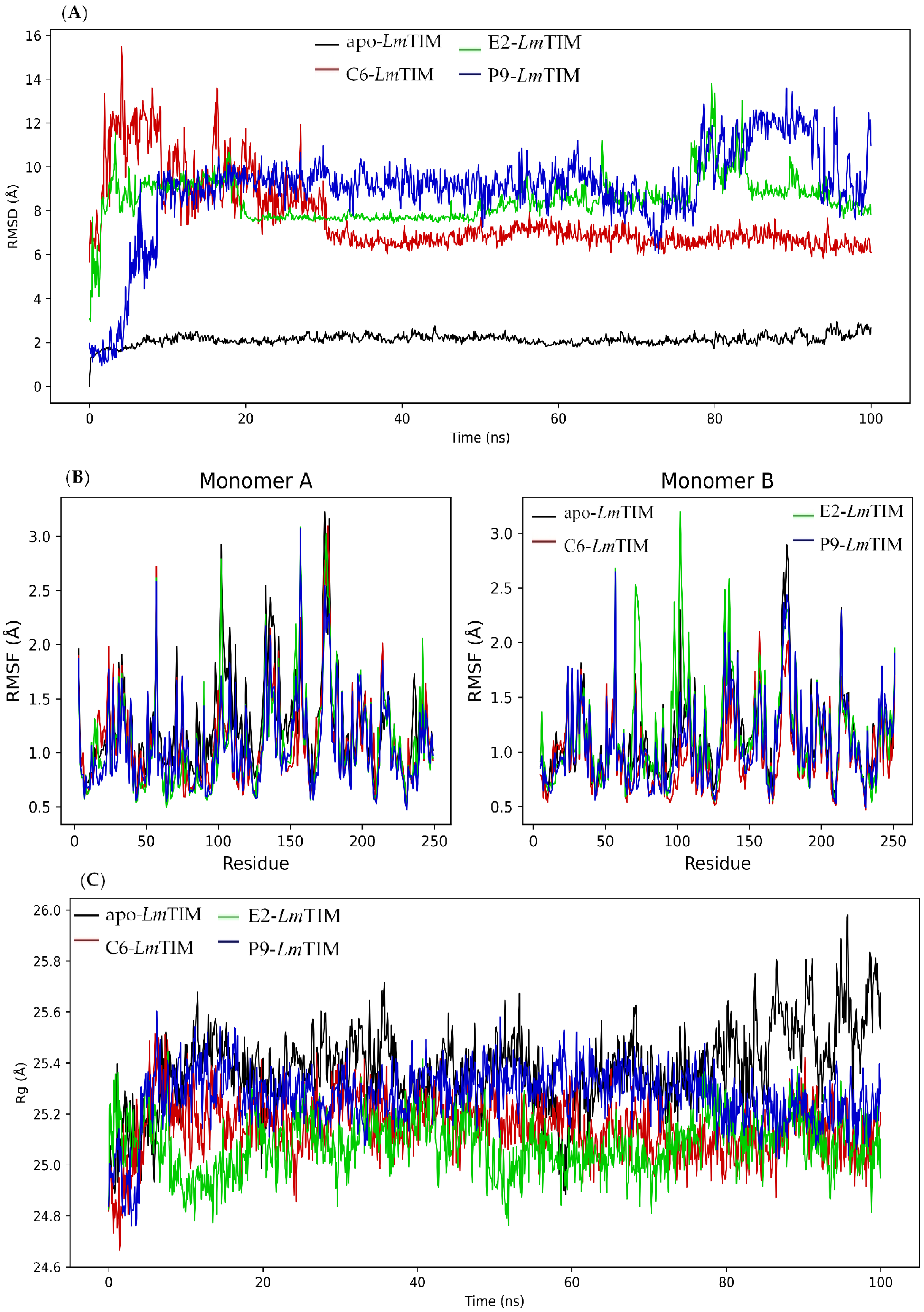
The analysis of the apo-LmTIM protein showed a RMSD with a minimum of 0.01 Å and a maximum of 3.12 Å, as well as a mean oscillation of 2.13 Å. The C6-LmTIM complex had an RMSD of 5.62 to 15.77 Å and a mean oscillation of 7.62 Å. The E2-LmTIM complex fluctuated from 2.61 Å to 13.86 Å with a mean oscillation of 8.48 Å. The E2-LmTIM complex showed a change in the initial position in the first 20 ns (Figure S1 of the Supplementary Material). After that, it remained stable until approximately 80 ns (with a RMSD mean of 3.65 Å), again observing a fluctuation between 80-82 ns and continuing stable until 100 ns. On the other hand, the P9-LmTIM complex showed a fluctuation of 0.92 to 14.25 Å and a mean oscillation of 9.01 Å (Figure 2A). The RMSD in the first 10 ns had a large fluctuation that can be attributed to a change in the initial position of the compound at the binding site (Figure S2 of the Supplementary Material). Subsequently, the complex had a stability of 10 to 80 ns (with a RMSD mean of 1.51 Å).
In general, the compounds P9, E2, and the control ligand in complex with LmTIM had a fluctuating behavior that suggests a change in the initial binding position [38,39]. The complex with the lowest RMSD and minimal differences between oscillations has been described as the most stable [40]. In this sense, the E2-LmTIM complex showed less oscillation and better stability after 20 ns than the P9-LmTIM complex. Both complexes showed a greater oscillation than the C6-LmTIM complex. Although an interaction profile analysis of molecular dynamics allowed us to observe that some initial interactions were maintained in both complexes (Figures S3 and S4 of the Supplementary Material).
The RMSF was also analyzed during molecular dynamics simulation (Figure 2B). The RMSF is a measure of the variation in the structure of a protein over time, calculated from a trajectory generated by molecular dynamics simulation. This measure allows evaluation of the stability and flexibility of the protein, as well as the influence of the interaction between the protein and the ligand on the stability of the system [41,42]. The RMSF results showed a similar fluctuation pattern between the apo-LmTIM and the protein in complex with the ligands. The apo-LmTIM protein showed a RMSF with a minimum of 0.52 Å, a maximum of 3.23 Å, and a mean oscillation of 1.20 Å (Figure 2B). The C6-LmTIM complex had a RMSF of 0.47 to 3.10 Å with a mean oscillation of 1.05 Å. The E2-LmTIM complex showed a fluctuation from 0.50 Å to 3.19 Å with a mean oscillation of 1.14 Å. The P9-LmTIM complex fluctuated from 0.47 to 3.08 Å with a mean oscillation of 1.07 Å.
We observed, in the RMSF calculation, that the high fluctuation in some regions according to the RMSD pattern, may be due to residues that are in constant movement in both monomers of the protein, such as the loops, with this movement being a factor that contributes to the elevation of the RMSD values. This finding has been described in the TIM of other species, such as Trypanosoma cruzi with approximately 82% identity to the LmTIM interface [43,44].
Finally, the rotation radius (Rg) of apo-LmTIM and the protein in complex with the compounds (C6, E2, and P9) (Figure 2C) was analyzed to predict the structural variations of the protein during the molecular dynamics analysis [45]. The free folding of LmTIM maintains an almost constant fluctuation between 24.79 and 25.98 Å with a difference in the oscillation of 1.19 Å during the 100 ns analyzed with a mean of 25.38 Å (Figure 2C). The C6-LmTIM complex fluctuated between 24.62 and 25.64 Å with a difference of 1.02 Å. The E2-LmTIM complex fluctuated from 24.62 to 25.45 Å with a difference of 0.83 Å, and the P9-LmTIM complex from 24.71 to 25.67 Å with a difference of 0.95 Å. Similar Rg values in the three complexes and greater compactness in their dynamics suggest that ligand interactions do not influence the variation of the LmTIM structure [46].

2.5. ADMET In Silico

Finally, an in silico analysis of the molecular, pharmacokinetic (SwissADME), and hepatotoxicity (ProTox-II) physicochemical properties of compounds P9 and E2 was performed. The results are shown in Table 5.
Compounds P9 and E2 complied with the Lipinski’s rule, which plays an important role in drug discovery and development [47,48]. On the other hand, the predictive study showed high human intestinal absorption of the two compounds. Furthermore, moderate solubility was predicted. However, these compounds are substrates of P-glycoprotein, an efflux pump that plays an important role in normal physiological detoxification and is associated with drug resistance [49]. Some antibiotics and anticancer drugs have been described that can be P-glycoprotein substrates and be used [50].
Hepatotoxicity predictions were negative, and regarding CYP450 inhibition, they showed that, according to structure, compound E2 inhibits two isoforms (1A2 and 2C19). Compound P9 is likely an inhibitor of the four CYP450 isoforms analyzed, giving rise to drug–drug interactions. It would be important to search for optimization in the bioavailability of this class of compounds with leishmanicidal potential.

2.6. Molecular Docking on HsTIM

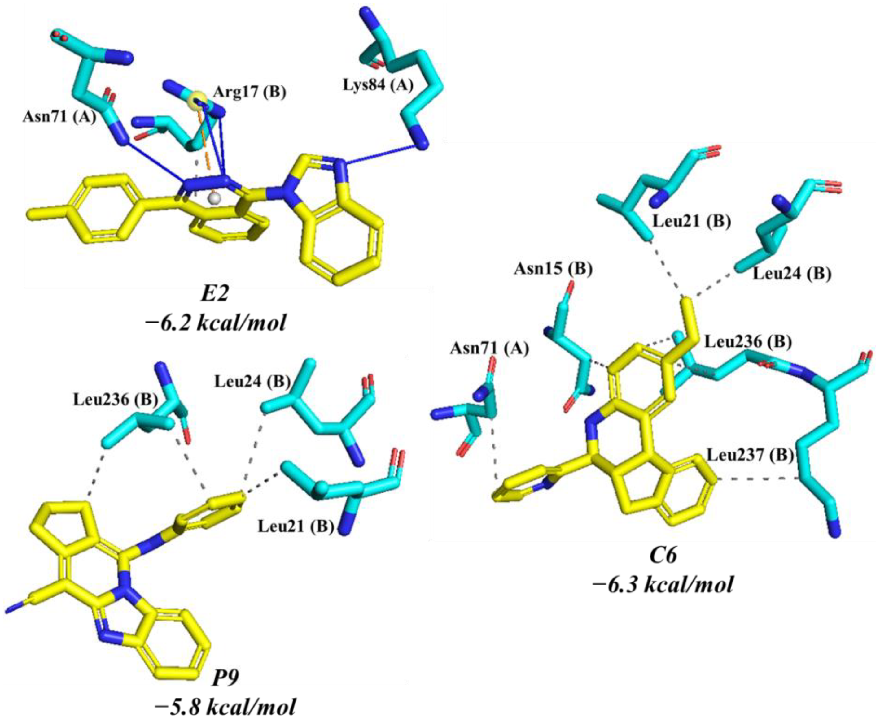
Compounds C6, E2, and P9 were evaluated against HsTIM by molecular docking (Figure 3) to establish the potential selectivity. Control compound C6 showed a docking score of −6.3 Kcal/mol and seven hydrophobic contacts with Asn71 in monomer A and Asn15, Leu21, Leu24, Leu236, Leu237 in monomer B. Compound P9 exhibited a docking score of −5.8 Kcal/mol. The phenyl ring of P9 was oriented towards the leucine triad (Leu21, Leu24, and Leu236 in the B monomer), which led to the formation of hydrophobic interactions between the carbons of the leucine residues and the carbons of the aromatic ring. Compound E2 presented a docking score of −6.2 Kcal/mol. It only showed interactions with three residues forming four hydrogen bonds, a hydrophobic interaction, and a salt bridge with Arg17, Asn71, and Lys84 in monomer A (Figure 3).

2.7. Molecular Dynamics Simulation on HsTIM

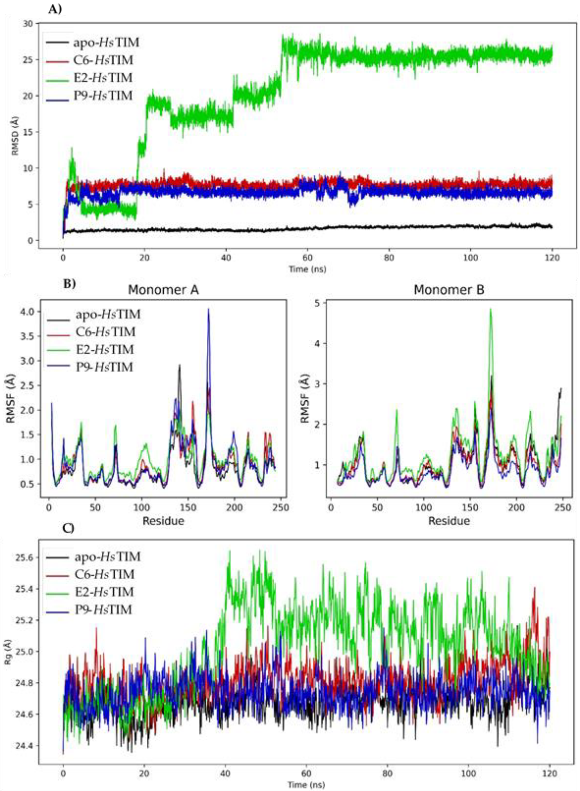
Molecular dynamics simulations were performed (Figure 4) to determine the stability of the complexes formed from the compounds C6, E2 and P9 at the HsTIM interface. The RMSD values of apo-HsTIM remained constant with a minimum fluctuation of 0.30 Å, a maximum of 2.49 Å, and a mean oscillation of 1.63 Å (Figure 4A). The RMSD value of the C6-HsTIM complex ranged from 0.77 to 9.92 Å with a mean range of 7.58 Å. The RMSD value for the complex with P9 was from 0.73 to 9.52 Å with a mean oscillation of 6.60 Å. The E2-HsTIM complex presented a RMSD of 1.14 to 28.64 Å with a mean oscillation of 20.24 Å, with this being the most unstable compound. Figure 4B shows the RMSF plot with large fluctuations in most regions according to the RMSD pattern.
In addition, the Rg (Figure 4C) was determined for the apo-HsTIM with values from 24.29 to 25.17 Å, with a mean oscillation of 24.68 Å. For the C6-HsTIM and P9-HsTIM complex, the Rg was very similar (24.35 and 24.36 Å) with a mean oscillation of 24.81 and 24.74 Å, respectively. E2-HsTIM complex presented a minimum fluctuation of 24.32 Å and a maximum of 25.73 Å with a mean oscillation of 24.74 Å.

3. Materials and Methods

3.1. Control Compounds

Six compounds (C1C6) with leishmanicidal or inhibitory activity against LmTIM were sketched in ChemDraw and saved in SDF format. Subsequently, they were minimized and converted to pdbqt format with OpenBabel.

3.2. Molecular Docking Analysis

The crystallographic structure of the LmTIM protein was obtained from the protein data bank (PDB) (http://www.pdb.org, accessed on 20 March 2020) [51] with the ID: 1AMK (resolution 1.83 Å), as well as the HsTIM structure with the ID: 4POC. The structures were prepared for molecular docking with the UCSF Chimera 1.14.1 DockPrep tool (The Regents of the University of California, Oakland, California) [52]. Additionally, the prepare_receptor4.py script from MGLTools 1.5.6 (Center for Computational Structural Biology, La Jolla, California) was used to add AutoDock atom types and Gasteiger charges to them.
The prediction of potential binding sites on LmTIM was first performed using the DoGSiteScorer tool (Center for Bioinformatics, Bundesstr, Germany) from the Proteins Plus server (https://proteins.plus/) (accessed on 23 March 2020) [53]. Subsequently, a blind molecular docking was performed. For this, the receptor was defined as rigid, and the docking protocol was setup up and performed with PyRx software, which works with AutoDock vina 1.1.2 [54]. For docking at the binding site, the conformational search space was determined by setting the coordinates to the center of the residues at the interface (X = −5.933, Y = −8.890, and Z = 7.297) using the PyRx software. The binding site on HsTIM was determined by overlap between LmTIM (ID: 1AMK) and HsTIM apoprotein (ID: 4POC) using UCSF Chimera. Based on binding site prediction, the dimer interface residues were selected for a guided molecular docking on both LmTIM and HsTIM as previously described [55].

3.3. Ligand-Based Virtual Screening

LBVS was carried out by substructure search using the benzimidazole scaffold in the ZINC15 database (https://zinc15.docking.org/) (accessed on 28 March 2020) [56]. Subsequently, the structures were obtained using SMILES representations. Lipinski’s rule was applied using the OpenBabel program. Finally, the structures were prepared for molecular docking at the LmTIM interface using the PyRx program, using the affinity energy of the compound C6 (−9.2 Kcal/mol) as the cutoff. Subsequently, through the PLIP web server, an interaction profile was generated for each of the complexes [57]. With the scikit-learn library and the DataWarrior program (https://openmolecules.org/datawarrior/) (accessed on 23 March 2020) [58,59], the compounds were clustered according to their interaction profiles and the kind structure similarity. Finally, two compounds were selected based on cost and availability for evaluation against L. mexicana promastigotes and molecular dynamics analysis.

3.4. In Vitro Leishmanicidal Activity

The leishmanicidal assay was carried out following the procedure reported by Muñoz et al. [60] and Inchausti et al. [61]. The strain of L. mexicana MHOM/MX/2011/Lacandona, kindly donated by Dr. Ingeborg Becker Fauser of the UNAM, was used, which was maintained in Schneider culture medium with 10% fetal bovine serum (FBS); parasites in the log phase of their growth cycle were transferred to a microplate (96 wells; 1 × 10⁶ parasites/well). Stock solutions of DMSO (blank), pentamidine (positive control), and benzimidazole derivatives were diluted in Schneider’s medium to <100 μg/mL, added to the plate, and incubated at 27 °C for 72 h. IC50 values (µg/mL) were obtained using the Biostat 2009 statistical program. Subsequently, the results were converted to micromolar. The benzimidazole derivatives (P9 and E2) were purchased from MolPort and worked up without further purification. The assay was performed in triplicate.

3.5. Molecular Dynamics Analysis

For the analysis of the molecular dynamics of compounds P9 and E2, the open source software package GROMACS 5.1.2 [62] was used. Protein ID: 1AMK was parameterized in the AMBER03 force field (ff94/ff99 modification by Duan et al. [63] with the GROMACS pdb2gmx software package. The protonation state of the protein, pH 7, was previously calculated with the PROPKA tool implemented in UCSF Chimera. On the other hand, the topology of the compounds was generated with ACPYPE Server (http://webapps.ccpn.ac.uk/acpype accessed 18 May 2020) [64], which is based on the General Amber Force Field (GAFF). The system was a dodecahedron with periodic boundary conditions. In addition to containing the protein–ligand complex, it was filled with TIP3P water molecules and the number of ions (Cl or Na+) necessary to have a neutral charge in the system. Before running the dynamics, the system was energetically minimized using the steepest descent algorithm. Then, two equilibrium steps were performed with 1000 kJ/mol nm2 constraints on the movement of light and heavy proteins and atoms. The first stage was at constant pressure, implementing the frog-jump method and the v-rescale thermostat to bring the system from 0 to 300 K. The second stage was performed at constant temperature again with the frog-jump method, but now with the Berendsen barostat method to bring the system from 1 to 2 bar. Both stages achieved a duration of 100 ps. Once the system was balanced, molecular dynamics was performed with a trajectory of 100 ns for LmTIM and 120 ns for HsTIM, where interactions and long-range forces were calculated with the particle-mesh Ewald (PME) method, establishing the Lennard-Jones and Coulomb contributions at 1.2. nm, balancing the system with samples at 100 ps. The stability of the complexes was determined using the GROMACS software tools and the root mean square deviation (RMSD) between the α carbons and the ligand was obtained, and the root mean square fluctuation (RMSF) of α carbons, together with two-dimensional structure and the radius of gyration (Rg), were calculated.3.6. Analysis of Molecular Physicochemical Properties
Compounds P9 and E2 were analyzed in silico with the SwissADME website (http://www.swissadme.ch/, accessed 20 August 2021) [65] to determine their physicochemical and pharmacokinetic properties, as well as their hepatotoxicity from the ProTox-II server (https://tox-new.charite.de/protox_II/, accessed 24 October 2021) [66].

4. Conclusions

In this work, a virtual screening based on the benzimidazole scaffold, and a molecular docking directed at the LmTIM dimer interface, allowed the prediction of 175 new benzimidazole derivatives with docking scores between 10.8 and −9.0 Kcal/mol on LmTIM. The in vitro evaluation against the promastigote forms of L. mexicana determined that compound E2 has better leishmanicidal activity than P9 and a value similar to pentamidine. Finally, a low affinity (−5.8 Kcal/mol) for E2 and the molecular dynamics studies at the HsTIM interface suggest selectivity on LmTIM. These results encourage us to continue the study of benzimidazole derivatives to obtain new and more selective leishmanicidal agents.

Supplementary Materials

The following supporting information can be downloaded at: https://www.mdpi.com/article/10.3390/ph16030390/s1, Figure S1: Molecular dynamics trajectory analysis of the E2-LmTIM complex; Figure S2:Molecular dynamics trajectory analysis of the P9-LmTIM complex; Figure S3: Interaction profile of the P9-LmTIM complex during molecular dynamics analysis; Figure S4: Interaction profile of the E2-LmTIM complex during molecular dynamics analysis.

Author Contributions

Conceptualization, G.R.; methodology, L.K.V.-J., A.J.-S., T.D.-M., M.J.C.-B. and L.D.G.-M.; software, L.K.V.-J., T.D.-M., E.O.-P. and A.J.-S.; validation, E.E.L.-R., E.R.-M., A.J.-S. and I.P.; formal analysis, E.O.-P.; resources, E.R.-M., E.E.L.-R. and G.R.; data curation, T.D.-M. and L.K.V.-J.; writing—original draft preparation, L.K.V.-J. and G.R.; writing—review and editing, L.D.G.-M., E.E.L.-R., M.J.C.-B., I.P., E.R.-M. and G.R.; supervision, M.J.C.-B., E.E.L.-R., I.P. and E.R.-M.; project administration, E.O.-P.; funding acquisition, M.J.C.-B., E.R.-M. and G.R. All authors have read and agreed to the published version of the manuscript.

Funding

This research was funded by “Secretaria de Investigación y Posgrado del Instituto Politecnico Nacional, grant number 20220935 and 20230935”.

Institutional Review Board Statement

Not applicable.

Informed Consent Statement

Not applicable.

Data Availability Statement

Date is contained in the article.

Acknowledgments

To the Secretaria de Investigacion y Posgrado del Instituto Politecnico Nacional and Proyectos de Desarrollo Tecnológico o innovación para alumnos del IPN 2022 for their support.

Conflicts of Interest

The authors declare no conflict of interest.

References

  1. Abadías-Granado, I.; Diago, A.; Cerro, P.A.; Palma-Ruiz, A.M.; Gilaberte, Y. Cutaneous and Mucocutaneous Leishmaniasis. Actas Dermosifiliogr. 2021, 27, 601–618. [Google Scholar] [CrossRef]
  2. Zamora-Ledesma, S.; Hernández-Camacho, N.; Sánchez-Moreno, M.; Ruiz-Piña, H.; Villagrán-Herrera, M.E.; Marín-Sánchez, C.; Carrillo-Angeles, I.G.; Jones, R.W.; Camacho-Macías, B. Seropositivity for Trypanosoma cruzi and Leishmania mexicana in dogs from a metropolitan region of Central Mexico. Vet. Parasitol. Reg. Stud. Rep. 2020, 22, 100459. [Google Scholar] [CrossRef] [PubMed]
  3. Pan American Health Organization (PAHO). Cutaneous and Mucosal Leishmaniasis. Available online: https://www3.paho.org/hq/index.php?option=com_content&view=article&id=6417:2012-leishmaniasis-cutanea-mucosa&Itemid=39345&lang=en (accessed on 10 May 2022).
  4. Ulloa, J.L.; Spina, R.; Casasco, A.; Petray, P.B.; Martino, V.; Sosa, M.A.; Frank, F.M.; Muschietti, L.V. Germacranolide-type sesquiterpene lactones from Smallanthus sonchifolius with promising activity against Leishmania mexicana and Trypanosoma cruzi. Parasit. Vectors. 2017, 10, 567. [Google Scholar] [CrossRef] [PubMed]
  5. World Health Organization (WHO). Control of the Leishmaniases. Report of a meeting of the WHO Expert Committee on the Control of Leishmaniases. WHO Technol. Rep. Ser. 2010, 949, 1–186. [Google Scholar]
  6. DNDi-Drugs for Neglected Diseases Initiative. Available online: http://www.dndi.org/diseases-projects/leishmaniasis/ (accessed on 31 May 2022).
  7. Matadamas-Martínez, F.; Hernández-Campos, A.; Téllez-Valencia, A.; Vázquez-Raygoza, A.; Comparán-Alarcón, S.; Yépez-Mulia, L.; Castillo, R. Leishmania mexicana Trypanothione Reductase Inhibitors: Computational and Biological Studies. Molecules 2019, 24, 3216. [Google Scholar] [CrossRef] [Green Version]
  8. De Sousa, L.R.; Wu, H.; Nebo, L.; Fernandes, J.B.; da Silva, M.F.; Kiefer, W.; Schirmeister, T.; Vieira, P.C. Natural products as inhibitors of recombinant cathepsin L of Leishmania mexicana. Exp. Parasitol. 2015, 156, 42–48. [Google Scholar] [CrossRef]
  9. Suresh, S.; Bressi, J.C.; Kennedy, K.J.; Verlinde, C.L.; Gelb, M.H.; Hol, W.G. Conformational changes in Leishmania mexicana glyceraldehyde-3-phosphate dehydrogenase induced by designed inhibitors. J. Mol. Biol. 2001, 309, 423–435. [Google Scholar] [CrossRef] [Green Version]
  10. Olivares-Illana, V.; Pérez-Montfort, R.; López-Calahorra, F.; Costas, M.; Rodríguez-Romero, A.; Tuena de Gómez-Puyou, M.; Gómez Puyou, A. Structural differences in triosephosphate isomerase from different species and discovery of a multi-trypanosomatid inhibitor. Biochemistry 2006, 45, 2556–2560. [Google Scholar] [CrossRef]
  11. Velanker, S.S.; Ray, S.S.; Gokhale, R.S.; Suma, S.; Balaram, H.; Balaram, P.; Murthy, M.R. Triosephosphate isomerase from Plasmodium falciparum: The crystal structure provides insights into antimalarial drug design. Structure 1997, 5, 751–761. [Google Scholar] [CrossRef] [Green Version]
  12. Vázquez-Raygoza, A.; Cano-González, L.; Velázquez-Martínez, I.; Trejo-Soto, P.; Castillo, R.; Hernández-Campos, A.; Hernández-Luis, F.; Oria-Hernández, J.; Castillo-Villanueva, A.; Avitia-Domínguez, C.; et al. Species-Specific Inactivation of Triosephosphate Isomerase from Trypanosoma brucei: Kinetic and Molecular Dynamics Studies. Molecules 2017, 22, 2055. [Google Scholar] [CrossRef] [Green Version]
  13. Gómez-Puyou, A.; Saavedra-Lira, E.; Becker, I.; Zubillaga, R.A.; Rojo-Domínguez, A.; Perez-Montfort, R. Using evolutionary changes to achieve species-specific inhibition of enzyme action-studies with triosephosphate isomerase. Chem. Biol. 1995, 2, 847. [Google Scholar] [CrossRef] [Green Version]
  14. Sanabria-Ayala, V.; Belmont, I.; Abraham, L. Triosephosphate isomerase of Taenia solium (TTPI): Phage display and antibodies as tools for finding target regions to inhibit catalytic activity. Parasitol. Res. 2015, 114, 55. [Google Scholar] [CrossRef]
  15. Saramago, L.; Gomes, H.; Aguilera, E.; Cerecetto, H.; González, M.; Cabrera, M.; Alzugaray, M.; daSilva Vaz Junior, I.; Nunes da Fonseca, R.; Aguirre-López, B.; et al. Novel and Selective Rhipicephalus microplus Triosephosphate Isomerase Inhibitors with Acaricidal Activity. Vet. Sci. 2018, 5, 74. [Google Scholar] [CrossRef] [Green Version]
  16. Juárez-Saldivar, A.; Barbosa-Cabrera, E.; Lara-Ramírez, E.E.; Paz-González, A.D.; Martínez-Vázquez, A.V.; Bocanegra-García, V.; Palos, I.; Campillo, N.E.; Rivera, G. Virtual Screening of FDA-Approved Drugs against Triose Phosphate Isomerase from Entamoeba histolytica and Giardia lamblia Identifies Inhibitors of Their Trophozoite Growth Phase. Int. J. Mol. Sci. 2021, 22, 5943. [Google Scholar] [CrossRef]
  17. Azahar, N.H.; Ab Dullah, S.; Abdullah, R.; Ahmat, N.; Md Akim, A.; Ab Hamid, H. Mutagenic Study of Benzimidazole Derivatives with (+S9) and without (-S9) Metabolic Activation. Int. J. Mol. Sci. 2019, 20, 4324. [Google Scholar] [CrossRef] [Green Version]
  18. Tonelli, M.; Novelli, F.; Tasso, B.; Vazzana, I.; Sparatore, A.; Boido, V.; Sparatore, F.; La Colla, P.; Sanna, G.; Giliberti, G.; et al. Antiviral activity of benzimidazole derivatives. III. Novel anti-CVB-5, anti-RSV and anti-Sb-1 agents. Bioorg. Med. Chem. 2014, 22, 4893–4909. [Google Scholar] [CrossRef]
  19. Picanço, G.A.; de Lima, N.F.; Gomes, T.C.; Fraga, C.M.D.; Alves, D.S.M.M.; Castillo, R.; da Costa, T.L.; Lino-Junior, R.S.; Ambrosio, J.; Vinaud, M.C. Partial inhibition of the main energetic pathways and its metabolic consequences after in vivo treatment with benzimidazole derivatives in experimental neurocysticercosis. Parasitology 2019, 146, 1578–1582. [Google Scholar] [CrossRef]
  20. Sirim, M.M.; Krishna, V.S.; Sriram, D.M.; Unsal Tan, O. Novel benzimidazole-acrylonitrile hybrids and their derivatives: Design, synthesis and antimycobacterial activity. Eur. J. Med. Chem. 2020, 188, 112010. [Google Scholar] [CrossRef]
  21. Tonelli, M.; Gabriele, E.; Piazza, F.; Basilico, N.; Parapini, S.; Tasso, B.; Loddo, R.; Sparatore, F.; Sparatore, A. Benzimidazole derivatives endowed with potent antileishmanial activity. J. Enzyme Inhib. Med. Chem. 2018, 33, 210–226. [Google Scholar] [CrossRef] [Green Version]
  22. Babkov, D.A.; Zhukowskaya, O.N.; Borisov, A.V.; Babkova, V.A.; Sokolova, E.V.; Brigadirova, A.A.; Litvinov, R.A.; Kolodina, A.A.; Morkovnik, A.S.; Sochnev, V.; et al. Towards multi-target antidiabetic agents: Discovery of biphenyl-benzimidazole conjugates as AMPK activators. Bioorg. Med. Chem. Lett. 2019, 29, 2443–2447. [Google Scholar] [CrossRef]
  23. Hsieh, C.Y.; Ko, P.W.; Chang, Y.J.; Kapoor, M.; Liang, Y.C.; Chu, H.L.; Lin, H.H.; Horng, J.C.; Hsu, M.H. Design and Synthesis of Benzimidazole-Chalcone Derivatives as Potential Anticancer Agents. Molecules 2019, 24, 3259. [Google Scholar] [CrossRef] [PubMed] [Green Version]
  24. Torres-Gómez, H.; Hernández-Núñez, E.; León-Rivera, I.; Guerrero-Alvarez, J.; Cedillo-Rivera, R.; Moo-Puc, R.; Argotte-Ramos, R.; Rodríguez-Gutiérrez, M.C.; Chan-Bacab, M.J.; Navarrete-Vázquez, G. Design, synthesis and in vitro antiprotozoal activity of benzimidazole-pentamidine hybrids. Bioorg. Med. Chem. Lett. 2008, 18, 3147–3151. [Google Scholar] [CrossRef] [PubMed]
  25. Nieto-Meneses, R.; Castillo, R.; Hernández-Campos, A.; Maldonado-Rangel, A.; Matius-Ruiz, J.B.; Trejo-Soto, P.J.; Nogueda-Torres, B.; Dea-Ayuela, M.A.; Bolás-Fernández, F.; Méndez-Cuesta, C.; et al. In vitro activity of new N-benzyl-1H-benzimidazol-2-amine derivatives against cutaneous, mucocutaneous and visceral Leishmania species. Exp. Parasitol. 2018, 184, 82–89. [Google Scholar] [CrossRef]
  26. Hernández-Chinea, C.; Carbajo, E.; Sojo, F.; Arvelo, F.; Kouznetsov, V.V.; Romero-Bohórquez, A.R.; Romero, P.J. In Vitro Activity of Synthetic tetrahydroindeno[2,1-c]quinolines on Leishmania Mexicana. Parasitol. Int. 2015, 64, 479–483. [Google Scholar] [CrossRef] [PubMed]
  27. Volkamer, A.; Kuhn, D.; Rippmann, F.; Rarey, M. DoGSiteScorer: A web server for automatic binding site prediction, analysis and druggability assessment. Bioinformatics 2012, 28, 2074–2075. [Google Scholar] [CrossRef] [Green Version]
  28. Breznik, M.; Ge, Y.; Bluck, J.P.; Briem, H.; Hahn, D.F.; Christ, C.D.; Mortier, J.; Mobley, D.L.; Meier, K. Prioritizing Small Sets of Molecules for Synthesis through in-silico Tools: A Comparison of Common Ranking Methods. Chem. Med. Chem. 2023, 18, e202200425. [Google Scholar] [CrossRef]
  29. Espinoza-Fonseca, L.M.; Trujillo-Ferrara, J.G. Toward a rational design of selective multi-trypanosomatid inhibitors: A computational docking study. Bioorg. Med. Chem. Lett. 2006, 16, 6288–6292. [Google Scholar] [CrossRef]
  30. Velázquez-López, J.M.; Hernández-Campos, A.; Yépez-Mulia, L.; Téllez-Valencia, A.; Flores-Carrillo, P.; Nieto-Meneses, R.; Castillo, R. Synthesis and trypanocidal activity of novel benzimidazole derivatives. Bioorg. Med. Chem. Lett. 2016, 26, 4377–4381. [Google Scholar] [CrossRef]
  31. Kurkcuoglu, Z.; Ural, G.; Demet Akten, E.; Doruker, P. Blind Dockings of Benzothiazoles to Multiple Receptor Confor-mations of Triosephosphate Isomerase from Trypanosoma cruzi and Human. Mol. Inform. 2011, 30, 986–995. [Google Scholar] [CrossRef]
  32. Téllez-Valencia, A.; Avila-Ríos, S.; Pérez-Montfort, R.; Rodríguez-Romero, A.; de Gómez-Puyou, M.T.; López-Calahorra, F.; Gómez-Puyou, A. Highly Specific Inactivation of Triosephosphate Isomerase from Trypanosoma Cruzi. Biochem. Biophys. Res. Commun. 2002, 295, 958–963. [Google Scholar] [CrossRef]
  33. Romo-Mancillas, A.; Téllez-Valencia, A.; Yépez-Mulia, L.; Hernández-Luis, F.; Hernández-Campo, A.; Castillo, R. The design and inhibitory profile of new benzimidazole derivatives against triosephosphate isomerase from Trypanosoma cruzi: A problem of residue motility. J. Mol. Graph Model. 2011, 30, 90–99. [Google Scholar] [CrossRef]
  34. Bansal, Y.; Silakari, O. The therapeutic journey of benzimidazoles: A review. Bioorg Med. Chem. 2012, 20, 6208–6236. [Google Scholar] [CrossRef]
  35. Beltran-Hortelano, I.; Alcolea, V.; Font, M.; Pérez-Silanes, S. The role of imidazole and benzimidazole heterocycles in Chagas disease: A review. Eur. J. Med. Chem. 2020, 206, 112692. [Google Scholar] [CrossRef]
  36. De Vivo, M.; Masetti, M.; Bottegoni, G.; Cavalli, A. Role of molecular dynamics and related methods in drug discovery. J. Med. Chem. 2016, 59, 4035–4061. [Google Scholar] [CrossRef]
  37. Arooj, M.; Shehadi, I.; Nassab, C.N.; Mohamed, A.A. Physicochemical stability study of protein-benzoic acid complexes using molecular dynamics simulations. Amino Acids 2020, 52, 1353–1362. [Google Scholar] [CrossRef]
  38. Espinosa-Bustos, C.; Ortiz-Pérez, M.; González-González, A.; Zarate, A.M.; Rivera, G.; Belmont-Díaz, J.A.; Saavedra, E.; Cuellar, M.A.; Vázquez, K.; Salas, C.O. New Amino Naphthoquinone Derivatives as Anti-Trypanosoma cruzi Agents Targeting Trypanothione Reductase. Pharmaceutics 2022, 14, 1121. [Google Scholar] [CrossRef]
  39. Vázquez-Jiménez, L.K.; Juárez-Saldivar, A.; Gómez-Escobedo, R.; Delgado-Maldonado, T.; Méndez-Álvarez, D.; Palos, I.; Bandyopadhyay, D.; Gaona-Lopez, C.; Ortiz-Pérez, E.; Nogueda-Torres, B.; et al. Ligand-Based Virtual Screening and Molecular Docking of Benzimidazoles as Potential Inhibitors of Triosephosphate Isomerase Identified New Trypanocidal Agents. Int. J. Mol. Sci. 2022, 23, 10047. [Google Scholar] [CrossRef]
  40. Méndez-Álvarez, D.; Herrera-Mayorga, V.; Juárez-Saldivar, A.; Paz-González, A.D.; Ortiz-Pérez, E.; Bandyopadhyay, D.; Pérez-Sánchez, H.; Rivera, G. Ligand-based virtual screening, molecular docking, and molecular dynamics of eugenol analogs as potential acetylcholinesterase inhibitors with biological activity against Spodoptera frugiperda. Mol. Divers. 2022, 26, 2025–2037. [Google Scholar] [CrossRef]
  41. Collier, T.A.; Piggot, T.J.; Allison, J.R. Molecular Dynamics Simulation of Proteins. Methods Mol. Biol. 2020, 2073, 311–327. [Google Scholar] [CrossRef]
  42. Fatriansyah, J.F.; Rizqillah, R.K.; Yandi, M.Y.; Fadilah, Sahlan, M. Molecular docking and dynamics studies on propolis sulabiroin-A as a potential inhibitor of SARS-CoV-2. J. King Saud Univ. Sci. 2022, 34, 101707. [Google Scholar] [CrossRef]
  43. Wierenga, R.K.; Noble, M.E. Comparison of the refined crystal structures of liganded and unliganded chicken, yeast and trypanosomal triosephosphate isomerase. J. Mol. Biol. 1992, 224, 1115–1126. [Google Scholar] [CrossRef] [PubMed]
  44. Ghosh, R.; Chakraborty, A.; Biswas, A.; Chowdhuri, S. Evaluation of green tea polyphenols as novel corona virus (SARS-CoV-2) main protease (Mpro) inhibitors-an in silico docking and molecular dynamics simulation study. J. Biomol. Struct. Dyn. 2021, 39, 4362–4374. [Google Scholar] [CrossRef] [PubMed]
  45. Wolf, A.; Kirschner, K.N. Principal component and clustering analysis on molecular dynamics data of the ribosomal L11·23S subdomain. J. Mol. Model. 2013, 19, 539–549. [Google Scholar] [CrossRef] [Green Version]
  46. Singh, S. Dynamics of heroin molecule inside the lipid membrane: A molecular dynamics study. J. Mol. Model. 2019, 25, 121. [Google Scholar] [CrossRef] [PubMed]
  47. Lipinski, C.A.; Lombardo, F.; Dominy, B.W.; Feeney, P.J. Experimental and computational approaches to estimate solubility and permeability in drug discovery and development settings. Adv. Drug. Deliv. Rev. 2001, 46, 3–26. [Google Scholar] [CrossRef]
  48. Benet, L.Z.; Hosey, C.M.; Ursu, O.; Oprea, T.I. BDDCS, the Rule of 5 and drugability. Adv. Drug Deliv. Rev. 2016, 101, 89–98. [Google Scholar] [CrossRef] [Green Version]
  49. Mollazadeh, S.; Sahebkar, A.; Hadizadeh, F.; Behravan, J.; Arabzadeh, S. Structural and functional aspects of P-glycoprotein and its inhibitors. Life Sci. 2018, 214, 118–123. [Google Scholar] [CrossRef]
  50. Waghray, D.; Zhang, Q. Inhibit or Evade Multidrug Resistance P-Glycoprotein in Cancer Treatment. J. Med. Chem. 2018, 61, 5108–5121. [Google Scholar] [CrossRef]
  51. Berman, H.M.; Kleywegt, G.J.; Nakamura, H.; Markley, J.L. The future of the Protein Data Bank. Biopolymers 2013, 99, 218–222. [Google Scholar] [CrossRef]
  52. Pettersen, E.F.; Goddard, T.D.; Huang, C.C.; Couch, G.S.; Greenblatt, D.M.; Meng, E.C.; Ferrin, T.E. UCSF Chimera-A visualization system for exploratory research and analysis. J. Comput. Chem. 2004, 25, 1605–1612. [Google Scholar] [CrossRef] [Green Version]
  53. Fährrolfes, R.; Bietz, S.; Flachsenberg, F.; Meyder, A.; Nittinger, E.; Otto, T.; Volkamer, A.; Rarey, M. ProteinsPlus: A web portal for structure analysis of macromolecules. Nucleic Acids Res. 2017, 45, W337–W343. [Google Scholar] [CrossRef] [Green Version]
  54. Trott, O.; Olson, A.J. AutoDock Vina: Improving the speed and accuracy of docking with a new scoring function, efficient optimization, and multithreading. J. Comput. Chem. 2010, 31, 455–461. [Google Scholar] [CrossRef] [Green Version]
  55. Téllez-Valencia, A.; Olivares-Illana, V.; Hernandez-Santoyo, A.; Perez-Montfort, R.; Costas, M.; Rodriguez-Romero, A.; López-Calahorra, F.; de Gómez-Puyou, M.T.; Gómez-Puyou, A. Inactivation of triosephosphate isomerase from Trypanosoma cruzi by an agent that perturbs its dimer interface. J. Mol. Biol. 2004, 341, 1355–1365. [Google Scholar] [CrossRef]
  56. Sterling, T.; Irwin, J.J. ZINC 15--Ligand Discovery for Everyone. J. Chem. Inf. Model. 2015, 55, 2324–2337. [Google Scholar] [CrossRef]
  57. Salentin, S.; Schreiber, S.; Joachim-Haupt, V.; Adasme, M.F.; Schroeder, M. PLIP: Fully automated protein-ligand interaction profiler. Nucleic. Acids. Res. 2015, 43, W443–W447. [Google Scholar] [CrossRef]
  58. Abraham, A.; Pedregosa, F.; Eickenberg, M.; Gervais, P.; Mueller, A.; Kossaifi, J.; Gramfort, A.; Thirion, B.; Varoquaux, G. Machine learning for neuroimaging with scikit-learn. Front. Neuroinform. 2014, 8, 14. [Google Scholar] [CrossRef] [Green Version]
  59. López-López, E.; Naveja, J.J.; Medina-Franco, J.L. DataWarrior: An evaluation of the open-source drug discovery tool. Expert. Opin. Drug. Discov. 2019, 14, 335–341. [Google Scholar] [CrossRef]
  60. Muñoz, V.; Moretti, C.; Sauvain, M.; Caron, C.; Porzel, A.; Massiot, G.; Richard, B.; Le Men-Olivier, L. Isolation of bis-indole alkaloids with antileishmanial and antibacterial activities from Peschiera van heurkii (Syn. Tabernamontana van heurkii). Planta Med. 1994, 60, 455–459. [Google Scholar] [CrossRef] [Green Version]
  61. Inchausti, A.; Yaluff, G.; Rojas de Arias, A.; Torres, S.; Ferreira, M.E.; Nakayama, H.; Schinini, A.; Lorenzen, K.; Anke, T.; Fournet, A. Leishmanicidal and trypanocidal activity of extracts and secondary metabolites from Besidiomycetes. Phytotherapy Res. 1997, 11, 193–197. [Google Scholar] [CrossRef]
  62. Van-Der-Spoel, D.; Lindahl, E.; Hess, B.; Groenhof, G.; Mark, A.E.; Berendsen, H.J. GROMACS: Fast, flexible, and free. J. Comput. Chem. 2005, 26, 1701–1718. [Google Scholar] [CrossRef]
  63. Duan, Y.; Wu, C.; Chowdhury, S.; Lee, M.C.; Xiong, G.; Zhang, W.; Yang, R.; Cieplak, P.; Luo, R.; Lee, T.; et al. A point-charge force field for molecular mechanics simulations of proteins based on condensed-phase quantum mechanical calculations. J. Comput. Chem. 2003, 24, 1999–2012. [Google Scholar] [CrossRef] [PubMed]
  64. Sousa-da-Silva, A.W.; Wim Vranken, F. ACPYPE AnteChamber PYthon Parser interface. BMC Res. Notes 2012, 5, 367. [Google Scholar] [CrossRef] [PubMed] [Green Version]
  65. Daina, A.; Michielin, O.; Zoete, V. SwissADME: A free web tool to evaluate pharmacokinetics, drug-likeness and medicinal chemistry friendliness of small molecules. Sci. Rep. 2017, 7, 42717. [Google Scholar] [CrossRef] [PubMed] [Green Version]
  66. Banerjee, P.; Eckert, A.O.; Schrey, A.K.; Preissner, R. ProTox-II: A webserver for the prediction of toxicity of chemicals. Nucleic Acids Res. 2018, 46, W257–W263. [Google Scholar] [CrossRef] [Green Version]
Figure 1. Prediction of the binding site of control compounds by the DoGSiteScorer web server and blind molecular docking on LmTIM. The protein is represented in gray color, and the dimer interface is represented in green color.
Pharmaceuticals 16 00390 g001
Figure 2. Molecular dynamics simulation of benzimidazole derivatives (E2 and P9) and control compound C6 in complex with LmTIM. (A) Plot of RMSD of the three complexes and apo-LmTIM. (B) Diagram of the RMSF of the three complexes and apo-LmTIM. (C) Diagram of Rg of the three complexes and apo-LmTIM.
Pharmaceuticals 16 00390 g002
Figure 3. Interaction profile and docking score of compounds C6, E2 and P9 at the HsTIM interface. Hydrogen bonds are shown as blue lines, hydrophobic interaction as gray dashed lines, and salt bridge interactions as yellow dashed lines. Interactions were obtained using the PLIP online server.
Pharmaceuticals 16 00390 g003
Figure 4. Molecular dynamics simulation of benzimidazole derivatives in complex with HsTIM. (A) Plot of RMSD of the three complexes and apo-HsTIM. (B) Diagram of the RMSF of the three complexes and apo-HsTIM. (C) Diagram of the Rg of the three complexes and apo-HsTIM.
Pharmaceuticals 16 00390 g004
Table 1. Docking scores and molecular interactions of compounds used as control ligands at the LmTIM interface.
Chemical Structure of Control Ligands
Pharmaceuticals 16 00390 i001
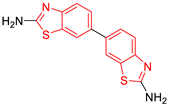
C1 DS= −7.2 Kcal/mol
1 IC50 = 0.71 μM [24]
HI: Ile69B, Ala70B, Lys71B, Glu105B, Ile109B; HB: Gln112B, Ala70B, Lys71B, Lys113B; π-S: Phe75B, Tyr102A, Tyr103A
Pharmaceuticals 16 00390 i002
C2 DS= −7.8 Kcal/mol
1 IC50 = 0.36 μM [24]
HI: Ile69B, Ala70B, Lys71B, Glu105B, Ile109B; HB: Gln112B, Ala70B, Lys71B, Lys113B; π-S: Phe75B, Tyr103A
Pharmaceuticals 16 00390 i003
C3 DS= −7.3 Kcal/mol
1 IC50 = 2.62 μM, 2 IC50 = 0.28 μM [25]
HI: Ile69B, Ala70B, Lys71B, Glu105B, Ile109B, Phe75B;
HB: Gln112B, Lys113B; π-S: Tyr103(A)
Pharmaceuticals 16 00390 i004
C4 DS= −7.1 Kcal/mol
1 IC50 = 3.21 μM, 2 IC50 = 0.26 μM [25]
HI: Ile69B, Lys71B, Glu105B, Ile109B; HB: Tyr103A
π-S: Phe75B, Tyr103A
Pharmaceuticals 16 00390 i005
C5 DS= −7.6 Kcal/mol
70% enzyme inhibition at 100 µM [10]
HI: Glu105B, Ile109B; HB: Tyr103A
Pharmaceuticals 16 00390 i006
C6 DS= −9.2 Kcal/mol
1 IC50 = 1.02 μg/mL, Cell line J774 = 187.0 μg/mL; SI = 183.3 [26]
HI: Ile69B, Phe75B, Ala70B, Tyr102A, Tyr103A, Ile109B, Glu105B
1 Promastigotes, 2 Amastigotes, DS: Docking score, HI: Hydrophobic interactions, HB: Hydrogen bonds, π–S: π-stacking interactions, SI: Selectivity index.
Table 2. Lead compounds from each group obtained by SBVS grouped by interaction profile.
GroupLead CompoundGroupLead Compound
1
(17 compounds)
Pharmaceuticals 16 00390 i007
ZINC000030028937 (P1) DS= −9.6 Kcal/mol
HI: Ala70B, Ile109B, Tyr102A, Ile69B, Lys71B; HB: Tyr103A, Lys71B, Ser72B, Ala74B; π-S: Phe75B, Tyr103A
6
(11 compounds)
Pharmaceuticals 16 00390 i008
ZINC000010116378 (P6) DS= −9.3 Kcal/mol
HI: Ile109B, Glu116B, Ala70B, Gln112B, Ile69B, Tyr102A, Phe75B; HB: Lys113B, Gln112B, Glu105B, Arg99B, Asn64B, Glu78B; π-S: Tyr103A, Tyr102A
2
(24 compounds)
Pharmaceuticals 16 00390 i009
ZINC000010125646 (P2) DS= −9.6 Kcal/mol
HI: Tyr103A, Tyr102A, Glu116B, Gln112B, Ile69B, HB: Ala70B, Lys113B; HB: Lys113B, Glu105B, Arg99B, Asn67B, Ala70B; π-S: Tyr103A, Tyr102A
7
(22 compounds)
Pharmaceuticals 16 00390 i010
ZINC000000182545 (P7) DS= −9.4 Kcal/mol
HI: Tyr102A, Ile69B, Phe75B, Pys71B, Ala70B, Ile109B, Glu105B; HB: Tyr103A;
π-S: Tyr103A
3
(17 compounds)
Pharmaceuticals 16 00390 i011
ZINC000030011443 (P3) DS= −9.6 Kcal/mol
HI: Lys71B, Ile109B, Glu105B, Ile69B, Ala70B; HB: Lys71B, Gln112B, Ala70B; π-S: Tyr103A
8
(9 compounds)
Pharmaceuticals 16 00390 i012
ZINC000030009197 (P8) DS= −9.8 Kcal/mol
HI: Ile109B, Glu105B, Ile69B, Phe75B; HB: Tyr103A, Gln112B, Tyr102A; π-S: Tyr103A
4
(14 compounds)
Pharmaceuticals 16 00390 i013
ZINC000010109617 (P4) DS= −10.3 Kcal/mol
HI: Tyr103A, Ile109B, Phe75B, Ala70B, Glu116B, Gln112B, Lys71B, Ile69B; HB: Ala70B, Gln112B, Glu105B, Arg99B, Tyr103B, Lys113B
9
(23 compounds)
Pharmaceuticals 16 00390 i014
ZINC000000183176 (P9) DS= −9.4 Kcal/mol
HI: Lys71B, Gln112B, Glu116B, Ala70B, Lys113B, Tyr103A, Tyr102A, Phe75B; HB: Gln112B, Lys113B;
π-S: Phe75B
5
(19 compounds)
Pharmaceuticals 16 00390 i015
ZINC000040058994 (P5) DS= −9.4 Kcal/mol
HI: Ile109B, Ala70B, Gln112B, Phe75B, Ile69B, Tyr102A, Tyr103A; HB: Lys113B, Tyr103B, Glu105B, Arg99B, Ala70B; π-S: Tyr103A
10
(19 compounds)
Pharmaceuticals 16 00390 i016
ZINC000030011418 (P10) DS= −9.7 Kcal/mol
HI: Lys113B, Ala70B, Phe75B, Tyr102A, Ile69B, Ile109B, Tyr103A; HB: Lys113B, Ala70B, Lys71B, Gln112B, Glu105B; π-S: Tyr102A
DS: Docking score, HI: Hydrophobic interactions, HB: Hydrogen bonds, π–S: π-stacking interactions.
Table 3. Lead compounds from each group obtained by SBVS grouped by structure.
GroupLead CompoundGroupLead Compound
1
(20 compounds)
Pharmaceuticals 16 00390 i017
ZINC000010116253 (E1) DS= −10.8 Kcal/mol
HI: Ile69B, Ile109B, Lys71B, Phe75B, Tyr102A;
HB: Gln112B; π-S: Tyr103A
6
(4 compounds)
Pharmaceuticals 16 00390 i018
ZINC000000086631 (E6) DS= −9.0 Kcal/mol
HI: Ile69B, Ile109B, Phe75B, Glu105B, Tyr102A; HB: Tyr102A; π-S: Tyr103A
2
(26 compounds)
Pharmaceuticals 16 00390 i019
ZINC000000087857 (E2) DS= −9.4 Kcal/mol
HI: Ile69B, Ile109B, Phe75B, Tyr103A, Gln112B, Glu116B; HB: Gln112B
7
(1 compound)
Pharmaceuticals 16 00390 i020
ZINC000010000495 (E7) DS= −9.7 Kcal/mol
HI: Ile69B, Ala70B, Ile109B, Phe75B, Tyr102A, Tyr103A, Gln112B; HB: Ala70B, Tyr103A, Lys113B, Lys71B
3
(102 compounds)
Pharmaceuticals 16 00390 i021
ZINC000000018961 (E3) DS= −9.3 Kcal/mol
HI: Ile69B, Ala70B, Ile109B; HB: Ala70B, Gln112B; π-S: Phe75B, Tyr103A
8
(4 compounds)
Pharmaceuticals 16 00390 i022
ZINC000010126510 (E8) DS= −9.5 Kcal/mol
HI: Ile69B, Ile109B, Phe75B, Tyr102A, Tyr103A; HB: Gln112B, Tyr103A, Tyr102A; π-S: Phe75B, Tyr102A
4
(4 compounds)
Pharmaceuticals 16 00390 i023
ZINC000000134117 (E4) DS= −9.3 Kcal/mol
HI: Ala70B, Ile109B, Lys71B, Phe75B, Glu105B; HB: Gln112B, Lys113B; π-S: Tyr103A
9
(3 compounds)
Pharmaceuticals 16 00390 i024
ZINC000010054868 (E9) DS= −9.4 Kcal/mol
HI: Ile69B, Ile109B, Tyr103A; HB: Tyr102A; π-S: Phe75B; SB: Lys113B
5
(6 compounds)
Pharmaceuticals 16 00390 i025
ZINC000010116541 (E5) DS= −9.2 Kcal/mol
HI: Ile69B, Lys71B, Glu105B; HB: Gln112B;
π-S: Phe75B, Tyr103A, Tyr102A
10
(2 compounds)
Pharmaceuticals 16 00390 i026
ZINC000020062149 (E10) DS= −9.1 Kcal/mol
HI: Ile69B, Lys71B, Phe75B, Glu105B, Tyr103A; HB: Gln112B; π-S: Phe75B; Halogen: Lys71B
DS= Docking score, HI: Hydrophobic interactions, HB: Hydrogen bonds, π–S: π-stacking interactions, SB: Salt bridge.
Table 4. Leishmanicidal activity of benzimidazole derivatives against L. mexicana promastigotes.
CompoundPromastigotes IC50 (µM)
P9Pharmaceuticals 16 00390 i027>148.63
E2Pharmaceuticals 16 00390 i0284.04 ± 0.39
Pentamidine2.23 ± 0.79
Table 5. Calculated physicochemical and pharmacokinetic parameters of benzimidazole derivatives by SwissADME.
Physicochemical PropertiesCompoundsPharmacokinetic PropertiesCompounds
P9E2P9E2
MW (g/mol) < 500324.38336.39Permeability BBBYesYes
Rotatable bonds < 1022GI AbsorptionElevatedElevated
Hydrogen bond acceptors < 1023P-gp subtrateYesYes
Hydrogen bond donors < 510CYP1A2 inhibitorYesYes
TPSA (Å2) < 14053.1243.60CYP2C19 inhibitorYesYes
Log P < 53.974.31CYP2C9 inhibitorYesNo
Log SModerately solubleModerately solubleCYP2D6 inhibitorYesNo
HepatotoxicityInactiveInactive
MW: molecular weight, TPSA: polar surface area, Log P: partition coefficient, Log S: solubility coefficient, Permeability BBB: permeability of the blood–brain barrier.
Disclaimer/Publisher’s Note: The statements, opinions and data contained in all publications are solely those of the individual author(s) and contributor(s) and not of MDPI and/or the editor(s). MDPI and/or the editor(s) disclaim responsibility for any injury to people or property resulting from any ideas, methods, instructions or products referred to in the content.