Article

Hybrid Dissolving Microneedle-Mediated Delivery of Ibuprofen: Solubilization, Fabrication, and Characterization

1
Department of Pharmacy, University of Peshawar, Peshawar 25000, Pakistan
2
Department of Pharmacy, CECOS University of IT and Emerging Sciences, Peshawar 25000, Pakistan
3
Department of Pharmacology and Toxicology, College of Pharmacy, Umm Al-Qura University, Makkah P.O. Box 715, Saudi Arabia
4
Department of Pharmacology and Toxicology, College of Pharmacy, King Saud University, Riyadh 11451, Saudi Arabia
5
Department of Pharmacy, Qurtaba University of Science and Information Technology, Peshawar 25000, Pakistan
*
Author to whom correspondence should be addressed.
Pharmaceuticals 2023, 16(5), 677; https://doi.org/10.3390/ph16050677
Submission received: 15 March 2023 / Revised: 27 April 2023 / Accepted: 27 April 2023 / Published: 30 April 2023
(This article belongs to the Section Pharmaceutical Technology)

Abstract

:
Microneedles have recently emerged as a promising platform for delivering therapeutic agents by disrupting the skin, resulting in improved and high drug delivery via this route. Ibuprofen is widely used topically and orally for chronic pain conditions; to avoid untoward gastric effects, topical application is preferred over the oral route. This study aimed to enhance the solubility of the poorly water-soluble ibuprofen using Soluplus (SP) as a solubilizer and to fabricate dissolving microneedle patches of the drug. The fabricated patches were compared with marketed oral and topical formulations of ibuprofen. A 432-fold increase was observed in the solubility of the drug at 8% SP. The FTIR studies revealed that the drug and polymers were compatible. MNs were of uniform morphology and released the drug in a predictable manner. The in vivo analysis on healthy human volunteers revealed a Cmax of 28.7 µg/mL ± 0.5 with a Tmax of 24 h and a MRT of 19.5 h, which was significantly higher than that observed for commercially available topical formulations. The prepared ibuprofen microneedles have higher bioavailability and MRT at a lower dose (165 µg) as compared to tablet and cream doses (200 mg).

Graphical Abstract

1. Introduction

Transdermal delivery provides an attractive alternative to other routes due to multiple advantages, including pain-free, non-invasive, easy self-administration by the patient, and the first-pass effect. Molecules smaller in size can easily pass through the skin’s epithelial barrier, enabling effective and easy penetration [1,2]. The biological barrier of the stratum corneum layer of the skin serves as a major barrier for smaller and macromolecules to penetrate through this route [3]. A variety of strategies have been explored to overcome this barrier and increase percutaneous absorption [4]. These strategies include the use of various chemical enhancers, liquid-powder jet injections, iontophoresis, electroporation, microdermabrasion, ultrasound, thermal ablation, and microneedles (MNs) [5]. Microneedle arrays have been widely studied and explored to enhance drug delivery through the transdermal route. Microneedles (MN) are minimally invasive micron-size devices that are long enough to painlessly by-pass the skin’s stratum corneum (SC), which serves as the principal barrier to drugs that are applied topically [6]. MNs of various geometries are made of various materials, such as silicon, metals, glass, and polymers. MNs are classified into four types: solid MNs, coated MNs, dissolving MNs, and hollow MNs. Each type of MN has distinct advantages and disadvantages. Dissolving MNs have gained immense attention as they are advantageous over the other types due to their ability to encapsulate sensitive molecules; they offer high drug loading; they are made of water-soluble, biodegradable, relatively inexpensive polymers that dissolve upon insertion into the skin, leaving no sharp biohazardous waste after use; they involve easy manufacturing techniques; and they offer an economically suitable option for mass production [7].
Poly vinyl alcohol (PVA) is a biocompatible polymer that is widely used in the pharmaceutical industry due to its low cost, good aqueous solubility, easy handling, and availability in a number of grades. PVA has been previously found to produce MNs that are safe to be used in humans and possess sufficient mechanical strength [8,9].
In this study, ibuprofen has been used as a model drug for incorporation into dissolving microneedles. ibuprofen is a chiral non-steroidal anti-inflammatory drug of the 2-aryl propionic acid class with a molecular weight of 206.29 g/mol, and its chemical formula is C13H18O2, as shown in Figure 1.
Different approaches have been used for transdermal ibuprofen delivery [10]. Ibuprofen, being a BCS class II drug, has poor aqueous solubility (less than 1 mg/mL) that results in low bioavailability and requires high doses and frequent administration as compared to its salt form, ibuprofen sodium, which has good aqueous dissolution, allowing faster absorption in the body [11,12,13]. Soluplus® (BASF SE, Ludwigshafen, Germany) (molecular weight range of 90,000–140,000 g/mol) and a critical micellar concentration of 7.6 mg/L is capable of improving the aqueous solubility of poorly soluble compounds such as ibuprofen. It can self-assemble into micelles above the CMC due to its amphipathic nature [14]. Previously various strategies had been applied to formulate dissolving microneedle arrays, one such work was conducted by Donnelly et al., who tested various aqueous polymeric blends for the fabrication of DMN arrays loaded with ibuprofen sodium, various gel formulations of different polymers such as Poly lactic acid (PLA), Eudragit, Gantrez, Poly vinyl alcohol (PVA), Poly vinyl pyrrolidone (PVP) and other were used as matrix forming materials with high loading of ibuprofen sodium but most of these conventional polymer blends including PVA resulted in improperly formed microneedle array patches, the problems such as insoluble drug-polymer aggregates, distinct separation of drug and polymer, stamp molded microneedle arrays, or unformed brittle needles were observed [15]. Several other studies have also been carried out for the fabrication of ibuprofen sodium microneedles using non-ionic surfactants such as Tween 80, Pluronic F88, and Lutrol F108 to study the effect of strength incorporation by these surfactants [16]. Our study is focused on resolving the poor aqueous solubility of ibuprofen, achieving a stable polymer-drug formulation with PVA, and translating the use of PVA dissolving MNs patches in healthy human volunteers for transdermal delivery of ibuprofen. PVA is a biocompatible polymer that is commercially available. It has been used in the fabrication of microneedles due to its optimum physical strength [17,18], good aqueous solubility [19], and biocompatibility [20,21]. The poor aqueous solubility of ibuprofen was addressed by incorporating it in Soluplus® micelles and using PVA as a matrix-forming material for loading into DMNs. This will significantly enhance ibuprofen bioavailability and patient compliance in arthritis patients who repeatedly need to take analgesics as part of their daily routine. To ensure effectiveness, the fabricated MNs will be compared with orally administered ibuprofen and topical ibuprofen-marketed formulations. The hypothesis of the study is to enhance the aqueous solubility of ibuprofen by using Soluplus® as a solubility enhancer, which will result in higher bioavailability after administering the prepared dissolvable MNs.

2. Results and Discussions

2.1. Fourier Transform Infrared Spectroscopy (FTIR)

FTIR spectra were obtained for ibuprofen, PVA, Soluplus®, and their physical mixtures. The % transmittance was measured in the spectral range of 4000 to 400 cm−1. The FTIR spectrum of pure ibuprofen showed well-defined and intense bands that are characteristic of ibuprofen at 1700 cm−1 (carbonyl stretching of the isopropionic acid group) and at 3000 cm−1 (hydroxyl stretching), as shown in Figure 2.
These peaks were retained in the physical mixture, showing no chemical interactions between the drug, PVA, and Soluplus. The C-H/O-H stretching of amine is characteristic between 3300 and 3400 cm−1 for PVA; this band is reduced in the physical mixture, showing physical interaction between the drug and the polymer.

2.2. Thermal Analysis (DSC/TGA)

Differential scanning calorimetry (DSC) and thermal gravimetric analysis (TGA) were performed for the pure drug, the excipients, and the physical mixtures. TGA analysis was performed to assess the thermal stability of ibuprofen and the other excipients. Ibuprofen was found to be thermally stable when heated up to 200 °C, while Soluplus (SP) remained stable until 300 °C, beyond which the polymer degradation initiated. The physical mixture of the drug, PVA, and SP retained thermal stability up to 210 °C, as shown in Figure 3a. The DSC curve of pure ibuprofen showed an endothermic peak at around 75 °C, indicating the melting point of the drug, with a second peak at around 250 °C showing its degradation. However, the physical mixture enhanced the thermal stability of the drug, with no drug degradation at 250 °C, as shown in Figure 3b.

2.3. Solubilization of Ibuprofen

The solubility of ibuprofen (n = 3) in distilled water and PBS at room temperature (~25 °C) was determined. Different Soluplus solutions were prepared to assess the solubility of ibuprofen with increasing concentrations of SP. The increase in Soluplus concentration resulted in increased solubility of the ibuprofen in both water and PBS. The highest solubility of the ibuprofen was observed in PBS at 8% Soluplus. The effect of different concentrations of Soluplus on enhancing the solubility of ibuprofen in water and PBS (7.4) at 37 °C is shown in Figure 4.
The amount of drug soluble in water and PBS without Soluplus was 18.7 and 58.8 µg/mL, respectively, which increased to 258.9 and 489.6 µg/mL with 8% of SP. The solubility of ibuprofen in both water and PBS in the presence of SP was significantly high (p = 0.001). Several studies have previously reported the use of SP as a solubility enhancer of BCS class II drugs due to its potential for micellization [22,23]. The results obtained in our study were in agreement with previously established solubility enhancement properties of SP [24]. The amphiphilic nature of SP enables the process of micellization to take place in aqueous solutions above its CMC value; this was achieved at a concentration of 0.08 g/mL of SP in PBS 7.4, which justifies the enhanced solubility of ibuprofen achieved in aqueous medium with SP [22]. A lipophilic compound can be incorporated in the core of micelles in an aqueous solution; it is assumed that the lipophilic portion of the forming polymer is included in the core and the polar portion forms the shell; thus, the localization of an ibuprofen molecule inside a micelle is dependent on its degree of lipophilicity, and ibuprofen is a highly lipophilic drug, making it an ideal candidate for micellization with SP [22].

2.4. Fabrication of Drug-Loaded Microneedles

The PDMS micro-molds were used to fabricate dissolving MNs by the solvent casting method. Different concentrations of PVA solutions (15%, 20%, 22.5%, and 25% w/v) were used for the fabrication process and analyzed for physical characterization and in vitro drug release. Depending on the physical properties and drug release profile, 25% w/v PVA was selected as the matrix-forming material to incorporate the drug. The polymer-drug solution was mixed w/w in a ratio of 1:1. Figure 5 is a representative photograph of the dissolving MN array.
The MNs were fabricated by vacuum filling the molds; a drug-free backing layer of the polymer formed the baseplate to hold the microneedle projections. The process of casting and fabrication was performed at room temperature by applying vacuum for 10 min to yield a 10 × 10 array comprising 100 individual MN projections per array, each with a height of 800 μm, a base width of 200 μm, and an interspacing of 500 μm on an array area of approximately 8 mm × 8 mm. The dimensions of the needles were confirmed with SEM.

2.5. Scanning Electron Microscopy (SEM)

SEM studies (Figure 6) revealed that the microneedle patch has a baseplate with 100 microneedle projections that are evenly distributed. As evident from the SEM image, the formed needles were of pyramid shape with a four-cornered base and a facet width of 200 µm that narrowed to a fine tip over a length of 800 µm. This was consistent with the standard sizes of the PDMS molds provided by Micropoint Technologies.

2.6. Mechanical Strength/Breaking Strength

The MN array must have enough mechanical strength to penetrate the skin without mechanical failure. The breaking strength of prepared dissolving MN patches was assessed using a universal testing machine (UTM) in compression mode, which continuously recorded the compression force against the deflection required for microneedle breakage (n = 3). The breaking strength of both placebo and drug-loaded MNs was measured. On application of a set force of 35 N, none of the needles fractured. The results obtained are in consonance with the previous studies [25].
The microneedle insertion force into the skin barrier is approximately 0.098 N/needle [26], hence the prepared MN patch had enough strength to effectively penetrate the skin’s stratum corneum. For microneedles to perform effectively, it is essential that they withstand a force that does not cause them to break easily [27,28]. A force of 350 N was applied to the MN patch; the needles were deformed at a force of 3.5 N/needle but did not break; this suggests that the microneedle patch was able to withstand significant force before reaching its breaking point. This was due to the good physical strength of PVA, as previously reported [29], and also to the high percent incorporation of the drug, which imparts additional strength to the formed MNs. It is considered that the addition of SP also imparted mechanical strength to the MNs based on previous studies reporting the impact of surfactants on the performance of MNs [16]. Figure 7a,b represents the SEM images of the MN patch after mechanical strength testing.

2.7. Insertion Studies

MN arrays were inserted using a spring applicator into eight-layer folded PF sheets as a model for measuring skin insertion [27,28]. Parafilm has been previously used in many studies to assess the insertion/mechanical properties of microneedles [30]. The MN array patch was inserted into the folded sheets and held for 30 s (n = 3); after removal, the sheets were observed for the total number of perforations created. The percent insertion of MNs in each layer was calculated using Equation (1). The thickness of a single parafilm was 130 µm; the eight-layer folded sheets presented a thickness of 1040 µm. All the needles had penetrated up to the third layer of parafilm, indicating 100% insertion to 390 µm depth. The MNs were able to insert 80% into the fourth layer.
MNs must pierce the human stratum corneum (10 to 20 μm) to serve as a substitute for common hypodermic needles, which is easily achieved by the MN array with a needle height of 800 μm; rather, the MNs go deep down to the dermis, approximately till 500 μm. This is consistent with a previous study [31]. Figure 8 shows the percent insertion of the array starting from the topmost first layer until the last eighth layer.

2.8. Drug Content

The drug content was calculated as the percentage recovery of ibuprofen from MN array patches in triplicates (n = 3) by using the formula given as Equation 2 with the help of HPLC. The calibration curve was obtained for ibuprofen standard solution and plasma for different concentrations of ibuprofen ranging from 0.5 to 5 µg/mL; the R2 values were 0.9998 and 0.9994 for standard and spiked plasma, respectively. The recovery was 100% ± 0.67. Each MN array patch contained a total of approximately 55 µg of the drug.

2.9. In Vitro Drug Release Profile

The cumulative drug release of ibuprofen from the MN patch (n = 3) is shown in Figure 9.
It suggested that the prepared MNs were able to release the drug in a controlled and predicted manner over a period of 48 h; the results displayed initial burst release, which was followed by sustained release of the drug, confirming a biphasic release pattern. The zero-order kinetic model best fitted the release data (R2 = 0.98) as evident in Table 1. The drug release from the MNs was through dissolution, as predicted by the Hixon–Crowell model (R2 = 0.98). The n value (1.8) obtained by fitting the Korsemyer and Peppas model acknowledged super-case II transport drug release [32,33]. The super-case II transport corresponds to zero-order kinetics [34] (time-independent release), which is related to the relaxation behavior of the polymer happening upon water permeation into the MNs system as the rate-controlling step [35]. The permeated water results in swelling of the matrix, which in turn causes the degradation and dissolution of the MNs.

2.10. In Vivo Release Studies

Comparative in vivo non-compartmental analysis (NCA) was carried out to estimate the pharmacokinetic parameters of the marketed oral formulation (200 mg tablet), the topical formulation (10% cream) of ibuprofen, and the fabricated dissolving MN patches. Three MN arrays were applied with the help of a spring applicator and taped with medical sticking to hold them in place for a period of 48 h, as shown in Figure 10.
Three dissolving MN patches were applied to the volar part of the wrist of all the volunteers (n = 18) in the study. The average amount of ibuprofen in each dissolving MN patch was approximately 55 µg. The pharmacokinetic parameters were determined for all three groups. The ANOVA results suggested significant differences among all the groups; to analyze the significance between each group, a post-hoc LSD test was performed. The pharmacokinetic parameters and the post-hoc significance values between MN patch vs. cream and tablet are shown in Table 2.
The plasma concentrations vs. time curve shown in Figure 11 indicated that the MN formulation can maintain the drug plasma concentration up to 48 h as compared to tablets and cream.
The t1/2, Cmax, Tmax, MRT, AUC, and Cl of prepared microneedles were significantly different from those of tablets and cream, as shown in Table 2. The t1/2 of MNs was significantly higher than that of tablets and cream. The higher half-life (p = 0.009 and 0.013), as shown in Table 2, is due to the low clearance of ibuprofen from the plasma with continuous and sustained drug release from the dissolving MNs over the studied time period. The p-value (0.000) for Cmax of the prepared MNs was significantly higher than that of the cream; this is due to the fact that the MNs pierced the stratum corneum, thus bypassing the major barriers posed in dermal drug delivery, as in the case of cream, ibuprofen permeates poorly through skin, resulting in low drug absorption and consequently lower plasma drug levels. The Cmax obtained after administration of MNs was five times higher than that obtained with cream. Despite the low dose of ibuprofen administered through MNs patches (i.e., 165 µg), a comparable Cmax value was obtained as compared to the tablet formulation (200 mg). This is attributed to the effective skin penetration properties of the microneedles, which deliver the drug deep down to the dermis and achieve optimum bioavailability, whereas the poor gastric absorption serves as a rate-limiting step towards the oral bioavailability of ibuprofen. The first-pass effect also leads to decreased plasma concentrations, as seen with the tablet formulation. The MNs demonstrated a higher bioavailability with an AUC0-t of 831.9 μg/mL*h as compared to tablets and cream, which were 157.84 and 43.90 μg/mL*h, respectively. The AUC of the MNs was 5.3 and 18.95 times higher than the commercial tablet and cream, respectively. The results were statistically significant, as evident from the p-value. The MRT observed with MNs was significantly higher (p = 0.000) than that observed with the other two formulations. This was due to the slow, sustained release of the drug from the MN patch, which delivered the drug over a longer period of time and had low clearance from the system; hence, the drug resided more in the plasma with a higher MRT value. The slow, controlled release of the drug led to slow clearance, which is significantly lower for MNs than that for tablets and cream. The pharmacokinetic parameters correlate with the in vitro dissolution profile of the MNs.

3. Materials and Methods

3.1. Materials and Analytical Instruments

Ibuprofen and Naproxen were purchased from Macklin Biochemical Co., Ltd. (Shanghai, China). Polydimethylsiloxane (PDMS) molds containing 100 microneedles with an 800 µm height, 200 µm base, and 500 µm pitch were purchased from Micropoint Technologies Pte, Ltd. (Singapore, Singapore). Poly vinyl alcohol 1500 (Lot: P01380a2) was obtained from Sigma Aldrich, Darmstadt, Germany. Soluplus (Lot: 85937736WO) was obtained from BASF SE (Ludwigshafen, Germany). Acetonitrile (Lot: 1878549) (purity >99.9%) was purchased from Fisher Scientific (Pittsburgh, PA, USA). Methanol (purity >99.9%) (Lot: 1905879) from MERCK, Sigma-Aldrich, Dermastdt, Germany, Dichloromethane (DCM) and potassium dihydrogen phosphate were purchased from Sigma Aldrich (St. Louis, MO, USA). Ortho-phosphoric acid (Lot: K3147213) (Scharlau Chemie, Barcelona, Spain), Sodium Bicarbonate (NaHCO3), 99.95% purity (Fluka), Sodium Chloride, Di-potassium hydrogen phosphate (Lot: 60221), Potassium Chloride (Lot: SZBD1740V) (Sigma Aldrich, Darmstadt, Germany), and distilled water (Millipore Ultrapure Water Sys. (Milford, CT, USA)) were also procured.
The Perkin Elmer Series 200 HPLC system (Norwalk, CT, USA) was used, which included an autosampler, vacuum degasser, pump, UV-visible detector, and Peltier column oven. To acquire and evaluate the experimental data, a Perkin–Elmer Total Chrom Workstation Version 6.3.1 connected to the HPLC system via a network chromatography interface (NCI 900) was used. The separation was achieved using a Welchrom® (Welch, Shanghai, China) C8, 5 µm, 4.6 × 150 mm analytical column fitted with a guard cartridge of matching chemistry.

3.2. Methodology

3.2.1. Preformulation Studies

FTIR

The FTIR spectra for the pure drugs ibuprofen, PVA, Soluplus, and their physical mixtures were obtained using the potassium bromide (KBr) disc technique on a PerkinElmer spectrum BX FTIR (PerkinElmer, Waltham, MA, USA). A spectral range of 4000–400 cm−1 at a resolution of 4 cm−1 was used for measuring the % transmittance.

Thermal Analysis (DSC/TGA)

Thermal analysis (DSC technique) recorded the phase transition behavior and enthalpy changes of ibuprofen, PVA, SP, and their physical mixtures. This was performed using the Simultaneous thermal analyzer STA 8000 (Perkin Elmer, Waltham, MA, USA). The process was carried out from 40 to 600 °C at a heating rate of 10 °C min−1, under a flow of nitrogen at a rate of 30 mL/min.
Thermogravimetric analysis (TGA) was performed to assess any weight loss against an increase in temperature by applying a heating rate 10 °C min–1 in the temperature range of 40–700 °C under an atmosphere of nitrogen with a flow rate of 40 mL min–1. The weight loss was measured to see if there were any changes in the thermal stability of the samples.

3.2.2. Solubilization of Ibuprofen

Ibuprofen is poorly soluble in water and other aqueous solvents. Its enhanced solubility was achieved by using a novel, amphiphilic, highly water-soluble caprolactam-poly vinyl acetate-polyethylene glycol graft copolymer (Soluplus®) to encourage micellization. Different concentrations of SP were used with water and PBS to select the optimum concentration of SP to achieve maximum solubility. The concentrations studied were 0%, 1%, 2%, 4%, 6%, and 8% w/v. Ibuprofen, 25% by weight, was gradually added to each 5 mL Soluplus aqueous dispersion in increments. The dispersions were covered with parafilm M and allowed to stir in a shaking water bath at 36 °C and 200 rpm for 24 h, centrifuged at 13,000 rpm and 10 °C for 1 h to separate the undissolved drug, and the supernatants were diluted with acetonitrile (ACN) and analyzed with HPLC-UV.

3.2.3. Preparation of Casting Material (Drug-Polymer Blend)

Initially, the drug-SP dispersion was prepared with 8% SP in PBS pH 7.4, as described in Section 3.2.2. Following that, a 25% w/v solution of PVA was prepared in distilled water by dissolving the required amount of PVA in 10 mL of pre-heated (80 °C) distilled water for 4 h, until a clear gel-like solution was obtained. It was then allowed to cool to room temperature. To serve as the microneedle casting material, the PVA gel solution and the drug-SP dispersion were mixed in a ratio of 1:1 w/w. The resulting blend was mixed over a magnetic stirrer at 150 rpm for 5 min at room temperature, and this was allowed to stand for 24 h to remove any bubbles before incorporation into PDMS molds.

3.2.4. Fabrication of Drug-Loaded Microneedles

The microneedle patches were prepared by the micromolding technique using a vacuum (−90 kPa) at 25 °C for 10 min. The drug-polymer blend (about 2 µL) was poured on the PDMS mold and subjected to vacuum using an oilless vacuum pump to allow efficient casting of the blend. The excess solution present on the surface of the mold was pipetted off. A drug-free backing layer of the PVA gel solution was poured on the mold and subjected to vacuum again to ensure optimum filling. The molds were allowed to dry at room temperature for 24 h, manually separated from the mold using adhesive tape, and stored in a desiccator until used. Placebo patches were also prepared in the same way. Figure 12 shows the schematic representation of the process.

3.3. Characterization of Ibuprofen Microneedles

3.3.1. Scanning Electron Microscopy (SEM)

SEM (JSM-5910, Jeol, Tokyo, Japan) was performed for the morphological characterization and shape of the drug-loaded MN patch. The MN patch was coated with gold and visualized under the Phenom™ SEM system to measure the needle morphology and dimensions.

3.3.2. Mechanical Strength/Breaking Strength

The UTM (universal testing machine) was used to assess the mechanical properties of prepared MN patches in triplicate (n = 3). The drug-loaded MN patch was placed on the flat, rigid aluminum block with the tips of the needles facing upwards. An axial force adjusted perpendicular to the block (i.e., parallel to the MN’s vertical axis) was applied using a stainless-steel cylindrical probe with a flat head of 10 mm diameter. This force pressed against the tips of the MN array at a constant speed of 1.1 mm/s and a set trigger force of 35 N. The UTM recorded the force required to move the metal cylinder as a function of distance, and the deformity in the MN patch was observed through microscopy.

3.3.3. Insertion Test

The skin simulant Parafilm M (PF) was used to evaluate the in vitro skin insertion capability of the prepared MN patches. The 10 × 10 array of MN patches was attached to a spring-operated applicator and then inserted into the stacked layers of PF (n = 3) after a sheet of Parafilm was folded to create an eight-layer film (1 mm thickness) [28]. After the MNs were removed, the punctured film was imaged under the microscope to calculate the percent insertion by the formula given as Equation (1).
Percent   Insertion = Number   of   holes   created   in   the   parafilm   ×   100 Total   no .   holes   microneedles

3.3.4. Drug Content

In order to determine the percentage recovery of ibuprofen from dissolving the MN array, a single microneedle patch was soaked in distilled water in a 15 mL Eppendorf tube and subjected to shaking in a heated shaking water bath for 24 h at 80 °C to completely dissolve the polymers PVA and Soluplus (n = 3). It was centrifuged for 15 min at 10 °C and 3000× g. The supernatant was discarded, and the precipitate containing ibuprofen was reconstituted with the mobile phase. This solution was sonicated, vortex-mixed, and filtered through a microfilter. The drug content and percentage recovery, as well as the in vitro and in vivo release of ibuprofen, were determined by HPLC. Briefly, a 50 μL sample was injected into the Welchrom® (Welch, Shanghai, China) column (C8, 4.6 150 mm), and the mobile phase was comprised of ACN:Phosphoric acid buffer, pH 2.3 (55:45 v/v), and was pumped with a flow rate of 1 mLmin−1. The UV detection was carried out at 220 nm, using naproxen sodium as an internal standard. The percent drug recovery was calculated by using the formula given in Equation 2.
Percent   drug   recovery = Absorbance   of   the   sample   MN   patch ×   100 Aborbance   of   standard   ( Ibu )

3.3.5. In Vitro Drug Release Profile

The drug-loaded microneedle patches in triplicate were assessed for drug release profiles (n = 3). The study was carried out using Franz cells with Parafilm M [36]. Three layers of Parafilm M stacked together were mounted on the Franz diffusion cell. The acceptor compartment was filled with the dissolution medium (phosphate buffer pH 7.4) and stirred at 600 rpm, maintaining the temperature at 37 ± 1 °C by circulating water via a peristaltic pump [15]. A drug-loaded microneedle patch was inserted on the stacked parafilm and secured by clamping between the donor and receptor chambers. An aliquot of 0.5 mL was collected from the sampling arm at predetermined time intervals (0.5, 1, 2, 3, 6, 12, 24, and 48 h.) and replaced with an equal volume of dissolution medium to maintain sink conditions. The drug content at different time intervals was assayed by HPLC method, as discussed.

3.3.6. In Vivo Release Studies

The in vivo study was carried out on healthy human volunteers. The study was approved by the Department of Pharmacy at the University of Peshawar ethical committee (505/EC-FLES-UOP/2022). The volunteers were divided into three groups (6 volunteers each n = 18). Group A was administered a 200 mg ibuprofen tablet, and Group B received an amount equivalent to 200 mg of ibuprofen in a 10% ibuprofen cream. Whereas group C was administered with the fabricated MN arrays containing 165 µg of the drug. Blood samples at predetermined time intervals (0, 0.5, 1, 2, 3, 6, 12, 24, and 48 h) were collected in ethylenediaminetetraacetic acid (EDTA) glass tubes and centrifuged at 1600× g for 10 min at 4 °C. Plasma was collected and stored at −20 °C for further analysis. A total amount of 200 µL of plasma was taken in an Eppendorf tube, and 10 µL (1µg/mL) of the internal standard (naproxen sodium) was added to it. The sample was vortex-mixed for 5 min, acidified with 120 µL of 1 M HCl, vortexed, followed by the addition of 500 µL of methanol to effect protein precipitation, and centrifuged at 10 °C for 10 min at 5500 rpm. The supernatant was collected, and the procedure was repeated once. The supernatant was dried under a stream of nitrogen at 40 °C. The dried sample was reconstituted with the mobile phase and analyzed for ibuprofen content with HPLC-UV, as discussed.

4. Conclusions

The approach of enhancing the solubility of the ibuprofen using Soluplus as a solubility enhancer and distributing the drug uniformly and evenly in the PVA polymer was successfully achieved by fabricating dissolving MNs with sufficient mechanical strength that were non-brittle and smooth, as evident from SEM images. The MNs delivered the drug in a predictable manner via dissolution of the polymer-drug matrix, as confirmed by the Hixon–Crowell model. The MNs successfully crossed the skin’s stratum corneum barrier, which resulted in active absorption of ibuprofen in plasma as compared to its passive permeation from the cream, which again resulted in higher bioavailability with a much lower dose (165 µg) as compared to tablets and cream (200 mg). It was concluded that the solubility achieved with SP by the direct dissolution method resulted in stable aqueous dispersions of the drug that are effectively incorporated into the fabrication process, overcoming the fragility and breakage of the MN patches that were observed by Donnelly et al. The FTIR study showed that the excipients used were compatible with ibuprofen. The thermal analysis also suggested that the MNs were stable enough, and the Soluplus and the polymer imparted no degradative effect on the thermal stability of ibuprofen. In vitro studies as well as in vivo pharmacokinetic evaluations confirmed the potential of MNs to provide sustained delivery of ibuprofen through the transdermal route. The high bioavailability and sustained release with lower doses will not only reduce the side effects but also reduce the cost of therapy and patient compliance.

Author Contributions

Conceptualization, T.H. and F.N.; methodology, T.H. and F.N.; validation, M.A.K. and S.P.; formal analysis, G.e.M., A.u.R., A.T.A. and W.H.A.; investigation, T.H.; resources, F.N.; data curation, T.H., M.A.K. and S.P.; writing—original draft preparation, T.H. and M.A.K.; writing—review and editing, T.H., F.N. and M.A.K.; visualization, T.H. and F.N.; supervision, F.N.; project administration, F.N., F.A. and W.H.A.; funding acquisition, F.N. and F.A. All authors have read and agreed to the published version of the manuscript.

Funding

This work was supported by researchers supporting project number RSP2023R235 at King Saud University, Riyadh, Saudi Arabia.

Institutional Review Board Statement

The study was approved by the Department of Pharmacy University of Peshawar ethical committee (505/EC-FLES-UOP/2022).

Informed Consent Statement

Investigations were carried out following the rules of the declaration of Helsinki of 1975. Informed consent was obtained for the study with human volunteers.

Data Availability Statement

Data is contained within the article.

Acknowledgments

The authors wish to thank the Centralized Resource Laboratory and the National Centre of Excellence in Geology, University of Peshawar, for technical support.

Conflicts of Interest

The authors declare no conflict of interest.

References

  1. Dubey, A.; Prabhu, P.; Kamath, J. Nano Structured lipid carriers: A Novel Topical drug delivery system. Int. J. PharmTech Res. 2012, 4, 705–714. [Google Scholar]
  2. Bos, J.D.; Meinardi, M.M. The 500 Dalton rule for the skin penetration of chemical compounds and drugs. Exp. Dermatol. 2000, 9, 165–169. [Google Scholar] [CrossRef] [PubMed]
  3. Alkilani, A.Z.; McCrudden, M.T.; Donnelly, R.F. Transdermal drug delivery: Innovative pharmaceutical developments based on disruption of the barrier properties of the stratum corneum. Pharmaceutics 2015, 7, 438–470. [Google Scholar] [CrossRef] [PubMed]
  4. Barry, B.W. Novel mechanisms and devices to enable successful transdermal drug delivery. Eur. J. Pharm. Sci. 2001, 14, 101–114. [Google Scholar] [CrossRef]
  5. Foldvari, M. Non-invasive administration of drugs through the skin: Challenges in delivery system design. Pharm. Sci. Technol. Today 2000, 3, 417–425. [Google Scholar] [CrossRef] [PubMed]
  6. Garland, M.J.; Migalska, K.; Mahmood, T.M.T.; Singh, T.R.R.; Woolfson, A.D.; Donnelly, R.F. Microneedle arrays as medical devices for enhanced transdermal drug delivery. Expert Rev. Med. Devices 2011, 8, 459–482. [Google Scholar] [CrossRef]
  7. Gera, A.K.; Burra, R.K. The Rise of Polymeric Microneedles: Recent Developments, Advances, Challenges, and Applications with Regard to Transdermal Drug Delivery. J. Funct. Biomater. 2022, 13, 81. [Google Scholar] [CrossRef]
  8. Chen, B.Z.; Ashfaq, M.; Zhang, X.P.; Zhang, J.N.; Guo, X.D. In Vitro and in vivo assessment of polymer microneedles for controlled transdermal drug delivery. J. Drug Target. 2018, 26, 720–729. [Google Scholar] [CrossRef]
  9. Wang, Q.L.; Ren, J.W.; Chen, B.Z.; Jin, X.; Zhang, C.Y.; Guo, X.D. Effect of humidity on mechanical properties of dissolving microneedles for transdermal drug delivery. J. Ind. Eng. Chem. 2018, 59, 251–258. [Google Scholar] [CrossRef]
  10. Herkenne, C.; Naik, A.; Kalia, Y.N.; Hadgraft, J.; Guy, R.H. Ibuprofen transport into and through skin from topical formulations: In vitro–in vivo comparison. J. Investig. Dermatol. 2007, 127, 135–142. [Google Scholar] [CrossRef]
  11. Legg, T.J.; Laurent, A.L.; Leyva, R.; Kellstein, D. Ibuprofen sodium is absorbed faster than standard ibuprofen tablets: Results of two open-label, randomized, crossover pharmacokinetic studies. Drugs R&d 2014, 14, 283–290. [Google Scholar]
  12. Oh, S.; Shin, W.S.; Kim, H.T. Effects of pH, dissolved organic matter, and salinity on ibuprofen sorption on sediment. Environ. Sci. Pollut. Res. 2016, 23, 22882–22889. [Google Scholar] [CrossRef] [PubMed]
  13. Rodriguez-Aller, M.; Guillarme, D.; Veuthey, J.-L.; Gurny, R. Strategies for formulating and delivering poorly water-soluble drugs. J. Drug. Deliv. Sci. Technol. 2015, 30, 342–351. [Google Scholar] [CrossRef]
  14. Pandey, A.; Mittal, A.; Chauhan, N.; Alam, S. Role of surfactants as penetration enhancer in transdermal drug delivery system. J. Mol. Pharm. Org. Process Res. 2014, 2, 2–7. [Google Scholar] [CrossRef]
  15. McCrudden, M.T.; Alkilani, A.Z.; McCrudden, C.M.; McAlister, E.; McCarthy, H.O.; Woolfson, A.D.; Donnelly, R.F. Design and physicochemical characterisation of novel dissolving polymeric microneedle arrays for transdermal delivery of high dose, low molecular weight drugs. J. Control. Release 2014, 180, 71–80. [Google Scholar] [CrossRef]
  16. Anjani, Q.K.; Sabri, A.H.B.; Utomo, E.; Domínguez-Robles, J.; Donnelly, R.F. Elucidating the impact of surfactants on the performance of dissolving microneedle array patches. Mol. Pharm. 2022, 19, 1191–1208. [Google Scholar] [CrossRef]
  17. Yang, S.; Feng, Y.; Zhang, L.; Chen, N.; Yuan, W.; Jin, T. A scalable fabrication process of polymer microneedles. Int. J. Nanomed. 2012, 12, 1415–1422. [Google Scholar]
  18. Liu, T.; Luo, G.; Xing, M. Biomedical applications of polymeric microneedles for transdermal therapeutic delivery and diagnosis: Current status and future perspectives. Adv. Ther. 2020, 3, 1900140. [Google Scholar] [CrossRef]
  19. He, R.; Niu, Y.; Li, Z.; Li, A.; Yang, H.; Xu, F.; Li, F. A hydrogel microneedle patch for point-of-care testing based on skin interstitial fluid. Adv. Healthc. Mater. 2020, 9, 1901201. [Google Scholar] [CrossRef]
  20. Oh, N.G.; Hwang, S.Y.; Na, Y.H. Fabrication of a PVA-Based Hydrogel Microneedle Patch. ACS Omega 2022, 7, 25179–25185. [Google Scholar] [CrossRef]
  21. Paradossi, G.; Cavalieri, F.; Chiessi, E.; Spagnoli, C.; Cowman, M.K. Poly(vinyl alcohol) as versatile biomaterial for potential biomedical applications. J. Mater. Sci. Mater. Med. 2003, 14, 687–691. [Google Scholar] [CrossRef] [PubMed]
  22. Pignatello, R.; Corsaro, R.; Bonaccorso, A.; Zingale, E.; Carbone, C.; Musumeci, T. Soluplus® polymeric nanomicelles improve solubility of BCS-class II drugs. Drug Deliv. Transl. Res. 2022, 12, 1991–2006. [Google Scholar] [CrossRef]
  23. Hussain, A.; Smith, G.; Khan, K.A.; Bukhari, N.I.; Pedge, N.I.; Ermolina, I. Solubility and dissolution rate enhancement of ibuprofen by co-milling with polymeric excipients. Eur. J. Pharm. Sci. 2018, 123, 395–403. [Google Scholar] [CrossRef] [PubMed]
  24. Shi, N.-Q.; Lai, H.-W.; Zhang, Y.; Feng, B.; Xiao, X.; Zhang, H.-M.; Li, Z.-Q.; Qi, X.-R. On the inherent properties of Soluplus and its application in ibuprofen solid dispersions generated by microwave-quench cooling technology. Pharm. Dev. Technol. 2018, 23, 573–586. [Google Scholar] [CrossRef] [PubMed]
  25. Tekko, I.A.; Chen, G.; Domínguez-Robles, J.; Thakur, R.R.S.; Hamdan, I.M.; Vora, L.; Larrañeta, E.; McElnay, J.C.; McCarthy, H.O.; Rooney, M. Development and characterisation of novel poly(vinyl alcohol)/poly(vinyl pyrrolidone)-based hydrogel-forming microneedle arrays for enhanced and sustained transdermal delivery of methotrexate. Int. J. Pharm. 2020, 586, 119580. [Google Scholar] [CrossRef] [PubMed]
  26. Makvandi, P.; Kirkby, M.; Hutton, A.R.; Shabani, M.; Yiu, C.K.; Baghbantaraghdari, Z.; Jamaledin, R.; Carlotti, M.; Mazzolai, B.; Mattoli, V. Engineering microneedle patches for improved penetration: Analysis, skin models and factors affecting needle insertion. Nano-Micro Lett. 2021, 13, 93. [Google Scholar] [CrossRef]
  27. Courtenay, A.J.; McAlister, E.; McCrudden, M.T.; Vora, L.; Steiner, L.; Levin, G.; Levy-Nissenbaum, E.; Shterman, N.; Kearney, M.-C.; McCarthy, H.O. Hydrogel-forming microneedle arrays as a therapeutic option for transdermal esketamine delivery. J. Control. Release 2020, 322, 177–186. [Google Scholar] [CrossRef]
  28. Bonfante, G.; Lee, H.; Bao, L.; Park, J.; Takama, N.; Kim, B. Comparison of polymers to enhance mechanical properties of microneedles for bio-medical applications. Micro Nano Lett. 2020, 8, 13. [Google Scholar] [CrossRef]
  29. Larrañeta, E.; Moore, J.; Vicente-Pérez, E.M.; González-Vázquez, P.; Lutton, R.; Woolfson, A.D.; Donnelly, R.F. A proposed model membrane and test method for microneedle insertion studies. Int. J. Pharm. 2014, 472, 65–73. [Google Scholar] [CrossRef]
  30. Nguyen, H.X.; Bozorg, B.D.; Kim, Y.; Wieber, A.; Birk, G.; Lubda, D.; Banga, A.K. Poly(vinyl alcohol) microneedles: Fabrication, characterization, and application for transdermal drug delivery of doxorubicin. Eur. J. Pharm. Biopharm. 2018, 129, 88–103. [Google Scholar] [CrossRef]
  31. Zhang, X.P.; Zhang, B.L.; Chen, B.Z.; Zhao, Z.Q.; Fei, W.M.; Cui, Y.; Guo, X.D. Dissolving microneedle rollers for rapid transdermal drug delivery. Drug. Deliv. Transl. Res. 2022, 12, 459–471. [Google Scholar] [CrossRef] [PubMed]
  32. Aguzzi, C.; Cerezo, P.; Salcedo, I.; Sánchez, R.; Viseras, C. Mathematical models describing drug release from biopolymeric delivery systems. Mater. Technol. 2010, 25, 205–211. [Google Scholar] [CrossRef]
  33. Patel, D.; Kumar, P.; Thakkar, H.P. Lopinavir metered-dose transdermal spray through microporated skin: Permeation enhancement to achieve therapeutic needs. J. Drug Deliv. Sci. Technol. 2015, 29, 173–180. [Google Scholar] [CrossRef]
  34. Laracuente, M.-L.; Marina, H.Y.; McHugh, K.J. Zero-order drug delivery: State of the art and future prospects. J. Control. Release 2020, 327, 834–856. [Google Scholar] [CrossRef] [PubMed]
  35. Siepmann, J.; Peppas, N.A. Modeling of drug release from delivery systems based on hydroxypropyl methylcellulose (HPMC). Adv. Drug Deliv. Rev. 2012, 64, 163–174. [Google Scholar] [CrossRef]
  36. Larrañeta, E.; Stewart, S.; Fallows, S.J.; Birkhäuer, L.L.; McCrudden, M.T.; Woolfson, A.D.; Donnelly, R.F. A facile system to evaluate in vitro drug release from dissolving microneedle arrays. Int. J. Pharm. 2016, 497, 62–69. [Google Scholar] [CrossRef]
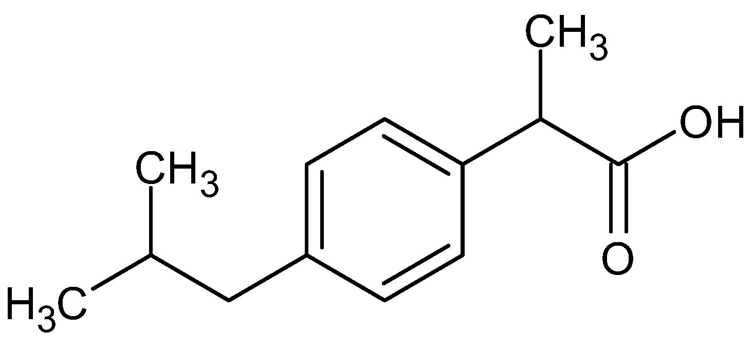
Figure 1. Molecular structure of ibuprofen.
Pharmaceuticals 16 00677 g001
Figure 2. FTIR spectra of the drug (Ibu), polymer (PVA), Soluplus (SP) and their physical mixture (Mix 1).
Pharmaceuticals 16 00677 g002
Figure 3. (a) TGA curves of ibuprofen, PVA, SP and their physical mixture (b) DSC thermogram of ibuprofen, polymers, and their physical mixture.
Pharmaceuticals 16 00677 g003
Figure 4. Solubility profile of ibuprofen in water and PBS with different concentrations of Soluplus.
Pharmaceuticals 16 00677 g004
Figure 5. Fabricated MN array patch of ibuprofen.
Pharmaceuticals 16 00677 g005
Figure 6. SEM images of the fabricated microneedle patch.
Pharmaceuticals 16 00677 g006
Figure 7. SEM Images of MN patch after applying a set force of 35 N show deformed tips (a) and (b) showing different magnifications of the array.
Pharmaceuticals 16 00677 g007
Figure 8. % Insertion of microneedle array across PF sheets.
Pharmaceuticals 16 00677 g008
Figure 9. Cumulative drug release profile of ibuprofen microneedles.
Pharmaceuticals 16 00677 g009
Figure 10. (a) Insertion of MN patch and (b) skin image after patch removal.
Pharmaceuticals 16 00677 g010
Figure 11. Plasma concentration versus time curve of ibuprofen tablet, ibuprofen cream, and ibuprofen MN patch.
Pharmaceuticals 16 00677 g011
Figure 12. Schematic representation of the fabrication process.
Pharmaceuticals 16 00677 g012
Table 1. R2 and k values obtained by fitting different kinetic models.
ModelR2 KnRegression Equation (y=)
Zero order0.982.220--2.2055x−2.2204
First order0.5720.280--0.0606x−0.2803
Hixson and Crowell0.98102.2--−2.2055x + 102.22
Higuchi0.9522.5--16.6x−22.5
Korsemeyer and Peppas0.947--1.8851.8853x−0.82
Table 2. Pharmacokinetic parameters for ibuprofen tablet, cream, and fabricated MN patch after NCA.
ParameterUnitTabletCreamIbu 25% MNsSig. (Post HOC Analysis)Sig. (Post HOC Analysis)
Mean ± SDMean ± SDMean ± SDMN- CreamMN-Tab
t1/2Hr2.48 ± 0.332.29 ± 0.165.2 ± 0.1* 0.009* 0.013
TmaxHr2.66 ± 0.41 2.33 ± 0.4124.0 ± 0.0* 0.000* 0.000
Cmaxμg/ml32.66 ± 1.735.73 ± 0.2428.7 ± 0.5* 0.000* 0.078
AUC0-tμg/mL·h157.84 ± 0.8343.90 ± 2.44831.9 ± 30.8* 0.000* 0.000
AUC0–∞μg/mL·h167.05 ± 4.7243.95 ± 2.45841.0 ± 31.1* 0.000* 0.000
AUMC0–∞μg/mL·h2780.63 ± 90.88259.23 ± 7.7316,387.0 ± 463.3* 0.000* 0.000
MRT0–∞H4.66 ± 0.425.91 ± 0.1919.5 ± 0.3* 0.000* 0.000
Cl/F(mg)/(μg/mL)/h4.94 ± 0.1418.85 ± 1.091.0 ± 0.0* 0.000* 0.000
* = The values are statistically significant (p < 0.05).
Disclaimer/Publisher’s Note: The statements, opinions and data contained in all publications are solely those of the individual author(s) and contributor(s) and not of MDPI and/or the editor(s). MDPI and/or the editor(s) disclaim responsibility for any injury to people or property resulting from any ideas, methods, instructions or products referred to in the content.