2.1. Bioactivity-Directed Isolation and Structure Elucidation of Cytotoxic Sesquiterpene Coumarin Ethers
Cytotoxic activity testing of the dichloromethane and methanol extracts of the roots of
Ferula huber-morathii has shown that the cytotoxic compounds were in the dichloromethane extract; in contrast, the methanol extract of the root did not show any cytotoxic activity at a concentration up to 100 µg/mL (
Table 1).
The cytotoxic dichloromethane extract of the roots of
Ferula huber-morathii was subjected to the bioactivity-directed fractionation on a Sephadex LH-20 column (
Supplementary Materials Figure S1). Initial fractions were combined based on their similar cytotoxic activities and thin layer chromatography (TLC) profiles. Then, each combined fraction group was subjected to further purification using various chromatographic techniques (i.e., RP-18 flash chromatography, prep. TLC on silicagel plates and prep. high-performance liquid chromatography (HPLC)). The following 15 sesquiterpene coumarin ethers were isolated from the aforementioned fractions: conferone (
1) [
26], conferol (
2) [
27], feselol (
3) [
27], badrakemone (
4) [
28], mogoltadone (
5) [
29], farnesiferol A (
6) [
30], farnesiferol A acetate (
7) [
31], gummosin (
8) [
32], ferukrin (
9) [
33], ferukrin acetate (
10) [
33], deacetylkellerin (
11) [
34], kellerin (
12) [
35], samarcandone (
13) [
36], samarcandin (
14) [
37], and samarcandin acetate (
15) (
Figure 1).
Due to the availability of limited NMR data for the isolated cytotoxic coumarins in the literature, extensive 2D NMR spectroscopic techniques, such as 2D
1H-
1H correlation spectroscopy (COSY), nuclear Overhauser effect spectroscopy (NOESY), heteronuclear single-quantum coherence (HSQC), heteronuclear multiple-bond connectivity (HMBC), (
Supplementary Materials Figures S2–S91) as well as high-resolution mass spectroscopy (HRMS) and optical rotation measurements (
Supplementary Materials Table S1), were used for the confirmation of their structures. However, many of the proton signals of bi-cyclic drimane sesquiterpene nucleus appear as overlapping signals in the upfield region of NMR spectra (
Table 2) and create ambiguity for the 2D NOESY stereochemical assignments of some of the sesquiterpene coumarins. Thus, most of the isolated compounds were synthesized by chemical transformations (
Supplementary Materials Figure S92), and structures of the parent and daughter compounds were unambiguously confirmed by comparing their spectroscopic data with those of semi-synthetic derivatives. Detailed
1H- and
13C-NMR data of all 15 cytotoxic coumarins are listed in
Table 2.
In order to illustrate the use of chemical transformation for the structure elucidation of
Ferula huber-morathii sesquiterpene coumarins, the determination of the structure of ferukrin (
9) will be described. The molecular formula of
9 was identified as C
24H
32O
5 from the ESI-MS peak observed at
m/
z 423.15 ([M+Na]
+, calc. for C
24H
32O
5Na
+), indicating nine degrees of unsaturation. The
1H-NMR,
13C-NMR, 2D COSY, HSQC, HMBC, and NOESY (
Supplementary Materials Figures S50–S55) indicated that the structure of
9 is a sesquiterpene coumarin derived from umbelliferone and a bicyclic drimane-type sesquiterpene triol by forming an etheric bond between the C-7 hydroxyl group of umbelliferone and C-11′ hydroxyl group of the drimane triol. Since the
1H-NMR signals of 10 cyclic aliphatic protons of the drimane sesquiterpene nucleus were clustered between the δ 1.30 and 1.80 ppm region of the
1H-NMR spectrum of
9, the stereochemistry data obtained from the 2D NOESY spectrum was ambiguous. However, the C-3′ hydroxyl group geminal proton (i.e., H-3′) appeared as a double of a doublet at δ 3.12 ppm (
J = 4.4, 11.5 Hz), indicating the equatorial orientation of the C-3′ hydroxyl group of the drimane triol nucleus. Due to the presence of both equatorially (i.e., compounds
1-
4) and axially (i.e., compounds
5–
8) oriented C-11′ drimane ethers in the dichloromethane root extract of
F. huber-morathii, identification of the spatial orientation of C-11′ methylene group of
9 was the key aspect of its structural determination. If the orientation of the C-11′ methylene group of
9 was equatorial (as observed with compounds
1–
4), then the structure of
9 should be isosamarcandin (
16). To explore whether the structure of
9 was isosamarcandin or not, samarcandin (
14) was transformed to isosamarcandin (
16) through an oxidoreductive chemical transformation (see
Supplementary Materials Figure S92). The
1H-NMR data of the semi-synthetic isosamarcandin (
16) (see
Supplementary Materials Figure S93) was not similar to that of
9. Thus, the structure of
9 was confirmed as ferukrin.
In a recent publication [
38], the presence of 4 daucane aromatic esters: ferutinin (
17), elaeochytrin A (
18), teferidin (
19), and feruhermonin C (
20), and 6 sesquiterpene coumarin ethers: mogoltavidin (
21), deacetylkellerin (
11), kellerin, farnesiferol A (
6), gummosin (
8), ferukrin acetate (
10), and a guaianetriol; teuclatriol (
22) (
Supplementary Materials Figure S94) was reported from the chloroform root extract of
Ferula huber-morathii. The potential aphrodisiac activity of some of these compounds was tested on male rats in comparison with sildenafil citrate as a positive reference compound, and ferutinin (
17) was determined as the most potent compound of the root extract, which was present at ca. 19.5% (
w/
w) in the chloroform extract of the root.
Ferutinin (
17) is a strong cytotoxic compound [
39]; however, during the bioactivity-directed isolation study, this compound was not found in the dichloromethane extract of the roots of
Ferula huber-morathii. Nevertheless, the absence of ferutinin (
17) and elaeochytrin A (
18) in the dichloromethane extract of the roots of
F. huber-morathii was further investigated by direct HPLC and TLC comparison of the ferutinin (
17) and elaeochytrin A (
18) reference compounds with the dichloromethane extracts of two
F. huber-morathii root samples collected from two different locations and times (i.e., Muş to Varto, 37 km from Zorabat village in June 1983 and between Erzurum and Varto, roadside in July 2017). The extract prepared from the old root samples was designated as the old extract, and the extract prepared from the root samples collected in 2017 was designated as the new extract; neither extract showed the presence of ferutinin (
17) and/or elaeochytrin A (
18) in its TLC and HPLC comparison chromatograms (
Supplementary Materials Figures S95 and S96). Moreover, the paper published by Baykan et al. [
39] describes the ferutinin content of the root extracts of several
Ferula species growing in Türkiye, including
F. huber-morathii, and does not list the presence of substantial quantities of ferutinin in the root extract of
F. huber-morathii. In addition, it should be noted that
F. huber-morathii is a member of subgenus
Dorematoides (Rgl. et Schmalh.) Korovin of the genus
Ferula, and from the chemotaxonomical point of view, phytochemical studies performed on the members of this subgenus revealed the presence of sesquiterpene coumarins, sesquiterpene chromones, sesquiterpene aryl compounds, phenylpropanoids, and guaiane sesquiterpenes, but daucane aromatic esters were never found in the
Ferula species of subgenus
Dorematoides [
13,
40,
41,
42,
43,
44,
45,
46,
47,
48,
49,
50,
51,
52,
53,
54]. The biosynthetic pathway of daucane sesquiterpenes is different [
19] than those of germacranes, elemanes, eudesmanes, and guaianes, and the aromatic esters of daucane sesquiterpenes are commonly found in the
Ferula species of subgenus
Euferula (Boiss.) Korovin [
9,
16,
18,
19,
20] and subgenus
Peucedanoides (Boiss.) Korovin [
12,
15,
23]. Thus, the presence of three daucane aromatic esters, ferutinin (
17), elaeochytrin A (
18), and teferidin (
19), in
F. huber-morathii is highly unlikely. In addition, the figure of structures of sesquiterpene coumarins reported in the same publication [
38] displays the structure of mogoltavidin (
21) as the enantiomer of deacetylkellerin (
11) (
Supplementary Materials Figure S94) that is not chromatographically separable by the analytical procedures described in the publication, and based on the basic organic chemistry rules, the spectroscopic data of enantiomeric compounds should be identical except for their opposite optical rotation values. However, the NMR spectra data for mogoltavidin (
21) and deacetylkellerin (
11) were reported as completely different spectra [
38]; thus, the proposed structure for mogoltavidin (
21) is incorrect. Careful examination of the reported
1H- and
13C-NMR spectra of mogoltavidin (
21) (i.e., SP 15 and SP 16) in the
supplementary data files of the previous publication [
38] and comparison with the
1H and
13C-NMR spectra of samarcandin (
14) shown in the supplementary files of the current publication (
Supplementary Materials Figures S80–S85) confirm the actual structure of mogoltavidin (
21) as samarcandin (
14).
2.2. Determination of the Absolute Configuration of Samarcandin 14
Samarcandin (
14), along with its keto derivative samarcandone (
13), was isolated from
Ferula samarcandica Korovin in 1968, but the initial structure proposed for samarcandin (
14) was incorrect [
36]. Several biological activities were attributed to samarcandin (
14), such as cytotoxic activity against the AGS (human gastric carcinoma) and WEHI-164 (fibrosarcoma) cancer cell lines [
37], activity against NCI yeast anticancer drug screen assay [
55], and potential antiviral [
56,
57] and aphrodisiac activities [
58]. The structure of samarcandin (
14) was revised based on NMR decoupling experiments [
59] and later by comparative optical rotation measurements of several sesquiterpene coumarin ethers, including various samarcandin derivatives and isomers [
60]. Nevertheless, the stereochemistry of samarcandin proposed in these publications was incorrect. The relative stereochemistry of samarcandin (
14) was established with a single crystal X-ray crystallographic analysis [
61]; however, without providing any supporting evidence, the relative stereochemistry was proposed as the absolute stereochemistry of samarcandin in this paper. In a more recent publication, the stereochemistry of samarcandin (
14) was reinvestigated by computational chemistry methods and X-ray crystallographic analysis. Despite the lack of required specific measurements and calculations for the establishment of absolute stereochemistry of samarcandin (
14), the enantiomer of samarcandin (i.e.,
ent-samarcandin,
23) was proposed as the absolute atomic structure of samarcandin [
37]. Close examination of the reviews of sesquiterpene coumarins in the literature also revealed the presence of similar confusion about the absolute stereochemistries of other sesquiterpene coumarins [
62,
63,
64,
65].
To identify the absolute stereochemistry of samarcandin (
14), the (
R)-MTPA ester of samarcandin
(24) (
Supplementary Materials Figure S99) was prepared, and its solid-state packing was investigated using X-ray crystallography. The molecular structure with the atom numbering scheme and the packing arrangement of the molecules are presented in
Figure 2 and
Supplementary Materials Figure S109. The asymmetric unit consists of one molecule of (
R)-MTPA ester of samarcandin (
24), as shown in
Figure 2. As expected, the coumarin moiety is nearly planar; the displacements of all ten atoms contained in the ring are less than 0.019(2) Å [for C(4)] from the least-squares plane. Two cyclohexane rings in the sesquiterpene parts of the samarcandin adopt a chair conformation with the spherical polar set values of Q = 0.5915(15) Å, θ = 7.67(15)°, and φ =2 52.4(11)° for the C11/C12/C13/C14/C15/C20 ring; these values are Q = 0.5383(16) Å, θ = 5.11(17)°, φ = 227.0(19)° for the C15/C16/C17/C18/C19/C20 ring. The O1−C1 and O6−C25 bond lengths of 1.213(2) Å and 1.192(2) Å, respectively, match the value for double bond C=O, while the bonds O2–C1 [1.3731(18) Å], O2–C9 [1.3772(18) Å], C7–O3 [1.3552(16) Å], O3–C10 [1.4390(18) Å], O4–C12 [1.4511(17) Å] and O5–C25 [1.3317(19) Å], O5–C17 [1.478(2) Å] correspond to the value for single C–O bonds. The MTPA moiety of the molecule adopts an extended conformation with torsion angle C17−O5−C25−O6 [3.0(2)°] and O5–C25–C26–C29 [68.99(15)°]. The trifluoro and methoxy groups attached to the C26 atom are twisted out of the phenyl ring plane with torsion angles of 142.46(18)° for C34–C29–C26–C27 and −102.92(18)° for C34–C29–C26–O7.
All of the intra- and intermolecular contacts were examined with Platon. Two types of intramolecular interactions, C−H···O and C−H···F, and one intermolecular hydrogen bond, O−H···O, were detected in the structure [O(4)−H(1)···O(1)i 0.87(3)Å 2.17(3)Å 3.028(2)Å 168(3)°] [Symmetry code: (i) −x, −1/2 + y, −z].
2.3. Cytotoxic Activities of the Sesquiterpene Coumarin Ethers Isolated from the Dichloromethane Extract of the Roots of Ferula huber-morathii
The cytotoxic activities of individual sesquiterpene coumarin ethers isolated from the roots of
F. huber-morathii were tested against the COLO 205, K-562, and MCF-7 cancer cell lines, as well as the non-cancerous human umbilical vein epithelial cells (HUVEC) cell line. The results are shown in
Table 3, which showed that compounds
4,
6,
8,
9,
10,
11,
13,
14, and
15 exhibited little or no cytotoxicity. On the contrary, compounds
1,
2,
3,
5, and
7 showed moderate or strong cytotoxicity. Moreover, compound
12 showed a moderate cytotoxic effect only for MCF-7 cell lines.
Based on the IC
50 values, conferol (
2) and mogoltadone (MOG,
5) are the most potent sesquiterpene coumarins against cancer cell lines. However, the selectivity indexes of mogoltadone (
5) for the COLO 205, K-562, and MCF-7 cells lines (>8.29; >12.46; >8.64, respectively) were higher than those of conferol (
2) (5.45; 1.73; 3.82, respectively). Therefore, the caspase-3, 8, and 9 activity analyses for the COLO 205, K-562, and MCF-7 cell lines were performed by mogoltadone (
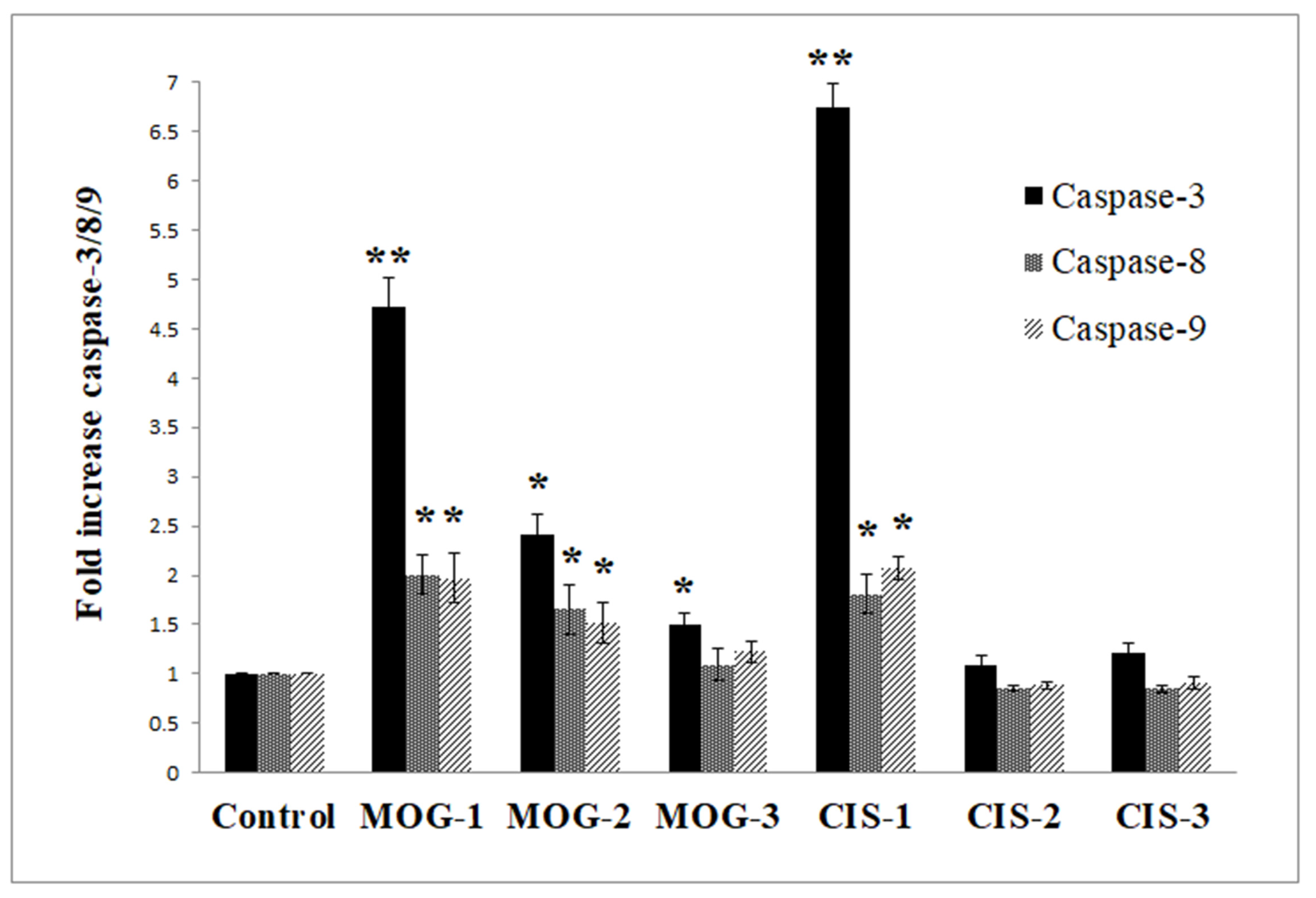
5). The data showed that the caspase-3, 8, and 9 activities were the highest in the COLO 205 cell line in comparison with the other cancer cell lines. Concerning the results for the COLO 205 cell line, mogoltadone (
5) dose-dependently increased the caspase-3, 8, and 9 activities. Furthermore, mogoltadone (
5) showed an increase in caspase-3 activity by 4.72-fold, in caspase-8 activity by 2-fold, and in caspase-9 activity by 1.97-fold when compared with the control at the highest concentration (131.58 µM) (
Figure 3). Cisplatin (CIS) was used as a positive control for the caspase activities. Based on these results, the cisplatin indicated an increase in the caspase-3 activity by 6.73-fold, in caspase-8 activity by 1.8-fold, and in caspase-9 activity by 2.07-fold compared with the control at the highest concentration (
Figure 3).
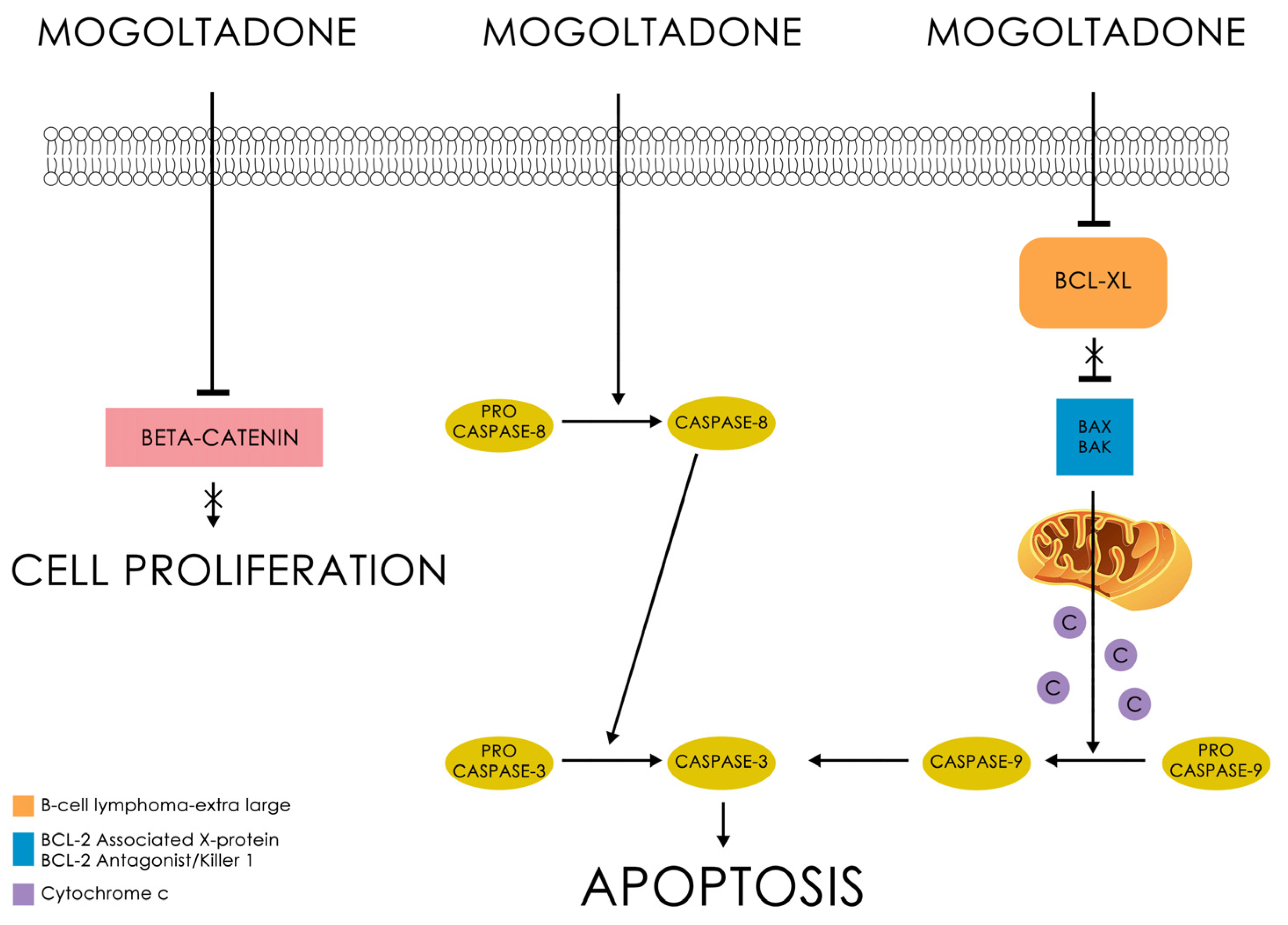
Western Blot analysis indicated that mogoltadone (
5) suppressed the level of Bcl-XL, an antiapoptotic protein, and decreased the procaspase-3 level in the COLO 205 cell line. The statistically significant effect for procaspase-3 was observed at a concentration of 131.58 µM. These data supported the results of the caspase-3 activity. Furthermore, the effect of mogoltadone (
5) on the β-catenin levels was also investigated and the results showed that mogoltadone decreased the β-catenin levels in the COLO 205 cell line. In contrast, mogoltadone (
5) did not have a statistically significant effect on the Bcl-XL, caspase-3, and β-catenin protein levels of the HUVEC cell line, which may explain the cytotoxic specificity of mogoltadone (
5) on cancer cell lines (
Figure 4).
The cytotoxicity evaluation of 15 cytotoxic sesquiterpene coumarins isolated from the dichloromethane extract of the roots of Ferula huber-morathii indicated that conferone (1), conferol (2), feselol (3), mogoltadone (5), and farnesiferol A acetate (7) show a remarkable cytotoxic effect on some cancer cell lines.
Previous cytotoxic activity studies performed on conferone (
1) showed that the highest cytotoxic activity of this compound was observed on the ovarian carcinoma cells (CH1) with an IC
50 of 7.8 µM [
29]. Conferone (
1) also showed moderate cytotoxic activity on colorectal (HT29; 20 µM), breast (MDA-MB-231; 30 µg/mL), colon (HCT116; 32 µM), ovarian (A2780; 32 µM), cervical (HeLa; 38 µM), lung (A549; 38 µM), cisplatin-resistant derivatives of the human ovarian (A2780P A2780/RCIS; 25 µM), melanoma (SK-MEL-28; 64 µM), and chronic myelogenous leukemia (K562; 86.12 µg/mL) cancer cell lines [
29,
66,
67,
68,
69,
70]. In addition, conferone (
1) showed very low cytotoxic activity (>100 µM) on the MCF-7 breast cancer cell line and no cytotoxic effect on bladder cancer cells (5637) [
46,
66,
69]. Based on the findings, conferone (
1) displayed a moderate cytotoxic effect on the COLO 205, K-562, and MCF-7 cancer cell lines, as well as on the non-cancerous HUVEC cell line.
In a study conducted by Soltani et al. [
67], the cytotoxic activities of feselol (
3) on the HCT-116, HeLa, A549, and A2780 cancer cell lines were found to be 28, 35, 26, and 20 µM, respectively. Furthermore, some studies reported that feselol (
3) did not show any cytotoxic activity on the bladder (TCC, 5637), K562, HeLa, and stomach (AGS) cancer cell lines [
14,
71,
72,
73,
74].
Thus far, there has been no cytotoxic activity reported for farnesiferol A acetate (7); however, the findings herein suggest that farnesiferol A acetate has moderate cytotoxic activity against the MCF-7 breast cancer cell line. Nevertheless, its cytotoxic effect was weak against the rest of the cell lines.
Few studies have explored the cytotoxic effect of mogoltadone (
5) on different cell lines [
29,
66]. A study carried out by Valiahdi et al. [
29] did not find any potent cytotoxic activity by mogoltadone (
5) against the A549 and SK-MEL-28 cell lines. However, against the ovarian carcinoma cell line (CH1), mogoltadone (
5) showed moderate cytotoxic effects [
29]. Similarly, another study suggested that mogoltadone (
5) had a low cytotoxic effect on the A2780/RCIS (>50) and MCF-7 (>100 µM) cancer cell lines [
14,
66]. Moreover, the combination of mogoltadone (
5) with cisplatin significantly enhanced the cisplatin cytotoxicity on A2780/RCIS cells [
66]. These results were similar to those of conferol (
2) [
75]. The current investigation indicated that mogoltadone (
5) showed a moderate cytotoxic effect on the COLO 205, MCF-7, and K-562 cancer cell lines, except the non-cancerous HUVEC cell line, which is the control cell line. As there are no mechanistic studies regarding the selective cytotoxic activity of mogoltadone (
5) reported in the literature, mogoltadone’s (
5) cytotoxic effect mechanism was also investigated in the present study. Hence, this is the first study to show that mogoltadone (
5) triggers the caspase activation and suppresses anti-apoptotic protein, Bcl-XL, and β-catenin. The accumulating evidence indicates that β-catenin has a central role in the malignant transformation of normal cells [
76]. Therefore, the inhibition of β-catenin supports the cytotoxic effect of the molecule (
Figure 5).

 ,
, 


