Article

Chaperone Activity and Protective Effect against Aβ-Induced Cytotoxicity of Artocarpus camansi Blanco and Amaranthus dubius Mart. ex Thell Seed Protein Extracts

by
David Sanchez-Rodriguez
,
Idsa Gonzalez-Figueroa
and
Merlis P. Alvarez-Berríos
*
Department of Science and Technology, Inter American University of Puerto Rico at Ponce, Ponce, PR 00715-1602, USA
*
Author to whom correspondence should be addressed.
Pharmaceuticals 2023, 16(6), 820; https://doi.org/10.3390/ph16060820
Submission received: 30 April 2023 / Revised: 29 May 2023 / Accepted: 29 May 2023 / Published: 31 May 2023
(This article belongs to the Section Natural Products)

Abstract

:
Alzheimer’s disease (AD) is the most common type of dementia and is listed as the sixth-leading cause of death in the United States. Recent findings have linked AD to the aggregation of amyloid beta peptides (Aβ), a proteolytic fragment of 39–43 amino acid residues derived from the amyloid precursor protein. AD has no cure; thus, new therapies to stop the progression of this deadly disease are constantly being searched for. In recent years, chaperone-based medications from medicinal plants have gained significant interest as an anti-AD therapy. Chaperones are responsible for maintaining the three-dimensional shape of proteins and play an important role against neurotoxicity induced by the aggregation of misfolded proteins. Therefore, we hypothesized that proteins extracted from the seeds of Artocarpus camansi Blanco (A. camansi) and Amaranthus dubius Mart. ex Thell (A. dubius) could possess chaperone activity and consequently may exhibit a protective effect against Aβ1–40-induced cytotoxicity. To test this hypothesis, the chaperone activity of these protein extracts was measured using the enzymatic reaction of citrate synthase (CS) under stress conditions. Then, their ability to inhibit the aggregation of Aβ1–40 using a thioflavin T (ThT) fluorescence assay and DLS measurements was determined. Finally, the neuroprotective effect against Aβ1–40 in SH-SY5Y neuroblastoma cells was evaluated. Our results demonstrated that A. camansi and A. dubius protein extracts exhibited chaperone activity and inhibited Aβ1–40 fibril formation, with A. dubius showing the highest chaperone activity and inhibition at the concentration assessed. Additionally, both protein extracts showed neuroprotective effects against Aβ1–40-induced toxicity. Overall, our data demonstrated that the plant-based proteins studied in this research work can effectively overcome one of the most important characteristics of AD.

1. Introduction

Alzheimer’s disease (AD) is considered the most common type of dementia and is listed as the sixth-leading cause of death in the United States [1]. Currently, 6.7 million Americans aged 65 and older are living with AD and it is expected that global AD patients will triple by 2050 [2,3]. AD is characterized by the formation of amyloid-beta (Aβ) peptide aggregates on senile plaques (SP) and neurofibrillary tangles formed by tau deposition [3,4,5]. Amyloid-beta (Aβ) peptide is a proteolytic fragment of 39–43 amino acid residues derived from the amyloid precursor protein (APP). SP are mainly composed of the 40- (Aβ1–40) and 42-residue-long peptides (Aβ1–42), where Aβ1–40 is the most abundant and Aβ1–42 the more aggregation-prone [6]. The aggregation of Aβ1–40 and Aβ1–42 into insoluble fibrils is considered the major pathological hallmark of AD [3,7,8]. It has proven to be toxic, leading to memory loss and cognitive decline over time. The amyloid cascade hypothesis of AD proposes that this aggregation is the primary event that ultimately leads to AD dementia [9].
Currently, no preventive or effective treatment for AD is available. Acetylcholinesterase inhibitors and N-methyl-D-aspartate receptor antagonists are the standard drugs for the treatment of AD and only provide temporary alleviation of the psychological and behavioral symptoms [10,11]. Therefore, new therapies to inhibit the aggregation or removal of amyloid-beta (Aβ) peptides are currently being explored as promising strategies for AD treatment [8,10,12,13,14,15]. For example, in 2021 the FDA approved a new drug (Aducanumab) through the accelerated approval program that removes amyloid deposits in the brain. Although this drug is a promising treatment, it induces severe side effects such as brain swelling or bleeding in the brain [16].
In recent decades, plant-based medications have gained significant attention for the treatment of neurological disorders [17,18,19]. The bioactive constituents of plants can exhibit therapeutic activity against AD, providing a new avenue for the discovery of drugs with minimal side effects. Studies have demonstrated the efficacy of different natural compounds of plant origin such as alkaloids, terpenoids and phenolic compounds against the aggregation of Aβ [17,20,21,22,23,24,25]. Bastianetto et al. [26] reported that the flavonoids present in the leaf extract of the Ginkgo biloba L. tree may be able to protect hippocampal cells against toxic effects induced by Aβ peptides. Extracts from Allium roseum L., a plant rich in organosulfur compounds and flavonoids, inhibited the fibrilization process of Aβ1–42 and provided a neuroprotective effect to SH-SY5Y human neuroblastoma cells by hindering the formation of mature fibrils [27]. Similarly, Lawsonia inermis L., Punica granatum L. and Pistacia lentiscus L. extracts inhibited the aggregation and induced neurotoxicity of Aβ1–42. Although these natural compounds have demonstrated in vitro and in vivo efficacy against this deadly disease, plants may also contain other bioactive molecules with superior anti-AD activity [28].
Several reports have revealed that chaperones and chaperone-like proteins have the ability to interfere with amyloid beta formation, preventing the toxicity that is associated with this process [29,30,31,32,33,34]. Chaperones play a key role in helping other proteins to maintain their correct shapes, preventing aggregation and its potentially cytotoxic effects [35]. Hochberga et al. [36] established that the chaperone αB-crystallin prevented Aβ fibril formation and consequently reduced its toxicity in HEK293, HeLa and PC12 cells. Arosio et al. [37] demonstrated that the molecular chaperones DNAJB6, Ssa1 and proSP-C Brichos were able to exert a protective function against the aggregation of Aβ1–42 through diverse mechanisms. Although the use of chaperones has shown promising results, they are highly toxic or reveal a lack of specificity [38]. Therefore, the exploration of plant-based proteins, especially those with chaperone activity, may be a key strategy to overcome one of the main hallmarks of AD.
Recent research has reported that Artocarpus and Amaranthus species contain potential bioactive molecules with pharmacological properties, such as antioxidant, anti-inflammatory, antibacterial and anticarcinogenic activities [39,40]. Artocarpus camansi Blanco (A. camansi) and Amaranthus dubius Mart. ex Thell (A. dubius), belonging to the Artocarpus and Amaranthus genera, respectively, are mainly distributed in the tropical regions, including Puerto Rico. These species have demonstrated remarkable anticancer and antioxidant properties [41,42]. To our knowledge, the chaperone activity and the neuroprotective effects of these plants against Aβ toxicity have not been evaluated in vitro. Seed plants are rich in proteins that are known to act as chaperones, such as heat shock proteins and the late embryogenesis abundant proteins [43,44]. Thus, in this study, the chaperone activity of seed protein extracts obtained from A. dubius and A. camansi was investigated. We hypothesize that A. camansi and A. dubius seed protein extracts may inhibit Aβ1–40 fibrils and provide a protective effect to SH-SY5Y human neuroblastoma cells due to their chaperone activity. To test this hypothesis, the enzymatic reaction of citrate synthase was used to determine the chaperone activity of each protein extract. Thioflavin T and DLS measurements were performed to test the ability of the extracts to inhibit the formation of Aβ1–40. In addition, neuroprotective studies using SH-SY5Y cells were carried out. Our results indicated that A. camansi and A. dubius seed protein extracts showed chaperone activity and were able to inhibit Aβ1–40 fibrillization, with A. dubius showing the highest efficacy. Moreover, they were able to provide protection to SH-SY5Y cells against Aβ1–40-induced toxicity. This neuroprotective effect may be attributed to the chaperone activity of the proteins present in the extracts, which inhibited the formation of toxic Aβ1–40 fibrils.

2. Results and Discussion

2.1. Preparation of Protein Extracts and Protein Concentration

Plants may provide a plethora of new therapeutic drugs for the treatment of AD [17,18,26]. In this study, seed protein extracts were obtained from A. camansi and A. dubius with the intention to evaluate their chaperone activity and in vitro therapeutic potential as anti-AD therapy. Proteins were extracted in 50 mM PBS (pH 7.4) and partially purified using ammonium sulfate precipitation and dialysis. The total protein concentrations of the A. camansi and A. dubius protein extracts were 6043.2 μg/mL and 675.7 μg/mL, respectively, as determined by the bicinchoninic acid (BCA) assay.
A. camansi, a type of plant (tree) that belongs to the family Moraceae [45], has larger seeds than A. dubius, a type of plant (leafy vegetable) from the family Amaranthaceae [46]. Large seed size has been shown to be associated with length of life cycle and habitat stability, while smaller seeds are characteristic of parasites that are species of plants that form seed banks and have higher distribution [47]. Having a large seed provides higher survivability for plants against drought and isolation on remote islands. This indicates that larger seeds have more resources towards survivability; hence, this could explain the notable protein concentration difference between the two protein extracts. Several studies have shown that A. camansi protein content ranges from 4.87 to 19.6% (dry weight), with high variability due to the plant’s location [48]. On the other hand, there is no information reported in the literature regarding the protein content of A. dubius seeds. To compare the potential inhibitory efficacy of both plant extracts against Aβ1–40 fibril formation under the same conditions, A. camansi and A. dubius seed protein extracts were used at the same concentration for further experiments.

2.2. Protein Profiling with SDS-PAGE

SDS-PAGE electrophoresis is widely used to determine the electrophoretic profile of representative samples including different species of plants [49]. To estimate the protein pattern of protein extracts obtained from A. dubius and A. camansi, SDS-PAGE electrophoresis was carried out. The results suggested that both extracts contained proteins with diverse molecular weight distributions (Figure 1). A. dubius protein bands ranged from approximately 75 kDa to 15 kDa, and there was a band above 250 kDa. Similarly, A. camansi bands ranged from approximately 100 kDa to 10 kDa. A band above 250 kDa was also observed.

2.3. CD Spectra and Estimation of Protein Secondary Structure Content

To obtain information about the secondary structure of the proteins present in the seed extracts, CD spectra were recorded and analyzed using the server BeStSel [50]. CD spectra for A. dubius protein extracts indicated a positive maximum at 196, suggesting the presence of β-sheet conformation, and a negative minimum at 208 nm, characteristic of α-Helix conformations (Figure 2). A. camansi spectra showed a negative minimum at 218 assigned to the β-sheet structure [51,52].
Secondary structure analysis using the BeStSel method suggested that the A. dubius protein extract consisted of 5.9% of α-helix, 25.6% of β-Sheet, 13.5% of β-Turn and 54.9% of “others” (Table 1). The protein extract obtained from A. camansi consisted of 36.8% of β-Sheet, 12.4% of β-Turn and 50.7% of “others”. Others, which include 310-helix, π-helix, β-bridge, bend, loop/irregular and invisible regions of the structure, accounted for the majority of the secondary structures in the A. camansi and A. dubius protein extracts.

2.4. Chaperone Activity of Seed Protein Extracts of A. camansi and A. dubius

Among plant bioactive molecules, proteins with chaperone activity are a promising anti-AD strategy due to their ability to inhibit the aggregation of Aβ peptide [29,30,34]. To elucidate if A. camansi and A. dubius protein extracts possess such a function, the enzymatic reaction of citrate synthase (CS) was used to measure their chaperone activity. The chaperone activity of a protein is determined by the protection of a client protein against loss of activity under stress conditions [53]. Therefore, the activity of CS in the presence or absence of A. camansi or A. dubius protein extracts at 44 °C for 40 min was determined. Activity of 100% was set to be the activity of CS without protein extracts before stress conditions. The results showed that the activity of CS decreased from 100% (before stress) to 11% (after stress) in the absence of the protein extracts (Figure 3). This result is consistent with previous reports where the activity of CS drops from 100% to under 20% in these conditions [53]. The protective effect of both protein extracts was evident when CS was exposed to temperature stress at the concentration assessed. The presence of A. camansi protein extract resulted in a significant increase in CS activity when compared with CS alone under stress conditions (66.7 ± 0.0% activity for CS + A. camansi and 11.1 ± 1.1% of activity for CS alone, p < 0.05). The enzymatic activity of CS is completely protected against temperature stress by the presence of A. dubius (144.4 ± 11.1% for CS + A. dubius compared with 11.1 ± 1.1% for CS alone, p < 0.05). The apparent activity of CS above 100% is a phenomenon that has been observed in several experiments with chaperones. It is believed that the reactivation of inactive species in the commercial enzyme product is mainly responsible for this increase [53,54]. It was observed that A. dubius protein extract can protect CS against temperature inactivation to a higher extent than A. camansi protein extract, suggesting that the concentration of proteins with chaperone activity is higher in A dubius. These results let us speculate that the two seed protein extracts studied in this work may possess potent inhibitory activity against Aβ fibril formation and a neuroprotective effect against Aβ-induced toxicity.

2.5. Inhibition of Aβ1–40 Fibrillation Using ThT Assay

The formation and accumulation of beta amyloid fibrils is one of the distinctive characteristic events in Alzheimer’s pathophysiology [55]. The inhibition effect of A. dubius and A. camansis protein extracts against Aβ1–40 fibril formation was examined using the fluorescent probe Thioflavin T at 37 °C. Thioflavin T is a fluorescent dye that exhibits enhanced fluorescence in the presence of amyloid fibrils, allowing it to act as an indicator for evaluating beta amyloid fibril formation and inhibition [56]. An increase in fluorescence intensity would be indicative of the presence of fibrils, while a decrease is indicative of inhibition. The results demonstrated that the seed protein extracts from A. dubius and A. camansi were effective in inhibiting Aβ1–40 fibrilization, with A. dubius extract showing the highest inhibition (Figure 4a). The fluorescence intensity of the control sample (Aβ1–40) occurred parabolically until reaching a plateau at 180 min (3 h), indicating the formation of amyloid beta fibrils. This trend is consistent with the literature, where amyloid fibril concentration increases in a parabolic manner until reaching a plateau [57,58]. The presence of protein extract from A. camansi demonstrated a 44% reduction in fluorescence intensity when compared with the untreated control (Figure 4a). On the other hand, the presence of A. dubius seed protein extract (Figure 4) demonstrated an 82% reduction in ThT fluorescence intensity when compared with control samples. After co-incubating Aβ1–40 peptide with the protein extracts for 13 h, the A. dubius and A. camansi protein extracts showed 69.0 ± 3.8% and 45.3 ± 2.5% inhibition of Aβ1–40 aggregation, respectively (Figure 4b). These results suggest that the high chaperone activity of A. dubius seed protein extract as shown in Figure 3 may be responsible for its superior inhibitory effect on Aβ1–40 aggregation at the concentration assessed.

2.6. Size Distribution Analysis with DLS

Dynamic light scattering (DLS) has been used to determine the size distribution of Aβ1–40 aggregates [59]. It provides information about the size for all molecules in a solution [60]. To further confirm the Aβ1–40 fibril inhibitory capacity of the seed protein extracts, the size distribution of Aβ1–40 incubated in the presence or absence of the A. camansis or A. dubius seed protein extracts was determined. Large diameters are considered as an indication of the presence and/or formation of amyloid fibrils and small diameters indicate a reduction in amyloid fibrillogenesis.
Untreated Aβ1–40 fibrils showed peaks at ~7048 nm and 332 nm (Figure 5). When Aβ1–40 was co-incubated with A. dubius protein extract, a small shift in the peak characteristic for A. dubius protein extract alone from ~103 nm to ~153 nm was observed (Figure 5a). An increasing number of reports have demonstrated that chaperones or proteins with chaperone activity can bind to oligomeric species of Aβ, interfering with the process of fibril formation [61,62,63]. Therefore, this peak shift may be attributed to the interaction of the Aβ1–40 peptides/oligomers with A. dubius proteins present in the solution, providing these proteins with the ability to inhibit the formation of small and large Aβ1–40 aggregates. The A. camansi-treated sample showed a shift in the peak corresponding to A. camansi alone from ~1821 to ~2322, also demonstrating its capacity to interact with Aβ1–40 peptide/oligomers (Figure 5b). An additional small peak at 450 nm was observed, indicating the ability of A. camansi extract to inhibit only the formation of large Aβ1–40 aggregates.
The DLS results were consistent with those obtained from ThT studies where both protein extracts demonstrated inhibitory properties towards Aβ1–40 fibrillogenesis, with A. dubius showing the highest inhibition efficacy. It has been reported that the inhibitory capacity of chaperones against amyloid beta fibril formation is concentration-dependent [64]. As displayed in Figure 3, A. dubius exhibited the highest chaperone activity; hence, this potent inhibitory effect may be due to the high concentration of chaperones in this protein extract. To our knowledge, there are no specific articles that address the inhibitory properties of A. dubius and A. camansi towards beta amyloid fibril formation. For the first time, we showed that A. dubius and A. camansi seed protein extracts are promising inhibitors of the Aβ fibrillation process, one of the main hallmarks of AD.

2.7. Hydrolysis of Seed Protein Extracts Using the Alcalase Enzyme

To confirm that proteins with chaperone activity present in the A. camansi and A. dubius seed extracts were responsible for inhibiting the fibrillization process of Aβ1–40 as demonstrated by the ThT and DLS results, the protein extracts were incubated with the proteolytic enzyme alcalase, which provides very extensive hydrolysis of plant proteins. If these proteins played a key role in fibril inhibition, the co-incubation of Aβ1–40 with alcalase-treated protein extracts would result in an increase in ThT fluorescence intensity (increase in amyloid fibril concentration) when compared with untreated seed protein extracts. The results showed that the protein breakdown induced by alcalase decreased the Aβ1–40 fibril inhibition activity of both extracts (Figure 6a). Increases of ~35% and ~100% in ThT fluorescence intensity were observed in the alcalase-treated A. camansi and A. dubius seed protein extract, respectively, when compared with untreated samples. This increase in fluorescence intensities may be attributed to the hydrolysis of key proteins with chaperone activity in the A. camansi and A. dubius samples responsible for fibril inhibition. After 13 h of incubation with the alcalase-treated protein extracts, the inhibition of Aβ1–40 aggregation decreased considerably when compared with the untreaded protein extracts, as shown in Figure 6b (A. camasi protein extract: 45.3 ± 2.5% for untreated sample and 25.9 ± 3.7% for alcalase-treated samples, p < 0.05; A. dubius protein extract: 69.0 ± 3.8% for untreated sample and 36.2 ± 5.7% for alcalase-treated sample, p < 0.05). Overall, our findings confirmed that the proteins that were hydrolyzed within the samples are important for the inhibition of Aβ1–40 fibril formation.

2.8. Cytotoxic Effect of Protein Extracts on SH-SY5Y Cells

To determine whether A. camansi and A. dubius seed protein extracts are cytotoxic, SH-SY5Y neuroblastoma cells were exposed to increasing concentrations of protein extracts for 48 h. Following this incubation period, cells were assayed for viability using a CellTiter 96® aqueous assay. The protein extract from A. camansi was not toxic to SH-SY5Y cells in the range assessed (1 μg/mL, 3 μg/mL, 6 μg/mL, 11 μg/mL, 46 μg/mL, 92 μg/mL, 184 μg/mL and 367 μg/mL). In contrast, the toxicity of the A. dubius protein extract was dose-dependent, and the viability of cells decreased at higher concentrations of the seed protein extract (92 μg/mL, 184 μg/mL and 367 μg/mL) (Figure 7). For neuroprotective studies, nontoxic protein extract concentrations were chosen (6 μg/mL, 11 μg/mL and 46 μg/mL).

2.9. Neuroprotective Effect of A. camansi and A. dubius Protein Extracts in SH-SY5Y Cells

To explore the promising neuroprotective effect of the A. camansi and A. dubius seed protein extracts against Aβ1–40 fibrils, Aβ1–40 (100 μM) was incubated at 37 °C in the presence or absence of protein extracts at the nontoxic concentrations of 46 μg/mL, 11 μg/mL or 6 μg/mL for 24 h. Afterwards, SH-SY5Y cells were co-incubated with these solutions for 48 h and the viability ratio was evaluated using the CellTiter 96® aqueous assay. The results revealed that Aβ1–40 fibrils at a concentration of 100 μM showed a significant decrease in cell viability relative to untreated cells (p < 0.05) (Figure 8). However, the co-incubation of Aβ1–40 with A. camansi or A. dubius protein extracts mitigated the cell mortality induced by Aβ1–40 amyloid fibrils at the three concentrations assessed. A statistically significant difference (p < 0.05) between the viability ratio of cells incubated with Aβ1–40 alone and cells incubated in the presence of the protein extracts was observed. This neuroprotective effect may be due to the chaperone activity of the proteins present in the extracts that were able to inhibit the formation of toxic Aβ1–40 fibrils. Overall, our results indicated that both protein extracts are a promising anti-AD therapy in the conditions tested.

3. Materials and Methods

3.1. Materials

Phosphate-buffered saline (PBS), ammonium sulphate, alcalase, citrate synthase from porcine heart, oxaloacetate, acetyl coenzyme A sodium salt, 5,5′-Dithiobis(2-nitrobenzoic acid) (DTNB), Tris-HCl buffer, thioflavin T (ThT) and all the materials for cell culture were purchased from Sigma-Aldrich (St Louis, MO, USA). Bicinchoninic acid (BCA) assay kit was obtained from Fisher Scientific (Hampton, NH, USA). Aβ1–40 was purchased from Anaspec (San Jose, CA, USA). Bio-Safe™ Coomassie stain, precision plus protein kaleidoscope standards and TGX precast gels were purchased from Bio-Rad (Hercules, CA, USA).

3.2. Plant Material

A. dubius and A. camansi were naturally cultivated and obtained from a local farm in Yauco, PR. Plants were identified by a local expert named David Sanchez Montalvo who has extensive knowledge in folkloric medicine of endemic and nonendemic plants found in Puerto Rico. The local names and aerial parts used of each selected plant are shown in Table 2.

3.3. Protein Extraction and Purification

Protein extraction and purification were performed using a method previously reported in the literature with some modifications [65]. Briefly, the two different types of seeds from Puerto Rican plants (Table 2) were collected in Yauco, PR, sun dried for 20 h and dried further using a lyophilizer. The seed of each plant was pulverized using a mortar and pestle and mixed with 50 mM PBS buffer (pH 7.4) using a 1:9 ratio (1 g of seed in 9 mL of buffer). Then, the extracts were centrifuged at 10,000 rpm at 4 °C for 20 min. After centrifugation, the protein crude extract (supernatant) was transferred to a fresh Eppendorf tube and saturated with ammonium sulphate until 80% saturation was achieved. The saturated crude extract was centrifuged at 12,000 rpm and 4 °C for 40 min, the supernatant was carefully discarded, and the pellet was resuspended in 50 mM PBS buffer and dialyzed using a Pur-A-Lyzer™ Maxi dialysis tube. The dialysis tube was first submerged into the 50 mM PBS buffer for 10 min. Afterwards, the protein solution was poured into the dialysis tube and placed in a beaker filled with 50 mM PBS buffer under continuous stirring. The buffer was changed three times every three hours. After the dialysis process, the protein solution was stored in small aliquots at −20 °C until use. The BCA protein assay kit was used to determine the concentration of protein in each seed extract [66].

3.4. SDS-PAGE Pattern of Seed Protein Extracts

The protein profile of the crude seed protein extracts was analyzed using sodium dodecyl sulfate polyacrylamide gel electrophoresis (SDS PAGE) according to the procedure described by Soto-Madrid et al. [67] with some modifications. For this, each seed protein extract (12 μg for A. dubius and 23 μg for A. camansi) was mixed with 8 μL of Laemmli buffer and incubated for 5 min at 95 °C. Samples and the prestained protein standard were loaded into TGX precast gels and run at 200 volts for 25 min in the Mini-PROTEAN Tetra cell electrophoresis module (Bio-Rad, Hercules, CA, USA). Afterwards, gels were stained with Bio-Safe™ Coomassie stain for two hours, and left destaining overnight.

3.5. Circular Dichroism Spectroscopy

Circular dichroism (CD) measurements of the protein extracts were collected using a J-1100 spectrophotometer (Jasco Inc., Tokyo, Japan) and a 1 mm path length cuvette thermostated at 20 °C. Each spectrum was measured from 190 to 250 nm, with six scans at a scanning speed of 200 nm/min, a 1.00 nm bandwidth and a spectral resolution of 0.1 nm. The CD spectra of the solvent were recorded and subtracted from the sample’s spectra. To estimate the major secondary conformations of the proteins present in the seed extracts, the server BeStSel was used [50].

3.6. Chaperone Activity Determination

Chaperone activity of the seed protein extracts was measured using a protocol developed by Hristozova et al. [53]. The protocol fits the format of a microplate reader and is based on the enzymatic reaction of citrate synthase (CS) and the ability of chaperones to protect its enzymatic activity under stress conditions (heat). First, the enzymatic activity of CS was evaluated before it was subjected to stress (heat). After this, 100 μL of a reaction solution containing 6 nM of CS, 0.45 mM of acetyl-coA, 0.5 mM of oxaloacetate and 0.1 mM of DTNB in mM50 mM Tris-HCl Buffer (pH 7.5) was followed for three minutes (samples were shaken and measurements were recorded every 20 s) at 30 °C and 412 nm using a TECAN Infinite M Plex microplate reader. To study the protective effect of the protein extracts against thermal deactivation of the CS, a reaction mixture containing CS and seed protein extracts in 50 mM Tris-HCl buffer at pH 7.5 was added to a 96-well plate and incubated at 44 °C for 40 min. After this period, acetyl-coA, oxaloacetate and DTNB were added to each heat-treated sample and immediately measured at 20 s intervals for three minutes with orbital shaking before each measurement using a TECAN Infinite M Plex microplate reader. Acetyl-coA, oxaloacetate and DTNB were added just before measurements to avoid thermal decomposition. The final volume of each well was 100 μL and contained the following final concentrations: CS (6 nM), acetyl-coA (0.45 mM), oxaloacetate (0.5 mM), DTNB (0.1 mM) and protein extracts (367 μg/mL). The absorbance of a sample blank for each extract (100 μL of protein extract at 367 μg/mL in 50 mM Tris-HCl buffer) was measured and subtracted from the sample’s measurements. The activity of CS was determined using the slope of the initial, linear phase of the curve [53]. Relative activity of the samples was calculated by determining the ratio between the activity of each sample and the activity of the enzyme before it was exposed to stress.

3.7. Thioflavin T Fluorescence Measurements

Thioflavin T is a benzothiazole dye that exhibits enhanced fluorescence upon binding to amyloid fibrils and is widely used to monitor their formation [56]. To evaluate the inhibition of Aβ1–40 fibrils by A. dubius and A. camansi seed protein extracts, thioflavin T studies were performed. In accordance with Sudhakar et al. [57], 1 mg of Aβ1–40 peptide was dissolved in 40 μL of a NH4OH solution at room temperature. Then, 960 μL of a 50 mM PBS buffer was added to obtain a final Aβ1–40 peptide concentration of 1 mg/mL (235.9 μM). A solution of ThT was prepared in 50 mM PBS (35 μM). Then, reaction mixtures with a final volume of 140 μL were prepared in a 96-well plate. Each well contained the following final concentrations: ThT (10 μM), Aβ1–40 peptide (40 μM) and protein extracts (367 μg/mL). Controls (Aβ1–40 in the absence of protein extracts) contained 50 mM PBS (pH 7.4) instead of the protein extracts. The fluorescence intensities of the reaction mixtures as a function of time were measured at 37 °C and at excitation and emission wavelengths of 446 and 490 nm, respectively, using a TECAN Infinite M Plex microplate reader. The plate was gently shaken before each measurement. The following equation was used to determine the percentage inhibition of Aβ1–40 aggregation [68]:
1 F a F b × 100
where Fb is the fluorescence intensity of Aβ1–40 in the absence of the protein extracts and Fa is the fluorescence intensity of Aβ1–40 in the presence of the protein extracts.

3.8. DLS Measurements of Aβ1–40 Fibrils in the Presence or Absence of Seed Protein Extracts

Because beta amyloid fibrils are clusters or aggregations of the beta amyloid peptide, it is expected that these molecules have an increased diameter [69]. Therefore, if the seed protein extracts have the capability to inhibit Aβ1–40 fibril formation in vitro, the diameter exhibited by the amyloid beta peptide in the presence of the plant extracts should be considerably lower when compared with the diameter in the absence of the extracts. For this experiment, 1 mg/mL Aβ1–40 peptide solution was prepared as described in Section 3.7. Then, the reaction mixtures were prepared in PBS and microcentrifuge tubes containing the following final concentrations: Aβ1–40 peptide (40 μM) and protein extracts (367 μg/mL). The final volume in each microcentrifuge tube was 140 μL. Samples were incubated for 20 h at 37 °C and DLS measurements were performed using a particle size analyzer (NanoPlus HD, Micromeritics Instrument Corporation, Norcross, GA, USA).

3.9. Alcalase Hydrolysis of Seed Protein Extracts

To evaluate if the proteins present in the extracts are the main ones responsible for inhibiting Aβ1–40 fibril formation, they were treated with the proteolytic enzyme alcalase, an endo-protease that allows the extensive hydrolysis of plant proteins [70]. This procedure was carried out in accordance with Parekh et al. [65] with some modifications. Alcalase was added to 120 μL of each protein extract using a 1:3 ratio. This mixture was incubated at 37 °C for 20 h. After treatment, the supernatant was recovered using centrifugation at 10,000 rpm for 5 min and used immediately for ThT fluorescence assay following the procedure established in Section 3.7.

3.10. Cell Culture

The neuroblastoma cell line SH-SY5Y, which has been used widely for neurotoxicity and neuroprotection studies, was purchased from Sigma-Aldrich. SH-SY5Y cells were cultured in 1:1 mixture of Eagle’s Minimum Essential Medium and Ham’s F12 medium supplemented with 15% FBS, 2mM L-glutamine, 1% nonessential amino acids, 1% streptomycin and 1% penicillin at 37 °C with 5% CO2 atmosphere. The culture media was changed every 2–3 days. The cells were used between passages 1 and 8 for all assays. All cell cultures were maintained in 75 cm2 cell culture flasks and the cells were passaged at 70–80% confluency every 3–5 days.

3.11. Cytotoxicity Studies of Seed Protein Extracts

A total of 12,500 cells/well were seeded in 96-well plates and allowed to adhere for 24 h at 37 °C with 5% CO2 atmosphere. After this period of time, the cell culture media was removed and cells were exposed to varying concentrations of A. dubius or A. camansi seed protein extracts (1 μg/mL, 3 μg/mL, 6 μg/mL, 11 μg/mL, 46 μg/mL, 92 μg/mL, 184 μg/mL and 367 μg/mL) for 48 h. Afterwards, cells were washed with cell culture media and cytotoxicity measurements were performed in a spectrophotometer at 490 nm using a CellTiter 96® aqueous assay. Nontoxic concentrations were selected for further experiments.

3.12. Neuroprotective Evaluation of Protein Extracts

For this experiment, Aβ1–40 was dissolved in sterile filtered H2O at 235.9 μM. A cytotoxicity test of Aβ1–40 on SH-SY5Y was performed to determine the optimal Aβ1–40 concentration for the assay and 100 µM Aβ1–40 was selected. These 100 μM Aβ1–40 samples were incubated at 37 °C with or without nontoxic concentrations of the seed protein extracts (6 μg/mL, 11 μg/mL and 46 μg/mL) for 24 h. After this incubation period, these solutions were added to SH-SY5Y cells (12,500 cell/well) that were allowed to adhere for 24 h and incubated for 48 h at 37 °C. Subsequently, cell viability was determined at 490 nm using a CellTiter 96® aqueous assay.

3.13. Statistical Analysis

Statistical analyses were conducted using Student’s t-test with two-tailed distributions (unequal variances). Differences were considered significant at p < 0.05. All experiments were performed in triplicate.

4. Conclusions

Our work demonstrated that A. dubius and A. camansi seed protein extracts exhibited chaperone activity and consequently were able to inhibit Aβ1–40 fibril formation as demonstrated by DLS and ThT measurements. The A. dubius sample demonstrated a higher efficacy in inhibiting fibril formation than the A. camansi protein extract. This result may be attributed to its higher chaperone activity at the concentration studied. Furthermore, the enzymatic hydrolysis study using alcalase confirmed that the proteins present in the samples played a key role in the inhibition of beta amyloid fibrillogenesis. At nontoxic concentrations (low concentrations), both protein extracts were able to protect SH-SY5Y cells against Aβ1–40-induced cytotoxicity. Taking into consideration that current treatments for AD induce severe side effects and only work to alleviate the psychological and behavioral symptoms associated with this disease, the protein extracts from A. dubius and A. camansi are promising candidates for AD treatment, especially considering that this approach may induce minimal side effects. Overall, our results proved the potential of these protein extracts as novel therapeutics for treating one of the hallmarks of Alzheimer’s disease, beta amyloid fibrillogenesis. This is the first time that the inhibitory capacity and neuroprotective effects of A. dubius and A. camansi seed protein extracts have been demonstrated. Future work will be focused on the fractionation of both protein extracts and exploring the efficacy of these fractions against Aβ-induced cytotoxicity. Additionally, the encapsulation of the protein extracts in drug delivery systems will be performed. These platforms will be further modified with ligands to target the brain.

Author Contributions

Conceptualization: M.P.A.-B., I.G.-F. and D.S.-R.; methodology: M.P.A.-B., I.G.-F. and D.S.-R.; software: M.P.A.-B.; validation: M.P.A.-B., I.G.-F. and D.S.-R.; formal analysis: M.P.A.-B., I.G.-F. and D.S.-R.; investigation: M.P.A.-B., I.G.-F. and D.S.-R.; resources: M.P.A.-B.; data curation: M.P.A.-B. and D.S.-R.; writing—original draft preparation: M.P.A.-B. and D.S.-R.; writing—review: M.P.A.-B. and D.S.-R. All authors have read and agreed to the published version of the manuscript.

Funding

We thank the following funding sources for their support: U.S Department of Education (Award number P425F200318) and National Science Foundation (Award number 2114401).

Institutional Review Board Statement

Not applicable.

Informed Consent Statement

Not applicable.

Data Availability Statement

Not applicable.

Acknowledgments

The authors thank the Inter American University of Puerto Rico, Ponce Campus for the material and infrastructure support. We also acknowledge Krystal Fornes, Paola Garcia and Mileysha Sanchez for helping with the chaperone activity determination experiment. The authors are grateful to Gabriele Haynes for critical reading of the manuscript.

Conflicts of Interest

The authors declare no conflict of interest.

References

  1. 2022 Alzheimer’s disease facts and figures. Alzheimers Dement. 2022, 18, 700–789. [CrossRef] [PubMed]
  2. Rajan, K.B.; Weuve, J.; Barnes, L.L.; McAninch, E.A.; Wilson, R.S.; Evans, D.A. Population estimate of people with clinical Alzheimer’s disease and mild cognitive impairment in the United States (2020–2060). Alzheimers Dement. 2021, 17, 1966–1975. [Google Scholar] [CrossRef] [PubMed]
  3. Yang, H.; Li, J.; Li, X.; Ma, L.; Hou, M.; Zhou, H.; Zhou, R. Based on molecular structures: Amyloid-beta generation, clearance, toxicity and therapeutic strategies. Front. Mol. Neurosci. 2022, 15, 927530. [Google Scholar] [CrossRef] [PubMed]
  4. Ricciarelli, R.; Fedele, E. The Amyloid Cascade Hypothesis in Alzheimer’s Disease: It’s Time to Change Our Mind. Curr. Neuropharmacol. 2017, 15, 926–935. [Google Scholar] [CrossRef]
  5. Sevigny, J.; Chiao, P.; Bussiere, T.; Weinreb, P.H.; Williams, L.; Maier, M.; Dunstan, R.; Salloway, S.; Chen, T.; Ling, Y.; et al. Addendum: The antibody aducanumab reduces Abeta plaques in Alzheimer’s disease. Nature 2017, 546, 564. [Google Scholar] [CrossRef]
  6. Gu, L.; Guo, Z. Alzheimer’s Abeta42 and Abeta40 peptides form interlaced amyloid fibrils. J. Neurochem. 2013, 126, 305–311. [Google Scholar] [CrossRef]
  7. Aleksis, R.; Oleskovs, F.; Jaudzems, K.; Pahnke, J.; Biverstål, H. Structural studies of amyloid-β peptides: Unlocking the mechanism of aggregation and the associated toxicity. Biochimie 2017, 140, 176–192. [Google Scholar] [CrossRef]
  8. Ruifang, E.; Shi, Y.; Wang, W.; Qi, M. Callistephin inhibits amyloid-β protein aggregation and determined cytotoxicity against cerebrovascular smooth muscle cells as an in vitro model of cerebral amyloid angiopathy. Arab. J. Chem. 2022, 15, 103605. [Google Scholar] [CrossRef]
  9. Reitz, C. Alzheimer’s disease and the amyloid cascade hypothesis: A critical review. Int. J. Alzheimers Dis. 2012, 2012, 369808. [Google Scholar] [CrossRef]
  10. Lannfelt, L.; Blennow, K.; Zetterberg, H.; Batsman, S.; Ames, D.; Harrison, J.; Masters, C.L.; Targum, S.; Bush, A.I.; Murdoch, R.; et al. Safety, efficacy, and biomarker findings of PBT2 in targeting Abeta as a modifying therapy for Alzheimer’s disease: A phase IIa, double-blind, randomised, placebo-controlled trial. Lancet Neurol. 2008, 7, 779–786. [Google Scholar] [CrossRef]
  11. Wilkinson, D.G.; Francis, P.T.; Schwam, E.; Payne-Parrish, J. Cholinesterase inhibitors used in the treatment of Alzheimer’s disease: The relationship between pharmacological effects and clinical efficacy. Drugs Aging 2004, 21, 453–478. [Google Scholar] [CrossRef] [PubMed]
  12. Adlard, P.A.; Cherny, R.A.; Finkelstein, D.I.; Gautier, E.; Robb, E.; Cortes, M.; Volitakis, I.; Liu, X.; Smith, J.P.; Perez, K.; et al. Rapid restoration of cognition in Alzheimer’s transgenic mice with 8-hydroxy quinoline analogs is associated with decreased interstitial Abeta. Neuron 2008, 59, 43–55. [Google Scholar] [CrossRef] [PubMed]
  13. Aisen, P.S.; Saumier, D.; Briand, R.; Laurin, J.; Gervais, F.; Tremblay, P.; Garceau, D. A Phase II study targeting amyloid-beta with 3APS in mild-to-moderate Alzheimer disease. Neurology 2006, 67, 1757–1763. [Google Scholar] [CrossRef] [PubMed]
  14. McLaurin, J.; Kierstead, M.E.; Brown, M.E.; Hawkes, C.A.; Lambermon, M.H.; Phinney, A.L.; Darabie, A.A.; Cousins, J.E.; French, J.E.; Lan, M.F.; et al. Cyclohexanehexol inhibitors of Abeta aggregation prevent and reverse Alzheimer phenotype in a mouse model. Nat. Med. 2006, 12, 801–808. [Google Scholar] [CrossRef] [PubMed]
  15. Opazo, C.; Luza, S.; Villemagne, V.L.; Volitakis, I.; Rowe, C.; Barnham, K.J.; Strozyk, D.; Masters, C.L.; Cherny, R.A.; Bush, A.I. Radioiodinated clioquinol as a biomarker for beta-amyloid: Zn complexes in Alzheimer’s disease. Aging Cell. 2006, 5, 69–79. [Google Scholar] [CrossRef]
  16. Vaz, M.; Silva, V.; Monteiro, C.; Silvestre, S. Role of Aducanumab in the Treatment of Alzheimer’s Disease: Challenges and Opportunities. Clin. Interv. Aging 2022, 17, 797–810. [Google Scholar] [CrossRef]
  17. Bhat, B.A.; Almilaibary, A.; Mir, R.A.; Aljarallah, B.M.; Mir, W.R.; Ahmad, F.; Mir, M.A. Natural Therapeutics in Aid of Treating Alzheimer’s Disease: A Green Gateway Toward Ending Quest for Treating Neurological Disorders. Front. Neurosci. 2022, 16, 884345. [Google Scholar] [CrossRef]
  18. Chen, X.; Drew, J.; Berney, W.; Lei, W. Neuroprotective Natural Products for Alzheimer’s Disease. Cells 2021, 10, 1309. [Google Scholar] [CrossRef]
  19. Hoi, C.P.; Ho, Y.P.; Baum, L.; Chow, A.H. Neuroprotective effect of honokiol and magnolol, compounds from Magnolia officinalis, on beta-amyloid-induced toxicity in PC12 cells. Phytother. Res. 2010, 24, 1538–1542. [Google Scholar] [CrossRef]
  20. Tuzimski, T.; Petruczynik, A. Determination of Anti-Alzheimer’s Disease Activity of Selected Plant Ingredients. Molecules 2022, 27, 3222. [Google Scholar] [CrossRef]
  21. Tan, M.A.; An, S.S.A. Neuroprotective potential of the oxindole alkaloids isomitraphylline and mitraphylline in human neuroblastoma SH-SY5Y cells. 3 Biotech 2020, 10, 517. [Google Scholar] [CrossRef] [PubMed]
  22. Vrabec, R.; Blunden, G.; Cahlikova, L. Natural Alkaloids as Multi-Target Compounds towards Factors Implicated in Alzheimer’s Disease. Int. J. Mol. Sci. 2023, 24, 4399. [Google Scholar] [CrossRef]
  23. Yoo, K.Y.; Park, S.Y. Terpenoids as potential anti-Alzheimer’s disease therapeutics. Molecules 2012, 17, 3524–3538. [Google Scholar] [CrossRef]
  24. Hamaguchi, T.; Ono, K.; Murase, A.; Yamada, M. Phenolic compounds prevent Alzheimer’s pathology through different effects on the amyloid-beta aggregation pathway. Am. J. Pathol. 2009, 175, 2557–2565. [Google Scholar] [CrossRef] [PubMed]
  25. Ono, K.; Li, L.; Takamura, Y.; Yoshiike, Y.; Zhu, L.; Han, F.; Mao, X.; Ikeda, T.; Takasaki, J.; Nishijo, H.; et al. Phenolic compounds prevent amyloid beta-protein oligomerization and synaptic dysfunction by site-specific binding. J. Biol. Chem. 2012, 287, 14631–14643. [Google Scholar] [CrossRef]
  26. Bastianetto, S.; Quirion, R. Natural extracts as possible protective agents of brain aging. Neurobiol. Aging 2002, 23, 891–897. [Google Scholar] [CrossRef]
  27. Boubakri, A.; Leri, M.; Bucciantini, M.; Najjaa, H.; Ben Arfa, A.; Stefani, M.; Neffati, M. Allium roseum L. extract inhibits amyloid beta aggregation and toxicity involved in Alzheimer’s disease. PLoS ONE 2020, 15, e0223815. [Google Scholar] [CrossRef] [PubMed]
  28. Dhouafli, Z.; Rigacci, S.; Leri, M.; Bucciantini, M.; Mahjoub, B.; Tounsi, M.S.; Wannes, W.A.; Stefani, M.; Hayouni, E.A. Screening for amyloid-β aggregation inhibitor and neuronal toxicity of eight Tunisian medicinal plants. Ind. Crops Prod. 2018, 111, 823–833. [Google Scholar] [CrossRef]
  29. Chen, G.; Andrade-Talavera, Y.; Tambaro, S.; Leppert, A.; Nilsson, H.E.; Zhong, X.; Landreh, M.; Nilsson, P.; Hebert, H.; Biverstal, H.; et al. Augmentation of Bri2 molecular chaperone activity against amyloid-beta reduces neurotoxicity in mouse hippocampus in vitro. Commun. Biol. 2020, 3, 32. [Google Scholar] [CrossRef] [PubMed]
  30. Kastenholz, B.; Garfin, D.E. Medicinal plants: A natural chaperones source for treating neurological disorders. Protein Pept. Lett. 2009, 16, 116–120. [Google Scholar] [CrossRef]
  31. Kastenholz, B.; Horst, B.; Horst, J. Can Plant-Made Copper Chaperones Heal Early Alzheimer’s Disease? Nat. Preced. 2011. [Google Scholar] [CrossRef]
  32. Lazarev, V.F.; Mikhaylova, E.R.; Guzhova, I.V.; Margulis, B.A. Possible Function of Molecular Chaperones in Diseases Caused by Propagating Amyloid Aggregates. Front. Neurosci. 2017, 11, 277. [Google Scholar] [CrossRef] [PubMed]
  33. Mansson, C.; Kakkar, V.; Monsellier, E.; Sourigues, Y.; Harmark, J.; Kampinga, H.H.; Melki, R.; Emanuelsson, C. DNAJB6 is a peptide-binding chaperone which can suppress amyloid fibrillation of polyglutamine peptides at substoichiometric molar ratios. Cell. Stress Chaperones 2014, 19, 227–239. [Google Scholar] [CrossRef]
  34. Zhang, H.; Xu, L.Q.; Perrett, S. Studying the effects of chaperones on amyloid fibril formation. Methods 2011, 53, 285–294. [Google Scholar] [CrossRef]
  35. Saibil, H. Chaperone machines for protein folding, unfolding and disaggregation. Nat. Rev. Mol. Cell. Biol. 2013, 14, 630–642. [Google Scholar] [CrossRef]
  36. Hochberg, G.K.; Ecroyd, H.; Liu, C.; Cox, D.; Cascio, D.; Sawaya, M.R.; Collier, M.P.; Stroud, J.; Carver, J.A.; Baldwin, A.J.; et al. The structured core domain of alphaB-crystallin can prevent amyloid fibrillation and associated toxicity. Proc. Natl. Acad. Sci. USA 2014, 111, E1562–E1570. [Google Scholar] [CrossRef]
  37. Arosio, P.; Michaels, T.C.; Linse, S.; Mansson, C.; Emanuelsson, C.; Presto, J.; Johansson, J.; Vendruscolo, M.; Dobson, C.M.; Knowles, T.P. Kinetic analysis reveals the diversity of microscopic mechanisms through which molecular chaperones suppress amyloid formation. Nat. Commun. 2016, 7, 10948. [Google Scholar] [CrossRef] [PubMed]
  38. Bernd, K. Phytochemical approach and bioanalytical strategy to develop chaperone-based medications. Open. Biochem. J. 2008, 2, 44–48. [Google Scholar] [CrossRef] [PubMed]
  39. Jagtap, U.B.; Bapat, V.A. Artocarpus: A review of its traditional uses, phytochemistry and pharmacology. J. Ethnopharmacol. 2010, 129, 142–166. [Google Scholar] [CrossRef]
  40. Baraniak, J.; Kania-Dobrowolska, M. The Dual Nature of Amaranth-Functional Food and Potential Medicine. Foods 2022, 11, 618. [Google Scholar] [CrossRef]
  41. House, N.C.; Puthenparampil, D.; Malayil, D.; Narayanankutty, A. Variation in the polyphenol composition, antioxidant, and anticancer activity among different Amaranthus species. S. Afr. J. Bot. 2020, 135, 408–412. [Google Scholar] [CrossRef]
  42. Silalahi, M. Keluwih (Artocarpus camansi Blanco): Potential utilization as foodstuff and its bioactivity. Biol. Pharm. Sci. 2022, 19, 310–315. [Google Scholar] [CrossRef]
  43. Bojorquez-Velazquez, E.; Barrera-Pacheco, A.; Espitia-Rangel, E.; Herrera-Estrella, A.; Barba de la Rosa, A.P. Protein analysis reveals differential accumulation of late embryogenesis abundant and storage proteins in seeds of wild and cultivated amaranth species. BMC Plant. Biol. 2019, 19, 59. [Google Scholar] [CrossRef]
  44. Kaur, H.; Petla, B.; Kamble, N.; Singh, A.; Rao, V.; Salvi, P.; Ghosh, S.; Majee, M. Differentially expressed seed aging responsive heat shock protein OsHSP18.2 implicates in seed vigor, longevity and improves germination and seedling establishment under abiotic stress. Front. Plant Sci. 2015, 6, 713. [Google Scholar] [CrossRef]
  45. Mukesh, S.; Sikarwar, B.J.H.; Subramaniam, K.; Valeisamy, B.D.; Yean, L.K.; Balaji, K. A Review on Artocarpus altilis (Parkinson) Fosberg (breadfruit). J. Appl. Pharm. Sci. 2014, 4, 091–097. [Google Scholar]
  46. Rodríguez, P.; Pérez, E.; Romel, G.; Dufour, D. Characterization of the protein’s fractions extracted from leaves of Amaranthus dubius (Amaranthus spp.). Afr. J. Food Sci. 2011, 5, 417–424. [Google Scholar]
  47. Fenner, M. Relationships Between Seed Weight, Ash Content, and Seedling Growth in Twenty-Four Species of Compositae. New Phytol. 1983, 95, 697–706. [Google Scholar] [CrossRef]
  48. Adeleke, R.A.; Abiodun, O.A. Nutritional composition of breadnut seeds (Artocarpus camansi). Afr. J. Agric. Res. 2010, 5, 1273–1276. [Google Scholar]
  49. Rehana Asghar, R.S.; Afzal, M.; Akhtar, S. Inter and Intra-Specific Variation in SDS-PAGE of Total Seed Protein in Rice (Oryza sativa L.) Germplasm. Pak. J. Biol. Sci. 2004, 7, 139–143. [Google Scholar] [CrossRef]
  50. Micsonai, A.; Wien, F.; Bulyaki, E.; Kun, J.; Moussong, E.; Lee, Y.H.; Goto, Y.; Refregiers, M.; Kardos, J. BeStSel: A web server for accurate protein secondary structure prediction and fold recognition from the circular dichroism spectra. Nucleic Acids Res. 2018, 46, W315–W322. [Google Scholar] [CrossRef]
  51. Xiao, B.; Liu, Y.; Luo, M.; Yang, T.; Guo, X.; Yi, H. Evaluation of the secondary structures of protein in the extracellular polymeric substances extracted from activated sludge by different methods. J. Environ. Sci. (China) 2019, 80, 128–136. [Google Scholar] [CrossRef] [PubMed]
  52. Wu, J.-H.; Wang, Z.; Xu, S.-Y. Preparation and characterization of sericin powder extracted from silk industry wastewater. Food Chem. 2007, 103, 1255–1262. [Google Scholar] [CrossRef]
  53. Hristozova, N.; Tompa, P.; Kovacs, D. A Novel Method for Assessing the Chaperone Activity of Proteins. PLoS ONE 2016, 11, e0161970. [Google Scholar] [CrossRef] [PubMed]
  54. Kovacs, D.; Agoston, B.; Tompa, P. Disordered plant LEA proteins as molecular chaperones. Plant. Signal. Behav. 2008, 3, 710–713. [Google Scholar] [CrossRef]
  55. Sun, X.; Chen, W.D.; Wang, Y.D. beta-Amyloid: The key peptide in the pathogenesis of Alzheimer’s disease. Front. Pharm. 2015, 6, 221. [Google Scholar] [CrossRef]
  56. Khurana, R.; Coleman, C.; Ionescu-Zanetti, C.; Carter, S.A.; Krishna, V.; Grover, R.K.; Roy, R.; Singh, S. Mechanism of thioflavin T binding to amyloid fibrils. J. Struct. Biol. 2005, 151, 229–238. [Google Scholar] [CrossRef]
  57. Sudhakar, S.; Kalipillai, P.; Santhosh, P.B.; Mani, E. Role of Surface Charge of Inhibitors on Amyloid Beta Fibrillation. J. Phys. Chem. C 2017, 121, 6339–6348. [Google Scholar] [CrossRef]
  58. Witter, S.; Witter, R.; Vilu, R.; Samoson, A. Medical Plants and Nutraceuticals for Amyloid-beta Fibrillation Inhibition. J. Alzheimers Dis. Rep. 2018, 2, 239–252. [Google Scholar] [CrossRef]
  59. Kannaian, B.; Sharma, B.; Phillips, M.; Chowdhury, A.; Manimekalai, M.S.S.; Adav, S.S.; Ng, J.T.Y.; Kumar, A.; Lim, S.; Mu, Y.; et al. Abundant neuroprotective chaperone Lipocalin-type prostaglandin D synthase (L-PGDS) disassembles the Amyloid-beta fibrils. Sci. Rep. 2019, 9, 12579. [Google Scholar] [CrossRef]
  60. Pryor, N.E.; Moss, M.A.; Hestekin, C.N. Unraveling the early events of amyloid-beta protein (Abeta) aggregation: Techniques for the determination of Abeta aggregate size. Int. J. Mol. Sci. 2012, 13, 3038–3072. [Google Scholar] [CrossRef]
  61. Mansson, C.; Arosio, P.; Hussein, R.; Kampinga, H.H.; Hashem, R.M.; Boelens, W.C.; Dobson, C.M.; Knowles, T.P.; Linse, S.; Emanuelsson, C. Interaction of the molecular chaperone DNAJB6 with growing amyloid-beta 42 (Abeta42) aggregates leads to sub-stoichiometric inhibition of amyloid formation. J. Biol. Chem. 2014, 289, 31066–31076. [Google Scholar] [CrossRef]
  62. Arimon, M.; Grimminger, V.; Sanz, F.; Lashuel, H.A. Hsp104 targets multiple intermediates on the amyloid pathway and suppresses the seeding capacity of Abeta fibrils and protofibrils. J. Mol. Biol. 2008, 384, 1157–1173. [Google Scholar] [CrossRef]
  63. Mannini, B.; Chiti, F. Chaperones as Suppressors of Protein Misfolded Oligomer Toxicity. Front. Mol. Neurosci. 2017, 10, 98. [Google Scholar] [CrossRef]
  64. Evans, C.G.; Wisén, S.; Gestwicki, J.E. Heat Shock Proteins 70 and 90 Inhibit Early Stages of Amyloid β-(1–42) Aggregation in Vitro*. J. Biol. Chem. 2006, 281, 33182–33191. [Google Scholar] [CrossRef]
  65. Parekh, M.I.K. Antimicrobial and Hemolytic Activity of Seed Protein Extracts from Selected Medicinal Plants against Tooth Decaying Microorganisms. Int. J. Res. Stud. Microbiol. Biotechnol. 2020, 6, 38–48. [Google Scholar]
  66. Walker, J.M. The bicinchoninic acid (BCA) assay for protein quantitation. Methods Mol. Biol. 1994, 32, 5–8. [Google Scholar] [CrossRef]
  67. Soto-Madrid, D.; Perez, N.; Gutierrez-Cutino, M.; Matiacevich, S.; Zuniga, R.N. Structural and Physicochemical Characterization of Extracted Proteins Fractions from Chickpea (Cicer arietinum L.) as a Potential Food Ingredient to Replace Ovalbumin in Foams and Emulsions. Polymers 2022, 15, 110. [Google Scholar] [CrossRef]
  68. Kanekiyo, T.; Ban, T.; Aritake, K.; Huang, Z.L.; Qu, W.M.; Okazaki, I.; Mohri, I.; Murayama, S.; Ozono, K.; Taniike, M.; et al. Lipocalin-type prostaglandin D synthase/beta-trace is a major amyloid beta-chaperone in human cerebrospinal fluid. Proc. Natl. Acad. Sci. USA 2007, 104, 6412–6417. [Google Scholar] [CrossRef]
  69. Serpell, L.C. Alzheimer’s amyloid fibrils: Structure and assembly. Biochim. Biophys. Acta 2000, 1502, 16–30. [Google Scholar] [CrossRef]
  70. Villanueva, A.; Clemente, A.; Bautista, J.; Millán, F. Production of an extensive sunflower protein hydrolysate by sequential hydrolysis with endo- and exo-proteases. Grasas Y Aceites 1999, 50, 472–476. [Google Scholar] [CrossRef]
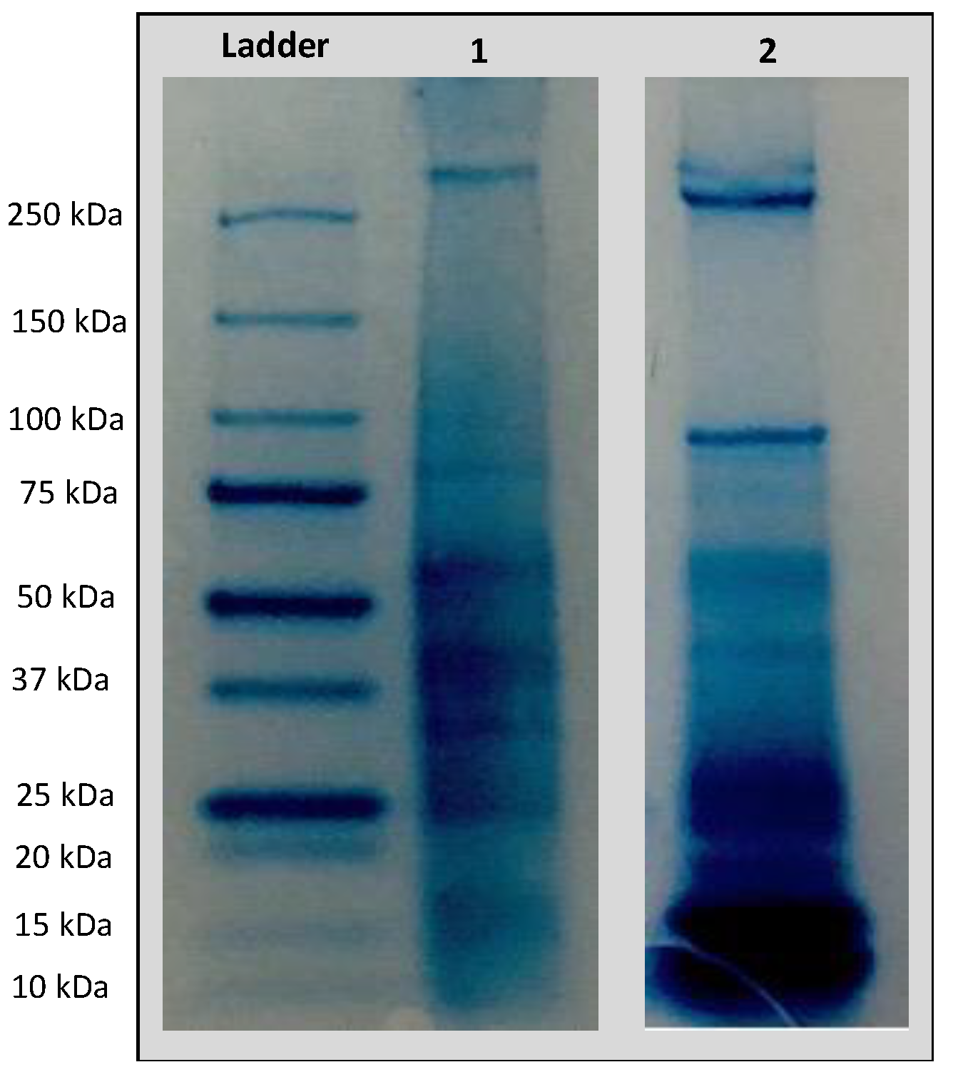
Figure 1. SDS-PAGE analysis of A. dubius (lane 1) and A. camansi (lane 2) protein extracts.
Pharmaceuticals 16 00820 g001
Figure 2. CD spectra of seed protein extracts obtained from (a) A. dubius and (b) A. camansi.
Pharmaceuticals 16 00820 g002
Figure 3. Chaperone activity of A. camansi or A. dubius protein extracts by evaluating their effect on the activity of CS under high-temperature stress conditions (44 °C for 40 min). The error bars represent the standard error of three independent experiments. * Statistical significance (p < 0.05) between A. camansi or A. dubius protein extract and CS alone at 44 °C for 40 min.
Pharmaceuticals 16 00820 g003
Figure 4. (a) Thioflavin T fluorescence intensity as a function of time for 40 μM Aβ1–40 (black), in the presence of 367 μg/mL of seed protein extracts obtained from A. camansi (red) or A. dubius (blue). (b) Inhibition of Aβ1–40 aggregation in the presence of A. camansi or A. dubius seed protein extracts. The error bars represent the standard error of three independent experiments.
Pharmaceuticals 16 00820 g004
Figure 5. Overlay of size distribution by intensity for untreated Aβ1–40 fibrils and Aβ1–40 fibrils treated with (a) A. dubius protein extract or (b) A. camansi protein extract.
Pharmaceuticals 16 00820 g005
Figure 6. (a) ThT fluorescence intensity as a function of time for 40 μM Aβ1–40 in the presence of 367 μg/mL of untreated or alcalase-treated seed protein extracts obtained from A. camansi or A. dubius. (b) Inhibition of Aβ1–40 aggregation in the presence of protein extracts treated or untreated with alcalase. The inhibition percentage of Aβ1–40 aggregation was calculated using the ThT fluorescence intensities of the control sample (Aβ1–40) and Aβ1–40 in the presence of treated or untreated seed protein extracts using Equation (1). Data are represented as the mean ± SE of four independent experiments. * p < 0.05 (Student’s t-test).
Pharmaceuticals 16 00820 g006
Figure 7. Dose–response curve for SH-SY5Y cells. Cells were exposed to varying concentrations of A. camansi (red) or A. dubius (blue) protein extracts (1–367 μg/mL) for 48 h. Error bars represent the standard error of the mean of three independent experiments.
Pharmaceuticals 16 00820 g007
Figure 8. Evaluation of the protective effect of A. camansi or A. dubius seed protein extracts against cytotoxicity induced by Aβ1–40 in SH-SY5Y cells. The viability ratio was determined after incubating SH-S5Y5 cells with Aβ1–40 fibrils in the presence or absence of A. camansi or A. dubius protein extracts for 48 h. Error bars represent the standard deviation of the mean of three independent experiments. * Statistical significance (p < 0.05) between Aβ1–40-treated cells and co-incubation of Aβ1–40 with A. camansi or A. dubius protein extracts.
Pharmaceuticals 16 00820 g008
Table 1. Protein secondary structure content obtained using the server BeStSel.
ConformationA. dubius (%)A. camansi (%)
α-Helix5.90.0
β-Sheet25.636.8
β-Turn13.512.4
Others54.950.7
Table 2. Scientific names, local names and parts used of the selected plants.
Scientific NamesLocal NamesParts Used
Amaranthus dubius Mart. ex ThellRed Spinach, Pig’s weed, BledoSeeds
Artocarpus camansi BlancoBreadnut, Pana de PepitaSeeds
Disclaimer/Publisher’s Note: The statements, opinions and data contained in all publications are solely those of the individual author(s) and contributor(s) and not of MDPI and/or the editor(s). MDPI and/or the editor(s) disclaim responsibility for any injury to people or property resulting from any ideas, methods, instructions or products referred to in the content.