Abstract
Heterocyclic nuclei have shown a wide variety of biological activities, highlighting their importance in drug discovery. Derivatives of 2,4-subsituted thiazolidine have a structural similarity with the substrates of tyrosinase enzymes. Hence, they can be used as an inhibitor to compete against tyrosine in the biosynthesis of melanin. This study is focused on design, synthesis, biological activities, and in silico studies of thiazolidine derivatives substituted at positions 2 and 4. The synthesized compounds were evaluated to determine the antioxidant activity and tyrosine inhibitory potential using mushroom tyrosinase. The most potent tyrosinase enzyme inhibitor was compound 3c having IC50 value 16.5 ± 0.37 µM, whereas compound 3d showed maximum antioxidant activity in a DPPH free radical scavenging assay (IC50 = 18.17 µg/mL). Molecular docking studies were conducted using mushroom tyrosinase (PDB ID: 2Y9X) to analyze binding affinities and binding interactions of the protein–ligand complex. Docking results indicated that hydrogen bonds and hydrophobic interactions were mainly involved in the ligand and protein complex. The highest binding affinity was found to be −8.4 Kcal/mol. These results suggest that thiazolidine-4-carboxamide derivatives could serve as lead molecules for development of novel potential tyrosinase inhibitors.
1. Introduction
Thiazolidine is a heterocyclic organic compound at position 1 with a thioether group and position 3 with an amine group. It is a five-membered saturated ring with the molecular formula of (CH2)3(NH)S. Schubert first reported the thiazolidine carboxylic acid formation as a result of the reaction of cysteine with different aldehydes [1,2]. Thiazolidine nuclei can be synthesized using various methods, as summarized in a study published in 2020 [3].
In mushrooms and vertebrates, melanin biosynthesis is catalyzed by a copper-containing enzyme, tyrosinase [4]. Tyrosinase obtained from Agaricus bisporus has shown structural similarity to mammalian tyrosinase [5,6]. This structural similarity has helped researchers conduct melanogenic studies using mushroom tyrosinase. Kojic acid, a reversible inhibitor of the tyrosinase enzyme, interacts with the binding pocket without any conformational changes in the protein. Major reported amino acids present in the active site are Val283, His263, and Phe264. Other amino residues of the active side include His244, Glu256, Asn260, and residues 279–282 and Ala286 [7,8,9].
The tyrosinase enzyme is an important enzyme responsible for the attachment of marine organisms to their substrates as well as the insect molting process. In humans, it has been found to be linked with the pathogenesis of certain neurodegenerative diseases, including Parkinson’s disease [10]. The imperative function of this enzyme is melanin biosynthesis. It activates this pathway by catalyzing two steps, indicating its importance in melanin formation. Initially, tyrosine is converted to 3,4-dihydroxyphenylalanine (l-DOPA) by hydroxylation. In the second step, l-DOPA is converted to o-dopaquinone through oxidation [11,12]. Melanin is one of the most widely distributed pigments in animals, plants, fungi, and bacteria. It is responsible for pigmentation and color patterns on the skin of mammalians and protects them from the harmful effects of UV radiation [13]. Melanin deposits have also been found in the brain, and it is interesting to know that excessive levels of tyrosine cause the accumulation of epidermal melanin which can result in sites of actinic damage, age spots, melisma, and malignant melanoma [14]. Different strategies have been designed to address this issue by reducing melanin hyperpigmentation, which includes autophagy induction, by disrupting the transfer of melanosomes from melanocytes to keratinocytes by inducing melanosome dematuration [15]. Another aspect is that the formation of melanin beneath the skin is attributed to excessive free radicals that progress through UV radiation. The disruption of this mechanism through the inhibitory action of antioxidant compounds is a major strategy for the regulation of melanin formation [16]. The tyrosinase enzyme also catalyzes the hydroxylation of monophenols to catechols and subsequent oxidation to o-quinones using oxygen. The success of these strategies is accompanied by side effects, which include redness, crusting, swelling, itching, stinging, burning, and unusual discoloration [17]. Inhibitors are usually developed to compete with tyrosine and l-DOPA in the substrate-binding region of the tyrosinase enzyme. The side effects associated with the existing therapeutic regimens have persuaded the researchers to identify alternative strategies including the identification of novel tyrosinase enzyme inhibitors to facilitate the inhibition of melanin formation. Despite the discovery of various semisynthetic and synthetic inhibitors that are used against hyperpigmentation, there is still a need for safer and more effective tyrosinase inhibitors [18,19].
Different modifications have been made to the thiazolidine nucleus at position 2 to achieve inhibitory activity against the tyrosinase enzyme. Young Mi Ha et al. synthesized thiazolidine-4-carboxylic acid analogs with substitution at carbon number 2 as novel tyrosinase inhibitors. The structure of derivatives was rationalized using structural features of a potent inhibitor of catechol oxidase enzyme, N-phenylthiourea, along with the substrate and product of tyrosinase [20]. Some other compounds containing thiazolidine-like nuclei also possess tyrosinase inhibitory activity. These compounds have benzothiazole, benzothiazoline, benzimidazole, and thiazoline nuclei instead of thiazolidine [17,18].
The thiazolidine nucleus can be modified in different ways to obtain a wide variety of derivatives. Although many derivatives of this nucleus have been synthesized and evaluated for different biological activities, which highlight its importance [21,22], new derivatives can be designed having potential biological activities. Thiazolidine-4-carboxylic derivatives reported so far have been modified at position 2 with a free carboxylic acid group at position 4. Position 4 can be further explored for designing compounds having potential biological effects. The main aim of this research was to design and synthesize a range of 2,4-substituted thiazolidine derivatives with potential tyrosinase inhibition by modifying the 4-carboxylic acid group. The antioxidant and tyrosinase inhibitory potential of the compounds synthesized was evaluated. Molecular docking evaluation was conducted to computationally validate the findings of this study. The rational for selecting tyrosinase activity was the structural similarity of 2-(substituted phenyl) thiazolidine-4-carboxamide derivatives with tyrosine, DOPA, and N-phenylthiourea. N-phenylthiourea has shown inhibitory potential against catechol oxidase enzyme, which belongs to the same type of enzymes as tyrosinase [23]. Since reactive oxygen species can increase tyrosinase expression [24], we investigated the antioxidant potential of the synthesized compounds. Compounds having antioxidant activity may exhibit enhanced inhibitory effects on melanin synthesis.
2. Results and Discussion
2.1. Chemistry
Scheme 1 represents the two steps involved in the synthesis of thiazolidine-4-carboxamide derivatives. In the first step, l-cysteine hydrochloride monohydrate was reacted with substituted benzaldehyde in the presence of sodium bicarbonate. This led to the formation of 2-(substituted phenyl) thiazolidine-4-carboxylic acids. The reaction was processed at room temperature. White-colored precipitates obtained were washed with water and ethanol. This was followed by drying in a vacuum desiccator to obtain the pure product. The reaction time and conditions were consistent with the previously reported schemes [20,25]. The reaction time ranged from 3 h to 12 h. The product formation was confirmed by comparing the melting points reported in a previous study [20]. The yield of the substituted thiazolidine nuclei (1–4) varied from 40 to 90%.
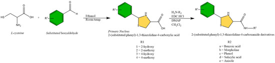
Scheme 1.
Synthetic route to 2-(substituted phenyl)-1,3-thiazolidine-4-carboxamide.
These nuclei (1–4) were used further to synthesize the carboxamide derivatives. Nuclei from the first step, 2-(substituted phenyl) thiazolidine-4-carboxylic acids (1–4), were reacted with an amine in the presence of N-(3-Dimethylaminopropyl)-N′-ethylcarbodiimide hydrochloride (EDC.HCl) and 4-Dimethylaminopyridine (DMAP) in dichloromethane. Stirring was carried out at room temperature for a time ranging from 4–24 h [26]. A gel-like product was obtained on reaction completion which was washed with methanol and water to obtain the product in powder form. The amines used were p-amino benzoic acid, morpholine, p-amino phenol, p-amino salicylic acid, and p-anisidine. As a result, five carboxamide derivatives were obtained numbered as a, b, c, d, and e, respectively. Nuclei with 2-methoxyphenyl (2) and 4-methoxyphenyl (4) gave a lesser yield with a longer reaction time owing to the steric hindrance, leading to the synthesis of limited derivatives, i.e., two for 2-methoxyphenyl (2a and 2d) and three derivatives in the case of 4-methoxyphenyl (4a, 4d and 4e). The yield of the final products varied from 20–72% (Table 1). In total, 14 derivatives were synthesized based on the four substituted thiazolidine nuclei and five amines. The purity of the synthesized compounds was confirmed through TLC. The chemo-informatics analysis of synthesized derivatives is presented in Table S1. Figure 1 represents the structures of all the synthesized compounds.

Table 1.
Physical properties of synthesized derivatives.
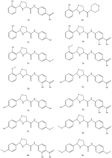
Figure 1.
Structures of the synthesized compounds.
Derivatives synthesized were characterized using spectral data from FTIR, 1H-NMR, and 13C-NMR (Figures S1–S8). The expected stretches were observed in the spectral data of FTIR. Further, chemical shift values from proton and carbon NMR were used for structural confirmation of the planned derivatives. The chemical shift values of thiazolidine protons were compared with the NMR results of the previously reported thiazolidine-4-carboxylic acid derivatives [20]. Observed protons of the thiazolidine nucleus included 2-H, 4-H, and 5-Ha, Hb. The proton of amine (NH) mostly gives a weak signal and is usually not reported. A peak of the proton of carboxamide was observed in all the compounds, which confirmed the structure of 4-carboxamide derivatives of the thiazolidine nuclei. A singlet was observed between the ranges of 8.112–8.998 ppm.
2.2. Biological Evaluation
2.2.1. Determination of Antioxidant Activity
The 2,2-Diphenyl-1-picrylhydrazyl (DPPH) radical was used to evaluate the antioxidant potential of the synthesized compounds. Different concentrations of the samples were employed in the subject assay. The maximum antioxidant potential was shown by the compound 2d (IC50 18.17 ± 1.0 µg/mL) in comparison with the antioxidant activity of the positive control, ascorbic acid (IC50 7.83 ± 0.5 µg/mL) (Figure 2). The compound 1d also showed significant antioxidant potential with an IC50 value of 18.27 ± 1.1 µg/mL. The principle of this assay is related to the conversion of the purple-colored free radical into the pale-yellow-colored reduced form, owing to the action of a proton donating or antioxidant agent [27]. The compound with the most prominent activity (2d) possessed hydroxyls at position 3 of the ring containing the carboxamide group, whereas the methoxy group was attached to position 2 of the benzene ring. Alternatively, compound 1d also acquired hydroxy groups on both mentioned positions. These groups are categorized as strong proton donors which tend to participate in the reduction in DPPH free radicals by donating protons.
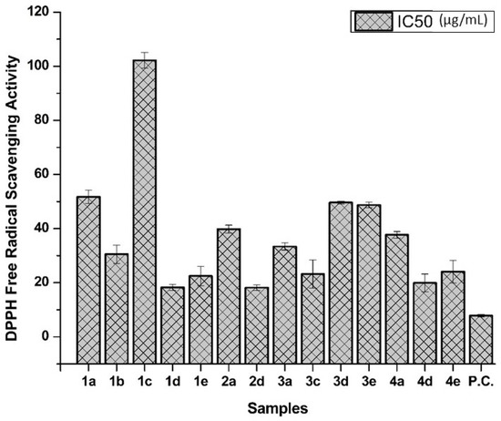
Figure 2.
The IC50 values of synthesized compounds in the DPPH free radical scavenging assay. The results are presented as mean ± SD. P.C. stands for positive control (ascorbic acid).
2.2.2. Tyrosinase Inhibition Studies
An evaluation of the inhibitory effect on the tyrosinase enzyme was conducted on mushroom tyrosinase. The results were compared with the competitive inhibitor of the tyrosinase enzyme, kojic acid (Table 2). Among the synthesized derivatives, the maximum inhibitory potential was observed in the case of compound 3c (IC50 16.5 ± 0.37 µM), compared with the IC50 value of the standard, kojic acid (15.9 ± 2.5 µM). Compounds 4e, 3e, 2d, and 2a also exhibited moderate activity. Compounds 3c, 3e, and 4e exhibited the presence of oxygen atoms at the para-position on both benzene rings in the form of hydroxyl and methoxy groups. The presence of oxygen atoms at these positions could be interacting with key amino acids and the copper ions of the enzyme, resulting in higher inhibitory potential. The remaining derivatives showed insignificant activity with values of IC50 varying between 98.5 ± 10.6 and 256 ± 16.3 µM.
Table 2.
Inhibitory activity of synthesized derivatives against the tyrosinase enzyme.
2.3. Molecular Docking
All synthesized compounds exhibited binding affinities higher than the reference compound, kojic acid. Important amino acids of the active site, His244, Glu256, Asn260, and residues 279–282 and Ala286, were involved in the ligand–protein interactions (Table 3) (Figure S9–S23). The distance of the binding interactions varied from 1.93 Å to 5.70 Å. The highest binding affinity was exhibited by compound 3c (−8.4 Kcal/mol) (Figure 3), while the lowest binding affinity was exhibited by compounds 1e and 3e (−6.9 Kcal/mol). Kojic acid exhibited a binding affinity of −5.5 Kcal/mol. Two derivatives of the 4-hydroxy primary nucleus, 3c and 3e, with lower IC50, represent the importance of the hydroxyl group at the fourth position. This activity was decreased by the modification of the carboxylic acid group towards the carboxamide end (compound 3a), probably owing to the bulky nature of the attached group. A similar pattern was observed in compounds 1a and 1d, owing to the carboxylic acid group and higher IC50 values. Derivatives with the hydroxyl group at the second position (1a–1e) exhibited higher IC50 values. This hydroxyl group can form intramolecular hydrogen bonding with the sulfur atom of the thiazolidine ring, resulting in poor interaction with the target enzyme. This observation was supported by the results of derivatives with the 2-methoxy group (2a and 2d). The 2-methoxy derivatives showed lower IC50 values in comparison with the 2-hydroxy derivatives. All the methoxy derivatives (2a, 2d, and 4e) exhibited lower IC50 values. Some thiazolidine derivatives have been previously synthesized, which also showed tyrosinase inhibitory potential supporting the rational of this study for selecting thiazolidine nucleus [20,28]. Thiazolidine derivatives synthesized in this study are novel through the modification of the carboxamide group at the fourth position of the nucleus.
Table 3.
Binding affinities and respective amino acids interacting with the ligand (synthesized derivatives).
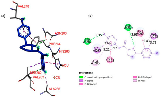
Figure 3.
Binding interactions between derivative 3c and mushroom tyrosinase active site (PDB-ID 2Y9X). (a) Binding conformation of compound 3c in 3D along with the binding interaction of the protein–ligand complex. (b) Interaction patterns in 2D indicating the type and distance of the interaction. Legend indicates various types of binding interactions present.
3. Materials and Methods
3.1. Instruments and Chemicals
Required chemicals and solvents were procured from Sigma-Aldrich (St. Louis, MO, USA), Merck (Kenilworth, NJ, USA), and Honeywell (Charlotte, NC, USA) with no further purification before their use. Spectrophotometric and elemental analysis was used for the characterization of the synthesized derivatives. Functional group detection was performed with a Bruker ALPHA FTIR spectrometer (Billerica, MA, USA), and structural analysis was conducted with 1HNMR and 13C NMR using a Bruker AM300 spectrophotometer (Billerica, MA, USA) with DMSO as the solvent. Melting points were also recorded with the help of the Gallenkamp melting point apparatus. Mushroom tyrosinase (EC 1.14.18.1) was purchased from Sigma-Aldrich, (St. Louis, MO, USA).
3.2. General Procedure for the Synthesis of the Thiazolidine Nucleus
A previously reported scheme was used for the synthesis of the thiazolidine nucleus [20,25]. A total of four 2-(substituted phenyl) thiazolidine-4-carboxylic acid derivatives were synthesized using differently substituted benzaldehydes. l-cysteine hydrochloride monohydrate (6.2 mmol) was added to 100 mL of water. Then, sodium bicarbonate (6.2 mmol) was added to the mixture with stirring. A solution of respective benzaldehyde (6.2 mmol) in ethanol (100 mL) was prepared separately and added to the reaction mixture. The mixture was stirred for 2.5 h at room temperature. The precipitates formed were collected through filtration and washed with cooled water and ethanol.
3.2.1. 2-(2-Hydroxyphenyl)-1,3-thiazolidine-4-carboxylic Acid (1)
White solid; mp(observed): 173–175 °C; mp(lit): 173.2–175.1 °C; yield: 90%; 1H-NMR (DMSO, δ ppm): 3.009 (d, J = 14.30 Hz, 1H, 5-Ha), 3.228 (d, J = 14.30 Hz, 1H, 5-Hb), 4.235 (dd, 1J = 9.37 Hz, 2J = 5.64 Hz, 1H, 4-H), 5.699 (s, 1H, 2-H), 6.525–7.625 (m, 4H, Ar-H), 9.868 (s, 1H, Ar-2′OH).
3.2.2. 2-(2-Methoxyphenyl)-1,3-thiazolidine-4-carboxylic Acid (2)
White solid; m.p.(observed): 137–139 °C; m.p.(lit): 137.7–139.2 °C; yield: 40%; 1H-NMR (DMSO, δ ppm): 3.495 (d, J = 14.31, 1H, 5-Ha), 3.551 (d, J = 14.31, 1H, 5-Hb), 3.795 (s, 3H, Ar-2′O CH3), 3.919 (dd, 1J = 9.52, 2J = 5.39, 1H, 4-H), 5.135 (s, 1H, 2-H), 6.953–8.049 (m, 4H, Ar-H).
3.2.3. 2-(4-Hydroxyphenyl)-1,3-thiazolidine-4-carboxylic Acid (3)
White solid; m.p.(observed): 161–164 °C; m.p.(lit): 161.8–164.4 °C; yield: 55%; 1H-NMR (DMSO, δ ppm): 3.404 (d, J = 14.29, 1H, 5-Ha), 3.621 (d, J = 14.29, 1H, 5-Hb), 3.693 (dd, 1J = 9.37, 2J = 5.64, 1H, 4-H), 5.236 (s, 1H, 2-H), 6.653–8.049 (m, 4H, Ar-H), 9.563 (s, 1H, Ar-4′OH).
3.2.4. 2-(4-Methoxyphenyl)-1,3-thiazolidine-4-carboxylic Acid (4)
White solid; m.p.(observed): 157–158 °C; m.p.(lit): 157.6–158.2 °C; yield: 45%; 1H-NMR (DMSO, δ ppm): 3.494 (d, J = 14.31, 1H, 5-Ha), 3.549 (d, J = 14.31, 1H, 5-Hb), 3.747 (s, 3H, Ar-4′OCH3), 3.887 (dd, 1J = 9.52, 2J = 5.39, 1H, 4-H), 5.147 (s, 1H, 2-H), 6.895–8.049 (m, 4H, Ar-H).
3.3. General Scheme for the Synthesis of Carboxamide Derivatives from Thiazolidine Nucleus
Carboxamide derivatives were synthesized from the thiazolidine nucleus by reacting with various amines [26]. 2-(substituted phenyl) thiazolidine-4-carboxylic acid (0.98 mmol), amine (1.12 mmol), DMAP (1.09 mmol), and EDC.HCl (1.08 mmol) were mixed in dichloromethane (2 mL). Then, stirring for 4 h was carried out at room temperature. The product was obtained in the form of a hard gel. This gel-type product was washed with methanol and water to obtain a purified product in the form of a powder.
3.3.1. 4-[2-(2-Hydroxyphenyl)-1,3-thiazolidine-4-carbonyl]-aminobenzoic Acid (1a)
Yellow solid; yield: 54%; m.p.: 135–136 °C; IR cm−1: 3500 (O-H), 3100 (N-H stretch), 1600 (C=O), 1500 (N-H bend), 1470 (C=C); 1H-NMR (DMSO, δ ppm): 3.009 (d, J = 14.30 Hz, 1H, 5-Ha), 3.228 (d, J = 14.30 Hz, 1H, 5-Hb), 4.235 (dd, 1J = 9.37 Hz, 2J = 5.64 Hz, 1H, 4-H), 5.699 (s, 1H, 2-H), 6.525–7.625 (m, 8H, Ar-H), 8.141 (s, 1H, amide-NH), 9.868 (s, 1H, Ar-2′OH), 11.295 (s, 1H, 4″COOH); 13C-NMR (DMSO, 100 MHz, δ ppm): 172.4 (CO-NH), 168.2 (COOH), 161.0 (C-2′), 136.4 (C-1′), 132.5 (C-3″, C-5″), 130.2 (C-4′), 127.4 (C-5′), 126.5 (C-1′), 126.0 (C-6′), 125.7 (C-4″), 119.0 (C-2″, C-6″), 118.2 (C-3′), 66.2 (C-2), 56.2 (C-4), 32.4 (C-5); Anal. Calcd. for C17H16N2O4S: C, 58.88; H, 3.95; N, 7.92; O, 17.96; S, 10.201. Found: C, 58.15; H, 3.30; N, 7.22; O, 18.40; S, 9.25.
3.3.2. [2-(2-Hydroxyphenyl)-1,3-thiazolidin-4-yl](morpholin-4-yl)-methanone (1b)
Orange solid; yield: 68%; m.p.: 128–129 °C; IR cm−1: 3600 (O-H), 3120 (N-H stretch), 1640 (C=O), 1590 (N-H bend), 1448 (C=C); 1H-NMR (DMSO, δ ppm): 3.393 (d, J = 14.34, 1H, 5-Ha), 3.598 (d, J = 14.34, 1H, 5-Hb), 3.624 (m, 8H, CH2), 3.712 (dd, 1J =9.37, 2J = 5.64, 1H, 4-H), 5.436 (s, 1H, 2-H), 6.658–7.398 (m, 8H, Ar-H); 13C-NMR (DMSO, 100 MHz, δ ppm): 170.2 (CO-NH), 160.5 (C-2′), 130.2 (C-4′), 129.5 (C-5′), 129.0 (C-1′), 128.6 (C-6′), 117.2 (C-3′), 66.8 (C-3″, C-5″), 64.8 (C-2), 57.4 (C-4), 44.5 (C-2″, C-6″), 31.6 (C-5); Anal. Calcd. for C14H18N2O3S: C, 57.12; H, 5.95; N, 8.85; O, 16.31; S, 9.74. Found: C, 56.95; H, 6.05; N, 8.45; O, 16.35; S, 9.80.
3.3.3. 2-(2-Hydroxyphenyl)-N-(4-hydroxyphenyl)-1,3-thiazolidine-4-carboxamide (1c)
Black solid; yield: 45%; m.p.: 109–111 °C; IR cm−1: 3530 (O-H), 3400 (N-H stretch), 1650(C=O), 1500 (N-H bend), 1410 (C=C); 1H-NMR (DMSO, δ ppm): 3.408 (d, J = 14.31, 1H, 5-Ha), 3.610 (d, J = 14.31, 1H, 5-Hb), 3.717 (dd, 1J = 9.37, 2J = 5.64, 1H, 4-H), 5.437 (s, 1H, 2-H), 6.658–7.398 (m, 8H, Ar-H), 8.518 (s, 1H, amide-NH), 9.218 (s, 1H, Ar-2′OH), 9.965 (s, 1H, Ar-4″OH); 13C-NMR (DMSO, 100 MHz, δ ppm): 174.8 (CO-NH), 162.0 (C-2′), 158.4 (C-4″), 137.8 (C-1″), 130.4 (C-4′), 128.4 (C-5′), 127.0 (C-1′), 125.8 (C-6′), 120.5 (C-5″, C-6″), 116.5 (C-3′), 113.2 (C-3″, C-5″), 66.2 (C-2), 55.4 (C-4), 30.6 (C-5). Anal. Calcd. for C16H16N2O3S: C, 59.85; H, 4.95; N, 9.12; O, 15.17; S, 9.03. Found: C, 59.70; H, 4.02; N, 9.50; O, 15.20; S, 10.10.
3.3.4. 2-Hydroxy-4-[2-(2-hydroxyphenyl)-1,3-thiazolidine-4-carbonyl]-aminobenzoic Acid (1d)
Yellow solid; yield: 68%; m.p.: 134–135 °C; IR cm−1: 3530 (O-H), 3150 (N-H stretch), 1643 (C=O), 1590 (N-H bend), 1400 (C=C); 1H-NMR (DMSO, δ ppm): 3.406 (d, J = 14.29, 1H, 5-Ha), 3.623 (d, J = 14.29, 1H, 5-Hb), 3.758 (dd, 1J = 9.37, 2J = 5.64, 1H, 4-H), 5.437 (s, 1H, 2-H), 6.308–8.023 (m, 8H, Ar-H), 8.123 (s, 1H, amide-NH), 9.225 (s, 1H, Ar-2′OH), 9.995 (s, 1H, Ar-3″OH), 11.598 (s, 1H, 4″-COOH); 13C-NMR (DMSO, 100 MHz, δ ppm): 173.5 (CO-NH),170.8 (COOH), 165.3 (C-3″), 161.0 (C-2′), 140.2 (C-1″), 133.4 (C-5″), 129.9 (C-4′), 128.8 (C-5′), 128.0 (C-1′), 126.0 (C-6′), 118.5 (C-6″), 115.8 (C-3′), 111.8 (C-4″), 101.1 (C-2″), 66.4 (C-2), 55.2 (C-4), 31.1 (C-5); Anal. Calcd. for C17H16N2O5S: C, 57.22; H, 5.04; N, 8.12; O, 21.10; S, 9.15. Found: C, 57.60; H, 5.45; N, 8.65; O, 21.18; S, 9.83.
3.3.5. 2-(2-Hydroxyphenyl)-N-(4-methoxyphenyl)-1,3-thiazolidine-4-carboxamide (1e)
Yellow solid; yield: 60%; m.p.: 83–84 °C; IR cm−1: 3600 (O-H), 3100 (N-H stretch), 1670 (C=O), 1490 (N-H bend), 1450 (C=C); 1H-NMR (DMSO, δ ppm): 3.152 (d, J = 14.04, 1H, 5-Ha), 3.324 (d, J = 14.04, 1H, 5-Hb), 3.797(s, 3H, 4″-OCH3), 4.358 (dd, 1J = 9.52, 2J = 5.39, 1H, 4-H), 5.298 (s, 1H, 2-H), 6.935–7.633 (m, 8H, Ar-H), 8.425 (s, 1H, amide-NH), 9.525 (s, 1H, Ar-2′OH); 13C-NMR (DMSO, 100 MHz, δ ppm): 173.6 (CO-NH), 162.0 (C-2′), 155.8 (C-4″), 138.4 (C-1″), 129.2 (C-4′), 128.4 (C-5′), 128.0 (C-1′), 127.2 (C-6′), 121.5 (C-2″. C-6″), 116.9 (C-3′), 113.5 (C-3″, C-5″), 66.5 (C-2), 56.6 (C-4), 55.0 (4′-OCH3), 31.8 (C-5). Anal. Calcd. for C17H18N2O3S: C, 61.80; H, 4.95; N, 7.98; O, 14.53; S, 10.03. Found: C, 61.75; H, 4.45; N, 7.42; O, 14.48; S, 10.60.
3.3.6. 4-[2-(2-Methoxyphenyl)-1,3-thiazolidine-4-carbonyl]-aminobenzoic Acid (2a)
White solid; yield: 20%; m.p.: 116–117 °C; IR cm−1: 3500 (O-H), 3220 (N-H stretch), 1600 (C=O), 1498 (N-H bend), 1380 (C=C); 1H-NMR (DMSO, δ ppm): 3.495 (d, J = 14.31, 1H, 5-Ha), 3.551 (d, J = 14.31, 1H, 5-Hb), 3.795 (s, 3H, Ar-2′O CH3), 3.919 (dd, 1J = 9.52, 2J = 5.39, 1H, 4-H), 5.135 (s, 1H, 2-H), 6.953–8.049 (m, 8H, Ar-H), 8.235 (s, 1H, amide-NH), 11.285 (s, 1H, 4″-COOH); 13C-NMR (DMSO, 100 MHz, δ ppm): 170.2 (CO-NH), 165.1 (COOH), 155.3 (C-2′), 138.4 (C-1″), 131.5 (C-3″, C-5″), 129.4 (C-4′), 128.5 (C-5′), 128.0 (C-1′), 127.8 (C-6′), 127.0 (C-4″), 117.9 (C-2″, C-6″), 114.8 (C-3′), 66.2 (C-2), 56.8 (C-4), 55.0 (2′-OCH3), 30.9 (C-5). Anal. Calcd. for C18H18N2O4S: C, 61.14; H, 4.92; N, 8.03; O, 17.86; S, 9.02. Found: C, 61.20; H, 4.95; N, 8.45; O, 17.90; S, 8.88.
3.3.7. 2-Hydroxy-4-[2-(2-methoxyphenyl)-1,3-thiazolidine-4-carbonyl]-aminobenzoic Acid (2d)
White solid; yield: 25%; m.p.: 112–113 °C; IR cm−1: 3510 (O-H), 3300 (N-H stretch), 1660 (C=O), 1489 (N-H bend), 1386 (C=C); 1H-NMR (DMSO, δ ppm): 3.496 (d, J = 14.31, 1H, 5-Ha), 3.551 (d, J = 14.31, 1H, 5-Hb), 3.795 (s, 3H, Ar-2′O CH3), 3.888 (dd, 1J = 9.52, 2J = 5.39, 1H, 4-H), 5.135 (s, 1H, 2-H), 6.953–7.395 (m, 8H, Ar-H), 8.586 (s, 1H, amide-NH), 9.228 (s, 1H, Ar-3″OH), 11.298 (s, 1H, 4″-COOH); 13C-NMR (DMSO, 100 MHz, δ ppm): 174.2 (CO-NH), 172.3 (COOH), 162.3 (C-3″), 158.3 (C-2′), 140.3 (C-1″), 132.3 (C-5″), 130.4 (C-4′), 128.5 (C-5′), 128.0 (C-1′), 126.8 (C-6′), 118.9 (C-6″), 114.5 (C-3′), 110.2 (C-4″), 100.4 (C-2″), 64.2 (C-2), 56.5 (C-4), 54.0 (2′-OCH3), 31.2 (C-5); Anal. Calcd. for C18H18N2O5S: C, 60.12; H, 5.02; N, 8.22; O, 20.13; S, 9.42. Found: C, 60.22; H, 5.22; N, 8.42; O, 21.24; S, 8.42.
3.3.8. 4-[2-(4-Hydroxyphenyl)-1,3-thiazolidine-4-carbonyl]-aminobenzoic Acid (3a)
Yellow solid; yield: 72%; m.p.: 164–166 °C; IR cm−1: 3550 (O-H), 3000 (N-H stretch), 1597 (C=O), 1508 (N-H bend), 1450 (C=C); 1H-NMR (DMSO, δ ppm): 3.404 (d, J = 14.29, 1H, 5-Ha), 3.621 (d, J = 14.29, 1H, 5-Hb), 3.693 (dd, 1J = 9.37, 2J = 5.64, 1H, 4-H), 5.236 (s, 1H, 2-H), 6.653–8.049 (m, 8H, Ar-H), 8.998 (s, 1H, amide-NH), 9.563 (s, 1H, Ar-4′OH), 12.004 (s, 1H, 4″COOH); 13C-NMR (DMSO, 100 MHz, δ ppm): 173.7 (CO-NH), 164.5 (COOH), 159.4 (C-4′), 138.4 (C-1″), 135.1 (C-1′), 131.2 (C-3″, C-5″), 127.2 (C-2′, C-6′), 125.1 (C-4″), 117.8 (C-2″, C-6”), 114.7 (C-3′, C-5′), 63.2 (C-2), 54.6 (C-4), 32.6 (C-5); Anal. Calcd. for C17H16N2O4S: C, 60.12; H, 5.14; N, 7.95; O, 18.58; S, 8.92. Found: C, 60.10; H, 5.40; N, 7.92; O, 18.40; S, 8.45.
3.3.9. N,2-Bis(4-hydroxyphenyl)-1,3-thiazolidine-4-carboxamide (3c)
Black solid; yield: 68%; m.p.: 174–176 °C; IR cm−1: 3580 (O-H), 3050 (N-H stretch), 1640 (C=O), 1512 (N-H bend), 1400 (C=C); 1H-NMR (DMSO, δ ppm): 3.408 (d, J = 14.30, 1H, 5-Ha), 3.615 (d, J = 14.30, 1H, 5-Hb), 3.692 (dd, 1J = 9.37, 2J = 5.64, 1H, 4-H), 5.326 (s, 1H, 2-H), 6.653–7.278 (m, 8H, Ar-H), 8.112 (s, 1H, amide-NH), 9.158 (s, 1H, Ar-4′OH), 9.998 (s, 1H, 4″OH); 13C-NMR (DMSO, 100 MHz, δ ppm): 170.2 (CO-NH), 157.5 (C-4′), 138.4 (C-1″), 134.2 (C-1′), 128.7 (C-2′, C-6′), 120.8 (C-2″, C-6″), 116.2 (C-3′, C5′), 114.2 (C-3″, C-5″), 64.3 (C-2), 55.6 (C-4), 32.4 (C-5); Anal. Calcd. for C16H16N2O3S: C, 59.89; H, 4.92; N, 9.12; O, 15.17; S, 9.24. Found: C, 59.95; H, 4.98; N, 9.22; O, 15.03; S, 10.02.
3.3.10. 2-Hydroxy-4-[2-(4-hydroxyphenyl)-1,3-thiazolidine-4-carbonyl]-aminobenzoic Acid (3d)
Yellow solid; yield: 68%; m.p.: 130–131 °C; IR cm−1: 3500 (O-H), 3100 (N-H stretch), 1600 (C=O), 1560 (N-H bend), 1450 (C=C); 1H-NMR (DMSO, δ ppm): 3.405 (d, J = 14.29, 1H, 5-Ha), 3.622 (d, J = 14.29, 1H, 5-Hb), 3.696 (dd, 1J = 9.37, 2J = 5.64, 1H, 4-H), 5.327 (s, 1H, 2-H), 6.307–8.023 (m, 8H, Ar-H), 8.534 (s, 1H, amide-NH), 9.125 (s, 1H, Ar-4′OH), 9.893 (s, 1H, Ar-3″OH), 11.518 (s, 1H, 4″COOH); 13C-NMR (DMSO, 100 MHz, δ ppm): 173.2 (CO-NH), 170.1 (COOH), 162.5 (C-3″), 158.4 (C-4′), 139.3 (C-1″), 135.8 (C-1′), 130.5 (C-5″), 128.8 (C-2′, C-6′), 118.0 (C-6″), 114.4 (C-3′, C-5′), 109.3 (C-4″), 101.2 (C-2″), 64.2 (C-2), 54.5 (C-4), 33.6 (C-5); Anal. Calcd. for C17H16N2O5S: C, 57.12; H, 5.15; N, 8.32; O, 21.95; S, 9.12. Found: C, 57.50; H, 5.35; N, 8.70; O, 21.80; S, 9.05.
3.3.11. 2-(4-Hydroxyphenyl)-N-(4-methoxyphenyl)-1,3-thiazolidine-4-carboxamide (3e)
Brown solid; yield: 65%; m.p.: 115–117 °C; IR cm−1: 3500 (O-H), 3100 (N-H stretch), 1600 (C=O), 1550 (N-H bend), 1440 (C=C); 1H-NMR (DMSO, δ ppm): 3.445 (d, J = 14.33, 1H, 5-Ha), 3.539 (d, J = 14.33, 1H, 5-Hb), 3.715 (s, 3H, 4″OCH3), 3.825 (dd, 1J = 9.52, 2J = 5.39, 1H, 4-H), 5.154 (s, 1H, 2-H), 6.653–7.275 (m, 8H, Ar-H), 8.224 (s, 1H, amide-NH), 9.212 (s, 1H, Ar-4′OH); 13C-NMR (DMSO, 100 MHz, δ ppm): 172.2 (COOH), 157.7 (C-4″), 155.4 (C-4′), 137.8 (C-1″), 134.2 (C-1′), 128.8 (C-2′, C-6′), 121.5 (C-2″, C-6″), 118.4 (C3′, C-5′), 115.2 (C-3″, C-5″), 64.2 (C-2), 57.2 (C-4), 55.0 (4″-OCH3), 32.2 (C-5); Anal. Calcd. for C17H18N2O3S: C, 61.80; H, 4.49; N, 9.05; O, 14.53; S, 10.12. Found: C, 61.78; H, 4.30; N, 9.35; O, 14.22; S, 10.63.
3.3.12. 4-[2-(4-Methoxyphenyl)-1,3-thiazolidine-4-carbonyl]-aminobenzoic Acid (4a)
Yellow solid; yield: 60%; m.p.: 94–95 °C; IR cm−1: 3610 (O-H), 3200 (N-H stretch), 1590 (C=O), 1550 (N-H bend), 1420 (C=C); 1H-NMR (DMSO, δ ppm): 3.494 (d, J = 14.31, 1H, 5-Ha), 3.549 (d, J = 14.31, 1H, 5-Hb), 3.747 (s, 3H, Ar-4′OCH3), 3.887 (dd, 1J = 9.52, 2J = 5.39, 1H, 4-H), 5.147 (s, 1H, 2-H), 6.895–8.049 (m, 8H, Ar-H), 8.152 (s, 1H, amide-NH), 11.892 (s, 1H, 4″COOH); 13C-NMR (DMSO, 100 MHz, δ ppm): 171.8 (CO-NH), 167.8 (COOH), 160.9 (C-4′), 136.2 (C-1″), 135.0 (C-1′), 131.4 (C-3″, C-5″), 129.4 (C-2′, C-6′), 128.2 (C-4″), 118.8 (C-2″, C-6″), 113.3 (C-3′, C-5′), 64.2 (C-2), 56.6 (4′-OCH3), 54.0 (C-4), 32.6 (C-5). Anal. Calcd. for, C18H18N2O4S: C, 59.92; H, 4.80; N, 8.13; O, 17.86; S, 9.21. Found: C, 59.75; H, 4.92; N, 8.66; O, 17.70; S, 8.84.
3.3.13. 2-Hydroxy-4-[2-(4-methoxyphenyl)-1,3-thiazolidine-4-carbonyl]-aminobenzoic Acid (4d)
White solid; yield: 66%; m.p.: 109–110 °C; IR cm−1: 3600 (O-H), 3120 (N-H stretch), 1645 (C=O), 1511 (N-H bend), 1420 (C=C); 1H-NMR (DMSO, δ ppm): 3.461 (d, J = 14.31, 1H, 5-Ha), 3.552 (d, J = 14.31, 1H, 5-Hb), 3.744 (s, 3H, Ar-4′OCH3), 3.861 (dd, 1J = 9.52, 2J = 5.39, 1H, 4-H), 5.148 (s, 1H, 2-H), 6.307–8.023 (m, 8H, Ar-H), 8.212 (s, 1H, amide-NH), 9.518 (s, 1H, Ar-3″OH), 12.001 (s, 1H, 4″-COOH); 13C-NMR (DMSO, 100 MHz, δ ppm): 171.2 (CO-NH), 170.1 (COOH), 163.4 (C-3″), 159.2 (C-4′), 135.4 (C-1″), 135.1 (C-1′), 130.7 (C-5″), 126.8 (C-2′, C-6′), 116.2 (C-6″), 114.0 (C-3′, C-5′), 110.1 (C-4″), 100.4 (C-2″), 64.8 (C-2), 54.8 (C-4), 52.4 (4-OCH3), 31.6 (C-5); Anal. Calcd. for C18H18N2O5S: C, 60.02; H, 5.11; N, 8.24; O, 20.98; S, 9.11. Found: C, 60.29; H, 5.22; N, 8.38; O, 21.40; S, 8.16.
3.3.14. N,2-Bis(4-methoxyphenyl)-1,3-thiazolidine-4-carboxamide (4e)
Brown solid; yield: 66%; m.p.: 129–131 °C; IR cm−1: 3150 (N-H stretch), 1600 (C=O), 1500 (N-H bend), 1410 (C=C); 1H-NMR (DMSO, δ ppm): 3.445 (d, J = 14.33, 1H, 5-Ha), 3.545 (d, J = 14.33, 1H, 5-Hb), 3.742 (s, 3H, Ar-2′OCH3), 3.763 (s, 3H, Ar-4″OCH3), 3.825 (dd, 1J = 9.52, 2J = 5.39, 1H, 4-H), 5.142 (s, 1H, 2-H), 6.637–7.297 (m, 8H, Ar-H), 8.512 (s, 1H, amide-NH); 13C-NMR (DMSO, 100 MHz, δ ppm): 171.5 (CO-NH), 159.8 (C-4′), 138.1 (C-1″), 135.0 (C-1′), 127.7 (C-2′, C-6′), 121.5 (C-2″, C-6″), 115.5 (C-3″, C-5″), 114.1 (C-3′, C-5′), 64.2 (C-2), 56.5 (C-4), 56.0(4′-OCH3, 4″-OCH3), 32.6 (C-5); Anal. Calcd. for C18H20N2O3S: C, 61.94; H, 4.95; N, 7.89; O, 13.94; S, 8.85. Found: C, 61.85; H, 4.50; N, 7.42; O, 13.78; S, 9.22.
3.4. Biological Evaluation
3.4.1. Determination of Antioxidant Activity
The synthesized compounds were evaluated for their antioxidant activity by measuring their ability to scavenge DPPH free radicals. A previously described protocol was followed for this purpose [27]. An amount of 20 µL of each of the four different dilutions of test samples (200, 66.66, 22.22, and 7.41 µg/mL) was added to 96-well plates containing 180 µL of DPPH solution. The absorbance at 517 nm was determined after incubating for 30 min at 37 °C. Ascorbic acid (1 mg/mL DMSO) was used as the positive control, and the assay was performed in triplicate. The IC50 values were also calculated using the % of inhibitions at different concentrations using Table Curve 2D Windows version 4.07.
3.4.2. Inhibitory Assay against Tyrosinase Enzyme
A tyrosinase inhibitory assay was conducted with the mushroom tyrosinase enzyme [29,30,31]. An amount of 20 µL of mushroom tyrosinase (30 U/mL) was combined with 140 µL (20 mM, pH 6.8) of phosphate buffer. An amount of 20 µL of inhibitor solution was added to a 96-well microplate. Then pre-incubation at room temperature was conducted for 10 min. A solution of l-DOPA (20 μL—0.85 mM) was added. This was followed by the incubation of the plate for 20 min at 25 °C. Using a microplate reader (OPTIMax, Tunable), the absorbance of dopa-chrome was recorded at 492 nm. Kojic acid and phosphate buffer served as the reference inhibitor and negative control, respectively.
The absorbance of dopa-chrome was measured at 492 nm using a microplate reader (OPTIMax, Tunable, San Jose, CA, USA). Phosphate buffer and kojic acid were used as the negative control and the reference inhibitor, respectively. The IC50 values were used to express the extent of inhibition. The experiments were performed in triplicate for each concentration.
3.5. In Silico Studies
Chemo-informatic analysis was performed using Molinspiration and ChemSketch [32]. Molecular docking was conducted using PyRX v0.8 [33]. A tyrosinase enzyme of mushroom origin having the PDB ID: 2Y9X was acquired from the RCSB Protein DataBank Site [7]. The search space for molecular docking was adjusted to the center with X: −7.4, Y: −23.5, and Z: −32.5 and dimensions of X: 61.33, Y: 57.4, and Z: 63. Discovery studio was used for the visualization of ‘.pdb’ files, cleaning of the protein molecule, and visualization of ligand–protein interactions in 3D and 2D. The structures of the ligands were drawn using ChemDraw v16 and converted into pdb files using Chem3D v16 [34].
4. Conclusions
This study presents the design and synthesis of a series of thiazolidine derivatives. The synthesized compounds were screened for their potential biological activity using molecular docking and in vitro antioxidant and tyrosinase inhibitory assays. were conducted to identify the potential biological activity of the synthesized compounds. Compound 2d showed the maximum antioxidant potential (18.17 ± 1.0 µg/mL). In the tyrosine inhibition assay, compound 3c exhibited significant activity with an IC50 value of 16.5 ± 0.37 µM. Molecular docking analysis led to an overview of the structural properties that can affect ligand–protein interactions. Hydrophobic interactions and hydrogen bonding were found to play a significant role. Compounds 4e, 3e, 2d, and 2a can also serve as leads for designing tyrosinase inhibitors by further optimizing their structures. These inhibitors have the potential to be used for treating tyrosinase-related disorders in various fields, including medicine, food, agriculture, and cosmetics. Future prospects may include in vivo analyses, such as skin depigmentation assay, and mechanism-based studies for evaluating the effects of the synthesized compounds in biological systems. Being novel in nature, these compounds can also be screened for other biological activities.
Supplementary Materials
The following supporting information can be downloaded at: https://www.mdpi.com/article/10.3390/ph16060835/s1; Table S1: Chemo-informatic analysis of synthesized derivatives; Figures S1–S8: NMR and FTIR spectra of selected compounds; Figures S9–23: Binding interactions between derivatives and mushroom tyrosinase active site.
Author Contributions
Conceptualization, H.N. and M.K.Z.; methodology, M.K.Z., Z.A. and H.N.; validation, Z.A. and N.A.; formal analysis, M.A.A.-M., M.A. (Mohammed Aufy) and M.A. (Madiha Ahmed); investigation, M.K.Z.; resources, Z.A., N.A. and M.A.A.-M.; data curation, M.A. (Madiha Ahmed); writing—original draft preparation, M.K.Z. and H.N.; writing—review and editing, M.A. (Madiha Ahmed) and M.A. (Mohammed Aufy); supervision, H.N.; project administration, Z.A. and N.A.; funding acquisition, M.A.A.-M. and M.A. (Mohammed Aufy). All authors have read and agreed to the published version of the manuscript.
Funding
This research was funded by King Saud University, Riyadh, Saudi Arabia under the Researchers Supporting Project, number (RSPD2023R725). The APC was funded by King Saud University, Riyadh, Saudi Arabia.
Institutional Review Board Statement
Not applicable.
Informed Consent Statement
Not applicable.
Data Availability Statement
Data are contained within the article and the Supplementary Materials.
Acknowledgments
The authors extend their appreciation to the Researchers Supporting Project, number (RSPD2023R725), King Saud University, Riyadh, Saudi Arabia, for the funding of this research.
Conflicts of Interest
The authors declare no conflict of interest.
References
- Schubert, M.P. Compounds of thiol acids with aldehydes. J. Biol. Chem. 1936, 114, 341–350. [Google Scholar] [CrossRef]
- Nawar, F.; Al-Asadi, R.; Abid, D. Synthesis, Antibacterial Activity and DFT Calculations of Some Thiazolidine-4-Carboxylic acid Derivatives and Their Complexes with Cu (II), Fe (II) and VO (II). Egypt. J. Chem. 2020, 63, 349–362. [Google Scholar] [CrossRef]
- Sahiba, N.; Sethiya, A.; Soni, J.; Agarwal, D.K.; Agarwal, S. Saturated five-membered thiazolidines and their derivatives: From synthesis to biological applications. Top. Curr. Chem. 2020, 378, 34. [Google Scholar] [CrossRef] [PubMed]
- Şöhretoğlu, D.; Sari, S.; Barut, B.; Özel, A. Tyrosinase inhibition by some flavonoids: Inhibitory activity, mechanism by in vitro and in silico studies. Bioorganic Chem. 2018, 81, 168–174. [Google Scholar] [CrossRef]
- Tsong-Min, C. Tyrosinase and tyrosinase inhibitors. J. Biocatal. Biotransformation 2012, 2, 327–333. [Google Scholar]
- Li, J.; Feng, L.; Liu, L.; Wang, F.; Ouyang, L.; Zhang, L.; Hu, X.; Wang, G. Recent advances in the design and discovery of synthetic tyrosinase inhibitors. Eur. J. Med. Chem. 2021, 224, 113744. [Google Scholar] [CrossRef]
- Ismaya, W.T.; Rozeboom, H.J.; Weijn, A.; Mes, J.J.; Fusetti, F.; Wichers, H.J.; Dijkstra, B.W. Crystal structure of Agaricus bisporus mushroom tyrosinase: Identity of the tetramer subunits and interaction with tropolone. Biochemistry 2011, 50, 5477–5486. [Google Scholar] [CrossRef]
- Ismaya, W.T.; Efthyani, A.; Lai, X.; Retnoningrum, D.S.; Rachmawati, H.; Dijkstra, B.W.; Tjandrawinata, R.R. A novel immune-tolerable and permeable lectin-like protein from mushroom Agaricus bisporus. Biochem. Biophys. Res. Commun. 2016, 473, 1090–1093. [Google Scholar] [CrossRef]
- De Luca, L.; Germanò, M.P.; Fais, A.; Pintus, F.; Buemi, M.R.; Vittorio, S.; Mirabile, S.; Rapisarda, A.; Gitto, R. Discovery of a new potent inhibitor of mushroom tyrosinase (Agaricus bisporus) containing 4-(4-hydroxyphenyl) piperazin-1-yl moiety. Bioorganic Med. Chem. 2020, 28, 115497. [Google Scholar] [CrossRef]
- Xia, L.; Idhayadhulla, A.; Lee, Y.R.; Wee, Y.-J.; Kim, S.H. Anti-tyrosinase, antioxidant, and antibacterial activities of novel 5-hydroxy-4-acetyl-2, 3-dihydronaphtho [1, 2-b] furans. Eur. J. Med. Chem. 2014, 86, 605–612. [Google Scholar] [CrossRef]
- Yu, F.; Pan, Z.; Qu, B.; Yu, X.; Xu, K.; Deng, Y.; Liang, F. Identification of a tyrosinase gene and its functional analysis in melanin synthesis of Pteria penguin. Gene 2018, 656, 1–8. [Google Scholar] [CrossRef] [PubMed]
- Jeon, N.-J.; Kim, Y.-S.; Kim, E.-K.; Dong, X.; Lee, J.-W.; Park, J.-S.; Shin, W.-B.; Moon, S.-H.; Jeon, B.-T.; Park, P.-J. Inhibitory effect of carvacrol on melanin synthesis via suppression of tyrosinase expression. J. Funct. Foods 2018, 45, 199–205. [Google Scholar] [CrossRef]
- Shoeva, O.Y.; Mursalimov, S.R.; Gracheva, N.V.; Glagoleva, A.Y.; Börner, A.; Khlestkina, E.K. Melanin formation in barley grain occurs within plastids of pericarp and husk cells. Sci. Rep. 2020, 10, 179. [Google Scholar] [CrossRef]
- Hanif, N.; Al-Shami, A.M.A.; Khalid, K.A.; Hadi, H. Plant-based skin lightening agents: A review. J. Phytopharm 2020, 9, 54–60. [Google Scholar] [CrossRef]
- Kwon, H.J.; Cho, Y.S. Pharmaceutical Composition for Preventing or Treating Autophagy-Related Diseases, Angiogenic Diseases or Melanin-Related Diseases. U.S. Patent 9,328,059, 3 May 2016. [Google Scholar]
- Momtaz, S.; Lall, N.; Basson, A. Inhibitory activities of mushroom tyrosine and DOPA oxidation by plant extracts. S. Afr. J. Bot. 2008, 74, 577–582. [Google Scholar] [CrossRef]
- Hu, H.; Yim, S.; Santhanam, U.; Lyga, J.W. Tyrosinase Inhibitors. Patents WO2014158943A1, 2 October 2016. [Google Scholar]
- Ullah, S.; Son, S.; Yun, H.Y.; Kim, D.H.; Chun, P.; Moon, H.R. Tyrosinase inhibitors: A patent review (2011–2015). Expert Opin. Ther. Pat. 2016, 26, 347–362. [Google Scholar] [CrossRef]
- Amaral, L.M.; Moniz, T.; Leite, A.; Oliveira, A.; Fernandes, P.; Ramos, M.J.; Araújo, A.N.; Freitas, M.; Fernandes, E.; Rangel, M. A combined experimental and computational study to discover novel tyrosinase inhibitors. J. Inorg. Biochem. 2022, 234, 111879. [Google Scholar] [CrossRef]
- Ha, Y.M.; Park, Y.J.; Lee, J.Y.; Park, D.; Choi, Y.J.; Lee, E.K.; Kim, J.M.; Kim, J.-A.; Park, J.Y.; Lee, H.J. Design, synthesis and biological evaluation of 2-(substituted phenyl) thiazolidine-4-carboxylic acid derivatives as novel tyrosinase inhibitors. Biochimie 2012, 94, 533–540. [Google Scholar] [CrossRef]
- Bajpai, P.; Jain, S.; Choubey, A. Thiazolidine, A Versatile ring in the treatment of various Diseases: A Systematic Review. Asian J. Res. Chem. 2020, 13, 283–286. [Google Scholar] [CrossRef]
- Das, A.; Ashraf, M.W.; Banik, B.K. Thione Derivatives as Medicinally Important Compounds. ChemistrySelect 2021, 6, 9069–9100. [Google Scholar] [CrossRef]
- Peng, Z.; Wang, G.; Zeng, Q.-H.; Li, Y.; Liu, H.; Wang, J.J.; Zhao, Y. A systematic review of synthetic tyrosinase inhibitors and their structure-activity relationship. Crit. Rev. Food Sci. Nutr. 2021, 62, 4053–4094. [Google Scholar] [CrossRef] [PubMed]
- Ullah, S.; Park, Y.; Park, C.; Lee, S.; Kang, D.; Yang, J.; Akter, J.; Chun, P.; Moon, H.R. Antioxidant, anti-tyrosinase and anti-melanogenic effects of (E)-2, 3-diphenylacrylic acid derivatives. Bioorganic Med. Chem. 2019, 27, 2192–2200. [Google Scholar] [CrossRef] [PubMed]
- Bilgicli, A.T.; Genc Bilgicli, H.; Hepokur, C.; Tüzün, B.; Günsel, A.; Zengin, M.; Yarasir, M.N. Synthesis of (4R)-2-(3-hydroxyphenyl) thiazolidine-4-carboxylic acid substituted phthalocyanines: Anticancer activity on different cancer cell lines and molecular docking studies. Appl. Organomet. Chem. 2021, 35, e6242. [Google Scholar] [CrossRef]
- Ohba, M.; Oka, T.; Ando, T.; Arahata, S.; Ikegaya, A.; Takagi, H.; Ogo, N.; Owada, K.; Kawamori, F.; Wang, Q. Discovery and synthesis of heterocyclic carboxamide derivatives as potent anti-norovirus agents. Chem. Pharm. Bull. 2016, 64, 465–475. [Google Scholar] [CrossRef] [PubMed]
- Ahmed, M.; Fatima, H.; Qasim, M.; Gul, B. Polarity directed optimization of phytochemical and in vitro biological potential of an indigenous folklore: Quercus dilatata Lindl. ex Royle. BMC Complement. Altern. Med. 2017, 17, 386. [Google Scholar] [CrossRef] [PubMed]
- Rezaei, M.; Mohammadi, H.T.; Mahdavi, A.; Shourian, M.; Ghafouri, H. Evaluation of thiazolidinone derivatives as a new class of mushroom tyrosinase inhibitors. Int. J. Biol. Macromol. 2018, 108, 205–213. [Google Scholar] [CrossRef]
- Ashraf, Z.; Rafiq, M.; Seo, S.-Y.; Babar, M.M.; Zaidi, N.-u.-S.S. Synthesis, kinetic mechanism and docking studies of vanillin derivatives as inhibitors of mushroom tyrosinase. Bioorganic Med. Chem. 2015, 23, 5870–5880. [Google Scholar] [CrossRef] [PubMed]
- Jung, H.J.; Noh, S.G.; Park, Y.; Kang, D.; Chun, P.; Chung, H.Y.; Moon, H.R. In vitro and in silico insights into tyrosinase inhibitors with (E)-benzylidene-1-indanone derivatives. Comput. Struct. Biotechnol. J. 2019, 17, 1255–1264. [Google Scholar] [CrossRef]
- Momtaz, S.; Mapunya, B.; Houghton, P.; Edgerly, C.; Hussein, A.; Naidoo, S.; Lall, N. Tyrosinase inhibition by extracts and constituents of Sideroxylon inerme L. stem bark, used in South Africa for skin lightening. J. Ethnopharmacol. 2008, 119, 507–512. [Google Scholar] [CrossRef]
- Cheminformatics, M. Calculation of molecular properties and bioactivity score. Comput. Softw. 2018. [Google Scholar]
- Dallakyan, S.; Olson, A.J. Small-molecule library screening by docking with PyRx. In Chemical Biology; Methods in Molecular Biology; Humana Press: New York, NY, USA, 2015; Volume 1263, pp. 243–250. [Google Scholar]
- Amparo, T.R.; Seibert, J.B.; Almeida, T.C.; Costa, F.S.; Silveira, B.M.; da Silva, G.N.; Dos Santos, O.D.; de Souza, G.H. In silico approach of secondary metabolites from Brazilian herbal medicines to search for potential drugs against SARS-CoV-2. Phytother. Res. 2021, 35, 4297–4308. [Google Scholar] [CrossRef] [PubMed]


Disclaimer/Publisher’s Note: The statements, opinions and data contained in all publications are solely those of the individual author(s) and contributor(s) and not of MDPI and/or the editor(s). MDPI and/or the editor(s) disclaim responsibility for any injury to people or property resulting from any ideas, methods, instructions or products referred to in the content. |
© 2023 by the authors. Licensee MDPI, Basel, Switzerland. This article is an open access article distributed under the terms and conditions of the Creative Commons Attribution (CC BY) license (https://creativecommons.org/licenses/by/4.0/).