
Nor-24-homoscalaranes, Neutrophilic Inflammatory Mediators from the Marine Sponge Lendenfeldia sp.
 , , , , and
Abstract
:
1. Introduction
2. Results and Discussion
3. Materials and Methods
3.1. General Experimental Procedures
3.2. Animal Material and Isolation of Compounds
3.3. In Silico Calculations
3.4. Preparation of Human Neutrophils
3.5. Measurement of Superoxide Anion (O2•−) Generation
3.6. Measurement of Elastase Release
3.7. Statistics
4. Conclusions
Supplementary Materials
Author Contributions
Funding
Institutional Review Board Statement
Informed Consent Statement
Data Availability Statement
Acknowledgments
Conflicts of Interest
References
- Galitz, A.; Cook, S.d.C.; Ekins, M.; Hooper, J.N.; Naumann, P.T.; De Voogd, N.J.; Wahab, M.A.; Wörheide, G.; Erpenbeck, D. Identification of an aquaculture poriferan “Pest with Potential” and its phylogenetic implications. Peer J 2018, 6, e5586. [Google Scholar] [CrossRef]
- Alvi, K.; Crews, P. Homoscalarane sesterterpenes from Lendenfeldia frondosa. J. Nat. Prod. 1992, 55, 859–865. [Google Scholar] [CrossRef] [PubMed]
- Chill, L.; Rudi, A.; Aknin, M.; Loya, S.; Hizi, A.; Kashman, Y. New sesterterpenes from Madagascan Lendenfeldia sponges. Tetrahedron 2004, 60, 10619–10626. [Google Scholar] [CrossRef]
- Kazlauskas, R.; Murphy, P.; Wells, R. Five new C26 tetracyclic terpenes from a sponge (Lendenfeldia sp.). Aust. J. Chem. 1982, 35, 51–59. [Google Scholar] [CrossRef]
- Peng, B.-R.; Lai, K.-H.; Chang, Y.-C.; Chen, Y.-Y.; Su, J.-H.; Huang, Y.M.; Chen, P.-J.; Yu, S.S.-F.; Duh, C.-Y.; Sung, P.-J. Sponge-derived 24-homoscalaranes as potent anti-inflammatory agents. Mar. Drugs 2020, 18, 434. [Google Scholar] [CrossRef] [PubMed]
- Peng, B.-R.; Lai, K.-H.; Chen, Y.-Y.; Su, J.-H.; Huang, Y.M.; Chen, Y.-H.; Lu, M.-C.; Yu, S.S.-F.; Duh, C.-Y.; Sung, P.-J. Probing anti-proliferative 24-homoscalaranes from a sponge Lendenfeldia sp. Mar. Drugs 2020, 18, 76. [Google Scholar] [CrossRef]
- Peng, B.-R.; Lai, K.-H.; Lee, G.-H.; Yu, S.S.-F.; Duh, C.-Y.; Su, J.-H.; Zheng, L.-G.; Hwang, T.-L.; Sung, P.-J. Scalarane-type sesterterpenoids from the marine sponge Lendenfeldia sp. alleviate inflammation in human neutrophils. Mar. Drugs 2021, 19, 561. [Google Scholar] [CrossRef]
- Sera, Y.; Adachi, K.; Shizuri, Y. A new epidioxy sterol as an antifouling substance from a Palauan marine sponge, Lendenfeldia chondrodes. J. Nat. Prod. 1999, 62, 152–154. [Google Scholar] [CrossRef]
- Dai, J.; Liu, Y.; Zhou, Y.-D.; Nagle, D.G. Cytotoxic metabolites from an Indonesian sponge Lendenfeldia sp. J. Nat. Prod. 2007, 70, 1824–1826. [Google Scholar] [CrossRef]
- Alahdal, A.M.; Asfour, H.Z.; Ahmed, S.A.; Noor, A.O.; Al-Abd, A.M.; Elfaky, M.A.; Elhady, S.S. Anti-helicobacter, antitubercular and cytotoxic activities of scalaranes from the red sea sponge Hyrtios erectus. Molecules 2018, 23, 978. [Google Scholar] [CrossRef]
- Cao, F.; Wu, Z.-H.; Shao, C.-L.; Pang, S.; Liang, X.-Y.; de Voogd, N.J.; Wang, C.-Y. Cytotoxic scalarane sesterterpenoids from the South China Sea sponge Carteriospongia foliascens. Org. Biomol. Chem. 2015, 13, 4016–4024. [Google Scholar] [CrossRef] [PubMed]
- Elhady, S.S.; Al-Abd, A.M.; El-Halawany, A.M.; Alahdal, A.M.; Hassanean, H.A.; Ahmed, S.A. Antiproliferative scalarane-based metabolites from the Red Sea sponge Hyrtios erectus. Mar. Drugs 2016, 14, 130. [Google Scholar] [CrossRef] [PubMed]
- Elhady, S.S.; El-Halawany, A.M.; Alahdal, A.M.; Hassanean, H.A.; Ahmed, S.A. A new bioactive metabolite isolated from the Red Sea marine sponge Hyrtios erectus. Molecules 2016, 21, 82. [Google Scholar] [CrossRef] [PubMed]
- Hahn, D.; Won, D.H.; Mun, B.; Kim, H.; Han, C.; Wang, W.; Chun, T.; Park, S.; Yoon, D.; Choi, H. Cytotoxic scalarane sesterterpenes from a Korean marine sponge Psammocinia sp. Bioorg. Med. Chem. Lett. 2013, 23, 2336–2339. [Google Scholar] [CrossRef]
- Hassan, M.H.; Rateb, M.E.; Hetta, M.; Abdelaziz, T.A.; Sleim, M.A.; Jaspars, M.; Mohammed, R. Scalarane sesterterpenes from the Egyptian Red Sea sponge Phyllospongia lamellosa. Tetrahedron 2015, 71, 577–583. [Google Scholar] [CrossRef]
- Jeon, J.-E.; Bae, J.; Lee, K.J.; Oh, K.-B.; Shin, J. Scalarane sesterterpenes from the sponge Hyatella sp. J. Nat. Prod. 2011, 74, 847–851. [Google Scholar] [CrossRef]
- Kaweetripob, W.; Mahidol, C.; Tuntiwachwuttikul, P.; Ruchirawat, S.; Prawat, H. Cytotoxic sesterterpenes from Thai marine sponge Hyrtios erectus. Mar. Drugs 2018, 16, 474. [Google Scholar] [CrossRef]
- Kwon, O.-S.; Kim, D.; Kim, C.-K.; Sun, J.; Sim, C.J.; Oh, D.-C.; Lee, S.K.; Oh, K.-B.; Shin, J. Cytotoxic scalarane sesterterpenes from the sponge Hyrtios erectus. Mar. Drugs 2020, 18, 253. [Google Scholar] [CrossRef]
- Lai, K.-H.; Liu, Y.-C.; Su, J.-H.; El-Shazly, M.; Wu, C.-F.; Du, Y.-C.; Hsu, Y.-M.; Yang, J.-C.; Weng, M.-K.; Chou, C.-H. Antileukemic scalarane sesterterpenoids and meroditerpenoid from Carteriospongia (Phyllospongia) sp., induce apoptosis via dual inhibitory effects on topoisomerase II and Hsp90. Sci. Rep. 2016, 6, 36170. [Google Scholar] [CrossRef]
- Lee, Y.-J.; Kim, S.-H.; Choi, H.; Lee, H.-S.; Lee, J.-S.; Shin, H.-J.; Lee, J. Cytotoxic furan-and pyrrole-containing scalarane sesterterpenoids isolated from the sponge Scalarispongia sp. Molecules 2019, 24, 840. [Google Scholar] [CrossRef]
- Lu, D.; Luo, X.-C.; Liu, J.; Wu, G.-L.; Yu, Y.; Xu, Y.-N.; Lin, H.-W.; Yang, F. Phyllofolactones NT, bioactive bishomoscalarane sesterterpenoids from the marine sponge Phyllospongia foliascens. Tetrahedron 2023, 137, 133382. [Google Scholar] [CrossRef]
- Shin, A.-Y.; Lee, H.-S.; Lee, J. Isolation of Scalimides A–L: β-alanine-bearing scalarane analogs from the marine sponge Spongia sp. Mar. Drugs 2022, 20, 726. [Google Scholar] [CrossRef]
- Shin, A.-Y.; Son, A.; Choi, C.; Lee, J. Isolation of scalarane-type sesterterpenoids from the marine sponge Dysidea sp. and stereochemical reassignment of 12-epi-phyllactone D/E. Mar. Drugs 2021, 19, 627. [Google Scholar] [CrossRef] [PubMed]
- Su, M.-Z.; Zhang, Q.; Yao, L.-G.; Wu, B.; Guo, Y.-W. Hyrtiosins F and G, two new scalarane sesterterpenes from the South China sea sponge Hyrtios erecta. J. Asian Nat. Prod. Res. 2022, 25, 741–747. [Google Scholar] [CrossRef]
- Tommonaro, G.; De Rosa, S.; Carnuccio, R.; Maiuri, M.C.; De Stefano, D. Marine sponge sesterpenoids as potent apoptosis-inducing factors in human carcinoma cell lines. In Handbook of Anticancer Drugs from Marine Origin; Springer: Cham, Switzerland, 2015; pp. 439–479. [Google Scholar]
- Tran, H.N.K.; Kim, M.J.; Lee, Y.-J. Scalarane sesterterpenoids isolated from the marine sponge Hyrtios erectus and their Cytotoxicity. Mar. Drugs 2022, 20, 604. [Google Scholar] [CrossRef] [PubMed]
- Yang, F.; Gan, J.-H.; Liu, X.-Y.; Lin, H.-W. Scalarane sesterterpenes from the Paracel islands marine sponge Hyrtios sp. Nat. Prod. Commun. 2014, 9, 763–764. [Google Scholar] [CrossRef]
- Yang, I.; Lee, J.; Lee, J.; Hahn, D.; Chin, J.; Won, D.H.; Ko, J.; Choi, H.; Hong, A.; Nam, S.-J. Scalalactams A–D, scalarane sesterterpenes with a γ-lactam moiety from a Korean Spongia sp. Marine sponge. Molecules 2018, 23, 3187. [Google Scholar] [CrossRef]
- Yang, I.; Nam, S.-J.; Kang, H. Two new scalaranes from a Korean marine sponge Spongia sp. Nat. Prod. Sci. 2015, 21, 289–292. [Google Scholar] [CrossRef]
- Yu, H.-B.; Hu, B.; Wu, G.-F.; Ning, Z.; He, Y.; Jiao, B.-H.; Liu, X.-Y.; Lin, H.-W. Phyllospongianes A–E, dinorscalarane sesterterpenes from the marine sponge Phyllospongia foliascens. J. Nat. Prod. 2023, 86, 1754–1760. [Google Scholar] [CrossRef]
- Zhang, H.; Crews, P.; Tenney, K.; Valeriote, F.A. Cytotoxic phyllactone analogs from the marine sponge Phyllospongia papyrecea. Med. Chem. 2017, 13, 295–300. [Google Scholar] [CrossRef]
- Chakraborty, K.; Francis, P. Hyrtioscalaranes A and B, two new scalarane-type sesterterpenes from Hyrtios erectus with anti-inflammatory and antioxidant effects. Nat. Prod. Res. 2021, 35, 5559–5570. [Google Scholar] [CrossRef] [PubMed]
- Francis, P.; Chakraborty, K. Anti-inflammatory scalarane-type sesterterpenes, erectascalaranes A–B, from the marine sponge Hyrtios erectus attenuate pro-inflammatory cyclooxygenase-2 and 5-lipoxygenase. Med. Chem. Res. 2021, 30, 886–896. [Google Scholar] [CrossRef]
- Kikuchi, H.; Tsukitani, Y.; Shimizu, I.; Kobayashi, M.; Kitagawa, I. Foliaspongin, an antiinflammatory bishomosesterterpene from the marine sponge Phyllospongia foliascens (Pallas). Chem. Pharm. Bull. 1981, 29, 1492–1494. [Google Scholar] [CrossRef]
- Kikuchi, H.; Tsukitani, Y.; Shimizu, I.; Kobayashi, M.; Kitagawa, I. Marine natural products. XI. An antiinflammatory scalarane-type bishomosesterterpene, foliaspongin, from the Okinawan marine sponge Phyllospongia foliascens (Pallas). Chem. Pharm. Bull. 1983, 31, 552–556. [Google Scholar] [CrossRef]
- Lee, S.-M.; Kim, N.-H.; Lee, S.; Kim, Y.-N.; Heo, J.-D.; Jeong, E.-J.; Rho, J.-R. Deacetylphylloketal, a new phylloketal derivative from a marine sponge, genus Phyllospongia, with potent anti-inflammatory activity in in vitro co-culture model of intestine. Mar. Drugs 2019, 17, 634. [Google Scholar] [CrossRef]
- Chang, L.C.; Otero-Quintero, S.; Nicholas, G.M.; Bewley, C.A. Phyllolactones A–E: New bishomoscalarane sesterterpenes from the marine sponge Phyllospongia lamellose. Tetrahedron 2001, 57, 5731–5738. [Google Scholar] [CrossRef]
- Choi, J.-H.; Lee, H.-S.; Campos, W.L. Scalarane-type Sesterterpenes from the Philippines Sponge Hyrtios sp. Ocean Polar Res. 2020, 42, 15–20. [Google Scholar]
- Daletos, G.; Ancheeva, E.; Chaidir, C.; Kalscheuer, R.; Proksch, P. Antimycobacterial metabolites from marine invertebrates. Arch. Pharm. 2016, 349, 763–773. [Google Scholar] [CrossRef]
- Hertzer, C.; Kehraus, S.; Böhringer, N.; Kaligis, F.; Bara, R.; Erpenbeck, D.; Wörheide, G.; Schäberle, T.F.; Wägele, H.; König, G.M. Antibacterial scalarane from Doriprismatica stellata nudibranchs (Gastropoda, Nudibranchia), egg ribbons, and their dietary sponge Spongia cf. agaricina (Demospongiae, Dictyoceratida). Beilstein J. Org. Chem. 2020, 16, 1596–1605. [Google Scholar] [CrossRef]
- Song, J.; Jeong, W.; Wang, N.; Lee, H.-S.; Sim, C.J.; Oh, K.-B.; Shin, J. Scalarane sesterterpenes from the sponge Smenospongia sp. J. Nat. Prod. 2008, 71, 1866–1871. [Google Scholar] [CrossRef]
- Chen, L.Y.; Peng, B.R.; Lai, G.Y.; Weng, H.J.; El-Shazly, M.; Su, C.H.; Su, J.H.; Sung, P.J.; Liao, C.P.; Lai, K.H. Chemometric-guided exploration of marine anti-neurofibroma leads. Front. Mar. Sci. 2022, 9, 930736. [Google Scholar] [CrossRef]
- Herrero-Cervera, A.; Soehnlein, O.; Kenne, E. Neutrophils in chronic inflammatory diseases. Cell. Mol. Immunol. 2022, 19, 177–191. [Google Scholar] [CrossRef] [PubMed]
- Wéra, O.; Lancellotti, P.; Oury, C. The dual role of neutrophils in inflammatory bowel diseases. J. Clin. Med. 2016, 5, 118. [Google Scholar] [CrossRef]
- Lai, Y.-Y.; Chen, L.-C.; Wu, C.-F.; Lu, M.-C.; Wen, Z.-H.; Wu, T.-Y.; Fang, L.-S.; Wang, L.-H.; Wu, Y.-C.; Sung, P.-J. New cytotoxic 24-homoscalarane sesterterpenoids from the sponge Ircinia felix. Int. J. Mol. Sci. 2015, 16, 21950–21958. [Google Scholar] [CrossRef] [PubMed]
- Lai, Y.-Y.; Lu, M.-C.; Wang, L.-H.; Chen, J.-J.; Fang, L.-S.; Wu, Y.-C.; Sung, P.-J. New scalarane sesterterpenoids from the Formosan sponge Ircinia felix. Mar. Drugs 2015, 13, 4296–4309. [Google Scholar] [CrossRef]
- Hii, C.S.; Marin, L.A.; Halliday, D.; Roberton, D.M.; Murray, A.W.; Ferrante, A. Regulation of human neutrophil-mediated cartilage proteoglycan degradation by phosphatidylinositol-3-kinase. Immunology 2001, 102, 59–66. [Google Scholar] [CrossRef] [PubMed]
- Chen, P.J.; Tseng, H.H.; Wang, Y.H.; Fang, S.Y.; Chen, S.H.; Chen, C.H.; Tsai, S.C.; Chang, Y.C.; Tsai, Y.F.; Hwang, T.L. Palbociclib blocks neutrophilic phosphatidylinositol 3-kinase activity to alleviate psoriasiform dermatitis. Br. J. Pharmacol. 2023, 180, 2172–2188. [Google Scholar] [CrossRef]
- Grimblat, N.; Zanardi, M.M.; Sarotti, A.M. Beyond DP4: An improved probability for the stereochemical assignment of isomeric compounds using quantum chemical calculations of NMR shifts. J. Org. Chem. 2015, 80, 12526–12534. [Google Scholar] [CrossRef]


 ) of relative structures of 1–4.
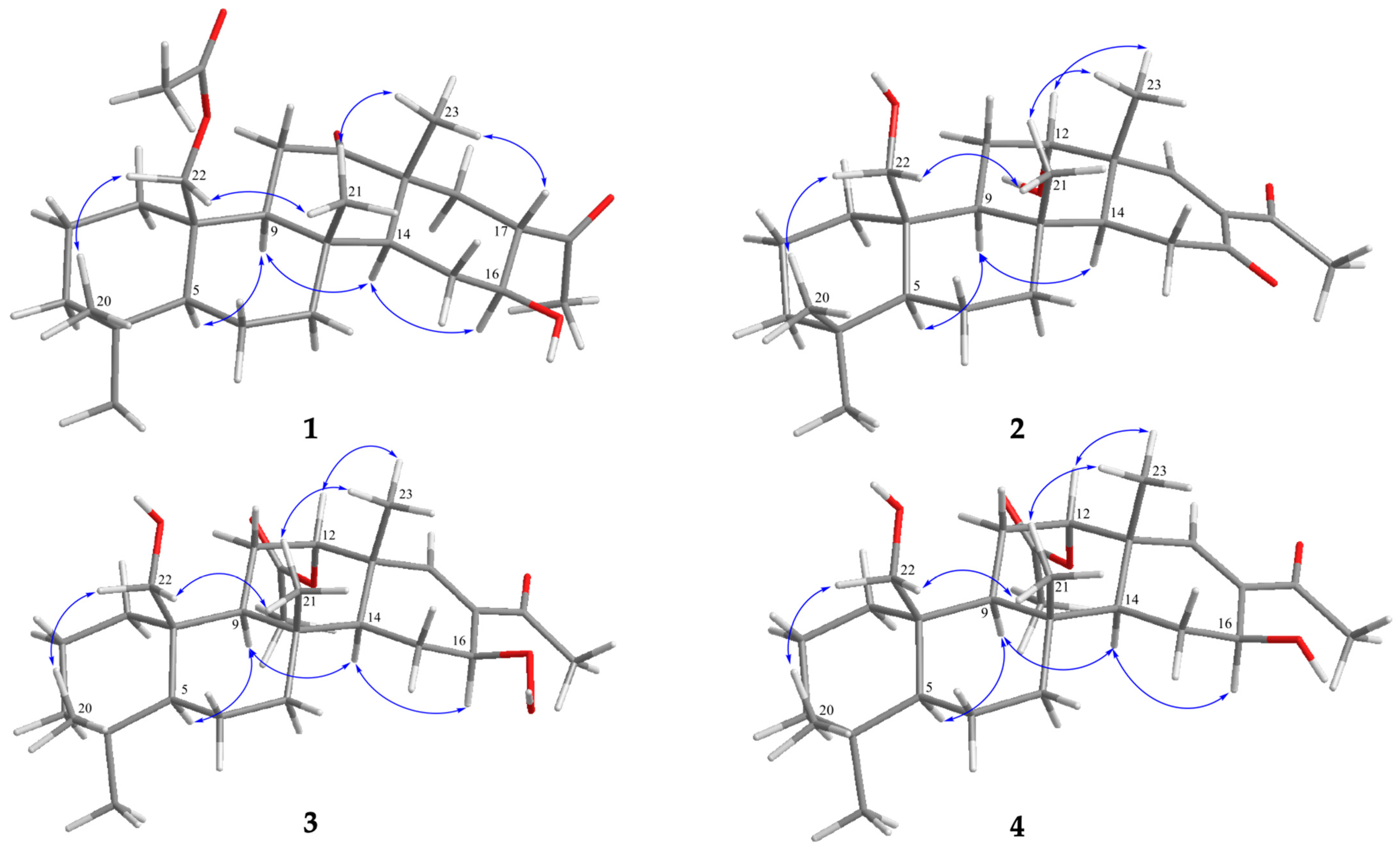

| 1 | 2 | |||
|---|---|---|---|---|
| C/H | δH (J in Hz) a | δC (mult.) b | δH (J in Hz) c | δC (mult.) d |
| 1 | 2.00 m; 0.77 ddd (12.4, 12.4, 4.4) e | 34.3, CH2 f | 2.13 m; 0.73 ddd (12.0, 12.0, 3.0) e | 34.1, CH2 f |
| 2 | 1.61 m; 1.48 m | 17.9, CH2 | 1.56 m; 1.37 m | 17.7, CH2 |
| 3 | 1.45 m; 1.17 m | 41.4, CH2 | 1.43 m; 1.19 m | 41.7, CH2 |
| 4 | 33.0, C | 33.0, C | ||
| 5 | 1.00 m | 56.8, CH | 1.03 dd (12.6, 2.4) | 56.9, CH |
| 6 | 1.61 m | 18.2, CH2 | 1.49 m | 18.4, CH2 |
| 7 | 1.95 m; 1.15 m | 41.5, CH2 | 1.77 ddd (12.6, 3.0, 3.0); 1.08 m | 41.1, CH2 |
| 8 | 37.2, C | 37.3, C | ||
| 9 | 1.32 m | 60.2, CH | 1.52 m | 51.8, CH |
| 10 | 41.0, C | 41.8, C | ||
| 11 | 3.97 dd (14.4, 14.4) 2.50 dd (14.4, 2.8) | 37.6, CH2 | 2.32 m; 1.86 ddd (16.2, 2.4, 2.4) | 28.3, CH2 |
| 12 | 214.4, C | 3.98 br s | 73.9, CH | |
| 13 | 48.5, C | 42.6, C | ||
| 14 | 1.19 m | 55.9, CH | 2.14 m | 47.5, CH |
| 15 | 1.98 m; 1.57 m | 27.6, CH2 | 2.53 m; 2.24 m | 34.9, CH2 |
| 16 | 3.80 ddd (9.6, 9.6, 3.2) | 70.9, CH | 198.2, C | |
| 17 | 2.49 m | 53.7, CH | 142.8, C | |
| 18 | 2.05 m | 36.8, CH | 7.55 s | 165.9, CH |
| 19 | 0.88 s | 33.7, CH3 | 0.86 s | 33.9, CH3 |
| 20 | 0.85 s | 21.8, CH3 | 0.77 s | 21.8, CH3 |
| 21 | 1.13 s | 16.0, CH3 | 1.11 s | 15.8, CH3 |
| 22 | 4.67 d (12.4) 4.21 dd (12.4, 1.2) | 64.5, CH2 | 4.06 d (11.4); 3.09 dd (11.4) | 62.8, CH2 |
| 23 | 1.26 s | 19.3, CH3 | 1.11 s | 18.6, CH3 |
| 24 | 212.4, C | 198.3, C | ||
| 25 | 2.19 s | 28.8, CH3 | 2.45 s | 30.7, CH3 |
| 22-OAc | 170.8, C | |||
| 2.06 s | 21.1, CH3 | |||
| 3 | 4 | |||
|---|---|---|---|---|
| C/H | δH (J in Hz) a | δC (mult.) b | δH (J in Hz) c | δC (mult.) d |
| 1 | 2.08 m; 0.58 ddd (13.2, 13.2, 2.8) e | 34.1, CH2 f | 2.07 m; 0.56 ddd (12.0, 12.0, 3.0) e | 34.2, CH2 f |
| 2 | 1.58 m | 17.8, CH2 | 1.56 m | 17.9, CH2 |
| 3 | 1.41 m; 1.17 m | 41.8, CH2 | 1.44 m; 1.17 m | 41.7, CH2 |
| 4 | 33.0, C | 33.0, C | ||
| 5 | 1.02 m | 56.8, CH | 0.99 dd (12.6, 2.4) | 57.0, CH |
| 6 | 1.56 m; 1.45 m | 18.3, CH2 | 1.58 m; 1.52 m | 18.4, CH2 |
| 7 | 1.91 m; 1.18 m | 41.2, CH2 | 1.92 m; 1.03 m | 41.4, CH2 |
| 8 | 36.8, C | 37.2, C | ||
| 9 | 1.41 m | 53.5, CH | 1.33 m | 53.5, CH |
| 10 | 41.8, C | 41.8, C | ||
| 11 | 2.35 m; 1.46 m | 21.9, CH2 | 1.91 m; 2.25 m | 25.1, CH2 |
| 12 | 4.97 d (2.8) | 76.2, CH | 4.96 dd (2.8, 2.8) | 76.6, CH |
| 13 | 41.7, C | 41.6, C | ||
| 14 | 1.86 dd (13.2, 2.0) | 43.6, CH | 1.50 m | 47.5, CH |
| 15 | 1.92 m; 2.29 m | 25.2, CH2 | 2.14 m; 1.50 m | 25.7, CH2 |
| 16 | 4.91 dd (4.0, 1.6) | 77.2, CH | 4.61 dd (8.8, 6.6) | 68.1, CH |
| 17 | 133.6, C | 132.4, C | ||
| 18 | 6.73 s | 155.7, CH | 6.59 s | 152.4, CH |
| 19 | 0.85 s | 33.8, CH3 | 0.88 s | 33.8, CH3 |
| 20 | 0.77 s | 21.9, CH3 | 0.76 s | 21.9, CH3 |
| 21 | 1.09 s | 16.6, CH3 | 1.10 s | 16.4, CH3 |
| 22 | 4.05 d (11.6); 3.91 dd (11.6) | 62.8, CH2 | 4.27 d (11.4); 3.90 dd (11.4) | 62.7, CH2 |
| 23 | 1.06 s | 19.4, CH3 | 1.20 s | 20.9, CH3 |
| 24 | 199.3, C | 202.1, C | ||
| 25 | 2.25 s | 25.8, CH3 | 2.25 s | 25.7, CH3 |
| 12-OAc | 170.8, C | 170.6, C | ||
| 2.03 s | 21.3, CH3 | 2.04 s | 21.3, CH3 | |
| 16-OOH | 9.27 s | |||
| Cald. Value a | Exp. Value | |
|---|---|---|
| Exp. 1 b | 21 | |
| Cald. 1-5S,8R,9S,10R,13R,14S,16S,17S | 31 | |
| Cald. 1-5R,8S,9R,10S,13S,14R,16R,17R | −31 | |
| Exp. 2 c | 61 | |
| Cald. 2-5S,8R,9S,10R,12S,13R,14S | 78 | |
| Cald. 2-5R,8S,9R,10S,12R,13S,14R | −78 | |
| Exp. 3 d | 41 | |
| Cald. 3-5S,8R,9S,10R,12S,13R,14S,16S | 15 | |
| Cald. 3-5R,8S,9R,10S,12R,13S,14R,16R | −15 | |
| Exp. 4 e | 76 | |
| Cald. 4-5S,8R,9S,10R,12S,13R,14S,16S | 103 | |
| Cald. 4-5R,8S,9R,10S,12R,13S,14R,16R | −103 |
| Compounds | Superoxide Anion Generation | Elastase Release | ||||
|---|---|---|---|---|---|---|
| IC50 (μM) | Inh% | IC50 (μM) | Inh% | |||
| 1 | 9.19 ± 1.06 | *** | 1.59 ± 1.90 | |||
| 2 | 3.98 ± 0.62 | 95.02 ± 3.42 | *** | 5.24 ± 0.28 | 95.47 ± 6.29 | *** |
| 3 | 12.12 ± 1.22 | *** | 9.70 ± 1.06 | *** | ||
| 4 | 4.46 ± 0.72 | 88.28 ± 5.25 | *** | 4.73 ± 0.40 | 98.00 ± 4.60 | *** |
| LY294002 | 2.66 ± 039 | 88.30 ± 5.00 | *** | 2.53 ± 0.53 | 86.71 ± 6.47 | *** |
Disclaimer/Publisher’s Note: The statements, opinions and data contained in all publications are solely those of the individual author(s) and contributor(s) and not of MDPI and/or the editor(s). MDPI and/or the editor(s) disclaim responsibility for any injury to people or property resulting from any ideas, methods, instructions or products referred to in the content. |
© 2023 by the authors. Licensee MDPI, Basel, Switzerland. This article is an open access article distributed under the terms and conditions of the Creative Commons Attribution (CC BY) license (https://creativecommons.org/licenses/by/4.0/).
Share and Cite
Peng, B.-R.; Zheng, L.-G.; Chen, L.-Y.; El-Shazly, M.; Hwang, T.-L.; Su, J.-H.; Lee, M.-H.; Lai, K.-H.; Sung, P.-J. Nor-24-homoscalaranes, Neutrophilic Inflammatory Mediators from the Marine Sponge Lendenfeldia sp. Pharmaceuticals 2023, 16, 1258. https://doi.org/10.3390/ph16091258
Peng B-R, Zheng L-G, Chen L-Y, El-Shazly M, Hwang T-L, Su J-H, Lee M-H, Lai K-H, Sung P-J. Nor-24-homoscalaranes, Neutrophilic Inflammatory Mediators from the Marine Sponge Lendenfeldia sp. Pharmaceuticals. 2023; 16(9):1258. https://doi.org/10.3390/ph16091258
Chicago/Turabian StylePeng, Bo-Rong, Li-Guo Zheng, Lo-Yun Chen, Mohamed El-Shazly, Tsong-Long Hwang, Jui-Hsin Su, Mei-Hsien Lee, Kuei-Hung Lai, and Ping-Jyun Sung. 2023. "Nor-24-homoscalaranes, Neutrophilic Inflammatory Mediators from the Marine Sponge Lendenfeldia sp." Pharmaceuticals 16, no. 9: 1258. https://doi.org/10.3390/ph16091258