Review

The Role of Small Molecules Containing Fluorine Atoms in Medicine and Imaging Applications

1
School of Science and Technology, Georgia Gwinnett College, 1000 University Center Lane, Lawrenceville, GA 30043, USA
2
Department of Chemistry, Petit Science Center, Georgia State University, 100 Piedmont Avenue SE, Atlanta, GA 30303, USA
3
Center for Diagnostics and Therapeutics, Georgia State University, 100 Piedmont Avenue SE, Atlanta, GA 30303, USA
*
Author to whom correspondence should be addressed.
Pharmaceuticals 2024, 17(3), 281; https://doi.org/10.3390/ph17030281
Submission received: 3 October 2023 / Revised: 12 February 2024 / Accepted: 17 February 2024 / Published: 22 February 2024
(This article belongs to the Special Issue Molecular Systems for the Delivery of Drugs and Contrast Agents)

Abstract

:
The fluorine atom possesses many intrinsic properties that can be beneficial when incorporated into small molecules. These properties include the atom’s size, electronegativity, and ability to block metabolic oxidation sites. Substituents that feature fluorine and fluorine-containing groups are currently prevalent in drugs that lower cholesterol, relieve asthma, and treat anxiety disorders, as well as improve the chemical properties of various medications and imaging agents. The dye scaffolds (fluorescein/rhodamine, coumarin, BODIPY, carbocyanine, and squaraine dyes) reported will address the incorporation of the fluorine atom in the scaffold and the contribution it provides to its application as an imaging agent. It is also important to recognize radiolabeled fluorine atoms used for PET imaging in the early detection of diseases. This review will discuss the many benefits of incorporating fluorine atoms into small molecules and give examples of fluorinated molecules used in the pharmaceutical industry and imaging techniques.

Graphical Abstract

1. Introduction

The value of fluorine-containing small molecules with pharmaceutical efficacy and the underlying importance of fluorine incorporation into these molecules is undeniable [1,2]. Every year, new fluoro-pharmaceuticals achieve FDA approval and are introduced into the market; this is an ongoing trend that has been observed over the past 20 years [3,4]. In 2021, nine fluorine-containing drugs were approved for use by the FDA, showing an increase in relevancy for the incorporation of fluorine in medicinal research [5]. The design, synthesis, and testing of medically viable fluorine-containing compounds have burgeoned over the past two decades, leading to numerous publications and patents.
Utilization of the fluorine atom and fluorine-containing functional groups in pharmaceuticals is very attractive to the medical field for a variety of reasons [2]. First, the fluorine atom is the second smallest “functional group” with a van der Waals radius of 1.47 Å [1,6]. Because fluorine’s size falls between that of a hydrogen atom (1.20 Å) and an oxygen atom (1.47 Å), it is often used as a bioisostere for these atoms [6,7,8]. It has been suggested that the trifluoromethyl group is comparable to an isopropyl group, even though its van der Waals volume is much smaller (-CF3: 39.8 Å3 vs. -CH(CH3)2: 56.2 Å3) [1]. The fact that the -CF3 substituent has rotational symmetry around the carbon–carbon bond, whereas the isopropyl group lacks rotational symmetry, helps account for this apparent discrepancy [9]. As shown in Figure 1, modeling studies reveal the relative similarity in sizes between stilbene and perfluorinated stilbene while highlighting the dramatic impact fluorine has on electrostatic potentials in the stilbene molecule [10]. Because the fluorine atom is the smallest halogen, an additional advantage is found in its ability (as well as fluorinated methyl groups) to fit into smaller pockets of space in comparison to the other halogens and their corresponding groups.
Fluorine’s high electronegativity, 4.0 on the Pauling scale, offers it other advantages over similarly sized atoms and functional groups when incorporated into small molecules. The C-F bond is considered the strongest bond in organic chemistry due to the electronegativity difference between the two atoms [11,12]. In addition, the highly electronegative nature of fluorine often alters the dipole moment of the overall molecule. Another molecular property affected by the electronegativity of the fluorine atom is the pKa [13]. The acidity of fluorinated molecules increases due to inductive effects brought about by fluorine’s electronegativity [7,14].
Additionally, benefits arising from the incorporation of one or more fluorine atoms into a compound often include alterations in drug biodistribution and drug–receptor binding, as well as enhancement of potency [15]. A significant advantage found upon fluorine substitution in drugs and imaging agents is the ability to modulate lipophilicity by adding or taking away fluorine atoms from the molecule in medical and biomedical imaging applications [16]. The tuning of lipophilicity helps the compounds to be absorbed and transported in vivo faster and more easily. This stems from the greater lipophilicity of the C-F bond relative to the C-H bond [17]. Because the fluorine atom is the smallest halogen, it (as well as fluorinated methyl groups) fits into receptor pockets and it contributes to blocking sites from metabolic oxidation more than that of other halogens [18]. The electronegativity and lipophilic properties of fluorine atoms incorporated into medicinal compounds have strong impacts on how the body will react to the molecules in ways such as the clearance rate, biodistribution route, and toxicity of the molecule. Figure 2 summarizes the key effects that fluorine substitution has on small molecules with potential pharmaceutical value.
Before introducing examples of fluorinated small molecules and their applications in medicine and imaging techniques, we will first discuss several key reactions by which fluorine and fluorinated functional groups are incorporated into compounds.

2. Common Reactions Incorporating Fluorine in Small Molecules

The introduction of fluorine and other fluorinated functional groups into the final drug architecture is most often accomplished via reactions with fluorinated precursors. Therefore, it is important to understand the reactions that are used to prepare these fluorinated precursor molecules. In Figure 3, a selection of processes used to prepare fluorobenzylic, fluoroalkyl, fluoroaromatic, and fluoroheteroaromatic systems is presented. For a more detailed discussion on current state-of-the-art organofluorine synthetic strategies, a review by Gouverneur et al. published in 2021 is illustrative [19]. The first reaction is performed at a large scale in industrial settings and involves hydrogen fluoride (HF) (Equation (1)). In this reaction, the fluoride anion serves as a nucleophilic substitute for other halogens occupying allylic and benzylic positions. [20]. Another reaction commonly used to incorporate fluorine is the Friedel–Crafts (FC) alkylation. Hydrogen fluoride, in this case, acts as both the FC catalyst and the fluorinating agent itself, resulting in the preparation of trifluoromethylated aromatic systems (Equation (2)). The carbon tetrachloride reagent in Figure 3, Equation (3), replaces a hydrogen atom on the benzene ring, followed by the substitution of fluorine for the three chlorine atoms.
Equation (4) is an example of the Swarts reaction. In this process, a metal fluoride species acts as a Lewis acid catalyst in removing a halide substituent and replacing it with a fluorine atom via the formation of a four-membered cyclic intermediate [21]. Equation (5) shows a green, highly regioselective fluorination of activated 2-aminopyridines using Selectfluor® [22]. Equation (6) shows electrophilic fluorination using acetyl hypofluorite (CH3COOF) as the fluorinating reactant [23].
Equations (7) and (8) are examples of electrophilic trifluoromethylations of activated aromatic and heteroaromatic substrates developed within the last two decades [20,24]. Equation (7) employs trifluoromethyltriethylsilane (TES-CF3) under Cu(I) catalysis to prepare trifluoromethylated aromatic and heteroaromatic molecules from the corresponding iodide starting material. The mechanism by which this reaction takes place is not fully understood but may involve a CF3-Cu complex. Equation (8) depicts aromatic trifluoromethylation by S-(trifluoromethyl)dibenzothiophenium tetrafluoroborate via a transition metal-catalyzed reaction. This reaction generates a product with the trifluoromethyl group introduced to the ortho position of benzene [25]. Finally, Equation (9) is an example of a highly regioselective heteroaromatic trifluoromethylation of a pyridine derivative [26]. In the next section, we discuss a number of the more important fluorinated pharmaceuticals. In addition to elaborating on the role that fluorine plays in altering molecular properties, we will also note how and when fluorine and functionalities containing fluorine are introduced into the drug’s molecular architecture.
Figure 3. Examples of fluorination and trifluoromethylation reactions involve benzylic, aromatic, alkyl, and heteroaromatic substrates [9,10,11,17,20,22,23,24,26,27].
Pharmaceuticals 17 00281 g003

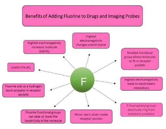
3. Characteristics of Pharmaceuticals Featuring Fluorine and Examples

Fluorinated therapeutics represent compounds available as drugs that use fluorine atoms to change the molecule’s overall lipophilicity, drug potency, and other factors. For many potentially viable pharmaceutical compounds, the clearance rate and clearance mechanism are critical considerations when designing and synthesizing molecules. Due to the extraordinary properties exhibited by the fluorine atom, drug designers continue to incorporate this atom into lead compounds. In the next section, the benefits of the properties of fluorine and fluorine-containing functional groups will be discussed.

3.1. Metabolic Oxidation

A primary reason to incorporate the fluorine atom, especially on aromatic and heterocyclic ring systems, is to combat the effect of metabolic oxidation which occurs when drugs are taken into the body and the process eliminates foreign molecules from the body as quickly as possible. The addition of a fluorine atom or fluorine-containing group often stops this metabolic pathway as the modified fluorobenzene does not fit into the active site of the monooxygenase. An additional benefit to adding fluorine to the para position on the benzene ring is that the inductive electron-withdrawing effect of the fluorine atom deactivates other positions on the ring against this particular metabolic pathway [9]. Slowing the oxidation process that the P450 protein performs allows for longer drug retention and thus increases effectiveness. Examples in the literature show compounds that have very rapid bodily clearances, but when fluorine is incorporated into the para position on a phenyl ring, the rate at which the compound is cleared slows by as much as 108-fold [28]. And, while the main function of the P450 protein is to initiate the metabolic process, oxygenizing a pharmaceutically active compound can turn it toxic [29]. Fluorine and chlorine can block this process from happening, but due to its minimal size, electronic withdrawing nature, and steric perturbation in active sites, fluorine is preferred over chlorine. Many therapeutics highlighted in this review article share the theme of a p-fluorophenyl group. The protection afforded by the para-substituted halogen reduces the potential toxicity when oxygenized and can increase drug effectiveness due to interactions with activation sites in proteins [9,30]. The strategy of p-fluorination has been thoroughly investigated and has been frequently used in drug design [29].

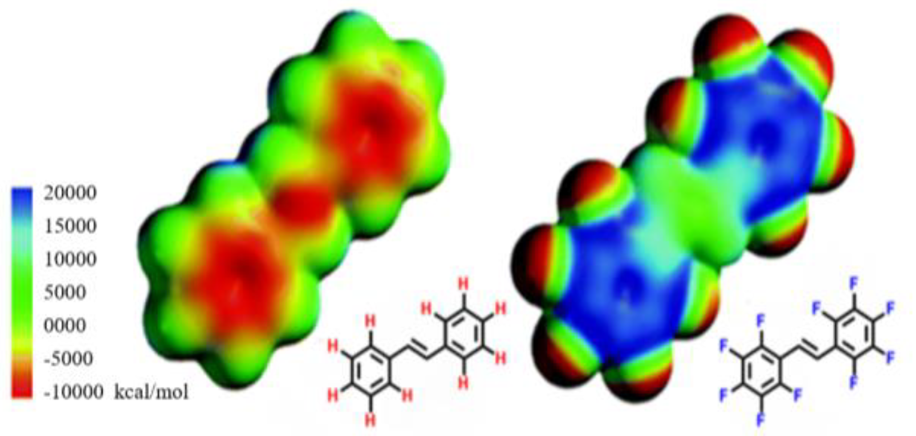
3.2. Electronic Considerations

While deactivating a drug against metabolic oxidation is possibly the most important consequence of incorporating fluorine into drugs, researchers can utilize fluorine atoms to alter compounds to achieve other strategic effects. Electronic considerations such as electronegativity, pKa modification, and lipophilicity tailoring are also motives that chemists take advantage of when designing fluorine-containing pharmaceutical compounds [4,15]. Electronegativity leads to several underlying effects on a molecule, including molecule and bond stability, dipole magnitude and direction, and electrostatic interactions with receptor sites. Here, several drugs are described that do not contain fluorine atoms for their metabolic effects but rather for their other properties.

3.3. Size Considerations

As mentioned previously, the size of fluorine-containing functional groups is very unique. Structurally, they take up more room than hydrogen but less than a hydroxyl group. The size of the functional groups can help direct a compound into its target pocket. Once the molecule is inside the pocket, the electronic characteristics of the fluorine then assist in adding to the potency and effectiveness of the drug.

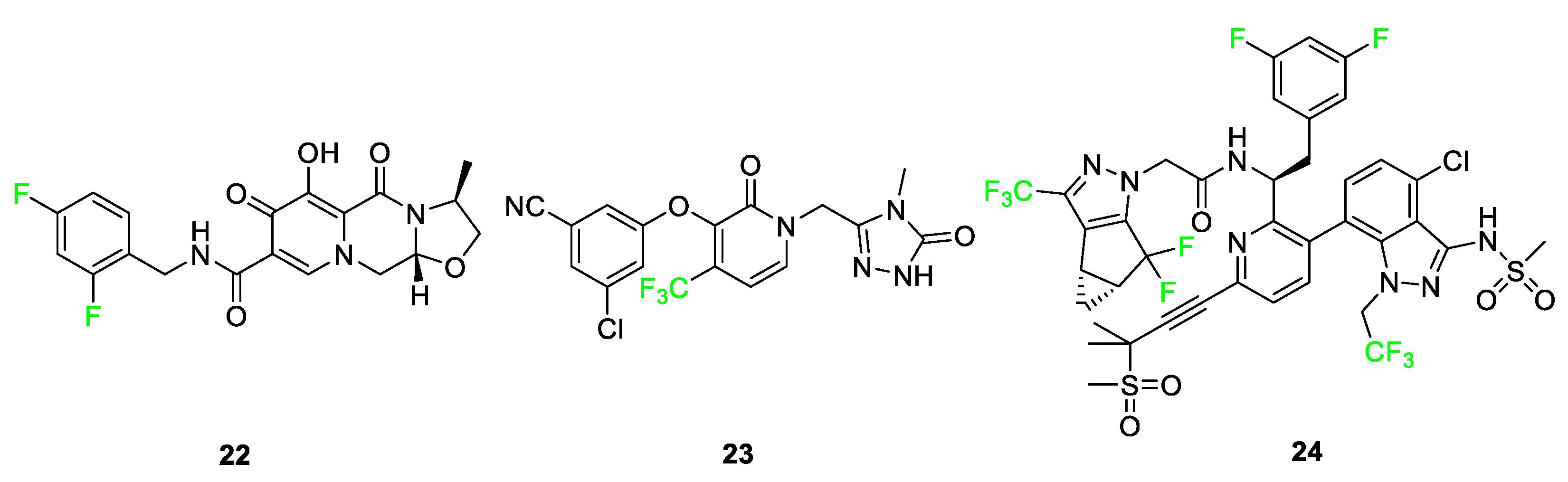
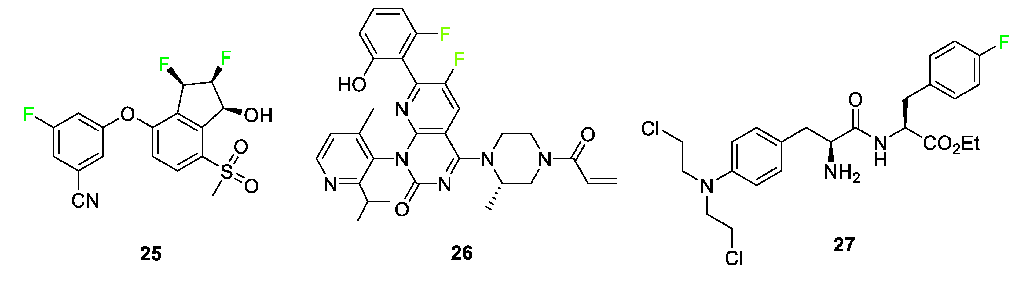
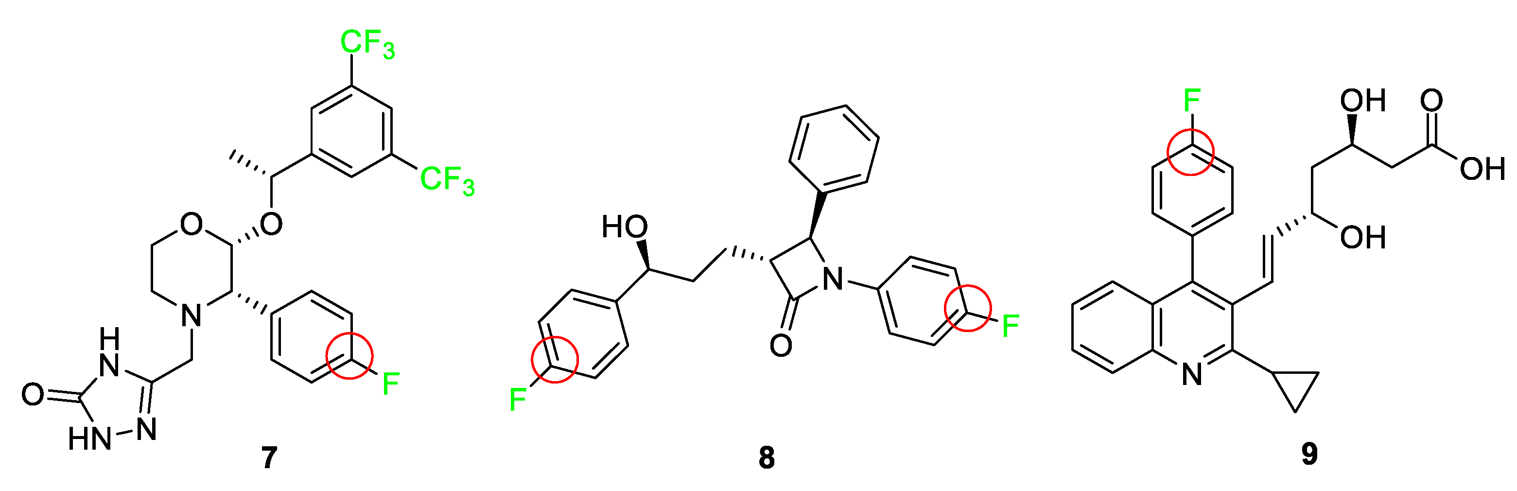
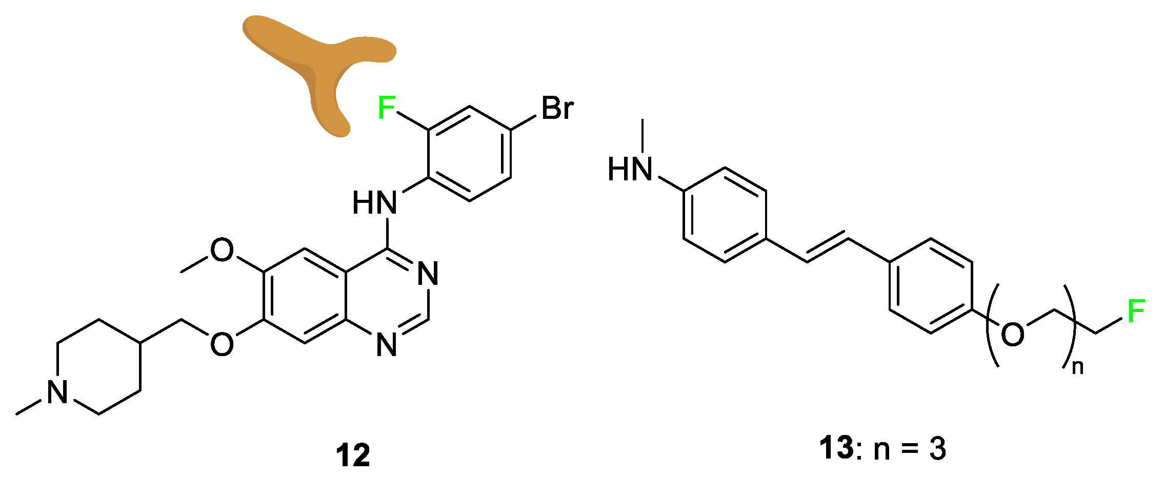
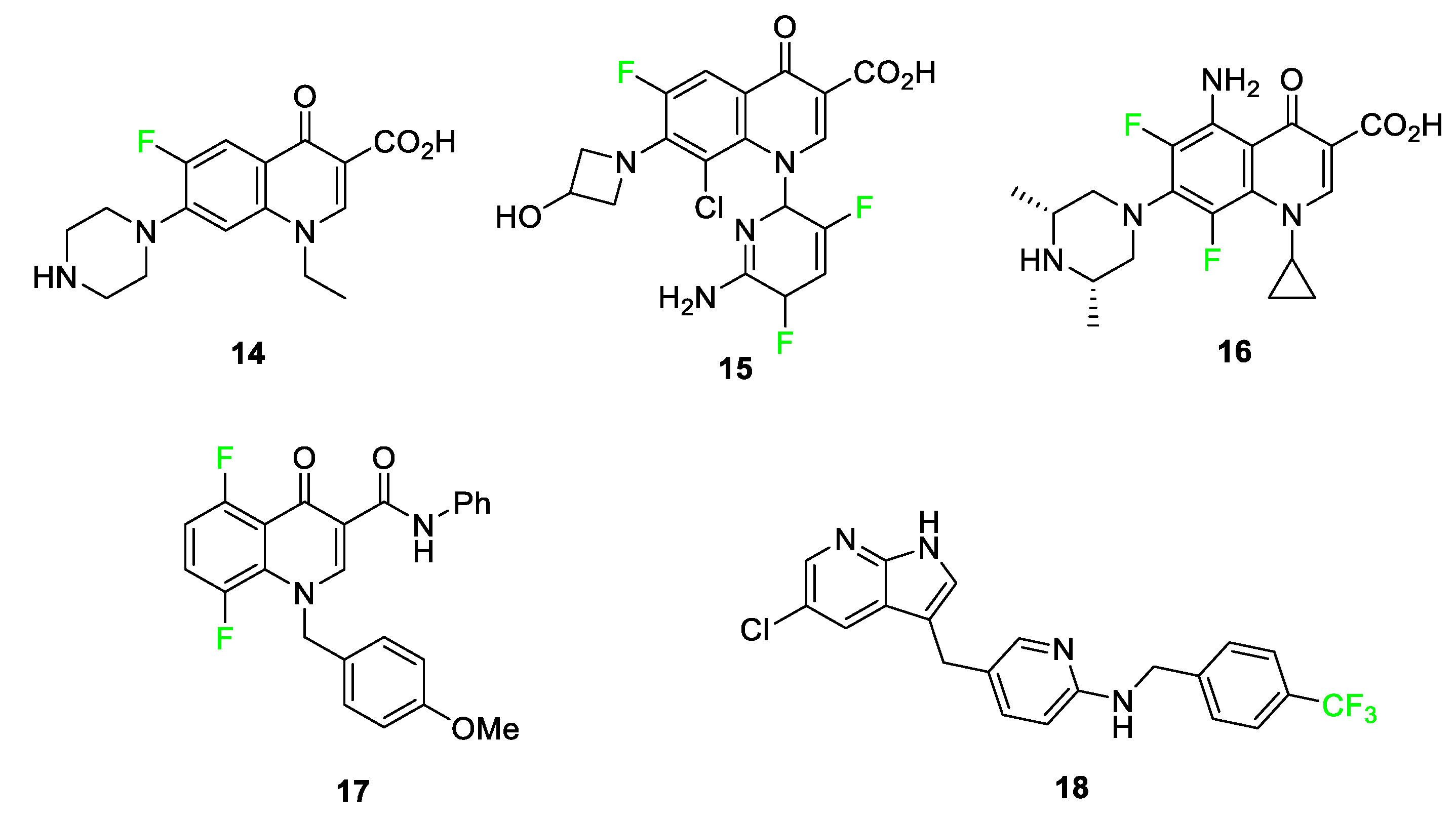
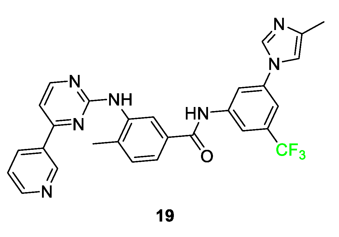
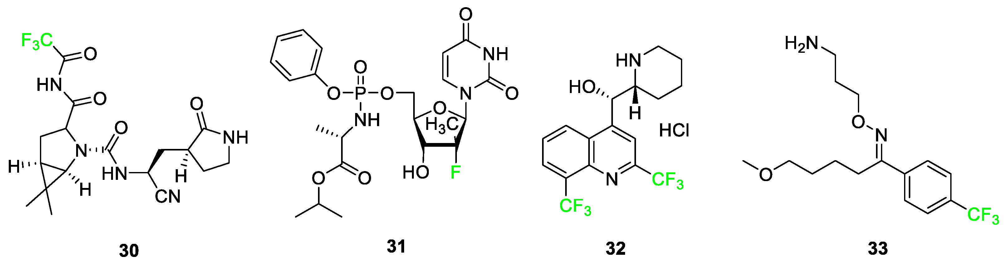
3.4. Examples of Pharmaceuticals Containing Fluorine

Numerous pharmaceuticals feature fluorine, including six examples seen in Figure 4: atorvastatin (Lipitor) 1, rosuvastatin (Crestor) 2, escitalopram (Lexapro) 3, fluticasone (Flonase®) 4, asciminib (ScemblixTM-AB001) 5, and atogepant (Qulipta®) 6. These drugs treat high cholesterol, depression, anxiety disorders, allergic rhinitis, chronic myelogenous leukemia (CML), and migraines, respectively [3,31]. These six drugs are used by patients in every demographic, and their functions affect millions of people worldwide; however, these compounds represent only a fraction of pharmaceuticals that contain at least one fluorine atom. In this section, pharmaceutical compounds will be described and categorized in detail, paying special attention to the features of the compounds containing fluorine atoms highlighted in Figure 4.
Atorvastatin 1 and rosuvastatin 2 are statin drugs used in the treatment of patients exhibiting high cholesterol [32]. Rosuvastatin 2, was developed from a desire to replace large, complex functional groups with simpler, achiral alternatives. The addition of the pyrimidine ring compared to other synthetic statins, such as atorvastatin 1, improves activity for inhibiting the targeted reductase. The p-fluorophenyl group on this molecule not only enhances its biological activity but also deactivates the ring against P450 monooxygenase.
Escitalopram 3 is a selective serotonin reuptake inhibitor used to treat major depression and anxiety disorders [33]. The drug targets the serotonin transporter and facilitates the reuptake of serotonin into the neurons for rapid antidepressant activity [34]. A binding study of 5-HT transporter (SERT) was conducted to determine affinity for the two potential binding sites and substituent selectivity [35]. The study determined that derivatives of escitalopram containing aromatic fluorine and the presence of the cyano group were significant for a twofold decrease in dissociation rate and had a high contribution to allosteric potency.
Figure 4. Six successful pharmaceutical drugs featuring fluorine [4,5,36,37].
Pharmaceuticals 17 00281 g004
Fluticasone 4 is used for relieving nasal symptoms such as sneezing, itching, and runny nose provoked by allergies; recent studies use this compound in combination with other medications to improve its effects against allergic rhinitis [38,39,40]. Also, fluticasone is combined with salmeterol and prescribed under the medication named Advair for the treatment of asthma as well as other obstructive airway diseases [41,42]. Fluticasone 4 containing the electron-withdrawing fluorine atom demonstrated greater topical activity compared to other analogs and increased glucocorticoid and mineralocorticoid effects [43].
Asciminib 5 is the active ingredient used in the treatment of CML containing a chiral compound with a difluorinated moiety [44]. CML is a disease associated with the overproduction of white blood cells in the bone marrow that arises from the BCR-ABL protein mutation leading to abnormal signaling pathways resulting in the growth and generation of leukemic cells [36,45]. These types of tyrosine kinase inhibitors work by binding to the ATP-binding site of BCR-ABL, thus transforming the disease into a controllable state during treatment [36,46]. The fluorine atoms in asciminib 5 interact with the carbonyl carbon of leucine-359 of the active site pocket, making the position of the fluorine atoms advantageous for improving the bioactivity of the overall compound [5].
Atogepant 6 is an orally administered calcitonin gene-related peptide (CGRP) receptor antagonist that aids in the treatment of migraines [37,47]. Substitution of the 1,3,5-trifluorobenzene moiety in place of a nonfluorinated benzene ring produces a four-fold increase in the affinity of atogepant 6 compared to the nonfluorinated benzene ring derivative [48,49,50].
Approved by the FDA in 2003, aprepitant (Emend) 7 combats chemotherapy-induced nausea and vomiting and serves as an example where the addition of a fluorine atom blocks oxidative metabolism and lowers the oxidation potential elsewhere on the ring (Figure 5) [10,51]. Ongoing clinical trials show promising results for the use of this drug in cancer treatment [52]. The 3,5-bis-(trifluoromethyl)phenyl group is a common feature with other NK1 receptor antagonists and improves penetration of the drug into the central nervous system [51]. The fluorine atom blocks the 4-position of the benzene ring from oxidation as well as deactivates the remainder of the ring positions.
Ezetimibe (Zetia®) 8 [53] is a drug that combats high cholesterol by targeting Niemann–Pick C1-like 1 protein (NPC1L1), thus lowering low-density lipoprotein (LDL); it features two p-fluoro substituents to block other sites on the ring from metabolic oxidation and improves metabolic stability [12,56]. Figure 5 shows the structure of ezetimibe 8 and highlights the blocking of sites because of fluorine atoms. The compound utilizes the fluorine atoms as a defense against aromatic hydroxylation as well as yielding a derivative that exhibits improved pharmacokinetic properties, which increases the activity of the drug significantly. Another property of the fluorine atom substitution that ezetimibe 8 takes advantage of is the increase in polarity of the overall molecule. Molecules with higher polarity are more susceptible to glucuronidation, a process that generally inactivates drugs. However, in the case of ezetimibe 8, glucuronidation improves the activity of the drug by recirculating the drug to the activation site and increasing the residence time [21]. Therefore, through the utilization of fluorine, researchers are able to increase lipophilicity and polarity, as well as block the oxidation sites for ezetimibe 8, which in turn exhibited a 50-fold increase in activity over the parent compound.
Synthetic statins are a class of drugs that must contain 4-fluorophenyl groups as structural requirements for biological activity. Examples include atorvastatin 1, rosuvastatin 2, and pitavastatin (Livalo) 9, as shown in Figure 4 and Figure 5. These drugs combat cholesterol problems by inhibiting hydroxymethylglutaryl-coenzyme A (HMG-CoA) reductase. All of these compounds feature a p-fluorophenyl moiety, as studies have shown that this substituent greatly surpasses the biological activity of all other functional groups tested [54,55]. Pitavastatin 9, a recent statin-type drug to enter the market, is completely synthetic. This HMG-CoA reductase inhibitor and low-density lipoprotein cholesterol (LDL-C) receptor inducer improves its pharmacokinetics, efficiency, and bioavailability by incorporating a p-fluoro group [57]. The 2-cyclopropyl-4-(4-fluorophenyl)quinoline pharmacophore differentiates it from other statins, and the highly functionalized heterocycle provides superior resistance to metabolism and prolongs its duration of action [58]. A modified version of pitavastatin 9 has been used for positron emission tomography (PET) imaging in vivo by incorporating a fluorine-18 atom [59].
As seen in Figure 6, fulvestrant (Faslodex) 11 utilizes the replacement of an n-ethyl functional group with a pentafluoroethyl moiety [60]. ICI 164,384 10, the original precursor, was developed for the treatment of breast cancer, specifically to combat the negative side effects of the receptor modulator Tamoxifen which increased the risk of the metastasis of associated tumors. The parent molecule did not display high levels of potency during in vivo testing. The introduction of the -CF2CF3 group in step 1 of the 12-step synthesis of compound 11, resulted in a five-fold increase in intrinsic potency relative to the parent molecule 10 [60]. The addition of the terminal pentafluoroethyl group as opposed to the ethyl end-chain increases the strength of the bonds and makes the compound more stable as well as increases hydrogen bonding with the receptor, thus increasing metabolic stability during estrogen receptor (ER) binding [60].
Multiple drugs use the added lipophilicity afforded by the substitution of a hydrogen atom with a fluorine atom; lipophilicity alteration can lead to greater drug uptake through cell membranes [61,62,63,64]. While fluorination of an aromatic or π-system increases lipophilicity, the addition of fluorine and trifluoromethyl groups on an n-alkyl chain does not have the same effect; some tailoring and chemical designs of aliphatic fluorination can decrease lipophilicity [65]. Vandetanib (Caprelsa) 12 [66], a drug that acts as an antagonist of the vascular endothelial growth factor receptor (VEGFR), is used as an oral kinase inhibitor for thyroid tumors. This is an example of a compound modified with fluorine to achieve the “Goldilocks” level of lipophilicity (Figure 7). Many derivatives of vandetanib 12 were tested to tailor the lipophilicity of the compound to increase its potency. Structure–activity relationship tests show that bromine at the C-4’ position was preferred, and that fluorine was optimal at the C-2’ position. The bromofluorophenyl group was analyzed as a residue matching these two cases, and it was shown that the fluorine atom leads the bromofluorophenyl group deep into the protein’s hydrophobic pocket, ultimately increasing the potency of the drug [66].
Compound 13, a specific AV-45 derivative with three additional polyethylene glycol (PEG) groups added to the compound, was tested alongside other chain lengths to determine how varying the hydrophilicity altered efficacy [67]. These compounds were being studied for amyloid beta (Aβ) plaque affinity in relation to combatting Alzheimer’s disease. Compound 13 displayed good Aβ binding coupled with high blood–brain barrier penetration. A range of fluoroethylene glycol (FPEG) lengths was studied to find the greatest uptake depending on the logP (lipophilicity). The result shows only a minimal change in uptake when modifying the PEG length below n = 5. However, when the FPEG group was replaced with a hydroxyl group, a significant increase in lipophilicity and, more importantly, a decrease in potency was observed [67].
Fluorine-containing quinolones, pyrimidoquinolines, and pyridyl-substituted indoles exhibit anti-cancer properties [68]. The fluorine atom and trifluoromethyl substituents play major roles in the anti-cancer, antimicrobial, and antituberculosis effectiveness of these compounds [69,70]. Adding fluorine to this class of small molecules increases the hydrophobicity of the compounds, which in turn, aids in the penetration into hydrophobic protein pockets. Examples of common amino acids that attract the hydrophobic fluorine group include leucine and phenylalanine [68]. More specific examples of these compounds 1418 are shown in Figure 8.
In Figure 9, nilotinib (TASIGNA®) 19, a derivative of Imatinib, is shown. The addition of the trifluoromethyl group resulted in the compound exhibiting 30 times the potency when compared to the nonfluorinated parent compound. Once inside the pocket of the Bcr-Abl tyrosine kinase inhibitor, the trifluoromethyl group interacts with the histidine and isoleucine side chain residues. By comparison, when an analog featuring a methyl substituent was tested in place of the trifluoromethyl, it showed a five-fold decrease in activity relative to compound 19 [55,72].
Other compounds that utilize the size of fluorine to advantage are lapatinib (TYKERB®) 20 and ivosidenib (TIBSOVO®) 21 (Figure 10) [73,74]. Lapatinib 20 is a human epidermal growth factor inhibitor and dual tyrosine kinase inhibitor for fighting breast cancer and other solid tumors. Groups larger than the featured m-fluorophenyl, e.g., the m-chlorophenyl group, displayed diminished drug activity for compound 20 analogs. Analogs that substituted -OH or -Br in place of the m-fluorophenyl group showed substantial decreases in inhibition. These findings led researchers to the conclusion that fluorine is essential to fit into the binding pocket as well as to the interactions that retain the drug in the pocket; X-ray crystallographic results support this conclusion [73]. Ivosidenib 21 is a drug developed to treat relapsed or refractory acute myeloid leukemia in adults with an isocitrate dehydrogenase 1 (IDH1) mutation. Fluorine substitution on the phenyl ring brought about desirable metabolic stability in observations from compound AGI-14100 containing two aromatic fluorine atoms; however, in the same study, compound 21 was determined to have more desirable properties upon changing the design to incorporate a nitrogen atom in the ring containing fluorine atom [74].
Figure 10. Lapatinib 20 and ivosidenib 21 utilize fluorine as an essential size requirement for potency [73,74].
Pharmaceuticals 17 00281 g010
Thus far, this review article has highlighted therapeutic agents and pharmaceutical compounds that feature fluorine atoms or fluorine-containing functional groups on small molecules. This special atom has become extremely important in drug development in recent decades and will continue to be an essential building block when designing and synthesizing drugs [4,75,76]. The characteristics of fluorine and the benefits of incorporation on pharmaceutical molecules, whether they be size considerations, electronic properties, or blocking of metabolic sites susceptible to the P450 monooxygenase, are undeniable in medicinal chemistry.
In the literature, fluorinated compounds including cabotegravir (Vocabria) 22, doravirine (PIFELTRO™) 23, and lenacapavir (SUNLENCA®) 24 are recent FDA-approved drugs that have been used in the development of HIV treatments (Figure 11) [14]. Cabotegravir 22 has been used in conjunction with rilpivirine as the cocktail under the name of Cabenuva [5,36,44]. The introduction of a fluorine atom to the benzene ring of cabotegravir compared to its analog improved its potency as an agent. In 2021, a study was conducted where it was used as an injectable in combination with rilpivirine to give it a long-lasting effect as treatment. Doravirine 23, like rilpivirine, is a non-nucleoside reverse transcriptase inhibitor (NNRTI) used in HIV treatment after it was FDA-approved in 2018 [47]. Its structure is a trifluorinated compound compared to an existing chlorine-containing analog; the modification to the structure shows improvements in half-life and plasma stability in studies conducted. Lenacapavir 24, FDA-approved in 2022, is used as a capsid protein inhibitor with pico-molar range potency and it showed significant viral suppression after some weeks when administered to patients. The introduction of fluorine to the structure anchors the conformation, thus improving the drug’s potency [47].
Anti-cancer medications are also highlighted as a sector where fluorinated compounds have been introduced (Figure 12). Belzutifan (WELIREG™) 25 is a hypoxia-inducible factor-2α inhibitor used for the treatment of Hippel–Lindau disease which is associated with the appearance of renal cell carcinoma, hemangioblastomas, and/or pancreatic neuroendocrine tumors [36]. Sotorasib 26 (LUMAKRAS®), an elective and irreversible covalent inhibitor B, is used to treat non-small cell lung cancer [36]. Sotorasib 26 interacts with a cysteine-12 residue of KRAS mutation, KRas G12C, to disrupt the signaling pathway. This KRAS mutation brings about the following tumors: lung adenocarcinoma, pancreatic ductal carcinoma, and colorectal carcinoma. The fluorine-containing version of the compound overcame the bioavailability issue observed for derivatives of the compound containing different halogens [5]. Melphalan flufenamide (Pepaxto®) 27, a drug approved in 2021 for use in the treatment of myeloma, is 10 times more pharmacologically active than melphalan [5,77]. The presence of the p-fluoro substituent on melphalan flufenamide 27 enhances metabolic stability over nonfluorinated melphalan analogs [78].
Figure 11. Fluorine-containing FDA-approved drugs for use in HIV treatment: cabotegravir 22, doravirine 23, and lenacapavir 24 [14,79].
Pharmaceuticals 17 00281 g011
Figure 12. Anti-cancer medications containing fluorine atoms: belzutifan 25, sotorasib 26, and melphalan flufenamide 27 [5,36].
Pharmaceuticals 17 00281 g012
The bioactivities of a venerable class of compounds, sulfonamides, have seen a resurgence in research interest over the past decade [80]. Numerous examples of fluorinated sulfonamides show enhanced biological efficacy over their nonfluorinated counterparts. The enhancements in effectiveness can arise from resistance to oxidation, electronic alterations, or size brought about by fluorine substitution. Tetrafluoropyridyl sulfonamide 28 (Figure 13) was found to be active against both Gram-positive (B. subtillis and S. pneumoniae) and Gram-negative (P. aeruginosa and E. coli) bacteria strains as well as effective against fungi (A. fumigatus). This sulfonamide was also active in a cytotoxic assay against the MCF-7 breast cancer and HepG2 hepatic cancer cell lines. Activity exceeded that of both cisplatin and 5-fluorouracil. Molecular docking shows that sulfonamide exhibits good interactions with amino acid residues on the mitogen-activated kinase active site. The parafluorophenyl sulfonamide 29 was found to be active in a cytotoxic assay against the HepG2 hepatic cancer cell lines with activity on par with cisplatin. Molecular docking shows that sulfonamide 29 exhibits good interactions with amino acid residues on the mitogen-activated kinase active site.
Some fluorinated drugs have been found to be relevant in the treatment of patients with mild to moderate symptoms of severe acute respiratory syndrome coronavirus 2, SARS-CoV-2. Nirmatrelvir (PAXLOVID™) 30 is a protease inhibitor containing a -CF3 group that is used to disrupt virus replication (Figure 14) [36]. In COVID-19 patients, it has been used alongside ritonavir to maintain the concentration of nirmatrelvir 30 during treatment. This combination of drugs was the first orally administered treatment approved by the FDA for the treatment of COVID-19. Other drugs such as ensitrelvir (Xocova®) and favipiravir (Avigan) are two promising antiviral compounds currently undergoing studies for potential use in the treatment of SARS-CoV-2.
Fluorinated drugs used to treat other diseases have been repurposed for potential uses in the treatment of SARS-CoV-2 such as sofosbuvir (SOVALDI®) 31, mefloquine HCl (Lariam®) 32, and fluvoxamine (Luvox®) 33, In 2013, sofosbuvir was used as a pro-antiviral drug for the treatment of the Hepatitis C virus, and in 2020, it was being studied for inhibition of RNA-dependent RNA polymerase in SARS-CoV-2 replication. Mefloquine HCl 32 was originally used as an antimalarial drug; studies conducted in 2021 adopted this drug for usage in the treatment of COVID-19 as the fluorinated version of the molecule increased its antiviral activity compared to derivative hydroxychloroquine (Figure 14). Fluvoxamine 33, originally used for obsessive-compulsive disorder (OCD) as a selective serotonin reuptake inhibitor, is being studied for its effect in reducing the need for hospitalization in COVID-19 patients [36].
In a recent review, Berrino et al. recounted the current state of research into the enhancement of carbonic anhydrase (CA) inhibition by fluorination of sulfonamides [81]. Several research teams have investigated [82,83,84,85] how aromatic fluorine substitution (compounds 34 and 35) as well as fluoroalkyl substitution (compounds 36 and 37) can improve CA II and CA IX inhibition (Figure 15).
The presence of fluorine in the para position of the aromatic rings in compounds 34 and 35 makes oxidation unlikely. As discussed earlier, in vitro inhibition studies show that compounds 34 and 35 inhibit the tumor-associated CA IX [82,83,84,85]. Compound 35 is in Phase 1b/II clinical trials as a CA IX inhibitor (with gemcitabine) for hypoxic solid tumor treatment. The fluorine(s) give the aryl group pharmacokinetic and pharmacodynamic properties not enjoyed by the nonfluorinated analogs. Additionally, X-ray studies of compound 35 have shown that the larger hydrophobic pocket of CA IX can better accommodate the tail of compound 35, leading to efficient hydrophobic interactions.
Substitution of fluorine on alkyl chains attached to either the aniline nitrogen or the sulfonylamino group can also enhance CA inhibition. Substitution of a 2-fluoropropyl moiety on the aniline nitrogen in compound 36 strongly enhances the inhibition potency relative to its nonfluorinated N-allylic precursor against CA II by a factor of 40 and against CA IX by a factor of 30. When the fluorinated chain is attached to the sulfonyl nitrogen, as in compound 37, the difluoro group enhances the inhibition of CA II and CA IX by a factor of 40.

4. Fluorine Incorporated into Dye Imaging Agents
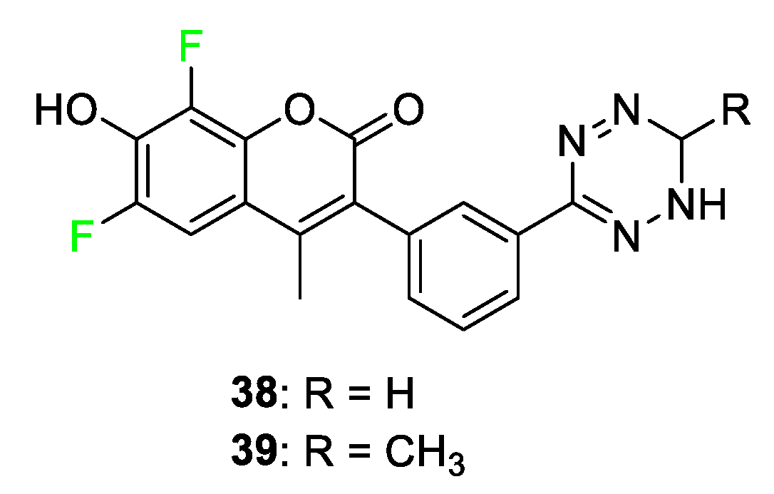
In addition to pharmaceuticals, another important medicinal chemistry application in which fluorine has found wide use is in imaging agents. Where the previous compounds were synthesized to fight and inhibit disease, the next section will focus on compounds that image and detect diseases. Imaging, both preoperative and in vivo, is another sector of medicine that has become a popular technique in recent years to assist in the fight against many diseases. First, we will present several classes of dyes that can be used for in vivo imaging and then consider several examples of fluorine-containing compounds that have been used in preoperative imaging. Fluorinated probes can illuminate several regions of the electromagnetic spectrum. Before presenting specific examples of fluorinated imaging agents, we will describe the benefits and shortcomings of a few different classes of biological imaging probes, including coumarin, fluorescein/rhodamine, boron–dipyrromethine (BODIPY), and cyanine dyes.
The electromagnetic spectrum spans from high-energy gamma rays to low-energy radio waves. The visible light spectrum lies between these extremes and is usually defined as having wavelengths of 500–600 nm. Dyes in this region are generally [86] used for surface imaging due to the higher probability of native tissue absorption in this range [87] when compared to near-infrared (NIR) dyes that absorb light at longer wavelengths. Because of this, very few examples of probes for biological imaging exist in this range. Dyes that fluoresce in this range can be used for purposes other than biological/medical applications, including solar cell construction.

4.1. Coumarin Dyes

The coumarin class of dye is a blue light-absorbing dye. Coumarin fluorophores are utilized for the diagnosis and imaging of diseases such as cancer. Coumarin molecules are small and biocompatible, and have a relatively high light quantum yield compared to other fluorescent dye classes [88]. Two examples of fluorine-incorporated visible light coumarin probes were discovered by Weissleder [71] and coworkers. The dyes, shown in Figure 16, contain two fluorine atoms on the coumarin derivatives 38 and 39. The coumarin dyes that contain fluorine substituents, in comparison to those without, show very comparable extinction coefficients, 19,000 to 16,000, while having a higher quantum yield, 49% to 41%, respectively [89]. The dyes effectively label the biological targets as well as feature optical flexibility and fast reactivity with the targets [89].

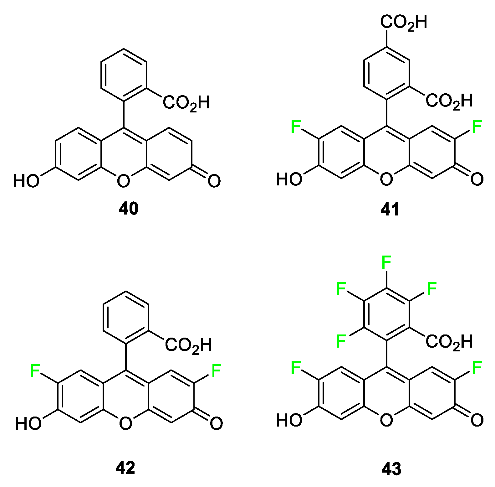
4.2. Fluorescein/Rhodamine Dyes

Fluorescein and rhodamine dyes consist of two important groups: the xanthene moiety, which acts as the fluorophore, and the benzene moiety, which provides the photoinduced electron transfer (PeT). PeT is a known mechanism in which the chromophore’s fluorescence is quenched with the electron transfer from the benzene donor to the xanthene acceptor [90,91]. The unsubstituted fluorescein dye 40 has a carboxyl group at position 2 of the benzene moiety, and until recently, it was believed that this acceptor group was essential to the molecule; this is based on results replacing the COO- group with hydrogen, reducing the quantum yield value by over 60% [92]. These xanthene-based dyes are seen as highly tunable and, therefore, are used as biological markers for DNA and proteins. Xanthene dyes do have some flaws, including being very pH-dependent; the forms in which the compounds can exist range from neutral to dianionic. A second shortcoming of the xanthene class of dyes stems from the wavelengths that these dyes absorb and fluoresce under 600 nm because there is high interference from tissue autofluorescence below 600 nm [93]. However, due to the high quantum yield and fluorescence tunability of these dyes, many of them still find use as biological markers, although their use as DNA stains is not performed in vivo [90,91].
Upon synthesizing a host of fluorinated fluorescein derivatives (Figure 17), it was found that these compounds exhibited some very interesting characteristics. While exhibiting slightly less molar absorptivity in comparison to the parent compound 40, the quantum yields of selected fluorinated xanthenes increased to nearly 100%. Another exciting fluorine-induced feature combats one of the drawbacks of this class of compounds as a whole: photobleaching. The bleaching value dropped from 17 (fluorescence percentage loss after 33 min) to as low as 4. It is hypothesized that this phenomenon likely stems from the triplet state lifetime of the molecule. The fluorine atoms, at the 2’ and 7’ position, in compounds 4143, shorten the triplet lifetime so that the likelihood of reaction with its quencher is decreased [94]. Although these compounds have not yet been tested in vivo, compounds 41 and 43 show great promise for use in bioconjugation in the future. When comparing these compounds’ absorbance wavelengths, compounds 41 and 42 are the same while a notable 18 nm redshift is observed for compound 43. Fluorophore 43 has a longer wavelength emission compared to fluorescein 40, and the report mentions its chemical properties (pKa, photostability, and high quantum yield), making it a useful compound for future investigation.
Rhodamine dyes 4447 have similar benefits and use in molecular imaging as the xanthene dye moiety as well as similar shortcomings (Figure 18). The core structure of rhodamine differs only in the two amine groups in place of the carbonyl and hydroxyl groups on the xanthene. This structural feature creates the limitation of pH dependency because the compounds can exist in neutral, zwitterionic, cationic, or dicationic forms. As in the case of the fluorinated fluorescein dyes, researchers likewise discovered an increased quantum yield with the fluorinated rhodamine dyes, along with prolonged photostability and decreased photobleaching of the dyes. Compounds 44 and 46 have similar absorbance and emission wavelengths; however, they differ in that compound 46 with two CH2CF3 groups attached to nitrogen has a higher reported quantum yield than compound 44. Hell and coworkers synthesized several rhodamine analogs featuring at least two fluorine atoms and discovered that this class of dyes shows great promise for stimulated emission depletion (STED) nanoscopy, a technique used for elucidating protein structures [95].

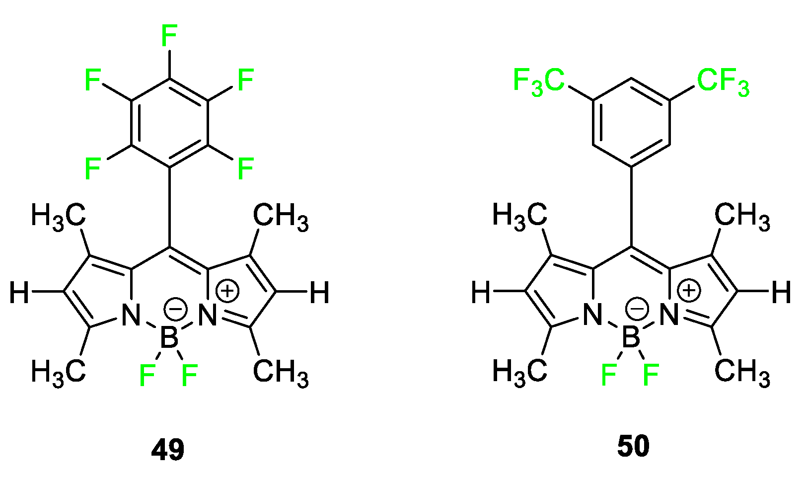
4.3. Boron–Dipyrromethene Dyes

The near-infrared region of light on the electromagnetic spectrum lies between 700 and 1000 nm. This region of light presents a unique range that allows for various uses in biomedical imaging. Because of the specific range in which the dyes absorb and fluoresce, NIR fluorophores avoid several potential problems concerning body imaging that were present with the coumarin, xanthene, and rhodamine dyes. Body tissue inherently fluoresces light in the 450–500 nm range, a phenomenon known as autofluorescence. Thus, imaging agents that fluoresce in this region exhibit a high background signal. The use of an NIR filter essentially eliminates tissue autofluorescence altogether [96] This makes higher-wavelength-fluorescing molecules superior in terms of signal-to-background noise ratios and molecular brightness. The upper end of the NIR range (near 1000 nm) is of limited usefulness due to water overtones that begin in this region. NIR light also has the ability to penetrate tissue for several centimeters [97]. This is due to the lower tissue absorbance and reduced scattering. Because of these factors, NIR fluorophores present themselves as the best potential in vivo imaging agents.
Many different classes of NIR fluorophores contain fluorine atoms. One of the most common types is BODIPY dye. The core structure of the BODIPY dye contains two fluorine atoms in the structure with a host of possible alteration sites. In recent studies, aza-BODIPY dyes have become increasingly popular in the literature due to their extended conjugation giving them more favorable optical properties for imaging [16]. Figure 19 features aza-BODIPY 48 highlighted in 2022 demonstrating dual imaging capabilities and potential for use in photodynamic therapy [98].
Although all BODIPY dyes feature fluorine attached to their core boron, very few BODIPY dyes further incorporate fluorine into the molecular architecture. Figure 20 outlines two examples, 49 and 50, which contain a perfluorophenyl ring and m-ditrifluoromethylbenzene ring, respectively, but the dyes were not tested for in vivo imaging [99]. The perfluorophenyl ring dye 49 has been used in studies to observe its reaction with thiols and amines, making them potential fluorophores for XPS/fluorescence labeling [100].

4.4. Carbocyanine Dyes

One class of NIR fluorophores that has yet to feature many lead compounds containing the fluorine atom for biological activity is the carbocyanine dye family. This class of dyes is often utilized in biomedical imaging. Several benefits arise from using carbocyanine dyes as an in vivo imaging agent. First, carbocyanine dyes are non-toxic to humans. In fact, Indocyanine green, a dye currently FDA-approved and used for medical imaging, remains one of the least toxic agents ever to be administered to humans [96]. Additionally, the core structure of the carbocyanine dye can easily be modified to match the wavelength needed: each double bond added between the indolium end units contributes to a wavelength increase of around 100 nm. While adding more carbon to the molecule does increase hydrophobicity, these dyes also have the advantage of having many different sites of modulation. This hydrophobicity increase caused by a longer carbon chain can be offset by adding a sulfonate or carboxylate group or another hydrophilic functional group to any of these sites.
Very few examples of carbocyanine dyes featuring the fluorine atom exist in the literature. Three examples of fluorocarbocyanine dyes are shown in Figure 21. Dye 51 is a fluorous amine-sensitive cyanine dye that was modified to observe fluorescence changes caused by the introduction of fluorine atoms to a known trimethine dye [101]. The results demonstrate that ratiometric and colorimetric property changes are observed upon exposure to amines. Dye 52 [102] selectively binds to G-quadruplex DNA; compound 53 [17] targets the thyroid and parathyroid glands. The fluorinated analog of the G-quadruplex DNA binding dye 52 shows a large increase in the thermal stability of the telomeric quadruplex. For the endocrine-targeting carbocyanine dyes, the uptake of the fluorinated molecule 53 was more than double that of any other substituent studied in the article, including other halogens and electron-donating groups. The fluorinated compounds exhibited a slower bodily clearance rate and were still observed in vivo after 4 h. The promising nature of these compounds for intraoperative imaging has led researchers to begin exploring these dyes as dual-modal molecules.

4.5. Squaraine Dyes

A significant portion of previous literature examples focuses on the use of squaraines in dye-sensitized solar cells (DSSCs). [103,104,105]. Squaraine dyes are growing in interest to the scientific community as many research groups are now finding biological applications for this dye scaffold [106,107]. One of the most promising properties of squaraine dyes is the relatively high quantum yield associated with this dye family compared to others: squaraine dyes are reported to have quantum yields between 20 and 40% [108]. They are also expected to have excellent molar absorptivity and high photobleaching thresholds. However, one of the problems related to squaraine dyes is the molecular stability around the oxycyclobutenolate ring in the center of the linker. Nevertheless, new examples of symmetrical and asymmetrical squaraine dyes have been synthesized and have demonstrated promising stability [105,109,110].
Figure 22 highlights a squaraine dye 54 [111] used for imaging ovarian cancer. This imaging agent stood out in cytotoxicity and NIR bioimaging studies when compared to analogs containing hydrogen, chlorine, or bromine atoms. Squaraine dye 55 [112] is used for labeling oligonucleotides. When compared to a nonfluorinated corresponding squaraine dye or dicyanosquaraine dye, the fluorine-containing dye 55 demonstrated improved photophysical properties and chemical stability.

5. Fluorine Incorporated into Molecules for Early Disease Detection Imaging Agents
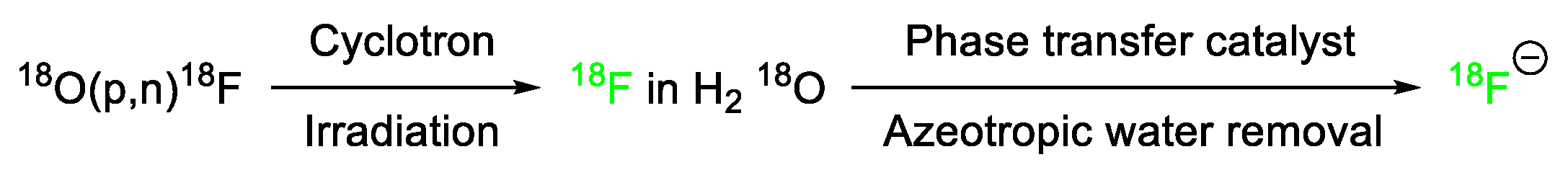
Due to the increased success rate of both surgery and chemotherapy during the early stages of tumor development, a need for enhanced detection methods has emerged. Visualization of diseased tissues gives doctors and surgeons insight into the answers to important questions such as: where is the diseased tissue, how big is the diseased mass, and what is the best course of action to combat the issue? Depending on the stage at which some diseases are detected, visualization of diseased tissues leads to curative measures. [97,113]. Currently, many imaging methods are used in the healthcare industry, including positron emission tomography (PET). These imaging methods aid healthcare professionals in assessing a patient’s specific situation and defining a course of action. Imaging agents can be used that will bind to specific tissues as well as give off energy in the form of positrons as emission radiation or radiative return, which is how the instrument obtains the visualization. While agents such as tagged proteins, nanoparticles, and metal delivery systems have been explored for use in these types of imaging modalities, small organic molecules are becoming popular for PET imaging.

Fluorine in PET

PET scans are presently among the most sensitive molecular imaging technique modalities [97]. The 18F-fluorine isotope has great utility in PET imaging due to its relatively long half-life (110 min.) in comparison to other positron-emitting atoms [114]. Additionally, fluorine-18 decays into oxygen-18, a non-toxic, non-radioactive nucleus that, when conjugated onto a sugar, is excreted through the kidneys and liver [53]. These advantages have driven the development of safe fluorinated contrast agents in use today [114].
Production of fluorine-18 requires a cyclotron, which irradiates 18O with protons. If the irradiation target is liquid H218O, an aqueous solution of 18F-fluoride results. The 18F-fluoride is then treated with a suitable salt such as the tetrabutylammonium cation or phase transfer catalyst such as Kryptofix2.2.2. The water is then removed by azeotropic distillation [115]. See Figure 23.
The 18F-fluoride may then be incorporated into molecules by nucleophilic aliphatic substitution (SN2) reactions or nucleophilic aromatic substitutions (SNAr). One advantage to this incorporation strategy is the high specific activity of fluoride-18 (~100 GBq/μmol) [116,117]. Figure 24 depicts several examples of radiotracers produced by nucleophilic substitutions using 18F-fluoride as the nucleophile.
The efficiency of SN2 nucleophilic substitutions depends on several factors, including substrate topology, leaving group ability and solvent choice. Ideally, substrates which allow for the nucleophile to approach the carbon bearing the leaving group (primary benzylic > primary aliphatic > secondary aliphatic) with minimal steric interference are preferred. Additionally, the selection of a satisfactory leaving group is paramount; the leaving group should facilitate the nucleophilic substitution [115]. In the examples depicted in Figure 24, both the triflate (OTf) and tosylate (OTs) leaving groups used in the preparation of 18F-FDG and 18F-FMISO, respectively, are exceptionally well suited as leaving groups [115]. Polar aprotic solvents are typically used for nucleophilic substitutions. In the figure above, acetonitrile (CH3CN) [118] served as the solvent to prepare 18F-FDG while dimethyl sulfoxide (DMSO) was the solvent used to prepare 18F-FMISO [119].
For nucleophilic aromatic substitutions (SNAr) to be successful, the aromatic ring system must contain electron-withdrawing groups positioned to enable mesomeric stabilization of the negative charge upon initial addition of the nucleophile. In the reaction depicted in Figure 24, the 18F-fluoride attacks the carbon bearing the -NO2 group (a typical leaving group for SNAr reactions). The aldehyde group ortho to the nucleophilic site of attack stabilizes the negative charge by resonance. The polar aprotic solvent dimethyl formamide (DMF) was the medium in which 18F-FDOPA was prepared [120].
While 18F-fluoride nucleophilic substitution reactions remain the preferred method for [18F]-labeled radiotracer production [116], electrophilic methods have been explored to enable C-18F bond formation in a variety of [18F]-labeled compounds that are used diagnostically [50,114,121,122,123,124,125,126,127,128,129,130,131]. Recent, thorough reviews of methodologies to form C-18F bonds by Chen et al. [115], Zhiyi and coworkers [132], and Lui et al. [133] provide excellent accounts of the state of the art in the production of [18F]-labeled compounds.
Since [18F]-FDG was approved for PET by the US Food and Drug Administration in 1999, more than thirty 18F-tagged compounds have been developed and found to be efficacious in PET applications for a variety of medical maladies [133]. The extensive and diverse uses of 18F-labeled compounds in PET have been well documented over the last twenty years, with more than 30,000 PET-related research articles and reviews published in the last decade alone, as a review by Crișan et al. notes [134]. This field of study remains vibrant, and there is no doubt that compounds labeled with 18F-fluorine will find additional utility in the PET area of medicine.

6. Summary and Outlook

The utilization of fluorine atoms in chemistry has increased in the past 30 years, and its viability and importance in drug development and bioimaging have become readily apparent to scientists [16]. Fluorine’s many unique characteristics and properties lend themselves to the use of this atom in combatting the many different problems medicinal chemists may face. Throughout recent years, we have witnessed the impact of fluorine-containing compounds on different aspects of medicine including compounds being used as antivirals in HIV and COVID-19 patients as well as being used for bacterial studies and anti-cancer studies [14,36].
The inherent size, electronegativity, and blocking of metabolic sites for drugs, along with the shortening of the triplet state for imaging chromophores, can be solved singularly by the addition of one or more fluorine atoms on a molecule. Similarly, in medicine, the addition of fluorine atoms to contrast agents is becoming increasingly prevalent in biomolecular imaging and preventative medicine. Other forms of imaging incorporate fluorine atoms as a radiolabel for increased use of fluorescent probes in multimodal imaging. A unique opportunity presents itself for researchers when considering the addition of fluorine on small molecules or imaging fluorophores: multipurpose molecules. Not only are the fluorinated chromophores useful intraoperatively and for drug tracking in the body, but radiolabeled compounds also have utility as 18F-radiolabels for PET and SPECT purposes. Through the research performed on small molecules containing fluorine atoms and functional groups containing fluorine, much has been learned about drug metabolic pathways and medical advancements both through pharmaceutical and imaging techniques.
While many fluorinated compounds are strategically designed to alter the physical properties and metabolism of overall molecules, not all compounds necessarily benefit from fluorination. Some reports have addressed the possibility of fluorinated compounds degrading during metabolic processes and found that these active agents can generate reactive intermediates, thus creating potentially indirect in vivo toxicity [135]. While this toxicity is an undesirable effect, some studies have used these effects for generating fluorine-facilitated selective toxins expected to be useful as antibiotics and anti-cancer agents. Research trends on fluorinated compounds continue to increase. Considering research trends, we anticipate more compounds to be introduced into the pharmaceutical market; medicinal chemists must consider the medicinal effect of fluorine moieties but also look into the possible byproducts and overall effects when designing compounds. It is also important to consider the limitations of drug design based on insufficient fluorine-containing precursor availability. Several fluorinated compounds developed in earlier studies introduced single fluorine atoms into the chemical structures and explored their bioactivity and chemical property changes. Current studies are further expanding on original designs and exploring the incorporation of fluorine on multiple aromatic and aliphatic moieties, as well as the addition of chiral centers and their overall effects on molecules and their applications. It is imperative for researchers in the field to continue the development of these reagents to introduce new possibilities for incorporating fluorinated compounds into the field of medicine and imaging.

Funding

M.H. acknowledges the funding from the National Institutes of Health under grants numbers R01EB034731 and R01RCA281098A1.

Data Availability Statement

Data sharing is not applicable.

Acknowledgments

The authors wishes to thank the Department of Chemistry at Georgia State University and the Brains and Behavior Grant, the Health Innovation Grant, and the Georgia Research Alliance Grant for their financial support. The authors would like to thank the seed grant from Georgia Gwinnett College.

Conflicts of Interest

The authors declare no conflicts of interest.

References

  1. Müller, K.; Faeh, C.; Diederich, F. Fluorine in Pharmaceuticals: Looking Beyond Intuition. Science 2007, 317, 1881–1886. [Google Scholar] [CrossRef] [PubMed]
  2. Shah, P.; Westwell, A.D. The role of fluorine in medicinal chemistry. J. Enzym. Inhib. Med. Chem. 2007, 22, 527–540. [Google Scholar] [CrossRef] [PubMed]
  3. Wang, Q.; Han, J.; Sorochinsky, A.; Landa, A.; Butler, G.; Soloshonok, V.A. The Latest FDA-Approved Pharmaceuticals Containing Fragments of Tailor-Made Amino Acids and Fluorine. Pharmaceuticals 2022, 15, 999. [Google Scholar] [CrossRef] [PubMed]
  4. Inoue, M.; Sumii, Y.; Shibata, N. Contribution of Organofluorine Compounds to Pharmaceuticals. ACS Omega 2020, 5, 10633–10640. [Google Scholar] [CrossRef] [PubMed]
  5. He, J.; Li, Z.; Dhawan, G.; Zhang, W.; Sorochinsky, A.E.; Butler, G.; Soloshonok, V.A.; Han, J. Fluorine-containing drugs approved by the FDA in 2021. Chin. Chem. Lett. 2022, 34, 107578. [Google Scholar] [CrossRef]
  6. Arntson, K.E.; Pomerantz, W.C.K. Protein-Observed Fluorine NMR: A Bioorthogonal Approach for Small Molecule Discovery. J. Med. Chem. 2016, 59, 5158–5171. [Google Scholar] [CrossRef] [PubMed]
  7. Smart, B.E. Fluorine substituent effects (on bioactivity). J. Fluor. Chem. 2001, 109, 3–11. [Google Scholar] [CrossRef]
  8. Bondi, A. van der Waals Volumes and Radii. J. Phys. Chem. 1964, 68, 441–451. [Google Scholar] [CrossRef]
  9. Purser, S.; Moore, P.R.; Swallow, S.; Gouverneur, V. Fluorine in medicinal chemistry. Chem. Soc. Rev. 2008, 37, 320–330. [Google Scholar] [CrossRef]
  10. Reichenbächer, K.; Süss, H.I.; Hulliger, J. Fluorine in crystal engineering—“The little atom that could”. Chem. Soc. Rev. 2005, 34, 22–30. [Google Scholar] [CrossRef]
  11. O’Hagan, D. Understanding organofluorine chemistry. An introduction to the C–F bond. Chem. Soc. Rev. 2008, 37, 308–319. [Google Scholar] [CrossRef] [PubMed]
  12. Aradi, K.; Kiss, L. Carbotrifluoromethylations of C−C Multiple Bonds (Excluding Aryl-and Alkynyltrifluoromethylations). Chem. A Eur. J. 2023, 29, e202203499. [Google Scholar] [CrossRef] [PubMed]
  13. Alle, T.; Joyasawal, S.; Oukoloff, K.; Long, K.; Owyang, Z.; Francisco, K.R.; Cahard, D.; Huryn, D.M.; Ballatore, C. Structure-property relationships of fluorinated carboxylic acid bioisosteres. Bioorganic Med. Chem. Lett. 2023, 91, 129363. [Google Scholar] [CrossRef]
  14. Han, S.; Lu, Y. Fluorine in anti-HIV drugs approved by FDA from 1981 to 2023. Eur. J. Med. Chem. 2023, 258, 115586. [Google Scholar] [CrossRef] [PubMed]
  15. Johnson, B.M.; Shu, Y.-Z.; Zhuo, X.; Meanwell, N.A. Metabolic and Pharmaceutical Aspects of Fluorinated Compounds. J. Med. Chem. 2020, 63, 6315–6386. [Google Scholar] [CrossRef]
  16. Casa, S.; Henary, M. Synthesis and Applications of Selected Fluorine-Containing Fluorophores. Molecules 2021, 26, 1160. [Google Scholar] [CrossRef]
  17. Hyun, H.; Park, M.H.; Owens, E.A.; Wada, H.; Henary, M.; Handgraaf, H.J.M.; Vahrmeijer, A.L.; Frangioni, J.V.; Choi, H.S. Structure-inherent targeting of near-infrared fluorophores for parathyroid and thyroid gland imaging. Nat. Med. 2015, 21, 192–197. [Google Scholar] [CrossRef]
  18. Zhou, P.; Zou, J.; Tian, F.; Shang, Z. Fluorine Bonding—How Does It Work In Protein−Ligand Interactions? J. Chem. Inf. Model. 2009, 49, 2344–2355. [Google Scholar] [CrossRef]
  19. Britton, R.; Gouverneur, V.; Lin, J.-H.; Meanwell, M.; Ni, C.; Pupo, G.; Xiao, J.-C.; Hu, J. Contemporary synthetic strategies in organofluorine chemistry. Nat. Rev. Methods Primers 2021, 1, 47. [Google Scholar] [CrossRef]
  20. Liang, T.; Neumann, C.N.; Ritter, T. Introduction of Fluorine and Fluorine-Containing Functional Groups. Angew. Chem. Int. Ed. 2013, 52, 8214–8264. [Google Scholar] [CrossRef]
  21. Preparation of Highly Fluorinated Compounds. In Fluorine in Organic Chemistry; Blackwell Publishing Ltd.: Hoboken, NJ, USA, 2004; pp. 23–46.
  22. Zhou, G.; Tian, Y.; Zhao, X.; Dan, W. Selective Fluorination of 4-Substituted 2-Aminopyridines and Pyridin-2(1H)-ones in Aqueous Solution. Org. Lett. 2018, 20, 4858–4861. [Google Scholar] [CrossRef] [PubMed]
  23. Lerman, O.; Tor, Y.; Hebel, D.; Rozen, S. A novel electrophilic fluorination of activated aromatic rings using acetyl hypofluorite, suitable also for introducing fluorine-18 into benzene. J. Org. Chem. 1984, 49, 806–813. [Google Scholar] [CrossRef]
  24. Furuya, T.; Kamlet, A.S.; Ritter, T. Catalysis for fluorination and trifluoromethylation. Nature 2011, 473, 470–477. [Google Scholar] [CrossRef] [PubMed]
  25. Sloop, J.C.; Holder, C.; Henary, M. Selective Incorporation of Fluorine in Pyrazoles. Eur. J. Org. Chem. 2015, 2015, 3405–3422. [Google Scholar] [CrossRef]
  26. Yang, X.; Sun, R.; Li, S.; Zheng, X.; Yuan, M.; Xu, B.; Jiang, W.; Chen, H.; Fu, H.; Li, R. Regioselective Direct C–H Trifluoromethylation of Pyridine. Org. Lett. 2020, 22, 7108–7112. [Google Scholar] [CrossRef]
  27. Liang, D.; Li, Y.; Gao, S.; Li, R.; Li, X.; Wang, B.; Yang, H. Amide-assisted radical strategy: Metal-free direct fluorination of arenes in aqueous media. Green Chem. 2017, 19, 3344–3349. [Google Scholar] [CrossRef]
  28. Park, B.K.; Kitteringham, N.R.; O’Neill, P.M. Metabolism of Fluorine-Containing Drugs. Annu. Rev. Pharmacol. Toxicol. 2001, 41, 443–470. [Google Scholar] [CrossRef] [PubMed]
  29. Nassar, A.-E.F.; Kamel, A.M.; Clarimont, C. Improving the decision-making process in the structural modification of drug candidates: Enhancing metabolic stability. Drug Discov. Today 2004, 9, 1020–1028. [Google Scholar] [CrossRef]
  30. Hagmann, W.K. The Many Roles for Fluorine in Medicinal Chemistry. J. Med. Chem. 2008, 51, 4359–4369. [Google Scholar] [CrossRef]
  31. Schoepfer, J.; Jahnke, W.; Berellini, G.; Buonamici, S.; Cotesta, S.; Cowan-Jacob, S.W.; Dodd, S.; Drueckes, P.; Fabbro, D.; Gabriel, T.; et al. Discovery of Asciminib (ABL001), an Allosteric Inhibitor of the Tyrosine Kinase Activity of BCR-ABL1. J. Med. Chem. 2018, 61, 8120–8135. [Google Scholar] [CrossRef]
  32. Szarek, M.; Amarenco, P.; Callahan, A.; DeMicco, D.; Fayyad, R.; Goldstein, L.B.; Laskey, R.; Sillesen, H.; Welch, K.M. Atorvastatin Reduces First and Subsequent Vascular Events Across Vascular Territories: The SPARCL Trial. J. Am. Coll. Cardiol. 2020, 75, 2110–2118. [Google Scholar] [CrossRef]
  33. Wang, J.; Sánchez-Roselló, M.; Aceña, J.L.; del Pozo, C.; Sorochinsky, A.E.; Fustero, S.; Soloshonok, V.A.; Liu, H. Fluorine in Pharmaceutical Industry: Fluorine-Containing Drugs Introduced to the Market in the Last Decade (2001–2011). Chem. Rev. 2014, 114, 2432–2506. [Google Scholar] [CrossRef]
  34. Zhang, X.; Ma, X.; Cheng, W.; Han, Y.; Mu, Q.; Li, J.; He, S. Effects of escitalopram oxalate plus low-dose trazodone on psychological state and quality of life in patients with treatment-refractory depression. Am. J. Transl. Res. 2023, 15, 1905–1912. [Google Scholar] [PubMed]
  35. Larsen, M.A.B.; Plenge, P.; Andersen, J.; Eildal, J.N.N.; Kristensen, A.S.; Bøgesø, K.P.; Gether, U.; Strømgaard, K.; Bang-Andersen, B.; Loland, C.J. Structure–activity relationship studies of citalopram derivatives: Examining substituents conferring selectivity for the allosteric site in the 5-HT transporter. Br. J. Pharmacol. 2016, 173, 925–936. [Google Scholar] [CrossRef] [PubMed]
  36. Chandra, G.; Singh, D.V.; Mahato, G.K.; Patel, S. Fluorine-a small magic bullet atom in the drug development: Perspective to FDA approved and COVID-19 recommended drugs. Chem. Pap. 2023, 77, 4085–4106. [Google Scholar] [CrossRef] [PubMed]
  37. Ailani, J.; Lipton, R.B.; Goadsby, P.J.; Guo, H.; Miceli, R.; Severt, L.; Finnegan, M.; Trugman, J.M. Atogepant for the Preventive Treatment of Migraine. N. Engl. J. Med. 2021, 385, 695–706. [Google Scholar] [CrossRef] [PubMed]
  38. Kariyawasam, H.H.; Scadding, G.K. Seasonal allergic rhinitis: Fluticasone propionate and fluticasone furoate therapy evaluated. J. Asthma Allergy 2010, 3, 19–28. [Google Scholar] [PubMed]
  39. Kumar, R.S.; Jain, M.K.; Kushwaha, J.S.; Patil, S.; Patil, V.; Ghatak, S.; Sanmukhani, J.; Mittal, R. Efficacy and Safety of Fluticasone Furoate and Oxymetazoline Nasal Spray: A Novel First Fixed Dose Combination for the Management of Allergic Rhinitis with Nasal Congestion. J. Asthma Allergy 2022, 15, 783–792. [Google Scholar] [CrossRef] [PubMed]
  40. Zhang, Y.; Wei, P.; Chen, B.; Li, X.; Luo, X.; Chen, X.; Xiang, M.; Li, L.; Zhao, S.; Xiao, X.; et al. Intranasal fluticasone furoate in pediatric allergic rhinitis: Randomized controlled study. Pediatr. Res. 2021, 89, 1832–1839. [Google Scholar] [CrossRef] [PubMed]
  41. Zhang, X.; Liu, M.; Mao, Y. Efficacy of Fluticasone and Salmeterol Dry Powder in Treating Patients with Bronchial Asthma and Its Effect on Inflammatory Factors and Pulmonary Function. Evid. Based Complement. Altern. Med. Ecam 2022, 2022, 8555417. [Google Scholar] [CrossRef]
  42. Prabhudesai, P.; Singh, B.P.; Agrawal, G.; Singh, A.K.; Jadhav, A.Y.; Patil, S.R.; Bhagat, S.; Patil, S.; Barkate, H. Fluticasone Furoate/Vilanterol Use Trends and Characteristics in Patients with Obstructive Airway Disease: A Real-World Study of 10,374 Patients from India. Cureus 2023, 15, e34825. [Google Scholar] [CrossRef]
  43. Crim, C.; Pierre, L.N.; Daley-Yates, P.T. A review of the pharmacology and pharmacokinetics of inhaled fluticasone propionate and mometasone furoate. Clin. Ther. 2001, 23, 1339–1354. [Google Scholar] [CrossRef]
  44. Ali, S.; Zhou, J. Highlights on U.S. FDA-approved fluorinated drugs over the past five years (2018–2022). Eur. J. Med. Chem. 2023, 256, 115476. [Google Scholar] [CrossRef]
  45. Lin, H.-C.; Saputra, F.; Audira, G.; Lai, Y.-H.; Roldan, M.J.M.; Alos, H.C.; Aventurado, C.A.; Vasquez, R.D.; Tsai, G.-J.; Lim, K.-H.; et al. Investigating Potential Cardiovascular Toxicity of Two Anti-Leukemia Drugs of Asciminib and Ponatinib in Zebrafish Embryos. Int. J. Mol. Sci. 2022, 23, 11711. [Google Scholar] [CrossRef]
  46. Deeks, E.D. Asciminib: First Approval. Drugs 2022, 82, 219–226. [Google Scholar] [CrossRef]
  47. Rizzo, C.; Amata, S.; Pibiri, I.; Pace, A.; Buscemi, S.; Palumbo Piccionello, A. FDA-Approved Fluorinated Heterocyclic Drugs from 2016 to 2022. Int. J. Mol. Sci. 2023, 24, 7728. [Google Scholar] [CrossRef] [PubMed]
  48. Dubowchik, G.M.; Conway, C.M.; Xin, A.W. Blocking the CGRP Pathway for Acute and Preventive Treatment of Migraine: The Evolution of Success. J. Med. Chem. 2020, 63, 6600–6623. [Google Scholar] [CrossRef] [PubMed]
  49. Martelletti, P.; Cipolla, F.; Capi, M.; Curto, M.; Lionetto, L. Atogepant. Calcitonin gene-related peptide (CGRP) receptor antagonist, Preventive treatment of migraine. Drugs Futur. 2020, 45, 285. [Google Scholar] [CrossRef]
  50. Benedetto Tiz, D.; Bagnoli, L.; Rosati, O.; Marini, F.; Sancineto, L.; Santi, C. New Halogen-Containing Drugs Approved by FDA in 2021: An Overview on Their Syntheses and Pharmaceutical Use. Molecules 2022, 27, 1643. [Google Scholar] [CrossRef] [PubMed]
  51. Di Fabio, R.; Alvaro, G.; Griffante, C.; Pizzi, D.A.; Donati, D.; Mattioli, M.; Cimarosti, Z.; Guercio, G.; Marchioro, C.; Provera, S.; et al. Discovery and Biological Characterization of (2R,4S)-1′-Acetyl-N-{(1R)-1-[3,5-bis(trifluoromethyl)phenyl]ethyl}-2-(4-fluoro-2-methylphenyl)-N-methyl-4,4′-bipiperidine-1-carboxamide as a New Potent and Selective Neurokinin 1 (NK1) Receptor Antagonist Clinical Candidate. J. Med. Chem. 2011, 54, 1071–1079. [Google Scholar]
  52. Wang, D.-S.; Hu, M.-T.; Wang, Z.-Q.; Ren, C.; Qiu, M.-Z.; Luo, H.-Y.; Jin, Y.; Fong, W.P.; Wang, S.-B.; Peng, J.-W.; et al. Effect of Aprepitant for the Prevention of Chemotherapy-Induced Nausea and Vomiting in Women: A Randomized Clinical Trial. JAMA Netw. Open 2021, 4, e215250. [Google Scholar] [CrossRef]
  53. Van Heek, M.; Farley, C.; Compton, D.S.; Hoos, L.; Alton, K.B.; Sybertz, E.J.; Davis, H.R., Jr. Comparison of the activity and disposition of the novel cholesterol absorption inhibitor, SCH58235, and its glucuronide, SCH60663. Br. J. Pharmacol. 2000, 129, 1748–1754. [Google Scholar] [CrossRef]
  54. Igel, M.; Sudhop, T.; von Bergmann, K. Pharmacology of 3-Hydroxy-3-Methylglutaryl-Coenzyme A Reductase Inhibitors (Statins), Including Rosuvastatin and Pitavastatin. J. Clin. Pharmacol. 2002, 42, 835–845. [Google Scholar] [CrossRef]
  55. Hak Lee, S.; Chung, N.; Kwan, J.; Kim, D.-I.; Ho Kim, W.; Jeong Kim, C.; Seung Kim, H.; Hoon Park, S.; Seog Seo, H.; Gu Shin, D.; et al. Comparison of the efficacy and tolerability of pitavastatin and atorvastatin: An 8-week, multicenter, randomized, open-label, dose-titration study in korean patients with hypercholesterolemia. Clin. Ther. 2007, 29, 2365–2373. [Google Scholar] [CrossRef]
  56. Xu, Q.; Deng, Y.; Xiao, J.; Liu, X.; Zhou, M.; Ren, Z.; Peng, J.; Tang, Y.; Jiang, Z.; Tang, Z.; et al. Three Musketeers for Lowering Cholesterol: Statins, Ezetimibe and Evolocumab. Curr. Med. Chem. 2021, 28, 1025–1041. [Google Scholar] [CrossRef]
  57. Saito, Y. Pitavastatin: An overview. Atheroscler. Suppl. 2011, 12, 271–276. [Google Scholar] [CrossRef]
  58. Saito, Y.; Yamada, N.; Teramoto, T.; Itakura, H.; Hata, Y.; Nakaya, N.; Mabuchi, H.; Tushima, M.; Sasaki, J.; Goto, Y.; et al. Clinical Efficacy of Pitavastatin, a New 3-Hydroxy-3-methylglutaryl Coenzyme A Reductase Inhibitor, in Patients with Hyperlipidemia. Arzneimittelforschung 2002, 52, 251–255. [Google Scholar] [CrossRef] [PubMed]
  59. Yagi, Y.; Kimura, H.; Okuda, H.; Ono, M.; Nakamoto, Y.; Togashi, K.; Saji, H. Evaluation of [18F]pitavastatin as a positron emission tomography tracer for in vivo organic transporter polypeptide function. Nucl. Med. Biol. 2019, 74–75, 25–31. [Google Scholar] [CrossRef] [PubMed]
  60. Wakeling, A.E.; Dukes, M.; Bowler, J. A Potent Specific Pure Antiestrogen with Clinical Potential. Cancer Res. 1991, 51, 3867–3873. [Google Scholar] [PubMed]
  61. Troup, R.I.; Jeffries, B.; Saudain, R.E.-B.; Georgiou, E.; Fish, J.; Scott, J.S.; Chiarparin, E.; Fallan, C.; Linclau, B. Skipped Fluorination Motifs: Synthesis of Building Blocks and Comparison of Lipophilicity Trends with Vicinal and Isolated Fluorination Motifs. J. Org. Chem. 2021, 86, 1882–1900. [Google Scholar] [CrossRef] [PubMed]
  62. Jeffries, B.; Wang, Z.; Felstead, H.R.; Le Questel, J.-Y.; Scott, J.S.; Chiarparin, E.; Graton, J.; Linclau, B. Systematic Investigation of Lipophilicity Modulation by Aliphatic Fluorination Motifs. J. Med. Chem. 2020, 63, 1002–1031. [Google Scholar] [CrossRef] [PubMed]
  63. Kürschner, M.; Nielsen, K.; von Langen, J.R.G.; Schenk, W.A.; Zimmermann, U.; Sukhorukov, V.L. Effect of Fluorine Substitution on the Interaction of Lipophilic Ions with the Plasma Membrane of Mammalian Cells. Biophys. J. 2000, 79, 1490–1497. [Google Scholar] [CrossRef] [PubMed]
  64. Yerien, D.E.; Bonesi, S.; Postigo, A. Fluorination methods in drug discovery. Org. Biomol. Chem. 2016, 14, 8398–8427. [Google Scholar] [CrossRef] [PubMed]
  65. Jeffries, B.; Wang, Z.; Graton, J.; Holland, S.D.; Brind, T.; Greenwood, R.D.R.; Le Questel, J.-Y.; Scott, J.S.; Chiarparin, E.; Linclau, B. Reducing the Lipophilicity of Perfluoroalkyl Groups by CF2–F/CF2–Me or CF3/CH3 Exchange. J. Med. Chem. 2018, 61, 10602–10618. [Google Scholar] [CrossRef] [PubMed]
  66. Ryan, A.J.; Wedge, S.R. ZD6474–a novel inhibitor of VEGFR and EGFR tyrosine kinase activity. Br. J. Cancer 2005, 92, S6–S13. [Google Scholar] [CrossRef] [PubMed]
  67. Kung, H.F.; Choi, S.R.; Qu, W.; Zhang, W.; Skovronsky, D. 18F Stilbenes and Styrylpyridines for PET Imaging of Aβ Plaques in Alzheimer’s Disease: A Miniperspective. J. Med. Chem. 2010, 53, 933–941. [Google Scholar] [CrossRef] [PubMed]
  68. Ghorab, M.M.; Ragab, F.A.; Heiba, H.I.; Arafa, R.K.; El-Hossary, E.M. Docking study, in vitro anticancer screening and radiosensitizing evaluation of some new fluorine-containing quinoline and pyrimidoquinoline derivatives bearing a sulfonamide moiety. Med. Chem. Res. 2011, 20, 388–400. [Google Scholar] [CrossRef]
  69. Kathrotiya, H.G.; Patel, M.P. Synthesis and identification of β-aryloxyquinoline based diversely fluorine substituted N-aryl quinolone derivatives as a new class of antimicrobial, antituberculosis and antioxidant agents. Eur. J. Med. Chem. 2013, 63, 675–684. [Google Scholar] [CrossRef]
  70. Mistry, S.N.; Valant, C.; Sexton, P.M.; Capuano, B.; Christopoulos, A.; Scammells, P.J. Synthesis and Pharmacological Profiling of Analogues of Benzyl Quinolone Carboxylic Acid (BQCA) as Allosteric Modulators of the M1 Muscarinic Receptor. J. Med. Chem. 2013, 56, 5151–5172. [Google Scholar] [CrossRef]
  71. Chen, D.; Zhang, Y.; Li, J.; Liu, Y. Exploratory Process Development of Pexidartinib through the Tandem Tsuji–Trost Reaction and Heck Coupling. Synthesis 2019, 51, 2564–2571. [Google Scholar] [CrossRef]
  72. Kantarjian, H.M.; Giles, F.; Gattermann, N.; Bhalla, K.; Alimena, G.; Palandri, F.; Ossenkoppele, G.J.; Nicolini, F.-E.; O’Brien, S.G.; Litzow, M.; et al. Nilotinib (formerly AMN107), a highly selective BCR-ABL tyrosine kinase inhibitor, is effective in patients with Philadelphia chromosome–positive chronic myelogenous leukemia in chronic phase following imatinib resistance and intolerance. Blood 2007, 110, 3540–3546. [Google Scholar] [CrossRef]
  73. Kuang, Y.-H.; Shen, T.; Chen, X.; Sodani, K.; Hopper-Borge, E.; Tiwari, A.K.; Lee, J.W.K.K.; Fu, L.-W.; Chen, Z.-S. Lapatinib and erlotinib are potent reversal agents for MRP7 (ABCC10)-mediated multidrug resistance. Biochem. Pharmacol. 2010, 79, 154–161. [Google Scholar] [CrossRef]
  74. Popovici-Muller, J.; Lemieux, R.M.; Artin, E.; Saunders, J.O.; Salituro, F.G.; Travins, J.; Cianchetta, G.; Cai, Z.; Zhou, D.; Cui, D.; et al. Discovery of AG-120 (Ivosidenib): A First-in-Class Mutant IDH1 Inhibitor for the Treatment of IDH1 Mutant Cancers. ACS Med. Chem. Lett. 2018, 9, 300–305. [Google Scholar] [CrossRef] [PubMed]
  75. Gupta, P.S. Roles of Fluorine in Drug Design and Drug Action. Lett. Drug Des. Discov. 2019, 16, 1089–1109. [Google Scholar] [CrossRef]
  76. Dabur, M.; Loureiro, J.A.; Pereira, M.C. Fluorinated Molecules and Nanotechnology: Future ‘Avengers’ against the Alzheimer’s Disease? Int. J. Mol. Sci. 2020, 21, 2989. [Google Scholar] [CrossRef]
  77. Dhillon, S. Melphalan Flufenamide (Melflufen): First Approval. Drugs 2021, 81, 963–969. [Google Scholar] [CrossRef] [PubMed]
  78. Lehmann, F.; Wennerberg, J. Melflufen: A Journey from Discovery to Multi-Kilogram Production. In Complete Accounts of Integrated Drug Discovery and Development: Recent Examples from the Pharmaceutical Industry Volume 3; American Chemical Society: Washington, DC, USA, 2020; Volume 1369, pp. 157–177. [Google Scholar]
  79. Mei, H.; Han, J.; Fustero, S.; Medio-Simon, M.; Sedgwick, D.M.; Santi, C.; Ruzziconi, R.; Soloshonok, V.A. Fluorine-Containing Drugs Approved by the FDA in 2018. Chem. A Eur. J. 2019, 25, 11797–11819. [Google Scholar] [CrossRef]
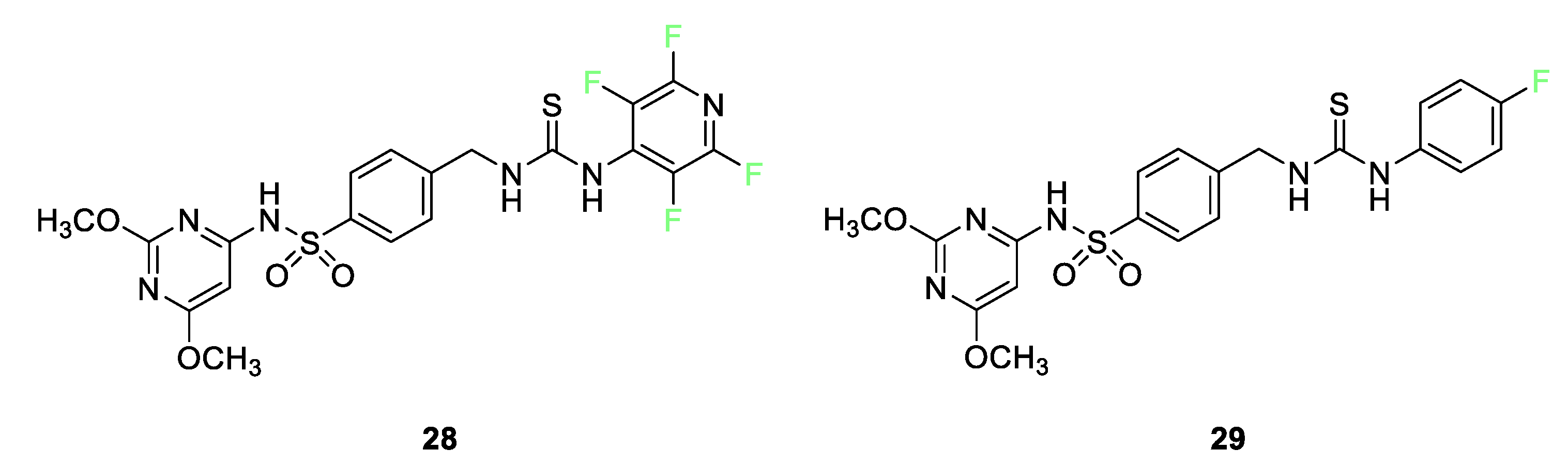
  80. El-Gaby, M.; Ammar, Y.A.; IH El-Qaliei, M.; Hussein, M.F.; Faraghally, F.A. Sulfonamides: Synthesis and the recent applications in Medicinal Chemistry. Egypt. J. Chem. 2020, 63, 5289–5327. [Google Scholar]
  81. Berrino, E.; Michelet, B.; Martin-Mingot, A.; Carta, F.; Supuran, C.T.; Thibaudeau, S. Modulating the Efficacy of Carbonic Anhydrase Inhibitors through Fluorine Substitution. Angew. Chem. Int. Ed. 2021, 60, 23068–23082. [Google Scholar] [CrossRef]
  82. Vullo, D.; Scozzafava, A.; Pastorekova, S.; Pastorek, J.; Supuran, C.T. Carbonic anhydrase inhibitors: Inhibition of the tumor-associated isozyme IX with fluorine-containing sulfonamides. The first subnanomolar CA IX inhibitor discovered. Bioorg. Med. Chem. Lett. 2004, 14, 2351–2356. [Google Scholar] [CrossRef]
  83. Pastorekova, S.; Vullo, D.; Casini, A.; Scozzafava, A.; Pastorek, J.; Nishimori, I.; Supuran, C.T. Carbonic anhydrase inhibitors: Inhibition of the tumor-associated isozymes IX and XII with polyfluorinated aromatic/heterocyclic sulfonamides. J. Enzym. Inhib. Med. Chem. 2005, 20, 211–217. [Google Scholar] [CrossRef]
  84. Pacchiano, F.; Aggarwal, M.; Avvaru, B.S.; Robbins, A.H.; Scozzafava, A.; McKenna, R.; Supuran, C.T. Selective hydrophobic pocket binding observed within the carbonic anhydrase II active site accommodate different 4-substituted-ureido-benzenesulfonamides and correlate to inhibitor potency. Chem. Commun. 2010, 46, 8371–8373. [Google Scholar] [CrossRef] [PubMed]
  85. Compain, G.; Martin-Mingot, A.; Maresca, A.; Thibaudeau, S.; Supuran, C.T. Superacid synthesis of halogen containing N-substituted-4-aminobenzene sulfonamides: New selective tumor-associated carbonic anhydrase inhibitors. Bioorg. Med. Chem. 2013, 21, 1555–1563. [Google Scholar] [CrossRef]
  86. Kobayashi, H.; Ogawa, M.; Alford, R.; Choyke, P.L.; Urano, Y. New Strategies for Fluorescent Probe Design in Medical Diagnostic Imaging. Chem. Rev. 2010, 110, 2620–2640. [Google Scholar] [CrossRef]
  87. Welsher, K.; Sherlock, S.P.; Dai, H. Deep-tissue anatomical imaging of mice using carbon nanotube fluorophores in the second near-infrared window. Proc. Natl. Acad. Sci. USA 2011, 108, 8943–8948. [Google Scholar] [CrossRef]
  88. Drexhage, K.H. Fluorescence Efficiency of Laser Dyes. J. Res. Natl. Bur. Stand. 1976, 80A, 421–428. [Google Scholar] [CrossRef]
  89. Meimetis, L.G.; Carlson, J.C.T.; Giedt, R.J.; Kohler, R.H.; Weissleder, R. Ultrafluorogenic Coumarin–Tetrazine Probes for Real-Time Biological Imaging. Angew. Chem. Int. Ed. 2014, 53, 7531–7534. [Google Scholar] [CrossRef] [PubMed]
  90. Lindqvist, L.; Lundeen, G.W. Radiationless Transitions in Xanthene Dyes. J. Chem. Phys. 1966, 44, 1711–1712. [Google Scholar] [CrossRef]
  91. Fink, D.W.; Willis, C.R. Structure and Internal Mixing in Xanthene Dyes. J. Chem. Phys. 1970, 53, 4720–4722. [Google Scholar] [CrossRef]
  92. Urano, Y.; Kamiya, M.; Kanda, K.; Ueno, T.; Hirose, K.; Nagano, T. Evolution of Fluorescein as a Platform for Finely Tunable Fluorescence Probes. J. Am. Chem. Soc. 2005, 127, 4888–4894. [Google Scholar] [CrossRef]
  93. Zhao, M.; Guo, Y.-S.; Xu, W.-N.; Zhao, Y.-F.; Xie, H.-Y.; Li, H.-J.; Chen, X.-F.; Zhao, R.-S.; Guo, D.-S. Far-red to near-infrared fluorescent probes based on silicon-substituted xanthene dyes for sensing and imaging. TrAC Trends Anal. Chem. 2020, 122, 115704. [Google Scholar] [CrossRef]
  94. Sun, W.-C.; Gee, K.R.; Klaubert, D.H.; Haugland, R.P. Synthesis of Fluorinated Fluoresceins. J. Org. Chem. 1997, 62, 6469–6475. [Google Scholar] [CrossRef]
  95. Mitronova, G.Y.; Belov, V.N.; Bossi, M.L.; Wurm, C.A.; Meyer, L.; Medda, R.; Moneron, G.; Bretschneider, S.; Eggeling, C.; Jakobs, S.; et al. New Fluorinated Rhodamines for Optical Microscopy and Nanoscopy. Chem. A Eur. J. 2010, 16, 4477–4488. [Google Scholar] [CrossRef] [PubMed]
  96. Frangioni, J.V. In vivo near-infrared fluorescence imaging. Curr. Opin. Chem. Biol. 2003, 7, 626–634. [Google Scholar] [CrossRef] [PubMed]
  97. Weissleder, R. Molecular Imaging in Cancer. Science 2006, 312, 1168–1171. [Google Scholar] [CrossRef] [PubMed]
  98. Li, A.; Peng, X.; Jiang, M.; Wu, T.; Chen, K.; Yang, Z.; Chen, S.; Zhou, X.; Zheng, X.; Jiang, Z.-X. Synthesis of trifluoromethylated aza-BODIPYs as fluorescence-19F MRI dual imaging and photodynamic agents. Org. Biomol. Chem. 2022, 20, 3335–3341. [Google Scholar] [CrossRef] [PubMed]
  99. Hecht, M.; Fischer, T.; Dietrich, P.; Kraus, W.; Descalzo, A.B.; Unger, W.E.; Rurack, K. Fluorinated Boron-Dipyrromethene (BODIPY) Dyes: Bright and Versatile Probes for Surface Analysis. ChemistryOpen 2013, 2, 25–38. [Google Scholar] [CrossRef]
  100. Vives, G.; Giansante, C.; Bofinger, R.; Raffy, G.; Guerzo, A.D.; Kauffmann, B.; Batat, P.; Jonusauskas, G.; McClenaghan, N.D. Facile functionalization of a fully fluorescent perfluorophenyl BODIPY: Photostable thiol and amine conjugates. Chem. Commun. 2011, 47, 10425–10427. [Google Scholar] [CrossRef]
  101. Kani, R.; Kubota, Y.; Inuzuka, T.; Funabiki, K. Aromatic fluorine atom-induced highly amine-sensitive trimethine cyanine dye showing colorimetric and ratiometric fluorescence change. RSC Adv. 2022, 12, 25587–25592. [Google Scholar] [CrossRef]
  102. Nanjunda, R.; Owens, E.A.; Mickelson, L.; Dost, T.L.; Stroeva, E.M.; Huynh, H.T.; Germann, M.W.; Henary, M.M.; Wilson, W.D. Selective G-Quadruplex DNA Recognition by a New Class of Designed Cyanines. Molecules 2013, 18, 13588–13607. [Google Scholar] [CrossRef]
  103. Bisht, R.; Sudhakar, V.; Mele Kavungathodi, M.F.; Karjule, N.; Nithyanandhan, J. Fused Fluorenylindolenine-Donor-Based Unsymmetrical Squaraine Dyes for Dye-Sensitized Solar Cells. ACS Appl. Mater. Interfaces 2018, 10, 26335–26347. [Google Scholar] [CrossRef] [PubMed]
  104. Al-horaibi, S.A.; Asiri, A.M.; El-Shishtawy, R.M.; Gaikwad, S.T.; Rajbhoj, A.S. Indoline and benzothiazole-based squaraine dye-sensitized solar cells containing bis-pendent sulfonate groups: Synthesis, characterization and solar cell performance. J. Mol. Struct. 2019, 1195, 591–597. [Google Scholar] [CrossRef]
  105. Galliano, S.; Novelli, V.; Barbero, N.; Smarra, A.; Viscardi, G.; Borrelli, R.; Sauvage, F.; Barolo, C. Dicyanovinyl and Cyano-Ester Benzoindolenine Squaraine Dyes: The Effect of the Central Functionalization on Dye-Sensitized Solar Cell Performance. Energies 2016, 9, 486. [Google Scholar] [CrossRef]
  106. Tatarets, A.L.; Fedyunyayeva, I.A.; Dyubko, T.S.; Povrozin, Y.A.; Doroshenko, A.O.; Terpetschnig, E.A.; Patsenker, L.D. Synthesis of water-soluble, ring-substituted squaraine dyes and their evaluation as fluorescent probes and labels. Anal. Chim. Acta 2006, 570, 214–223. [Google Scholar] [CrossRef]
  107. Markova, L.I.; Terpetschnig, E.A.; Patsenker, L.D. Comparison of a series of hydrophilic squaraine and cyanine dyes for use as biological labels. Dye. Pigment. 2013, 99, 561–570. [Google Scholar] [CrossRef]
  108. Yadav, Y.; Owens, E.; Nomura, S.; Fukuda, T.; Baek, Y.; Kashiwagi, S.; Choi, H.S.; Henary, M. Ultrabright and Serum-Stable Squaraine Dyes. J. Med. Chem. 2020, 63, 9436–9445. [Google Scholar] [CrossRef] [PubMed]
  109. Ilina, K.; MacCuaig, W.M.; Laramie, M.; Jeouty, J.N.; McNally, L.R.; Henary, M. Squaraine Dyes: Molecular Design for Different Applications and Remaining Challenges. Bioconjug. Chem. 2020, 31, 194–213. [Google Scholar] [CrossRef]
  110. Mayerhöffer, U.; Gsänger, M.; Stolte, M.; Fimmel, B.; Würthner, F. Synthesis and Molecular Properties of Acceptor-Substituted Squaraine Dyes. Chem. A Eur. J. 2013, 19, 218–232. [Google Scholar] [CrossRef]
  111. Fukuda, T.; Yokomizo, S.; Casa, S.; Monaco, H.; Manganiello, S.; Wang, H.; Lv, X.; Ulumben, A.D.; Yang, C.; Kang, M.-W.; et al. Fast and Durable Intraoperative Near-infrared Imaging of Ovarian Cancer Using Ultrabright Squaraine Fluorophores. Angew. Chem. Int. Ed. 2022, 61, e202117330. [Google Scholar] [CrossRef]
  112. Renard, B.-L.; Aubert, Y.; Asseline, U. Fluorinated squaraine as near-IR label with improved properties for the labeling of oligonucleotides. Tetrahedron Lett. 2009, 50, 1897–1901. [Google Scholar] [CrossRef]
  113. Etzioni, R.; Urban, N.; Ramsey, S.; McIntosh, M.; Schwartz, S.; Reid, B.; Radich, J.; Anderson, G.; Hartwell, L. The case for early detection. Nat. Rev. Cancer 2003, 3, 243–252. [Google Scholar] [CrossRef]
  114. Ametamey, S.M.; Honer, M.; Schubiger, P.A. Molecular Imaging with PET. Chem. Rev. 2008, 108, 1501–1516. [Google Scholar] [CrossRef] [PubMed]
  115. Jacobson, O.; Kiesewetter, D.; Chen, X. Fluorine-18 Radiochemistry, Labeling Strategies and Synthetic Routes. Bioconjugate Chem. 2015, 26, 1–18. [Google Scholar] [CrossRef] [PubMed]
  116. Coenen, H.H. Fluorine-18 labeling methods: Features and possibilities of basic reactions. In PET Chemistry: The driving Force in Molecular Imaging; Springer: Berlin/Heidelberg, Germany, 2007; pp. 15–50. [Google Scholar]
  117. Ido, T.; Wan, C.N.; Casella, V.; Fowler, J.S.; Wolf, A.P.; Reivich, M.; Kuhl, D.E. Labeled 2-deoxy-D-glucose analogs. 18F-labeled 2-deoxy-2-fluoro-D-glucose, 2-deoxy-2-fluoro-D-mannose and 14C-2-deoxy-2-fluoro-D-glucose. J. Label. Compd. Radiopharm. 1978, 14, 175–183. [Google Scholar] [CrossRef]
  118. Hamacher, K.; Coenen, H.H.; Stöcklin, G. Efficient stereospecific synthesis of no-carrier-added 2-[18F]-fluoro-2-deoxy-D-glucose using aminopolyether supported nucleophilic substitution. J. Nucl. Med. 1986, 27, 235–238. [Google Scholar] [PubMed]
  119. Grierson, J.; Link, J.; Mathis, C.; Rasey, J.; Krohn, K. A Radiosynthesis of fluorine- 18 Fluoromisonidazole. J. Nucl. Med. 1989, 30, 343–350. [Google Scholar]
  120. Shen, B.; Ehrlichmann, W.; Uebele, M.; Machulla, H.; Reischl, G. Automated synthesis of n.c.a. [18F]FDOPA via nucleophilic aromatic substitution with [18F]fluoride. Appl. Radiat. Isot. 2009, 67, 1650–1653. [Google Scholar] [CrossRef]
  121. Choi, S.R.; Golding, G.; Zhuang, Z.; Zhang, W.; Lim, N.; Hefti, F.; Benedum, T.E.; Kilbourn, M.R.; Skovronsky, D.; Kung, H.F. Preclinical Properties of 18F-AV-45: A PET Agent for Aβ Plaques in the Brain. J. Nucl. Med. 2009, 50, 1887–1894. [Google Scholar] [CrossRef]
  122. Zhang, W.; Oya, S.; Kung, M.-P.; Hou, C.; Maier, D.L.; Kung, H.F. F-18 Polyethyleneglycol stilbenes as PET imaging agents targeting Aβ aggregates in the brain. Nucl. Med. Biol. 2005, 32, 799–809. [Google Scholar] [CrossRef]
  123. Qiao, L.; Fisher, E.; McMurray, L.; Milicevic Sephton, S.; Hird, M.; Kuzhuppilly-Ramakrishnan, N.; Williamson, D.J.; Zhou, X.; Werry, E.; Kassiou, M.; et al. Radiosynthesis of (R,S)-[18F]GE387: A Potential PET Radiotracer for Imaging Translocator Protein 18 kDa (TSPO) with Low Binding Sensitivity to the Human Gene Polymorphism rs6971. ChemMedChem 2019, 14, 982–993. [Google Scholar] [CrossRef]
  124. Alauddin, M.M.; Conti, P.S.; Fissekis, J.D. Synthesis of [18F]-labeled 2′-deoxy-2′-fluoro-5-methyl-1-β-D-arabinofuranosyluracil ([18F]-FMAU). J. Label. Compd. Radiopharm. 2002, 45, 583–590. [Google Scholar] [CrossRef]
  125. Hendricks, J.A.; Keliher, E.J.; Wan, D.; Hilderbrand, S.A.; Weissleder, R.; Mazitschek, R. Synthesis of [18F]BODIPY: Bifunctional Reporter for Hybrid Optical/Positron Emission Tomography Imaging. Angew. Chem. Int. Ed. 2012, 51, 4603–4606. [Google Scholar] [CrossRef]
  126. Huang, X.; Gillies, R.J.; Tian, H. Synthesis of [18F] 4-amino-N-(3-chloro-4-fluorophenyl)-N′-hydroxy-1,2,5-oxadiazole-3-carboximidamide (IDO5L): A novel potential PET probe for imaging of IDO1 expression. J. Label. Compd. Radiopharm. 2015, 58, 156–162. [Google Scholar] [CrossRef] [PubMed]
  127. Sromek, A.W.; Zhang, S.; Akurathi, V.; Packard, A.B.; Li, W.; Alagille, D.; Morley, T.J.; Baldwin, R.; Tamagnan, G.; Neumeyer, J.L. Convenient synthesis of 18F-radiolabeled R-(−)-N-n-propyl-2-(3-fluoropropanoxy-11-hydroxynoraporphine. J. Label. Compd. Radiopharm. 2014, 57, 725–729. [Google Scholar] [CrossRef]
  128. Graham, T.J.A.; Lambert, R.F.; Ploessl, K.; Kung, H.F.; Doyle, A.G. Enantioselective Radiosynthesis of Positron Emission Tomography (PET) Tracers Containing [18F]Fluorohydrins. J. Am. Chem. Soc. 2014, 136, 5291–5294. [Google Scholar] [CrossRef] [PubMed]
  129. Nielsen, M.K.; Ugaz, C.R.; Li, W.; Doyle, A.G. PyFluor: A Low-Cost, Stable, and Selective Deoxyfluorination Reagent. J. Am. Chem. Soc. 2015, 137, 9571–9574. [Google Scholar] [CrossRef]
  130. An, F.-F.; Kommidi, H.; Chen, N.; Ting, R. A Conjugate of Pentamethine Cyanine and 18F as a Positron Emission Tomography/Near-Infrared Fluorescence Probe for Multimodality Tumor Imaging. Int. J. Mol. Sci. 2017, 18, 1214. [Google Scholar] [CrossRef]
  131. Heo, Y.-A. Flotufolastat F 18: Diagnostic First Approval. Mol. Diagn. Ther. 2023, 27, 631–636. [Google Scholar] [CrossRef]
  132. Michler, E.; Kaiser, D.; Eleftheriadou, K.; Falkenburger, B.; Kotzerke, J.; Hoberück, S. Comparison of 6-[18F]FDOPA PET with Nigrosome 1 detection in patients with parkinsonism. EJNMMI Res. 2021, 11, 16. [Google Scholar] [CrossRef]
  133. Liu, Z.; Sun, Y.; Liu, T. Recent Advances in Synthetic Methodologies to Form C-18F Bonds. Front. Chem. 2022, 10, 883866. [Google Scholar] [CrossRef]
  134. Crișan, G.; Moldovean-Cioroianu, N.S.; Timaru, D.-G.; Andrieș, G.; Căinap, C.; Chiș, V. Radiopharmaceuticals for PET and SPECT Imaging: A Literature Review over the Last Decade. Int. J. Mol. Sci. 2022, 23, 5023. [Google Scholar] [CrossRef] [PubMed]
  135. O’Hagan, D.; Young, R.J. Future challenges and opportunities with fluorine in drugs? Med. Chem. Res. 2023, 32, 1231–1234. [Google Scholar] [CrossRef]
Figure 1. The relative sizes of stilbene and decafluorobenzene visualized through Spartan calculations. Figure used with permission [10]. Copyright 2005 Royal Society of Chemistry.
Pharmaceuticals 17 00281 g001
Figure 2. Effects of fluorine incorporation on drugs and imaging probes.
Pharmaceuticals 17 00281 g002
Figure 5. The drugs aprepitant 7, ezetimibe 8, and pitavastatin 9 with a fluorine atom and trifluoromethyl groups strategically block potential metabolic oxidation sites, highlighted by the red circles [51,53,54,55].
Pharmaceuticals 17 00281 g005
Figure 6. The parent molecule 10 and the FDA-approved drug derivative, fulvestrant 11 [60].
Pharmaceuticals 17 00281 g006
Figure 7. Vandetanib 12 and AV-45 derivative 13 use the fluorine atom to tailor the lipophilicity of the overall compound or certain parts of the compound [66,67].
Pharmaceuticals 17 00281 g007
Figure 8. Norfloxacin (Noroxin) 14, delafloxacin (BAXDELA®) 15, sparfloxacin (Zagam) 16, 5,8-difluoro-1-(4-methoxybenzyl)-4-oxo-N-phenyl-1,4-dihydroquinoline-3-carboxamide 17, and pexidartinib (TURALIO®) 18 [69,70,71].
Pharmaceuticals 17 00281 g008
Figure 9. Nilotinib 19 is a compound that inhibits a tyrosine kinase inhibitor in patients with chronic myelogenous leukemia [72].
Pharmaceuticals 17 00281 g009
Figure 13. Tetrafluoropyridyl sulfonamide 28 and parafluorophenyl sulfonamide 29 [80].
Pharmaceuticals 17 00281 g013
Figure 14. COVID-related fluorine-containing drugs: nirmatrelvir 30, sofosbuvir 31, mefloquine HCl 32, and fluvoxamine 33 [36].
Pharmaceuticals 17 00281 g014
Figure 15. Fluorine-containing sulfonamides is used for inhibition of carbonic anhydrase.
Pharmaceuticals 17 00281 g015
Figure 16. Two fluorinated coumarin dyes, 38 and 39, fluoresce near 450 nm [89].
Pharmaceuticals 17 00281 g016
Figure 17. Fluorescein 40 and fluorine-containing derivatives 4043 show decreased photobleaching and comparable quantum yield [94].
Pharmaceuticals 17 00281 g017
Figure 18. Rhodamine derivatives featuring fluorine and trifluoroethyl groups, compounds 4447 [95].
Pharmaceuticals 17 00281 g018
Figure 19. The structure of aza−BODIPY dye 48 [98].
Pharmaceuticals 17 00281 g019
Figure 20. Examples of BODIPY dyes 49 and 50 feature perfluorophenyl or 3,5−ditrifluoromethylphenyl groups on top of the two fluorine atoms already on the molecule [99].
Pharmaceuticals 17 00281 g020
Figure 21. Representative fluorinated carbocyanine dyes 5153 [17,101,102].
Pharmaceuticals 17 00281 g021
Figure 22. Representative fluorinated squaraine dyes used for biological applications [111,112].
Pharmaceuticals 17 00281 g022
Figure 23. Production of 18F−fluoride.
Pharmaceuticals 17 00281 g023
Figure 24. Selected 18F−aliphatic and 18F−aromatic nucleophilic substitutions [117,118,119,120].
Pharmaceuticals 17 00281 g024
Disclaimer/Publisher’s Note: The statements, opinions and data contained in all publications are solely those of the individual author(s) and contributor(s) and not of MDPI and/or the editor(s). MDPI and/or the editor(s) disclaim responsibility for any injury to people or property resulting from any ideas, methods, instructions or products referred to in the content.

