Article

Current Perspectives on Biological Screening of Newly Synthetised Sulfanilamide Schiff Bases as Promising Antibacterial and Antibiofilm Agents

1
Department of Pharmaceutical Chemistry, Faculty of Pharmacy, Carol Davila University of Medicine and Pharmacy, 6 Traian Vuia Str., 020950 Bucharest, Romania
2
Costin D. Nenitzescu Institute of Organic and Supramolecular Chemistry, 202 B Splaiul Independentei, 060023 Bucharest, Romania
3
Department of Drug Control, Faculty of Pharmacy, Carol Davila University of Medicine and Pharmacy, 6 Traian Vuia Str., 020950 Bucharest, Romania
4
Faculty of Biology, University of Bucharest, Splaiul Independenței 91-95, 050095 Bucharest, Romania
5
Research Institute of the University of Bucharest (ICUB), University of Bucharest, Șoseaua Panduri 90, 050663 Bucharest, Romania
6
National Institute Research and Development for Biological Sciences, Splaiul Independenței 296, 060031 Bucharest, Romania
7
Department of Anatomy, Animal Physiology and Biophysics, Faculty of Biology, University of Bucharest, Splaiul Independentei 91-95, 050095 Bucharest, Romania
8
Physiology Department, Carol Davila University of Medicine and Pharmacy, 020021 Bucharest, Romania
9
Neurosurgery Department 1, The University Emergency Hospital of Bucharest, Splaiul Independenței 169, 050098 Bucharest, Romania
10
National Institute for Chemical-Pharmaceutical Research and Development, 112 Vitan Av., 031299 Bucharest, Romania
*
Author to whom correspondence should be addressed.
Pharmaceuticals 2024, 17(4), 405; https://doi.org/10.3390/ph17040405
Submission received: 15 February 2024 / Revised: 8 March 2024 / Accepted: 19 March 2024 / Published: 22 March 2024
(This article belongs to the Section Medicinal Chemistry)

Abstract

:
Growing resistance to antimicrobials, combined with pathogens that form biofilms, presents significant challenges in healthcare. Modifying current antimicrobial agents is an economical approach to developing novel molecules that could exhibit biological activity. Thus, five sulfanilamide Schiff bases were synthesized under microwave irradiation and characterized spectroscopically and in silico. They were evaluated for their antimicrobial and antibiofilm activities against both Gram-positive and Gram-negative bacterial strains. Their cytotoxic potential against two cancer cell lines was also determined. Gram-positive bacteria were susceptible to the action of these compounds. Derivatives 1b and 1d inhibited S. aureus’s growth (MIC from 0.014 mg/mL) and biofilm (IC from 0.029 mg/mL), while compound 1e was active against E. faecalis’s planktonic and sessile forms. Two compounds significantly reduced cell viability at 5 μg/mL after 24 h of exposure (1d—HT-29 colorectal adenocarcinoma cells, 1c—LN229 glioblastoma cells). A docking study revealed the increased binding affinities of these derivatives compared to sulfanilamide. Hence, these Schiff bases exhibited higher activity compared to their parent drug, with halogen groups playing a crucial role in both their antimicrobial and cytotoxic effects.

1. Introduction

Continuous efforts to create novel antimicrobial agents are required due to the escalating resistance observed in microbials towards current medications [1]. Additionally, pathogens that form biofilms present significant challenges in the field of medical care [2]. While traditional antibiotic treatments prove effective against planktonic cells, their impact on biofilm populations, responsible for persistent and device-related infections, is minimal or non-existent [3,4,5].
An economic strategy for developing new therapeutic agents is modifying existing drugs in order to improve their activity or induce new properties. Sulfonamides are a well-known class of synthetic bacteriostatic agents, analogues of para-aminobenzoic acid, which competitively inhibit dihydropteroate synthetase (DHPTS), a key enzyme in folic acid metabolism in bacterial cells [6,7]. Their spectrum is large and comprises microorganisms that cannot use folate directly from the environment: both Gram-positive and Gram-negative bacteria, including Nocardia, Actinomyces spp., Plasmodium and Toxoplasma species [8]. In recent years, they have received some interest due to their increased resistance to more potent agents [9].
Schiff bases, the condensation products of carbonyl compounds (aldehydes and ketones) with primary amines, have exhibited different biological effects [10], including antibacterial [11] and antibiofilm effects [12]. The >C=N- (imine) bond is polar, capable of forming hydrogen bonds and coordinative interactions. It may serve as a linker between two scaffolds, resulting hybrid molecules, or as a key structural feature of metal complexes, which are important for biological activities [13].
The literature has documented the presence of sulfonamide Schiff bases, either as simple molecules or ligands in metal complexes [14]. Promising results have been obtained from coupling bacteriostatic sulfonamides with different aromatic aldehydes. Returning to benzaldehyde Schiff bases, the designed molecules have been evaluated for their antimicrobial action [15,16,17,18,19] and for their inhibition of biologically relevant enzymes: human carbonic anhydrase (CA) (and its tumor-related isoforms—CA IX, CA XII) [20,21,22], acetylcholinesterase (AChE) [23,24] and urease [25].
The structure–activity relationship of selected compounds investigated for their antimicrobial effect is presented in Figure 1, and those for their anticancer properties in Figure 2.
For antibacterial activity, the effect varied according to the starting amine and the nature and position of the substituents of the aromatic aldehydes (Figure 1). As a general rule, the derivatives exhibited greater potency than their parent drugs and were bactericidal in action. The Schiff bases of 4-aminobenzenesulfonamides are active against Gram-positive cocci, especially Staphylococci, including resistant strains and clinical isolates (MIC from 3.91 μM) [15,16,17]. Sulfadiazine Schiff bases may exhibit antifungal and antimycobacterial properties [16]. Mafenide derivatives, on the other hand, have a wider spectrum, comprising Gram-positive and Gram-negative bacteria (MIC from 7.81 μM and 15.62 μM, respectively), mycobacteria (MIC from 3.91 μg/mL), yeast and molds (MIC from 3.91 μM) [19]. Their biofilm inhibition potential has been evaluated in a few cases and it was modest compared with their antibacterial effect [15].
3,5-Dihalogenated-salicylaldehyde moieties are optimal for antibacterial and antifungal activity; the heavier the halogen, the greater the effect. The cytotoxicity of HepG2 varies in the same way [15,16,17,19]. The hydroxyl group in the ortho position (X) (Figure 1) is not necessary for antimycobacterial activity, changes in its position and its substitution with acetyl are tolerated [19]. In the case of mafenide derivatives, the exchanging of a benzylidene moiety with 5-nitro-furan or 5-nitro-thiophen is beneficial for their antimicrobial activity, but this increases their cytotoxicity [19]. Substituents of the sulfamoyl group (R) (Figure 1) influenced their activity in a lesser extent; heterocycles like 4,6-dimethylpyrimidine and thiazole were preferred over 5-methylisoxazole [15,18]. The imine bond serves as a linker between the two scaffolds. A reduction of the imine bond or its substitution are detrimental to antibacterial activity. In contrast, substitution is tolerated for a retained antimycobacterial effect [19].
CA IX and CA XII are transmembrane enzymes overexpressed in tumors, such as renal cell carcinoma (CA IX) [26] and breast and brain tumors (CA XII) [27]. Sulfonamides are known inhibitors of these isoforms, as they are able to bind to the Zn2+ of the catalytic site of the enzyme [28]. The Schiff bases of different sulfonamides and benzaldehydes have been assayed for their selective inhibition of these isoforms [20,21,22]. The sulfonamide part offers them an affinity to carbonic anhydrase, whereas substituents of benzaldehyde influence their potency and selectivity for a specific isoform (Figure 2) [20,21,22]. A bridge of two atoms is tolerated between the amino group and benzenesulfonamide ring [20]. The imine bond links the two scaffolds. A reduction to amine is tolerated, and even beneficial in some cases, for enzyme inhibition [21]. The imine nitrogen may be implicated in coordinative interactions, with ruthenium complexes being of special interest [29,30]. Regarding the Schiff bases’ mechanism of anticancer action, there is a notable reduction in cell viability, an induction of apoptosis and an up-regulation of reactive oxygen species’ production in cancer cell lines over-expressing CA IX/XII isoforms [31].
Figure 2. Structure–activity relationship of selected sulfonamide-derived Schiff bases’ anticancer (CA inhibition) effect [20,21,22,29,30].
Pharmaceuticals 17 00405 g002
Taking all of this into account, this study presents the microwave-assisted organic synthesis of five halogenated and non-halogenated sulfanilamide-derived Schiff bases, as well as their spectroscopic and in silico analysis. Their drug- and lead-likeness properties, as well as their ADME (absorption, distribution, metabolism and elimination) profiles, were computed. A comprehensive set of toxicological profiles for the compounds 1ae, in human and other species, was also determined.
The derivatives were evaluated for their antibacterial and antibiofilm activities against both Gram-positive and Gram-negative strains. Additionally, an in vitro cytotoxicity screening was performed on two cancer cell lines (colorectal adenocarcinoma and glioblastoma). The results were compared with those obtained for the parent drug to understand the impact of derivatization on their pharmacological profile. To elucidate the possible mechanism of action and to identify the relationship between their chemical structure and biological activities (SAR), an in silico analysis was conducted, as well as a docking study against relevant target enzymes.

2. Results

2.1. Chemistry and Spectral Data

The synthesis of five Schiff base derivatives (1ae) of sulfanilamide (2) with different substituted benzaldehydes (3ae) was performed, with ethanol or methanol as a solvent and glacial acetic acid as a catalyst, under microwave irradiation (Scheme 1). TLC and spectral analyses (FT-IR, NMR) confirmed their condensation. The products were characterized by their appearance and melting points.
There are some common features in the IR spectra of the analyzed compounds that confirm their condensation. First of all, they do not exhibit the characteristic bands of an amino group (3500–3300 cm−1), which are seen in the spectrum of sulfanilamide. Secondly, bands associated with an azomethine group (1625.7–1619.0 cm−1) appeared in the spectra of all derivatives. Corresponding stretching vibrations for NH2(SO2), an aromatic ring and SO2-NH2 are present in the spectra of both sulfanilamide and its derivatives (Table 1).
The 1H-NMR spectra confirmed the formation of an azomethine bond. All of the compounds presented the characteristic chemical shift of a singlet corresponding to azomethine hydrogen (8.86–8.61 ppm). In 13C-NMR’s spectra, the chemical shift of the azomethine carbon appeared in the range of 161.7–157.3 ppm.
(E)-4-{[(4-phenoxyphenyl)methylidene]amino}benzene-1-sulfonamide (1a)
The compound was prepared using 1 mmol of 4-phenoxybenzaldehyde, 1 mmol of sulfanilamide, 3 mL of methanol and 1 drop of glacial acetic acid. The vial, still closed, was cooled on an ice bath and then in a refrigerator for several hours. The crude product was filtered under vacuum, washed with cold methanol and let to dry. It was recrystallized from methanol. Formula: C19H16N2O3S. Molecular weight: 352.41. Appearance: white translucid polyhedric crystals. Yield: 0.267 g (76%). m.p. 162–164 °C. Rf 0.25.
FT-IR (ATR, cm−1, solid sample): 3357 w, 3263 w, 1626 w, 1570 m, 1481 m, 1414 m, 1335 m, 1252 s, 1153 vs, 1094 s, 905 m, 881 m, 836 s, 690 vs, 551 vs.
1H-NMR (DMSO-d6, δ ppm, J Hz): 8.60 (s, 1H, H-8); 7.97 (d, 8.8, 2H, H-14,10); 7.84 (d, 8.5, 2H, H-4,6); 7.46 (dd, 8.5, 7.6, 2H, H-17, 19); 7.37 (d, 8.5, 2H, H-3,7); 7.33 (s, NH2); 7.32 (d, 6.9, 2H, H-10,14); 7.24 (tt, 7.3, 1.7, 1H, H-18); 7.15–7.10 (m, 4H, H-11,13; H-16,20).
13C-NMR (DMSO-d6, δ ppm): 161.7 (C-8); 160.2 (C-12); 155.4 (C-15); 154.7 (C-2); 141.0 (C-5); 131.1 (C-10,14); 130.6 (C-9); 130.3 (C-17,19); 126.9 (C-4,6); 124.5 (C-18); 121.2 (C-3,7); 119.7 (C-11,13); 117.9 (C-16,20).
(E)-4-{[(2-bromophenyl)methylidene]amino}benzene-1-sulfonamide (1b)
The compound was prepared using 2.5 mmol of each reactant (sulfanilamide and 2-bromobenzaldehyde), 3.5 mL of ethanol and two drops of glacial acetic acid. Crystals started to separate after the opening of the vial, and they were filtered and washed with cold ethanol. The solid was recrystallized from ethanol. Obtained spectral data are in accordance with those previously reported [32]. Formula: C13H11BrN2O2S. Molecular weight: 339.21. Appearance: white crystals. Yield: 0.610 g (72%). m.p. 164–166 °C (lit. 174–176 °C [32]).
FT-IR (ATR, cm−1, solid sample): 3293 w, 3065 w, 1614 m, 1580 m, 1486 w, 1433 w, 1338 s, 1276 m, 1156 vs, 1093 m, 1028 m, 842 s, 828 s, 751 s, 718 s, 557 vs.
1H-NMR (DMSO-d6, δ ppm, J Hz): 8.78 (s, 1H, H-8); 8.15 (dd, 7.1, 2.3, 1H, H-11(14)); 7.88 (d, 8.5, 2H, H-4,6); 7.82 (dd, 7.5, 1.7, 1H, H-14(11)); 7.50–7.55 (m, 2H, H-12,13); 7.42 (d, 8.5, 2H, H-3,7); 7.38 (s, 2H, NH2).
13C-NMR (DMSO-d6, δ ppm): 160.8 (C-8); 154.0 (C-2); 141.7 (C-5); 133.7 (C-9); 133.6 (C-11(14)); 133.4 (C-14(11)); 129.0 (C-12,13); 128.3 (C-13,12); 127.7 (C-4,6); 125.6 (C-10); 121.3 (C-3,7).
(E)-4-{[(2,6-dichlorophenyl)methylidene]amino}benzene-1-sulfonamide (1c)
The compound was prepared using 2 mmol of each reactant (sulfanilamide and 2,6-dichlorobenzylaldehyde), 3 mL of methanol and 2 drops of glacial acetic acid. The crude mixture was cooled on an ice bath. The vial was opened and the content transferred into a beaker. After the evaporation of the solvent, the yellow semisolid mixture was recrystallized from ethanol. Formula: C13H10Cl2N2O2S. Molecular weight: 329,20. Appearance: white amorphous solid. Yield: 0.551 g (83%). m.p. 168–171 °C. Rf 0.64; 0.77.
FT-IR (ATR, cm−1, solid sample): 3313 m, 3078 w, 1644 s, 1587 m, 1561 m, 1488 w, 1435 m, 1339 vs, 1171 m, 1152 vs, 1093 s, 833 vs, 778 vs, 557 vs.
1H-NMR (DMSO-d6, δ ppm, J Hz): 8.75 (s, 1H, H-8); 7.90 (d, 8.8, 2H, H-4,6); 7.52–7.63 (m, 3H, H-11,12,13); 7.39 (s, 2H, NH2); 7.38 (d, 8.8, 2H, H-3,7).
13C-NMR (DMSO-d6, δ ppm): 158.3 (C-8); 153.6 (C-2); 141.9 (C-5); 134.1 (C-10; C-14); 132.2 (C-12); 131.8 (C-9); 129.2 (C-11, C-13); 127.1 (C-4, C-6); 121.0 (C-3, C-7).
(E)-4-{[(3,5-dichlorophenyl)methylidene]amino}benzene-1-sulfonamide (1d)
The compound was prepared using 2 mmol of each reactant (sulfanilamide and 3,5-dichlorobenzylaldehyde), 3 mL of ethanol and 2 drops of glacial acetic acid. The reaction mixture was cooled in a refrigerator for several hours. The vial was opened and its contents were transferred into a beaker. After the evaporation of the solvent, the remaining solid was recrystallized from ethanol. Formula: C13H10Cl2N2O2S. Molecular weight: 329,20. Appearance: white crystalline solid. Yield: 0.609 g (92%). m.p. 171.8–172.7 °C. Rf 0,61.
FT-IR (ATR, cm−1, solid sample): 3312 w, 1621 w, 1588 w, 1564 m, 1511 w, 1422 w, 1336 m, 1153 s, 1094 w, 836 w, 664 m, 639 m, 568 m.
1H-NMR (DMSO-d6, δ ppm, J Hz): 8.66 (s, 1H, H-8); 7.97 (d, 2.1, 2H, H-10,14); 7.72 (d, 8.5, 2H, H-4,6); 7.83 (t, 2.1, 1H, H-12); 7.42 (d, 8.5, 2H, H-3,7); 7.38 (2H, NH2).
13C-NMR (DMSO-d6, δ ppm): 160.3 (C-8); 153.4 (C-2); 141.9 (C-5); 138.9 (C-9); 134.7 (C-11,13); 131.0 (C-12); 127.2 (C-10,14); 127.0 (C-4,6); 121.4 (C-3,7).
(E)-4-{[(2,3,5-trichlorophenyl)methylidene]amino}benzene-1-sulfonamide (1e)
The compound was prepared using 1 mmol of each reactant (sulfanilamide and 2,3,5-trichlorobenzaldehyde), 3 mL of methanol and 1 drop of glacial acetic acid. After the reaction, a white solid was formed. The mixture was first cooled on an ice bath and then put in a refrigerator. The reaction vial was opened and the product was filtered under vacuum, washed with cold methanol and then let to dry. Formula: C13H9Cl3N2O2S. Molecular weight: 363,65. Appearance: white amorphous solid. Yield: 0.290 g (80%). m.p. 207–209 °C. Rf 0.57; 0.63.
FT-IR (ATR, cm−1, solid sample): 3325 w, 3245 w, 3068 w, 1619 w, 1551 w, 1412 w, 1330 m, 1155 s, 1097 m, 889 w, 828 m, 754 m, 527 m.
1H-NMR (DMSO-d6, δ ppm, J Hz): 8.86 (s, 1H, H-8); 8.10 (d, 2.6, 1H, H-14(12)); 8.09 (d, 2.6, 1H, H-12(14)); 7.89 (d, 8.5, 2H, H-4,6); 7.48 (d, 8.5, 2H, H-3,7); 7.39 (2H, NH2).
13C-NMR (DMSO-d6, δ ppm): 157.3 (C-8); 153.0 (C-2); 141.9 (C-5); 135.6 (C-10); 133.8 (C-9); 132.6 (C-14); 132.1 (C-11,13); 127.4 (C-4,6); 126.6 (C-12); 121.6 (C-3,7).

2.2. In Silico Studies

2.2.1. Assessment of the Compounds’ Drug- and Lead-Likeness Features

The results generated from the medicinal chemistry filtering (Lipinski, Ghose, Veber and Egan) analyses and bioavailability scoring are presented in Table 2. For the mentioned compounds, their physico-chemical properties, such as their hydrophobicity, an account of their hydrogen bond donor/acceptor atoms, an account of the number of rotatable bonds they have, and their polar molecular surface were computed.

2.2.2. Pharmacokinetics and Pharmacogenomics Profiles of Molecules 1ae

Furthermore, the ADME-predicted properties of compounds 1ae were evaluated (Table 3), with emphasis on (i) their human oral bioavailability, (ii) blood–brain barrier (BBB) and central nervous system (CNS) permeability, and (iii) inhibition of renal organic cation transporter 2 (OCT2) and organic anion transporters (OATP1B1, OATP1B3). Also, in Table 3, the pharmacogenomic profiles of compounds 1ae are mentioned.
The predicted toxicological profiles of synthesized Schiff bases 1ae, in human and other species, are presented in Table 4.

2.2.3. Computational Pharmacodynamic Profiles of Molecules 1ae

From the SuperPred database, the most significant molecular targets for compounds 1ae were extracted. The molecular targets with a model accuracy greater than 70% and a probability of interaction of at least 98% were selected (Table 5).

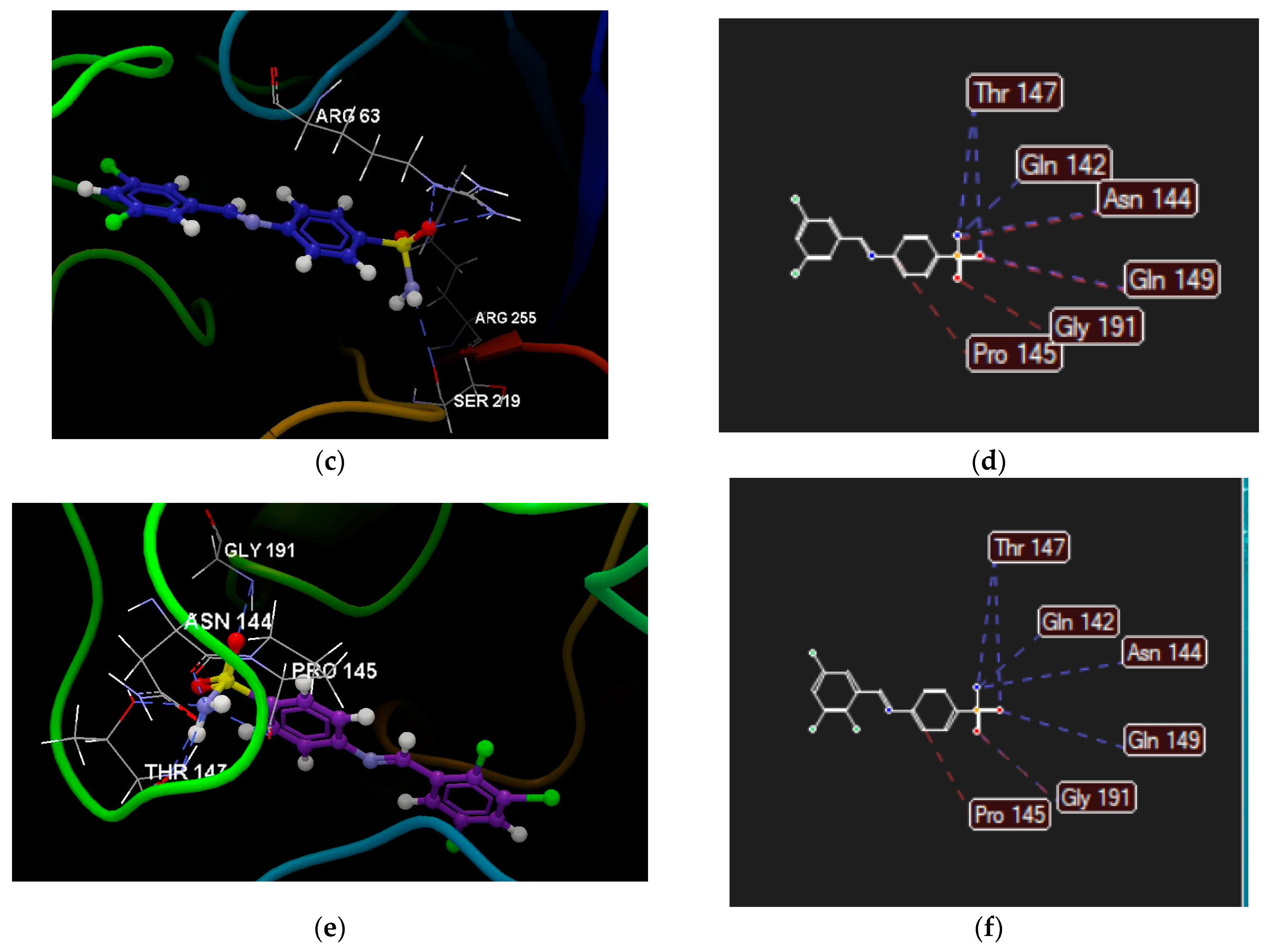
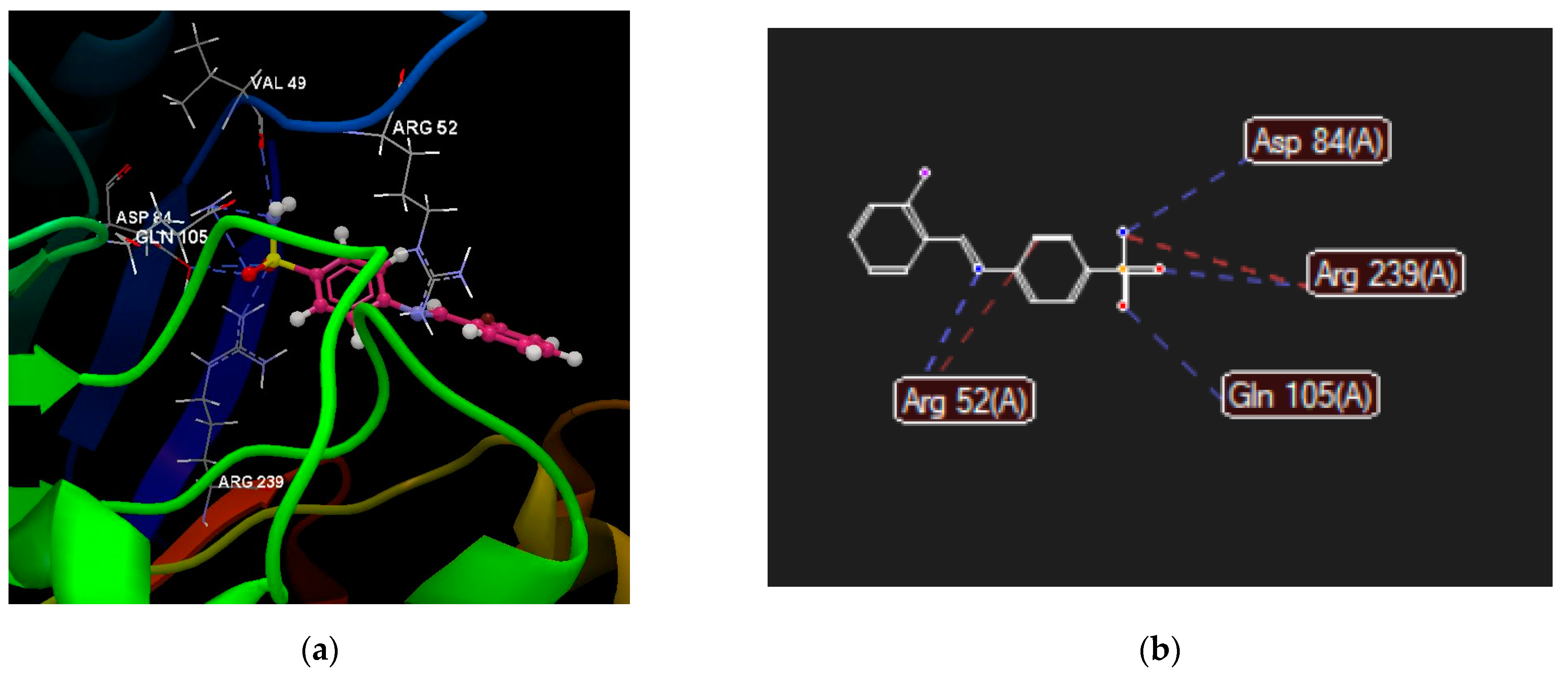
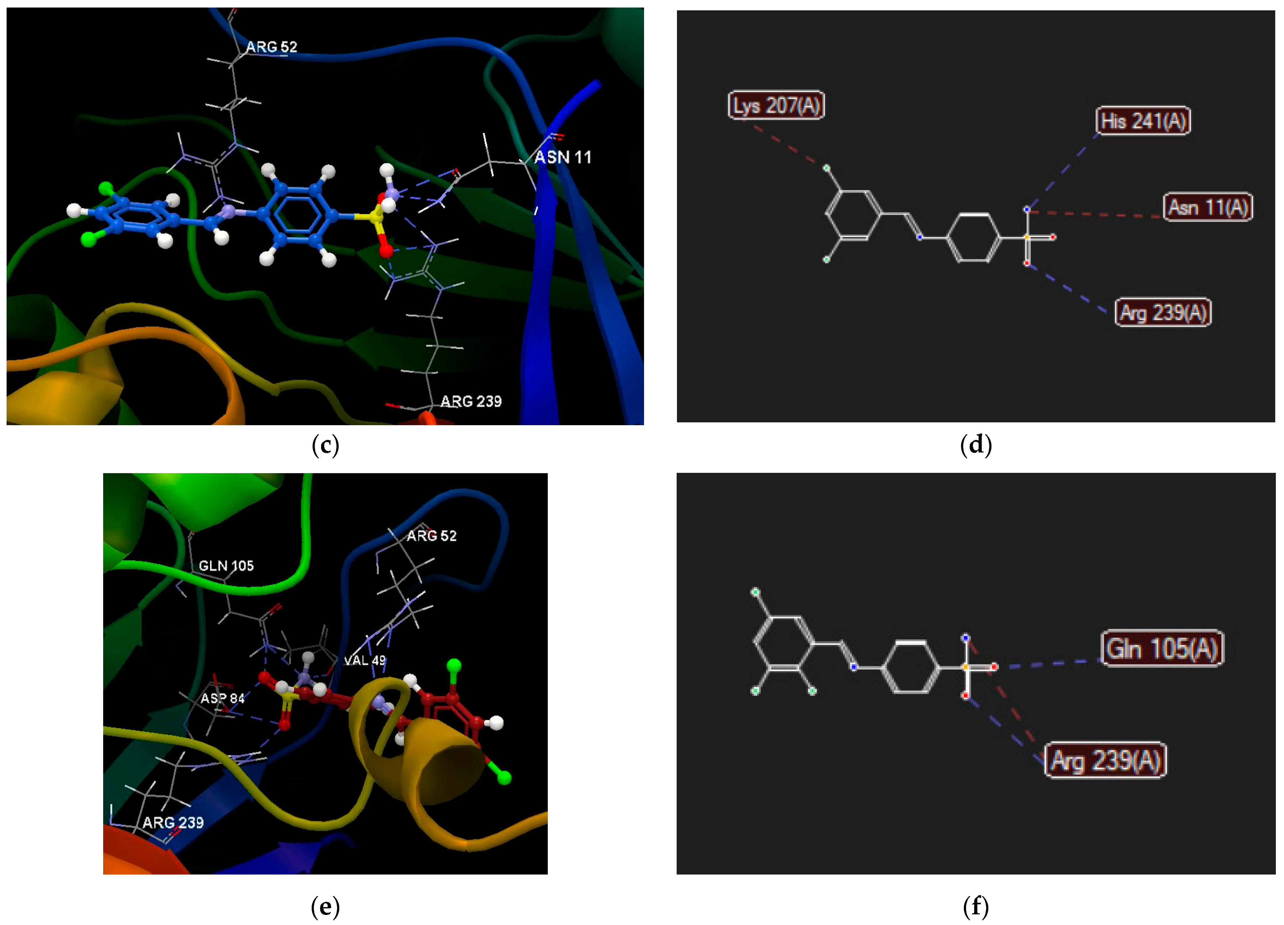
2.2.4. Molecular Docking

In order to understand the possible mechanisms of antimicrobial action for Schiff bases 1ae, a docking study was performed on two dihydropteroate synthetases: E. coli (PDB ID 1AJ0), a ternary complex with sulfanilamide (Figure S26a) [35], and S. aureus (PDB ID 1AD4), complex with the natural ligand hydroxymethylene-pterin-pyrophosphate (Figure S26b) [36].
The calculated docking scores of Schiff bases 1ae are presented comparatively in Figure 3.
For the most potent antibacterial compounds (1b, 1d, 1e), their interactions with the active site of the amino acid residues of selected enzymes are shown in Figure 4 and Figure 5.

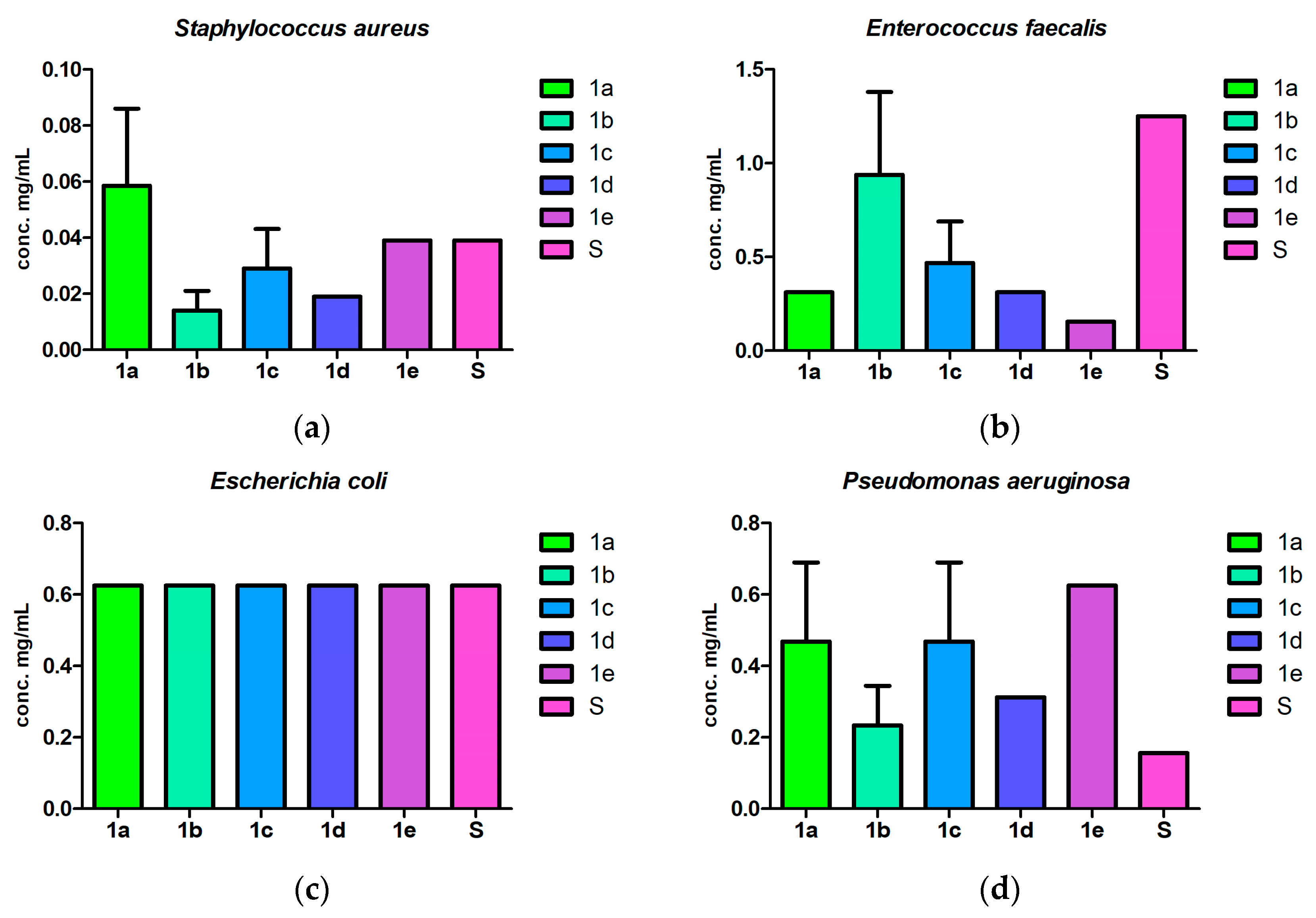
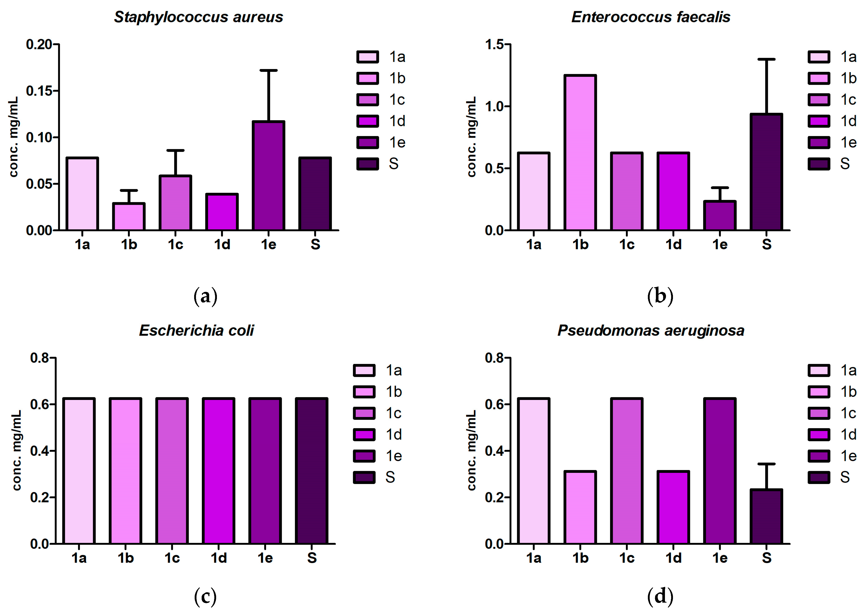
2.3. Antibacterial and Antibiofilm Screening

An antibacterial assay of the synthesized Schiff bases was performed both qualitatively (agar diffusion method, Figure S30) and quantitively (minimum inhibitory concentration, MIC) on Gram-positive and Gram-negative bacterial strains (Figure 6, Table S5). Their antibiofilm potential was determined measuring the MIC values of the bacterial adherence to inert substrata (Figure 7, Table S5).
Compounds 1c and 1d exhibited limited inhibition zone formation around the inoculation site for the tested Gram-positive bacteria, whereas the other derivatives had no impact on bacterial proliferation (Figure S30).
Regarding quantitative evaluation, S. aureus was the most susceptible tested strain, with MIC values from 0.009 to 0.078 mg/mL. The Schiff bases also exhibited more significant activity than sulfanilamide against E. faecalis, with compound 1e being the most potent (MIC 0.156 mg/mL). In the case of Gram-negative bacteria, their activity was reduced. All tested substances inhibited E. coli at the same concentration, 0.625 mg/mL, whereas, on P. aeruginosa, the Schiff bases were less active than their parent amine (Figure 6, Table S5).
The tested compounds were able to inhibit bacterial adherence at concentrations equal to or two times higher than their MIC. A trend was conserved, with S. aureus being the most susceptible (Figure 7, Table S5).

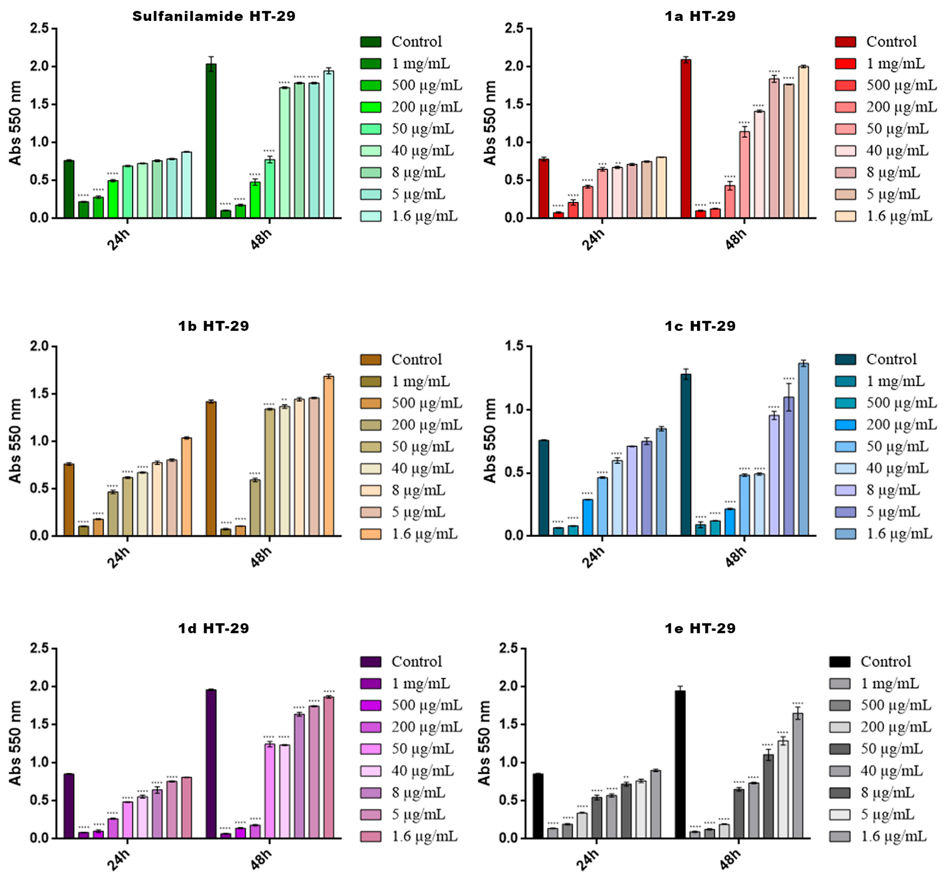
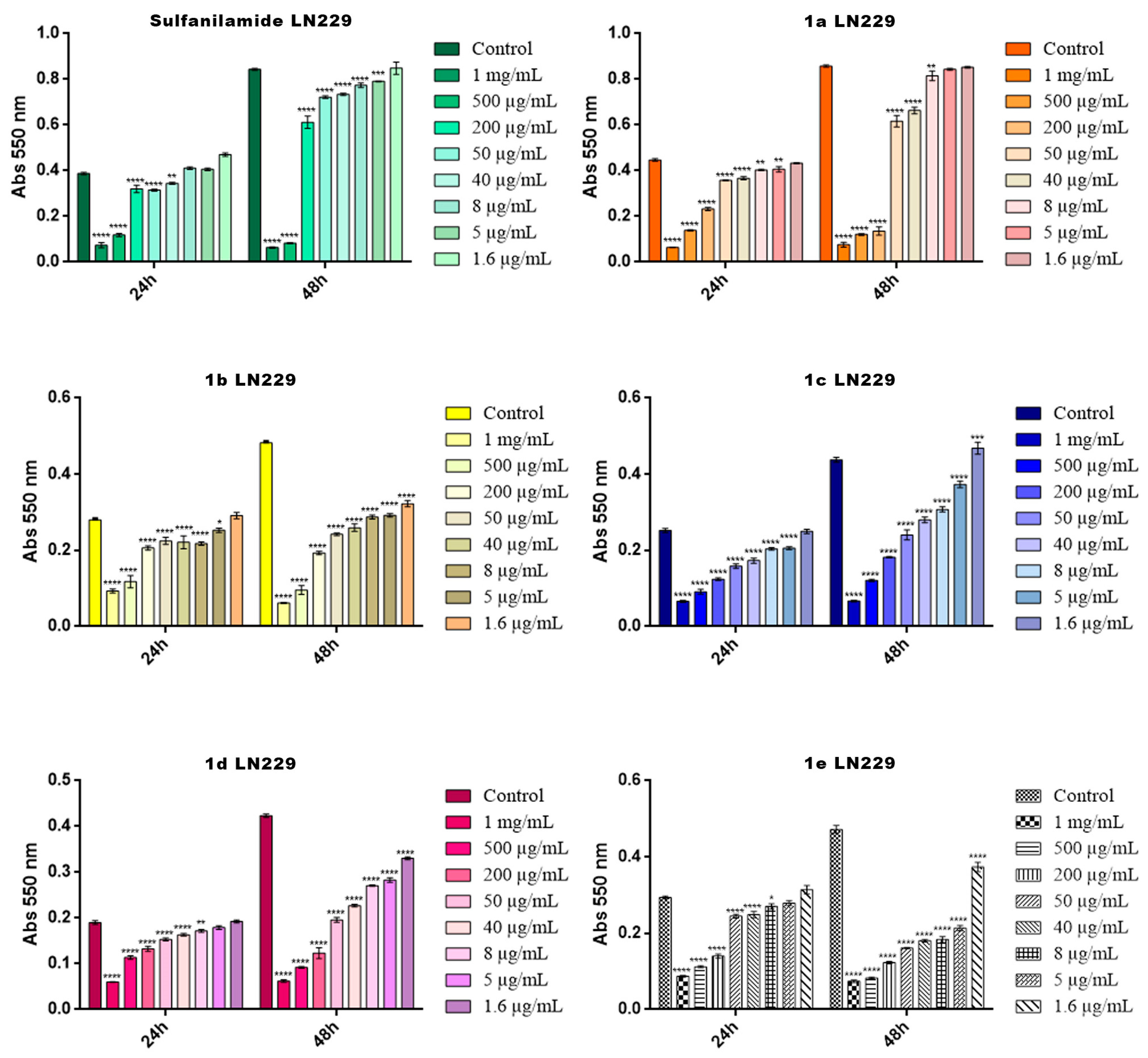
2.4. Cytotoxicity Screening

The anticancer potential of the new compounds was investigated, in terms of their cytotoxicity, on HT-29 adenocarcinoma cells and LN229 glioblastoma cells (Table S6). To assess this, a spectrophotometric MTT assay was employed and the resulting data were statistically analyzed and graphically represented, as seen in Figure 8 and Figure 9, using GraphPad Prism 6.0. Software.
Table S6 presents the first (lowest) doses that induced a statistically significant decrease in the cells’ viability in both cell lines at 24 h and 48 h.

3. Discussion

Compounds 1ae comply with drug-likeness rules (Table 2), indicating their potential drug effect and good bioavailability. Bioavailability is a crucial indicator in drug absorption. All compounds recorded a bioavailability of 0.55, which means that, at a physiological pH, 55% of the compound is expected to reach circulation in an unchanged or active form [37].
The summary of pharmacokinetic profiles presented in Table 3 revealed that (i) all compounds 1ae exhibited excellent intestinal absorption, (ii) a very good BBB permeability (log BBB varied from −0.90 to −0.55) and (iii) practical CNS permeability, varying from −2.16 to −1.94. Among the analyzed Schiff bases, compound 1b presents suitable permeability for the BBB, while derivative 1e showed good permeability in the nervous system and BBB.
The pharmacogenomic profiles of derivatives 1ae (Table 3) can be summarized as follows: (i) none of the compounds interact with CYP2D6, (ii) only compound 1a may be a possible inhibitor of CYP2C9 and (iii) all derivatives may inhibit CYP3A4 and CYP1A2, but they are not substrates. Significant results were recorded for the elimination phase. Having acidic sulfonamide group, compounds 1ae are inhibitors of the hepatic organic anion transporters (OATP1B1, OATP1B3), but not of the renal (OCT2) or hepatic cation transporters (OCT1).
The toxicity profiles of the compounds (Table 4) indicate that (i) all compounds presented affinities for aromatase and estrogen receptors; (ii) all but 1d showed an affinity for the glucocorticoid receptor; (iii) all compounds could be associated with mitochondrial and hepatic toxicities, but not reproductive toxicity or eye irritation; and (iv) all halogenated derivatives are indicated to be toxic to the respiratory system. The p-phenoxy derivative 1a is the only one associated with carcinogenicity, mutagenic toxicity, androgen receptor binding and human ether-a-go-go-related gene inhibition.
Regarding their predicted pharmacodynamic profiles (Table 5), the most common molecular targets for Schiff bases 1ae are the endoplasmic reticulum-associated amyloid beta-peptide-binding protein (ERAB) (1ad), carbonic anhydrase XII (CA XII), carbonic anhydrase IX (CA IX) (1c1e) and cyclooxygenase-2 (COX-2) (1ac, 1e). These results coincide with the usual targets of sulfonamide-based molecules. Other possibly indicated targets were the dual specificity protein kinase CLK4 for compound 1e, cyclin-dependent kinase 1 for derivative 1c and transcription intermediary factor 1-alpha for 1a and 1b.
Regarding microbiological screening, the isomers 1c and 1d were the only ones that showed a small inhibition zone around the inoculum for Gram-positive bacteria on the disc diffusion test (Figure S34). No effect was observed on the Gram-negative representatives.
According to the antibacterial assay (Figure 6, Table S5), the Schiff bases exhibited selectivity towards S. aureus. Halogen substitution appears to be important for their antimicrobial activity. Derivative 1b ((E)-4-{[(2-bromophenyl)methylidene]amino}benzene-1-sulfonamide) was the most potent of the series (MIC 0.014 mg/mL), followed by 1d (4-{[(3,5-dichlorophenyl)methylidene]amino}benzene-1-sulfonamide) (MIC 0.019 mg/mL). On the other hand, E. faecalis was more susceptible to the action of Schiff bases than to that of sulfanilamide. Compound 1e ((E)-4-{[(2,3,5-trichlorophenyl)methylidene]amino}benzene-1-sulfonamide) was the most active in this case (MIC 0.156 mg/mL). The phenoxy derivative 1a showed only low activity on S. aureus and a moderate effect on E. faecalis.
None of the compounds, or sulfanilamide, had any effect on E. coli. For P. aeruginosa, the derivatives were all less active than sulfanilamide (MIC 0.156 mg/mL), with 1b and 1d performing slightly better than the rest (MIC 0.234 mg/mL and 0.312 mg/mL, respectively).
Similar results were observed for the antibiofilm assay (Figure 7, Table S5). Schiff bases 1b (MIC 0.029 mg/mL) and 1d (MIC 0.039 mg/mL) were more potent against S. aureus’s bacterial adherence than sulfanilamide (MIC 0.078 mg/mL). Compound 1e was active against the E. faecalis biofilm (MIC 0.234 mg/mL), surpassing its parent amine (MIC 0.938 mg/mL). In contrast, on the P. aeruginosa biofilm, sulfanilamide performed better than its derivatives (MIC 0.234 mg/mL), with 1b and 1d being the most potent (MIC 0.312 mg/mL).
The preference of the compounds for Gram-positive bacteria over Gram-negative ones is not surprising. P. aeruginosa is naturally resistant to sulfonamides as it contains efflux pumps [3]. Other Gram-negative bacteria usually acquire resistance through mutations in folP, the gene responsible for the codification of DHPTS, which leads to altered enzymes that have low or no affinity to sulfonamides, thus bypassing the drug [4,38]. Four types of sulfonamide resistance genes, all plasmid-borne, have been discovered in Gram-negative bacteria: sul1, which is present in the class 1 integron; sul2, which is integron independent; sul3, which may be integron-linked [38,39]; and sul4, which is present in E. coli [40].
The cytotoxicity screening revealed that the Schiff bases generated a significant decrease in cell viability at low doses both on HT-29 colorectal adenocarcinoma cells as well as on LN229 glioblastoma cells, indicating their high cytotoxic potential on these cancer cell lines.
The highest toxicity on HT-29 colorectal adenocarcinoma cells was displayed by compound 1d, which induced a statistically significant decrease in the cells’ viability (****, p < 0.0001) at 5 μg/mL after 24 h of exposure and at 1.6 μg/mL after 48 h of exposure (Table S6, Figure 8). Regarding the LN229 glioblastoma cells, the highest toxicity was displayed by compound 1c, which induced a statistically significant decrease in the cells’ viability (****, p < 0.0001) at 5 μg/mL after 24 h of exposure and at 1.6 μg/mL after 48 h of exposure (Table S6, Figure 9). The same compounds are also indicated to be CA IX and CA XII inhibitors through molecular target prediction. Sulfanilamide was the least cytotoxic in both determinations.
The docking study on E. coli DHPTS revealed the increased binding affinities of Schiff bases 1ae compared to co-crystallized sulfanilamide (Figure 3). The latter interacts with amino acids of the active site primarily through hydrogen bonds: its sulfamoyl group with Arg63 and Ser219 and its amino group with Thr62 (Table S1). Schiff base derivatives form hydrogen bonds using their free sulfamoyl group and steric interactions using their benzene rings, e.g., Pro145 (Figure 4 and Figure 5). Their additional benzene ring can contribute to supplementary interactions with the active site, e.g., the phenoxy group of compound 1a forms hydrogen bonds with Arg63 (Table S2).
In the case of S. aureus DHPTS, the docking scores of our compounds are comparable or lower to those obtained with the natural ligand, pterin-pyrophosphate (Figure 3). Besides their sulfamoyl group, their imine group is also implicated in hydrogen bonds with amino acid Arg52 (Figure 6 and Figure 7). In addition, substituents on the second bezene ring contribute to binding to the active site: the phenoxy group forms hydrogen bonds with Arg239 and steric interactions with Asp84 and Met128 (Table S4); the chloro group in the metha position forms steric interactions with Lys207 (Figure 7), while the ortho position is not favorable (1c—the lowest scores). These observations are in correlation with the results obtained by in vivo antimicrobial tests. Even though compound 1a generally had the highest scores among the tested derivatives, it did not exhibit an antibacterial effect.
Combining all of the above, some structure–activity relationships may be drawn. Changing the amino group of the sulfonamide derivatives to azomethine contributes to their antibacterial activity; in some cases, it may even enhance the effect. The imine group is able to form hydrogen bonds with the active site. Halogen substitution proves beneficial for their activity, with the 2-bromo, 3,5-dichloro and 2,3,5-trichloro derivatives demonstrating antibacterial and antibiofilm potential. Additionally, a dichloro substitution on benzene ring appears to enhance their cytotoxicity towards cancer cell lines. Thus, compound 1d presents the most promising antibacterial and anticancer activity. These findings align with results reported in the literature for similar compounds [15,19,22].

4. Materials and Methods

4.1. Chemistry

4.1.1. General Information

All reactions were performed under microwave irradiation using a Biotage® Initiator Classic 2.0 (Biotage, Uppsala, Sweden), in sealed 2–5 mL reaction vials, under magnetic stirring and a very high absorbance level. The reagents and the solvents were purchased from Sigma-Aldrich, USA (4-phenoxybenzaldehyde); Sigma-Aldrich, Buchs, Switzerland (2,6-dichlorobenzaldehyde); Sigma-Aldrich, Schnelldorf, Germany (sulfanilamide); Merck Schuchardt, Hohenbrunn, Germany (2-bromobenzaldehyde, 3,5-dichlorobenzaldehyde, 2,3,5-trichlorobenzaldehyde); Chemical Company S.A., Iași, Romania (ethanol, methanol); and Chimopar Tranding SRL, Bucharest, Romania (glacial acetic acid).
For thin-layer chromatography (TLC), the methodology indicated in reference [41] was followed. Glass TLC plates coated with silica gel 60, 20 cm × 20 cm (Merck, Darmstadt, Germany), were used as the stationary phase, while the mobile phase was a mixture of chloroform–methanol (9:1, v/v) (migration distance of 13 cm). The spots were visualized using an acidified potassium permanganate solution. The reference substances were sulfanilamide, 2,6-dichlorobenzaldehyde, 2,3,5-trichlorobenzaldehyde and 2-bromobenzaldehyde.
The infrared spectra were recorded on a JASCO FT/IR-4200 equipped with ATR PRO 450S (diamond crystal). Absorption maxima were reported in wavenumbers (cm−1), using the range 400–4000 cm−1, with transmittance recorded on the abscissa. The following abbreviations were used: w (weak), m (medium), s (strong), and vs (very strong).
1H NMR and 13C NMR spectra were recorded on a Varian Gemini 300BB instrument (Varian Medical Systems, Palo Alto, CA, USA) operating at two frequencies, 300 MHz for proton and 75 MHz for carbon-13 NMR. The solvent used was DMSO-d6. The chemical shifts are reported in parts per million (ppm, δ scale) (internal standard—tetramethylsilane, (CH3)4Si, TMS) and all coupling constant (J) values are in Hertz (Hz). The following abbreviations were used to explain the multiplicities of the 1H signals: s (singlet), d (doublet), t (triplet), m (multiplet), dd (doublet doublet) and tt (triplet triplet). The data order is the following: for 1H NMR—chemical shifts, multiplicity, coupling constants, number of protons and signal attribution; for 13C NMR—chemical shifts, signal attribution.
Melting points (uncorrected) (m.p.) were determined via an open capillary method using an Electrothermal 9100 apparatus (Bibby Scientific Ltd., Stone, UK).

4.1.2. Synthesis and Characterization of the Compounds

The employed synthesis protocol is an adaptation of one previously described [42]. A clean, dry reaction vial (2–5 mL) with a stir bar was charged with sulfanilamide (1–2 mmol), stoichiometric quantities of benzaldehydes, anhydrous methanol or ethanol (3–3.5 mL) and 1–2 drops of glacial acetic acid. Part of the alcohol was used to disperse the reactants and the rest to wash clean the vial. The vial was capped and placed in the reactor cavity. The mixture was stirred for 5 min and then irradiated at 90 °C for 30 min (level of absorbance—very high). Then, the reaction mixture was cooled on an ice bath and/or left in a refrigerator for several hours. The product was filtered under vacuum and washed with ice-cold ethanol or methanol. In some cases, it was first necessary to evaporate part of the solvent and then to filter. The products were purified via recrystallization, from either ethanol (1bd) or methanol (1a), in the presence of decolorizing carbon.
Thin-layer chromatography was performed to verify the status of the reaction; 0.2% ethanol solutions of the tested mixtures and references were prepared and a migration distance of 13 cm was set. All benzaldehydes migrated with the front, while sulfanilamide had a retention factor of 0.32.

4.2. Computational Strategy

4.2.1. Molecular Modeling of Chemical Structures 1ae

The 3D structure and Simplified Molecular Input Line Entry (SMILES) file of the compounds were obtained for further bioinformatics and cheminformatics analyses using Spartan’20 software (2022) (Wavefunction, Inc., Irvine, CA, USA) [43]. The energies of the structures were minimized using Forcefield MMFF94x with a gradient of 0.05. After minimization, Gasteiger partial charges were applied to all compounds [44].

4.2.2. Assessment of Compounds’ Drug- and Lead-Likeness Features

To evaluate their drug- and lead-likeness feature, the compounds 1ae were computed, under medicinal chemistry rules, to Lipinski [45], Ghose [46], Veber [47] and Egan [48] filters using the SwissADME web service [49]. These rules impose the following criteria: (i) the Lipinski rule—the molecular weight should not be more than 500 g/mol, the partition coefficient between octanol and water (Log P(o/w) not more than 5, the number of hydrogen bond acceptors should not exceed 10 and the number of hydrogen bond donors should not exceed 5; (ii) the Ghose filter—the molecular weight should be between 160 and 480 g/mol, the Log P(o/w) should be between −0.4 and 5.6, the molar refractivity should be between 40 and 130 m3/mol and the number of atoms should be between 20 and 70; (iii) the Veber rule—the number of rotatable bonds not more than 10 and the total polar surface area should not be greater than 140 Å2; and (iv) the Egan filter—the Log P(o/w) should not be more than 5.88 and the total polar surface area should not be greater than 131 Å2.

4.2.3. Computational Pharmacokinetics, Pharmacogenomics and Toxicological Profiles of Sulfanilamide Schiff Bases 1ae

The SMILES files of compounds 1ae were used to predict their pharmacokinetic and pharmacogenomic profiles (absorption, distribution, excretion) using the pkCSM [34] and admetSAR2.0 database [33]. From a large set of pharmacokinetic and pharmacogenomic items, the most relevant were chosen, expressed as numerical and categorical variables: (i) intestinal absorption; (ii) blood–brain barrier (BBB) permeability; (iii) central nervous system permeability (Log CNS); plasma protein binding; and inhibition of the OCT receptor at the renal level. The potential of the compounds 1ae to serve as inhibitors or substrates of the cytochromes involved in the metabolism of many drugs, such as CYP2D6, CYP3A4, CYP1A2, CYP2C19 and CYP2C9, was investigated. A significant number of items representing toxicity were analyzed: ames (mutagenesis), carcinogenicity, toxicity to different species (crustacea, bees, fish), nephrotoxicity, hepatotoxicity, cardiotoxicity, mitochondrial toxicity and toxicity to nuclear receptors.

4.2.4. Computational Pharmacodynamic Profiles of Molecules 1ae

The pharmacodynamic features and possible molecular mechanisms by which compounds 1ae modulated the human enzymes involved in different disorders were investigated using bioinformatics. In this context, the SuperPred database [50] was used to establish the probability of binding of Schiff bases 1ae to human enzymes based on logistic regression machine learning models. The binding data and resulting model accuracy are based on 646 human targets. In addition, the therapeutic indications of the predicted targets were identified.

4.2.5. Molecular Docking

Two software were used for this assay: the CLC Drug Discovery Workbench 2.4 (2015) [51] and Molegro Virtual Docker (2019) [52]. The structures of the compounds were prepared using Spartan’20 software [53]. The E isomer of the Schiff bases was chosen for docking. The protein structures were imported from the Protein Data Bank (http://www.rcsb.org/ (accessed on 3 January 2024).
The docking protocol was carried out according to each software’s requirements. It can be summarized as follows: extraction of the co-crystallized ligand, identification of the binding site and binding pocket, redocking of the ligand and project validation. The hydrogen bonds between co-crystallized and amino acids’ residues were identified and the group of interactions was established. The new ligands were then introduced into the project, and their interactions and docking scores were determined.

4.3. Antibacterial and Antibiofilm Bioactivity Screening

4.3.1. General Information

In order to assess the antibacterial and antibiofilm activity of the tested compounds, the following assays were performed: a qualitative evaluation of their antibacterial activity (agar diffusion method) and quantitative evaluations of minimum inhibitory concentration (MIC) and their minimum inhibitory concentrations against bacterial adherence to inert substrata. Four representative bacterial strains were selected: Escherichia coli ATCC 25922, Pseudomonas aeruginosa ATCC 27853 (Gram-negative), Enterococcus faecalis ATCC 29212 and Staphylococcus aureus ATCC 25923 (Gram-positive).

4.3.2. Methods

The qualitative evaluation of the compounds’ antibacterial activity was conducted using the agar diffusion method, following CLSI (Clinical and Laboratory Standard Institute) guidelines. A 0.5 McFarland inoculum was dispersed on Mueller Hinton agar plates, and 5 µL of each compound (at a concentration of 10 mg/mL in DMSO) was placed on the agar surface. Following overnight incubation at 37 °C, the antibacterial activity was quantified as the inhibition zone surrounding the area where the compound was applied, and its diameter was measured using a ruler.
The quantitative evaluation of antibacterial activity was conducted as follows: Serial binary dilutions of the test compounds were prepared in 96-well plates, resulting in concentrations ranging from 5 to 0.009 mg/mL. A bacterial suspension of 106 CFU/mL (colonies forming units) was added. Following overnight incubation at 37 °C, their MICs were determined as the lowest concentration that inhibited bacterial growth, by reading their optical densities at 620 nm using a Multiskan FC Thermo Scientific spectrophotometer (Thermo Scientific, Waltham, MA, USA). The assay was performed in duplicate, and the results were presented as the mean ± standard deviation (SD).
To evaluate the antibiofilm activity of the compounds, after determining their MICs, the contents of the 96-well plates were discarded, and the wells were washed three times with phosphate-buffered saline. The biofilms were then fixed with methanol, stained with a 1% crystal violet solution for 20 min and treated with a 33% acetic acid solution for 15 min. Their absorbance at 492 nm was measured using a plate-reading spectrophotometer (Multiskan FC Thermo Scientific). This assay was also performed in duplicate, and the results were presented as the mean ± SD.

4.4. In Vitro Cytotoxicity on Cancer Cells

4.4.1. General Information

The cytotoxic potential of the compounds on cancer cells was investigated in vitro by evaluating the cells’ viability after 24 h and 48 h of exposure to several concentrations of the compounds. For this, the following two cancer cell lines were employed: HT-29 (ATCC® HTB-38™)—colorectal adenocarcinoma cells and LN229 (ATCC® CRL-2611™)—glioblastoma cells. These cells were grown in Dulbecco’s modified Eagle medium (DMEM), supplemented with 10% FBS and 1% penicillin/streptomycin mixture (10,000 units/mL penicillin and 10 mg/mL streptomycin), in standard culturing conditions (37 °C, humidified atmosphere of 80RH and 5% CO2).

4.4.2. Methods

Cell viability was quantified by an MTT spectrophotometric assay. Initially the cells were seeded at a density of 103 cells/cm2 in flat-bottomed 96 well plates and allowed to adhere for 24 h in complete culture medium under standard culture conditions. The following day, the culture medium was replaced with treatments consisting of the following concentrations of compounds prepared in complete culture medium: 1 mg/mL; 500 μg/mL; 200 μg/mL; 50 μg/mL; 40 μg/mL; 8 μg/mL; 5 μg/mL; and 1,6 μg/mL. Fresh complete culture medium was added on control monolayers. After 24 h and 48 h, respectively, the treatments were removed and the monolayers were incubated at 37 °C for 4 h with a 1 mg/mL MTT (3-(4,5-dimethilthiazol-2-il)-2,5-dipheniltetrazolium bromide) solution freshly prepared in serum-free DMEM. The resulting formazan crystals formed by the metabolically active cells were solubilized in 2-propanol, and the absorbance of the resulting solution was determined at 550 nm using a FlexStation III multimodal reader. The spectrophotometric data were statistically analyzed by applying a two-way ANOVA and Bonferroni test and graphically illustrated using GraphPad Prism 6.0. All the experiments were conducted in triplicate and the results are represented as the mean of three independent experiments (n = 3). All the data are expressed as mean ± standard error of the mean. A p-value of ≤0.05 was considered statistically significant.

5. Conclusions

A series of five Schiff base derivatives of sulfanilamide was successfully synthesized using microwave irradiation. All the derivatives comply with drug-likeness rules, presenting acceptable predicted pharmacokinetic and toxicologic properties. Proteins such as CA IX, CA XII, COX-2 and ERAB were identified as possible molecular targets of compounds 1ae.
The Schiff bases were evaluated for their antibacterial, antibiofilm and cytotoxicity activity. Gram-positive bacteria were susceptible to the activity of Schiff bases in all assays, whereas Gram-negative strains remained unaffected. The halogenated Schiff bases exhibited better activity than sulfanilamide. Compounds 1b and 1d were the most potent against S. aureus’s growth and bacterial adherence, whereas derivative 1e showed the greatest inhibition of E. faecalis’s planktonic and sessile forms.
All Schiff bases presented a statistically significant reduction of HT-29 colorectal adenocarcinoma cells and LN229 glioblastoma cells’ viability, with higher levels of cytotoxicity on the LN229 glioblastoma cells. Moreover, compound 1d exerted the highest cytotoxic effect on HT-29 colorectal adenocarcinoma cells, while compound 1c exhibited the highest cytotoxicity towards LN229 glioblastoma cells.
This research aligns with our objective of developing new antimicrobial and antibiofilm agents. The identification of these promising molecules encourages further exploration of the valuable biological potential of sulfonamide-derived Schiff bases.

Supplementary Materials

The following supporting information can be downloaded at https://www.mdpi.com/article/10.3390/ph17040405/s1, Figure S1: NMR atom numbering in molecules 1ae; Figure S2: FT-IR spectrum of 1a; Figure S3: 1H-NMR spectrum of 1a; Figure S4: 13C-NMR spectrum of 1a; Figure S5: COSY spectrum (Correlated Spectroscopy) of 1a; Figure S6: COSY spectrum (Correlated Spectroscopy) of 1a; Figure S7: HSQC spectrum (Heteronuclear Single Quantum Coherence) of 1a; Figure S8: HSQC spectrum of 1a; Figure S9: FT-IR spectrum of 1b; Figure S10: 1H-NMR spectrum of 1b; Figure S11: 13C-NMR spectrum of 1b; Figure S12: FT-IR spectrum of 1c; Figure S13: 1H-NMR spectrum of 1c; Figure S14: 13C-NMR spectrum of 1c; Figure S15: FT-IR spectrum of 1d; Figure S16: 1H-NMR spectrum of 1d; Figure S17: 13C-NMR spectrum of 1d; Figure S18: HSQC spectrum of 1d; Figure S19: HSQC spectrum of 1d; Figure S20: HSQC spectrum of 1d; Figure S21: HSQC spectrum of 1d; Figure S22: HSQC spectrum of 1d; Figure S23: FT-IR spectrum of 1e; Figure S24: 1H-NMR spectrum of 1e; Figure S25: 13C-NMR spectrum of 1e; Figure S26: The crystal structures of the ternary complex of E. coli dihydropteroate synthase (1AJ0) (a) and of S. aureus dihydropteroate synthase (1AD4) (b); Figure S27: TUBE 3D structures of compounds 1ae. The numbering is generated by Spartan software; Figure S28: Group interactions of selected compounds 1b, 1d, 1e with the active site of 1AJ0; Figure S29: Group interactions of selected compounds 1b, 1d, 1e with the active site of 1AD4 (chain A); Figure S30: Qualitative evaluation of the antimicrobial activity of the sulfanilamide-derived compounds; Table S1: Docking scores of selected ligands in the active site of 1AJ0 (CLC Drug Discovery Workbench); Table S2: Docking scores of selected ligands in the active site of 1AJ0 (Molegro Virtual Docker); Table S3: Docking scores of selected ligands in the active site of 1AD4 (CLC Drug Discovery Workbench); Table S4: Docking scores of selected ligands in the active site of 1AJ0 (Molegro Virtual Docker); Table S5: Presentation of minimum inhibitory concentrations for antibacterial activity and bacterial adherence assay of Schiff bases 1ae; Table S6: Presentation of the lowest doses required to induce a statistically significant response in terms of a decrease in HT-29 and LN229 cells’ viability after 24 h and 48 h of treatment with Schiff base 1ae.

Author Contributions

Conceptualization, M.C., D.C.N. and C.L. (Carmen Limban); methodology, M.C., D.C.N., C.L. (Carmen Limban), C.D., A.-M.C., G.A.G., M.P., M.S., C.L. (Cristina Larion), S.A., C.M., M.-C.C., A.D., A.H., B.G. and L.P.; software, S.A. and L.P.; validation, G.A.G., M.P., M.S., C.L. (Cristina Larion), A.H., B.G. and L.P.; formal analysis, G.A.G., M.P., M.S., C.L. (Cristina Larion), A.H. and B.G.; investigation, M.C., D.C.N., C.L. (Carmen Limban), C.D., A.-M.C., G.A.G., M.P., M.S., C.L. (Cristina Larion), S.A., C.M., M.-C.C., A.D., A.H., B.G. and L.P.; resources, D.C.N., C.D., A.-M.C., G.A.G., M.P., S.A., A.H., B.G. and L.P.; data curation, M.C.; writing—original draft preparation, M.C., C.D., G.A.G., M.P., S.A., C.M., A.H., B.G. and L.P.; writing—review and editing, M.C., D.C.N., C.L. (Carmen Limban) and A.-M.C.; visualization, M.C., G.A.G., M.P., S.A., C.M., A.H., B.G. and L.P.; supervision, D.C.N. and C.L. (Carmen Limban); funding acquisition, M.C., D.C.N., M.-C.C., A.D., A.H. and B.G. All authors have read and agreed to the published version of the manuscript.

Funding

This research was funded by the Ministry of Research, Innovation and Digitization, CNCS UEFISCDI, grant number 104TE/2022—PN-III-P1-1.1-TE-2021-1660 within PNCDI III.

Institutional Review Board Statement

Not applicable.

Informed Consent Statement

Not applicable.

Data Availability Statement

Data is contained within the article and Supplementary Materials.

Acknowledgments

NUCLEU Program, which is implemented with the support of MCDI, project no. PN 23-28 01 01.

Conflicts of Interest

The authors declare no conflicts of interest.

References

  1. New WHO Report Highlights Progress, but Also Remaining Gaps, in Ensuring a Robust Pipeline of Antibiotic Treatments to Combat Antimicrobial Resistance (AMR). Available online: https://www.who.int/news/item/15-05-2023-new-who-report-highlights-progress-but-also-remaining-gaps-in-ensuring-a-robust-pipeline-of-antibiotic-treatments-to-combat-antimicrobial-resistance-(amr) (accessed on 23 May 2023).
  2. Høiby, N.; Bjarnsholt, T.; Moser, C.; Bassi, G.L.; Coenye, T.; Donelli, G.; Hall-Stoodley, L.; Holá, V.; Imbert, C.; Kirketerp-Møller, K.; et al. ESCMID* Guideline for the Diagnosis and Treatment of Biofilm Infections 2014. Clin. Microbiol. Infect. 2015, 21, S1–S25. [Google Scholar] [CrossRef] [PubMed]
  3. Jorge, P.; Magalhães, A.P.; Grainha, T.; Alves, D.; Sousa, A.M.; Lopes, S.P.; Pereira, M.O. Antimicrobial Resistance Three Ways: Healthcare Crisis, Major Concepts and the Relevance of Biofilms. FEMS Microbiol. Ecol. 2019, 95, fiz115. [Google Scholar] [CrossRef] [PubMed]
  4. Darby, E.M.; Trampari, E.; Siasat, P.; Gaya, M.S.; Alav, I.; Webber, M.A.; Blair, J.M.A. Molecular Mechanisms of Antibiotic Resistance Revisited. Nat. Rev. Microbiol. 2023, 21, 280–295. [Google Scholar] [CrossRef]
  5. Ceri, H.; Gorman, S.P.; Gilmore, B.F. Microbial Biofilms: Consequences for Health. In Hugo and Russell’s Pharmaceutical Microbiology; Denyer, S.P., Hodges, N., Gorman, S.P., Gilmore, B.F., Eds.; Wiley-Blackwell: Hoboken, NJ, USA, 2011; pp. 121–130. ISBN 9781444330632. [Google Scholar]
  6. Patrick, G.L. Antibacterial Agents. In An Introduction to Medicinal Chemistry; Oxford University Press: Oxford, UK, 2017; pp. 425–432. ISBN 9780198749691. [Google Scholar]
  7. Werth, B.J. Sulfonamides. Available online: https://www.msdmanuals.com/professional/infectious-diseases/bacteria-and-antibacterial-drugs/sulfonamides?query=sulfonamideantibiotics (accessed on 23 February 2023).
  8. Mercer, M.A. Sulfonamides and Sulfonamide Combinations Use in Animals. Available online: https://www.msdvetmanual.com/pharmacology/antibacterial-agents/sulfonamides-and-sulfonamide-combinations-use-in-animals (accessed on 23 February 2023).
  9. Kaye, K.S.; Gales, A.C.; Dubourg, G. Old Antibiotics for Multidrug-Resistant Pathogens: From in Vitro Activity to Clinical Outcomes. Int. J. Antimicrob. Agents 2017, 49, 542–548. [Google Scholar] [CrossRef]
  10. Younus, H.A.; Saleem, F.; Hameed, A.; Al-Rashida, M.; Al-Qawasmeh, R.A.; El-Naggar, M.; Rana, S.; Saeed, M.; Khan, K.M. Part-II: An Update of Schiff Bases Synthesis and Applications in Medicinal Chemistry-a Patent Review (2016–2023). Expert Opin. Ther. Pat. 2023, 33, 841–864. [Google Scholar] [CrossRef]
  11. Ceramella, J.; Iacopetta, D.; Catalano, A.; Cirillo, F.; Lappano, R.; Sinicropi, M.S. A Review on the Antimicrobial Activity of Schiff Bases: Data Collection and Recent Studies. Antibiotics 2022, 11, 191. [Google Scholar] [CrossRef]
  12. Coandă, M.; Limban, C.; Nuță, D.C. Small Schiff Base Molecules—A Possible Strategy to Combat Biofilm-Related Infections. Antibiotics 2024, 13, 75. [Google Scholar] [CrossRef]
  13. Hameed, A.; al-Rashida, M.; Uroos, M.; Abid Ali, S.; Khan, K.M. Schiff Bases in Medicinal Chemistry: A Patent Review (2010–2015). Expert Opin. Ther. Pat. 2017, 27, 63–79. [Google Scholar] [CrossRef]
  14. Zafar, W.; Sumrra, S.H.; Hassan, A.U.; Chohan, Z.H. A Review on ‘Sulfonamides’: Their Chemistry and Pharmacological Potentials for Designing Therapeutic Drugs in Medical Science. J. Coord. Chem. 2023, 76, 546–580. [Google Scholar] [CrossRef]
  15. Krátký, M.; Konečná, K.; Janoušek, J.; Janďourek, O.; Maixnerová, J.; Kalivodová, S.; Trejtnar, F.; Vinšová, J. Sulfonamide-Salicylaldehyde Imines Active against Methicillin- and Trimethoprim/Sulfonamide-Resistant Staphylococci. Future Med. Chem. 2021, 13, 1945–1962. [Google Scholar] [CrossRef]
  16. Krátký, M.; Dzurková, M.; Janoušek, J.; Konečná, K.; Trejtnar, F.; Stolaříková, J.; VinŠová, J. Sulfadiazine Salicylaldehyde-Based Schiff Bases: Synthesis, Antimicrobial Activity and Cytotoxicity. Molecules 2017, 22, 1573. [Google Scholar] [CrossRef]
  17. Krátký, M.; Vinšová, J.; Volková, M.; Buchta, V.; Trejtnar, F.; Stolaříková, J. Antimicrobial Activity of Sulfonamides Containing 5-Chloro-2- Hydroxybenzaldehyde and 5-Chloro-2-Hydroxybenzoic Acid Scaffold. Eur. J. Med. Chem. 2012, 50, 433–440. [Google Scholar] [CrossRef]
  18. Mondal, S.; Mandal, S.M.; Mondal, T.K.; Sinha, C. Structural Characterization of New Schiff Bases of Sulfamethoxazole and Sulfathiazole, Their Antibacterial Activity and Docking Computation with DHPS Protein Structure. Spectrochim. Acta—Part A Mol. Biomol. Spectrosc. 2015, 150, 268–279. [Google Scholar] [CrossRef]
  19. Krátký, M.; Konečná, K.; Šimková, A.; Janďourek, O.; Maixnerová, J.; Stolaříková, J.; Vejsová, M.; Voxová, B.; Trejtnar, F.; Vinšová, J. Improving the Antimicrobial Activity of Old Antibacterial Drug Mafenide: Schiff Bases and Their Bioactivity Targeting Resistant Pathogens. Future Med. Chem. 2023, 15, 255–274. [Google Scholar] [CrossRef]
  20. Sarikaya, B.; Ceruso, M.; Carta, F.; Supuran, C.T. Inhibition of Carbonic Anhydrase Isoforms I, II, IX and XII with Novel Schiff Bases: Identification of Selective Inhibitors for the Tumor-Associated Isoforms over the Cytosolic Ones. Bioorganic Med. Chem. 2014, 22, 5883–5890. [Google Scholar] [CrossRef] [PubMed]
  21. Durgun, M.; Turkmen, H.; Ceruso, M.; Supuran, C.T. Synthesis of 4-Sulfamoylphenyl-Benzylamine Derivatives with Inhibitory Activity against Human Carbonic Anhydrase Isoforms I, II, IX and XII. Bioorg. Med. Chem. 2016, 24, 982–988. [Google Scholar] [CrossRef]
  22. Koyuncu, I.; Tülüce, Y.; Slahaddin Qadir, H.; Durgun, M.; Supuran, C.T. Evaluation of the Anticancer Potential of a Sulphonamide Carbonic Anhydrase IX Inhibitor on Cervical Cancer Cells. J. Enzyme Inhib. Med. Chem. 2019, 34, 703–711. [Google Scholar] [CrossRef]
  23. Taslimi, P.; Işık, M.; Türkan, F.; Durgun, M.; Türkeş, C.; Gülçin, İ.; Beydemir, Ş. Benzenesulfonamide Derivatives as Potent Acetylcholinesterase, α-Glycosidase, and Glutathione S-Transferase Inhibitors: Biological Evaluation and Molecular Docking Studies. J. Biomol. Struct. Dyn. 2020, 39, 5449–5460. [Google Scholar] [CrossRef]
  24. Işık, M.; Demir, Y.; Durgun, M.; Türkeş, C.; Necip, A.; Beydemir, Ş. Molecular Docking and Investigation of 4-(Benzylideneamino)- and 4-(Benzylamino)-Benzenesulfonamide Derivatives as Potent AChE Inhibitors. Chem. Pap. 2020, 74, 1395–1405. [Google Scholar] [CrossRef]
  25. Hamad, A.; Khan, M.A.; Rahman, K.M.; Ahmad, I.; Ul-Haq, Z.; Khan, S.; Shafiq, Z. Development of Sulfonamide-Based Schiff Bases Targeting Urease Inhibition: Synthesis, Characterization, Inhibitory Activity Assessment, Molecular Docking and ADME Studies. Bioorg. Chem. 2020, 102, 104057. [Google Scholar] [CrossRef] [PubMed]
  26. Dorai, T.; Sawczuk, I.S.; Pastorek, J.; Wiernik, P.H.; Dutcher, J.P. The Role of Carbonic Anhydrase IX Overexpression in Kidney Cancer. Eur. J. Cancer 2005, 41, 2935–2947. [Google Scholar] [CrossRef] [PubMed]
  27. Waheed, A.; Sly, W.S. Carbonic Anhydrase XII Functions in Health and Disease. Gene 2017, 623, 33–40. [Google Scholar] [CrossRef]
  28. Krungkrai, J.; Supuran, C.T. The Alpha-Carbonic Anhydrase from the Malaria Parasite and Its Inhibition. Curr. Pharm. Des. 2008, 14, 631–640. [Google Scholar] [CrossRef] [PubMed]
  29. Maji, M.; Acharya, S.; Bhattacharya, I.; Gupta, A.; Mukherjee, A. Effect of an Imidazole-Containing Schiff Base of an Aromatic Sulfonamide on the Cytotoxic Efficacy of N,N-Coordinated Half-Sandwich Ruthenium(II) p-Cymene Complexes. Inorg. Chem. 2021, 60, 4744–4754. [Google Scholar] [CrossRef]
  30. Arshad, J.Z.; Tabassum, S.; Kiani, M.S.; Arshad, S.; Hashmi, M.A.; Majeed, I.; Ali, H.; Shah, S.S.A. Anticancer Properties of Ru and Os Half-Sandwich Complexes of N,S Bidentate Schiff Base Ligands Derived from Phenylthiocarbamide. Chem. Asian J. 2023, 18, e202300804. [Google Scholar] [CrossRef] [PubMed]
  31. Koyuncu, I.; Temiz, E.; Durgun, M.; Kocyigit, A.; Yuksekdag, O.; Supuran, C.T. Intracellular PH-Mediated Induction of Apoptosis in HeLa Cells by a Sulfonamide Carbonic Anhydrase Inhibitor. Int. J. Biol. Macromol. 2022, 201, 37–46. [Google Scholar] [CrossRef]
  32. Elie, J.; Vercouillie, J.; Arlicot, N.; Lemaire, L.; Bidault, R.; Bodard, S.; Hosselet, C.; Deloye, J.B.; Chalon, S.; Emond, P.; et al. Design of Selective COX-2 Inhibitors in the (Aza)Indazole Series. Chemistry, In Vitro Studies, Radiochemistry and Evaluations in Rats of a [18F] PET Tracer. J. Enzyme Inhib. Med. Chem. 2019, 34, 1–7. [Google Scholar] [CrossRef]
  33. Yang, H.; Lou, C.; Sun, L.; Li, J.; Cai, Y.; Wang, Z.; Li, W.; Liu, G.; Tang, Y. AdmetSAR 2.0: Web-Service for Prediction and Optimization of Chemical ADMET Properties. Bioinformatics 2019, 35, 1067–1069. [Google Scholar] [CrossRef]
  34. Pires, D.E.V.; Blundell, T.L.; Ascher, D.B. PkCSM: Predicting Small-Molecule Pharmacokinetic and Toxicity Properties Using Graph-Based Signatures. J. Med. Chem. 2015, 58, 4066–4072. [Google Scholar] [CrossRef]
  35. Achari, A.; Somers, D.O.; Champness, J.N.; Bryant, P.K.; Rosemond, J.; Stammers, D.K. Crystal Structure of the Anti-Bacterial Sulfonamide Drug Target Dihydropteroate Synthase. Nat. Struct. Biol. 1997, 4, 490–497. [Google Scholar] [CrossRef]
  36. Hampele, I.C.; D’Arcy, A.; Dale, G.E.; Kostrewa, D.; Nielsen, J.; Oefner, C.; Page, M.G.P.; Schönfeld, H.-J.; Stüber, D.; Then, R.L. Structure and Function of the Dihydropteroate Synthase from Staphylococcus Aureus11Edited by R. Huber. J. Mol. Biol. 1997, 268, 21–30. [Google Scholar] [CrossRef] [PubMed]
  37. Martin, Y.C. A Bioavailability Score. J. Med. Chem. 2005, 48, 3164–3170. [Google Scholar] [CrossRef] [PubMed]
  38. Sköld, O. Sulfonamide Resistance: Mechanisms and Trends. Drug Resist. Updat. 2000, 3, 155–160. [Google Scholar] [CrossRef]
  39. Poey, M.E.; Azpiroz, M.F.; Laviña, M. On Sulfonamide Resistance, Sul Genes, Class 1 Integrons and Their Horizontal Transfer in Escherichia Coli. Microb. Pathog. 2019, 135, 103611. [Google Scholar] [CrossRef]
  40. Razavi, M.; Marathe, N.P.; Gillings, M.R.; Flach, C.F.; Kristiansson, E.; Joakim Larsson, D.G. Discovery of the Fourth Mobile Sulfonamide Resistance Gene. Microbiome 2017, 5, 160. [Google Scholar] [CrossRef]
  41. Poole, C. Thin-Layer Chromatography. In Clarke’s Analysis of Drugs and Poisons; Moffat, A.C., Osselton, M.D., Widdop, B., Watts, J., Eds.; Pharmaceutical Press: London, UK, 2011; pp. 611–634. ISBN 978 0 85369 711 4. [Google Scholar]
  42. Avram, S.; Udrea, A.M.; Nuta, D.C.; Limban, C.; Balea, A.C.; Caproiu, M.T.; Dumitrascu, F.; Buiu, C.; Bordei, A.T. Synthesis and Bioinformatic Characterization of New Schiff Bases with Possible Applicability in Brain Disorders. Molecules 2021, 26, 4160. [Google Scholar] [CrossRef]
  43. Shao, Y.; Molnar, L.F.; Jung, Y.; Kussmann, J.; Ochsenfeld, C.; Brown, S.T.; Gilbert, A.T.B.; Slipchenko, L.V.; Levchenko, S.V.; O’Neill, D.P.; et al. Advances in Methods and Algorithms in a Modern Quantum Chemistry Program Package. Phys. Chem. Chem. Phys. 2006, 8, 3172–3191. [Google Scholar] [CrossRef] [PubMed]
  44. Avram, S.; Movileanu, L.; Mihailescu, D.; Flonta, M.-L. Comparative Study of Some Energetic and Steric Parameters of the Wild Type and Mutants HIV-1 Protease: A Way to Explain the Viral Resistance. J. Cell. Mol. Med. 2002, 6, 251–260. [Google Scholar] [CrossRef]
  45. Lipinski, C.A.; Lombardo, F.; Dominy, B.W.; Feeney, P.J. Experimental and Computational Approaches to Estimate Solubility and Permeability in Drug Discovery and Development Settings. Adv. Drug Deliv. Rev. 2001, 46, 3–26. [Google Scholar] [CrossRef]
  46. Ghose, A.K.; Viswanadhan, V.N.; Wendoloski, J.J. A Knowledge-Based Approach in Designing Combinatorial or Medicinal Chemistry Libraries for Drug Discovery. 1. A Qualitative and Quantitative Characterization of Known Drug Databases. J. Comb. Chem. 1999, 1, 55–68. [Google Scholar] [CrossRef]
  47. Veber, D.F.; Johnson, S.R.; Cheng, H.Y.; Smith, B.R.; Ward, K.W.; Kopple, K.D. Molecular Properties That Influence the Oral Bioavailability of Drug Candidates. J. Med. Chem. 2002, 45, 2615–2623. [Google Scholar] [CrossRef] [PubMed]
  48. Egan, W.J.; Merz, K.M.; Baldwin, J.J. Prediction of Drug Absorption Using Multivariate Statistics. J. Med. Chem. 2000, 43, 3867–3877. [Google Scholar] [CrossRef] [PubMed]
  49. Daina, A.; Michielin, O.; Zoete, V. SwissADME: A Free Web Tool to Evaluate Pharmacokinetics, Drug-Likeness and Medicinal Chemistry Friendliness of Small Molecules. Sci. Rep. 2017, 7, 1–13. [Google Scholar] [CrossRef]
  50. Nickel, J.; Gohlke, B.O.; Erehman, J.; Banerjee, P.; Rong, W.W.; Goede, A.; Dunkel, M.; Preissner, R. SuperPred: Update on Drug Classification and Target Prediction. Nucleic Acids Res. 2014, 42, W26–W31. [Google Scholar] [CrossRef]
  51. QIAGEN Aarhus A/S. CLC Drug Discovery Workbench, Version 2.4; Silkeborgvej 2 Prismet; QIAGEN Aarhus A/S: Aarhus, Denmark, 2015. [Google Scholar]
  52. Molegro Virtual Docker, Version 2019; Molexus IVS, Rørt: Odder, Denmark. 2019. Available online: http://www.molexus.io (accessed on 3 January 2024).
  53. SPARTAN’20, Version 2022; Wavefunction, Inc.: Irvine, CA, USA, 2022; Available online: www.wavefun.com (accessed on 3 January 2024).
Figure 1. Structure–activity relationship of selected sulfonamide-derived Schiff bases’ antimicrobial effect [15,16,17,18,19].
Pharmaceuticals 17 00405 g001
Scheme 1. Preparation of sulfanilamide Schiff base derivatives 1ae. Radical R is: 4-PhO (a); 2-Br (b); 2,6-diCl (c); 3,5-diCl (d); 2,3,5-triCl (e).
Pharmaceuticals 17 00405 sch001
Figure 3. Comparative docking scores of Schiff bases 1ae for 1AJ0 and 1AD4. * Calculated using the CLC Drug Discovery Workbench, ** Calculated using the Molegro Virtual Docker.
Pharmaceuticals 17 00405 g003
Figure 4. Interactions between compounds 1b, 1d, 1e and the amino acids of the active site of E. coli DHPTS (1AJ0). (a) Hydrogen bonds of 1b (CLC Drug Discovery Workbench); (b) hydrogen bonds (blue) and steric interactions (red) of 1b (Molegro Virtual Docker); (c) hydrogen bonds of 1d (CLC Drug Discovery Workbench); (d) hydrogen bonds (blue) and steric interactions (red) of 1d (Molegro Virtual Docker); (e) hydrogen bonds of 1e (CLC Drug Discovery Workbench); (f) hydrogen bonds (blue) and steric interactions (red) of 1e (Molegro Virtual Docker).
Pharmaceuticals 17 00405 g004aPharmaceuticals 17 00405 g004b
Figure 5. Interactions between compounds 1b, 1d, 1e and the amino acids of the active site of S. aureus DHPTS (1AD4). (a) Hydrogen bonds of 1b (CLC Drug Discovery Workbench); (b) hydrogen bonds (blue) and steric interactions (red) of 1b (Molegro Virtual Docker); (c) hydrogen bonds of 1d (CLC Drug Discovery Workbench); (d) hydrogen bonds (blue) and steric interactions (red) of 1d (Molegro Virtual Docker); (e) hydrogen bonds of 1e (CLC Drug Discovery Workbench); (f) hydrogen bonds (blue) and steric interactions (red) of 1e (Molegro Virtual Docker).
Pharmaceuticals 17 00405 g005aPharmaceuticals 17 00405 g005b
Figure 6. Minimum inhibitory concentration (MIC) values of the compounds (mean ± SD) against (a) Staphylococcus aureus ATCC 25923; (b) Enterococcus faecalis ATCC 29212; (c) Escherichia coli ATCC 25922; and (d) Pseudomonas aeruginosa ATCC 27853. Corresponding notations: S (sulfanilamide).
Pharmaceuticals 17 00405 g006
Figure 7. Minimum inhibitory concentrations against bacterial adherence to the inert substrata of the tested compounds (mean ± SD): (a) Staphylococcus aureus ATCC 25923; (b) Enterococcus faecalis ATCC 29212; (c) Escherichia coli ATCC 25922; (d) Pseudomonas aeruginosa ATCC 27853. Corresponding notations: S (Sulfanilamide).
Pharmaceuticals 17 00405 g007
Figure 8. Graphical representation of the HT-29 adenocarcinoma cells’ viability after 24 h and 48 h exposure to sulfanilamide, 1a, 1b, 1c, 1d and 1e treatments at 1 mg/mL, 500 μg/mL, 200 μg/mL, 50 μg/mL, 40 μg/mL, 8 μg/mL, 5 μg/mL and 1.6 μg/mL, as compared to untreated cells (**** = p < 0.0001 sample versus untreated cells; *** = p < 0.001 sample versus untreated cells and ** = p < 0.01 sample versus untreated cells).
Pharmaceuticals 17 00405 g008
Figure 9. Graphical representation of the LN229 glioblastoma cells’ viability after 24 h and 48 h exposure to sulfanilamide, 1a, 1b, 1c, 1d and 1e treatments at 1mg/mL, 500 μg/mL, 200 μg/mL, 50 μg/mL, 40 μg/mL, 8 μg/mL, 5 μg/mL and 1.6 μg/mL, as compared to untreated cells (**** = p < 0.0001 sample versus untreated cells; *** = p < 0.001 sample versus untreated cells, ** = p < 0.01 sample versus untreated cells and * = p < 0.05 sample versus untreated cells).
Pharmaceuticals 17 00405 g009
Table 1. FT-IR (ATR) spectroscopy of sulfanilamide and its Schiff base derivatives.
Compoundsν (NH2)
(cm−1)
ν (NH2-SO2)
(cm−1)
ν (C=N)
(cm−1)
ν (Aromatic Ring)
(cm−1)
ν (-SO2-N)
(cm−1)
Sulfanilamide3476
3373
3265-1627
1594
1504
1311
1144
1a-3357
3263
16261570
1481
1335
1153
1b-329316141580
1486
1338
1156
1c-331316441587
1561
1488
1339
1152
1d-331216211588
1564
1511
1336
1153
1e-3325
3245
161915511330
1155
Table 2. Drug- and lead-likeness profiles of Schiff base derivatives 1ae.
Compound1a1b1c1d1e
Parameter
Lipinski #violations00000
Ghose #violations00000
Veber #violations00000
Egan #violations00000
Muegge #violations00000
Bioavailability Score0.550.550.550.550.55
Lead-likeness
#violations
1
Violations: MW > 350 (1a, 1e)
0002
Violations: MW > 350, XLOGP3 > 3.5 (1e)
Lipophilicity (XLOGP3)3.322.483.043.043.67
Table 3. Computational pharmacokinetics and pharmacogenomic profiles of sulfanilamide Schiff base derivatives 1ae [33,34].
Compound1a1b1c1d1e
Parameter
Human intestinal absorptionYesYesYesYesYes
Plasma protein binding0.831.040.950.921.09
BBB permeability (logBBB)−0.66−0.55−0.72−0.72−0.90
CNS permeability (logPS)−2.16−2.16−2.06−2.06−1.94
OCT2 inhibitorNoNoNoNoNo
OCT1 inhibitorNoNoNoNoNo
OATP1B1 inhibitorYesYesYesYesYes
OATP1B3 inhibitorYesYesYesYesYes
CYP1A2 inhibitorYesYesYesYesYes
CYP2C19 inhibitorYesNoNoNoNo
CYP2C9 inhibitor/substrateYes/NoNo/NoNo/NoNo/NoNo/No
CYP2D6 inhibitor/substrateNo/NoNo/NoNo/NoNo/NoNo/No
CYP3A4 inhibitor/substrateYes/NoYes/NoYes/NoYes/NoYes/No
Table 4. Toxicity predictions of sulfanilamide Schiff base derivatives 1ae [33].
Compound1a1b1c1d1e
Property
Human Toxicity
Ames mutagenesisYesNoNoNoNo
Androgen receptor bindingYesNoNoNoNo
Aromatase bindingYesYesYesYesYes
Estrogen receptor bindingYesYesYesYesYes
Glucocorticoid receptor bindingYesYesYesNoYes
Human ether-a-go-go-related
gene inhibition
YesNoNoNoNo
Carcinogenicity (binary)YesNoNoNoNo
Mitochondrial toxicityYesYesYesYesYes
HepatotoxicityYesYesYesYesYes
NephrotoxicityYesNoYesNoYes
Reproductive toxicityNoNoNoNoNo
Respiratory toxicityNoYesYesYesYes
Eye corrosionNoNoNoNoNo
Eye irritationNoNoNoNoNo
Other species toxicity
Honey bee toxicityNoNoNoNoNo
Crustacea aquatic toxicityYesNoYesYesYes
Fish aquatic toxicityYesYesYesYesYes
Table 5. The molecular targets, probability of interaction, and model accuracy of sulfanilamide Schiff base derivatives 1ae.
CompoundsHuman TargetsProbability (%)Model
Accuracy
1aEndoplasmic reticulum-associated amyloid beta-peptide-binding protein10070
Transcription intermediary factor 1-alpha9896
Cyclooxygenase-29890
1bEndoplasmic reticulum-associated amyloid beta-peptide-binding protein99.670
Cyclooxygenase-29890
Transcription intermediary factor 1-alpha9896
1cEndoplasmic reticulum-associated amyloid beta-peptide-binding protein9970
Carbonic anhydrase XII9997
Carbonic anhydrase IX9999
Cyclin-dependent kinase 19892
Cyclooxygenase-29890
1dEndoplasmic reticulum-associated amyloid beta-peptide-binding protein99.170
Carbonic anhydrase XII9997
Carbonic anhydrase IX9899
1eCarbonic anhydrase XII9997
Cyclooxygenase-29990
Carbonic anhydrase IX9999
Dual specificity protein kinase CLK49894
Disclaimer/Publisher’s Note: The statements, opinions and data contained in all publications are solely those of the individual author(s) and contributor(s) and not of MDPI and/or the editor(s). MDPI and/or the editor(s) disclaim responsibility for any injury to people or property resulting from any ideas, methods, instructions or products referred to in the content.

